Embed Size (px)
Citation preview

S1
SUPPORTING INFORMATION
Dye-conjugated Complementary Lipophilic Nucleosides as Useful Probes to Study Association Processes by
Fluorescence Resonance Energy TransferM. J. Mayoral,*,a J. Camacho-García,a E. Magdalena-Estirado,a M. Blanco-Lomas,a E. Fadaei,a
C. Montoro-García,a D. Serrano-Molinaa and D. González-Rodríguez*,a,b
a Nanostructured Molecular Systems and Materials Group, Departamento de Química Orgánica, Universidad Autónoma de Madrid, 28049 Madrid, Spain
b Institute for Advanced Research in Chemical Sciences (IAdChem), Universidad Autónoma de Madrid, 28049 Madrid, Spain
SUPPORTING INFORMATION
TABLE OF CONTENTS
S0A. Experimental Section ..........................................................................................................................................2
S0B. 1H NMR Spectra ....................................................................................................................................................7
S1. Cyclic voltammograms of reference compounds ........................................................................................13
S2. Absorption, emission, excitation and temperature-dependent emission spectra of nucleoside donor and acceptor molecules and their binary mixtures ............................................................................14
S3A. Analysis of the Titration Emission Data to obtain Ka................................................................................17
S3B. Analysis of the NMR Titration data to obtain Ka.........................................................................................19
Electronic Supplementary Material (ESI) for Organic & Biomolecular Chemistry.This journal is © The Royal Society of Chemistry 2017

S2
S0A. Experimental Section
General Methods
Column chromatography was carried out on silica gel Merck-60 (230-400 mesh, 60 Å), and TLC on aluminium sheets precoated with silica gel 60 F254 (Merck). NMR spectra were recorded with a BRUKER AVANCE-II (300 MHz) instrument and BRUKER DRX 500 MHz. The temperature was actively controlled at 298 K. Chemical shifts are measured in ppm using the signals of the deuterated solvent as the internal standard [CDCl3 calibrated at 7.26 ppm (1H) and 77.0 ppm (13C), DMSO-d6 calibrated at 2.50 ppm (1H) and 39.5 ppm (13C) and DMF-d7 calibrated at 8.03 ppm (1H) and 163.2 ppm (13C). FAB MS spectra were recorded on a VG AutoSpec instrument, MALDI-TOF MS/HRMS on a Bruker Reflex III spectrometer and ESI-MS spectra were obtained from an Applied Biosystems QSTAR equipment. UV-Visible experiments were conducted using a JASCO V-660 apparatus at room temperature. Emission spectra were recorded in a JASCO FP-8600 equipment. Fluorescence quantum yields were obtained following the comparative method of Williams et al.1 at room temperature. A solution of anthracene in deoxygenated cyclohexane was used as standard2 in the case of donor benzodithiophene species, as their absorption and emission range are similar to each other. Donor species were dissolved in deoxygenated toluene. The emission spectra of the corresponding compounds were measured using an excitation wavelength of 350 nm. On the other hand, for acceptor BODIPY species a solution of cresyl violet perchlorate in deoxygenated ethanol was used as standard.2 Acceptor species were dissolved in deoxygenated toluene. The emission spectra of the corresponding compounds were measured using an excitation wavelength of 540 nm. Electrochemical measurements were carried out with an AUTOLAB electrochemistry system in a three-electrode cell under N2 atmosphere in anhydrous deoxygenated CH2Cl2 containing 0.1 M tetrabutylammonium hexafluorophosphate (TBAPF6) as the supporting electrolyte at room temperature. Cyclic and square wave voltammetry (CV and SWV, respectively) studies were carried out using a scan rate of 100 mVs-1. Polycrystalline Pt was used as working electrode, the counter electrode was a Pt gauze, and the reference electrode was a silver wire quasi-reference electrode. Ferrocene (Fc) was used as internal standard, and all potentials in this work are referenced to the ferrocene couple (Fc+/Fc).
Starting materials. Chemicals were purchased from commercial suppliers and used without further purification. Hygroscopic reagents were dried in a vacuum oven before use. Reaction solvents were thoroughly dried before use using standard methods. The synthesis and characterization of 5- (pyrimidines) or 8- (purines) ethynyl-substituted nucleosides equipped with lipophilic riboses (G1, C1, AA1 and U1), and the mononucleosidic compounds G, C, AA and U, have been previously reported by us.3 2,6-dibromobenzo[1,2-b:4,5-b’]dithiophene (CAS number: 909280-97-3) was purchased from TCI Europe N.V. The synthesis of the diiodinated BODIPY I-a-I was carried out as described in literature procedures.4 The synthesis of the rest of the compounds reported in this manuscript is detailed below.
1 A. T. R. Williams, S. A. Winfield and J. N. Miller, Analyst, 1983, 108, 1067.2 http://omlc.org/spectra/PhotochemCAD/index.html3 J. Camacho-García, C. Montoro-García, A. M. López-Pérez, N. Bilbao, S. Romero-Pérez and D. González-Rodríguez, Org. Biomol.
Chem., 2015, 13, 4506.4 (a) M. Mao, J.-B. Wang, Z.-F. Xiao, S.-Y. Dai and Q.-H. Song, Dyes Pigments, 2012, 94, 224; (b) L. Wang, J.-W. Wang, A.-j. Cui, X.-X.
Cai, Y. Wan, Q. Chen, M.-Y.He and W. Zhang, RSC Adv., 2013, 3, 9219.

S3
Synthesis
General procedure for the Sonogashira coupling reaction
A dry THF/NEt3 or DMF/NEt3 (4:1) mixture (5 mL) was subjected to deoxygenation by three freeze-pump-thaw cycles with argon. It was then poured over a round-bottom flask containing the corresponding amount of the compound bearing the ethynyl group, the right proportion of halogenated species, Pd(PPh3)4 (0.02 eq., 0.0025 g) and CuI (0.01 eq., 0.0002 g). The resulting mixture was stirred for 12 hours under argon atmosphere at the corresponding temperature for each case. Once completed, the mixture was filtrated over a celite plug and the solvent was evaporated under reduced pressure. The residue was purified by silica gel column chromatography using the respective eluent to give the desired products. The slight modifications of this procedure are remarked in each case.
N N
C5H11
BF F
S
S
S
S
a-I (93%)
N N
C5H11
BF F
Br I
S
SBr
N N
C5H11
BF F
IBr I
(2.2 eq)
(0.25 eq) (0.25 eq)DMF/NEt3 (4:1) dry,Pd(PPh3)4, CuI,60ºC, Ar atm.
THF/NEt3 (4:1) dry,Pd(PPh3)4, CuI,40ºC, Ar atm.
DMF/NEt3 (4:1) dry,Pd(PPh3)4, CuI,60ºC, Ar atm.
(2.2 eq)THF/NEt3 (4:1) dry,Pd(PPh3)4, CuI,40ºC, Ar atm.
a (57%)
I-a-I
d-Br (48%)
d (43%)
Br-d-Br
Scheme S1. Synthesis of compounds d, a, d-Br and d-I from dibromobithiophene Br-d-Br and diiodoBOPIPY I-a-I.
d-Br. This compound was prepared from 2,6-dibromobenzo[1,2-b:4,5-b’]dithiophene (3 eq., 0.114 g), 1-(tert-butyl)-4-ethynylbenzene (1 eq., 0.015 mL), and a dry DMF/NEt3 (4:1) mixture as solvent. The reaction mixture was stirred at 60ºC overnight. The crude was purified using cyclohexane/toluene (10:1) as eluent. d-Br was obtained as a white solid in 48% yield. 1H NMR (300 MHz, CDCl3) δ (ppm) 8.08 (s, 1H), 8.07 (s, 1H), 7.49 (d, J = 8.7 Hz, 3H), 7.39 (d, J = 8.2 Hz, 2H), 7.34 (s, 1H), 1.34 (s, 9H). HRMS (MALDI): Calculated for C22H17BrS2: 423.9955 [M]. Found: 423.9950 [M].
d. This compound was prepared from 2,6-dibromobenzo[1,2-b:4,5-b’]dithiophene (1 eq., 0.114 g), 1-(tert-butyl)-4-ethynylbenzene (2.2 eq., 0.13 mL), and a dry DMF/NEt3 (4:1) mixture as solvent. The reaction mixture was stirred at 60ºC overnight. The crude was purified using cyclohexane/toluene (10:1) as eluent. d was obtained as a yellow solid in 43% yield. 1H NMR (300 MHz, C2D2Cl4) δ (ppm) 8.18 (s, 2H), 7.53 (d, J = 9.0 Hz, 4H), 7.42 (d, J = 8.4 Hz, 4H), 7.29 (s, 2H), 1.34 (s, 18H). HRMS (MALDI): Calculated for C34H30S2: 502.1789 [M]+. Found: 502.1783 [M]+.

S4
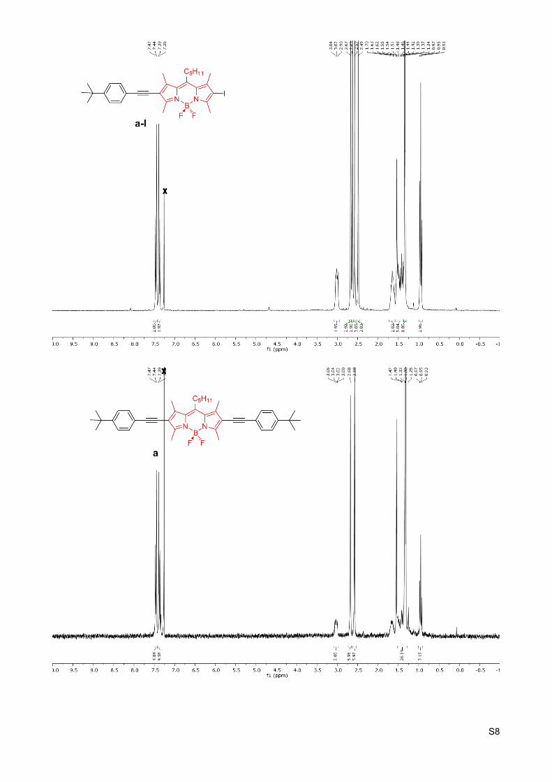
a-I. This compound was prepared from 4,4’-difluoro-1,3,5,7-tetrametil-8-pentil-4-bora-3a,4a-diaza-s-indacene (3 eq., 0.187 g), 1-(tert-butyl)-4-ethynylbenzene (1 eq., 0.015 mL), and a dry THF/NEt3 (4:1) mixture as solvent. The reaction mixture was stirred at 40ºC overnight. The crude was purified using chloroform/methanol (100:1) as eluent. a-I was obtained as a purple solid in 93% yield. 1H NMR (300 MHz, CDCl3) δ (ppm) 7.46 (d, J = 8.2 Hz, 2H), 7.38 (d, J = 8.3 Hz, 2H), 3.09 – 2.95 (m, 2H), 2.67 (s, 3H), 2.63 (s, 3H), 2.57 (s, 3H), 2.49 (s, 3H), 1.73 – 1.58 (m, 2H), 1.58 – 1.37 (m, 5H), 1.34 (s, 9H), 0.95 (t, J = 7.1 Hz, 3H). 13C NMR (76 MHz, CDCl3) δ (ppm) 157.3, 154.9, 151.7, 147.0, 141.7, 141.6, 132.0, 131.2, 131.0, 125.5, 120.4, 116.9, 96.8, 86.1, 81.0, 34.9, 32.6, 31.6, 31.3, 29.2, 22.6, 18.9, 16.2, 15.4, 14.1, 13.8. HRMS (MALDI): Calculated for C30H36BF2IN2: 600.1984 [M]+. Found: 600.1992 [M]+.
a. This compound was prepared from 4,4’-difluoro-1,3,5,7-tetrametil-8-pentil-4-bora-3a,4a-diaza-s-indacene (1 eq., 0.187 g), 1-(tert-butyl)-4-ethynylbenzene (2.2 eq., 0.13 mL), and a dry THF/NEt3 (4:1) mixture as solvent. The reaction mixture was stirred at 40ºC overnight. The crude was purified using chloroform/methanol (20:1) as eluent. a was obtained as a purple solid in 57% yield. 1H NMR (300 MHz, CDCl3) δ (ppm) 7.46 (d, J = 8.5 Hz, 4H), 7.38 (d, J = 8.6 Hz, 4H), 3.09 – 2.97 (m, 2H), 2.68 (s, 6H), 2.58 (s, 6H), 1.42-1.25 (m, 18H), 0.95 (t, J = 7.1 Hz, 3H). 13C NMR (76 MHz, CDCl3) δ (ppm) 156.8, 151.6, 141.0, 132.3, 131.4, 131.2, 125.6, 125.5, 120.5, 96.6, 81.2, 77.5, 34.9, 32.6, 31.7, 31.3, 31.2, 22.6, 15.2, 14.2, 13.7. HRMS (FAB+): Calculated for C42H49BF2N2: 630.3957 [M]+. Found: 630.3994 [M]+.
d-Br
DMF/NEt3 (4:1) dry,Pd(PPh3)4, CuI,60ºC, Ar atm.
THF/NEt3 (4:1) dry,Pd(PPh3)4, CuI,40ºC, Ar atm.
BASE
G1 / AA1 / C1 / U1
G1 C1 U1
a-I
S
S
N N
C5H11
BF F
BASE BASE
BASE
G1 / AA1 / C1 / U1
N
N
N
NH
O
NH2
O
O
OOO
N
NO
O
O
O
H2N
O
O
NHO
O
N
O
O
O O
O
dG (56%) dAA (82%) dC(60%) dU (50%) aG (88%) aAA (76%) aC(92%) aU (84%)
AA1N
N
N
N
NH2
O
O
OO
NH2
Si
Scheme S2. Synthesis of mononucleosides dG, dAA, dC, dU, aG, aAA, aC and aU via Sonogashira coupling reaction between d-Br or a-I and ethynyl-nucleobases G1, AA1, C1 and U1.
dG. This compound was prepared from d-Br (1 eq., 0.045 g), G1 (1.1 eq., 0.052 g), and a dry DMF/NEt3 (4:1) mixture as solvent. The reaction mixture was stirred at 60ºC overnight. The crude was purified using chloroform/methanol (40:1) as eluent. dG was obtained as a yellow solid in 56% yield. 1H NMR (300 MHz, CDCl3) δ (ppm) 8.15 (s, 1H), 8.10 (s, 1H), 7.68 (s, 1H), 7.54 – 7.34 (m, 5H), 6.53 (s, 1H), 6.27 (s, 1H), 5.46 (s, 1H), 5.07 (m, 1H), 4.80 (m, 1H), 4.41 (m, 1H), 4.11 (m, 1H), 3.65 (s, 1H), 1.42 (s, 3H), 1.34 (s, 9H), 1.26 (s, 3H), 1.18 (s, 9H). 13C NMR (76 MHz, DMSO-d6) δ (ppm) 177.1, 154.3, 152.4, 150.4, 138.1, 137.4, 137.3, 137.2, 131.2, 130.8, 128.5, 125.7, 123.7, 121.6, 120.4, 117.9, 113.4, 96.0, 85.4, 83.4, 81.4, 64.1, 38.1, 34.7, 30.8, 27.0, 26.7, 25.3. HRMS (MALDI): Calculated for C42H41N5O6S2: 775.2498 [M]+. Found: 798.2396 [M+Na]+.

S5
dAA. This compound was prepared from d-Br (1 eq., 0.045 g), AA1 (1.1 eq., 0.056 g), and a dry DMF/NEt3 (4:1) mixture as solvent. The reaction mixture was stirred at 60ºC overnight. The crude was purified using chloroform/methanol (40:1) as eluent. dAA was obtained as a yellow solid in 82% yield. 1H NMR (300 MHz, CDCl3) δ (ppm) 8.21 (s, 1H), 8.17 (s, 1H), 7.70 (s, 1H), 7.52 (d, J = 8.2 Hz, 3H), 7.42 (d, J = 8.4 Hz, 2H), 6.29 (d, J = 2.1 Hz, 1H), 5.68 (dd, J = 6.4, 2.3 Hz, 1H), 5.51 (s, 2H), 5.11 (dd, J = 6.5, 3.4 Hz, 1H), 4.79 (s, 2H), 4.38-4.24 (m, 1H), 3.82 (m, 2H), 1.67 (s, 3H), 1.45 (s, 3H), 1.36 (s, 9H), 0.87 (s, 9H), 0.02 (s, 3H), 0.00 (s, 3H). 13C NMR (76 MHz, CDCl3 and a drop of DMSO-d6) δ (ppm) 160.2, 155.8, 152.0, 150.6, 138.0, 137.6, 137.6, 136.7, 131.0, 130.1, 129.8, 129.3, 127.1, 125.1, 124.6, 120.8, 118.7, 116.8, 116.1, 114.5, 113.6, 96.0, 89.4, 87.8, 87.3, 84.1, 82.7, 82.0, 82.0, 63.2, 62.4, 34.5, 30.8, 29.3, 27.0, 25.5, 25.3, 18.0. HRMS (MALDI): Calculated for C43H48N6O4S2Si: 804.2948 [M]+. Found: 827.2828 [M+Na]+.
dC. This compound was prepared from d-Br (1 eq., 0.045 g), C1 (1.1 eq., 0.031 g), and a dry DMF/NEt3 (4:1) mixture as solvent. The reaction mixture was stirred at 60ºC overnight. The crude was purified using chloroform/methanol (40:1) as eluent. dC was obtained as a yellow solid in a 60% yield. 1H NMR (300 MHz, CDCl3) δ (ppm) 8.16 (s, 1H), 8.15 (s, 1H), 7.83 (s, 1H), 7.50 (d, J= 6.6 Hz, 4H), 7.40 (d, J = 8.3 Hz, 2H), 5.86 (s, 1H), 5.77 (s, 1H), 4.98-4.96 (m, 1H), 4.82 – 4.79 (m, 1H), 4.44-4.36 (m, 3H), 2.61 (hept, J= 7.1 Hz, 1H), 1.58 (s, 3H), 1.36 (s, 3H), 1.34 (s, 9H), 1.18 (dd, J = 6.9, 1.2 Hz, 6H). 13C NMR (75 MHz, DMSO-d6) δ 175.8, 163.8, 152.9, 152.3, 147.6, 137.5, 137.4, 137.2, 137.0, 131.2, 128.8, 128.5, 125.7, 123.1, 123.0, 117.2, 117.2, 113.0, 95.8, 93.4, 89.3, 87.6, 84.6, 82.3, 80.6, 63.8, 56.0, 34.6, 33.1, 30.8, 26.9, 25.1, 18.8, 18.7, 18.5. HRMS (MALDI): Calculated for C40H39N3O6S2: 721.2280 [M]+. Found: 744.2172 [M+Na]+.
dU. This compound was prepared from d-Br (1 eq., 0.045 g), U1 (1.1 eq., 0.044 g), and a dry DMF/NEt3 (4:1) mixture as solvent. The reaction mixture was stirred at 60ºC overnight. The crude was purified using chloroform/methanol (40:1) as eluent. dU was obtained as a yellow solid in a 50% yield. 1H NMR (300 MHz, CDCl3) δ (ppm) 8.44 (s, 1H), 8.12 (d, J = 4.4 Hz, 2H), 7.74 (s, 1H), 7.55 – 7.46 (m, 4H), 7.39 (d, J = 8.5 Hz, 2H), 5.85 (d, J = 2.3 Hz, 1H), 4.91 (dd, J = 6.4, 2.3 Hz, 1H), 4.79 (dd, J = 6.4, 3.7 Hz, 1H), 4.43 (m, 1H), 4.36 (d, J = 4.0 Hz, 2H), 2.65 (hept, J = 7.0 Hz, 1H), 1.60 (s, 3H), 1.38 (s, 3H), 1.34 (s, 9H), 1.20 (dd, J = 7.0, 3.3 Hz, 6H). 13C NMR (76 MHz, CDCl3) δ (ppm) 176.6, 160.4, 152.5, 148.7, 143.7, 138.2, 138.1, 138.0, 137.5, 131.5, 129.0, 127.5, 125.6, 125.0, 122.9, 119.5, 116.8, 116.5, 115.1, 100.3, 96.3, 93.7, 87.9, 85.8, 85.0, 82.5, 80.7, 63.7, 35.0, 34.1, 31.3, 27.3, 27.0, 25.4, 19.2, 19.1. HRMS (MALDI): Calculated for C40H38N2O7S2: 722.2120 [M]+. Found: 722.2116 [M]+.
aG. This compound was prepared from a-I (1 eq., 0.066 g), G1 (1.1 eq., 0.052 g), and a dry THF/NEt3 (4:1) mixture as solvent. The reaction mixture was stirred at 40ºC overnight. The crude was purified using chloroform/methanol (40:1) as eluent. aG was obtained as a purple solid in 88% yield. 1H NMR (300 MHz, CDCl3 and a drop of DMSO-d6) δ (ppm) 7.51 – 7.35 (m, 4H), 6.31 (s, 1H), 6.12 (s, 2H), 5.55 – 5.39 (m, 1H), 5.12 – 4.97 (m, 1H), 4.97 – 4.83 (m, 1H), 4.43-4.38 (m, 1H), 4.06 – 3.94 (m, 1H), 3.73 – 3.58 (m, 1H), 3.11 – 2.96 (m, 2H), 2.70 (s, 6H), 2.59 (s, 6H), 1.56 (s, 3H), 1.43 (s, 3H), 1.39-1.12 (m, 24H), 0.96 (t, J = 7.2 Hz, 3H). HRMS (MALDI): Calculated for C50H60BF2N7O6: 903.4666 [M]+. Found: 926.4585 [M+Na]+.
aAA. This compound was prepared from a-I (1 eq., 0.066 g), AA1 (1.1 eq., 0.056 g), and a dry THF/NEt3 (4:1) mixture as solvent. The reaction mixture was stirred at 40ºC overnight. The crude was purified using chloroform/methanol (40:1) as eluent. aAA was obtained as a purple solid in 76% yield. 1H NMR (300 MHz, CDCl3) δ (ppm) 7.57 – 7.35 (m, 4H), 6.31 (s, 1H), 5.79 (d, J = 4.7 Hz, 1H), 5.53 – 5.33 (m, 2H), 5.10 (d, J = 9.0 Hz, 1H), 4.75 (s, 2H), 4.37 – 4.22 (m, 1H), 4.01 (d, J = 7.3 Hz, 1H), 3.88 – 3.63 (m, 2H), 3.08 (t, J = 10.5 Hz, 2H), 2.72 (s, 6H), 2.63 (s, 6H), 1.62 (s, 3H), 1.44 (s, 3H), 1.37– 0.84 (m, 18H), -0.01 (s, 6H). HRMS (MALDI): Calculated for C51H67BF2N8O4Si: 932.5116 [M]+. Found: 955.5021 [M+Na]+.

S6
aC. This compound was prepared from a-I (1 eq., 0.066 g), C1 (1.1 eq., 0.046 g), and a dry THF/NEt3 (4:1) mixture as solvent. The reaction mixture was stirred at 40ºC overnight. The crude was purified using chloroform/methanol (40:1) as eluent. aC was obtained as a purple solid in 92% yield. 1H NMR (300 MHz, CDCl3 and a drop of DMSO-d6) δ (ppm) δ 7.95 (s, 1H), 7.73 (s, 1H), 7.56 – 7.32 (m, 4H), 5.85 (s, 1H), 5.71 (s, 1H), 5.01 (d, J = 6.1 Hz, 1H), 4.89 – 4.77 (m, 1H), 4.51 – 4.27 (m, 3H), 3.16 – 2.93 (m, 2H), 2.69-2.53 (m, 13H), 1.74 – 1.22 (m, 21H), 1.15 (d, J = 6.9 Hz, 6H), 1.00 – 0.89 (m, 3H). 13C NMR (76 MHz, CDCl3 and a drop of DMSO-d6) δ (ppm) 176.5, 164.8, 155.6, 153.9, 151.7, 148.07, 145.2, 142.1, 140.7, 131.9, 131.2, 130.9, 125.4, 120.2, 117.3, 114.4, 114.1, 97.1, 96.0, 91.6, 85.9, 85.5, 81.2, 80.7, 64.2, 34.8, 33.9, 32.5, 31.7, 31.2, 29.7, 28.9, 27.2, 25.3, 22.5, 19.1, 18.9, 15.3, 15.2, 14.0, 13.7, 13.6. HRMS (MALDI): Calculated for C48H58BF2N5O6: 849.4448 [M]+. Found: 872.4316 [M+Na]+.
aU. This compound was prepared from a-I (1 eq., 0.066 g), U1 (1.1 eq., 0.046 g), and a dry THF/NEt3 (4:1) mixture as solvent. The reaction mixture was stirred at 40ºC overnight. The crude was purified using chloroform/methanol (40:1) as eluent. aU was obtained as a purple solid in 84% yield. 1H NMR (300 MHz, CDCl3) δ (ppm) 8.09 (s, 1H), 7.54 (s, 1H), 7.42 – 7.26 (m, 4H), 5.71 (d, J = 2.2 Hz, 1H), 4.87 (dd, J = 6.4, 2.2 Hz, 1H), 4.72 (dd, J = 6.4, 3.8 Hz, 1H), 4.40 – 4.22 (m, 3H), 3.01 – 2.89 (m, 2H), 2.60 (s, 3H), 2.56 (s, 3H), 2.53 (m, 1H), 2.50 (s, 3H), 2.48 (s, 3H), 1.52 (s, 3H), 1.48 – 1.32 (m, 6H), 1.30 (s, 3H), 1.26 (s, 9H), 1.10 (dd, J = 7.0, 3.8 Hz, 6H), 0.87 (t, J = 7.1 Hz, 3H).13C NMR (76 MHz, CDCl3) δ (ppm) 176.6, 160.6, 157.8, 156.3, 151.7, 148.7, 148.0, 142.5, 141.7, 141.3, 131.3, 125.5, 120.4, 115.0, 101.2, 97.0, 94.3, 87.5, 86.1, 85.2, 84.9, 80.9, 63.8, 34.9, 34.0, 32.6, 31.7, 31.3, 29.0, 27.3, 25.4, 22.6, 19.2, 19.1, 15.3, 15.3, 14.1, 13.8, 13.7. HRMS (MALDI): Calculated for C48H57BF2N4O7: 850.4288 [M]+. Found: 873.4201 [M+Na]+.

S7
S0B. 1H NMR Spectra
S8

S9

S10

S11

S12

S13
S1. Cyclic voltammograms of reference compounds
Figure S1. Cyclic voltammetry curves of reference compounds d, a, G, C, AA and U at a scan rate of 100 mVs-1 in anhydrous deoxygenated CH2Cl2 at Pt working electrode. Supporting electrolyte 0.1 M TBAPF6. Ferrocene (Fc) was used as internal standard.

S14
S2. Absorption, emission, excitation and temperature-dependent emission spectra of nucleoside donor and acceptor molecules and their binary mixtures
Figure S2A. a) Absorption, (b) emission ( exc = 353 nm) and (c) excitation ( em = 353, 612 nm) spectra of 𝜆 𝜆compounds dG and aC and their 1:1 mixtures dG+aC. d) Emission spectra ( exc = 353 nm) of the dG+aC 1:1 𝜆mixtures as a function of temperature in the 0-80 ºC range. Arrows indicate the evolution of donor and acceptor emission maxima when decreasing temperature. In all cases the concentration energy donor and acceptor compounds was set at C = 1x10⁻⁵ M in toluene.
a) b)
c) d)
Figure S2B. a) Absorption, (b) emission ( exc = 368 nm) and (c) excitation ( em = 434, 608 nm) spectra of 𝜆 𝜆compounds dC and aG and their 1:1 mixtures dC+aG. d) Emission spectra ( exc = 368 nm) of the dC+aG 1:1 𝜆mixtures as a function of temperature in the (-5)-75ºC range. Arrows indicate the evolution of donor and acceptor emission maxima when decreasing temperature. In all cases the concentration energy donor and acceptor compounds was set at C = 1x10⁻⁵ M in toluene.
S15
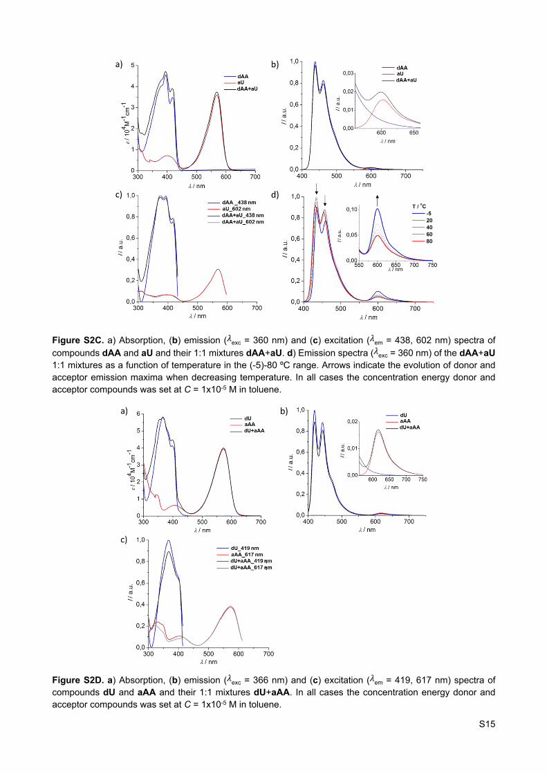
Figure S2C. a) Absorption, (b) emission ( exc = 360 nm) and (c) excitation ( em = 438, 602 nm) spectra of 𝜆 𝜆compounds dAA and aU and their 1:1 mixtures dAA+aU. d) Emission spectra ( exc = 360 nm) of the dAA+aU 𝜆1:1 mixtures as a function of temperature in the (-5)-80 ºC range. Arrows indicate the evolution of donor and acceptor emission maxima when decreasing temperature. In all cases the concentration energy donor and acceptor compounds was set at C = 1x10⁻⁵ M in toluene.
Figure S2D. a) Absorption, (b) emission ( exc = 366 nm) and (c) excitation ( em = 419, 617 nm) spectra of 𝜆 𝜆compounds dU and aAA and their 1:1 mixtures dU+aAA. In all cases the concentration energy donor and acceptor compounds was set at C = 1x10⁻⁵ M in toluene.

S16
Figure S2E. a) Absorption, (b) emission ( exc = 373 nm) and (c) excitation ( em = 429, 602 nm) spectra of 𝜆 𝜆compounds dG and aU and their 1:1 mixtures dG+aU. d) Emission spectra ( exc = 373 nm) of the dG+aU 1:1 𝜆mixtures as a function of temperature in the (-5)-75 ºC range. Arrows indicate the evolution of donor and acceptor emission maxima when decreasing temperature. In all cases the concentration energy donor and acceptor compounds was set at C = 1x10⁻⁵ M in toluene.
Figure S2F. a) Absorption, (b) emission ( exc = 394 nm) and (c) excitation ( em = 438, 607 nm) spectra of 𝜆 𝜆compounds dAA and aC and their 1:1 mixtures dAA+aC. d) Emission spectra ( exc = 394 nm) of the dAA+aC 𝜆1:1 mixtures as a function of temperature in the (-5)-80 ºC range. Arrows indicate the evolution of donor and

S17
acceptor emission maxima when decreasing temperature. In all cases the concentration energy donor and acceptor compounds was set at C = 1x10⁻⁵ M in toluene.
S3A. Analysis of the Titration Emission Data to obtain Ka
ReactLab EQUILIBRIA is a program developed and commercialized by Jplus Consulting Pty Ltd (http://jplusconsulting.com/; 8 Windsor Road, East Fremantle, WA 6158, Australia). It allows for the global fitting of multiwavelength spectroscopic data in equilibrium titration measurements to chemical reaction schemes, and determines all equilibrium constants in the underlying mechanism. ReactLab™ algorithms fit complete reaction models directly to multivariate data and delivers all the required parameters in one step. The analysis also yields the concentration distributions of all species and the individual spectra of all the participating species. The program, including all algorithms and the GUI frontend has been developed in Matlab and compiled to produce the final deployable application.
A wavelength region in the absorption spectra (from 410 to 460 nm; each wavelength representing one set of data) was fitted by this software. However, only 4 selected wavelengths are plotted (see Figure S3A and Table S1).

S18
Figure S3A. Fitting of the emission data of (a) dG+aC, (b) dC+aG, (c) dAA+aU and (d) dU+aAA in toluene by the ReactLab™ EQUILIBRIA software.
The 1:1 binding constants have been also calculated using a custom written global nonlinear regression analysis program developed by P. Thordarson5 within the Matlab R2012b package utilizing the Simplex algorithm.6
The standard errors (SEy) were calculated by:
𝑆𝐸𝑦 = ∑(𝑦𝑑𝑎𝑡𝑎 ‒ 𝑦𝑐𝑎𝑙𝑐)2
𝑁 ‒ 𝑘
Where N is the number of data points and k the number of parameters to be fitted.
The calculated Ka values are shown in Table S1.
S3B. Analysis of the NMR Titration data to obtain Ka5 P. Thordarson, Chem. Soc. Rev. 2011, 40, 1305.6 (a) J. A. Nelder and R. Mead, Comp. J. 1965, 7, 308; (b) J. C. Lagarias, J. A. Reeds, M. H. Wright and P. E. Wright, SIAM J. Optim.
1998, 9, 112.

S19
The 1:1 binding constants of the NMR experiments have been also calculated using the Matlab® scripts developed by P. Thordarson5 (Figure S3B and Table S1).
Figure S3B. NMR data of the (a) G-C and (b) AA-U pairs7 in toluene fitted with the Matlab® scripts developed by P. Thordarson.5
Table S1. Calculated association contants (Ka, M-1) between complementary nucleosides in toluene
7 C. Montoro-García, J. Camacho-García, A. M. López-Pérez, M. J. Mayoral, N. Bilbao and D. González-Rodríguez, Angew. Chem. Int. Ed., 2016, 55, 223.
aG aC aAA aU
5.0·105 [a]dG 4.1·105 (SEy = 10.040)[b]
1.2·105 (SEy = 0.010)[c]
3.0·105 [a]
4.2·105 (SEy = 9.395)[b]dC
2.0·103 [a]dAA 7.4·102 (SEy = 57.631)[b]
7.8·103 (SEy = 0.014)[c]
1.6·103 [a]dU 8.1·102 (SEy = 4.065)[b]
[a] Association constants calculated from emission data and fitted with ReactLabTM EQUILIBRIA.[b] Emission data fitted with the Matlab® scripts developed by P. Thordarson.5 [c] NMR data fitted with the Matlab® scripts developed by P. Thordarson. In this case, nucleosides G, C, AA, U (see Figure 1) were employed instead.