Embed Size (px)
Citation preview
10.22592/ode2017n.esp.p46
Institucional
Taller 5 - Peri-implantitis
Diego Sales 1
Judith Esquenazi2
Martín Minvielle3
Ernesto Andrade2
Magdalena Mayol2
1Cátedra de Rehabilitación, Prostodoncia Fija y TTM. Facultad de Odontología.
Universidad de la República. Uruguay. [email protected]
2Cátedra de Periodoncia. Universidad de la República. Uruguay. 3Cátedra de Rehabilitación, Prostodoncia Fija y TTM. Departamento de
Implantología Oral y Maxilofacial. Facultad de Odontología. Universidad de la
República. Uruguay.
Fecha de recibido: 28.03.2017 - Fecha de aceptado: 11.07.2017
MODALIDAD DE TRABAJO DEL TALLER DE PERI-IMPLANTITIS.
La misma se dividió en 3 etapas:1) Desarrollo de temáticas guía, selección y
envío de artículos, 2) Recepción, evaluación y devolución de artículos y 3)
Taller propiamente dicho.
PRIMERA ETAPA: dio inicio en febrero de 2014, donde se formularon 5
preguntas guía con el objetivo de ordenar la discusión del taller. Ellas fueron:
Diagnóstico, Epidemiología, Factores de riesgo, Antecedentes Periodontales y
Terapéutica de la Peri-Implantitis. Se realizó una búsqueda bibliográfica de
artículos en PUBMED, EMBASE, LILACS y COCHRANE, en el lapso
comprendido del año 2000 a la fecha, tanto en inglés como en portugués,
siendo utilizados los siguientes descriptores: peri-implantitis, peri implant
disease, perimplantitis, risk factor, epidemiology, incidence, prevalecence,
treatment, therapeutics, combinado con operadores booleanos. En su mayoría,
los artículos recuperados consistieron en estudios retrospectivos,
transversales, revisiones sistemáticas, consensos, escasos meta-análisis y
estudios prospectivos. Fueron descartados reportes de casos así como
revisiones de autor. Por parte de los responsables del taller se seleccionaron
los artículos más relevantes, dejando de lado los consensos y se enviaron vía
email a los participantes del taller.
SEGUNDA ETAPA: consistió en la recepción y análisis de artículos
seleccionados por los participantes del taller. Los que cumplían con los criterios
de inclusión, fueron redistribuidos entre los talleristas. La actividad del taller se
desarrolló de 8:30 a 12:30hs dedicándole un tiempo aproximado de 45 minutos
a cada una de las preguntas guía propuestas. Luego de la discusión, el
evaluador realizó un resumen y análisis de los artículos seleccionados
destacando además si las conclusiones extraídas guardaban relación con la
evidencia resultante de la bibliografía considerada.
Directores del taller: Dres. Judith Esquenazi y Diego Sales. Secretario: Dr.
Martín Minvielle. Evaluador: Dr. Ernesto Andrade.
Participantes del taller: Dres.: Adriana Ramos, Carolina Verolo, Jorge Cabrera,
Luis Arroyo, Magdalena Mayol, María José Quintana, Ricardo Kaufmann,
Verónica Foglino, Virginia Papone, Michel Bittencourt, Carolina Aldaya, Alessia
Molinari, Sebastián Pérez.
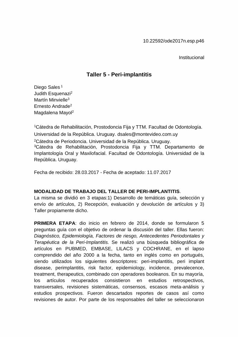
Pregunta Nº 1- ¿Cómo diagnosticamos: Mucositis y Peri-implantitis?
El término Peri-implantitis fue introducido por Mombelli en 1987 (1) y luego
modificado en el 1er Taller Europeo de Periodoncia para describir una
enfermedad inflamatoria que conduce a la pérdida ósea alrededor de los
implantes2. Las enfermedades peri-implantarias incluyen además: la
Mucositis, descripta como una lesión inflamatoria en la mucosa que rodea
al implante, sin pérdida ósea concomitante3. La literatura presenta
discrepancias en cuanto a los parámetros clínicos que se deben
considerar (Fig. 1).
Fig. 1
El tejido conectivo adyacente al implante presenta abundantes fibras
colágenas, es relativamente acelular y avascular, presentando características
histológicas semejantes al tejido cicatricial. La disposición de las fibras
colágenas es paralela al eje implantar, dando por resultado una conexión más
lábil y una progresión de la enfermedad más rápida para perimplantitis
comparado con la enfermedad periodontal(4).
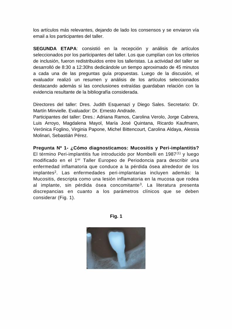
Clasificación de Peri-implantitis(5)
Temprana: Profundidad de sondaje >4 mm, con sangrado y/o supuración en
>2 sitios del implante y pérdida ósea < 25% del largo del implante (Fig. 2)
Fig. 2
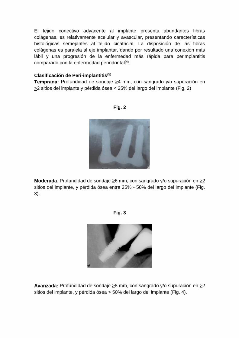
Moderada: Profundidad de sondaje >6 mm, con sangrado y/o supuración en >2
sitios del implante, y pérdida ósea entre 25% - 50% del largo del implante (Fig.
3).
Fig. 3
Avanzada: Profundidad de sondaje >8 mm, con sangrado y/o supuración en >2
sitios del implante, y pérdida ósea > 50% del largo del implante (Fig. 4).
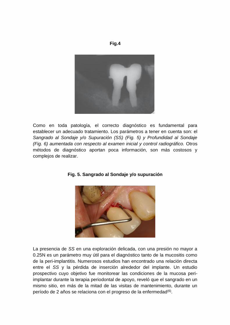
Fig.4
Como en toda patología, el correcto diagnóstico es fundamental para
establecer un adecuado tratamiento. Los parámetros a tener en cuenta son: el
Sangrado al Sondaje y/o Supuración (SS) (Fig. 5) y Profundidad al Sondaje
(Fig. 6) aumentada con respecto al examen inicial y control radiográfico. Otros
métodos de diagnóstico aportan poca información, son más costosos y
complejos de realizar.
Fig. 5. Sangrado al Sondaje y/o supuración
La presencia de SS en una exploración delicada, con una presión no mayor a
0.25N es un parámetro muy útil para el diagnóstico tanto de la mucositis como
de la peri-implantitis. Numerosos estudios han encontrado una relación directa
entre el SS y la pérdida de inserción alrededor del implante. Un estudio
prospectivo cuyo objetivo fue monitorear las condiciones de la mucosa peri-
implantar durante la terapia periodontal de apoyo, reveló que el sangrado en un
mismo sitio, en más de la mitad de las visitas de mantenimiento, durante un
período de 2 años se relaciona con el progreso de la enfermedad(6).
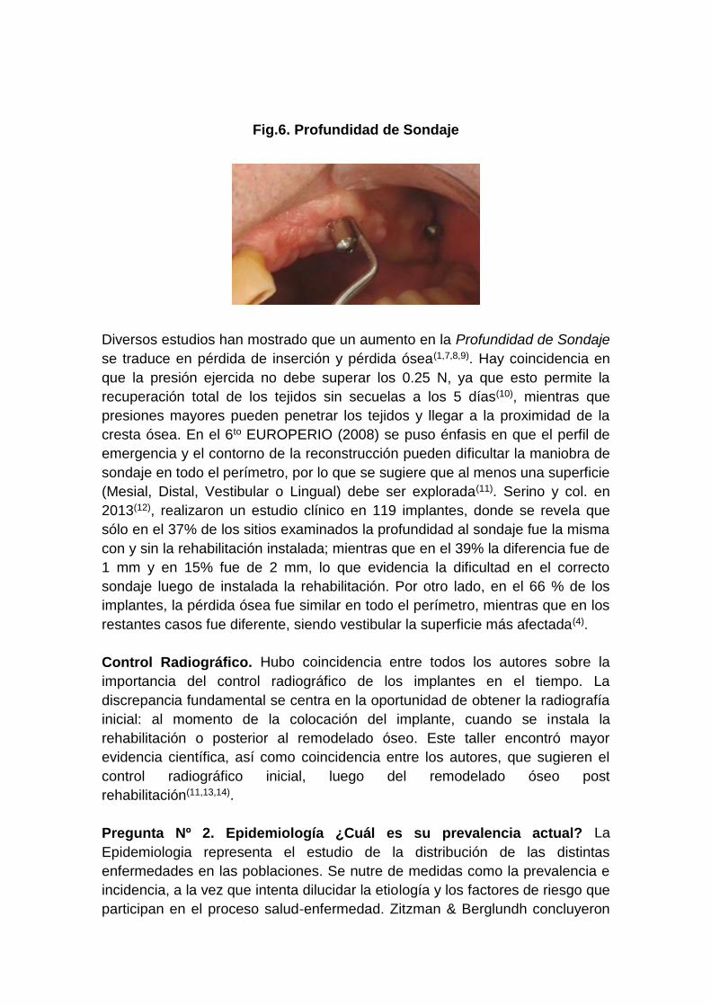
Fig.6. Profundidad de Sondaje
Diversos estudios han mostrado que un aumento en la Profundidad de Sondaje
se traduce en pérdida de inserción y pérdida ósea(1,7,8,9). Hay coincidencia en
que la presión ejercida no debe superar los 0.25 N, ya que esto permite la
recuperación total de los tejidos sin secuelas a los 5 días(10), mientras que
presiones mayores pueden penetrar los tejidos y llegar a la proximidad de la
cresta ósea. En el 6to EUROPERIO (2008) se puso énfasis en que el perfil de
emergencia y el contorno de la reconstrucción pueden dificultar la maniobra de
sondaje en todo el perímetro, por lo que se sugiere que al menos una superficie
(Mesial, Distal, Vestibular o Lingual) debe ser explorada(11). Serino y col. en
2013(12), realizaron un estudio clínico en 119 implantes, donde se revela que
sólo en el 37% de los sitios examinados la profundidad al sondaje fue la misma
con y sin la rehabilitación instalada; mientras que en el 39% la diferencia fue de
1 mm y en 15% fue de 2 mm, lo que evidencia la dificultad en el correcto
sondaje luego de instalada la rehabilitación. Por otro lado, en el 66 % de los
implantes, la pérdida ósea fue similar en todo el perímetro, mientras que en los
restantes casos fue diferente, siendo vestibular la superficie más afectada(4).
Control Radiográfico. Hubo coincidencia entre todos los autores sobre la
importancia del control radiográfico de los implantes en el tiempo. La
discrepancia fundamental se centra en la oportunidad de obtener la radiografía
inicial: al momento de la colocación del implante, cuando se instala la
rehabilitación o posterior al remodelado óseo. Este taller encontró mayor
evidencia científica, así como coincidencia entre los autores, que sugieren el
control radiográfico inicial, luego del remodelado óseo post
rehabilitación(11,13,14).
Pregunta Nº 2. Epidemiología ¿Cuál es su prevalencia actual? La
Epidemiologia representa el estudio de la distribución de las distintas
enfermedades en las poblaciones. Se nutre de medidas como la prevalencia e
incidencia, a la vez que intenta dilucidar la etiología y los factores de riesgo que
participan en el proceso salud-enfermedad. Zitzman & Berglundh concluyeron
que la prevalencia de peri-implantitis oscila entre un 28% - 56% de los
individuos (12%- 40% de los implantes). Por otro lado, la mucositis peri-
implantaria está presente en aproximadamente el 80% de los sujetos y 50% de
los implantes(3,11). Recientemente, un meta-análisis a partir de 9 artículos
publicados hasta el año 2012 con 6283 implantes en 1497 pacientes evidenció
que un 9,6% de los implantes presenta peri-implantitis, mientras que
considerando al individuo como unidad experimental la cifra alcanza el
18,8%(15). Hasta el año 2002, solo el 40-60% de los artículos recuperados
aportó datos de sobre “nuevos casos de peri-implantitis”; a su vez, la falta de
homogeneidad en los parámetros clínicos y para-clínicos utilizados para el
registro de la enfermedad impidió extraer resultados concluyentes sobre la
incidencia de la patología considerada(16). El estudio de la Enfermedad
Periimplantar (Mucositits y Periimplantitis) desde la óptica de la epidemiologia
debe tomar en consideración los siguientes aspectos:
la mayoría de los estudios utilizaron muestras para conveniencia del
investigador (pudiendo ser tendenciosas) y no seleccionadas por azar (no
aleatorias), lo que impide la generalización de los resultados a las
poblaciones (validez externa)(45);
se observa inexistencia de datos iniciales (ej.: radiografías) que permitan
valorar la pérdida ósea y estimar así la evolución;
desde una perspectiva epidemiológica, es inapropiado extraer datos de
prevalencia de peri-implantitis solo a partir de un elemento o variable aislada
(por ej.: sangrado al sondaje)(46);
falta de unanimidad en la definición de peri-implantitis y en los umbrales de
cada variable utilizada (sangrado y profundidad al sondaje) utilizada, lo que
dificulta la comparación entre los estudios(23, 46)
la inclusión de fumadores y personas con periodontitis modifica la prevalencia
de enfermedad, y no siempre estas sub-poblaciones están adecuadamente
registradas y consideradas dentro de los estudios;
la utilización del “implante” en vez del “sujeto” como elemento utilizado para
cuantificar la enfermedad (unidad experimental de análisis), no representa
cabalmente la frecuencia de la enfermedad ya que puede subestimar la
patología existente;
la mayor parte de los estudios publicados hasta el momento registran los casos
de peri-implantitis con 5 años posterior a la carga del implante; sin embargo
algunos autores consideran insuficiente este lapso de tiempo para el
desarrollo de dicha enfermedad, siendo mencionados hasta 10 años(16)
existen carencias en la información que se presenta en los artículos publicados
ya que hay datos o información que no son reportados(47).
Pregunta Nº 3. ¿Cuáles son sus factores de riesgo? Para identificar un
factor de riesgo verdadero son necesarios estudios prospectivos que permitan
un seguimiento claro de una población específica y establezcan una relación
temporal entre un factor de exposición y la enfermedad. Se han reportado muy
pocos estudios con estas características y los datos disponibles generalmente
surgen a partir de estudios transversales y series de casos que permiten así
identificar indicadores de riesgo(17,18).
¿Qué condiciones generales, locales e individuales pueden favorecer la
aparición de peri-implantitis? El taller concluyó que los indicadores de riesgo
asociados con mayor evidencia son:
Tabaquismo: un meta-análisis que compara fumadores con no fumadores,
identifica al tabaquismo como un indicador de riesgo significativo para la
pérdida ósea marginal, comparado con no fumadores. Esto debería ser
considerado en la información proporcionada al paciente previo al
tratamiento, así como en la etapa de mantenimiento donde es posible
detectar en forma temprana cambios negativos en los tejidos peri-
implantario(17,19). El hábito de fumar tiene efectos deletéreos a largo plazo
sobre la respuesta inflamatoria e inmunitaria, así como, sobre la
cicatrización, con menor producción de colágeno, disfunción de fibroblastos,
reducción de la circulación periférica y función de neutrófilos y macrófagos
comprometida(20).
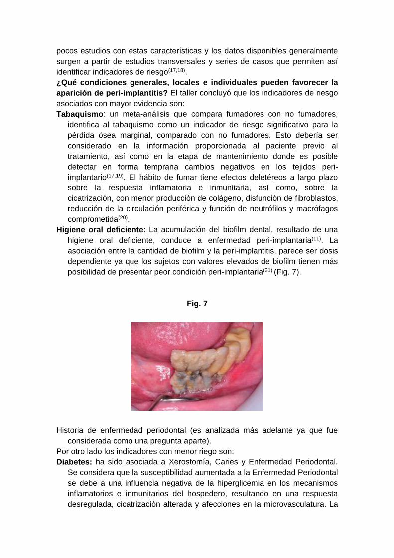
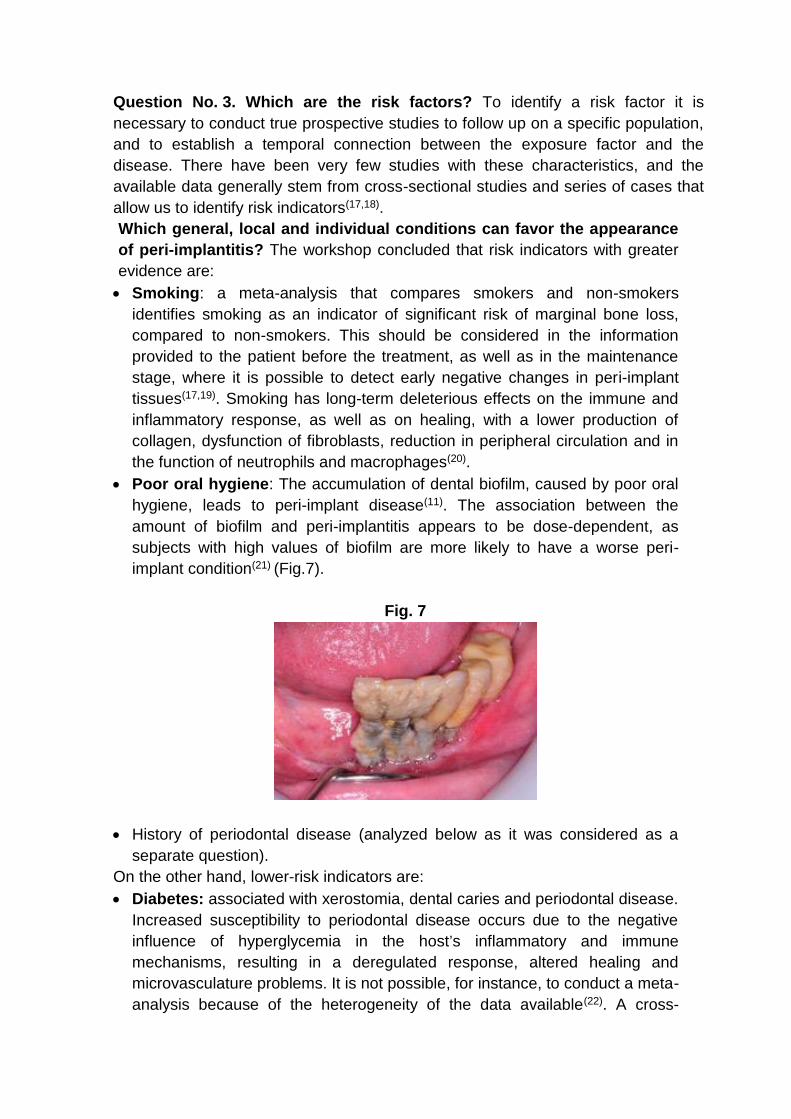
Higiene oral deficiente: La acumulación del biofilm dental, resultado de una
higiene oral deficiente, conduce a enfermedad peri-implantaria(11). La
asociación entre la cantidad de biofilm y la peri-implantitis, parece ser dosis
dependiente ya que los sujetos con valores elevados de biofilm tienen más
posibilidad de presentar peor condición peri-implantaria(21) (Fig. 7).
Fig. 7
Historia de enfermedad periodontal (es analizada más adelante ya que fue
considerada como una pregunta aparte).
Por otro lado los indicadores con menor riego son:
Diabetes: ha sido asociada a Xerostomía, Caries y Enfermedad Periodontal.
Se considera que la susceptibilidad aumentada a la Enfermedad Periodontal
se debe a una influencia negativa de la hiperglicemia en los mecanismos
inflamatorios e inmunitarios del hospedero, resultando en una respuesta
desregulada, cicatrización alterada y afecciones en la microvasculatura. La
heterogeneidad de los datos disponibles impide por ejemplo la realización
de meta-análisis22. Un estudio transversal en 212 sujetos, con 578
implantes, incluyó 29 diabéticos. Los resultados de este estudio, muestran
que el control metabólico inadecuado en diabéticos aumenta el riesgo de
desarrollar peri-implantitis. Se diagnosticó peri-implantitis en el 24,13% de
los diabéticos, mientras que en los no diabéticos fue significativamente
menor 6,56%(21).
Sobrecarga oclusal: los reportes son controversiales, ya que no hay
unanimidad en la definición de sobrecarga oclusal. Los implantes se
consideran más lábiles que los dientes al enfrentarse a fuerzas no axiales,
por la falta de ligamento periodontal(23). El stress excesivo puede ocasionar
micro fracturas y eventual pérdida ósea. Una revisión sistemática reciente(24)
sugirió que la sobrecarga oclusal está asociada positivamente con la
reabsorción ósea marginal periimplantaria. Sin embargo el biofilm sigue
siendo el factor causal clave. Se necesitan más investigaciones con una
definición precisa de sobre carga oclusal(23).
Alcoholismo: Un estudio prospectivo de 3 años, evaluó la posible relación
entre la pérdida ósea periimplantaria marginal, el consumo de tabaco y
alcohol. El análisis multivariado mostro que la pérdida ósea estaba
relacionada significativamente con el consumo de >10grs diarios de
alcohol(25).
Cemento residual: en un estudio prospectivo se incluyeron 39 pacientes
portadores de 42 implantes, rehabilitados con coronas unitarias
cementadas, con peri-implantitis. 12 de ellos tenían además 20 implantes
rehabilitados bajo las mismas circunstancias, pero sin peri implantitis (este
segundo grupo de implantes se utilizó como control). Para explorar el área
subgingival se utilizó un endoscopio dental (Dental View, Irvine, CA). Como
resultado se encontró cemento residual en 34 de los 42 implantes test y en
ninguno de los controles, siendo removido mediante diferentes técnicas
(quirúrgicas y no quirúrgicas). Luego de 30 días, se observó resolución de
los signos de enfermedad peri implantaría en 25 de los 33 implantes del
grupo experimental. Por lo tanto, el exceso de cemento y su incompleta
remoción en la superficie subgingival del implante determina un medio
susceptible para la colonización bacteriana y el desarrollo una respuesta
inflamatoria(26).
Superficie del implante: al momento actual no hay evidencia de que las
características superficiales del implante puedan tener influencia
significativa en la iniciacion de la peri-implantitis(27).
Ausencia de encía queratinizada: La deficiencia de mucosa queratinizada
alrededor de los implantes, parece estar relacionada a parámetros clínicos
de inflamación y a la acumulación de biolfim. Sin embargo, en base a la
evidencia disponible actualmente, la cantidad de mucosa queratinizada en
relación al desarrollo de la peri implantitis permanece controversial(28,29).
Pregunta Nº 4. Antecedentes Periodontales y la enfermedad peri-
implantar. Desde hace algunos años, surge en la literatura un número
creciente de trabajos, que tienen como objetivo analizar las relaciones entre la
implantología y la periodoncia. La implantologia nacida para la rehabilitación de
pacientes totalmente edéntulos, posteriormente también se ocupó de pacientes
parcialmente dentados. El fracaso tardío de los implantes, parece estar
principalmente correlacionado con el tipo de flora bacteriana peri-implantaria y
las condiciones del huésped(30). El biofilm asociado a los implantes con peri-
implantitis, es semejante al que habita en los pacientes con enfermedad
periodontal avanzada(1,31) existiendo estudios que evidencian patógenos
periodontales colonizando la superficie de los implantes(32,33). Especies como
Porphyromonas Gingivalis, Prevotella Intermedia y Aggregatibacter
Actinomycemcomitans recolectadas a partir de bolsas periodontales avanzadas
(asociadas con signos de inflamación) pueden traslocarse a sitios peri-
implantarios(34,35). La presencia de un biofilm de particular virulencia, se vincula
con la aparición de peri-implantitis, dependiendo de la susceptibilidad del
hospedero y la presencia de factores de riesgo (mencionados anteriormente).
Considerando lo anteriormente mencionado, nos cabe preguntar, si la
susceptibilidad a la periodontitis, aumenta la susceptibilidad a la peri-implantitis,
incluso en pacientes que fueron tratados por paradenciopatias. Karoussis(36) fue
pionero en considerar la posibilidad de que personas con historia de
enfermedad periodontal sean más propensos a desarrollar peri-implantitis,
comparando, con individuos sin historia de enfermedad periodontal.
El análisis de la literatura destaca la existencia de dos categorías de estudios:
a) los que investigan el éxito de la terapia implantar en el paciente con historia
de enfermedad periodontal sin comparación con los pacientes que no la
padecieron y b) los que ofrecen una comparación directa entre pacientes con
historia de enfermedad periodontal destructiva y aquellos sin antecedentes de
la misma. Varias revisiones sistemáticas(37,38,39,40,41,41,43), así como un estudio
prospectivo con 10 años en promedio de seguimiento muestran que las
personas con historia de enfermedad periodontal tienen más probabilidades
de desarrollar peri-implantitis que sin antecedentes de ella(44).
Pregunta Nº 5. ¿Cuáles son las técnicas terapéuticas de la peri-implantitis
y cuál es su previsibilidad? Las terapias propuestas para el manejo de la
periimplantitis están basadas en la evidencia disponible del tratamiento de la
enfermedad periodontal, por lo que los pasos básicos para su resolución son:
control de la infección, terapia no quirúrgica y quirúrgica y terapia de apoyo.
Para facilitar la oseointegración, la mayoría de los fabricantes de implantes
presentan en el mercado superficies moderadamente rugosas para aumentar la
superficie de contacto lo que representaría una desventaja si el implante queda
expuesto al medio bucal. La rugosidad de la superficie y su composición
química, al igual que el diseño de implantes roscados, tienen un impacto
significativo en la acumulación de Biofilm Oral(48). El diseño de la prótesis
puede dificultar la correcta limpieza mecánica y el tratamiento de los implantes
infectados, por lo que la corrección de la restauración es fundamental para el
acceso de los elementos de higiene del paciente(49).
Basados en los conceptos mencionados anteriormente, numerosos estudios
proponiendo diferentes tratamientos como desbridamiento mecánico, uso de
antisépticos, antibióticos locales y sistémicos tanto como acceso quirúrgico y
realización de procedimientos regenerativos han sido empleados con
resultados diversos. Los intentos por comparar la información disponible en la
literatura que permitan extraer conclusiones robustas han fallado por
inexsitencia de datos y por no completar los criterios estrictos de un estudio
controlado aleatorizado. La ausencia de un verdadero grupo control, el no
disponer de un número significativo de pacientes con características clínicas
comparables, además de razones éticas, marcan la dificultad de realizar
ensayos controlados aleatorizados(50). Una revisión sistemática en la cual se
incluyeron 9 ensayos clínicos controlados aleatorizados, donde se intentó
identificar el tratamiento más efectivo concluyó que no había evidencia
confiable que sugiriera qué tratamiento era superior(51). Se recomienda
comenzar con terapia no quirúrgica. Esto permite evaluar la respuesta tisular
inicial así como el cumplimiento del paciente con las medidas de autocuidado,
lo que determinara en gran medida el éxito de las opciones terapéuticas
propuestas. Estudios donde diversas modalidades de tratamientos no
quirúrgicos fueron comparados (desbridamiento mecánico con curetas de
titanio y pulido con copas de goma vs. desbridamiento mecánico con
dispositivos ultrasónicos y pulido con copas de goma con administración de
antimicrobianos) se analizaron a partir de variables como el sangrado al
sondaje, profundidad al sondaje y los depósitos de biofilm. En su evaluación al
mes, tres y seis meses no se hallaron resultados clínicamente relevantes que
demostraran superioridad entre los tratamientos propuestos(52). Por otra parte,
la utilización de dispositivos de laser (Er:YAG)(53) y la aplicación de glicina en
polvo a presión limitada(54) al ser testeadas con el desbridamiento mecánico no
evidenciaron diferencias ni clínica ni estadísticamente significativas. Sin
embargo, la terapia no quirúrgica por sí sola es insuficiente en la mayoría de
los casos de periimplantitis(52). El biofilm y los depósitos calcificados deben ser
removidos para permitir la cicatrización y reducir el riesgo de progresión de la
enfermedad. El protocolo quirúrgico que implica un acceso quirúrgico,
desbridamiento, descontaminación de la superficie, irrigación con suero
fisiológico y el uso de antibióticos sistémicos ha sido evaluado, con resultados
positivos mantenidos por 12 meses(55). Por otro lado estudios realizados por
Romeo en 2007, mencionan técnicas en las que se practica la implantoplastia,
es decir el pulido de la superficie del implante con piedras de diamante, gomas
y pastas siliconadas, reposicionando el colgajo apicalmente(56).
En resumen, de lo expuesto en el taller de periimplantitis del primer congreso
de Implantología de la Facultad de Odontología de la Universidad de la
República Oriental del Uruguay se concluye que: 1) el tratamiento quirúrgico
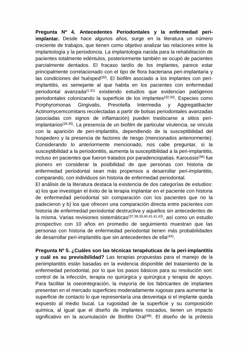
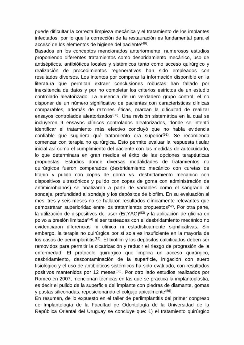
debe siempre ser precedido por el tratamiento no quirúrgico basado en el
desbridamiento mecánico y la utilización de antisépticos como la clorhexidina al
0,12% con una espera de 3 meses antes de comenzar con el tratamiento
quirúrgico (Figs. 8, 9).
Fig. 8
Fig. 9
2) El tratamiento quirúrgico que consiste en: a) la apertura de un colgajo
mucoperióstico con la eliminación del tejido de granulación; b) El tratamiento de la
superficie del implante ya sea con gasa con suero, gasa con clorhexidina al 0,2%,
gasa con H2O2 al 3%, antibiótico local sobre la superficie del implante, spray de
bicarbonato de sodio o glicina, Er: YAG laser, curetaje mecánico o implantoplastia;
c) técnicas regenerativas o resectivas; d) antibiótico administrado por vía sistémica
(Amoxicilina 500mg y Metronidazol 500mg por 7 días); e) aplicación de clorhexidina
al 0,12% hasta retomar la higiene mecánica; f) terapia de Apoyo de 3 a 6 meses.
Las técnicas de tratamiento de superficie nombradas han sido analizadas en
numerosos estudios clínicos y no se ha comprobado aun la superioridad de
alguna de ellas. Por otro lado, en la mayoría de los casos las secuelas estéticas
son evidentes, comprometiendo el éxito del tratamiento implanto-protésico. Pese
a que resultados a corto plazo fueron reportados en varios estudios, falta de
resolución de la enfermedad, recurrencia de la misma y pérdida de implantes a
pesar del tratamiento fueron también reportados. Hoy en día no tenemos
certezas del éxito del tratamiento de la Peri Implantitis a mediano y largo plazo.
Son entonces necesarios estudios prospectivos más extensos en el tiempo que
uniformicen criterios como definición, severidad de la enfermedad,
heterogeneidad del diseño, tiempo del seguimiento y criterios de inclusión o
exclusión.
En cuanto al mantenimiento debe existir un control exhaustivo de biofilm y re
motivación. En el examen clínico de los tejidos peri-implantares se debe sondear
para controlar su profundidad buscando sangrado o supuración al sondaje. La
remoción de la prótesis con este fin puede ser necesaria. Se debe incluir además
el análisis oclusal, pesquisando facetas de desgaste o falta de estructura. Se
debe complementar con aplicaciones de antisépticos(57).
Referencias
1. Mombelli A, van Oosten MA, Schurch E, Land NP. The microbiota associated
with successful or failing osseointegrated titanium implants. Oral Microbiol
Immunol. 1987;2:145–51.
2. Albrektsson, T. & Isidor F. Consensus report: implant therapy. Quintessence.
1994;365-369.
3. Zitzmann NU, Berglundh T. Definition and prevalence of peri-implant
diseases. J Clin Periodontol. 2008;35:286–91.
4. Berglundh T, Zitzmann NU, M Donati. Are peri-implantitis lesions different
from periodontitis lesions? J Clin Periodontol. 2011;38(mar):188 – 202.
5. Froum SJ, Rosen PS. A proposed classification for peri-implantitis. Int J
Periodontics Restorative Dent. 2012; 32: 533–40.
6. Luterbacher, S, Heitz-Mayfield LJA, Brägger Urs, Lang NP. Diagnostic
characteristics of clinical and microbiological tests for monitoring periodontal
and peri-implant mucosal tissue conditions during supportive periodontal
therapy (SPT). Clin Oral Implant Res. 2000; 11(dec): 521 -529.
7. Hultin M, Gustafsson A, Hallström H, Johansson L-Å, Ekfeldt A, Klinge B.
Microbiological findings and host response in patients with peri-implantitis.
Clin Oral Implant Res. 2002;13:349–58.
8. Schou S, Holmstrup P, Stoltze K, Hjørting-Hansen E, Kornman K. Ligature-
induced marginal inflammation around osseointegrated implants and
ankylosed teeth. Clin Oral Implant Res. 1993;4(1):12 – 22.
9. Fransson C, Wennström J, Berglundh T. Clinical characteristics at implants
with a history of progressive bone loss. Clin Oral Implants Res.
2008;19:142–7.
10. Etter TH, Håkanson I, Lang NP, Trejo PM, Caffesse RG. Healing after
standardized clinical probing of the perlimplant soft tissue seal: a
histomorphometric study in dogs. Clin Oral Implants Res. 2002;13:571–80.
11. Lindhe J, Meyle J. Peri-implant diseases: Consensus Report of the Sixth
European Workshop on Periodontology. J Clin Periodont 2008: 282–5.
12. Serino G, Turri A, Lang NP. Probing at implants with peri-implantitis and its
relation to clinical peri-implant bone loss. Clin Oral Implants Res
2013;24:91–5.
13. Lang NP, Berglundh T. Periimplant diseases: where are we now?--
Consensus of the Seventh European Workshop on Periodontology. J Clin
Periodontol 2011: 178–81.
14. De Bruyn H, Vandeweghe S, Ruyffelaert C, Cosyn J, Sennerby L.
Radiographic evaluation of modern oral implants with emphasis on crestal
bone level and relevance to peri-implant health. Periodontol 2000.
2013;62:256–70.
15. Atieh M a, Alsabeeha NHM, Faggion CM, Duncan WJ. The frequency of
peri-implant diseases: a systematic review and meta-analysis. J Periodontol
2013;84:1586–98.
16. Berglundh T, Persson L, Klinge B. A systematic review of the incidence of
biological and technical complications in implant dentistry reported in
prospective longitudinal studies of at least 5 years. J Clin Periodontol.
2002;29 Suppl 3:197–212; discussion 232–233.
17. Heitz-Mayfield LJA. Peri-implant diseases: diagnosis and risk indicators. J
Clin Periodontol. 2008;35:292–304.
18. Rocchietta I, Nisand D. A review assessing the quality of reporting of risk
factor research in implant dentistry using smoking, diabetes and periodontitis
and implant loss as an outcome: critical aspects in design and outcome
assessment. J Clin Periodontol 2012;39:114–21.
19. Strietzel FP, Reichart PA, Kale A, Kulkarni M, Wegner B, Küchler I.
Smoking interferes with the prognosis of dental implant treatment: a
systematic review and meta-analysis. J Clin Periodontol. 2007;34:523–44.
20. Palmer RM, Wilson RF, Hasan AS, Scott DA. Mechanisms of action of
environmental factors--tobacco smoking. J Clin Periodontol. 2005;32 Suppl
6:180–95.
21. Ferreira SD, Silva GLM, Cortelli JR, Costa JE, Costa FO. Prevalence and
risk variables for peri-implant disease in Brazilian subjects. J Clin
Periodontol. 2006;33:929–35.
22. Bornstein MM, Cionca N, Mombelli A. Systemic conditions and treatments
as risks for implant therapy. Int J Oral Maxillofac Implants. 2009;24
Suppl:12–27.
23. Academy Report: Peri-Implant Mucositis and Peri-Implantitis: A Current
Understanding of Their Diagnoses and Clinical Implications. J Periodontol
2013 Mar 28;84(4):436–43.
24. Fu J-H, Hsu Y-T, Wang H-L. Identifying occlusal overload and how to deal
with it to avoid marginal bone loss around implants. Eur J Oral Implantol
2012;5 Suppl:S91–103.
25. Galindo-Moreno P, Fauri M, Avila-Ortiz G, Fernández-Barbero JE, Cabrera-
León A, Sánchez-Fernández E. Influence of alcohol and tobacco habits on
peri-implant marginal bone loss: a prospective study. Clin Oral Implants Res.
2005;16:579–86.
26. Wilson TG. The positive relationship between excess cement and peri-
implant disease: a prospective clinical endoscopic study. J Periodontol.
2009;80:1388–92.
27. Renvert S, Polyzois I, Claffey N. How do implant surface characteristics
influence periimplant disease? J Clin Periodont 2011; 38 (suppl 11): 214–22.
28. Gobbato L, Avila-Ortiz, Sohrabi K, Wang C, Karimbux N. The effect of
keratinized mucosa width on peri-implant health: a systematic review. Int J
Oral Maxillofac Implant. 2013;28(6):1536–45.
29. Lin G-H, Chan H-L, Wang H-L. The significance of keratinized mucosa on
implant health: a systematic review. J Periodontol 2013;84(12):1755–67.
30. Van Steenberghe D, Quirynen M. Reproducibility and detection threshold of
peri-implant diagnostics. Adv Dent Res. 1993;7:191–5.
31. Papaioannou W, Quirynen M, Van Steenberghe D. The influence of
periodontitis on the subgingival flora around implants in partially edentulous
patients. Clin Oral Implants Res. 1996;7:405–9.
32. Leonhardt A, Adolfsson B, Lekholm U, Wikström M, Dahlén G. A
longitudinal microbiological study on osseointegrated titanium implants in
partially edentulous patients. Clin Oral Implants Res. 1993;4:113–20.
33. Quirynen M, De Soete M, van Steenberghe D. Infectious risks for oral
implants: a review of the literature. Clin Oral Implants Res. 2002;13:1–19.
34. Mombelli A, Marxer M, Gaberthüel T, Grunder U, Lang NP. The microbiota
of osseointegrated implants in patients with a history of periodontal disease.
J Clin Periodontol. 1995;22:124–30.
35. Leonhardt A, Renvert S, Dahlén G. Microbial findings at failing implants.
Clin Oral Implants Res. 1999;10:339–45.
36. Karoussis IK, Salvi GE, Heitz-Mayfield LJA, Brägger U, Hämmerle CHF,
Lang NP. Long-term implant prognosis in patients with and without a history
of chronic periodontitis: a 10-year prospective cohort study of the ITI Dental
Implant System. Clin Oral Implants Res. 2003;14:329–39.
37. Van der Weijden GA, van Bemmel KM, Renvert S. Implant therapy in
partially edentulous, periodontally compromised patients: a review. J Clin
Periodontol. 2005;32:506–11.
38. Schou S, Holmstrup P, Worthington H V, Esposito M. Outcome of implant
therapy in patients with previous tooth loss due to periodontitis. Clin Oral
Implants Res. 2006;17 Suppl 2:104–23.
39. Karoussis IK, Kotsovilis S, Fourmousis I. A comprehensive and critical
review of dental implant prognosis in periodontally compromised partially
edentulous patients. Clin Oral Implants Res. 2007;18:669–79.
40. Klokkevold PR, Han TJ. How do smoking, diabetes, and periodontitis affect
outcomes of implant treatment? The International journal of oral & maxillofacial
implants. 2007. p. 173–202.
41. Ong CTT, Ivanovski S, Needleman IG, Retzepi M, Moles DR, Tonetti MS, et
al. Systematic review of implant outcomes in treated periodontitis subjects. J
Clin Periodontol. 2008;35:438–62.
42. Renvert S, Persson GR. Periodontitis as a potential risk factor for peri-
implantitis. J Clin Periodontol. 2009;36 Suppl 1:9–14.
43. Safii SH, Palmer RM, Wilson RF. Risk of implant failure and marginal bone
loss in subjects with a history of periodontitis: A systematic review and meta-
analysis. Clin Impl Dent Related Res 2010; 12(3): 165–74.
44. Roccuzzo M, De Angelis N, Bonino L, Aglietta M. Ten-year results of a
three-arm prospective cohort study on implants in periodontally
compromised patients. Part 1: implant loss and radiographic bone loss. Clin
Oral Implants Res. 2010;21:490–6.
45. Tomasi C, Derks J. Clinical research of peri-implant diseases--quality of
reporting, case definitions and methods to study incidence, prevalence and
risk factors of peri-implant diseases. J Clin Periodontol 2012;39 (Suppl
1):207–23.
46. Mombelli A, Müller N, Cionca N. The epidemiology of peri-implantitis. Clin
Oral Implants Res 2012;23 (Suppl 6):67–76.
47. Meijer H, Raghoebar G. Quality of reporting of descriptive studies in implant
dentistry. Critical aspects in design, outcome assessment and clinical
relevance. J Clin Periodontol 2012; 39(Suppl 12): 108–13.
48. Teughels W, Assche N Van, Sliepen I, Quirynen M. Effect of material
characteristics and/or surface topography on biofilm development. Clin Oral
Imp Res. 2006; (suppl 6):68–81.
49. Renvert S, Samuelsson E, Lindahl C, Persson GR. Mechanical non-surgical
treatment of peri-implantitis: A double-blind randomized longitudinal clinical
study. I: Clinical results. J Clin Periodontol. 2009;36:604–9.
50. Heitz-Mayfield LJA, Mombelli A. The Therapy of Peri-implantitis: A
Systematic Review. Int J Oral Maxillofac Implants 2014;29 (Suppl):325–45.
51. Esposito M, Grusovin MG, Worthington H V. Treatment of peri-implantitis:
what interventions are effective? A Cochrane systematic review. Eur J Oral
Implantol 2012;5 (Suppl):S21–41.
52. Renvert S, Roos-Jansåker AM, Claffey N. Non-surgical treatment of peri-
implant mucositis and peri-implantitis: A literature review. J Clin Periodontol
2008; 35(Suppl 8): 305–15.
53. Schwarz F, Sculean A, Rothamel D, Schwenzer K, Georg T, Becker J.
Clinical evaluation of an Er:YAG laser for nonsurgical treatment of
periimplantitis: A pilot study. Clin Oral Implants Res. 2005;16:44–52.
54. Moëne R, Décaillet F, Andersen E, Mombelli A. Subgingival plaque removal
using a new air-polishing device. J Periodontol. 2010;81:79–88.
55. Heitz-Mayfield LJA, Salvi GE, Mombelli A, Faddy M, Lang NP. Anti-infective
surgical therapy of peri-implantitis. A 12-month prospective clinical study.
Clin Oral Implants Res. 2012;23:205–10.
56. Romeo E, Lops D, Chiapasco M, Ghisolfi M, Vogel G. Therapy of peri-
implantitis with resective surgery. A 3-year clinical trial on rough screw-
shaped oral implants. Part II: Radiographic outcome. Clin Oral Implants Res.
2007;18:179–87.
57. Schwarz F, Sahm N, Iglhaut G, Becker J. Impact of the method of surface
debridement and decontamination on the clinical outcome following
combined surgical therapy of peri-implantitis: a randomized controlled clinical
study. J Clin Periodontol 2011;38:276–84.
Workshop 5 - Peri-implantitis
Working methodology at the peri-implantitis workshop. The workshop was
divided into three stages: 1) development of guiding topics, selection and delivery
of articles, 2) reception, evaluation and returning of articles, and 3) the workshop
itself.
FIRST STAGE: The process began in February 2014, when five guiding questions
were formulated to organize the workshop discussion. These were: Diagnosis,
Epidemiology, Risk Factors, Periodontal Disease History and Peri-Implantitis
Treatment. A literature search was performed in PUBMED, EMBASE, LILACS and
COCHRANE, from 2000 to date, in both English and Portuguese, using the
following descriptors: peri-implantitis, peri-implant disease, perimplantitis, risk
factor, epidemiology, incidence, prevalence, treatment, therapeutics, combined with
boolean operators. Most articles were cross-sectional, retrospective studies,
systematic reviews, consensus statements, with few meta-analyses and
prospective studies. Case reports and author reviews were excluded. Workshop
leaders selected the most relevant articles, excluding consensus statements, and
emailed the papers selected to the workshop participants.
SECOND STAGE: workshop participants received and analyzed the articles
selected. The articles that met the inclusion criteria were distributed among the
workshop participants. The workshop took place from 8:30 to 12:30hs, and 45
minutes were devoted to each guiding question. After the debate, the reviewer
summarized and analyzed the selected articles, stating if the conclusions drawn
were consistent with the evidence resulting from the literature considered.
Workshop leaders: Drs. Judith Esquenazi and Diego Sales. Secretary: Dr. Martín
Minvielle. Reviewer: Dr. Ernesto Andrade
Workshop participants: Drs. Adriana Ramos, Carolina Verolo, Jorge Cabrera, Luis
Arroyo, Magdalena Mayol, María José Quintana, Ricardo Kaufmann, Verónica
Foglino, Virginia Papone, Michel Bittencourt, Carolina Aldaya, Alessia Molinari,
Sebastián Pérez.
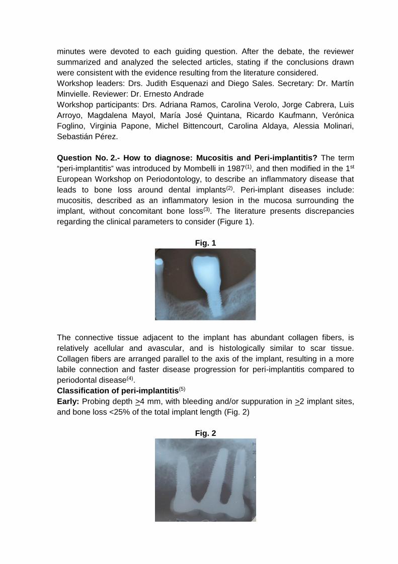
Question No. 2.- How to diagnose: Mucositis and Peri-implantitis? The term
“peri-implantitis” was introduced by Mombelli in 1987(1), and then modified in the 1st
European Workshop on Periodontology, to describe an inflammatory disease that
leads to bone loss around dental implants(2). Peri-implant diseases include:
mucositis, described as an inflammatory lesion in the mucosa surrounding the
implant, without concomitant bone loss(3). The literature presents discrepancies
regarding the clinical parameters to consider (Figure 1).
Fig. 1
The connective tissue adjacent to the implant has abundant collagen fibers, is
relatively acellular and avascular, and is histologically similar to scar tissue.
Collagen fibers are arranged parallel to the axis of the implant, resulting in a more
labile connection and faster disease progression for peri-implantitis compared to
periodontal disease(4).
Classification of peri-implantitis(5)
Early: Probing depth >4 mm, with bleeding and/or suppuration in >2 implant sites,
and bone loss <25% of the total implant length (Fig. 2)
Fig. 2
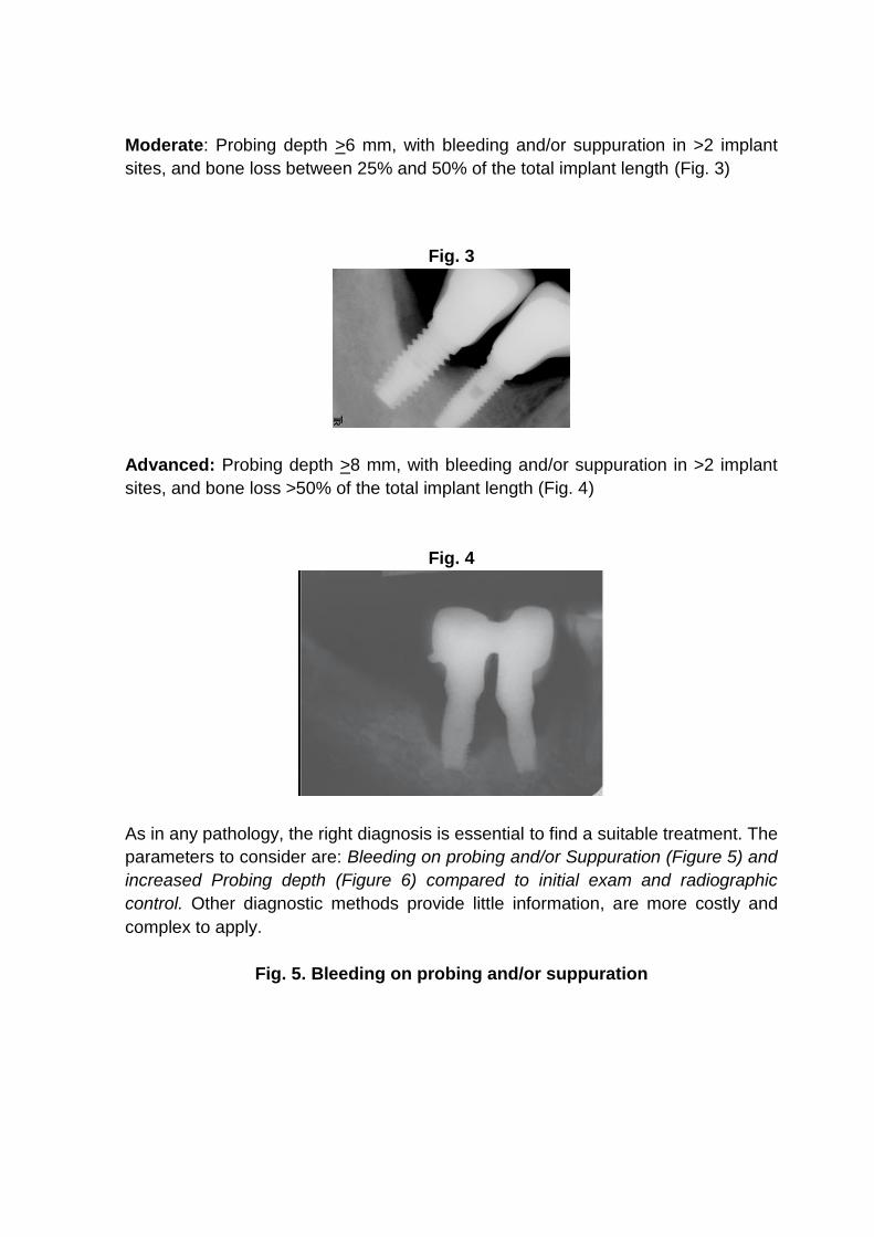
Moderate: Probing depth >6 mm, with bleeding and/or suppuration in >2 implant
sites, and bone loss between 25% and 50% of the total implant length (Fig. 3)
Fig. 3
Advanced: Probing depth >8 mm, with bleeding and/or suppuration in >2 implant
sites, and bone loss >50% of the total implant length (Fig. 4)
Fig. 4
As in any pathology, the right diagnosis is essential to find a suitable treatment. The
parameters to consider are: Bleeding on probing and/or Suppuration (Figure 5) and
increased Probing depth (Figure 6) compared to initial exam and radiographic
control. Other diagnostic methods provide little information, are more costly and
complex to apply.
Fig. 5. Bleeding on probing and/or suppuration
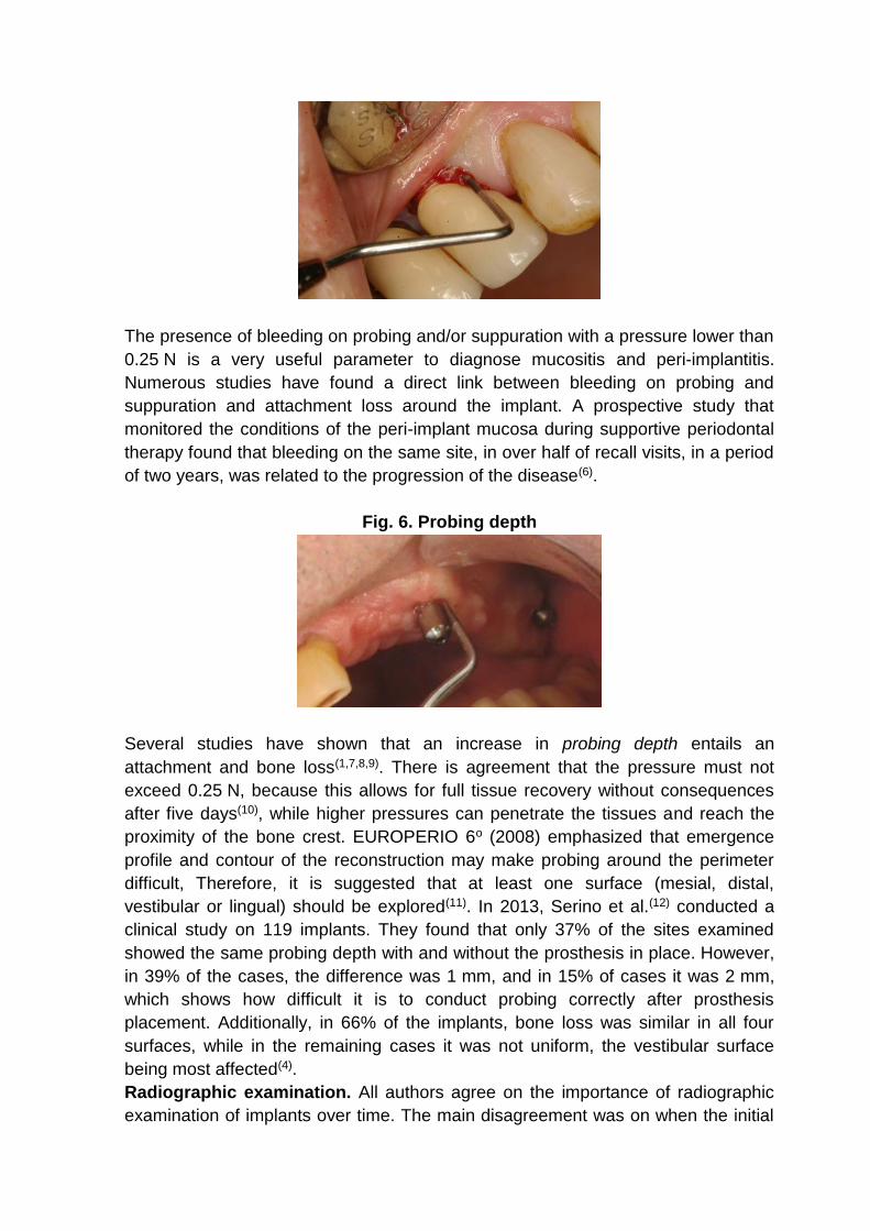
The presence of bleeding on probing and/or suppuration with a pressure lower than
0.25 N is a very useful parameter to diagnose mucositis and peri-implantitis.
Numerous studies have found a direct link between bleeding on probing and
suppuration and attachment loss around the implant. A prospective study that
monitored the conditions of the peri-implant mucosa during supportive periodontal
therapy found that bleeding on the same site, in over half of recall visits, in a period
of two years, was related to the progression of the disease(6).
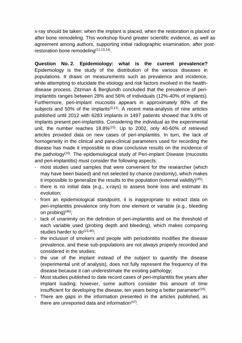
Fig. 6. Probing depth
Several studies have shown that an increase in probing depth entails an
attachment and bone loss(1,7,8,9). There is agreement that the pressure must not
exceed 0.25 N, because this allows for full tissue recovery without consequences
after five days(10), while higher pressures can penetrate the tissues and reach the
proximity of the bone crest. EUROPERIO 6o (2008) emphasized that emergence
profile and contour of the reconstruction may make probing around the perimeter
difficult, Therefore, it is suggested that at least one surface (mesial, distal,
vestibular or lingual) should be explored(11). In 2013, Serino et al.(12) conducted a
clinical study on 119 implants. They found that only 37% of the sites examined
showed the same probing depth with and without the prosthesis in place. However,
in 39% of the cases, the difference was 1 mm, and in 15% of cases it was 2 mm,
which shows how difficult it is to conduct probing correctly after prosthesis
placement. Additionally, in 66% of the implants, bone loss was similar in all four
surfaces, while in the remaining cases it was not uniform, the vestibular surface
being most affected(4).
Radiographic examination. All authors agree on the importance of radiographic
examination of implants over time. The main disagreement was on when the initial
x-ray should be taken: when the implant is placed, when the restoration is placed or
after bone remodeling. This workshop found greater scientific evidence, as well as
agreement among authors, supporting initial radiographic examination, after post-
restoration bone remodeling(11,13,14).
Question No. 2. Epidemiology: what is the current prevalence?
Epidemiology is the study of the distribution of the various diseases in
populations. It draws on measurements such as prevalence and incidence,
while attempting to elucidate the etiology and risk factors involved in the health-
disease process. Zitzman & Berglundh concluded that the prevalence of peri-
implantitis ranges between 28% and 56% of individuals (12%-40% of implants).
Furthermore, peri-implant mucositis appears in approximately 80% of the
subjects and 50% of the implants(3,11). A recent meta-analysis of nine articles
published until 2012 with 6283 implants in 1497 patients showed that 9.6% of
implants present peri-implantitis. Considering the individual as the experimental
unit, the number reaches 18.8%(15). Up to 2002, only 40-60% of retrieved
articles provided data on new cases of peri-implantitis. In turn, the lack of
homogeneity in the clinical and para-clinical parameters used for recording the
disease has made it impossible to draw conclusive results on the incidence of
the pathology(16). The epidemiological study of Peri-implant Disease (mucositis
and peri-implantitis) must consider the following aspects:
- most studies used samples that were convenient for the researcher (which
may have been biased) and not selected by chance (randomly), which makes
it impossible to generalize the results to the population (external validity)(45);
- there is no initial data (e.g., x-rays) to assess bone loss and estimate its
evolution;
- from an epidemiological standpoint, it is inappropriate to extract data on
peri-implantitis prevalence only from one element or variable (e.g., bleeding
on probing)(46);
- lack of unanimity on the definition of peri-implantitis and on the threshold of
each variable used (probing depth and bleeding), which makes comparing
studies harder to do(23,46);
- the inclusion of smokers and people with periodontitis modifies the disease
prevalence, and these sub-populations are not always properly recorded and
considered in the studies;
- the use of the implant instead of the subject to quantify the disease
(experimental unit of analysis), does not fully represent the frequency of the
disease because it can underestimate the existing pathology;
- Most studies published to date record cases of peri-implantitis five years after
implant loading; however, some authors consider this amount of time
insufficient for developing the disease, ten years being a better parameter(16).
- There are gaps in the information presented in the articles published, as
there are unreported data and information(47).
Question No. 3. Which are the risk factors? To identify a risk factor it is
necessary to conduct true prospective studies to follow up on a specific population,
and to establish a temporal connection between the exposure factor and the
disease. There have been very few studies with these characteristics, and the
available data generally stem from cross-sectional studies and series of cases that
allow us to identify risk indicators(17,18).
Which general, local and individual conditions can favor the appearance
of peri-implantitis? The workshop concluded that risk indicators with greater
evidence are:
Smoking: a meta-analysis that compares smokers and non-smokers
identifies smoking as an indicator of significant risk of marginal bone loss,
compared to non-smokers. This should be considered in the information
provided to the patient before the treatment, as well as in the maintenance
stage, where it is possible to detect early negative changes in peri-implant
tissues(17,19). Smoking has long-term deleterious effects on the immune and
inflammatory response, as well as on healing, with a lower production of
collagen, dysfunction of fibroblasts, reduction in peripheral circulation and in
the function of neutrophils and macrophages(20).
Poor oral hygiene: The accumulation of dental biofilm, caused by poor oral
hygiene, leads to peri-implant disease(11). The association between the
amount of biofilm and peri-implantitis appears to be dose-dependent, as
subjects with high values of biofilm are more likely to have a worse peri-
implant condition(21) (Fig.7).
Fig. 7
History of periodontal disease (analyzed below as it was considered as a
separate question).
On the other hand, lower-risk indicators are:
Diabetes: associated with xerostomia, dental caries and periodontal disease.
Increased susceptibility to periodontal disease occurs due to the negative
influence of hyperglycemia in the host’s inflammatory and immune
mechanisms, resulting in a deregulated response, altered healing and
microvasculature problems. It is not possible, for instance, to conduct a meta-
analysis because of the heterogeneity of the data available(22). A cross-
sectional study of 212 subjects, with 578 implants, included 29 diabetics. The
results of this study show that inadequate metabolic control in diabetic
patients increases the risk of peri-implantitis. Peri-implantitis was diagnosed
in 24.13% of diabetic patients, whereas in non-diabetics it was significantly
less than 6.56%(21).
Occlusal overload: The reports are controversial, as there is no unanimity in
the definition of occlusal overload. Implants are considered more labile than
teeth when facing axial forces given their lack of periodontal ligament(23).
Excessive stress can cause micro fractures and bone loss. A recent
systematic review(24) suggests that occlusal overload is positively associated
with marginal peri-implant bone resorption. However, biofilm remains the key
causal factor. More research is needed to define, with precision, what
occlusal loading is(23).
Alcoholism: A prospective three-year study evaluated the possible
connection between marginal peri-implant bone loss and tobacco and alcohol
consumption. The multivariate analysis showed that bone loss was
significantly associated with the consumption of >10g of alcohol per day(25).
Residual cement: a prospective study included 39 patients with 42 implants,
with peri-implantitis, which had been restored with single cemented crowns.
Twelve of them also had 20 restored implants under the same
circumstances, but without peri-implantitis (this second group of implants was
used as control group). A dental endoscope was used to explore the
subgingival area (DentalView, Irvine, CA). As a result, residual cement was
found in 34 of the 42 implants, and in none of the control implants. The
cement was removed using different techniques (surgical and non-surgical).
After 30 days, the signs of peri-implant disease had disappeared in 25 of the
33 implants placed in the experimental group. Therefore, excess cement and
its incomplete removal from the subgingival surface of the implant provide an
environment susceptible to bacterial colonization and the development of an
inflammatory response(26).
Surface of the implant: at present there is no evidence that the surface
characteristics of the implant may have a significant influence on the onset
the peri-implantitis(27).
Absence of keratinized gingiva: The deficiency of keratinized mucosa
around the implants seems to be related to clinical parameters of
inflammation and biofilm accumulation. However, based on the evidence
currently available, the amount of keratinized mucosa related to the
development of the peri-implantitis remains controversial(28.29).
Question No. 4. Periodontal and peri-implant disease history. In the last few
years, there has been an increase in the number of studies that analyze the
relationship between implantology and periodontics. Implantology, created for the
rehabilitation of fully edentulous patients, was later used to treat partially
edentulous patients. Late implant failure seems to be strongly connected to the
type of peri-implant bacterial flora and the conditions of the host(30). The biofilm
associated with implants with peri-implantitis is similar to that found in patients with
advanced periodontal disease(1,31). There are studies that show periodontal
pathogens colonizing the surface of the implants(32,33). Species such as
Porphyromonas gingivalis, Prevotella intermedia and Aggregatibacter
actinomycetemcomitans collected from advanced periodontal pockets (associated
with signs of inflammation) can transfer to peri-implant sites(34,35). The presence of
particularly virulent biofilm is linked with the appearance of peri-implantitis,
depending on the susceptibility of the host and the presence of risk factors (see
above). Considering the above, we should ask ourselves if susceptibility to
periodontitis increases the susceptibility to peri-implantitis, even in patients who
have been treated for periodontitis. Karoussis(36) was a pioneer in considering the
possibility that people with a history of periodontal disease are more likely to
develop peri-implantitis, compared to individuals without a history of periodontal
disease.
The analysis of the literature highlights the existence of two categories of studies:
a) those researching the success of the therapy in patients with a history of
periodontal disease compared to patients who did not suffer from it, and b) those
providing a direct comparison between patients with a history of destructive
periodontal disease and those with no history of such disease. Several systematic
reviews(37,38,39,40,41,41,43), as well as a prospective study with an average follow-up
period of 10 years, show that people with a history of periodontal disease are more
likely to develop peri-implantitis(44).
Question No. 5. Which are the therapeutic techniques to treat peri-
implantitis and how predictable are they? The therapies proposed to
manage peri-implantitis are based on the available evidence from the treatment
of periodontal disease. Therefore, the basic steps for resolution are: infection
control, surgical and non-surgical therapy, and supportive therapy. To facilitate
osseointegration, most implant manufacturers present moderately rough
surfaces in the market to increase the contact surface, which would be a
disadvantage if the implant is exposed to the oral environment. The roughness
of the surface and its chemical composition, as well as the design of threaded
implants, have a significant impact on the accumulation of oral biofilm(48). The
design of the prosthesis can hinder proper mechanical cleaning and treatment
of infected implants. Therefore, restoration adjustment is essential for
successful patient hygiene(49).
Based on the concepts mentioned above, numerous studies suggesting
different treatments such as mechanical debridement, use of antiseptics, local
and systemic antibiotics as well as surgical access and regenerative procedures
have been used, with varying results. The attempts to compare the information
available in the literature to draw solid conclusions have failed as there are no
data, and because the strict criteria of randomized controlled studies are not
met. It is difficult to have randomized controlled trials given the absence of a
true control group, the lack of a significant number of patients with similar
clinical features, and also for ethical reasons(50). A systematic review including
nine randomized controlled clinical studies, where researchers tried to identify
the most effective treatment, concluded that there was no reliable evidence to
suggest which treatment was the best one(51). Non-surgical therapy is
recommended as the first step. This allows us to evaluate the initial tissue
response and the patient's compliance with self-care measures, which will
largely determine the success of the therapeutic options proposed. Studies
where various forms of non-surgical treatments were compared (mechanical
debridement with titanium curettes and polishing with rubber cups vs.
mechanical debridement with ultrasonic devices and polishing with rubber cups
with antimicrobial administration) were analyzed based on variables such as
bleeding on probing, probing depth and biofilm deposits. In the follow-up
appointments after one, three and six months, no clinically relevant outcomes
were found to support that one treatment is better than the others(52).
Furthermore, when testing the use of laser devices (Er:YAG)(53) and the
application of glycine powder at limited pressure(54) with mechanical
debridement, researchers found no clinically or statistically significant
differences. However, non-surgical therapy alone is insufficient in most
peri-implantitis cases(52). Biofilm and calcified deposits must be removed to
allow healing and reduce the risk of disease progression. The surgical protocol
that involves surgical access, debridement, decontamination of the surface,
irrigation with saline and the use of systemic antibiotics has been evaluated,
with positive results over a 12-month period(55). On the other hand, studies
conducted by Romeo in 2007 mention implantoplasty techniques, i.e. polishing
the implant surface with diamond stones, rubbers and silicone materials,
repositioning the flap apically(56).
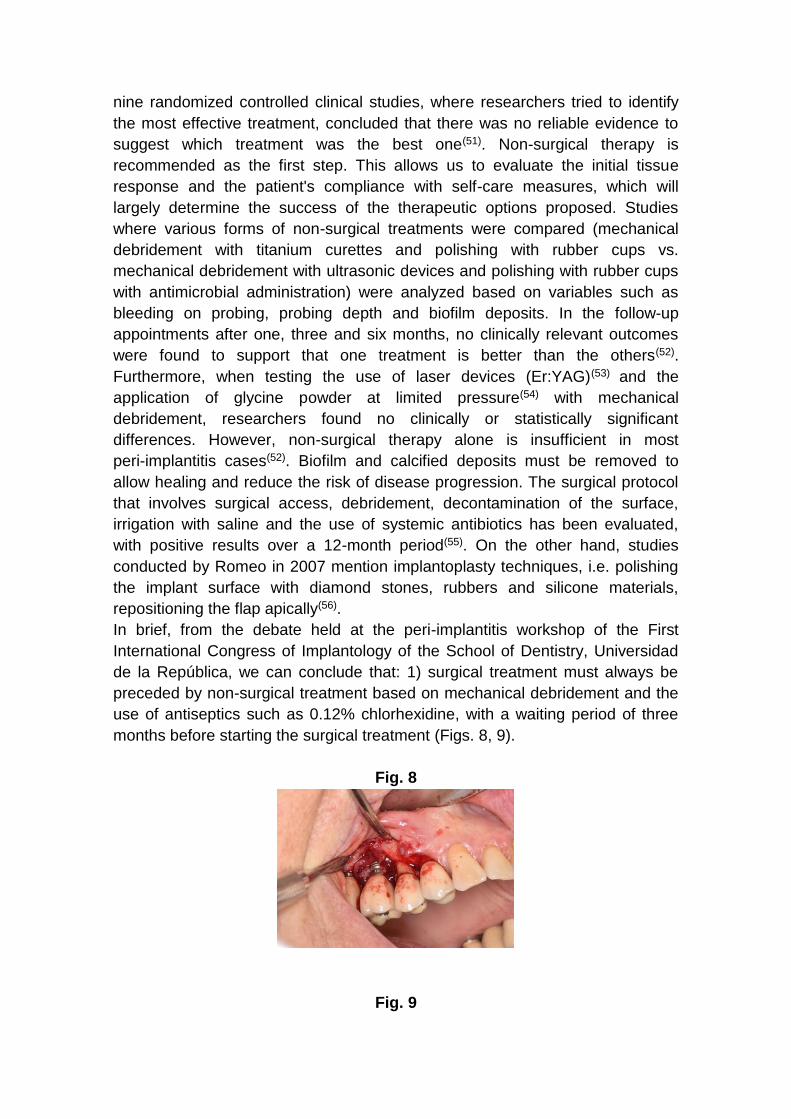
In brief, from the debate held at the peri-implantitis workshop of the First
International Congress of Implantology of the School of Dentistry, Universidad
de la República, we can conclude that: 1) surgical treatment must always be
preceded by non-surgical treatment based on mechanical debridement and the
use of antiseptics such as 0.12% chlorhexidine, with a waiting period of three
months before starting the surgical treatment (Figs. 8, 9).
Fig. 8
Fig. 9
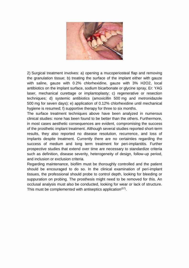
2) Surgical treatment involves: a) opening a mucoperiosteal flap and removing
the granulation tissue; b) treating the surface of the implant either with gauze
with saline, gauze with 0.2% chlorhexidine, gauze with 3% H2O2, local
antibiotics on the implant surface, sodium bicarbonate or glycine spray, Er: YAG
laser, mechanical curettage or implantoplasty; c) regenerative or resection
techniques; d) systemic antibiotics (amoxicillin 500 mg and metronidazole
500 mg for seven days); e) application of 0.12% chlorhexidine until mechanical
hygiene is resumed; f) supportive therapy for three to six months.
The surface treatment techniques above have been analyzed in numerous
clinical studies: none has been found to be better than the others. Furthermore,
in most cases aesthetic consequences are evident, compromising the success
of the prosthetic implant treatment. Although several studies reported short-term
results, they also reported no disease resolution, recurrence, and loss of
implants despite treatment. Currently there are no certainties regarding the
success of medium and long term treatment for peri-implantitis. Further
prospective studies that extend over time are necessary to standardize criteria
such as definition, disease severity, heterogeneity of design, follow-up period,
and inclusion or exclusion criteria.
Regarding maintenance, biofilm must be thoroughly controlled and the patient
should be encouraged to do so. In the clinical examination of peri-implant
tissues, the professional should probe to control depth, looking for bleeding or
suppuration on probing. The prosthesis might need to be removed for this. An
occlusal analysis must also be conducted, looking for wear or lack of structure.
This must be complemented with antiseptics application(57).