Embed Size (px)
Citation preview

Talonavicular Joint Abnormalities and Walking Ability
of Patients with Rheumatoid Arthritis
Noriyoshi Miyamoto , Masuo Senda, Masanori Hamada, Yoshimi Katayama,
Atsushi Kinosita, Kensuke Uchida, and Hajime Inoue
Department of Orthopaedic Surgery, Okayama University Graduate School of
Medicine and Dentistry, Okayama 700-8558, Japan
Rheumatoid arthritis(RA)is often associated with deformities of the feet, and foot pain often arises
in the talonavicular joint of patients with RA. The object of this study was to assess the relationship
between magnetic resonance imaging (MRI)findings of the talonavicular joint and walking ability.The subjects were 35 RA patients (10 feet in 5 males and 56 feet in 30 females)aged 34-87 years(mean:70 years±12.1), with a disease duration from 1-54 years (mean:14 years±12.1). MRI
findings were classified as follows:Grade 1, almost normal;Grade 2, early articular destruction;Grade 3, moderate articular destruction;Grade 4, severe articular destruction;and Grade 5, bony
ankylosis dislocation. Walking ability was classified into one of 9 categories ranging from normal
gait to bedridden status according to the system of Fujibayashi. As the grade of MRI images became
higher the walking ability decreased, and these parameters showed a correlation by Spearman’s
rank correlation coefficient analysis( =0.003). Thus, in the present cohort group of patients with
RA, the deterioration of walking ability increased with the severity of destruction of the
talonavicular joint.
Key words: rheumatoid arthritis, magnetic resonance imaging, talonavicular joint, walking ability
D eformity of the foot occurs frequently in patients
with rheumatoid arthritis (RA). Bone and joint
deformities are gradually caused by the thickening and
proliferation of the synovium and articular capsule and
loosening of muscles and ligaments. For abnormalities of
the hip or knee joint in RA patients, favorable results
have long been obtained by using artificial joints. In
contrast to the hip or knee, few reports have been
published on abnormalities of the foot in RA. In particu-lar, there have been few studies based on X-ray and
magnetic resonance imaging (MRI)examination of foot
abnormalities in RA. Mark et al.[1-4]reported that
deformity and pain of the talonavicular joint are common
in RA patients. Higashi et al.[5, 6]reported that
deformity of the foot occurs due to the depression of the
vertical axis arch. Such deformity may progress depend-ing on the severity of talonavicular joint subluxation and
may come to have a considerable influence on walking.The talonavicular joint thus has a significant influence on
quality of life(QOL)in RA patients. To date, however,there have been no few extensive evaluations of this joint.And while both total knee replacement (TKR)and total
hip replacement (THR)are established treatments, the
best therapy for foot abnormalities in RA remains contro-versial. There is thus need of further investigation into
deformity of the talonavicular joint in patients with RA.
Received April 18,2003;accepted November 26,2003.Corresponding author.Phone:+81-86-273-6073;Fax:+81-86-273-6073
http://www.lib.okayama-u.ac.jp/www/acta/
Acta Med. Okayama, 2004
Vol. 58, No. 2, pp. 85-9 0
Original Article
Copyrightc2004 by Okayama University Medical School.

We recently classified talonavicular joint abnormalities
based on MRI findings and evaluated their relationship to
walking ability in a general cohort. The objective of the
present study was to extend our exploration of the
relationship between MRI findings of the talonavicular
joint and walking ability to patients with RA.
Materials and Methods
We studied 66 feet of 35
patients with RA who had not undergone surgical treat-ment of the foot. There were 10 feet in 5 men and 56 feet
in 30 women, and the patients ranged in age from 34 to
87 years (mean age:70 years±12.1). The duration of
RA varied from 1 to 54 years (mean:14 years±12.1).The patients had been treated with nonsteroidal anti-inflammatory agents, steroids, gold preparations, and
immunosuppressants. There were 8 patients who had
been on steroid therapy with methylprednisolone(2 mg/1
day)for at least 1 year. Two patients had undergone
unilateral total hip replacement and 4 patients had received
unilateral total knee replacement. Laboratory test values
for rheumatoid factor(RAF), C reactive protein(CRP),and erythrocyte sedimentation rate (ESR), as well as
X-ray and MRI views of the talonavicular joints, were
reviewed. Walking ability was assessed using Fujibaya-shi’s classification[7].
Lateral X-ray films of the
talonavicular joint and views obtained at 30 degrees from
the ankle axis were reviewed. Talonavicular joint abnor-malities were classified according to Larsen’s
classification.[8]
Using a
brace, the foot was fixed with the ankle joint at 0 degrees,and sagittal and coronal T1-and T2-weighted images were
obtained(Fig. 1). The sagittal T2-weighted images were
used for evaluation. The MRI apparatus was a Toshiba
MRT-50GP(0.5 T). The routine study protocol consist-ed of T1-weighted images(reverse time(TR)/echo time(TE)=600-650/20-25 ms) and T2-weighted images(TR/TE=600-650/20-25 ms). The conditions were as
follows:matrix, 128×256;field of volume(FOV), 18
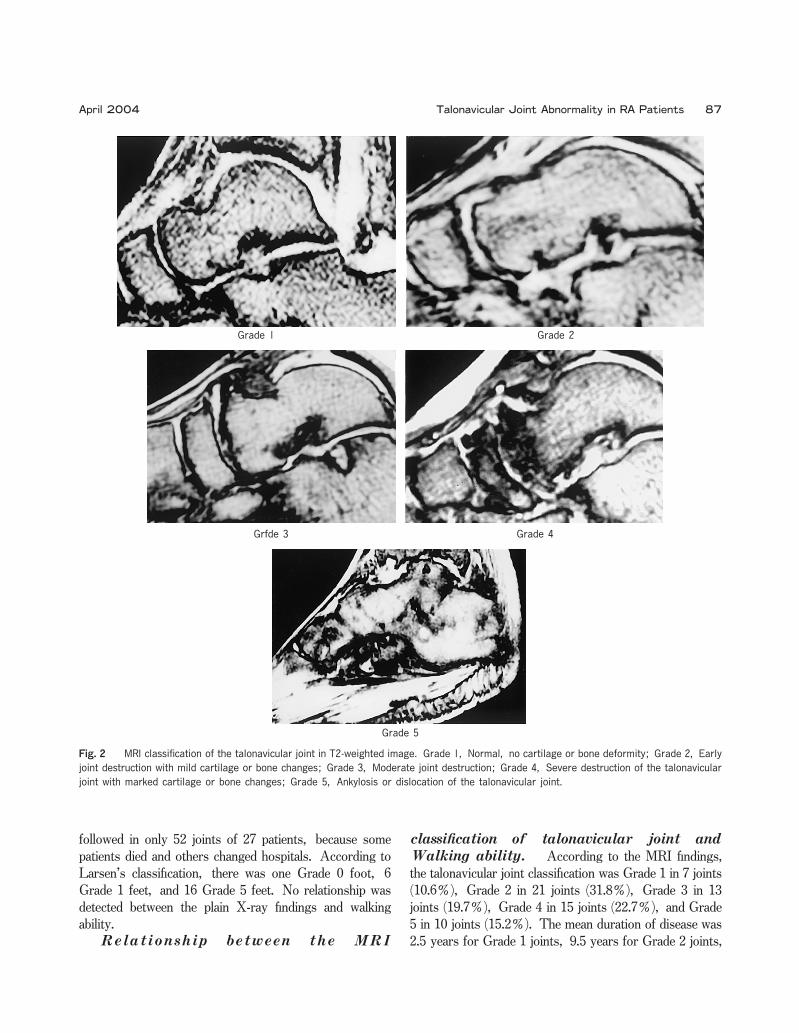
cm;slice, 5-mm thick with a 0.7-mm gap.The severity of talonavicular joint destruction was
classified as Grade 1-5 on the basis of MRI findings.Grade 1 joints showed no cartilage or bone deformity.Early joint destruction with mild cartilage or bone changes
was classified as Grade 2, while moderate joint destruc-tion was classified as Grade 3. Severe destruction of the
talonavicular joint with marked cartilage or bone changes
was classified as Grade 4, while ankylosis or dislocation
of the joint was classified as Grade 5(Fig. 2).All MRI scorings were reviewed by 2 independent
orthopaedists on the same day.
Walking
ability was classified according to the method of Fujibaya-shi[7]as follows:Class 2 patients walked normally.Class 3a patients could walk 500 to 1,000 m, but were
unable to ascend stairs, Class 3b patients could walk with
a walking stick, Class 3c patients could walk only in the
hospital garden, and Class 3d patients could walk only
indoors. Class 4a patients could walk minimally, Class 4b
patients could not use a wheelchair effectively, and Class
4d patients were bedridden.
Statistical analysis was
performed using Spearman’s rank correlation analysis
procedure, with the level of significance set at P<0.05.
Results
The
RA factor values ranged from(-)to (2+), the CRP
values from 0.15-11.9 (mean:4.12±3.31), and the ESR
values(1-h value)from 1.0-124(mean:55.5±25.4). No
correlation was found between abnormalities of the
talonavicular joint on MRI and any of the laboratory test
values.The X-ray changes could be
Miyamoto et al. Acta Med. Okayama Vol. 58, No. 2 86
Fig.1 Plastic gyps fixation. The foot was fixed with the ankle joint
at 0 degrees.
followed in only 52 joints of 27 patients, because some
patients died and others changed hospitals. According to
Larsen’s classification, there was one Grade 0 foot, 6
Grade 1 feet, and 16 Grade 5 feet. No relationship was
detected between the plain X-ray findings and walking
ability.
According to the MRI findings,the talonavicular joint classification was Grade 1 in 7 joints(10.6 ), Grade 2 in 21 joints (31.8 ), Grade 3 in 13
joints(19.7 ), Grade 4 in 15 joints(22.7 ), and Grade
5 in 10 joints(15.2 ). The mean duration of disease was
2.5 years for Grade 1 joints, 9.5 years for Grade 2 joints,
Talonavicular Joint Abnormality in RA Patients April 2004
Grade 1 Grade 2
Grfde 3 Grade 4
Grade 5
Fig.2 MRI classification of the talonavicular joint in T2-weighted image. Grade 1, Normal, no cartilage or bone deformity;Grade 2, Early
joint destruction with mild cartilage or bone changes;Grade 3, Moderate joint destruction;Grade 4, Severe destruction of the talonavicular
joint with marked cartilage or bone changes;Grade 5, Ankylosis or dislocation of the talonavicular joint.
87

13 years for Grade 3 joints, 17 years for Grade 4 joints,and 28 years for Grade 5 joints.Among patients with a Grade 1 joint on MRI, there
was one patient who was Class 2 and one who was Class
3a with respect to walking ability, and these patients had
almost no problems with walking. Among patients with a
Grade 2 talonavicular joint, walking ability was judged as
Class 2 in 2 patients, Class 3a in 2, Class 3b in 3, Class
3c in 2, Class 4c in 1, and Class 4d in 1. The patients
with Class 4c or Class 4d walking performance had
cerebral infarction as well as RA. Among patients with a
Grade 3 talonavicular joint, the walking performance was
Class 3a in 1 patient, Class 3b in 3, Class 3c in 3, and
Class 4b in 1. Among patients with a Grade 4
talonavicular joint, the walking performance was Class 3a
in 2 patients, Class 3b in 1, Class 3c in 1, Class 3d in
1, Class 4a in 1, and Class 4d in 1. Among patients with
a Grade 5 talonavicular joint, walking performance was
Class 4a in 1 patient and Class 4d in 5 patients(Fig. 3).Among patients in whom the MRI views of the
bilateral talonavicular joints were obtained, both joints
were usually of the same grade. When the 2 joints were
of different grades, the joint with the higher grade was
evaluated.Using Spearman’s rank correlation analysis, a
significant correlation was detected between the MRI
classification of talonavicular joint abnormalities and
walking performance(P=0.003).
Discussion
Deformity of the foot occurs relatively often in RA
and tends to cause disturbance of daily activities. Calabro
et al.[9]reported that valgus flat foot is a common
deformity in patients with RA and that talonavicular joint
abnormality is the main cause of such deformity. Mark
and Wigren et al.[1, 10]reported that deformity and
pain of the talonavicular joint are frequent problems
among RA patients with valgus flat foot. Cracchiolo,Elbor, and Hollinshead[11, 12, 13]proposed that the
talonavicular joint participate the deformity and pain of the
foot. The deformity and pain due to this joint are located
at the crest of the arch of the foot, often consist of plantar
flexion and dorsiflexion of the ankle. Because the ankle
joint is not surrounded by muscles, it sustains the body
weight on the articular surfaces of the bones(tibia, fibula,talus, and navicilar), and pressure is transmitted to the
talonavicular joint. Therefore, talonavicular joint deform-ity is gradually caused by such pressure. Higashi et al.[5]stated that shearing of the talonavicular joint can
rapidly cause depression of the vertical axis of the foot
arch, resulting in deformity of the foot. Therefore, we
considered that the talonavicular joint was important in the
etiology of foot pain in RA patients. Evaluation of this
joint is important when assessing pedal involvement by
RA. Despite these facts, there have been no few recent
reports on MRI evaluation of the talonavicular joint. We
considered that MRI may be an accurate method for the
assessment of talonavicular joint abnormalities, since it
more precisely visualizes the joint space, bones, and
synovial membrane than plain X-ray. When MRI was
performed to assess the talonavicular joint in our RA
patients, the ankle joint was fixed at 0 degrees. We
initially devised a wooden brace to fix the foot, but
switched to plastic-brace fixation because our patients with
advanced RA had pes equines and suffered severe pain
during MRI. Our use of 0-degree fixation may have
affected the results and thus may represent a limitation of
the present study. Although both T1-weighted and
T2-weighted images were obtained, the latter were more
suitable for investigation of synovial inflammation prolifer-ation and joint destruction. In addition, intra-and extra-articular soft tissues and effusions can be quantified by
T2-weighted images.We investigated the severity of synovial inflammation,
narrowing of the joint space, and bone destruction, and
classified the changes using a 5-grade scale. Among
patients with Grade 1 changes on MRI, there was mild
bone destruction and, in most cases, a walking perfor-mance of Class 2 or 3-i.e., no influence on daily activities.Most patients with Grade 2 changes on MRI were Class
Fig.3 MRI classification of talonavicular joint abnormalities were
significantly correlated with walking ability(P=0.003 by Spearman’s
rank correlation analysis).
Miyamoto et al. Acta Med. Okayama Vol. 58, No. 2 88

2 or 3 with respect to walking status, although 2 patients
with Grade 2 changes were almost bedridden due to
cerebral infarction and were Class 4 with respect to
walking. Patients with Grade 3 or Grade 4 changes on
MRI had advanced joint destruction and often were
confined to a wheelchair. Most patients with Grade 5
changes on MRI were bedridden. The significant correla-tion between MRI grade and walking performance sug-gested that the severity of talonavicular joint changes had
an influence on walking ability.The hip and knee joint have been extensively studied
in RA patients, and there are established treatment
modalities for damages to these joints, including total
joint replacement. However, deformity of the foot has an
impact on activities of daily living (ADL)in RA. It is
thus necessary to pay attention to the talonavicular joint as
well as the hip and knee joints. It may be beneficial to
perform surgical fixation when the joint is Grade 3 or
worse in order to reduce pain[3, 14, 15]. MRI
revealed advanced bone changes and Grade 3 joints, even
in patients who were Larsen‘s Grade 2 on plain X-ray
films. Thus, MRI showed the condition of the joint more
precisely than plain X-ray (Fig. 4), and MRI was
considered to be a more appropriate tool for assessing
talonavicular joint destruction in RA. Akagi et al.[16]
reported that foot disease was rarely the main factor
causing impairment of ADL and that there was no
consistent influence of deformity or X-ray changes of the
foot on ADL. However, we showed a correlation
between the MRI severity of talonavicular joint changes
and Fujibayashi’s classification of walking performance(Spearman’s rank correlation analysis, P=0.003). This
difference in results was likely due to the fact that Akagi
et al. used X-ray film data, whereas we used MRI data,which more precisely visualizes pathological changes of
the bones and joints. In conclusion, not only the hip and
knee joints, but also the talonavicular joint can influence
ADL and walking ability in patients with RA. It is
therefore necessary to pay more attention to the
talonavicular joint when assessing RA patients.
References
1. Figgie MP, Sobel M and Geppert MJ:The hind foot;in Surgical
Treatment of Rheumatoid Arthritis, Sculco TP ed, 1 th Ed, Mosby,New York(1992)pp 273-313.
2. Vidigal E, Jacoby RK, Dixon A, Ratliff AH and Kirkup J:The foot in
chronic rheumatoid arthritis. Ann Rheum Dis (1975)34:292-297.3. Ljung P, Kaij J, Knutson K, Pettersson H and Rydholm U:
Talonavicular arthrodesis in the rheumatoid foot. Foot Ankle (1992)13:313-316.
4. Vaino K:The rheumatoid foot. A clinical study with pathological and
Lersen III Grade 2
Lersen II Grade 3
Fig.4 MRI visualized the talonavicular joint more precisely than plain X-ray.
89 Talonavicular Joint Abnormality in RA Patients April 2004

roentgenographical comments. Ann Chir Gynaecol Suppl(1956)45:1.5. Higashi B, Yamamoto T, Kimura T, Oti T and Ono K:Natural course
and surgical treatment of hind foot deformity in Rheumatoid Arthritis.Rinsyouryumati(Clin Rheumatol)(1991)4:121-127(in Japanese).
6. Katsuki I and Torisu T:The pathomechanics of the deformity of
rheumatoid foot. Seikeisaigaigeka (Orthopaedic Surgery and
Traumatology)(1984)11:1605-1609 (in Japanese).7. Fujibayashi H, Gouda H, Maeno K, Kobayashi M, Fukumoto K,
Kitakawa T, Yokota M, Hamaguti M and Wada M:Effect of Rehabilita-tion on the arthritis patients;Due to follow-up studies. Rigakuryouhou
To Sagyouryouhou (The Japanese journal of physical therapy and
occupational therapy)(1977)11:209-217(in Japanese).
8. Larsen A, Dale K and Eek M:Radiographic evaluation of rheumatoid
arthritis and related conditoins by standard reference films. Acta
Radiol Diagn(1977)18:481-491.9. Calabro JJ:A critical evaluation of the diagnotic features of the feet
in rheumatoid arthritis. Arthrits Rheum(1962)5:19-29.
10. Wigren A:Operative treatment with special regard to the hind foot.
Arthritis Rheum(1987)11:100-103.11. Cracchiolo A:Surgical for rheumatoid disease. Instr Course Lect
(1984)33:386-411.12. Elbar JE, Thomas WH, Weinfeld MS and Potter TA:Talonavicular
arthrodesis for rheumatoid arthritis of the hindfoot. Orthop Clin North
Am(1976)7:821-826.
13. Hollinshead WH:Functional anatomy of the limbs and back. 3rd Ed,WB Saunders company, Philadelphia(1969)p 342.
14. Figgie MP, O’malley MJ, Ranawat CR, Inglis AE and Sculco TP:Triple
arthrodesis in rheumatoid arthrits. Cin Orthop(1993)292:250-254.15. Cracchiolo A IIIrd, Pearson S, Kitaoka H and Grace D:Hindfoot
arthrodesis in adults utilizing a dowel graft technique. Clin Orthop(1990)257:193-203.
16. Akagi S, Saitou T, Morimoto T, Sugano H and Ogawa R:Hind
deformity and its signnificance for ADL in rheumatoid arthritis patients
of long duration. Seikeisaigaigeka (Orthopaedic Surgery and
Traumatology)(1993)36:1623-1630(in Japanese).
Miyamoto et al. Acta Med. Okayama Vol. 58, No. 2 9 0