Embed Size (px)
Citation preview
Targeted enrichment of ancient pathogens yieldingthe pPCP1 plasmid of Yersinia pestis from victimsof the Black DeathVerena J. Schuenemanna,1, Kirsten Bosb,1, Sharon DeWittec, Sarah Schmedesd, Joslyn Jamiesonb, Alissa Mittnika,Stephen Forrestb, Brian K. Coombese, James W. Woodf,g, David J. D. Earne,h, William Whitei,2, Johannes Krausea,j,3,and Hendrik N. Poinarb,e,3
aInstitut für Naturwissenschaftliche Archäologie, University of Tübingen, 72072 Tübingen, Germany; bMcMaster Ancient DNA Centre, Department ofAnthropology, McMaster University, Hamilton, ON, Canada L8S 4L8; cDepartment of Anthropology, University at Albany, State University of New York,Albany, NY 12222; dInstitute of Investigative Genetics, University of North Texas Health Science Center, Fort Worth, TX 76107; eMichael G. DeGroote Institutefor Infectious Disease Research, McMaster University, Hamilton, ON, Canada L8S 4L8; fDepartment of Anthropology, Pennsylvania State University, UniversityPark, PA 16802; gPopulation Research Institute, Pennsylvania State University, University Park, PA 16802; hDepartment of Mathematics and Statistics,McMaster University, Hamilton, ON, Canada L8S 4L8; iCentre for Human Bioarcheology, Museum of London, London EC2Y 5HN, United Kingdom; and jHumanGenetics Department, Medical Faculty, University of Tübingen, 72076 Tübingen, Germany
Edited by Francisco Mauro Salzano, Instituto de Biociencias, Porto Alegre, RS, Brazil, and approved July 22, 2011 (received for review March 30, 2011)
Although investigations of medieval plague victims have identi-fied Yersinia pestis as the putative etiologic agent of the pan-demic, methodological limitations have prevented large-scalegenomic investigations to evaluate changes in the pathogen’s vir-ulence over time. We screened over 100 skeletal remains fromBlack Death victims of the East Smithfield mass burial site (1348–1350, London, England). Recent methods of DNA enrichment cou-pled with high-throughput DNA sequencing subsequently permit-ted reconstruction of ten full human mitochondrial genomes (16kb each) and the full pPCP1 (9.6 kb) virulence-associated plasmidat high coverage. Comparisons of molecular damage profiles be-tween endogenous human and Y. pestis DNA confirmed its au-thenticity as an ancient pathogen, thus representing the longestcontiguous genomic sequence for an ancient pathogen to date.Comparison of our reconstructed plasmid against modern Y. pestisshows identity with several isolates matching the Medievalis bio-var; however, our chromosomal sequences indicate the victimswere infected with a Y. pestis variant that has not been previouslyreported. Our data reveal that the Black Death in medieval Europewas caused by a variant of Y. pestis that may no longer exist, andgenetic data carried on its pPCP1 plasmid were not responsible forthe purported epidemiological differences between ancient andmodern forms of Y. pestis infections.
ancient DNA | paleopathology
The Black Death of 1347–1351 in Europe was one of the mostcataclysmic events in history, and it is arguably “one of the
most dramatic examples ever of emerging or reemerging disease”(ref. 1, p. 971). The disease is assumed to have been a particularlyintense pandemic of bubonic and pneumonic plague caused by theGram-negative bacillus Yersinia pestis. In this model, the medievalplague is considered the second of three pandemic waves, startingwith the Plague of Justinian in A.D. 541 and culminating in the20th century pandemic, which is still responsible for 2,000 cases/yworldwide (2) and is regarded as reemerging (3). Similarities indisease manifestation, mortality rates, and geographical distribu-tion are generally cited as factors relating the three pandemics,though some scholars have argued that the second wave was toodistinct in terms of its purported symptoms, epidemiology, andtime of year of peak mortality to warrant such a connection. Thesediscussants have argued in favor of other potential microbialassociations with the medieval disease, including Bacillus anthracis(4), a filovirus (5), or a pathogen that has since become extinct (6).Ancient DNA was sought to address the above controversy,
although failed attempts to replicate initial work showing thepresence of Y. pestis in purported victims of the medieval pan-demic ignited skepticism regarding the identity of its etiologic
agent (7, 8). Recent publications seem to have settled the con-troversy with the amplification of short segments of Y. pestisDNA by PCR-based approaches in several skeletal collectionsfrom time periods associated with medieval plague outbreaks (9,10), though none of these collections can be conclusively asso-ciated with the purported initial disease outbreak in medievalEurope of 1347–1351. A global survey of modern Y. pestis var-iants suggests that the ancient forms possess a unique andancestral phylogenetic placement (9, 11). Although standardmolecular methods can permit identification and limited phylo-genetic resolution of ancient microbes, successful characteriza-tion of long contiguous stretches of authentic pathogen DNAwill facilitate greater insight into the molecular architecture ofancient host–pathogen interactions. For Y. pestis, such an ap-proach might be informative in addressing the noted differencesbetween ancient and modern forms of the disease (4, 5, 12, 13).PCR-based approaches are ill-suited for large-scale genetic
investigations of ancient DNA owing to their preferential am-plification of less damaged templates that derive from exogenouscontaminants and the highly fragmented nature of endogenousmolecules (14, 15). In contrast, targeted enrichment strategies(16) in combination with high-throughput DNA sequencing al-low for long stretches of ancient DNA to be reconstructed froma complex metagenomic background, and it is clearly the wayforward for ancient pathogen research. The authenticity of en-dogenous ancient sequences can then be determined by lookingfor patterns of nucleotide damage typical of ancient DNA (15,17). To show the suitability of these methods for analyses ofancient pathogens, we have identified virulence-associated Y.
Author contributions: K.B., S.D., B.K.C., J.W.W., D.J.D.E., W.W., J.K., and H.N.P. designedresearch; V.J.S., K.B., S.D., S.S., J.J., A.M., S.F., J.W.W., and J.K. performed research; B.K.C.and W.W. contributed new reagents/analytic tools; V.J.S., K.B., J.K., and H.N.P. analyzeddata; and K.B., J.K., and H.N.P. wrote the paper.
The authors declare no conflict of interest.
This article is a PNAS Direct Submission.
See Commentary on page 15669.
Freely available online through the PNAS open access option.
Data deposition: The sequence reported in this paper has been deposited in the Genbankdatabase (accession nos. HE576978–HE576987).1V.J.S. and K.B. contributed equally to this work.2Deceased December, 2010.3To whom correspondence may be addressed. E-mail: [email protected] [email protected].
See Author Summary on page 15673.
This article contains supporting information online at www.pnas.org/lookup/suppl/doi:10.1073/pnas.1105107108/-/DCSupplemental.
E746–E752 | PNAS | September 20, 2011 | vol. 108 | no. 38 www.pnas.org/cgi/doi/10.1073/pnas.1105107108
Dow
nloa
ded
by g
uest
on
Feb
ruar
y 18
, 202
2
pestis DNA fragments by PCR and independently replicatedthese results through subsequent targeted DNA enrichment (18)and high-throughput sequencing for several skeletal samplesfrom victims securely dated to the initial medieval Black Deathpandemic of 1348–1350 in London, England. The data presentedhere represent both the oldest and longest assembled authenticsequences from an ancient pathogen, and in turn, they suggestthat the Black Death was caused by a Y. pestis variant that har-bors a pPCP1 plasmid found in some modern isolates.
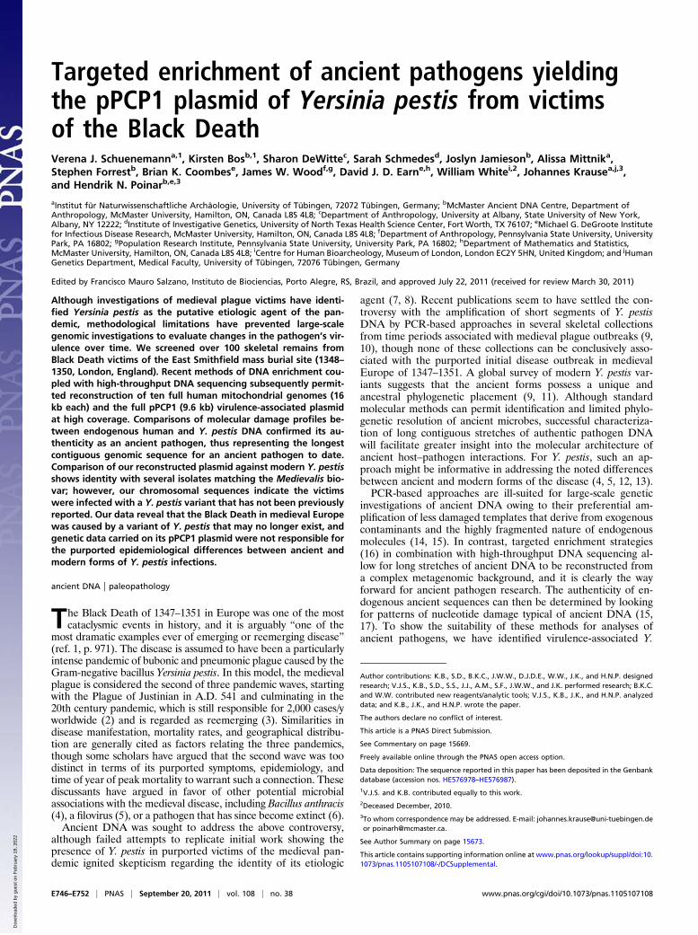
ResultsY. pestis DNA Preservation in Skeletal Remains. Complete infor-mation regarding skeletal screening is available in SI Materials andMethods. In total, we screened DNA extracts from 109 samples—53 bones and 46 teeth from the East Smithfield (ES) collectionas well as 10 samples from St. Nicholas Shambles (SNS) thatserved as negative controls (Fig. 1 and Table S1). Total DNAcontent measured fluorometrically revealed less DNA in teeththan in bone, with dental extracts yielding on average 1,065 and664 pg/μL for supernatant and pellet, respectively, compared with1,956 and 2,280 pg/μL for bone. Quantitative PCR (qPCR) resultsshowed the presence of amplifiable Y. pestis pla DNA in 5.7%of bones (3/53) and 37% of teeth (17/46) (Figs. S1 and S2). Thecaf1M assay yielded expected products in 5 of 17 teeth that con-tained amplifiable pla, although this finding is expected based onY. pestis plasmid numbers where the pPCP1 plasmid outnumbersthe pMT1 by an estimated 100 to 1 (19). No Y. pestis DNA wasdetected in any of the negative control samples. DNA sequencesfrom the qPCR products contained both G to A and C to Ttransitions, damage patterns typical of ancient DNA (17). Despitetheir lower whole DNA content, dental samples were consistentlya richer source for Y. pestis DNA than bone, and higher copynumbers were frequently observed in the EDTA supernatants asopposed to the pellets (Table S1). This finding is consistent withwhat one might expect from a blood-borne pathogen, where DNAlikely resides in the desiccated blood vessels of the pulp chamber.All amplifiable DNA was extremely low in quantity, with a maxi-mum estimated copy number of 30 copies/μL.
Multiplex PCR Data for the pla Gene. Using a multiplex PCR ap-proach, we were successful in sequencing 78.3% of the pla geneand its flanking intergenic spacers at a minimum of four timescoverage (Dataset S1). Missing regions were caused by a lack ofexpected PCR product. Cloned sequence data revealed 63 sitesshowing DNA damage reflected in the predominance of C to T(53.97%) and G to A (30.16%) transitions, which accounted for84.13% of all aberrant positions. No transversions were ob-served. To distinguish damage from potential polymorphisms, allsequences were confirmed by cloned data from two independentamplified libraries. Wherever products from these two librariesdid not match, cloned sequence data from a third amplified li-
brary was sought. This process permitted us to resolve all tran-sitions with the exception of three, where single nucleotidedifferences from published sequences were present in a subset ofclones from two independently amplified libraries (Fig. S3). ThePCR data did not contain the Microtus-specific C to T (20) atposition 1,109 in Dataset S1, and it does not show the T to Ctransition previously reported in an ancient sequence for thisregion in the work by Raoult et al. (7) (Dataset S1, position 809).
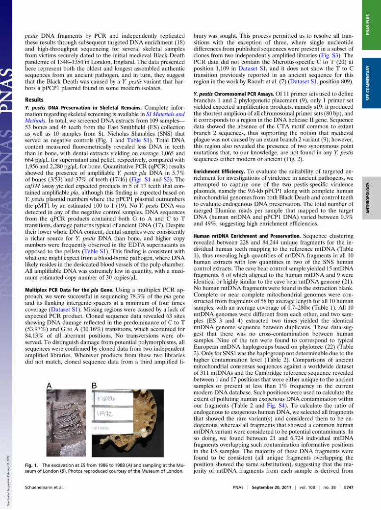
Y. pestis Chromosomal PCR Assays. Of 11 primer sets used to definebranches 1 and 2 phylogenetic placement (9), only 1 primer setyielded expected amplification products, namely s19: it producedthe shortest amplicon of all chromosomal primer sets (80 bp), andit corresponds to a region in the DNA helicase II gene. Sequencedata showed the absence of the CTA motif common to extantbranch 2 sequences, thus supporting the notion that medievalplague was not caused by an extant branch 2 variant (9); however,this region also revealed the presence of two synonymous pointmutations that, to our knowledge, are not found in any Y. pestissequences either modern or ancient (Fig. 2).
Enrichment Efficiency. To evaluate the suitability of targeted en-richment for investigations of virulence in ancient pathogens, weattempted to capture one of the two pestis-specific virulenceplasmids, namely the 9.6-kb pPCP1 along with complete humanmitochondrial genomes from both Black Death and control teethto evaluate endogenous DNA preservation. The total number ofmerged Illumina reads per sample that mapped to the targetDNA (human mtDNA and pPCP1 DNA) varied between 0.3%and 49%, suggesting high enrichment efficiencies.
Human mtDNA Enrichment and Preservation. Sequence clusteringrevealed between 228 and 84,244 unique fragments for the in-dividual human teeth mapping to the reference mtDNA (Table1), thus revealing high quantities of mtDNA fragments in all 10human extracts with low quantities in two of the SNS humancontrol extracts. The cave bear control sample yielded 15 mtDNAfragments, 6 of which aligned to the human mtDNA and 9 wereidentical or highly similar to the cave bear mtDNA genome (21).No human mtDNA fragments were found in the extraction blank.Complete or near complete mitochondrial genomes were con-structed from fragments of 58 bp average length for all 10 humansamples, with an average coverage of 0.7–280× (Table 1). All 10mtDNA genomes were different from each other, and two sam-ples (ES 3 and 4) extracted two times yielded the identicalmtDNA genome sequence between duplicates. These data sug-gest that there was no cross-contamination between humansamples. Nine of the ten were found to correspond to typicalEuropean mtDNA haplogroups based on phylotree (22) (Table2). Only for SNS1was the haplogroup not determinable due to thehigher contamination level (Table 2). Comparisons of ancientmitochondrial consensus sequences against a worldwide datasetof 311 mtDNAs and the Cambridge reference sequence revealedbetween 1 and 17 positions that were either unique to the ancientsamples or present at less than 1% frequency in the currentmodern DNA database. Such positions were used to calculate theextent of polluting human exogenous DNA contamination withinour fragments (Table 2 and Fig. S4). To calculate the ratio ofendogenous to exogenous human DNA, we selected all fragmentsthat showed the rare variant(s) and considered them to be en-dogenous, whereas all fragments that showed a common humanmtDNA variant were considered to be potential contaminants. Inso doing, we found between 21 and 6,724 individual mtDNAfragments overlapping such contamination informative positionsin the ES samples. The majority of these DNA fragments werefound to be consistent (all unique fragments overlapping theposition showed the same substitution), suggesting that the ma-jority of mtDNA fragments from each sample is derived from
Fig. 1. The excavation at ES from 1986 to 1988 (A) and sampling at the Mu-seum of London (B). Photos reproduced courtesy of the Museum of London.
Schuenemann et al. PNAS | September 20, 2011 | vol. 108 | no. 38 | E747
ANTH
ROPO
LOGY
PNASPL
US
SEECO
MMEN
TARY
Dow
nloa
ded
by g
uest
on
Feb
ruar
y 18
, 202
2
a single biological source (Fig. S4) (15, 23). Individual mtDNAfragments were compared against the corresponding consensussequence, and nucleotide substitutions were recorded for eachposition along the DNA fragment. Based on these estimates,levels of polluting human DNA could be calculated, althoughthese rates were very low for all samples (Table 2). In supportof this notion, fragments identified as endogenous DNAcontained degradation patterns typical for ancient templates(Fig. S5), again suggesting preservation of endogenous humanDNA in all 10 medieval teeth.
Y. pestis DNA Enrichment and Preservation. We found between 42and 36,986 unique fragments mapping to a portion of the Y.pestis pPCP1 plasmid reference genome in all libraries, includingthe pre-Black Death human control, the cave bear sample, andthe extraction blank (Table 1). All reads in the non-ES extractsmapped to a region between positions 3,000 and 4,200 of thereference pPCP1 that shows a high similarity to expression vec-tors that are used for recombinant enzyme production. This re-gion was found to be problematic in a previous Y. pestis pPCP1study using shotgun sequencing (24); hence, it is likely that rem-nants of the expression vectors are being captured by our enrich-
Fig. 2. Positional relationship of the sequences reportedfor medieval Y. pestis. (A) Quantitative PCR assay for thecaf1M locus of the pMT1 plasmid. (B) Chromosomal se-quence data for s19 (9) showing positions of two synony-mous substitutions in the DNA helicase II gene and absenceof the CTA SNP required for branch 2 designation (boxednucleotides). (C) Solexa consensus (minimum of two-foldcoverage) compared with 14 modern Y. pestis variantsshown with positional information for genes on the PCP1plasmid (rop, plasmid replication regulatory protein; pim,pesticin immunity protein; pst, pesticin; pla, plasminogenactivator gene). Transcriptional polarity (45) is indicated byarrows. Identity is shown in green; the region between3,000 and 4,200 is not considered (in the text). (D) Align-ment of the medieval Y. pestis pPCP1 consensus against 14modern variants showing presence of an indel common tobranch 2 and ancestral isolates.
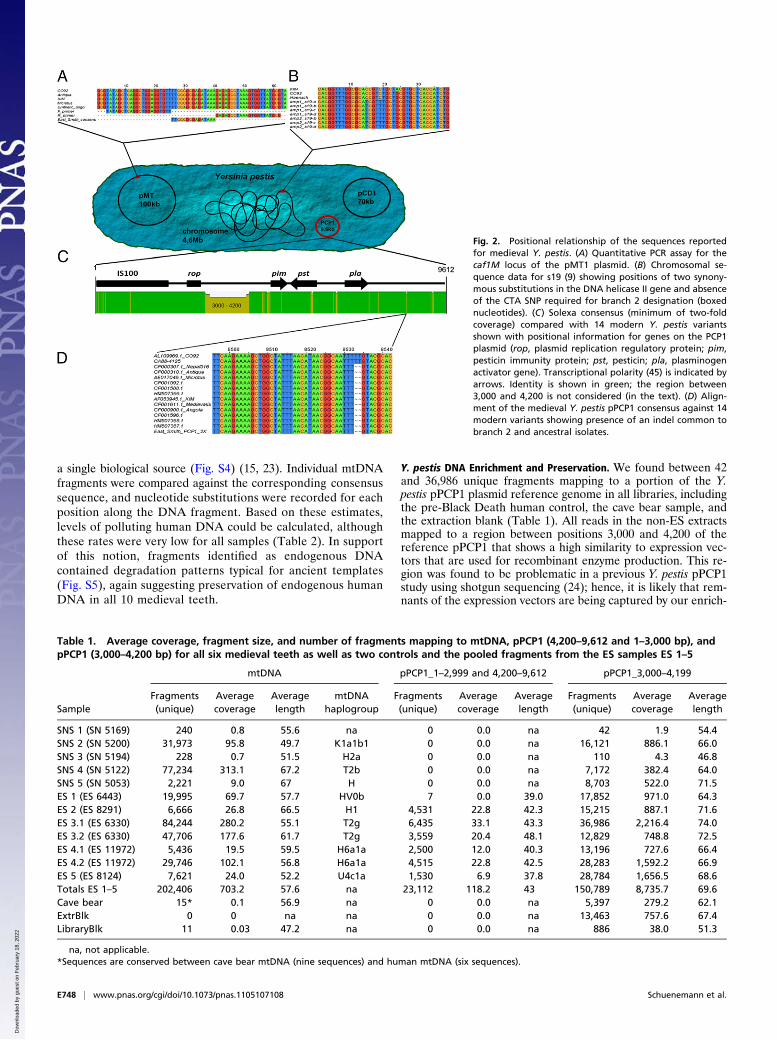
Table 1. Average coverage, fragment size, and number of fragments mapping to mtDNA, pPCP1 (4,200–9,612 and 1–3,000 bp), andpPCP1 (3,000–4,200 bp) for all six medieval teeth as well as two controls and the pooled fragments from the ES samples ES 1–5
mtDNA pPCP1_1–2,999 and 4,200–9,612 pPCP1_3,000–4,199
SampleFragments(unique)
Averagecoverage
Averagelength
mtDNAhaplogroup
Fragments(unique)
Averagecoverage
Averagelength
Fragments(unique)
Averagecoverage
Averagelength
SNS 1 (SN 5169) 240 0.8 55.6 na 0 0.0 na 42 1.9 54.4SNS 2 (SN 5200) 31,973 95.8 49.7 K1a1b1 0 0.0 na 16,121 886.1 66.0SNS 3 (SN 5194) 228 0.7 51.5 H2a 0 0.0 na 110 4.3 46.8SNS 4 (SN 5122) 77,234 313.1 67.2 T2b 0 0.0 na 7,172 382.4 64.0SNS 5 (SN 5053) 2,221 9.0 67 H 0 0.0 na 8,703 522.0 71.5ES 1 (ES 6443) 19,995 69.7 57.7 HV0b 7 0.0 39.0 17,852 971.0 64.3ES 2 (ES 8291) 6,666 26.8 66.5 H1 4,531 22.8 42.3 15,215 887.1 71.6ES 3.1 (ES 6330) 84,244 280.2 55.1 T2g 6,435 33.1 43.3 36,986 2,216.4 74.0ES 3.2 (ES 6330) 47,706 177.6 61.7 T2g 3,559 20.4 48.1 12,829 748.8 72.5ES 4.1 (ES 11972) 5,436 19.5 59.5 H6a1a 2,500 12.0 40.3 13,196 727.6 66.4ES 4.2 (ES 11972) 29,746 102.1 56.8 H6a1a 4,515 22.8 42.5 28,283 1,592.2 66.9ES 5 (ES 8124) 7,621 24.0 52.2 U4c1a 1,530 6.9 37.8 28,784 1,656.5 68.6Totals ES 1–5 202,406 703.2 57.6 na 23,112 118.2 43 150,789 8,735.7 69.6Cave bear 15* 0.1 56.9 na 0 0.0 na 5,397 279.2 62.1ExtrBlk 0 0 na na 0 0.0 na 13,463 757.6 67.4LibraryBlk 11 0.03 47.2 na 0 0.0 na 886 38.0 51.3
na, not applicable.*Sequences are conserved between cave bear mtDNA (nine sequences) and human mtDNA (six sequences).
E748 | www.pnas.org/cgi/doi/10.1073/pnas.1105107108 Schuenemann et al.
Dow
nloa
ded
by g
uest
on
Feb
ruar
y 18
, 202
2
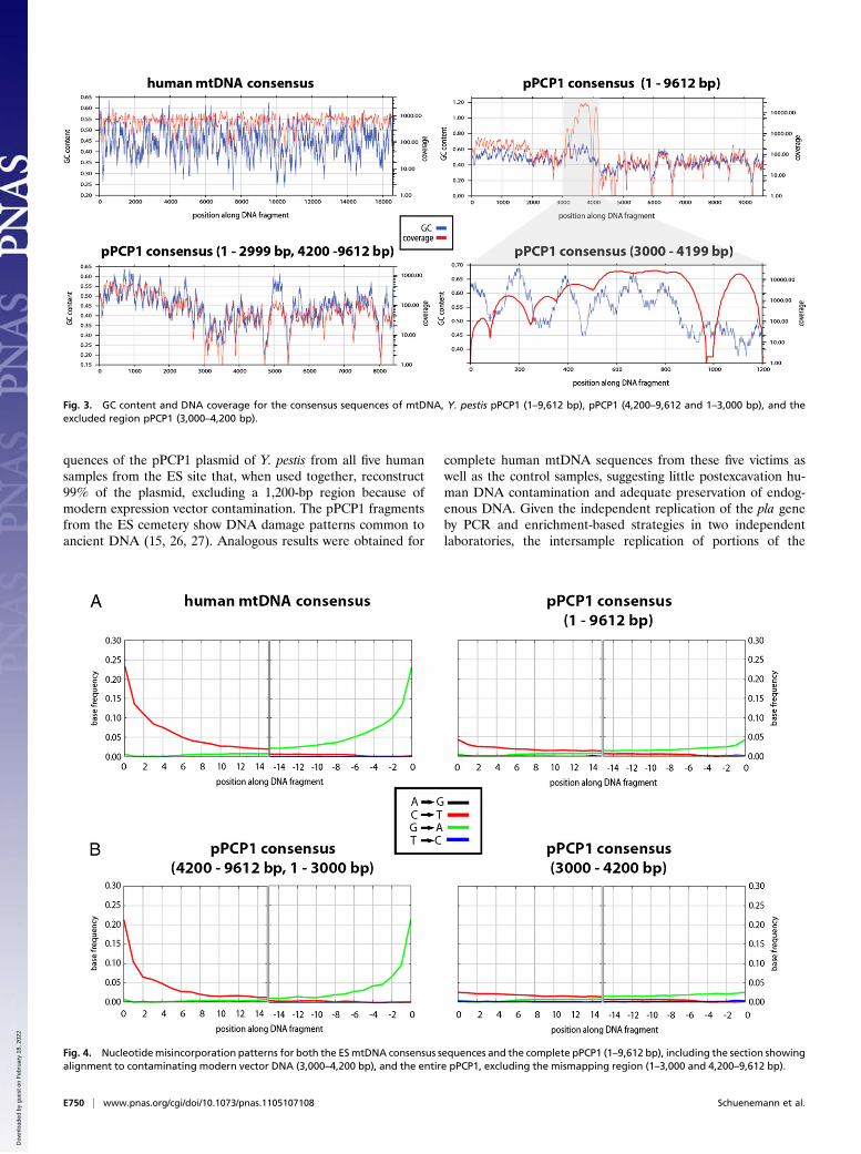
ment approach. Apart from these conserved motifs found acrossall samples and controls, the pre-Black Death human controlsamples (SNS) showed not a single unique fragment homologousto the pPCP1 sequence, whereas the five ES samples containedbetween 7 and 6,435 unique fragments that mapped to the Y. pestispPCP1 plasmid (Table 1). Because all five individuals from theES cemetery likely died from the same strain of Y. pestis, uniquepPCP1 fragments were pooled to reconstruct a consensus se-quence at maximum coverage. The pooled consensus has an av-erage coverage of 43 bp, ranging from 0× to 702×, and an averagefragment length of 36 bp (Table 1). This finding may be whya previous study using the ES collection that targeted regions inexcess of 130 bp (8) was unsuccessful in replicating some originalwork with plague samples (7, 25). Not surprisingly, the threeregions with the poorest coverage had a lowGC content (%) (Fig.3 and Fig. S6). Shotgun sequence data for ancient human samples(23) show similar patterns, suggesting a preservation bias to highGCcontent and thus likely ruling out enrichment artifacts.Despitelower coverage of AT-rich regions, the consensus sequence covers∼99% of the entire pPCP1 at a minimum of 2× (Table 3).We tested the authenticity of the pPCP1 DNA by analyzing
the substitution patterns along the DNA fragments and foundthe same nucleotide misincorporation pattern that was observedwithin the mtDNA fragments from the five medieval samples ofthe ES site. The predominant C to T substitutions found at the 5′ends and G to A substitutions at the 3′ ends are typical of ancientDNA (15, 26, 27). This pattern is not seen in the fragmentsmapping to the vector region of the pPCP1 plasmid, lendingadditional support to the notion that the majority of these
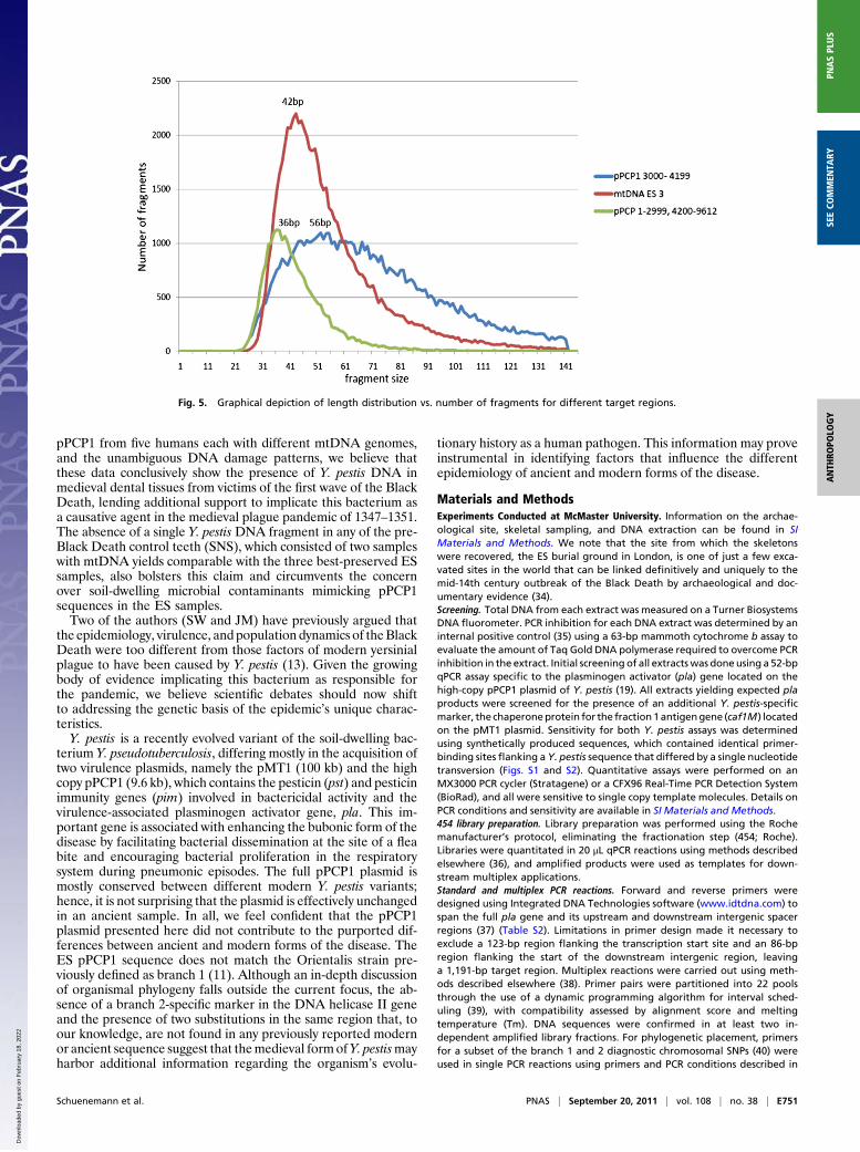
fragments are indeed derived from contaminating expressionvectors of the supplied reagents (Fig. 4 and Fig. S7). Fragmentlength distribution also indicates longer fragments obtained forthis region, which is consistent with their coming from a moderncontaminant source (Fig. 5).Phylogenetic comparison of the 8,299-bp 2× consensus pPCP1
plasmid with modern Y. pestis reveals sequence identity with 11of 14 extant Y. pestis strains; it did not match sequences typedas Microtus (AE017046.1), Angola (CP000900.1), and Medie-valis (CP001611.1), although it does match the KIM pPCP1(AF053945.1). It also resolves the three ambiguous positionsidentified by the multiplex approach of the pla gene and repli-cates the pla sequence described above. Considering indelpolymorphisms, the ES strain does not contain the TT insertioncommon to the Orientalis biovar (AL109969.1 and CA88-4125).
DiscussionThe last several decades have introduced human populations toa historically unprecedented number of emerging or reemerginginfectious diseases, mostly facilitated by anthropogenic factorssuch as globalization of trade and human travel, changes in localecology, and antibiotic resistance (28). Genetic diversity in path-ogens is known to be a major source of phenotypic diversity under-lying disease dynamics (29), hence microbial changes cannot beignored as a potential driving force influencing host–pathogeninteractions (30, 31). Genetic investigations of ancient microbesmay provide much needed data to elucidate how the virulenceof our close microbial companions has evolved over time. Po-tential factors that influenced the coevolution of humans andtheir pathogens in the past can be of great value to elucidate thedynamics of host–pathogen relationships in our new era ofemerging infections (32, 33).Here we report the presence of short DNA fragments (<60
bp) from two pestis-specific plasmids as well as one chromosomalsequence showing synonymous substitutions from individualsfrom a well-documented Black Death mass burial ground from1348 to 1350. These results were obtained in a facility that hasnot been previously exposed to modern sources of Y. pestis, andthe sequence data contain a damage pattern that is characteristicof ancient DNA (26). Using a targeted DNA capture approachcombined with high-throughput sequencing, we obtained se-
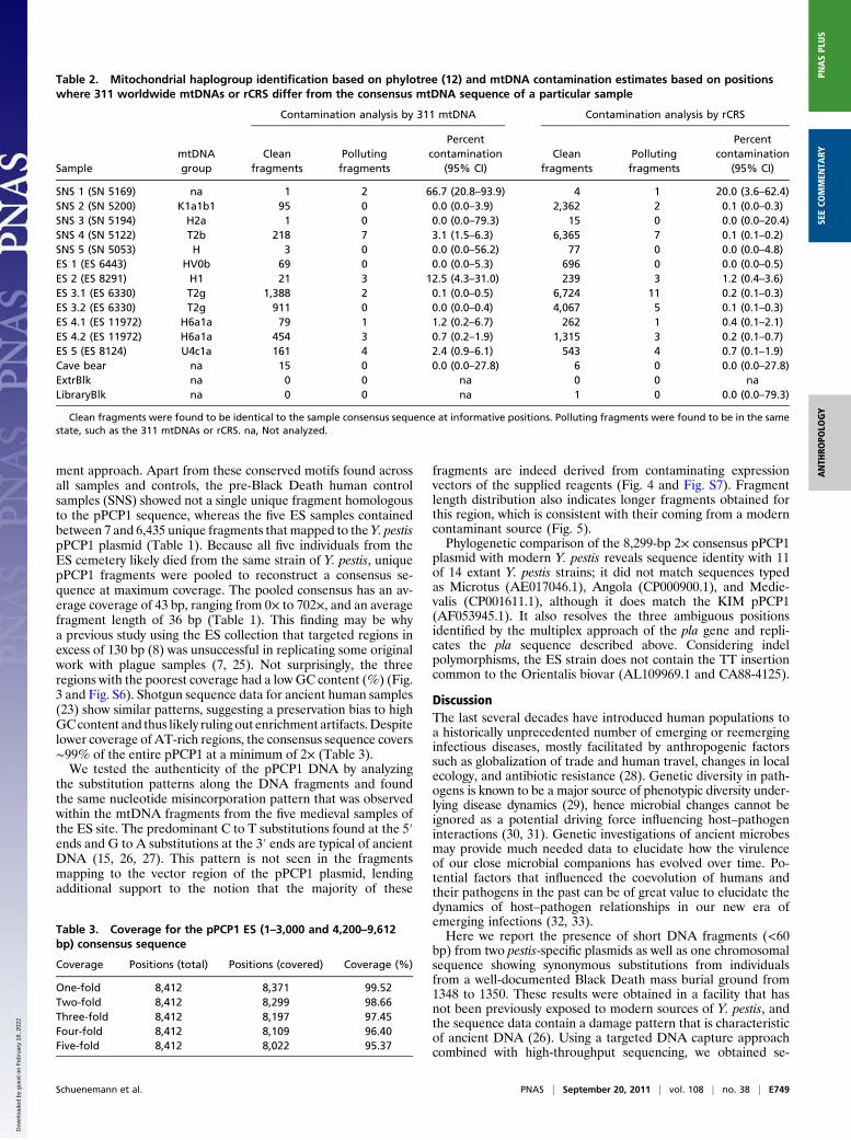
Table 2. Mitochondrial haplogroup identification based on phylotree (12) and mtDNA contamination estimates based on positionswhere 311 worldwide mtDNAs or rCRS differ from the consensus mtDNA sequence of a particular sample
Contamination analysis by 311 mtDNA Contamination analysis by rCRS
SamplemtDNAgroup
Cleanfragments
Pollutingfragments
Percentcontamination
(95% CI)Clean
fragmentsPollutingfragments
Percentcontamination
(95% CI)
SNS 1 (SN 5169) na 1 2 66.7 (20.8–93.9) 4 1 20.0 (3.6–62.4)SNS 2 (SN 5200) K1a1b1 95 0 0.0 (0.0–3.9) 2,362 2 0.1 (0.0–0.3)SNS 3 (SN 5194) H2a 1 0 0.0 (0.0–79.3) 15 0 0.0 (0.0–20.4)SNS 4 (SN 5122) T2b 218 7 3.1 (1.5–6.3) 6,365 7 0.1 (0.1–0.2)SNS 5 (SN 5053) H 3 0 0.0 (0.0–56.2) 77 0 0.0 (0.0–4.8)ES 1 (ES 6443) HV0b 69 0 0.0 (0.0–5.3) 696 0 0.0 (0.0–0.5)ES 2 (ES 8291) H1 21 3 12.5 (4.3–31.0) 239 3 1.2 (0.4–3.6)ES 3.1 (ES 6330) T2g 1,388 2 0.1 (0.0–0.5) 6,724 11 0.2 (0.1–0.3)ES 3.2 (ES 6330) T2g 911 0 0.0 (0.0–0.4) 4,067 5 0.1 (0.1–0.3)ES 4.1 (ES 11972) H6a1a 79 1 1.2 (0.2–6.7) 262 1 0.4 (0.1–2.1)ES 4.2 (ES 11972) H6a1a 454 3 0.7 (0.2–1.9) 1,315 3 0.2 (0.1–0.7)ES 5 (ES 8124) U4c1a 161 4 2.4 (0.9–6.1) 543 4 0.7 (0.1–1.9)Cave bear na 15 0 0.0 (0.0–27.8) 6 0 0.0 (0.0–27.8)ExtrBlk na 0 0 na 0 0 naLibraryBlk na 0 0 na 1 0 0.0 (0.0–79.3)
Clean fragments were found to be identical to the sample consensus sequence at informative positions. Polluting fragments were found to be in the samestate, such as the 311 mtDNAs or rCRS. na, Not analyzed.
Table 3. Coverage for the pPCP1 ES (1–3,000 and 4,200–9,612bp) consensus sequence
Coverage Positions (total) Positions (covered) Coverage (%)
One-fold 8,412 8,371 99.52Two-fold 8,412 8,299 98.66Three-fold 8,412 8,197 97.45Four-fold 8,412 8,109 96.40Five-fold 8,412 8,022 95.37
Schuenemann et al. PNAS | September 20, 2011 | vol. 108 | no. 38 | E749
ANTH
ROPO
LOGY
PNASPL
US
SEECO
MMEN
TARY
Dow
nloa
ded
by g
uest
on
Feb
ruar
y 18
, 202
2
quences of the pPCP1 plasmid of Y. pestis from all five humansamples from the ES site that, when used together, reconstruct99% of the plasmid, excluding a 1,200-bp region because ofmodern expression vector contamination. The pPCP1 fragmentsfrom the ES cemetery show DNA damage patterns common toancient DNA (15, 26, 27). Analogous results were obtained for
complete human mtDNA sequences from these five victims aswell as the control samples, suggesting little postexcavation hu-man DNA contamination and adequate preservation of endog-enous DNA. Given the independent replication of the pla geneby PCR and enrichment-based strategies in two independentlaboratories, the intersample replication of portions of the
Fig. 4. Nucleotidemisincorporation patterns for both the ESmtDNA consensus sequences and the complete pPCP1 (1–9,612 bp), including the section showingalignment to contaminating modern vector DNA (3,000–4,200 bp), and the entire pPCP1, excluding the mismapping region (1–3,000 and 4,200–9,612 bp).
Fig. 3. GC content and DNA coverage for the consensus sequences of mtDNA, Y. pestis pPCP1 (1–9,612 bp), pPCP1 (4,200–9,612 and 1–3,000 bp), and theexcluded region pPCP1 (3,000–4,200 bp).
E750 | www.pnas.org/cgi/doi/10.1073/pnas.1105107108 Schuenemann et al.
Dow
nloa
ded
by g
uest
on
Feb
ruar
y 18
, 202
2
pPCP1 from five humans each with different mtDNA genomes,and the unambiguous DNA damage patterns, we believe thatthese data conclusively show the presence of Y. pestis DNA inmedieval dental tissues from victims of the first wave of the BlackDeath, lending additional support to implicate this bacterium asa causative agent in the medieval plague pandemic of 1347–1351.The absence of a single Y. pestis DNA fragment in any of the pre-Black Death control teeth (SNS), which consisted of two sampleswith mtDNA yields comparable with the three best-preserved ESsamples, also bolsters this claim and circumvents the concernover soil-dwelling microbial contaminants mimicking pPCP1sequences in the ES samples.Two of the authors (SW and JM) have previously argued that
the epidemiology, virulence, andpopulation dynamics of theBlackDeath were too different from those factors of modern yersinialplague to have been caused by Y. pestis (13). Given the growingbody of evidence implicating this bacterium as responsible forthe pandemic, we believe scientific debates should now shiftto addressing the genetic basis of the epidemic’s unique charac-teristics.Y. pestis is a recently evolved variant of the soil-dwelling bac-
terium Y. pseudotuberculosis, differing mostly in the acquisition oftwo virulence plasmids, namely the pMT1 (100 kb) and the highcopy pPCP1 (9.6 kb), which contains the pesticin (pst) and pesticinimmunity genes (pim) involved in bactericidal activity and thevirulence-associated plasminogen activator gene, pla. This im-portant gene is associated with enhancing the bubonic form of thedisease by facilitating bacterial dissemination at the site of a fleabite and encouraging bacterial proliferation in the respiratorysystem during pneumonic episodes. The full pPCP1 plasmid ismostly conserved between different modern Y. pestis variants;hence, it is not surprising that the plasmid is effectively unchangedin an ancient sample. In all, we feel confident that the pPCP1plasmid presented here did not contribute to the purported dif-ferences between ancient and modern forms of the disease. TheES pPCP1 sequence does not match the Orientalis strain pre-viously defined as branch 1 (11). Although an in-depth discussionof organismal phylogeny falls outside the current focus, the ab-sence of a branch 2-specific marker in the DNA helicase II geneand the presence of two substitutions in the same region that, toour knowledge, are not found in any previously reported modernor ancient sequence suggest that themedieval formofY. pestismayharbor additional information regarding the organism’s evolu-
tionary history as a human pathogen. This information may proveinstrumental in identifying factors that influence the differentepidemiology of ancient and modern forms of the disease.
Materials and MethodsExperiments Conducted at McMaster University. Information on the archae-ological site, skeletal sampling, and DNA extraction can be found in SIMaterials and Methods. We note that the site from which the skeletonswere recovered, the ES burial ground in London, is one of just a few exca-vated sites in the world that can be linked definitively and uniquely to themid-14th century outbreak of the Black Death by archaeological and doc-umentary evidence (34).Screening. Total DNA from each extract was measured on a Turner BiosystemsDNA fluorometer. PCR inhibition for each DNA extract was determined by aninternal positive control (35) using a 63-bp mammoth cytochrome b assay toevaluate the amount of Taq Gold DNA polymerase required to overcome PCRinhibition in the extract. Initial screening of all extractswas doneusing a 52-bpqPCR assay specific to the plasminogen activator (pla) gene located on thehigh-copy pPCP1 plasmid of Y. pestis (19). All extracts yielding expected plaproducts were screened for the presence of an additional Y. pestis-specificmarker, the chaperoneprotein for the fraction1antigengene (caf1M) locatedon the pMT1 plasmid. Sensitivity for both Y. pestis assays was determinedusing synthetically produced sequences, which contained identical primer-binding sites flanking a Y. pestis sequence that differed by a single nucleotidetransversion (Figs. S1 and S2). Quantitative assays were performed on anMX3000 PCR cycler (Stratagene) or a CFX96 Real-Time PCR Detection System(BioRad), and all were sensitive to single copy template molecules. Details onPCR conditions and sensitivity are available in SI Materials and Methods.454 library preparation. Library preparation was performed using the Rochemanufacturer’s protocol, eliminating the fractionation step (454; Roche).Libraries were quantitated in 20 μL qPCR reactions using methods describedelsewhere (36), and amplified products were used as templates for down-stream multiplex applications.Standard and multiplex PCR reactions. Forward and reverse primers weredesigned using Integrated DNA Technologies software (www.idtdna.com) tospan the full pla gene and its upstream and downstream intergenic spacerregions (37) (Table S2). Limitations in primer design made it necessary toexclude a 123-bp region flanking the transcription start site and an 86-bpregion flanking the start of the downstream intergenic region, leavinga 1,191-bp target region. Multiplex reactions were carried out using meth-ods described elsewhere (38). Primer pairs were partitioned into 22 poolsthrough the use of a dynamic programming algorithm for interval sched-uling (39), with compatibility assessed by alignment score and meltingtemperature (Tm). DNA sequences were confirmed in at least two in-dependent amplified library fractions. For phylogenetic placement, primersfor a subset of the branch 1 and 2 diagnostic chromosomal SNPs (40) wereused in single PCR reactions using primers and PCR conditions described in
Fig. 5. Graphical depiction of length distribution vs. number of fragments for different target regions.
Schuenemann et al. PNAS | September 20, 2011 | vol. 108 | no. 38 | E751
ANTH
ROPO
LOGY
PNASPL
US
SEECO
MMEN
TARY
Dow
nloa
ded
by g
uest
on
Feb
ruar
y 18
, 202
2
ref. 9. Primer sequences, PCR conditions, and the consensus sequence aredescribed in SI Materials and Methods.
Experiments Conducted at the Max Planck Institute in Leipzig, Germany. DNAextraction and enrichment. To independently replicate the PCR-amplified Y.pestis DNA from the ES collection and test the suitability of enrichmentstrategies and subsequent targeted high-throughput sequencing for ancientpathogens, several teethwere analyzed at theMax Planck Institute in Leipzig,Germany. Five dental roots from mature molars from the ES collection werechosen based on pla copy numbers and analyzed alongside five dental rootsfrom St. Nicholas Shambles (Table S1), and an ancient cave bear sample wasused as a cross-contamination control. An aliquot from each DNA extract wasused to produce libraries using a modified Illumina multiplex protocol (41).Endogenous mitochondrial DNAwas captured in parallel from all libraries. Toenrich for target DNA, we used long-range PCR products as bait for molecularcapture through hybridization (18). Additional details on extraction, librarypreparation, and enrichment are available in SI Materials and Methods.Solexa sequencing and analysis (Leipzig). Theenriched librarypoolwassequencedon an Illumina Genome Analyzer IIx using the manufacturer’s protocols formultiplex sequencing with modifications. The raw reads were aligned to thePhiX174 reference sequence toobtain a trainingdataset for thebase caller Ibis(42). Raw sequences called by Ibis 1.1.1 werefiltered for the individual indexesas previously described (43). The paired-end reads were subjected to a fusionprocess (including removal of adapter sequences and adaptor dimers) by re-quiring at least an 11-nt overlap between the two reads. In the overlappingsequence, quality scores were combined, and the base with the highest basequality score was called. Only sequences merged in this way were used foradditional analysis. The sequencing data were analyzed starting from QSEQ
sequence files and CIF intensity files from the Illumina Genome Analyzer RTA1.6 software. Enrichment efficiency was calculated by mapping all 37,502,405merged reads to the revised Cambridge Reference Sequence (rCRS) for humanmtDNA and the Y. pestis CO92 pPCP1 plasmid using a custom iterative map-ping assembler (23). From this information, the fraction of total reads thatmapped to the target DNA was calculated.Phylogenetic analysis. The consensus sequences for the mitochondrial genomefragments were analyzed using a custom Perl script to identify individualhaplogroups based on phylotree (22). All pPCP1 fragments from the ESsamples were combined into a single consensus sequence, and they werealigned and compared by eye with 14 modern pPCP1 sequences using thesoftware Muscle (44) (SI Materials and Methods).
ACKNOWLEDGMENTS. We thank Tomislav Maricic and Martin Kircher fortechnical expertise and analysis assistance. We thank Svante Pääbo for accessto the clean room and laboratory facilities of the Max Planck Institute forEvolutionary Anthropology in Leipzig, Germany. We also thank Rebecca Red-fern and Jelena Bekvalac for providing access to skeletal samples and facilitiesfor sampling at the Museum of London, Elizabeth Carniel for providing mod-ernYersinia pestisDNA, the entire team at theMcMaster Ancient DNACentrefor helpful comments on the design and implementation of laboratory work,and D. Ann Herring and Debi Poinar for helpful comments on earlier versionsof the manuscript. Financial support was provided by the Social Sciences andHumanities Research Council of Canada, the Canadian Institute for HealthResearch, Canada Research Chairs program, theMichael G. DeGroote Institutefor Infectious Disease Research, McMaster University, the University at AlbanyCenter for Social and Demographic Analysis, and the Human Genetics Depart-ment, Medical Faculty, University of Tübingen, Germany.
1. Wheelis M (2002) Biological warfare at the 1346 siege of Caffa. Emerg Infect Dis 8:971–975.
2. World Health Organization (2011) Zoonotic Infections. Available at www.who.int/vaccine_research/diseases/zoonotic/en/index3.html. Accessed August 11, 2011.
3. Stenseth NC, et al. (2008) Plague: Past, present, and future. PLoS Med 5:e3.4. Twigg G (1984) The Black Death: A Biological Reappraisal (Batsford Academic,
London).5. Scott S, Duncan CJ (2001) The Biology of Plagues (Cambridge University Press,
Cambridge, UK).6. Cohn SK (2003) The Black Death Transformed: Disease and Culture in Early
Renaissance Europe (Arnoldohn, London).7. Raoult D, et al. (2000) Molecular identification by “suicide PCR” of Yersinia pestis as
the agent of medieval black death. Proc Natl Acad Sci USA 97:12800–12803.8. Gilbert MT, et al. (2004) Absence of Yersinia pestis-specific DNA in human teeth from
five European excavations of putative plague victims. Microbiology 150:341–354.9. Haensch S, et al. (2010) Distinct clones of Yersinia pestis caused the black death. PLoS
Pathog 6:e1001134.10. Wiechmann I, Harbeck M, Grupe G (2010) Yersinia pestis DNA sequences in late
medieval skeletal finds, Bavaria. Emerg Infect Dis 16:1806–1807.11. Morelli G, et al. (2010) Yersinia pestis genome sequencing identifies patterns of
global phylogenetic diversity. Nat Genet 42:1140–1143.12. Wood JW, Ferrell RJ, Dewitte-Aviña SN (2003) The temporal dynamics of the
fourteenth-century Black Death: New evidence from English ecclesiastical records.Hum Biol 75:427–448.
13. Wood JW, DeWitte-Aviña SN (2003) Was the Black Death yersinial plague? LancetInfect Dis 3:327–328.
14. Pääbo S, et al. (2004) Genetic analyses from ancient DNA. Annu Rev Genet 38:645–679.
15. Krause J, et al. (2010) A complete mtDNA genome of an early modern human fromKostenki, Russia. Curr Biol 20:231–236.
16. Burbano HA, et al. (2010) Targeted investigation of the Neandertal genome by array-based sequence capture. Science 328:723–725.
17. Hofreiter M, Jaenicke V, Serre D, Haeseler Av A, Pääbo S (2001) DNA sequences frommultiple amplifications reveal artifacts induced by cytosine deamination in ancientDNA. Nucleic Acids Res 29:4793–4799.
18. Maricic T, Whitten M, Pääbo S (2010) Multiplexed DNA sequence capture ofmitochondrial genomes using PCR products. PLoS One 5:e14004.
19. Parkhill J, et al. (2001) Genome sequence of Yersinia pestis, the causative agent ofplague. Nature 413:523–527.
20. Song Y, et al. (2004) Complete genome sequence of Yersinia pestis strain 91001, anisolate avirulent to humans. DNA Res 11:179–197.
21. Krause J, et al. (2008) Mitochondrial genomes reveal an explosive radiation of extinctand extant bears near the Miocene-Pliocene boundary. BMC Evol Biol 8:220–232.
22. van Oven M, Kayser M (2009) Updated comprehensive phylogenetic tree of globalhuman mitochondrial DNA variation. Hum Mutat 30:E386–E394.
23. Green RE, et al. (2008) A complete Neandertal mitochondrial genome sequencedetermined by high-throughput sequencing. Cell 134:416–426.
24. Plunkett G, Anderson BD, Burland V, Cabot EL, Glasner JD, et al. (2007) Yersinia pestisCA88-4125 whole genome shotgun sequencing project, direct submission to NCBI,accession number NZ_ABCD01000008.1.
25. Drancourt M, Aboudharam G, Signoli M, Dutour O, Raoult D (1998) Detection of 400-year-old Yersinia pestis DNA in human dental pulp: An approach to the diagnosis ofancient septicemia. Proc Natl Acad Sci USA 95:12637–12640.
26. Briggs AW, et al. (2007) Patterns of damage in genomic DNA sequences froma Neandertal. Proc Natl Acad Sci USA 104:14616–14621.
27. Brotherton P, et al. (2007) Novel high-resolution characterization of ancient DNAreveals C > U-type base modification events as the sole cause of post mortemmiscoding lesions. Nucleic Acids Res 35:5717–5728.
28. Barrett R, Kuzawa CW, McDade T, Armelagos GJ (1998) Emerging and re-emerginginfectious diseases: The third epidemiologic transition. Annu Rev Anthropol 27:247–271.
29. Lawrence JG (2005) Common themes in the genome strategies of pathogens. CurrOpin Genet Dev 15:584–588.
30. Morse SS (1995) Factors in the emergence of infectious diseases. Emerg Infect Dis 1:7–15.
31. Pybus OG, Rambaut A (2009) Evolutionary analysis of the dynamics of viral infectiousdisease. Nat Rev Genet 10:540–550.
32. Baum J, Khalina Bar-Gal G (2003) Emerging Pathogens Archaeology, Ecology, &Evolution of Infectious Diseases, eds Greenblat C, Spigleman M (Oxford UniversityPress, Oxford), pp 67–78.
33. Gandon S, Buckling A, Decaestecker E, Day T (2008) Host-parasite coevolution andpatterns of adaptation across time and space. J Evol Biol 21:1861–1866.
34. Grainger I, Hawkins D (1988) Excavations at the Royal Mint site 1986–1988. LondArchaeol 5:429–436.
35. King CE, Debruyne R, Kuch M, Schwarz C, Poinar HN (2009) A quantitative approachto detect and overcome PCR inhibition in ancient DNA extracts. Biotechniques 47:941–949.
36. Meyer M, et al. (2008) From micrograms to picograms: Quantitative PCR reduces thematerial demands of high-throughput sequencing. Nucleic Acids Res 36:e5.
37. Kim TJ, et al. (2007) Direct transcriptional control of the plasminogen activator geneof Yersinia pestis by the cyclic AMP receptor protein. J Bacteriol 189:8890–8900.
38. Krause J, et al. (2006) Multiplex amplification of the mammoth mitochondrialgenome and the evolution of Elephantidae. Nature 439:724–727.
39. Bos KI, Forrest SA, Poinar HN (2009) Metagenomics and ancient human disease:Efficient targeted retrieval of ancient pathogen DNA for high-throughput sequencingapplications. Presented at the 37th Annual Meeting for the Canadian Association forPhysical Anthropology.
40. Achtman M, et al. (2004) Microevolution and history of the plague bacillus, Yersiniapestis. Proc Natl Acad Sci USA 101:17837–17842.
41. Meyer M, Kircher M (2010) Illumina sequencing library preparation for highlymultiplexed target capture and sequencing. Cold Spring Harb Protoc 2010:pdb.prot5448.
42. Kircher M, Stenzel U, Kelso J (2009) Improved base calling for the Illumina GenomeAnalyzer using machine learning strategies. Genome Biol 10:R83.
43. Meyer M, Stenzel U, Myles S, Prüfer K, Hofreiter M (2007) Targeted high-throughputsequencing of tagged nucleic acid samples. Nucleic Acids Res 35:e97.
44. Edgar RC (2004) MUSCLE: Multiple sequence alignment with high accuracy and highthroughput. Nucleic Acids Res 32:1792–1797.
45. Rakin A, Boolgakowa E, Heesemann J (1996) Structural and functional organizationof the Yersinia pestis bacteriocin pesticin gene cluster. Microbiology 142:3415–3424.
E752 | www.pnas.org/cgi/doi/10.1073/pnas.1105107108 Schuenemann et al.
Dow
nloa
ded
by g
uest
on
Feb
ruar
y 18
, 202
2