Embed Size (px)
Citation preview
3102 דצמבר תשע"ד טבת
הריון רעלת של מהפתוגנזה כחלק בשליה האוטופאגיה תהליך
Autophagy in the placenta as part of the pathogenesis
of preeclampsia
תמר צ'סלהעבודת הגמר של התלמידה
1-3013022-2ת.ז:
[email protected]כתובת דוא"ל:
, כמילוי חלק מהדרישות לשם קבלת תואר דוקטור לרפואה
העברית והדסה, ירושלים.בית הספר לרפואה של האוניברסיטה מטעם
.3102", אוגוסט Placentaכמאמר בעיתון " מההתפרסהעבודה
"Expression profiling of autophagy associated genes in placentas of preeclampsia". Placenta. 2013 Oct;34(10):959-62. doi: 10.1016/j.placenta.2013.07.069. Epub 2013 Aug 15. PMID: 23953864
העבודה נעשתה בהדרכתן של:
, האדם שליית חקרל המרכזיחידת ההפריה החוץ גופית, -רונית קוכמן-חיימוב פרופ'
, ירושלים "הצופים הר הדסה" ח"ביה, העברית האוניברסיטה, ויולדות נשים מחלקת
, ויולדות נשים מחלקת, האדם שליית חקרל המרכז, דבורה וואהל-מןגולד ר"ד
, ירושלים. "הצופים הר הדסה" ח"ביה, העברית האוניברסיטה
T.cesla - Autophagy in the placenta as part of the pathogenesis of preeclampsia
3
Introduction:
Preeclampsia is a multi-system disorder characterized by hypertension and proteinuria in
the last half of pregnancy. Although most affected pregnancies deliver at term or near term
with good maternal and fetal outcomes, Preeclampsia is a potentially dangerous syndrome
with increased risk for maternal and/or fetal mortality or serious morbidity. Preeclampsia
occurs in up to 7.5 percent of pregnancies worldwide.
The pathophysiology of preeclampsia likely involves both maternal and fetal/placental
factors. Abnormalities in the development of placental vasculature early in pregnancy may
result in relative placental underperfusion or ischemia, which then leads to release of
antiangiogenic factors into the maternal circulation that alter maternal systemic endothelial
function and cause hypertension and other manifestations of the disease. However, the
molecular basis for placental dysregulation of these pathogenic factors remains unknown.
The pathogenesis of preeclampsia, is thought to originate in the placenta (1). Characteristics
often associated with preeclampsia, including an excessive inflammatory response (2),
premature placental aging (Tenney-Parker change), metabolic syndrome, hypoxia and
placental insufficiency are reminiscent of features of impaired autophagy.
'Autophagy' (in Latin = "self eating") refers to any intracellular process that involves the
degradation of cytosolic components by the lysosome. There are at least three distinct
autophagic pathways: Macroautophagy, Microautophagy and Chaperone-mediated
autophagy. Macroautophagy, here referred to as 'autophagy' ,a lysosomal pathway of
cellular component degradation, is known to play an essential role in such diverse processes
as cell survival under stress and nutrient deprivation, cell differentiation, development,
immunity and clearance of defective macromolecules. In addition to its role in normal
cellular homeostasis, defects in autophagy are implicated in pathologies such as
neurodegeneration, cancer, inflammation and aging (3-5). On the one hand, autophagy is an
essential cellular process through which defective or harmful proteins are eliminated. On the
other hand, it is a process through which cells can survive under stress conditions by
digesting cellular organelles and proteins for reuse. Although previously described as a death
pathway, autophagy is now considered an important survival phenomenon in response to
environmental stressors to which most organs are exposed.
Although possible roles for autophagy in placental function have recently been suggested (6,
7), Studies of autophagy in the placenta are still lagging. Two main players in the regulation
of autophagy, Beclin-1 (which plays a pivotal role in the regulation of autophagy by

T.cesla - Autophagy in the placenta as part of the pathogenesis of preeclampsia
2
combining with positive and negative co-factors) and LC3- ll (which required for the
formation of the autophagosomal membranes), have been studied in preeclampsia. LC3-ll
was found to be increased in placentas of preeclampsia, while Beclin-1 was not (8).
To elucidate a possible association between autophagy and the clinical syndrome of
preeclampsia we undertook an expression profile analysis with a directed focus of
autophagy associated genes from placentas of preeclamptic pregnancies as compared to
controls. We used microarray data including publicly available data from the NCBI Gene
Expression Omnibus (GEO) platform, to analyze over 40 genes known to be regulated in
autophagy. We compared datasets from 106 normal and 78 preeclamptic placental samples
to determine whether they displayed statistically significant differential expression of these
autophagy-related genes.
Materials and Methods:
1. Expression profile analysis: Five datasets were examined for a total of 78
preeclamptic and 106 control placental samples (Table 1). Datasets were carefully
chosen for validation that was performed through additional methods such at RT-
PCR. Additionally, at least two of the genes known to be associated with
preeclampsia, FLT1 (fms-like tyrosine kinase-one) and ENG (endoglin)(9), were found
to be upregulated in all the third trimester datasets that we used. Analysis of
whole genome expression transcriptional profiling with RMA (quantile
based) normalization using the PARTEK GENOMIC SUITE 6.6. PCA
(principal component analysis) was performed to remove outlier samples. Unpaired
t-tests were performed to detect statistically significant differentially expressed
genes. 43 genes known to have a role in the macroautophagy pathway (Table 2)
found in the "Amigo – Gene Ontology" Database
http://amigo.geneontology.org/cgi-bin/amigo/term-
assoc.cgi?gptype=all&speciesdb=all&taxid=9606&evcode=all&term_assocs=all&ter
m=GO%3A0016236&session_id=9416amigo1353235318&action=filter were
analyzed with a t test. p <0.05 was considered statistically significant.
T.cesla - Autophagy in the placenta as part of the pathogenesis of preeclampsia
4
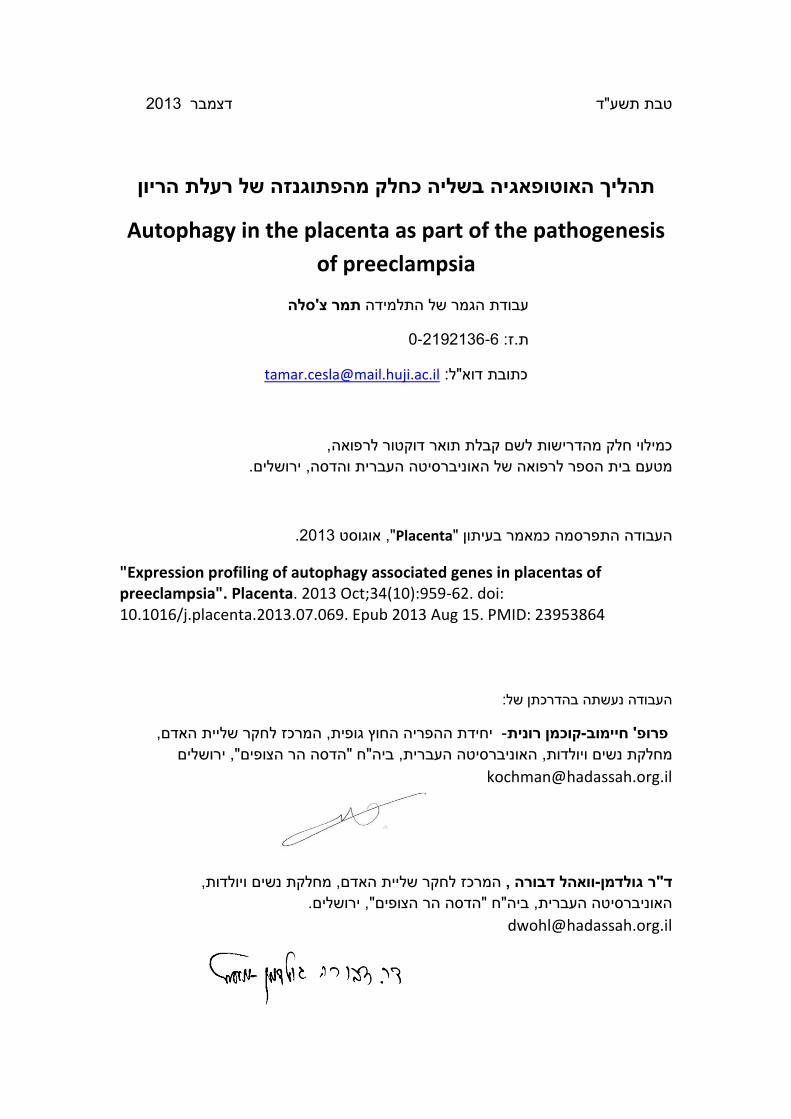
2. Immunohistochemistry: Immunohistochemistry was performed on archival FFPE
normal third trimester (4 sections) and preeclamptic (8 sections) placental sections
as well as a 17 day secretory endometrium sample. The KIAA1324 antibody (Sigma
prestige antibody produced in rabbit) was used (as being the most upregulated gene
in Preeclamptic placenta, see results section) according to the manufacturer's
instructions with citrate buffer antigen retrieval (1:100 dilution). The secondary
antibody was Zytomed (Germany) systems HRP one-step polymer, antimouse/
rabbit/ rat. Color detection was with AEC (Zymed, CA) and GVA (Zymed, CA)
mounting was used.
Results:
We analyzed five different microarray datasets, from five different studies, (10-14) (Winn et
al. 2009, Sitras et al. 2009, Herse et al. 2012, Tsai et. al 2011, Founds et al. 2009), comprising
gene expression data of placental tissue. Each dataset included both preeclamptic and
control samples. Detailed information on the datasets is shown in Table 1. We analyzed each
dataset separately for several reasons, but mainly because each study defined preeclampsia
differently as regards severe (10, 11) or mild (12-14) disease.
The five datasets were created using three discrete microarray platforms: Affymetrix Human
Genome U133A and U133B Array (10, 14), Illumina human-6 v2.0 expression beadchip (12,
13), and ABI Human Genome Survey Microarray Version2 (11).
Table 1 shows that the five datasets differ in the number of samples collected. In one
dataset (14) the tissue collected differs from the others, as it was obtained via chorionic
villus sampling at first trimester and not placental sampling at delivery.
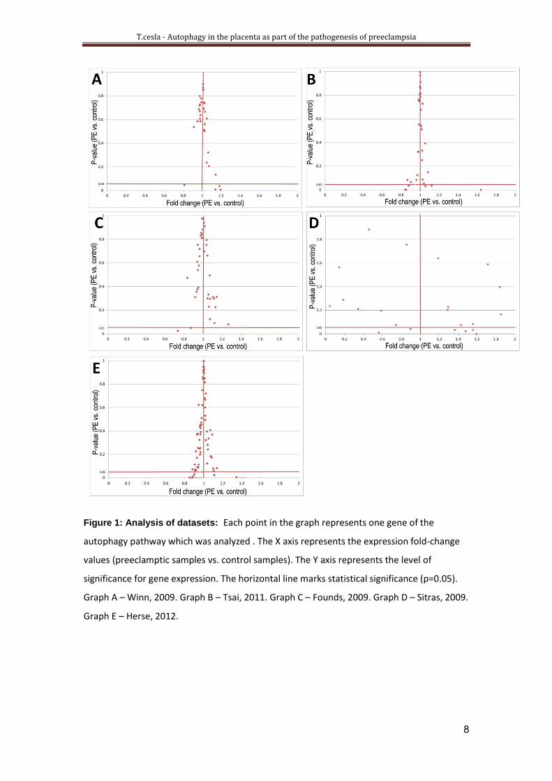
t-test did not reveal a statistical difference between autophagy associated gene expression
of normal and preeclamptic placental samples. Of the five datasets, very few autophagy
genes differed significantly between the two groups, and only three of them had a fold
change higher than 1.5 (in preeclamptic samples vs. control). They were the genes
KIAA1324, WDR45L (which are involved in the vacuole formation stage of macroautophagy)
and NPC1 (which is one of the negative regulators of macroautophagy). The gene mTOR

T.cesla - Autophagy in the placenta as part of the pathogenesis of preeclampsia
5
(FRAP1) displayed a 2-fold change in one of the datasets but was not significant (p=0.0624)
(Figure 1A-E).
The gene KIAA1324 displayed a 1.6 fold overexpression in preeclampsia as compared to
control samples. The gene, also known as EIG121, is a transmembrane protein that is
estrogen-induced and is overexpressed in endometrial carcinoma (15) . Overexpression of
tetracycline inducible EIG121 in stably transfected cells coupled with starvation was found to
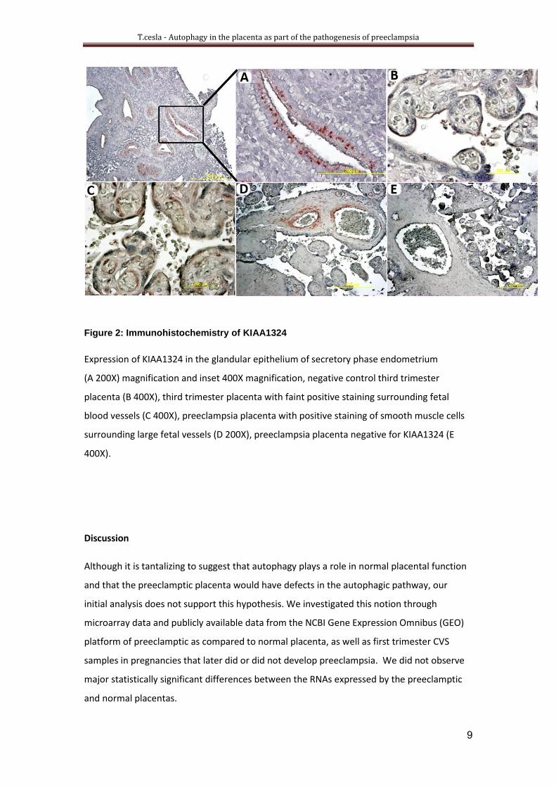
upregulate autophagy (16). To investigate KIAA1324 protein expression in placenta we
performed immunohistochemistry on normal third trimester and first pregnancy severe
preeclampsia placental sections. As a positive control for the antibody we used secretory
phase endometrium. As expected, staining was observed in the glandular epithelium (Figure
2A). The serum only control was negative (Figure 2B). In three of four normal third trimester
placenta staining was observed surrounding fetal vessels; In six of eight of the preeclampsia
placentas we observed staining in the smooth muscle cells surrounding fetal vessels Figure
(Figure 2C,D). Overall however, the preeclamptic placental staining appeared less intense
than the staining intensity observed in the normal sections.

T.cesla - Autophagy in the placenta as part of the pathogenesis of preeclampsia
2
Table 1. Characteristics of the five datasets used in the analysis
Platform Sampling Procedure Number of Cases Study Author, year
Affymetrix
Human Genome
U133A and
U133B Array.
Basal plate biopsies of preterm labor (24-36 weeks) and preterm severe preeclampsia (24-36 weeks).
12 severe preterm
PE, 11 control
preterm delivery
GSE14722 Winn, 2009
Illumina human-
6 v2.0 expression
beadchip
The placental sample, excluding fetal membranes, was obtained within 5 cm of the placental umbilical insertion site.
23 preeclampsia,
37 control
GSE25906 Tsai, 2011
Affymetrix
Human Genome
U133 Plus 2.0
Array
A nested case control study of CVS tissues collected at 10–12 weeks’ gestation in 160 patients with singleton fetuses.
4 CVS samples of
PE, 8 matched
controls
GSE12767 Founds,
2009
ABI Human
Genome Survey
Microarray
Version 2
Chorionic tissue was dissected from a standardized location – approximately 2 cm from the umbilical cord insertion, from the middle layer of placenta midway between maternal and fetal surfaces.
17 preeclampsia,
26 control
GSE10588 Sitras, 2009
Affymetrix
Human Genome
U133 Plus 2.0
Array
Placental biopsies were excised from
a macroscopically normal looking,
centrally located cotyledon, omitting
the decidual layer.
22 preeclampsia,
24 control
Biobank
Collection Oslo
University
Hospital
Herse, 2012

T.cesla - Autophagy in the placenta as part of the pathogenesis of preeclampsia
7
Table 2. Autophagy related genes and function
Induction Nucleation Expansion Vacuole
formation
Co-
Regulators
of
Autophagy
and
Apoptosis:
Degradation
of misfolded
proteins
Positive
regulation of
Macroautophagy
Negative
regulators of
Macroautophagy
mTOR RB1CC1 ATG3 ATG9A CLN3 HDAC6 LARP1 LZTS1
ULK1 ATG101 ATG4B ATG9B SQSTM1 PAFAH1B2 NPC1
BECN1 ATG4C ATG13 SIRT1 NRBP2
PIK3C3 ATG5 MAP1LC3
A
SUPT5H
ATG14 ATG7 NBR1 WAC
ATG12 PSEN1 TP53INP2
ATG16L1 WDR45 TRIM13
WDR45L
WIPI1
WIPI2
TP53INP2
TBC1D25
KIAA1324
T.cesla - Autophagy in the placenta as part of the pathogenesis of preeclampsia
8
Figure 1: Analysis of datasets: Each point in the graph represents one gene of the
autophagy pathway which was analyzed . The X axis represents the expression fold-change
values (preeclamptic samples vs. control samples). The Y axis represents the level of
significance for gene expression. The horizontal line marks statistical significance (p=0.05).
Graph A – Winn, 2009. Graph B – Tsai, 2011. Graph C – Founds, 2009. Graph D – Sitras, 2009.
Graph E – Herse, 2012.
T.cesla - Autophagy in the placenta as part of the pathogenesis of preeclampsia
1
Figure 2: Immunohistochemistry of KIAA1324
Expression of KIAA1324 in the glandular epithelium of secretory phase endometrium
(A 200X) magnification and inset 400X magnification, negative control third trimester
placenta (B 400X), third trimester placenta with faint positive staining surrounding fetal
blood vessels (C 400X), preeclampsia placenta with positive staining of smooth muscle cells
surrounding large fetal vessels (D 200X), preeclampsia placenta negative for KIAA1324 (E
400X).
Discussion
Although it is tantalizing to suggest that autophagy plays a role in normal placental function
and that the preeclamptic placenta would have defects in the autophagic pathway, our
initial analysis does not support this hypothesis. We investigated this notion through
microarray data and publicly available data from the NCBI Gene Expression Omnibus (GEO)
platform of preeclamptic as compared to normal placenta, as well as first trimester CVS
samples in pregnancies that later did or did not develop preeclampsia. We did not observe
major statistically significant differences between the RNAs expressed by the preeclamptic
and normal placentas.
T.cesla - Autophagy in the placenta as part of the pathogenesis of preeclampsia
01
This was a surprising result as autophagy not only plays a role in normal cellular homeostasis
but plays a role during nutrient starvation, which could help explain placental function
during nutritional variability in maternal blood. Furthermore, considering the known
contributions of autophagy to processes such as aging, immunity, inflammation, and cell
survival, it seemed reasonable to suspect an association between defects in autophagy and
preeclampsia.
However, our findings are not without caveats. Among the possible mechanisms that may
diminish the need for autophagy in the placenta is placental reserve. Placental reserve is
essential for fetal survival as fluctuations in nutrient availability are to be expected.
Placental reserve is highly dependent on transport efficiency and placental size. Along with
the placental capacity of adaptability of active transport, placental reserve could offer a way
to meet fetal nutritional demands in times of limited nutrient availability (17).
Another possibility that may explain our negative results is that while not rising to statistical
significance, the small differences observed for some of the genes (less than 1.5 fold) may be
enough to trigger the autophagy pathway and perhaps to sustain it. These small RNA
differences could reflect larger differences if the data could be analyzed in protein arrays or
by western blot. In other words, the significance of defective autophagy in preeclampsia
may be revealed on the level of protein expression but not on RNA (4). In addition, marked
differences in gene expression may be observed according to the number of samples
collected per placenta, whether they were pooled or not, or the quality of the RNA: all of
these factors, as well as the anatomical site where the placental sample was taken, may
impact the results(18).
The placental samples used in this current investigation were from samples (either third
trimester or chorionic villous sampling) where the number of extravillous trophoblasts
would constitute only a minor fraction of the cell types. Therefore, if the defect in autophagy
in preeclampsia was to be found in extravillous trophoblasts then it would not be observed
by this methodology. Furthermore, since the basis for development of preeclampsia is
thought to be in the first trimester of pregnancy, with defective remodeling by extravillous
trophoblast of uteroplacental spiral arteries then perhaps a defect in autophagy expression
would not be detectable in the third trimester samples evaluated from four of five sources
used here for the array data (19).

T.cesla - Autophagy in the placenta as part of the pathogenesis of preeclampsia
00
Summary:
Autophagy, a mechanism of cell survival during times of stress, may be involved in normal
placental maintenance, and in addition to its role in normal cellular homeostasis, defects in
autophagy are implicated in pathologies such as neurodegeneration, cancer, inflammation
and aging (3-5). Characteristics often associated with preeclampsia, an hypertensive disorder
of pregnancy, including an excessive inflammatory response (2), premature placental aging
(Tenney-Parker change), metabolic syndrome, hypoxia and placental insufficiency are
reminiscent of features of impaired autophagy. Thus, the disruption of autophagy might
contribute to the pathophysiology of preeclampsia. However, studies of autophagy in the
placenta are still lagging.
To elucidate a possible association between autophagy and the clinical syndrome of
preeclampsia we undertook an expression profile analysis with a directed focus of
autophagy associated genes from placentas of preeclamptic pregnancies as compared to
controls. We used microarray data including publicly available data through the NCBI Gene
Expression Omnibus (GEO) platform, to analyze over 40 genes known to be regulated in
autophagy. We compared datasets from 106 normal and 78 preeclamptic placental samples
to determine whether they display statistically significant differential expression of these
genes.
Of the five datasets, very few autophagy genes differed significantly between the two
groups, and only three of these (KIAA1324, WDR45L and NPC1) had a fold change higher
than 1.5 (in preeclamptic samples vs. control).
To investigate KIAA1324 protein expression in placenta we performed
immunohistochemistry on normal third trimester and first pregnancy severe preeclampsia
placental sections. Overall however, the preeclamptic placental staining appeared less
intense than the staining intensity observed in the normal sections.
Although preeclampsia displays many of the features suggestive of altered autophagy,
including premature placental aging and placental insufficiency, defective placental
autophagy as a cause of preeclampsia is not supported by whole placental tissue microarray
differential expression profiling. We suggest that further investigations and alternate
approaches be taken to unveil the role that autophagy may play in normal placental function
and possible dysfunction in preeclampsia.
T.cesla - Autophagy in the placenta as part of the pathogenesis of preeclampsia
03
:סיכום
אוטופגיה הינו מנגנון של שרידות תאים בעתות עקה ורעב, וייתכן ומהווה חלק מיכולת השליה
התקינה להזנת העובר ברחם. תהליך אוטופאגיה פגום, הוכח כמעורב בפתולוגיות כגון סרטן, דלקת,
. בנוסף, מאפיינים רבים של רעלת הריון, ביניהם תהליך (3-5) נוירודגנרטיביים הזדקנות ותהליכים
תית, הינם מצבים דלקתי מוגבר, הזדקנות שלייתית מוקדמת, היפוקסיה ומצב של אי ספיקה שליי
. על כן, הועלתה ההשערה כי לתהליך (7 ,6)בהם ישנן עדויות לכך שתהליך האוטופגיה לקוי
האוטופגיה תפקיד בפתוגנזה של רעלת הריון.
כדי לחקור השערה זו, ולמצוא קשר בין תהליך האוטופאגיה לבין תסמונת רעלת ההריון, ביצענו
, על מנת NCBI Gene Expression Omnibus (GEO) platform -בו השתמשנו במידע המופיע ב מחקר
גנים, הידועים כשייכים לתהליך האוטופאגיה. במחקר ביצענו השוואה בין 41לנתח ביטויים של מעל
דגימות משליות של נשים שחלו ברעלת הריון, זאת על 78 -דגימות משליות תקינות ו 012 -נתונים מ
מנת לבדוק האם יהיה הבדל סטטיסטי מובהק בין ביטוי הגנים הללו בין שתי הקבוצות.
באנליזה נמצא כי מעט מאוד גנים היו שונים בצורה מובהקת בין שתי הקבוצות, ורק שלושה מהם
(KIAA1324, WDR45L ו- NPC1( היו בעלי השתנות )fold changeהגבוהה מ )- בשליות 0.5(
, אך לא היה מובהק 3הראה ביטוי הגבוה פי mTOR (FRAP1)ה"חולות" אל מול אלה הביקורת(. הגן
סטטיסטית.
על חתכים בשליה, ביצענו צביעה אימונוהיסטוכימית KIAA1324 -על מנת לחקור את ביטוי חלבון ה
משליות "תקינות" מטרימסטר שלישי, ומשליות של נשים שחלו ברעלת הריון חמורה. הצביעה
בשליות ה"חולות" הייתה חלשה מאשר זו שנצפתה בחתכים התקינים.
על כן, על אף שרעלת הריון מבטאת הרבה מן המאפיינים המתאימים לאיבר בו תהליך האוטופאגיה
וקדמת ואי ספיקה שלייתית, ההשערה לפיה פגם בתהליך האוטופגיה , כגון הזדקנות מהינו פגוע
בשליה הינו סיבה להתפתחות של רעלת הריון, לא נתמך על ידי מחקרנו.

T.cesla - Autophagy in the placenta as part of the pathogenesis of preeclampsia
02
References:
1. Redman CW, Sargent IL. Placental stress and pre-eclampsia: a revised view.
Placenta. 2009;30 Suppl A:S38-42.
2. Redman CW, Sargent IL. Immunology of pre-eclampsia. Am J Reprod
Immunol.63(6):534-43.
3. Levine B, Mizushima N, Virgin HW. Autophagy in immunity and
inflammation. Nature. 2011;469(7330):323-35.
4. Mizushima N, Levine B. Autophagy in mammalian development and
differentiation. Nat Cell Biol. 2010;12(9):823-30.
5. Rubinsztein DC, Marino G, Kroemer G. Autophagy and aging. Cell.
2011;146(5):682-95.
6. Bildirici I, Longtine MS, Chen B, Nelson DM. Survival by self-destruction: A
role for autophagy in the placenta? Placenta.33(8):591-8.
7. Saito S, Nakashima A. Review: The role of autophagy in extravillous
trophoblast function under hypoxia. Placenta .2112.
8. Oh SY, Choi SJ, Kim KH, Cho EY, Kim JH, Roh CR. Autophagy-related
proteins, LC3 and Beclin-1, in placentas from pregnancies complicated by
preeclampsia. Reprod Sci. 2008;15(9):912-20.
9. Levine RJ, Lam C, Qian C, Yu KF, Maynard SE, Sachs BP, et al. Soluble
endoglin and other circulating antiangiogenic factors in preeclampsia. N Engl J Med.
2006;355(10):992-1005.
11. Winn VD, Gormley M, Paquet AC, Kjaer-Sorensen K, Kramer A, Rumer KK,
et al. Severe preeclampsia-related changes in gene expression at the maternal-fetal
interface include sialic acid-binding immunoglobulin-like lectin-6 and pappalysin-2.
Endocrinology. 2009;150(1):452-62.
11. Sitras V, Paulssen RH, Gronaas H, Leirvik J, Hanssen TA, Vartun A, et al.
Differential placental gene expression in severe preeclampsia. Placenta.
2009;30(5):424-33.
12. Herse F, Lamarca B, Hubel CA, Kaartokallio T, Lokki AI, Ekholm E, et al.
Cytochrome P450 subfamily 2J polypeptide 2 expression and circulating
epoxyeicosatrienoic metabolites in preeclampsia. Circulation. 2012;126(25):2990-9.
13. Tsai S, Hardison NE, James AH, Motsinger-Reif AA, Bischoff SR, Thames
BH, et al. Transcriptional profiling of human placentas from pregnancies complicated
by preeclampsia reveals disregulation of sialic acid acetylesterase and immune
signalling pathways. Placenta. 2011;32(2):175-82.
14. Founds SA, Conley YP, Lyons-Weiler JF, Jeyabalan A, Hogge WA, Conrad
KP. Altered global gene expression in first trimester placentas of women destined to
develop preeclampsia. Placenta .2119;31(1:)15-24.
15. Deng L, Broaddus RR, McCampbell A, Shipley GL, Loose DS, Stancel GM,
et al. Identification of a novel estrogen-regulated gene, EIG121, induced by hormone
replacement therapy and differentially expressed in type I and type II endometrial
cancer. Clin Cancer Res. 2005;11(23):8258-64.
16. Deng L, Feng J, Broaddus RR. The novel estrogen-induced gene EIG121
regulates autophagy and promotes cell survival under stress. Cell Death Dis.1:e32.
17. Fowden AL, Ward JW, Wooding FP, Forhead AJ, Constancia M.
Programming placental nutrient transport capacity. J Physiol. 2006;572(Pt 1):5-15.
18. Wyatt SM, Kraus FT, Roh CR, Elchalal U, Nelson DM, Sadovsky Y. The
correlation between sampling site and gene expression in the term human placenta.
Placenta. 2005;26(5):372-9.

T.cesla - Autophagy in the placenta as part of the pathogenesis of preeclampsia
04
19. Goldman-Wohl D, Yagel S. Preeclampsia--a placenta developmental biology
perspective. J Reprod Immunol. 2009;82(2):96-9.


![Research Paper HO-1 induced autophagy protects against IL ... · induce apoptosis of the nucleus pulposus cells (NPCs) in the degenerative intervertebral disc [5, 6]. Autophagy is](https://img.pdfslide.tips/doc/110x75/5e72f110b749c078843e28fa/research-paper-ho-1-induced-autophagy-protects-against-il-induce-apoptosis-of.jpg)