Embed Size (px)
Citation preview
Tejidos animales y sistemas de órganos
Homeostasis en animales
� Las partes del cuerpo interaccionan para:• Coordinar y controlar las partes individuales• Adquirir y distribuir materiales a las celulas y
disponer de los desperdicios• Proteger a los tejidos contra heridas y ataques• Reproducir, alimentar y proteger la progenie en
su desarrollo temprano• Mantener el ambiente interno (homeostasis )
Organización del cuerpo animal
� Tejido• Conjunto de celulas que interaccionan y las
sustancias extracelulares que conducen algunatarea
� Organo• Unidad estructural de 2 o mas tejidos
organizados para conducir alguna tarea
� Sistemas de órganos• 2 o mas organos que interactuan para una tarea
comun
Las células animales se unen mediante variostipos de unión celular
� Uniones adherentes• Mantienen a las celulas unidas en lugares
especificos.
� Uniones estrechas• Cierran el paso de fluidos entre celula y celula;
esos fluidos son forzados a pasar por el interior de las celulas.
� Uniones de hendiduras• Permiten el paso de iones y moleculas pequenas
del citoplasma de una celula al citoplasma de otra celula adyacente.
Cuatro tipos de tejidos principales en animales
� Tejido epitelial
� Tejido conectivo
� Tejido muscular
� Tejido nervioso
Tejido epitelial
� Epitelio• Una capa de celulas que cubre la superficie
exterior y recubre los ductos y cavidades internasdel animal.
� Membrana basal• Una matriz extracelular que adhiere el epitelio al
tejido subyacente.
� Microvellos• Proyecciones de epitelio absorbente parecidas a
dedos
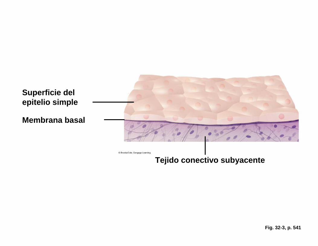
Fig. 32-3, p. 541
Superficie del epitelio simple
Membrana basal
Tejido conectivo subyacente
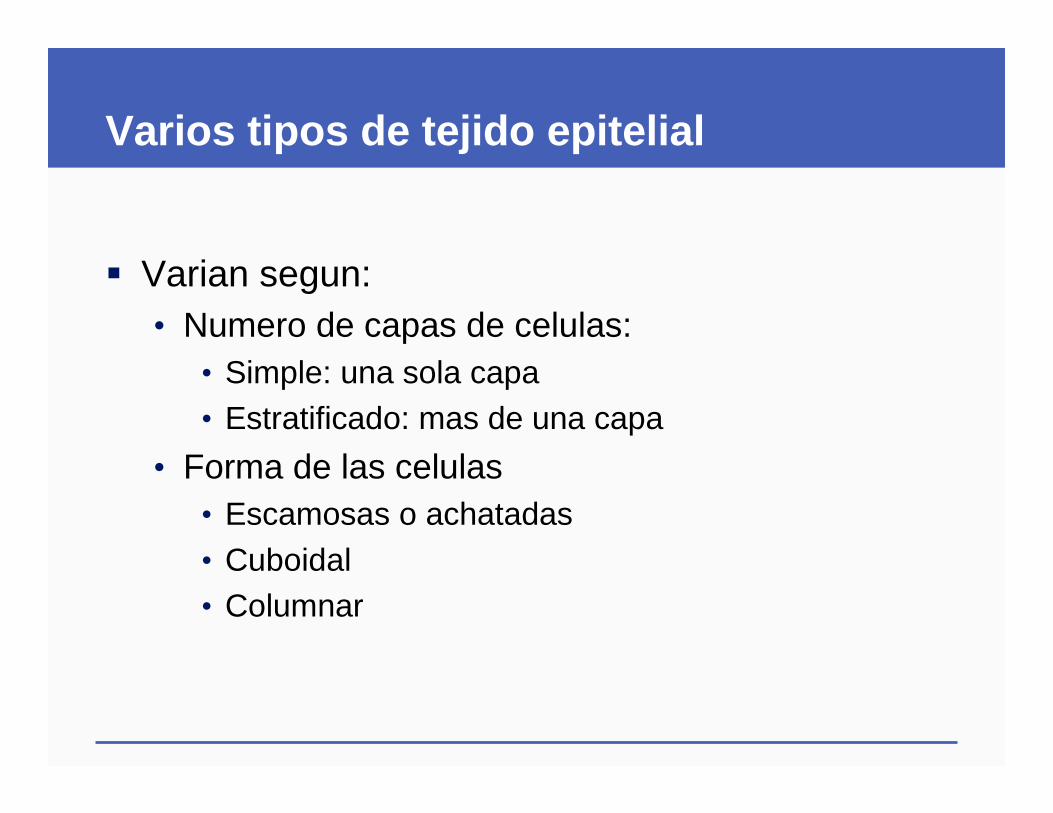
Varios tipos de tejido epitelial
� Varian segun:• Numero de capas de celulas:
• Simple: una sola capa• Estratificado: mas de una capa
• Forma de las celulas• Escamosas o achatadas• Cuboidal• Columnar
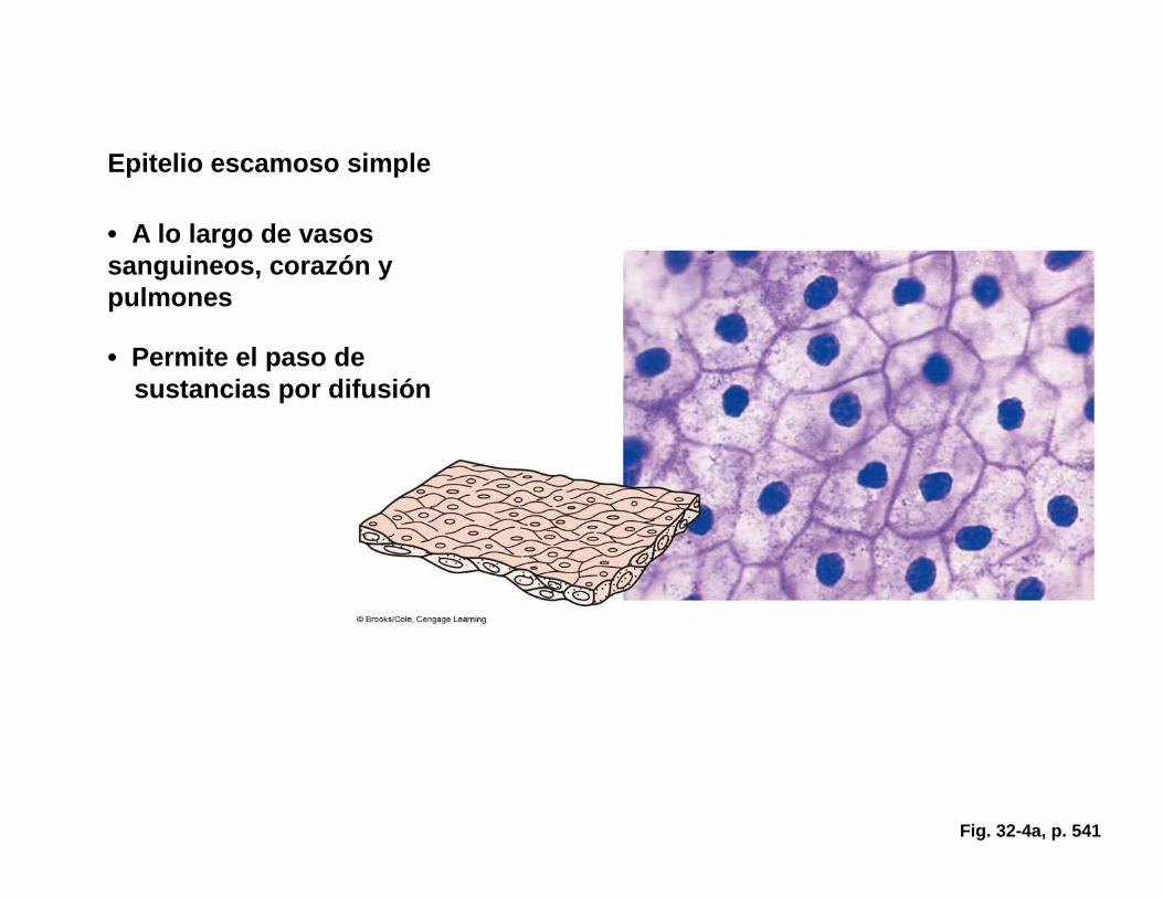
Fig. 32-4a, p. 541
Epitelio escamoso simple
• A lo largo de vasossanguineos, corazón y pulmones
• Permite el paso de sustancias por difusión
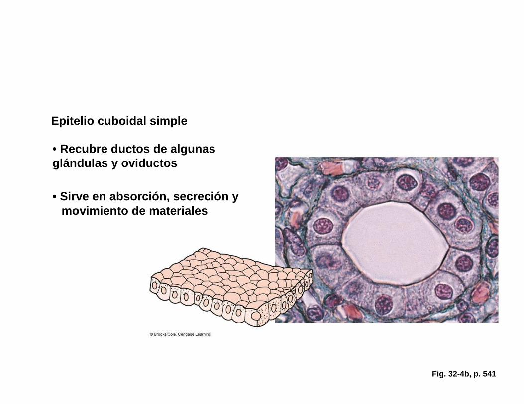
Fig. 32-4b, p. 541
Epitelio cuboidal simple
• Recubre ductos de algunasglándulas y oviductos
• Sirve en absorción, secreción y movimiento de materiales
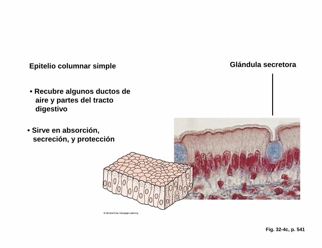
Fig. 32-4c, p. 541
Epitelio columnar simple Glándula secretora
• Recubre algunos ductos de aire y partes del tractodigestivo
• Sirve en absorción, secreción, y protección
Glandular Epithelium
� Glands• Organs that release substances onto the skin, or
into a body cavity or interstitial fluid
� Exocrine glands (glands with ducts)• Deliver secretions to an external or internal
surface (saliva, milk, earwax, digestive enzymes)
� Endocrine glands (no ducts)• Secrete hormones which are carried in blood
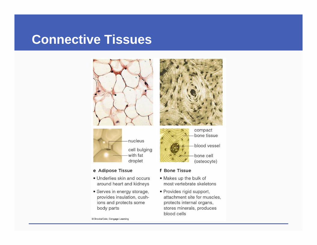
32.3 Connective Tissues
� Connective tissues consist of cells and the extracellular matrix they secrete
� Connective tissues connect body parts and provide structural and functional support to other body tissues
Soft Connective Tissues
� Loose connective tissue• Fibroblasts secrete a matrix of complex
carbohydrates with fibers dispersed widely through the matrix
� Dense connective tissue (dense collagen fibers)• Dense irregular: Supports skin, internal organs• Dense regular: Ligaments and tendons
Specialized Connective Tissues
� Cartilage : Rubbery extracellular matrix, supports and cushions bones
� Adipose tissue : Fat filled cells, stores energy, cushions and protect organs
� Bone : Rigid support, muscle attachment, protection, mineral storage, blood production
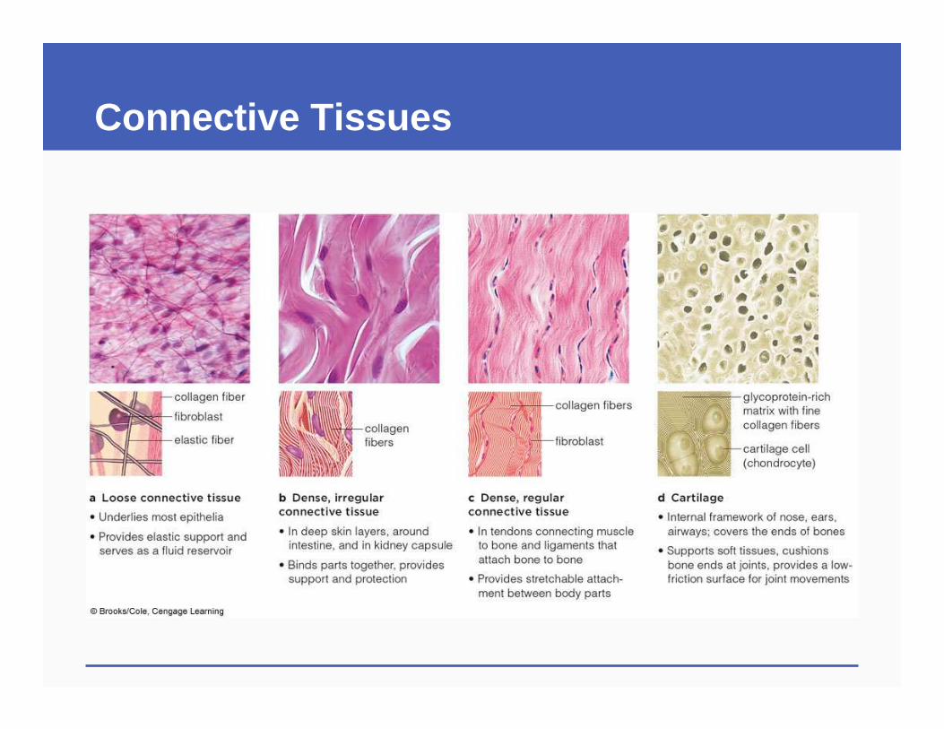
Connective Tissues
Connective Tissues
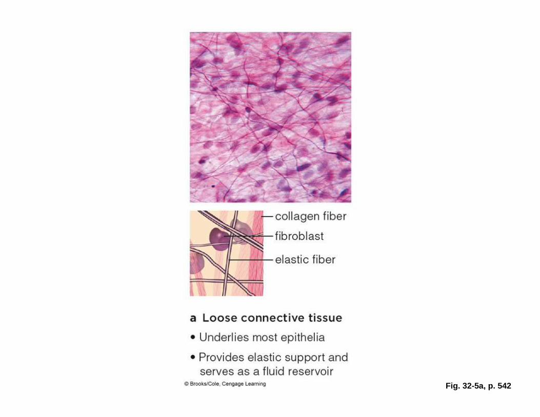
Fig. 32-5a, p. 542
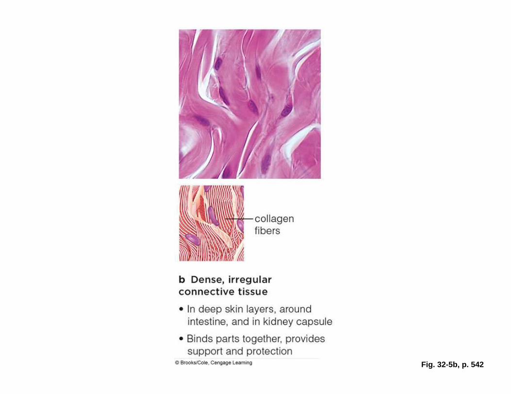
Fig. 32-5b, p. 542
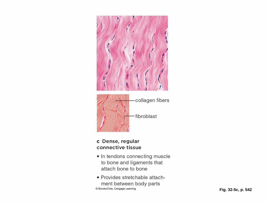
Fig. 32-5c, p. 542
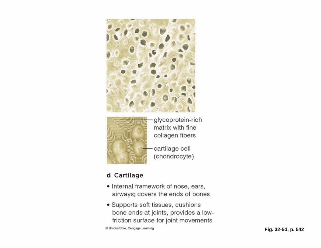
Fig. 32-5d, p. 542
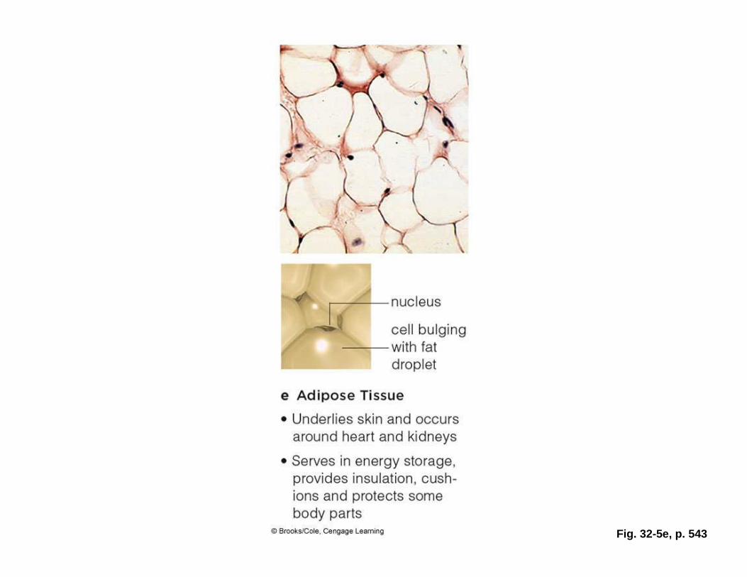
Fig. 32-5e, p. 543
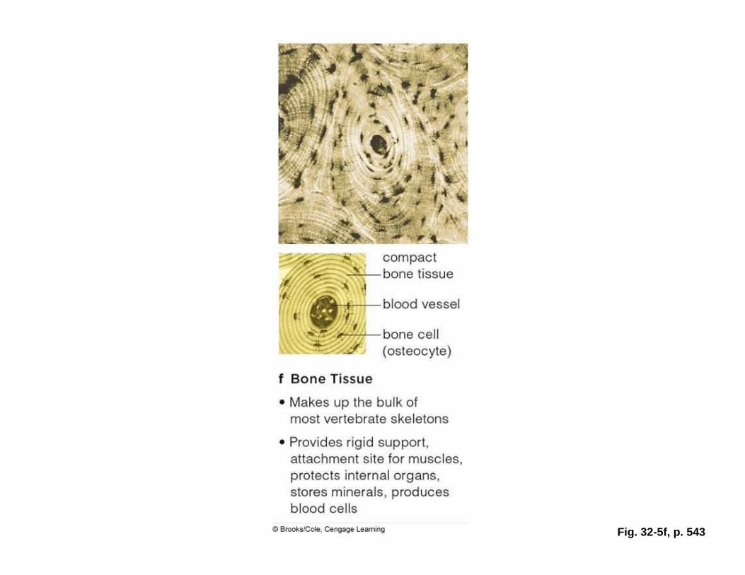
Fig. 32-5f, p. 543
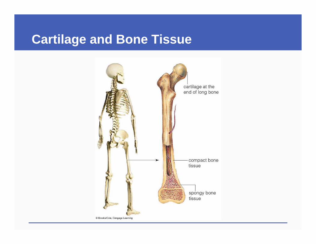
Cartilage and Bone Tissue
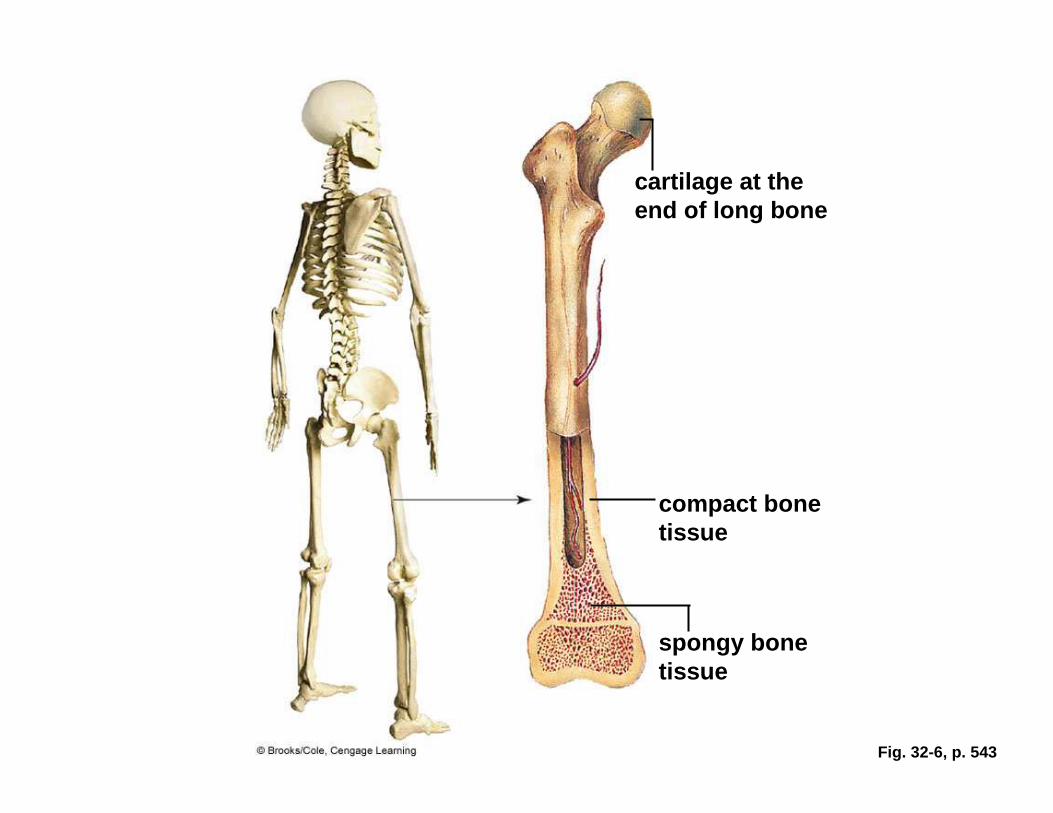
Fig. 32-6, p. 543
cartilage at the end of long bone
compact bone tissue
spongy bone tissue
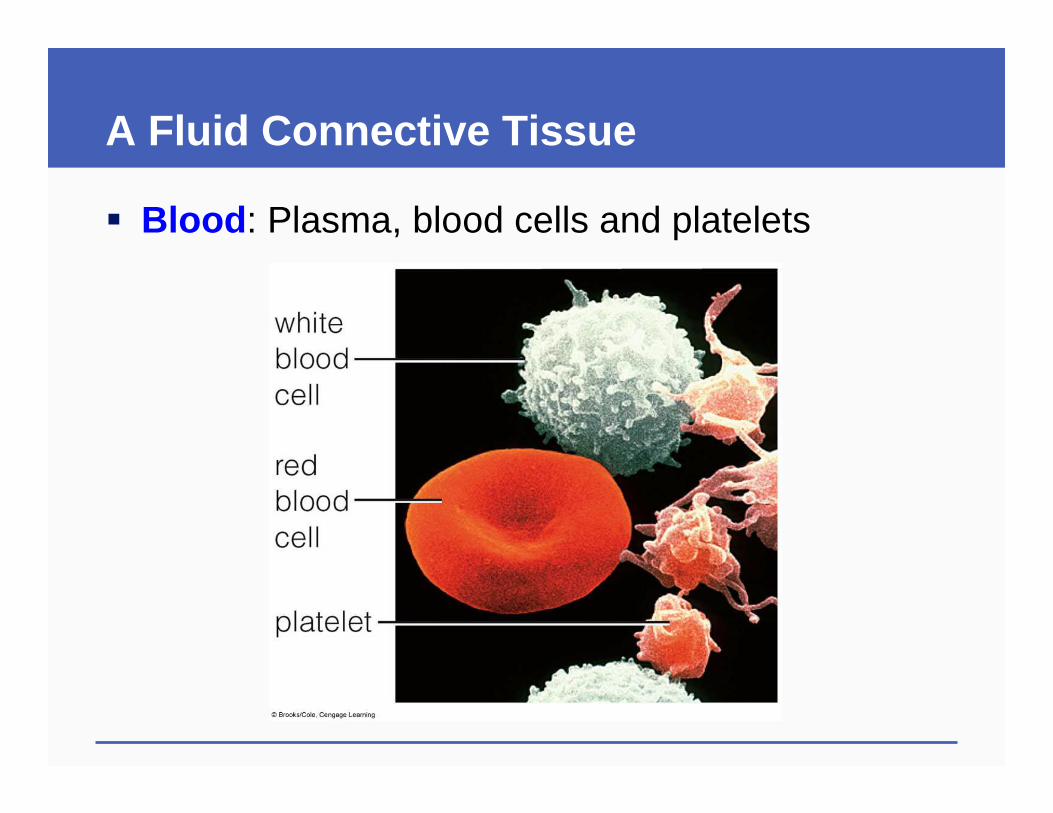
A Fluid Connective Tissue
� Blood : Plasma, blood cells and platelets
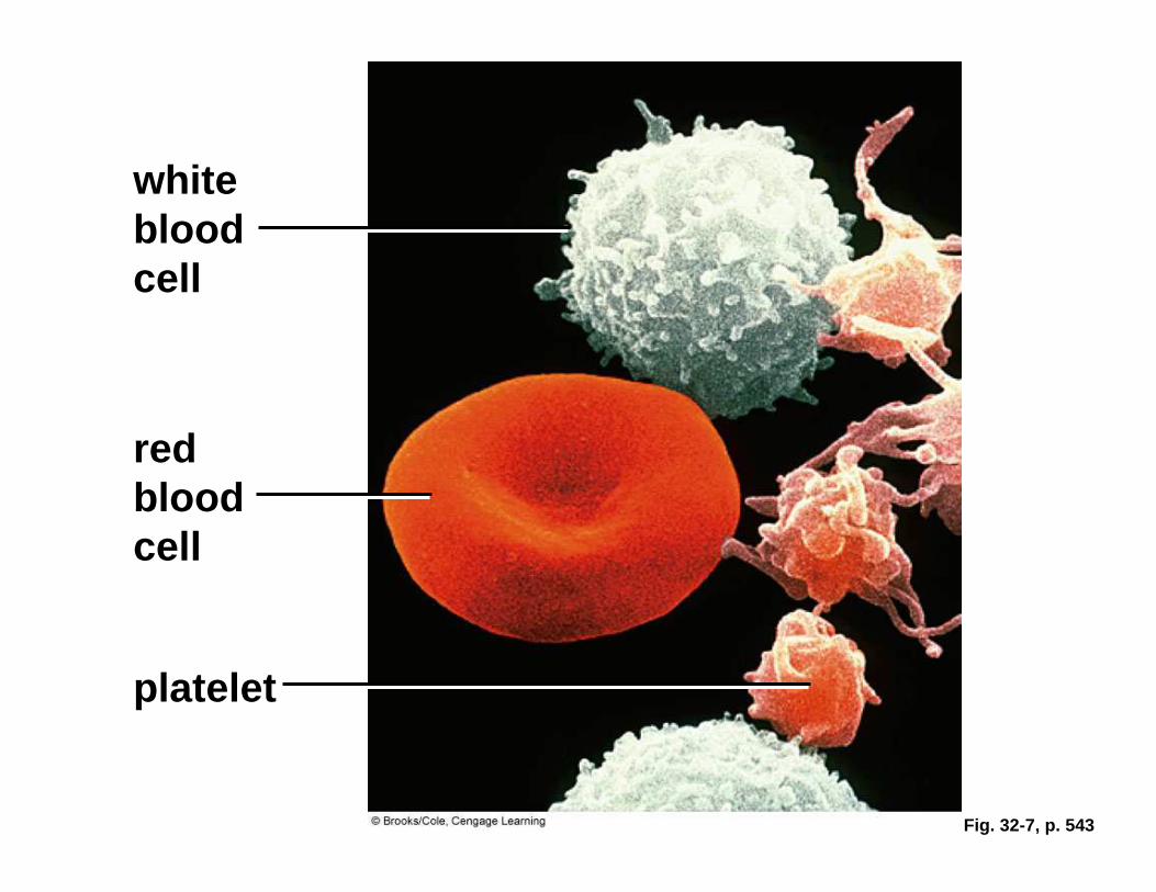
Fig. 32-7, p. 543
white blood cell
red blood cell
platelet
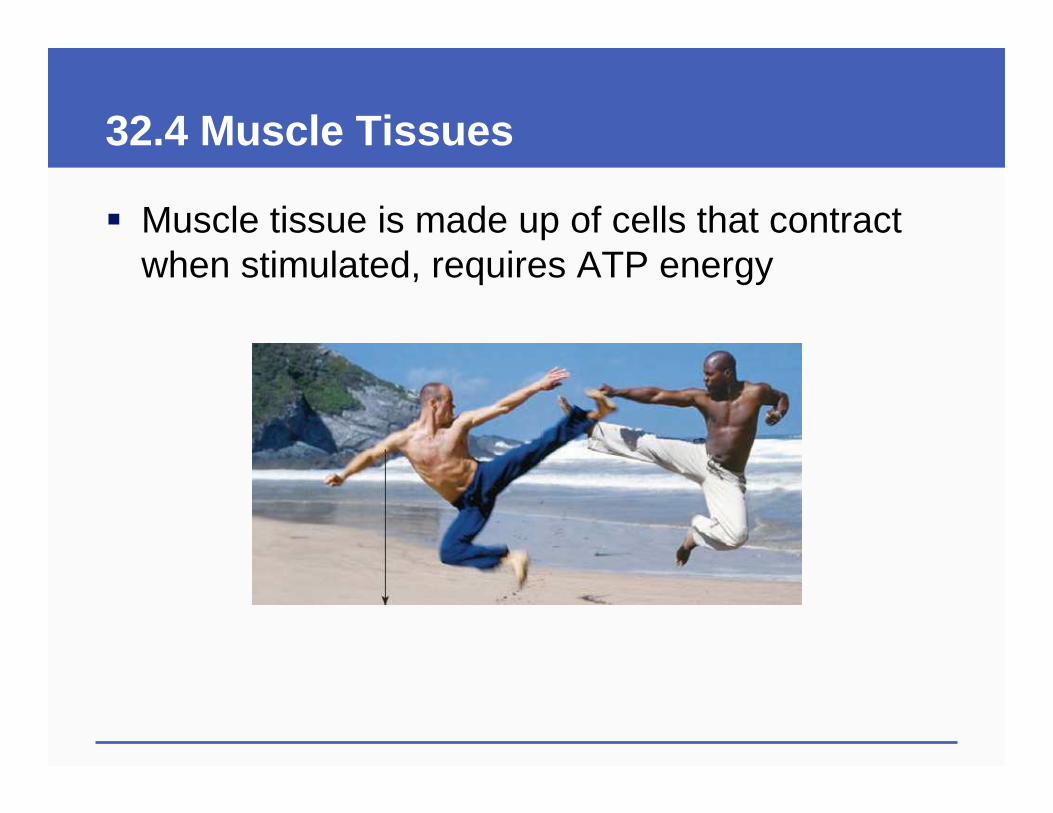
32.4 Muscle Tissues
� Muscle tissue is made up of cells that contract when stimulated, requires ATP energy
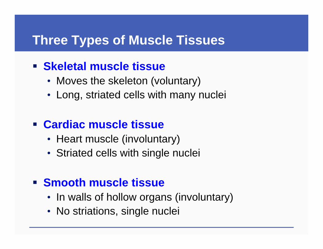
Three Types of Muscle Tissues
� Skeletal muscle tissue• Moves the skeleton (voluntary)• Long, striated cells with many nuclei
� Cardiac muscle tissue• Heart muscle (involuntary)• Striated cells with single nuclei
� Smooth muscle tissue• In walls of hollow organs (involuntary)• No striations, single nuclei
32.5 Nervous Tissue
� Nervous tissue• Consists of specialized signaling cells (neurons )
and cells that support them (neuroglial cells )
� Nervous tissue detects internal and external stimuli, and coordinates responses to stimuli
Neurons
� Neurons• Excitable cells with long cytoplasmic extensions• Send and receive electrochemical signals
� Three types of neurons• Sensory neurons are excited by specific stimuli• Interneurons integrate sensory information• Motor neurons relay commands from brain and
spinal cord to muscles and glands
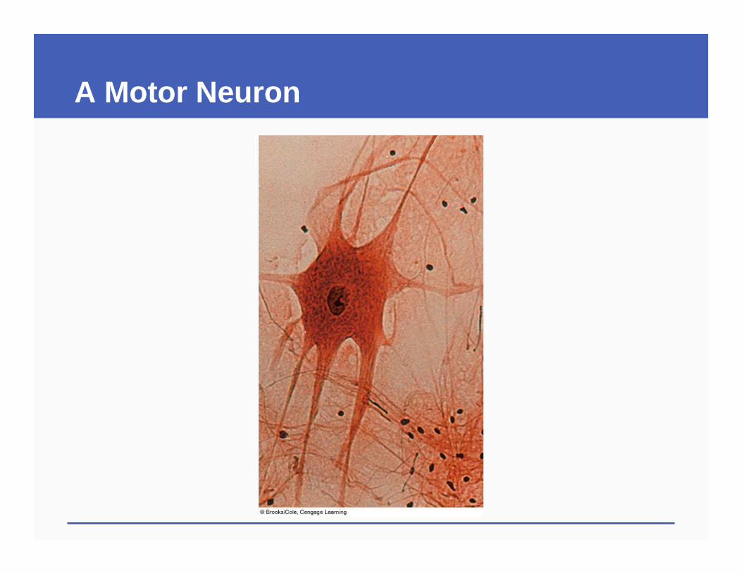
A Motor Neuron
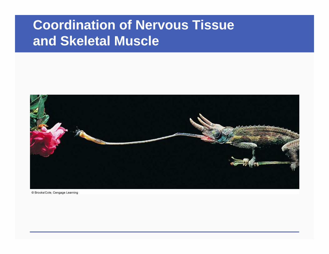
Coordination of Nervous Tissue and Skeletal Muscle
32.2-32.5 Key Concepts Types of Animal Tissues
� Epithelial tissue covers the body’s surface and lines its internal tubes
� Connective tissue provides support and connects body parts
� Muscle tissue moves the body and its parts
� Nervous tissue detects internal and external stimuli and coordinates responses
32.6 Overview of Major Organ Systems
� In vertebrates, organs arise from three embryonic germ layers• Ectoderm (outermost layer) forms nervous tissue
and epithelium of skin• Mesoderm (middle layer) forms muscle,
connective tissue, and lining of body cavities• Endoderm (innermost layer) forms epithelium of
gut and lungs
Body Cavities and Directional Terms
Body Cavities and Directional Terms
Body Cavities and Directional Terms
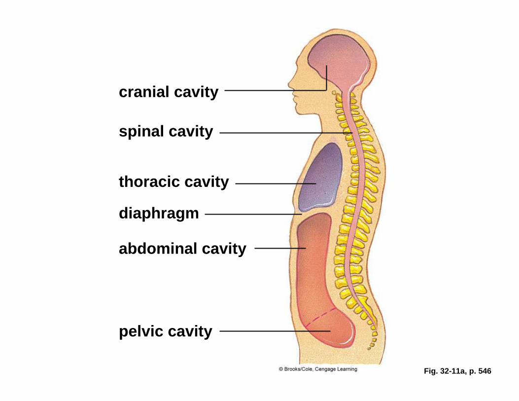
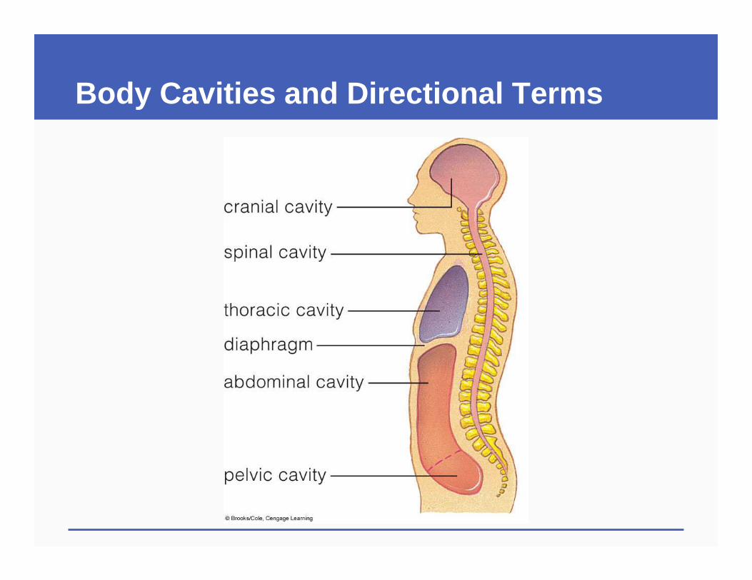
Fig. 32-11a, p. 546
cranial cavity
spinal cavity
thoracic cavity
diaphragm
abdominal cavity
pelvic cavity
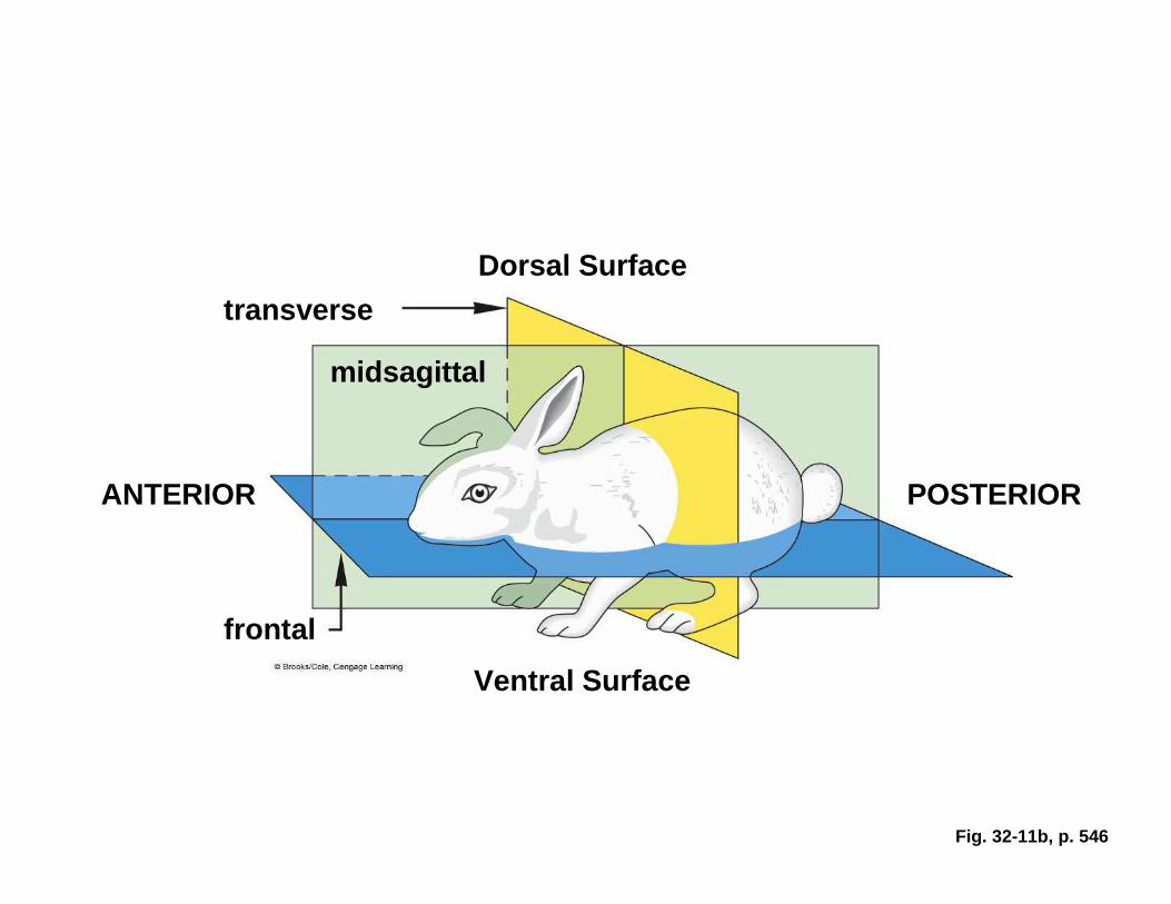
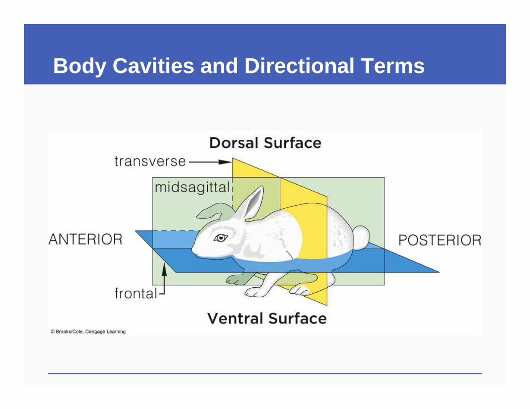
Fig. 32-11b, p. 546
Dorsal Surface
transverse
midsagittal
ANTERIOR POSTERIOR
frontal
Ventral Surface
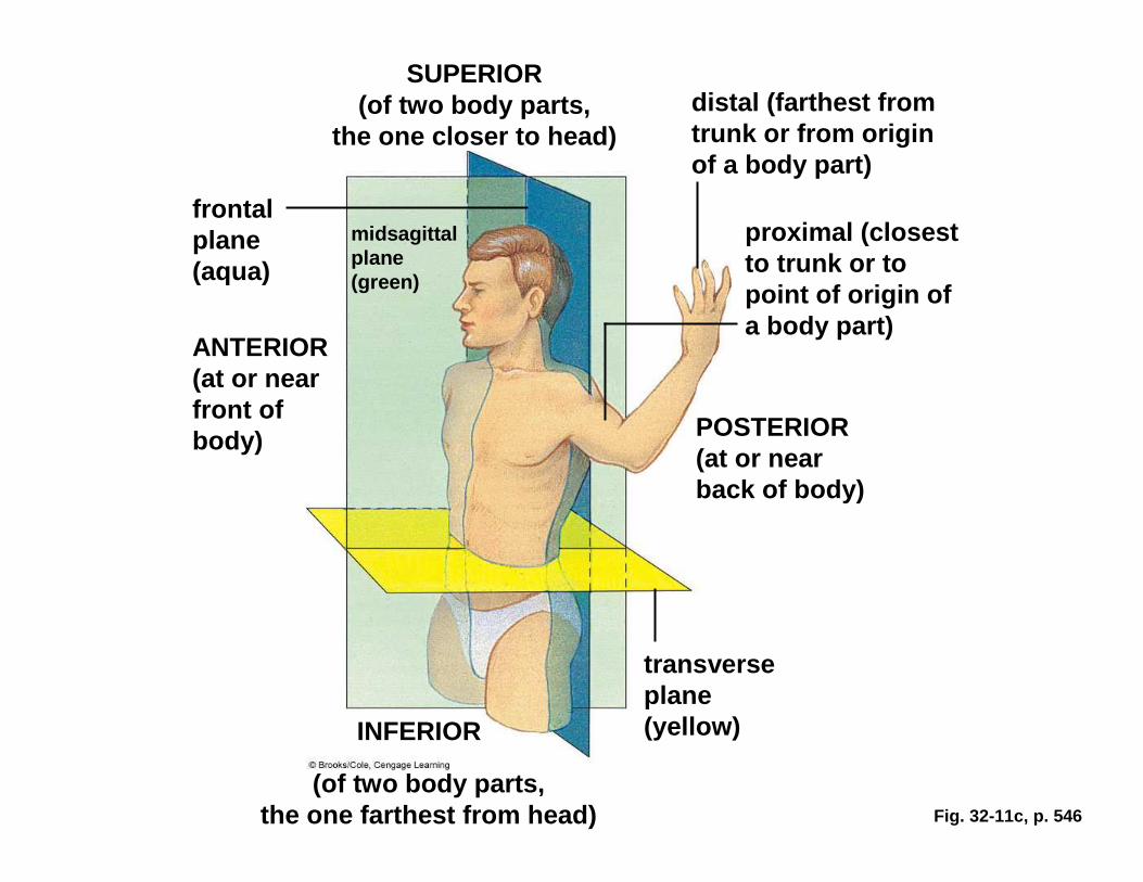
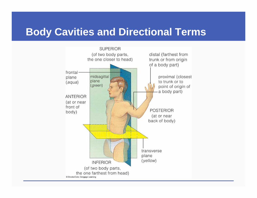
Fig. 32-11c, p. 546
SUPERIOR (of two body parts,
the one closer to head)distal (farthest from trunk or from origin of a body part)
frontal plane (aqua)
midsagittalplane (green)
proximal (closest to trunk or to point of origin of a body part)
ANTERIOR (at or near front of body) POSTERIOR
(at or near back of body)
transverse plane (yellow)INFERIOR
(of two body parts, the one farthest from head)
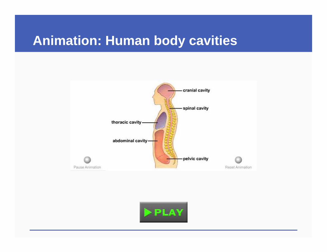
Animation: Human body cavities
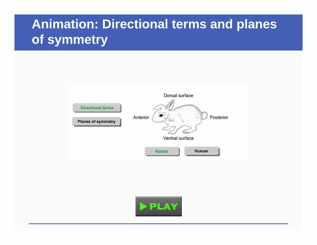
Animation: Directional terms and planes of symmetry
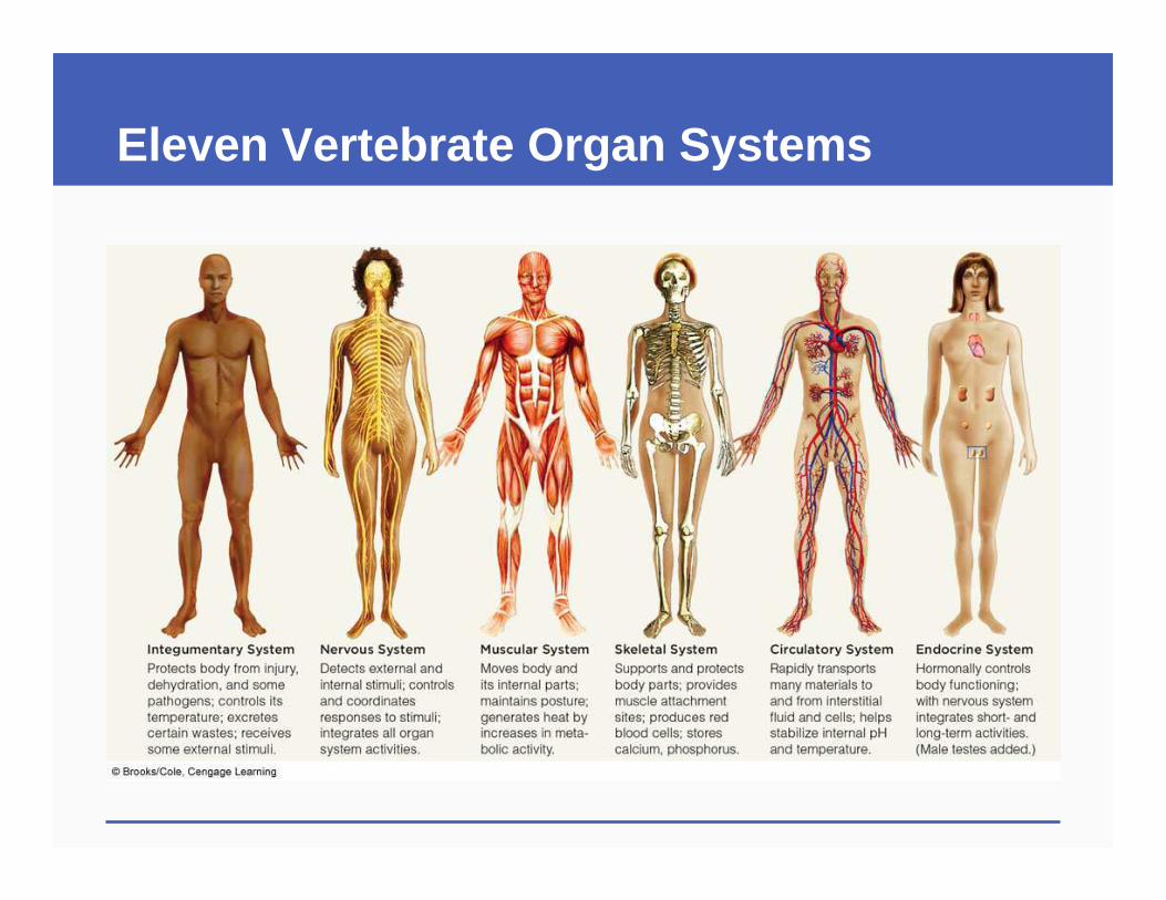
Eleven Vertebrate Organ Systems
Eleven Vertebrate Organ Systems
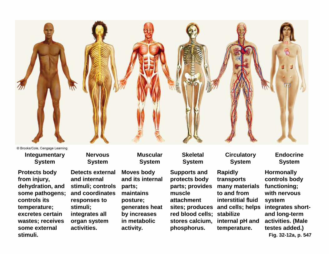
Fig. 32-12a, p. 547
IntegumentarySystem
Nervous System
Muscular System
Skeletal System
Circulatory System
Endocrine System
Protects body from injury, dehydration, and some pathogens; controls its temperature; excretes certain wastes; receives some external stimuli.
Detects external and internal stimuli; controls and coordinates responses to stimuli; integrates all organ system activities.
Moves body and its internal parts; maintains posture; generates heat by increases in metabolic activity.
Supports and protects body parts; provides muscle attachment sites; produces red blood cells; stores calcium, phosphorus.
Rapidly transports many materials to and from interstitial flflflfluidand cells; helps stabilize internal pH and temperature.
Hormonally controls body functioning; with nervous system integrates short-and long-term activities. (Male testes added.)
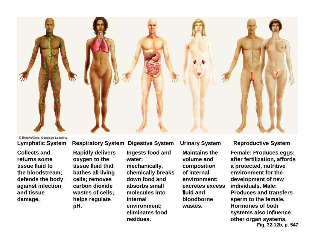
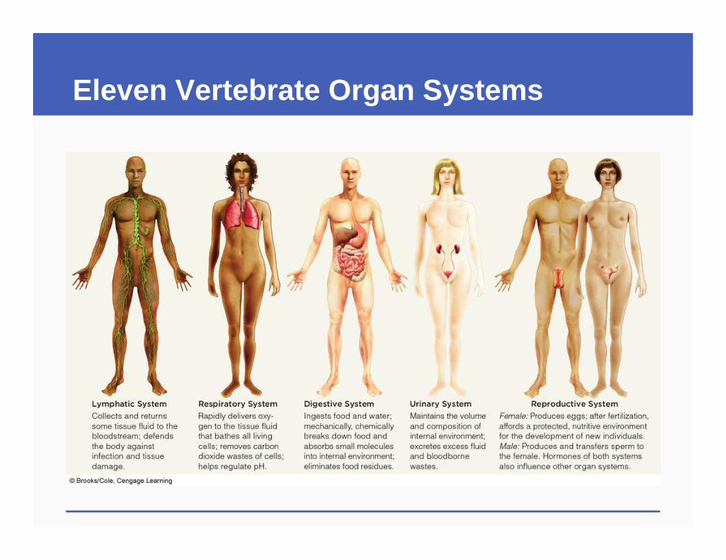
Fig. 32-12b, p. 547
Lymphatic System Respiratory System Digestive System U rinary System Reproductive System
Collects and returns some tissue flflflfluid to the bloodstream; defends the body against infection and tissue damage.
Rapidly delivers oxygen to the tissue flflflfluid that bathes all living cells; removes carbon dioxide wastes of cells; helps regulate pH.
Ingests food and water; mechanically, chemically breaks down food and absorbs small molecules into internal environment; eliminates food residues.
Maintains the volume and composition of internal environment; excretes excess flflflfluid and bloodbornewastes.
Female: Produces eggs; after fertilization, affords a protected, nutritive environment for the development of new individuals. Male: Produces and transfers sperm to the female. Hormones of both systems also in flflflfluenceother organ systems.
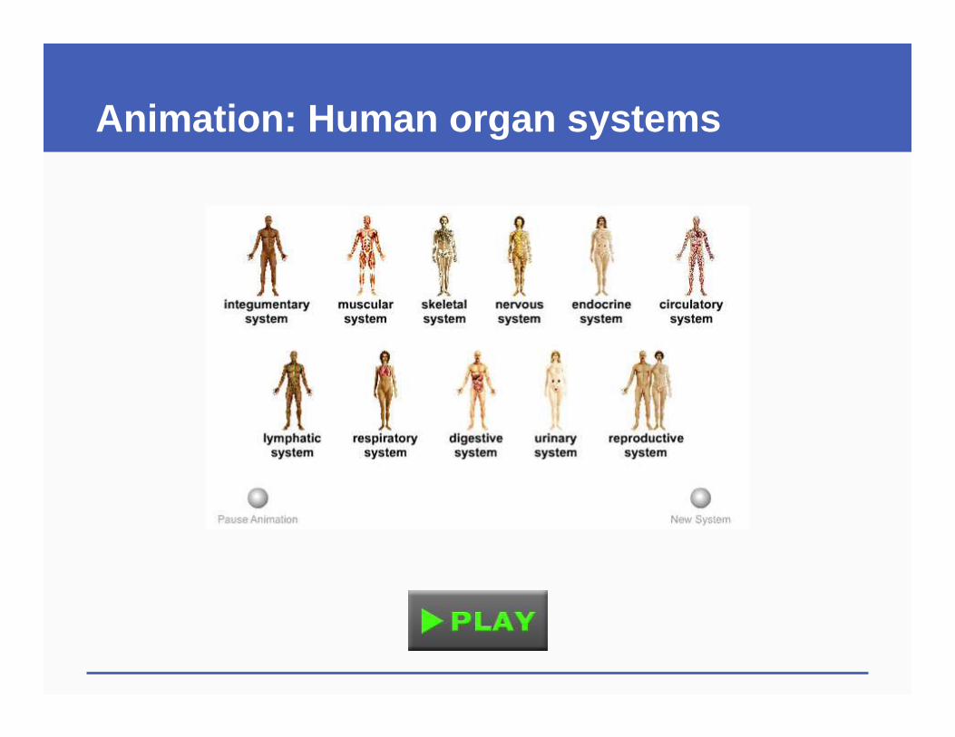
Animation: Human organ systems
32.6 Key Concepts Organ Systems
� Vertebrate organ systems compartmentalize the tasks of survival and reproduction for the body as a whole
� Different systems arise from ectoderm, mesoderm, and endoderm, the primary tissue layers that form in the early embryo
32.7 Vertebrate Skin—Example of an Organ System
� Skin is the body’s interface with the environment• Sensory receptors, barrier against pathogens,
internal temperature control, water conservation
� Vertebrate skin is made up of all four tissue types arranged in two layers: • Outer epidermis contain keratinocytes• Deeper dermis contains nerves, blood and lymph
vessels, hair follicles and glands
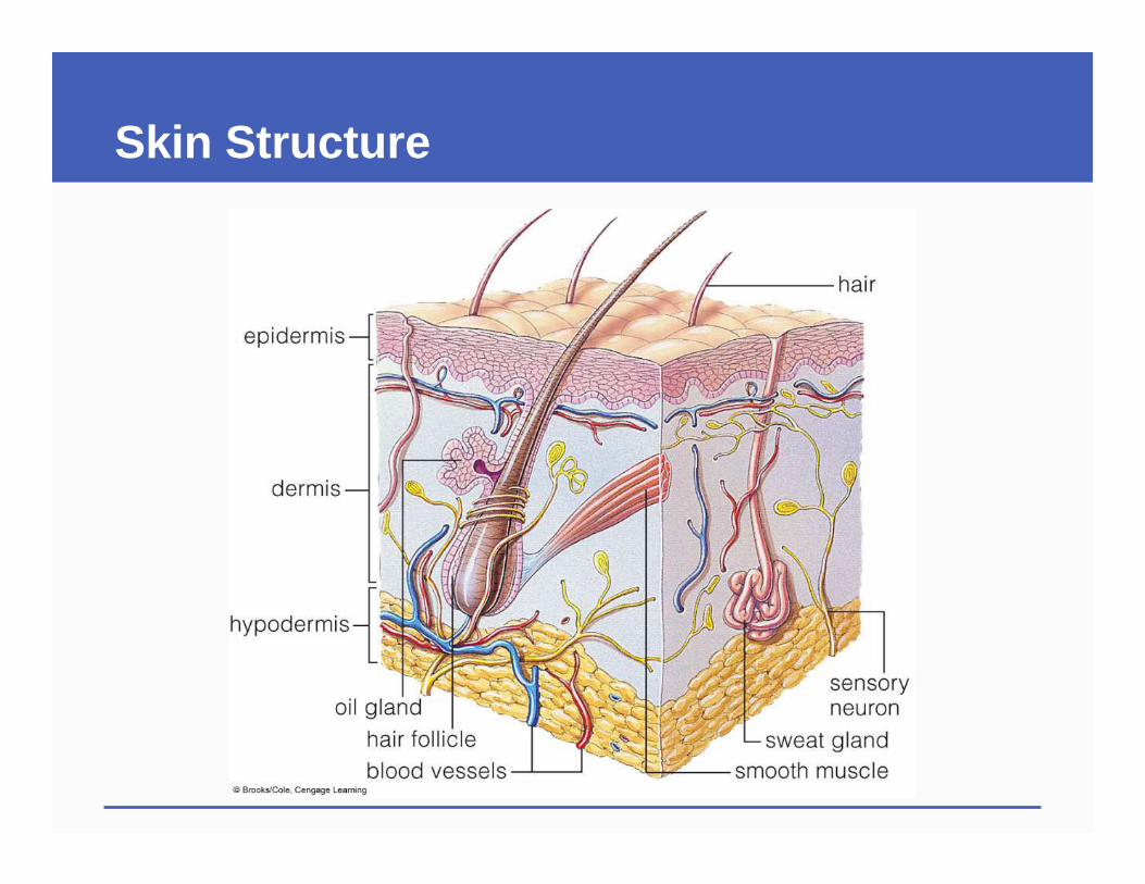
Skin Structure
Skin Structure
Skin Structure
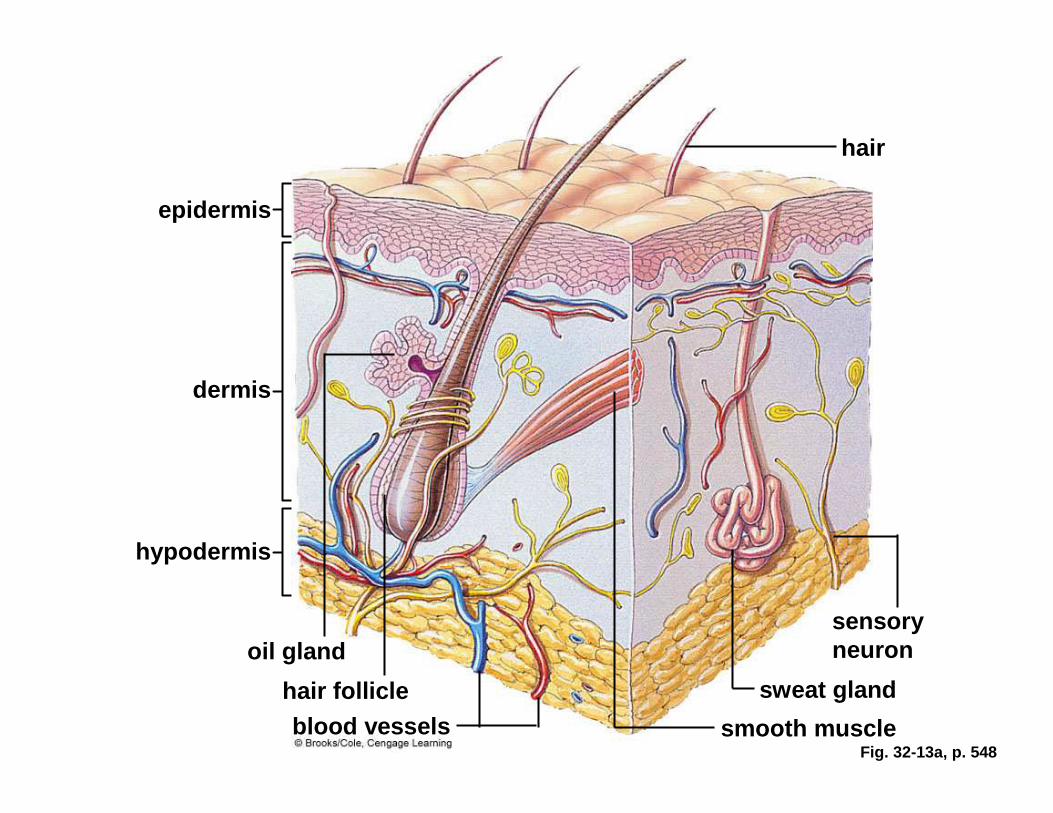
Fig. 32-13a, p. 548
hair
epidermis
dermis
hypodermis
sensory neuronoil gland
hair follicle sweat gland
blood vessels smooth muscle
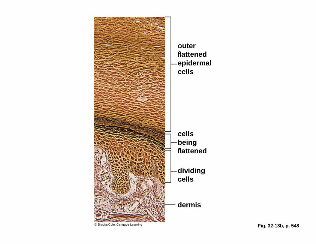
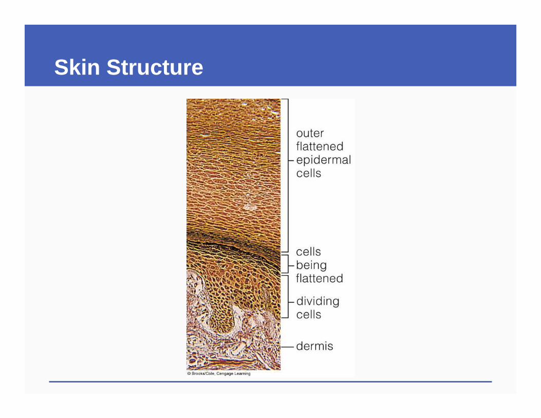
Fig. 32-13b, p. 548
outer flflflflattenedepidermal cells
cells being flflflflattened
dividing cells
dermis
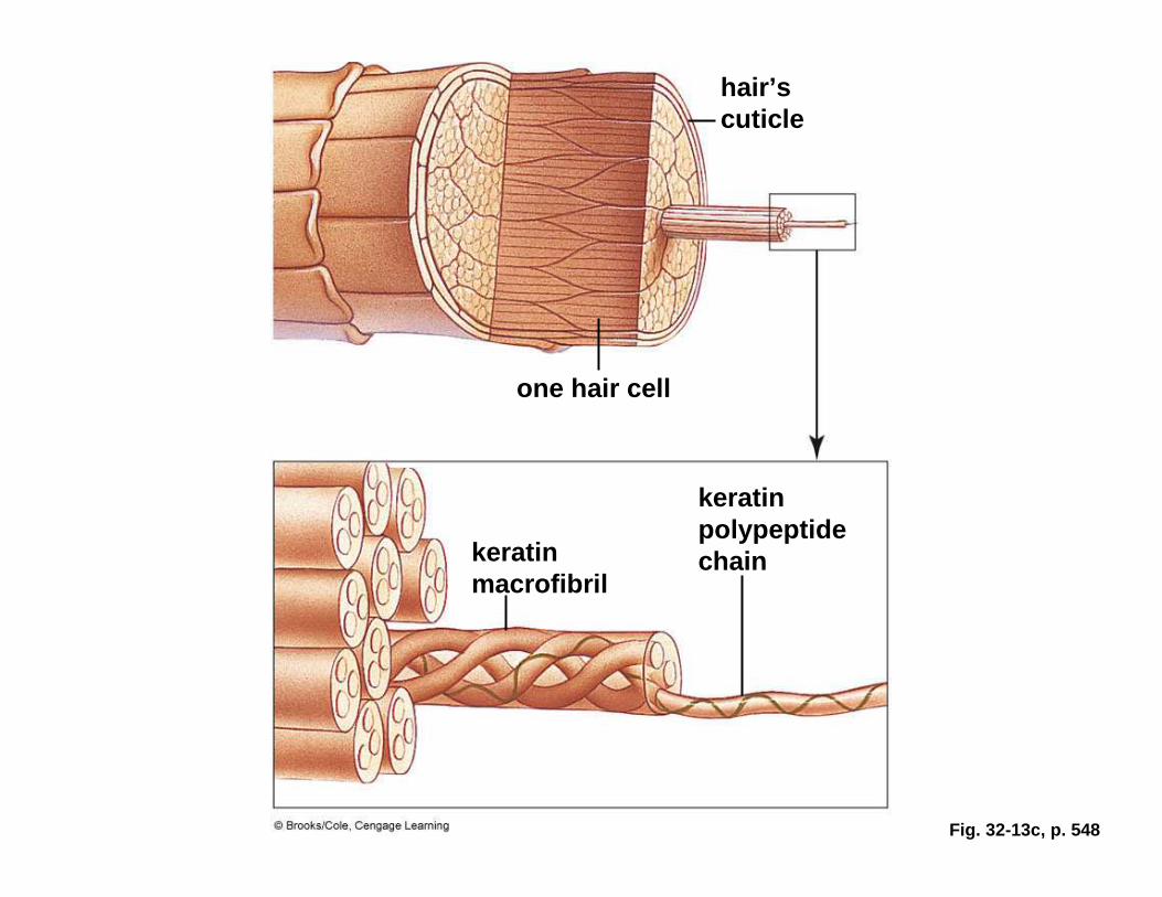
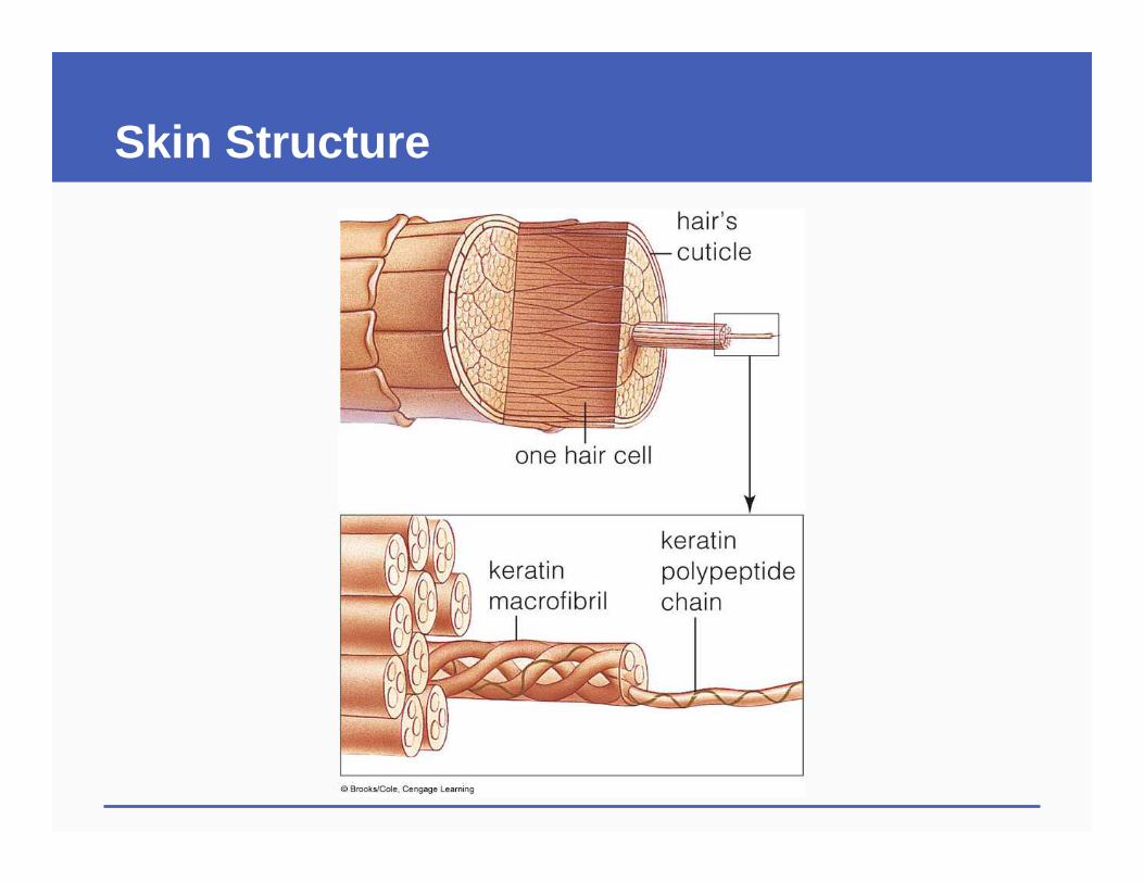
Fig. 32-13c, p. 548
hair’s cuticle
one hair cell
keratin macrofibril
keratin polypeptide chain
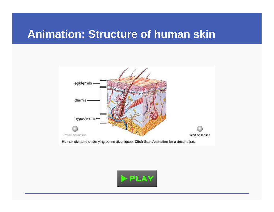
Animation: Structure of human skin
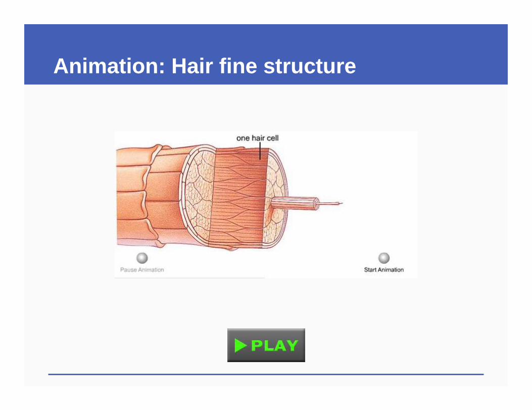
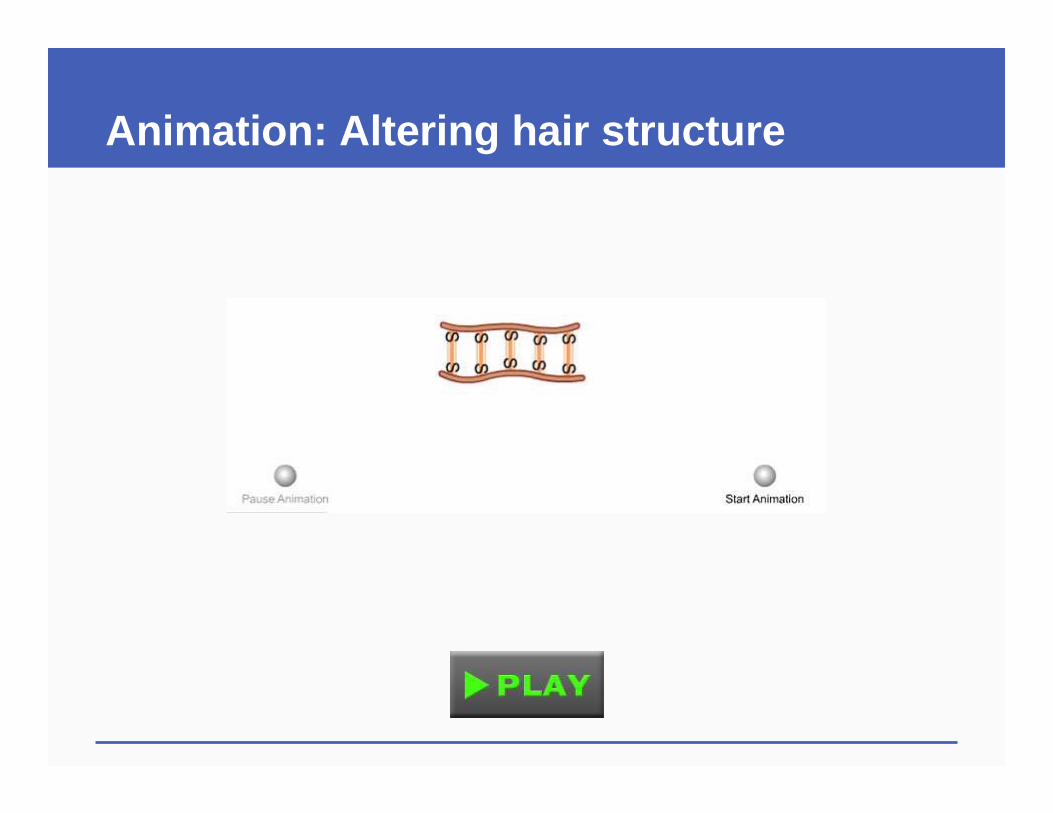
Animation: Hair fine structure
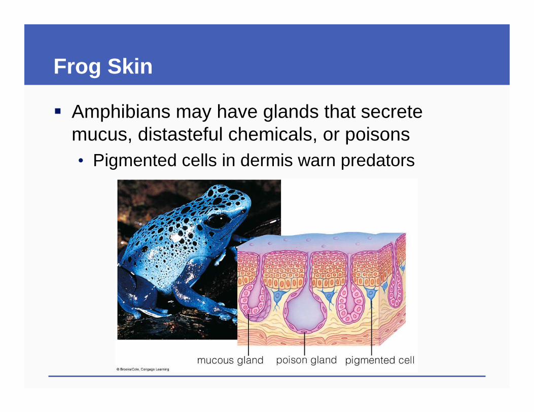
Frog Skin
� Amphibians may have glands that secrete mucus, distasteful chemicals, or poisons• Pigmented cells in dermis warn predators
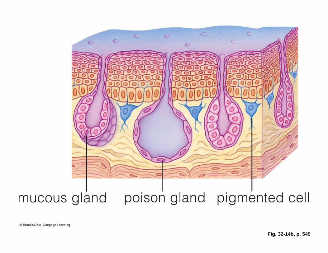
Fig. 32-14b, p. 549
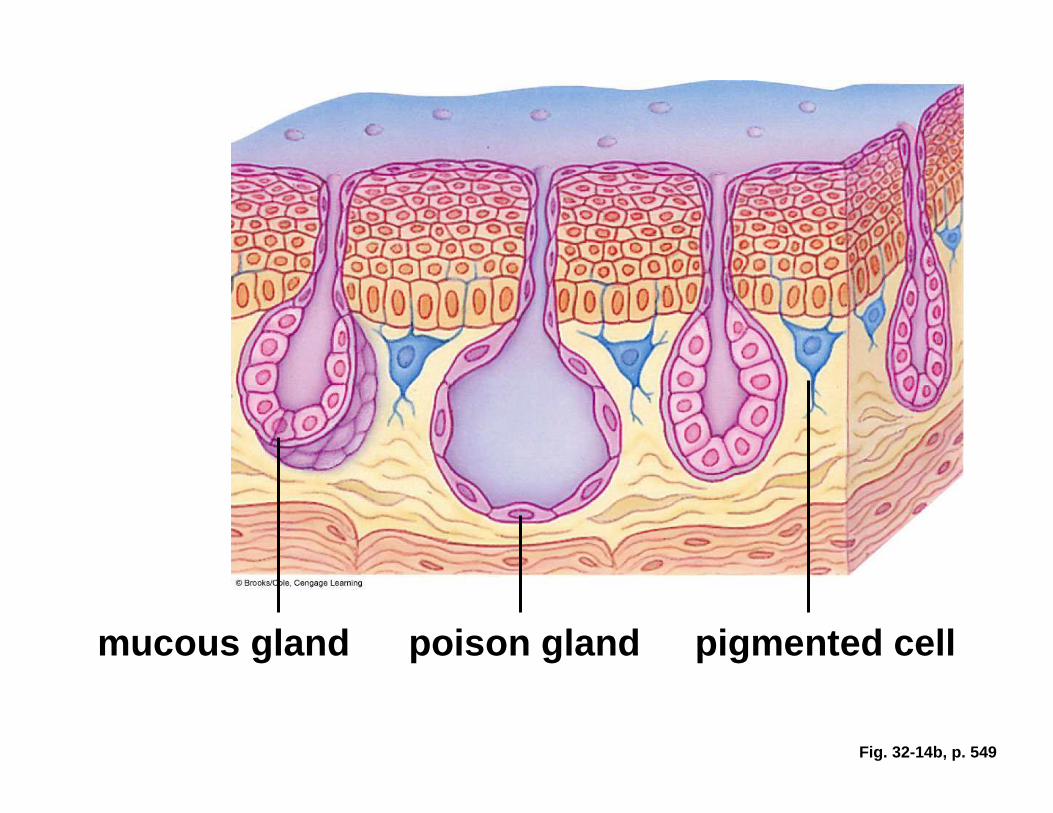
Fig. 32-14b, p. 549
mucous gland poison gland pigmented cell
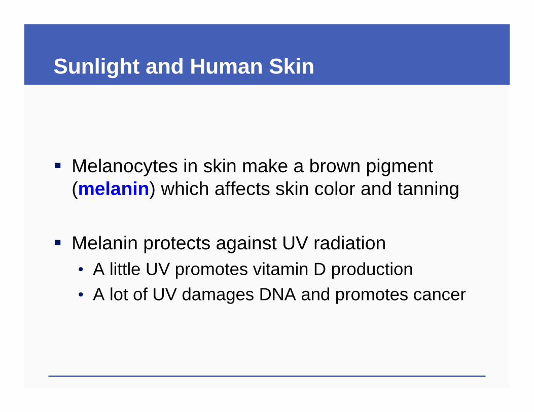
Sunlight and Human Skin
� Melanocytes in skin make a brown pigment (melanin ) which affects skin color and tanning
� Melanin protects against UV radiation• A little UV promotes vitamin D production• A lot of UV damages DNA and promotes cancer
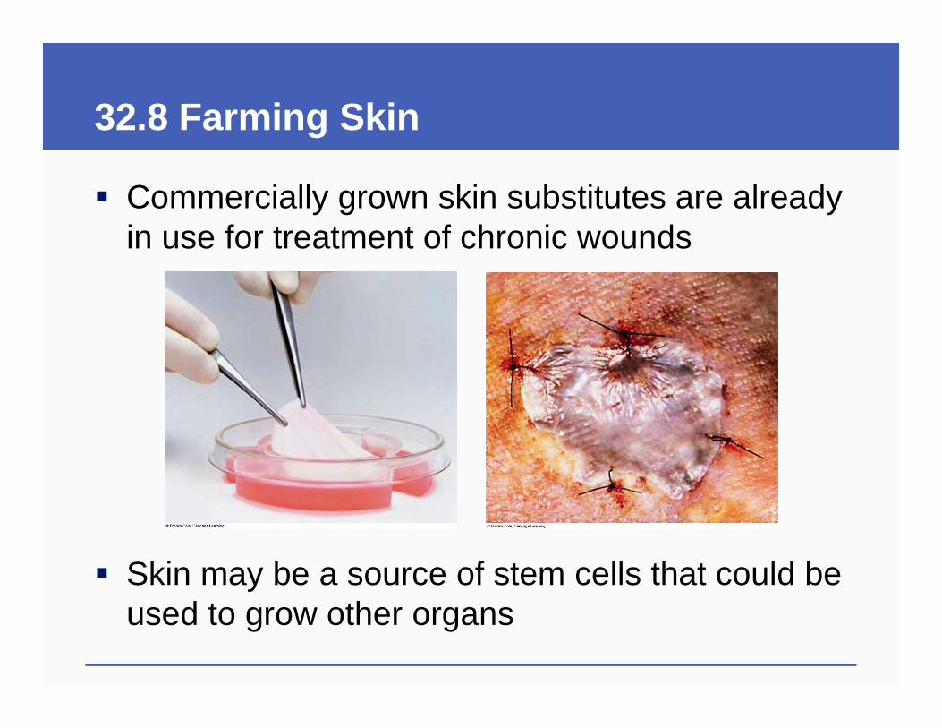
32.8 Farming Skin
� Commercially grown skin substitutes are already in use for treatment of chronic wounds
� Skin may be a source of stem cells that could be used to grow other organs
32.7-32.8 Key ConceptsA Closer Look at Skin
� Skin is an example of an organ system
� It includes epithelial layers, connective tissue, adipose tissue, glands, blood vessels, and sensory receptors
� It helps protect the body, conserve water, control temperature, excrete wastes, and detect external stimuli
Animation: Altering hair structure
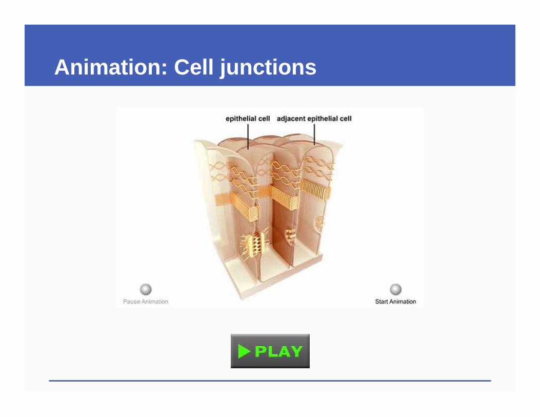
Animation: Cell junctions
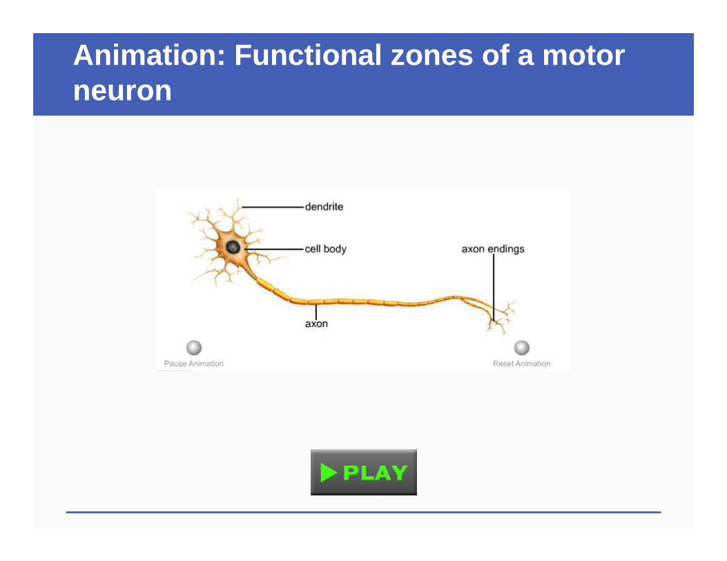
Animation: Functional zones of a motor neuron
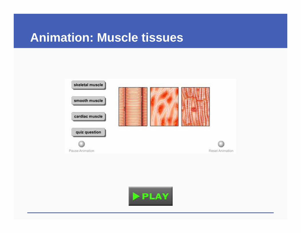
Animation: Muscle tissues
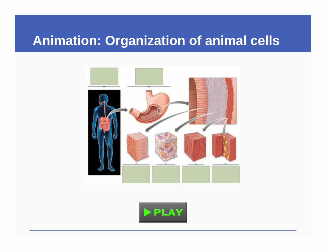
Animation: Organization of animal cells
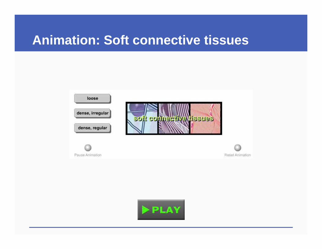
Animation: Soft connective tissues
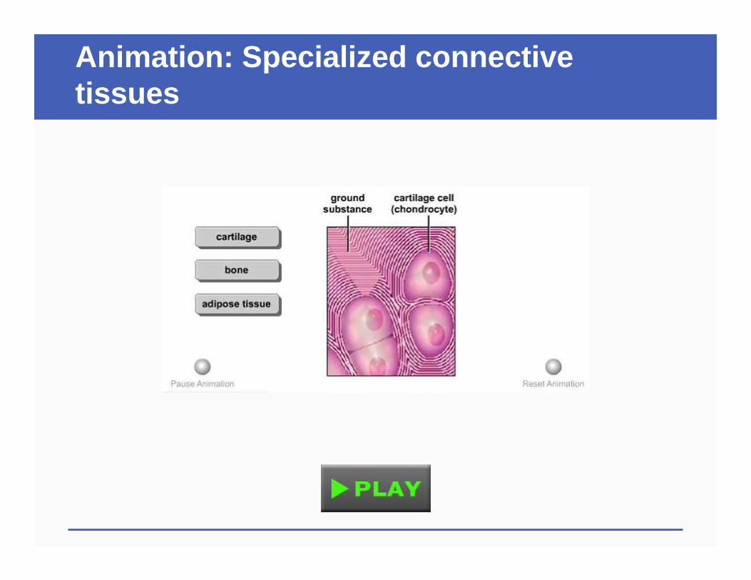
Animation: Specialized connective tissues
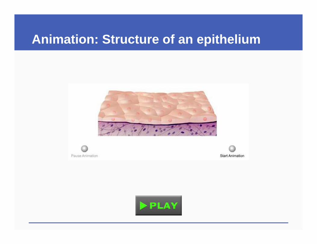
Animation: Structure of an epithelium
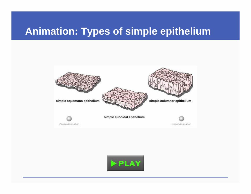
Animation: Types of simple epithelium

ABC video: A Saving Graft

ABC video: New Hands
Video: Stem Cells