Embed Size (px)
Citation preview
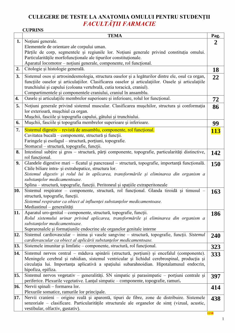
1
CULEGERE DE TESTE LA ANATOMIA OMULUI PENTRU STUDENȚII
FACULTĂȚII FARMACIE
CUPRINS
TEMA Pag.
1. Noțiuni generale.
Elementele de orientare ale corpului uman.
Părţile de corp, segmentele şi regiunile lor. Noţiuni generale privind constituţia omului.
Particularităţile morfofuncţionale ale tipurilor constituţionale.
Aparatul locomotor – noţiuni generale, componente, rol funcţional.
2
2. Citologie și histologie generală. 18 3. Sistemul osos şi artrosindesmologia, structura oaselor şi a legăturilor dintre ele, osul ca organ,
funcţiile oaselor şi articulaţiilor. Clasificarea oaselor şi articulaţiilor. Oasele şi articulaţiile
trunchiului şi capului (coloana vertebrală, cutia toracică, craniul).
Compartimentele şi componentele craniului, craniul în ansamblu.
22
4. Oasele şi articulaţiile membrelor superioare şi inferioare, rolul lor funcţional. 72 5. Noţiuni generale privind sistemul muscular. Clasificarea muşchilor, structura şi conformaţia
lor exterioară, muşchiul ca organ.
Muşchii, fasciile şi topografia capului, gâtului şi trunchiului.
86
6. Muşchii, fasciile şi topografia membrelor superioare şi inferioare. 99 7. Sistemul digestiv – revistă de ansamblu, componente, rol funcţional.
Cavitatea bucală – componente, structură şi funcţii.
Faringele şi esofagul – structură, porţiuni, topografie.
Stomacul – structură, topografie, funcţii.
113
8. Intestinul subţire şi gros – structură, părţi componente, topografie, particularităţi distinctive,
rol funcţional. 142
9. Glandele digestive mari – ficatul şi pancreasul – structură, topografie, importanţă funcţională.
Căile biliare intra- şi extrahepatice, structura lor.
Sistemul digestiv şi rolul lui în aplicarea, transformările şi eliminarea din organism a
substanţelor medicamentoase.
Splina – structură, topografie, funcţii. Peritoneul și spațiile extraperitoneale
150
10. Sistemul respirator – componente, structură, rol funcţional. Glanda tiroidă şi timusul –
structură, topografie, funcţii.
Sistemul respirator ca obiect al influenţei substanţelor medicamentoase.
Mediastinul – generalități
163
11. Aparatul uro-genital – componente, structură, topografie, funcţii.
Rolul sistemului urinar privind aplicarea, transformările şi eliminarea din organism a
substanţelor medicamentoase.
Suprarenalele şi formaţiunile endocrine ale organelor genitale interne
186
12. Sistemul cardiovascular – inima şi vasele sangvine – structură, topografie, funcţii. Sistemul
cardiovascular ca obiect al aplicării substanţelor medicamentoase. 240
13. Sistemele imunitar şi limfatic – componente, structură, rol funcţional. 323 14. Sistemul nervos central – măduva spinării (structură, porţiuni) şi encefalul (componente).
Meningele cerebral şi rahidian, sistemul ventricular şi lichidul cerebrospinal, producţia şi
circulaţia lui. Importanţa aplicativă a spaţiului subarahnoidian. Hipotalamusul endocrin,
hipofiza, epifiza.
333
15. Sistemul nervos vegetativ – generalităţi. SN simpatic şi parasimpatic – porţiuni centrale şi
periferice. Plexurile vegetative. Lanţul simpatic – componente, topografie, ramuri. 397
16. Nervii spinali – formarea lor.
Plexurile somatice, ramurile lor principale. 414
17. Nervii cranieni – origine reală şi aparentă, tipuri de fibre, zone de distribuire. Sistemele
senzoriale – clasificare. Particularităţile structurale ale organelor de simţ (vizual, acustic,
vestibular, olfactiv, gustativ).
438
1218
2
TESTE
Noțiuni generale. Elementele de orientare ale corpului uman. Părţile de corp, segmentele
şi regiunile lor. Noţiuni generale privind constituţia omului.
Particularităţile morfofuncţionale ale tipurilor constituţionale.
Aparatul locomotor – noţiuni generale, componente, rol funcţional.
1. CS. Anatomia ca știință studiază:
A. Forma și structura organismului uman
B. Filo- și ontogeneza organismului uman
C. Modificările condiționate de interacțiunea corpului cu mediul extern
D. Schimbările condiționate de vârstă și gen
E. Toate enumerate.
SC. Anatomy as a science studies:
A. The shape and structure of the human body
B. Phylo- and ontogenesis of the human body
C. Changes conditioned by interaction of the human being with the environment
D. Changes conditioned by age and gender
E. All mentioned above.
CS. Анатомия как наука изучает:
A. Форму и строение человеческого организма
B. Фило- и онтогенез человеческого организма
C. Изменения обусловленные взаимосвязью человека с окружающей средой
D. Изменения обусловленные возрастом и полом человека
E Все вышеперечисленные
2. CM. Numiți metodele de investigație a anatomiei omului viu:
A. Metode senzoriale directe (axate pe simțirile naturale)
B. Metode senzoriale mediate (bazate pe dispozitive și aparate)
C. Metode experimentale pe animale de laborator
D. Metoda de disecție anatomică
E. Metoda microscopică a țesutului bioptic.
MC. Name the methods of examination on a living person:
A. Direct sensory methods (based on natural sensory organs/filings)
B. Paraclinical sensory methods (based on divices)
C. Experimental methods on laboratory animals
D. Anatomical dissection (preparation)
E. Microscopic methods or tissues byopsy
CM. Назовите методы изучения анатомии на живом человеке:
A. Клинические методы исследования (на основании естественных чувств)
B. Параклинические методы исследования (лабораторные и инструментальные)
C. Экспериментальные методы исследуемые на лабораторных животных
D. Метод рассечения (препарирование)
E. Mикроскопический метод исследования биоптического материала (ткани)
3. CS. Definiți noțiunea de ”organ”:
A. Reprezintă o structură bine diferențiată a corpului uman, care ocupă în el un loc
determinat
B. Reprezintă o structură bine diferențiată a corpului uman și are o funcție anumită
C. Reprezintă o structură bine diferențiată a corpului uman cu o formă anumită
D. Reprezintă o structură bine diferențiată a corpului uman, constituită din câteva tipuri de
țesuturi
E. Reprezintă o structură bine diferențiată a corpului uman, care ocupă în el un loc

3
determinat, are o formă și funcție anumită, constituită din câteva tipuri de țesuturi.
SC. Give definition of an ”organ”:
A. An organ represents a well diferentiated structure of the human body with a determined
location
B. An organ represents a well diferentiated structure of the human body with a specific
function
C. An organ represents a well diferentiated structure of the human body having a specific
shape
D. An organ represents a well diferentiated structure of the human body, that is built up from
several tissues
E. An organ represents a well diferentiated structure of the human body, with a determined
location, shape and function, and it is built up of several tisuues.
CS. Дайте определение понятия ”oрган”:
A. Орган представляет собой хорошо дифференцированную структуру человеческого
тела, занимающий в нем определенное место
B. Орган представляет собой хорошо дифференцированную структуру человеческого
тела имеющий определенную функцию
C. Орган представляет собой хорошо дифференцированную структуру человеческого
тела имеющий определенную форму
D. Орган представляет собой хорошо дифференцированную структуру человеческого
тела, состоящий из нескольких видов тканей
E. Орган представляет собой хорошо дифференцированную структуру человеческого
тела, занимающий в нем определенное место, имеющий определенную форму и
функцию, состоящий из нескольких видов тканей.
4. CM. Enumerați tipurile constituționale la om:
A. Normostenic, astenic și hiperstenic
B. Diolihomorf, mezomorf și brahimorf
C. Hipotrofic, mezotrofic și hipertrofic
D. Hipodinamic, mezodinamic și hiperdinamic
E. Feminin și masculin.
MC. Point out the human's constitutional types:
A. Normostenic, astenic and hyperstenic
B. Dolichomorphic, mesomorphic and brachimorphic
C. Hypotrophic, mesotrophic and hypertrophic
D. Hypodynamic, mosodynamic and hyperdynamic
E. Female and male
CM. Перечислите типы телосложения человека:
A. Астенический, гиперстенический и нормостенический
B. Мезоморфный, брахиморфный, долихоморфный
C. Гипотрофический, мезотрофический и гипертрофический
D. Гиподинамический, мезодинамический и гипердинамический
E. Женский и мужской
5. CS. Definiți noțiunea de ”poziție anatomică”:
A. Corpul uman în poziție verticală, cu capul aranjat sub unghi drept
B. Membrele superioare și cele inferioare aliniate într-o linie
C. Extremitățile membrelor superioare și inferioare sunt amplasate pe o circumferință
D. Fața trebuie să fie orientată în sus
E. Corpul uman în poziție verticală, palmele în supinație, membrele inferioare paralele lipite.
4
CS. Give definition of ”anatomical position”:
A. The human body is in a vertical position and its head forms a 90º angle with the body
B. The upper and lower limbs are aligned on the same line
C. The upper and lower limbs are located on a circumference
D. The face must be turned upright
E. The human body is in a vertical position, the palms are supinated and the lower limbs are
parallel and close to each other.
CS. Дайте определение понятия ”исходное анатомическое положение”:
A. Расположение тела в вертикальном положении, голова находится под углом 90º
B. Верхние и нижние конечности расположены на одной линии
C. Верхние и нижние конечности расположены на одной окружности
D. Лицо обращено вверх
E. Тело в вертикальном положении, ладони супинированы, нижние конечности
параллельно и вместе
6. CM. Planul mediosagital:
A. Trece prin axa longitudinală și sagitală a corpului
B. Divide corpul în jumătăți simetrice
C. Departajează componentele mediale și laterale ale formațiunilor corpului
D. Este perpendicular la planurile parasagitale
E. Corespunde grosimii corpului.
MC. The mediosagittal plan:
A. Passes through the longitudinal and sagittal axes of the human body
B. Divides the human body into simmetrical halves
C. Separates the medial components of the human body from the lateral ones
D. It passes perpendicullarly to the parasagittal plans
E. It corresponds to the width of the body.
CM. Срединная сагиттальная плоскость:
A. Проходит через продольную и сагиттальную оси тела
B. Делит тело на две симметричные половины
C. Делит медиальную и латеральную части тела
D.Ориентированна перпендикулярно к парасагиттальным плоскостям
E. Соответствует толщине тела
7. CM. Enumerați tipurile de ținută:
A. Cifotică
B. Redresată
C. Grbovită
D. Lordotică
E. Tonică.
MC. Name the types of human posture:
A. Kyphotic
B. Straighten/flat/plane
C. Hunchbacked
D. Lordotic
E. Tonic.
CM. Перечислите типы осанки:
A. Кифотическая
B. Плоская
C. Сутулая
5
D. Лордотическая
E. Тоническая.
8. CM. Viscerele se proiectează pe peretele anterior al abdomenului:
A. În epigastru
B. În hipogastru
C. În regiunea inghinală
D. În regiunea parasternală
E. În regiunea hipocondriacă stângă.
MC. The viscera project on the anterior abdominal wall in the following regions:
A. Epigastrium
B. Hypogastrium
C. Inguinal region
D. Parasternal region
E. Left hypochondrium.
CM. Внутренности проецироруются на передней стенке живота в следующие
области:
A. Надчревье
B. Подчревье
C. В паховой области
D. В парастернальной области
E. В левое подреберье
9. CM. Care din perioadele de vârstă la om sunt postnatale:
A. De sugar
B. Adolescența
C. De organogeneză
D. Segmentarea
E. Toate enumerate.
MC. Name the postnatal periods of the human life:
A. Infant
B. Puberty
C. Organogenesis
D. Segmentation
E. All mentioned above.
СМ. К постнатальным периодам человека относятся:
A. Грудной период
B. Юность
C. Органогенез
D. Сегментация
E. Все перечисленные.
10. CS. Norma reprezintă:
A. Un diapazon de devieri, în anumite limite de la indicii statistici, neînsoțite de dereglări
funcționale – forma cea mai frecventă din punct de vedere statistic.
B. Structura cea mai rațională și avantajoasă a organismului/organelor, adecvată condițiilor
concrete ale mediului.
C. Abaterea de la structura și/sau funcțiile specifice pentru specia biologică respectivă,
rezultată din perturbarea embriogenezei/morfogenezei, care provoacă dereglări funcționale ale
acestora.
D. Acele formațiuni anatomice, care au fost caracteristice strămoșilor îndepărtați ai omului.
6
E. Modul particular de prezentare a unei formațiuni anatomice, apărută ca rezultat al abaterilor
în dezvoltare, care nu depășește limitele normei.
SC. The norm is:
A. A range of deviations within certain limits of statistical indexes, which are not accompanied
by functional disorders - the most common from statistical point of view.
B. The most rational and useful structure of the body/organ adequite for life within
corresponding envitonmental conditions
C.Deviation from the specific structure or/and function inhereted in the respective biological
species, which appeared due to disturbances of embryogenesis / morphogenesis, leading to
functional disorders
D. Anatomical structures that were characteristic to our ancestors
E. A particular (individual) way of manifestation of an anatomical structure that appeared as a
result of deviations of development, but not exeeding the normal limits.
CS. Норма представляет:
A. Диапазон отклонений, в определенных пределах от статистических показателей, не
сопровождающиеся функциональными расстройствами (средняя арифметическая
целого ряда изменений – наиболее статистики распространенная форма
B. Самая рациональная и полезная структура тела/органа, соответствующая конкретным
условиям окружающей среды
C. Отклонение от структуры и/или функции присущее соответствующему
биологическому виду, возникшее вследствие нарушения эмбриогенеза /морфогенеза,
ведущее к нарушению функции этих
D. Aнатомические образования, свойственные далеким предкам человека.
E. Индивидуальное проявление анатомического образования, возникшее в результате
отклонений в процессе внутриутробного развития, не выходящее за пределы нормы
11. CS. Variantă a normei (varitas) este:
A. Un diapazon de devieri, în anumite limite, de la indicii statistici, neînsoțite de dereglări
funcționale – forma cea mai frecventă din punct de vedere statistic.
B. Structura cea mai rațională și avantajoasă a organismului/organelor, adecvată condițiilor
concrete ale mediului.
C. Formațiunea anatomică, care a fost caracteristică strămoșilor îndepărtați ai omului.
D. Modul particular de prezentare a unei formațiuni anatomice, apărută ca rezultat al abaterilor
în dezvoltare, care nu depășește limitele normei.
E. Abaterea de la structura și/sau funcțiile specifice pentru specia biologică respectivă,
rezultată din perturbarea embriogenezei/morfogenezei.
SC. The variant of norm (varitas) is:
A. A range of deviations within certain limits of statistical indexes, that are not accompanied
by functional disorders - the most common from statistical point of view
B. The most rational and useful structure of the body/organ adequite for life within
corresponding envitonmental conditions
C.Anatomical structures that were characteristic to our ancestors
D. A particular (individual) way of manifestation of an anatomical structure that appeared as a
result of deviations of development, but not exeeding the normal limits
E. Deviation from the specific structure or/and function inhereted in the respective biological
species, which appeared due to disturbances of embryogenesis / morphogenesis.
CS. Вариант нормы (varitas) это:
A. Диапазон отклонений, в определенных пределах, от статистических показателей, не
сопровождающиеся функциональными расстройствами – наиболее распространенная
форма с точки зрения статистики
B. Самая рациональная и полезная структура тела/органа, соответствующая конкретным
7
условиям окружающей среды
C. Анатомические образования, свойственные далеким предкам человека.
D. Индивидуальное проявление анатомического образования, возникшее в результате
отклонений в процессе внутриутробного развития, не выходящее за пределы нормы
E. Отклонение от структуры и/или функции присуще для соответствующего
биологического вида, возникшее вследствие нарушения эмбриогенеза /морфогенеза
12. CS. Variabilitatea anatomică individuală determină:
A. Diapazonul de devieri, în anumite limite, de la indicii statistici, neînsoțite de dereglări
funcționale – media aritmetică a unei game de varietăți
B. Abaterea de la structura și/sau funcțiile specifice pentru specia biologică respectivă,
rezultată din perturbarea embriogenezei/morfogenezei
C. Structura cea mai rațională și avantajoasă a organismului/organelor, adecvată condițiilor
concrete ale mediului.
D. Acele formațiuni anatomice, care au fost caracteristice strămoșilor îndepărtați ai omului.
E. Modul particular de prezentare a unei formațiuni anatomice, apărută ca rezultat al abaterilor
în dezvoltare, care nu depășește limitele normei.
SC. Individual anatomical variability determines:
A. A range of deviations within certain limits of statistical indexes, that are not accompanied
by functional disorders – the average of a row of variations
B. Deviation from the specific structure or/and function inhereted in the respective biological
species, which appeared due to disturbances of embryogenesis/morphogenesis
C. The most rational and useful structure of the body/organ adequite for life within
corresponding envitonmental conditions
D. Anatomical structures that were characteristic to our ancestors
E. A particular (individual) way of manifestation of an anatomical structure that appeared as a
result of deviations of development, but not exeeding the normal limits.
CS. Индивидуальная анатомическая изменчивость определяет:
A. Диапазон отклонений, в определенных пределах, от статистических показателей, не
сопровождающиеся функциональными расстройствами – средняя арифметическая
целого ряда изменений
B. Отклонение от структуры и/или функции присуще для соответствующего
биологического вида, возникшее вследствие нарушения эмбриогенеза/морфогенеза
C. Самая рациональная и полезная структура тела/органа, соответствующая конкретным
условиям окружающей среды
D. Анатомические образования, свойственные далеким предкам человека
E. Индивидуальное проявление анатомического образования, возникшее в результате
отклонений в процессе внутриутробного развития, не выходящее за пределы нормы.
13. CS. Noțiunea de atavism se referă la:
A. Abaterea de la structura și/sau funcțiile specifice pentru specia biologică respectivă,
rezultată din perturbarea embriogenezei/morfogenezei acelei/altei formațiuni anatomice
B. Modul particular de prezentare a unei formațiuni anatomice, apărută ca rezultat al abaterilor
în dezvoltare, care nu depășește limitele normei
C. Structura cea mai rațională și avantajoasă a organismului/organelor, adecvată condițiilor
concrete ale mediului.
D. Acele formațiuni anatomice, care au fost caracteristice strămoșilor îndepărtați ai omului.
E. Diapazonul de devieri, în anumite limite, de la indicii statistici, neînsoțite de dereglări
funcționale – forma cea mai frecventă din punct de vedere statistic.
SC. What an atavism is:
A. Deviation from the specific structure or/and function inhereted in the respective biological
species, which appeared due to disturbances of embryogenesis/morphogenesis of one or
8
another anatomical structure
B. A particular (individual) way of manifestation of an anatomical structure that appeared as a
result of deviations of development, but not exeeding the normal limits
C. The most rational and useful structure of the body/organ adequite for life within
corresponding envitonmental conditions
D. Anatomical structures that were characteristic to our ancestors
E. A range of deviations within certain limits of statistical indexes, that are not accompanied
by functional disorders - the most common from statistical point of view.
CS. Определение атавизм относится к:
A. Отклонение от структуры и/или функции присуще для соответствующего
биологического вида, возникшее вследствие нарушения эмбриогенеза /морфогенеза того
/иного анатомического образования
B. Индивидуальное проявление анатомического образования, возникшее в результате
отклонений в процессе внутриутробного развития, не выходящее за пределы нормы
C. Самая рациональная и полезная структура тела / органа, соответствующая
конкретным условиям окружающей среды
D. Анатомические образования, свойственные далеким предкам человека
E. Диапазон отклонений, в определенных пределах, от статистических показателей, не
сопровождающиеся функциональными расстройствами – наиболее распространенная
статистическая форма.
14. CS. Anomalia (anomalos) reprezintă:
A. Diapazonul de devieri, în anumite limite, de la indicii statistici, neînsoțite de dereglări
funcționale – forma cea mai frecventă din punct de vedere statistic.
B. Modul particular de prezentare a unei formațiuni anatomice, apărută ca rezultat al abaterilor
în dezvoltare, care nu depășește limitele normei
C. Structura cea mai rațională și avantajoasă a organismului/organelor, adecvată condițiilor
concrete ale mediului.
D. Acele formațiuni anatomice, care au fost caracteristice strămoșilor îndepărtați ai omului.
E. Abaterea de la structura єi/sau funcțiile specifice pentru specia biologică respectivă,
rezultată din perturbarea embriogenezei/morfogenezei acelei/altei formațiuni anatomice, care
provoacă dereglări funcționale ale acestora.
SC. An abnormality (anomalos) is:
A. A range of deviations within certain limits of statistical indexes, that are not accompanied
by functional disorders - the most common from statistical point of view
B. A particular (individual) way of manifestation of an anatomical structure that appeared as a
result of deviations of development, but not exeeding the normal limits
C.The most rational and useful structure of the body/organ adequite for life within
corresponding envitonmental conditions
C.Anatomical structures that were characteristic to our ancestors
D. A particular (individual) way of manifestation of an anatomical structure that appeared as a
result of deviations of development, but not exeeding the normal limits
E. Deviation from the specific structure or/and function inhereted in the respective biological
species, which appeared due to disturbances of embryogenesis/morphogenesis, of one or
another anatomical structure, leading to functional disorders.
CS. Аномалия (anomalos) представляет:
A. Диапазон отклонений, в определенных пределах, от статистических показателей, не
сопровождающиеся функциональными расстройствами – наиболее распространенная
форма с точки зрения статистики
B. Индивидуальное проявление анатомического образования, возникшее в результате
отклонений в процессе внутриутробного развития, не выходящее за пределы нормы
C. Самая рациональная и полезная структура тела / органа, соответствующая
9
конкретным условиям окружающей среды
D. Анатомические образования, свойственные далеким предкам человека.
E. Отклонение от структуры и/или функции присуще для соответствующего
биологического вида, возникшее вследствие нарушения эмбриогенеза/морфогенеза того
/другого анатомического образования, ведущее к нарушению функции этих.
15. CS. Constituția se definește ca:
A. Totalitatea caracterelor de ordin psihic și somatic ale unui individ, care se exteriorizează
prin particularități morfologice, funcționale, de randament, rezistență precum și reacția
individului la diferite influențe nocive și patologice
B. Totalitatea caracterelor de ordin psihic și somatic ale unui individ, care se exteriorizează
prin particularități funcționale
C. Totalitatea caracterelor de ordin psihic și somatic ale indivizilor, care se exteriorizează prin
particularități de rezistență
D. Totalitatea caracterelor de ordin psihic și somatic ale indivizilor, care se exteriorizează prin
stabilități certe morfologice, funcționale, de randament și rezistență
E. Totalitatea caracterelor de ordin psihic și somatic ale unui individ, care se exteriorizează
prin stabilități certe morfologice, funcționale și prin reacția individului la diferite influențe
nocive și patologice
CS. Дайте определение понятия ”телосложение”:
A. Совокупность психических и соматических характеристик индивидуума, которые
проявляются морфологическими и функциональными особенностями, эффективности
работы, прочностью, а также индивидуальной реакции на различных вредных и
патологических влияний
B. Совокупность психических и соматических характеристик индивидуума, которые
проявляются функциональными особенностями
C. Совокупность психических и соматических характеристик индивидуума, которые
проявляются особенностями сопротивления
D. Совокупность психических и соматических характеристик индивидуума, которые
проявляются определенной стабильностью – морфологической, функциональной,
эффективностью и прочностью
E. Совокупность психических и соматических характеристик индивидуума, которые
проявляются явной морфологической и функциональной стабильностью, а также
реакцией на различных вредных и патологических влияний
16. CM. Nomenclatura Anatomică Internațională:
A. Include termenii care determină poziția, dimensiunile organelor și locația lor
B. Include termenii care determină mișcările din diferite segmente corporale
C. De obicei au origine a limbajului autohton
D. Este o listă strictă, fără schimbări ulterioare
E. Își ia naștere din Grecia Antică.
MC. International Anatomical Terminology:
A. Includes antomical terms that determine the position, dimensions and location of the organs
B. Includes antomical terms that determine the movement of different segments of the body
C. It includes a list of anatomical terms in Romanian language
D. It is a strict list of anatomical terms and no changes are allowed
E. It was found in the Ancient Greece.
CM. Международная Анатомическая Номенклатура:
A. Включает список анатомических терминов, определяющие положение, размеры
органов и их расположение
B. Включает список анатомических терминов, определяющие движения различных
частей тела
10
C. В ней приведен полный список анатомических терминов на румынском языке
D. Включает список анатомических терминов без их последующих изменений
E. Родилась в Древней Греции.
17. CM. Ținuta gârbovită se caracterizează prin:
A. Lordoză cervicală bine pronunțată, lordoză lombară - redusă
B. Lordoză cervicală redusă, lordoză lombară - bine pronunțată
C. Tipică pentru perioada de senilitate
D. Tipică pentru perioada de mică copilărie
E. Limitarea mișcărilor coastelor duce la micșorarea volumului cutiei toracice.
CM. The following features are characteristic for slouching (hunchbacked) position of
the human body:
A. Significant cervical lordosis, and poor pronounced lumbar lordosis
B. Poor cervical lordosis, and significant lumbar lordosis
C. It is typic for senile people
D. It is typic for early childhood
E. The limited movement of the ribs leads to the diminuation of the thoracic cage volume.
CM. Сутулая осанка характеризуется:
A. Хорошо выраженным шейным лордозом и слабо намеченным поясничным лордозом
B. Слабо намеченным шейным лордозом и хорошо выраженным поясничным лордозом
C. Типична старческому возрасту
D. Типична младшему школьному возрасту
E. Ограничение движения ребер ведет к уменьшению обьема грудной клетки.
18. CM. Planurile de orientare ale corpului uman:
A. Sagital
B.Ventral
C. Frontal
D. Transversal
E. Dorsal.
CM. Plans of the human body are:
A. Sagittal
B.Ventral
C. Frontal
D. Transverse
E. Dorsal.
CM. Плоскости для ориентирования тела человека:
A. Сагиттальная
B. Вентральная
C. Фронтальная
D. Поперечная
E. Дорсальная.
19. CM. Axe de orientare ale corpului uman:
A. Longitudinal
B. Sagital
C. Transversal
D. Ventral
E. Dorsal.
CM. Axes of the human body are:
11
A. Longitudinal
B. Sagittal
C. Transverse
D. Ventral
E. Dorsal.
CM. Оси для ориентирования тела человека:
A. Продольная
B. Сагиттальная
C. Поперечная
D. Вентральная
E. Дорсальная
20. CS. La determinarea vârstei biologice se ține cont de criteriile:
A. Somatice
B. Endocrine
C. Scheletice
D. Clinice
E. Toate menționate.
CS. The following criteria must be taken into consideration when determining the
biological age:
A. Somatic
B. Endocrine
C. Skeletal
D. Clinical
E. All mentioned above.
CS. Биологический возраст определяется следующими критериями: A. Соматическими
B. Эндокринными
C. Скелетными
D. Клиническими
E. Все вышеуказанные.
21. CS. Când au loc salturile de creștere:
A. Prima jumătate de dezvoltare intrauterină
B. A doua jumătate de dezvoltare intrauterină
C. La vârsta de 4-7 ani
D. La vârsta de 14-16 ani
E. La vârsta de 19-24 ani.
SC. Periods of growth:
A. The first half of the intrauterine development
B. The second half of the intrauterine development
C. At 4-7 years of age
D. At 14-16 years of age
E. At 19-24 years of age.
CS. Периоды роста: A. В первой половине развития внутриутробной жизни
B. Во второй половине развития внутриутробной жизни
C. В возрасте 4-7 лет
D. В возрасте 14-16 лет
E. В возрасте 19-24 лет
12
22. CM. Axe de orientare ale corpului uman:
A. Longitudinal
B. Sagital
C. Transversal
D. Ventral
E. Dorsal.
CM. Axes of the human body are:
A. Longitudinal
B. Sagittal
C. Transverse
D. Ventral
E. Dorsal.
CM. В анатомии пользуются следующими осями: A. Продольная
B. Сагиттальная
C. Поперечная
D. Вентральная
E. Дорсальная
23. CM. Etagele peretelui anterior al abdomenului:
A. Cranial
B. Epigastrul
C. Caudal
D. Mezogastrul
E. Hipogastrul.
CM. Regions of the anterior abdominal wall:
A. Cranial
B. Epigastrium
C. Caudal
D. Mesogastrium
E. Hypogastrium.
CM. Этажи передней стенки живота:
A. Краниальный
B. Надчревный
C. Каудальный
D. Чревье
E. Подчревье
24. CM. Sunt zone anatomoclinice ale peretelui ventral al abdomenului:
A. Laterale abdominale: dreaptă şi stângă
B. Ombilicală
C. Hipocondriacă dreaptă şi stângă
D. Pubiană
E. Lombară.
MC. On the anterior abdominal wall the following anatomoclinical regions are
distinguished:
A. Right and left lateral regions
B. Ombilical region
C. Right and left hypocondriums
D. Pubic region
13
E. Lumbar region.
CM. Анатомоклинические области передней стенки живота:
A. Латеральные: правая и левая
B. Пупочная
C. Правая и левая подрѐберные
D. Лобковая
E. Поясничная.
25. CM. Sunt zone anatomoclinice ale peretelui ventral al abdomenului:
A. Medială
B. Laterală
C. Inghinală dreaptă şi stângă
D. Epigastrică propriu-zisă
E. Lombară.
MC. The following anatomoclinical zones of the ventral abdominal wall are
distinguished:
A. Medial region
B. Lateral region
C. Right and left inguinal
D. Epigarstric region
E. Lumbar region.
CM. Анатомоклинические области передней стенки живота (вентральной):
A. Медиальная
B. Латеральная
C. Правая и левая паховые
D. Собственно надчревная
E. Поясничная
26. CM. Enumeraţi sistemele de organe a aparatului locomotor:
A. Ligamentar
B. Artrosindesmologia
C. Miologia
D. Cartilaginos
E. Osos.
MC. Name the systems of organs of the locomotor apparatus:
A. Ligamentary
B. Arthrosyndesmology
C. Miology
D. Cartilaginous
E. Osteology.
CM. Перечислите системы органов опорно-двигательного аппарата:
A. Связочный аппарат
B. Артросиндесмология
C. Миология
D. Хрящевой аппарат
E. Костная система
27. CM. Partea pasivă a aparatului locomotor include:
A. Oasele
B. Muşchii
14
C. Articulaţiile
D. Tendoanele muşchilor
E. Fasciile musculare.
MC. The passive part of the locomot apparatus includes:
A. Bones
B. Muscles
C. Joints
D. Muscles tendons
E. Muscles fasciae.
CM. Пассивная часть опорно-двигательного аппарата включает:
A. Кости
B. Мышцы
C. Суставы
D. Сухожилия мышц
E. Фасции мышц.
28. CS. Partea activă a aparatului locomotor include:
A. Muşchii
B. Oasele
C. Articulaţiile
D. Tendoanele muşchilor
E. Fasciile musculare.
SC. The active part of the locomotor apparatus includes:
A. Muscles
B. Bones
C. Joints
D. Muscles tendons
E. Muscles fasciae.
CS. Активная часть опорно-двигательного аппарата включает:
A. Мышцы
B. Кости
C. Суставы
D. Сухожилия мышц
E. Фасции мышц
29. CM. Formaţiunile anatomice ale scheletului dur:
A. Oase
B. Articulaţii
C. Ţesut cartilaginos
D. Tunicile conjunctive ale viscerelor
E. Muşchii.
CM. The anatomical formations of the hard skeleton are:
A. Bones
B. Joints
C. Cartilaginous tissue
D. Conjunctive coats of the viscera
E. Muscles.
CМ. Анатомические образования твѐрдого скелета:
A. Кости
15
B. Суставы
C. Хрящевая ткань
D. Соединительные оболочки внутренних органов
E. Мышцы
30. CM. Oasele sunt depozite pentru:
A. Săruri minerale
B. De sânge
C. De calciu
D. Acid citric
E. Toate false.
CM. The bones are storage of:
A. Mineral solts
B. Blood
C. Calcium
D. Citric acid
E. All are false.
CМ. Кости являются депо для:
A. Минеральных солей
B. Крови
C. Кальция (соли кальция)
D. Цитровая кислота
E. Неправильные ответы
31. CM. Proprietăţile fizice ale oaselor:
A. Rezistenţa
B. Elasticitatea
C. Contractilitate
D. Conductibilitate
E. Excitabilitate.
CM. Physical properties of bones are:
A. Resistence
B. Elasticity
C. Contractibility
D. Conductibility
E. Excitability.
CМ. Физические свойства костей:
A. Прочность
B. Эластичность (упругость)
C. Сократительность
D. Проводимость
E. Возбудимость
32. CM. Funcţiile biologice ale osului:
A. Imunitară
B. De sprigin
C. Hematopoetică
D. Participă la schimbul de substanţe
E. Rezervă de calciu.
CM. The biological functions of bones are:
16
A. Imunitary
B. Support
C. Haematopoietic
D. Participation in exchanges of substances
E. Calcium storage.
CМ. Биологические функции костей:
A. Иммунная
B. Опорная
C. Кроветворная
D. Участвуют в обмене веществ
E. Депонируют соли кальция
33. CM. În dezvoltarea oaselor pot fi evidenţiate etapele:
A. Embrionară
B. Membranoasă
C. Cartilagionasă
D. Osoasă
E. Fetală.
CM. In the development of bones the following stages are distinguished:
A. Embryonic
B. Membranous
C. Cartilaginous
D. Bony
E. Fetal.
CМ. Этапы развития костей:
A. Эмбриональный
B. Перепончатый
C. Хрящевой
D. Костный
E. Фетальный.
34. CM. Modalităţile de osteogeneză:
A. Terţiară
B. Osoasă
C. Desmală (primară)
D. Condrală (secundară)
E. Musculară.
CM. Types of osteogenesis are:
A. Tertiary
B. Bony
C. Desmal (primary)
D. Chondral (secondary)
E. Muscular.
CМ. Типы остеогенеза:
A. Третичный
B. Костный
C. Десмальный (первичный)
D. Хондральный (вторичный)
E. Мышечный
17
35. CM. Funcţiile articulaţiilor sunt de:
A. Unire a oaselor într,un schelet integru
B. Creştere
C. Amortizare
D. Locomoţie
E. Toate false.
CM. Functions of the joints are:
A. Joining of bones into a skeleton
B. Growth
C. Amortization
D. Locomotion
E. All are false.
CМ. К функциям суставов относятся:
A. Соединение костей в один скелет
B. Рост
C. Амортизирующая
D. Рычаги при движении
E. Ответы неправильные
36. CM. După localizare în corp există oase:
A. Craniului
B. Trunchiului
C. Membrelor
D. Polimorfe
E. Sesamoide.
CM. According to the topography the bones are classified into:
A. Bones of the skull
B. Bones of the trunk
C. Bones of the limbs
D. Polimorphic bones
E. Sesamoid bones.
CМ. По локализации кости делят на:
A. Кости черепа
B. Кости туловища
C. Кости конечностей
D. Полиморфные
E. Сесамовидные
37. CM. După dezvoltare există oase:
A. Desmale (primare)
B. Condrale (secundare)
C. Condro-desmale
D. Tubulare
E. Plate.
CM. According to the development the bones are classified into:
A. Desmal (primary) bones
B. Chondral (secundary) bones
C. Chondro-desmal bones
D. Tubular bones
E. Flat bones.
18
CМ. По развитию кости делят на:
A. Десмальные (первичные)
B. Хондральные (вторичные)
C. Хондро-десмальные
D. Трубчатые
E. Плоские.
38. CM. Oasele aerofore (pneumatice) sunt următoarele:
A. Occipitalul
B. Nazale
C. Maxila
D. Sfenoidul
E. Frontalul
CM. The following bones belong to the pneumatic ones:
A. Occipital
B. Nasal
C. Maxilla
D. Sphenoid
E. Frontal.
CM. Воздухоносными являются следующие кости:
А. Затылочная
В. Носовые
С. Верхняя челюсть
D. Клиновидная
Е. Лобная
Citologie
și histologie generală
39. CM. Membranele biologice elementare sunt:
A. Plasmalema
B. Citolema
C. Endomembranele
D. Glicocalexul
E. Biomembranele speciale.
CM. The elementary biological membranes are, as follows:
A. Plasmalemma
B. Cytolemma
C. Endomembranes
D. Glycocalyx
E. Special biomembranes.
CМ. Элементарными биологическими мембранами являются:
A. Клеточная мембрана (плазмалемма)
B. Цитолемма
C. Эндомембраны
D. Гликокаликс
E. Специальные биомембраны
40. CM. Citolema este constituită din:
A. Glicocalix
B. Plasmalemă
19
C. Citoplasmă
D. Elementele citoscheletului
E. Organite celulare.
CM. Cytolemma is built up of:
A. Glycocalyx
B. Plasmalemma
C. Cytoplasm
D. Elements of the cytoskeleton
E. Cell organelles.
CМ. Клеточная мембрана (или цитолемма) состоит из :
A. Гликокаликса
B. Плазмолеммы
C. Цитоплазмы
D. Элементов цитоскелета
E. Клеточные органеллы
41. CM. Funcţiile principale ale citolemei sunt:
A. Recepţie
B. Protecţie
C. Barieră
D. Transport
E. Sinteză.
CM. The main functions of the cytolemma are:
A. Reception
B. Protection
C. Barrier
D. Transportation
E. Synthesis.
CМ. Основные функции клеточной мембраны (цитолеммы) :
A. Чувствительная или рецепторная
B. Защитная
C. Барьерная
D. Транспортная
E. Синтез
42. CM. Citoreceptorii pentru substanţe exogene servesc la detectarea:
A. Toxinelor microbiene
B. Bacteriilor
C. Antigenilor „self”
D. Hormonilor
E. Virusurilor.
CM. Cytoreceptors for exogenic substances serve as detectors for:
A. Microbial toxines
B. Bacteriae
C. Self antigens
D. Hormones
E. Viruses.
CМ. Мембранные рецепторы, взаимодействующие с экзогенными веществами,
служат для определения:
20
A. Бактериальных токсинов
B. Бактерий
C. Антигенов типа „self”
D. Гормонов
E. Вирусов
43. CM. Selectaţi organitele de tip general:
A. Microvilii
B. Complexul Golgi
C. Mitocondriile
D. Ribozomii
E. Cilii.
CM. Select the general organelles:
A. Microvilli
B. Golgi apparatus
C. Mitochondria
D. Ribosomes
E. Cilia.
CМ. Выделите органеллы общего типа:
A. Микроворсинки
B. Комплекс Гольджи
C. Митохондрии
D. Рибосомы
E. Ворсинки
44. CM. Selectaţi funcţiile complexului Golgi:
A. De împachetare a substanţelor sintetizate
B. De sinteză a enzimelor lizozomale
C. De segregare a substanţelor sintetizate
D. De sinteză a proteinelor de secreţie
E. De dezintoxicare.
CM. Select the functions of the Golgi apparatus:
A. Collection of the synthetized substances
B. Synthesis of the hydrolitic lysosomal enzymes
C. Segregation of synthetized substances
D. Synthesis of secretory proteins
E. Detoxification.
CМ. Выделите функции комплекса Гольджи:
A. «Упаковка» синтезируемых веществ
B. Синтез гидролитических ферментов лизосом
C. Сегрегация (отделение) синтезируемых веществ
D. Синтез секреторных белков
E. Дезинтоксикация
45. CM. Selectaţi organitele amembranare:
A. Peroxizomii
B. Ribozomii
C. Reticulul endoplasmatic rugos
D. Centrozomul
E. Citoscheletul.
21
CM. Select the non-mebrane organelles:
A. Peroxisomes
B. Ribosomes
C. Rough endoplasmatic reticulum
D. Centrosom
E. Cytoskeleton.
CМ. Выделите/выберите немембранные органеллы:
A. Пероксисомы
B. Рибосомы
C. Гранулярный эндоплазматический ретикулум (сеть)
D. Клеточный центр (центросома)
E. Цитоскелет
46. CM. Fazele mitozei sunt:
A. Diachineza
B. Metafaza
C. Profaza
D. Leptotena
E. Anafaza.
CM. The phases of the mitosis are:
A. Diakinesis
B. Metaphase
C. Prophase
D. Leptotene (leptonema)
E. Anaphase
CМ. Основные фазы митоза являются:
A. Диакенез
B. Метафаза
C. Профаза
D. Лептотена
E. Анафаза
47. CM. Componentele structurale ale nucleului celular sunt:
A. Lamina nucleară
B. Cromatina
C. Astrosfera
D. Nucleolema
E. Nucleolul.
CM. The structural components of the cell nucleus are:
A. Nuclear lamina
B. Chromatin
C. Astrosphere
D. Nucleolemma
E. Nucleolus.
CМ. Составляющим клеточного ядра являются:
A. Ядерная пластинка
B. Хроматин
C. Астросфера
D. Ядерная оболочка (муклеолема)
E. Ядрышко
22
48. CS. Deplasarea sincronă a cromatidelor spre polii celulei are loc în:
A. Profază
B. Metafază
C. Anafază
D. Telofază
E. Citokineză.
CS. Synchronic movement of the cell towards the cell poles occurs in the following phase:
A. Prophase
B. Metaphase
C. Anaphase
D. Telophase
E. Cytokinesis.
CS. Синхронное смещение хроматид в сторону клеточных полюсов в следующей
фазе:
A. Профазе
B. Метафазе
C. Анафазе
D. Телофазе
E. В фазе цитокинеза
Sistemul osos şi artrosindesmologia, structura oaselor şi a legăturilor dintre ele,
osul ca organ, funcţiile oaselor şi articulaţiilor.
Clasificarea oaselor şi articulaţiilor.
Oasele şi articulaţiile trunchiului şi capului (coloana vertebrală, cutia toracică, craniul).
Compartimentele şi componentele craniului, craniul în ansamblu.
49. CM. Referitor la oasele tubulare, indicați enunțurile corecte:
A. Sunt lungi și scurte
B. Fac parte din oasele trunchiului
C. Cele lungi au epifiză proximală și distală
D. Epifizele posedă fețe articulare acoperite cu cartilaj
E. Către oasele tubulare scurte se referă cele carpiene și tarsiene.
CM. Concerning the tubular bones the following statements are true:
A. Are divided into long and short
B. They belong to the bones of the trunk
C. The long tubular bones have a proximal and distal diaphysis
D. The epiphyses posses articular surfaces covered by cartilage
E. The carpal and tarsal bones are reffered to short tubular bones.
CM. Относительно трубчатых костей. Укажите правильные ответы:
А. Могут быть длинными и короткими
В. Являются составляющими костей туловища
С. Длинные трубчатые кости имеют два эпифиза: проксимальный и дистальный
D. Эпифизы несут суставные поверхности покрытые суставным хрящом
Е. Кости запястья и предплюсны являются короткими трубчатыми костями
50. CM. Oasele plate:
A. Participă la formarea cutiei toracice
B. Sunt component ale centurilor membrelor superioare și inferioare și craniului
C. La ele predomină țesutul osos compact
D. Pot fi confundate cu oasele sesamoide
E. La ele predomină țesutul osos spongios.
23
CM. The flat bones:
A. Participate in formation of the thoracic cage
B. Are components of the upper and lower limbs girdles and of the skull
C. They consist mainly of compact bony tossue
D. They might be confused with spongy bones
E. They consists mainly of spongy bony tissue.
СМ. Плоские кости:
А. Участвуют в образовании грудной клетки
В. Являются составляющими поясов и черепа
С. Построены преимущественно из компактного костного вещества
D. Могут быть приняты за сесамовидные кости
Е. Состоит преимущественно из губчатого костного вещества
51. CM. Oasele mixte:
A. Diferă după formă și structură
B. Sunt componente ale bazei craniului
C. Se întâlnesc în regiunea carpiană și tarsiană
D. Din ele fac parte vertebrele
E. La ele predomină substanța compactă.
CM. The mixed bones:
A. They differ by shape and structure
B. They are components of the basis of the skull
C. They can be found in the tarsal and carpal regions
D. The vertebrae belong to mixed bones
E. They are built up predominantly of compact boby tissue.
CM. Смешанные кости:
А. Различаются по форме и строению
В. Являются составляющими основания черепа
С. Встречаются на уровне запястья и предплюсны
D. К ним относятся позвонки
Е. Построены преимущественно из компактного вещества
52. CM. Oasele pneumatice:
A. Au diafiză și două epifize
B. Se întâlnesc la membre
C. Sunt componente ale craniului
D. Posedă cavități umplute cu un lichid vâscos
E. Au cavități umplute cu aer.
CM. The pneumatic bones:
A. They have a diaphysis and two epiphyses
B. They are located in the limbs
C. They are components of the skull
D. They contain cavities filled with viscous fluid
E. They contain air cavities.
СМ. Воздухоносные кости:
А. Имеют диафиз и два эпифиза
В. Встречаются на уровне конечностей
С. Являются составляющими черепа
D. Содержат полости, заполненные вязкой жидкостью
24
Е. Содержат воздухоносные пазухи.
53. CS. Osteonul reprezintă:
A. Lamelele osoase din jurul diafizei
B. Lamelele osoase din jurul canalului nutritiv
C. Lamelele osoase din jurul canalului medular
D. Lamelele osoase din jurul canalului Havers
E. Lamelele osoase din jurul metafizei.
CS. An osteon consists of: A. Bony lamellae located around the diaphysis
B. Bony lamellae located around the nutrient canal
C. Bony lamellae located around the spinal canal
D. Bony lamellae located around the haversian canal
E. Bony lamellae located around the metaphysis.
CS. Остеон представляет:
А. Костные пластинки вокруг диафиза
В. Костные пластинки вокруг питательного канала
С. Костные пластинки вокруг костномозгового канала
D. Костные пластинки вокруг гаверсового канала
Е. Костные пластинки вокруг метафиза
54. CS. Creşterea osului în grosime are loc pe contul:
A. Cartilajului hialin
B. Cartilajului fibros
C. Periostului
D. Metafizei
E. Fasciei.
CS. Bone growth in thickness occurs due to:
A. Hyaline cartilage
B. Fibrous cartilage
C. Periosteum
D. Metaphysis
E. Fasciae.
CS. Кость растет в толщину за счет:
А. Гиалинового хряща
В. Волокнистого хряща
С. Надкостницы
D. Метафиза
Е. Фасции
55. CS. Creşterea osului în lungime are loc pe contul:
A. Endostului
B. Periostului
C. Cartilajului articular
D. Cartilajului metaepifizar
E. Pericondrului.
CS. Bone growth in length occurs due to:
A. Endosteum
B. Periosteum
C. Articular cartilage
25
D. Metaepiphyseal cartilage
E. Perichondrium.
CS. Кость растет в толщину за счет:
А. Гиалинового хряща
В. Волокнистого хряща
С. Надкостницы
D. Метафизарного хряща
Е. Фасции
56. CS. Punctele de osificare primare apar:
A. În prima jumătate a perioadei intrauterine
B. Imediat după naștere
C. În a doua jumătate a perioadei intrauterine
D.Până la vârsta de 8 ani
E. După vârsta de 10 ani.
CS. Primary ossification points appear:
A. In the first half of intrauterine period
B. Immediately after birth
C. During the second half of intrauterine period
D. By the age of 8
E. After the age of 10.
CS. Первичные точки окостенения появляются:
А. В первой половине внутриутробной жизни
В. Непосредственно после рождения
С. Во второй половине внутриутробной жизни
D. В возрасте до 8 лет
Е. В возрасте после 10 лет
57. CS. Punctele de osificare secundare apar:
A. În prima jumătate a perioadei intrauterine
B. Imediat după naștere
C. În a doua jumătate a perioadei intrauterine
D.Până la vârsta de 8 ani
E. După vârsta de 10 ani.
CS. Secundary ossification points appear:
A. In the first half of intrauterine period
B. Immediately after birth
C. During the second half of intrauterine period
D. By the age of 8
E. After the age of 10.
CS. Вторичные точки окостенения появляются:
А. В первой половине утробной жизни
В. Непосредственно после рождения
С. Во второй половине утробной жизни
D. В возрасте до 8 лет
Е. В возрасте после 10 лет
58. CS. Punctele de osificare auxiliare/adăugătoare apar:
A. În prima jumătate a perioadei intrauterine
B. Imediat după naștere
26
C. În a doua jumătate a perioadei intrauterine
D.Până la vârsta de 8 ani
E. După vârsta de 10 ani.
CS. Auxilliary ossification points appear: A. During the first half of intrauterine life
B. Immediately after birth
C. During the second half of intrauterine life
D. Until the age of 8 years
E. After the age of 10 years.
CS. Добавочные точки окостенения появляются в:
А. В первой половине утробной жизни
В. После рождения
С. Во второй половине утробной жизни
D. В возрасте до 8 лет
E. В возрасте после 10 лет
59. CS. Oasele carpiene şi tarsiene sunt:
A. Tubulare
B. Spongioase
C. Plate
D. Mixte
E. Aerofore.
CS. Carpal and tarsal bones are:
A. Tubular
B. Spongy
C. Flat
D. Mixed
E. Pneumatic.
CS. Кости запястья и предплюсны являются:
А. Трубчатыми костями
В. Губчатыми костями
С. Плоскими костями
D. Смешанными костями
Е. Воздухоносными костями
60. CS. Realizează funcţii de pârghii:
A. Oasele tubulare
B. Oasele spongioase
C. Oasele plate
D. Oasele mixte
E. Oasele aerofore.
CS. Bones that perform functions of levers are: A. Tubular
B. Spongy
C. Flat
D. Mixed
E. Pneumatic.
CS. Выполняют функции рычагов:
А. Трубчатые кости
27
В. Губчатые кости
С. Плоские кости
D. Смешанные кости
Е. Воздухоносные кости
61. CS. Diplöe reprezintă:
A. Substanţa spongioasă a epifizelor
B. Substanţa spongioasă a oaselor carpiene
C. Substanţa spongioasă a oaselor craniului
D. Substanţa spongioasă a sternului
E. Substanţa spongioasă a vertebrelor.
CS. Diplöe is: A. Spongy substance of the epiphyses
B. Spongy substance of the carpal bones
C. Spongy substance of the skull bones
D. Spongy substance of the sternum
E. Spongy substance of the vertebrae.
CS. Diplöe представляет:
А. Губчатое вещество эпифизов
В. Губчатое вещество костей запястья
С. Губчатое вещество костей черепа
D. Губчатое вещество грудины
Е. Губчатое вещество позвонков
62. CS. Sunt localizate în vecinătatea articulaţiilor sau în tendoanele unor muşchi:
A. Oasele fonticulare
B. Oasele plate
C. Oasele suturare
D. Oasele spongioase scurte
E. Oasele sesamoide
CS. The structures located near the joints or inside the muscular tendons are: A. Fonticular bones
B. Flat bones
C. Sutural bones
D. Short spongy bones
E. Sesamoid bones
CS. Располагаются вблизи суставов или в толще сухожилий отдельных мышц:
А. Родничковые кости
В. Плоские кости
С. Шовные кости
D. Короткие губчатые кости
Е. Сесамовидные кости
63. CS. Îndeplinesc rolul de pârghii de viteză:
A. Oasele tubulare lungi
B. Oasele plate
C. Oasele suturare
D. Oasele spongioase scurte
E. Oasele sesamoide.
CS. The bones that perform the function of speed levers are:
28
A. Long tubular bones
B. Flat bones
C. Sutural bones
D. Short spongy bones
E. Sesamoid bones.
CS. Выполняют роль рычага скорости:
А. Длинные трубчатые кости
В. Плоские кости
С. Шовные кости
D. Короткие губчатые кости
Е. Сесамовидные кости
64. CS. Posedă diafiză:
A. Oasele tubulare lungi
B. Oasele plate
C. Oasele suturare
D. Oasele spongioase scurte
E. Oasele sesamoide.
CS. The bones that have diaphysis are: A. Long tubular bones
B. Flat bones
C. Sutural bones
D. Short spongy bones
E. Sesamoid bones.
CS. Имеют диафиз:
А. Длинные трубчатые кости
В. Плоские кости
С. Шовные кости
D. Короткие губчатые кости
Е. Сесамовидные кости
65. CS. La care din vertebrele cervicale lipseşte apofiza spinoasă?
A. C 3
B. C 2
C. C 6
D. C 1
E. C 7
CS. Which of the cervical vertebrae does not have spinous process?
A. C3
B. C2
C. C6
D. C1
E. C7
CS. У каких из шейных позвонков отсутствует остистый отросток:
А. С3
В. С2
С. С6
D. С1
Е. С7
29
66. CS. Sunt rudimentare:
A. Vertebrele toracice
B. Vertebrele cervicale
C. Vertebrele lombare
D. Vertebrele sacrale
E. Vertebrele coccigiene.
CS. Rudimentary vertebrae are: A. Thoracic vertebrae
B. Cervical vertebrae
C. Lumbar vertebrae
D. Sacral vertebrae
E. Coccygeal vertebrae.
CS. Рудиментарными являются:
А. Грудные позвонки
В. Шейные позвонки
С. Поясничные позвонки
D. Крестцовые позвонки
Е. Копчиковые позвонки или копчиковая кость.
67. CS. Au corpul masiv în formă de bob:
A. Vertebrele toracice
B. Vertebrele cervicale
C. Vertebrele lombare
D. Vertebrele sacrale
E. Vertebrele coccigiene.
CS. Vertebrae that have bean-shaped massive body are: A. Thoracic vertebrae
B. Cervical vertebrae
C. Lumbar vertebrae
D. Sacral vertebrae
E. Coccygeal vertebrae.
CS. Имеют массивное тело бобовидной формы:
А. Грудные позвонки
В. Шейные позвонки
С. Поясничные позвонки
Д. Крестцовые позвонки
Е. Копчиковые позвонки
68. CS. În adolescenţă formează un singur os:
A. Vertebrele cervicale
B. Vertebrele toracice
C. Vertebrele lombare
D. Vertebrele sacrale
E. Vertebrele coccigiene.
CS. Vertebrae forming a single bone in adolescence are the: A. Thoracic vertebrae
B. Cervical vertebrae
C. Lumbar vertebrae
D. Sacral vertebrae
E. Coccygeal vertebrae.
30
CS. В юности образуют единую кость:
А. Грудные позвонки
В. Шейные позвонки
С. Поясничные позвонки
D. Крестцовые позвонки
Е. Копчиковые позвонки
69. CS. Are tubercul carotidian:
A. Atlasul
B. Axisul
C. Vertebra cervicală VI
D. Vertebra toracică I
E. Vertebrele lombare
CS. The vertebra that has the carotid tubercle is the: A. Atlas
B. Axis
C. VI-th cervical vertebra
D. I-st thoracic vertebra
E. Lumbar vertebrae.
CS. Имеет сонный бугорок:
А. Атлант
В. Осевой позвонок
С. VI шейный позвонок
D. I грудной позвонок
Е. Поясничные позвонки.
70. CS. Are feţele articulare superioare localizate pe corp:
A. Atlasul
B. Axisul
C. Vertebra cervicală VI
D. Vertebra toracică I
E. Vertebra lombară I
CM. The vertebra that has the superior articular facets on its body is:
A. Atlas
B. Axis
C. VI cervical vertebra
D. I thoracic vertebra
E. I lumbar vertebra.
CS. Имеют суставные поверхности, расположенные в сагиттальной плоскости: А. Атлант
В. Осевой позвонок
С. VI шейный позвонок
D. I грудной позвонок
Е. I поясничный позвонок
71. CS. Nu are corp:
A. Atlasul
B. Axisul
C. Vertebra cervicală VI
D. Vertebra toracică I
31
E. Vertebra lombară V.
CS. It does not have a body: A. Atlas
B. Axis
C. VI-th cervical vertebra
D. I-st thoracic vertebra
E. V-th lumbar vertebra
CS. Не имеет тело:
А. Атлант
В. Осевой позвонок
С. VI шейный позвонок
D. I грудной позвонок
Е. V поясничный позвонок
72. CS. Promontoriul este format de către:
A. Ultima vertebră cervicală şi T1
B. Ultima vertebră toracică şi L1
C. Ultima vertebră lombară şi S1
D. Ultima vertebră sacrală şi Co1
E. Vertebrele T6 si T7
CS. Promontorium is formed by the:
A. The last cervical and T1 vertebrae
B. The last thoracic and L1 vertebrae
C. The last lumbar and S1 vertebrae
D. The last sacral and Co1 vertebrae
E. The T6 and T7 vertebrae
CS. Мыс образован:
А. Последним шейным позвонком и T1
В. Последним грудным позвонком и L1
C. Последним поясничным позвонком и S1
D. Последним крестцовым позвонком и Сo10
Е. Позвонком T6 и T7
73. CS. Un rol funcţional al curburilor coloanei vertebrale este:
A. De amortizare
B. De consolidare a vertebrelor
C. De sprijin
D. De fixare a membrelor
E. De protecţie.
CS. The functional role of the spinal curvatures is: A. Shock absorption
B. Consolidation of the vertebrae
C. Support
D. Fixation of the limb
E. Protection
CS. Физиологическая роль изгибов позвоночного столба является:
А. Амортизирующая
В. Консолидация позвонков
С. Опорная
32
D. Фиксация конечностей
Е. Защитная
74. CS. Care dintre curburile coloanei vertebrale apare la vârsta de 2-3 luni a dezvoltării
postnatale?
A. Lordoza cervicală
B. Scolioza toracală
C. Lordoza lombară
D. Cifoza sacrală
E. Cifoza toracală.
CS. Which spinal curvature forms at the age of 2-3 months of postnatal development? A. Cervical lordosis
B. Thoracic scoliosis
C. Lumbar lordosis
D. Sacral kyphosis
E. Thoracic kyphosis.
CS. Какие кривизны позвоночного столба появляются в 2-3 месячном периоде
постнатального развития? А. Шейный лордоз
В. Грудной сколиоз
С. Поясничный лордоз
D. Крестцовый кифоз
Е. Грудной кифоз
75. CS. Toracele este plat:
A. La sportivi
B. La brahimorfi
C. La persoanele senile
D. La mezomorfi
E. La dolicomorfi.
CS.The thorax is flat: A. In athletes
B. In brachimorphic type
C. In senile people
D. In mesomorphic type
E. In dolichomorphic type.
CS. Плоская грудная клетка имеется: А. У спортсменов
В. У брахиоморфных
С. У лиц старческого возраста
D. У мезоморфных
Е. У долихоморфных
76. CS. Are aperturile superioară şi inferioară:
A. Coloana vertebrală
B. Sternul
C. Cutia toracică
D. Sacrul
E. Coastele.
SC. The structures containing the superior and inferior apertures are the:
33
A. Vertebral column
B. Sternum
C. Thoracic cage
D. Sacrum
E. Ribs.
CS. Имеют верхнюю и нижнюю апертуры:
А. Позвоночный столб
В. Грудина
С. Грудная клетка
D. Крестец
Е. Ребра
77. CS. Os triunghiular cu o bază şi un vârf:
A. Coloana vertebrală
B. Sternul
C. Cutia toracică
D. Sacrul
E. Coasta XII.
CS. Which bone is tiangular in shape and has a base and an apex: A. Vertebral column
B. Sternum
C. Thorasic cage
D. Sacrum
E. XII rib.
CS. Имеет основание и верхушку:
А. Позвоночный столб
В. Грудина
С. Грудная клетка
D. Крестец
Е. XII ребро
78. CS. Constă din manubriu, corp, apofiză xifoidă:
A. Coloana vertebrală
B. Sternul
C. Cutia toracică
D. Sacrul
E. Coasta XII.
CS. It consists of manubrium, body and xiphoid process: A. Vertebral column
B. Sternum
C. Thoracic cage
D. Sacrum
E. The XII-th rib
CS. Состоит из рукоятки, тела и мечевидного отростка:
А. Позвоночный столб
В. Грудина
С. Грудная клетка
D. Крестец
Е. XII ребро
34
79. CS. Şanţul intertubercular se află pe:
A. Scapulă
B. Claviculă
C. Humerus
D. Radius
E. Ulnă
CS. The intertubercular groove is located on the:
A. Scapula
B. Clavicle
C. Humerus
D. Radius
E. Ulna.
CS. Межбугорковая борозда находится на:
А. Лопатке
В. Ключице
С. Плечевой кости
D. Лучевой кости
Е. Локтевой кости
80. CS. Apofiza coracoidă se află pe:
A. Scapulă
B. Claviculă
C. Humerus
D. Radius
E. Ulnă
CS. The coracoid process is located on the:
A. Scapula
B. Clavicle
C. Humerus
D. Radius
E. Ulna.
CS. Клювовидный отросток находиться на:
А. Лопатке
В. Ключице
С. Плечевой кости
D. Лучевой кости
Е. Локтевой кости
81. CS. Acromionul e parte componentă a:
A. Scapulei
B. Claviculei
C. Humerusului
D. Radiusului
E. Ulnei
CS. The acromion is a component part of the: A. Scapula
B. Clavicle
C. Humerus
D. Radius
E. Ulna
35
CS. Акромион является составной частью:
А. Лопатки
В. Ключицы
С. Плечевой кости
D. Лучевой кости
Е. Локтевой кости
82. CS. Şanţul nervului ulnar se află pe:
A. Scapulă
B. Claviculă
C. Humerus
D. Radius
E. Ulnă
CS. The groove of the ulnar nerve is located on the:
A. Scapula
B. Clavicle
C. Humerus
D. Radius
E. Ulna.
CS. Борозда локтевого нерва находится на:
А. Лопатке
В. Ключице
С. Плечевой кости
D. Лучевой кости
Е. Локтевой кости
83. CS. Care dintre oasele membrului inferior sunt sesamoide?
A. Astragalul
B. Rotula
C. Cuboidul
D. Cuneiformul medial
E. Navicularul
CS. Which of the bones of the lower limb are sesamoid bones?
A.Talus
B. Patella (or knee-cap)
C. Cuboid bone
D. Medial cuneiform bone
E. Navicular bone.
CS. Какие кости нижней конечности являются сесамовидными:
А. Таранная кость
В. Надколенник
С. Кубовидная кость
D. Медиальная клиновидная кость
Е. Ладьевидная кость
84. CS. În piramida temporalului se află canalele, cu excepţia:
A. Canalului carotid
B. Canalului nervului facial
C. Canaliculului timpanic
D. Canalului nervului hipoglos
36
E. Canalului nervului pietros mare.
CS. Which of the following canals is not placed inside of the temporal pyramid: A. Carotid canal
B. Canal of the facial nerve
C. Tympanic canalicle
D. Canal of the hypoglossal nerve
E. Canal of the greater petrosal nerve
CS. В пирамиде височной кости находятся каналы, кроме:
А. Сонного канала
В. Канала лицевого нерва
С. Барабанного канальца
D. Канала подъязычного нерва
Е. Канала большого каменистого нерва
85. CS. Orbita comunică cu fosa pterigopalatină prin:
A. Orificiul rotund.
B. Orificiul palatin mare.
C. Fisura orbitară inferioară
D. Fisura orbitară superioară
E. Canalul pterigoid.
CS. The orbit communicates with the pterygopalatine fossa through the: A. Round foramen
B. Greater palatine foramen
C. Inferior orbital fissure
D. Superior orbital fissure
E. Pterygoid canal.
CS. Глазница сообщается с крыловидно-небной ямкой через: А. Круглое отверстие
В. Большое небное отверстие
С. Нижнюю подглазничную щель
D. Верхнюю подглазничную щель
Е. Крыловидный канал
86. CS. Ce reprezintă fontanelele?
A. Porţiuni cartilaginoase ale calvariei
B. Porţiuni membranoase ale oaselor calvariei
C. Suturile calvariei
D. Dereglări ale osteogenezei
E. Fisuri ale calvariei.
CS. The fontanelles are the: A. Cartilaginous parts of the calvaria
B. Membranous parts of the calvaria
C. Sutures of the calvaria
D. Disorders of osteogenesis
E. Fissures of the calvaria.
CS. Что собой представляют роднички?
А. Хрящевые участки свода черепа
В. Перепончатые участки свода черепа
С. Швы свода черепа
37
D. Нарушения остеогенеза
Е. Щели свода черепа
87. CS. Canalul pterigoid ține de:
A. Osul parietal
B. Osul temporal
C. Osul frontal
D. Osul sfenoid
E. Osul occipital.
CS. The pterygoid canal belongs to the: A. Parietal bone
B. Temporal bone
C. Frontal bone
D. Sphenoid bone
E. Occipital bone.
CS. Крыловидный канал находится на:
А. Теменной кости
В. Височной кости
С. Лобной кости
D. Клиновидной кости
Е. Затылочной кости
88. CS. Canalul optic trece prin:
A. Osul parietal
B. Osul temporal
C. Osul frontal
D. Osul sfenoid
E. Osul occipital.
CS. The optic canal passes through the: A. Parietal bone
B. Temporal bone
C. Frontal bone
D. Sphenoid bone
E. Occipital bone.
CS. Зрительный канал проходит через:
А. Теменную кость
В. Височную кость
С. Лобную кость
D. Клиновидную кость
Е. Затылочную кость
89. CS. Canalul facial se deschide în exterior prin:
A. Hiatus canalis nervi petrosi majoris
B. Porus acusticus internus
C. Foramen stylomastoideum
D. Fissura petrosquamosa
E. Foramen spinosum
CS.The outlet of the facial canal is the: A. Hiatus canalis nervi petrosi majoris
B. Porus acusticus internus
38
C. Foramen stylomastoideum
D. Fissura petrosquamosa
E. Foramen spinosum.
CS. Лицевой канал кнаружи открывается в: А. Hiatus canalis nervi petrosi majoris
B. Porus acusticus internus
C. Foramen stylomastoideum
D. Fissura petrosquamosa
E. Foramen spinosum.
90. CS. Canalul hipoglos trece prin:
A. Osul parietal
B. Osul temporal
C. Osul frontal
D. Osul sfenoid
E. Osul occipital.
CS.The hypoglossal canal passes through the: A. Parietal bone
B. Temporal bone
C. Frontal bone
D. Sphenoid bone
E. Occipital bone.
CS. Подъязычный канал проходит через:
А. Теменную кость
В. Височную кость
С. Лобную кость
D. Крыловидную кость
Е. Затылочную кость
91. CS. Partea timpanică se asociază cu:
A. Osul temporal
B. Osul occipital
C. Osul sfenoid
D. Osul frontal
E. Osul parietal
CS. The tympanic part is associated with the: A. Temporal bone
B. Occipital bone
C. Sphenoid bone
D. Frontal bone
E. Parietal bone.
CS. Барабанная часть ассоциируется с :
А. Височной костью
В. Затылочной костью
С. Клиновидной костью
D. Лобной костью
Е. Теменной костью
92. CS. Posterior cavitatea nazală se deschide prin:
A. Canaliculul mastoidian
39
B. Canalul musculotubar
C. Canaliculul coardei timpanice
D. Canaliculul timpanic
E. Coane.
CS. The posterior opening of the nasal cavity is the: A. Mastoid canalicle
B. Musculotubal canal
C. Chorda tympani canalicle
D. Tympanic canalicle
E. Choanae
CS. Сзади полость носа открывается посредством:
А. Сосцевидного канальца
В. Мышечно-трубным каналом
С. Канальцем барабанной струны
D. Барабанным канальцем
Е. Хоанами
93. CS. Canalul nazolacrimal se deschide:
A. În meatul nazal inferior
B. Pe peretele medial al orbitei
C. La baza apofizei zigomatice
D. Pe piramida osului temporal
E. În meatul nazal mediu.
CS. The nasolacrimal canal opens: A. Into the inferior nasal meatus
B. On the medial wall of the orbit
C. At the base of the zygomatic process
D. On the pyramid of the temporal bone
E. Into the middle nasal meatus
CS. Носослезный канал открывается:
А. В нижний носовой ход
В. На медиальной стенке глазницы
С. У основания скулового отростка
D. На пирамиде височной кости
Е. В средний носовой ход
94. CS. Cavitatea nazală comunică cu cea bucală prin:
A. Canaliculul mastoidian
B. Canalul incisiv
C. Canalul musculotubar
D. Canaliculul timpanic
E. Canalul carotid.
CS. The nasal cavity communicates with the oral one through the: A. Mastoid canalicle
B. Incisive canal
C. Musculotubal canal
D. Tympanic canalicle
E. Carotid canal.
CS. Полость носа сообщается с полостью рта через:
40
А. Сосцевидный каналец
В. Резцовый канал
С. Мышечно-трубный канал
D. Барабанный каналец
Е. Сонный канал
95. CS. Unghiul Louis se află la:
A. Cutia toracică
B. Craniu
C. Bazin
D. Coloana vertebrală
E. Membrul superior.
CS. The angle of Louis is located on the: A. Thoracic cage
B. Skull
C. Pelvis
D. Vertebral column
E. Upper limb
CS. Угол Louis находится:
А. На грудной клетке
В. В черепе
С. В области таза
D. В позвоночном столбе
Е. На верхней конечности
96. CM. Osul realizează:
A. Funcția hematopoetică
B. Funcția de sprijin.
C. Funcția de protecție.
D. Funcția de locomoție.
E. Funcția de limfopoeză
CM. The functions of bones are:
A. Hematopoiesis
B. Support
C. Protection
D. Locomotion
E. Lymphopoiesis.
CM. Кость выполняет:
А. Кроветворную функцию
В. Опорную функцию
С. Защитную функцию
D. Локомоторную функцию
Е. Функцию лимфопоэза
97. CM. Care termeni anatomici țin de unitatea morfofuncțională a țesutului osos.
A. Oseina
B. Osteonul
C. Măduva osoasă roșie
D. Osteocitul
E. Sistemul haversian.
41
CM. The anatomical terms related to the morphofunctional unit of bone tissue are: A. Ossein
B. Osteon
C. Red bony marrow
D. Osteocyte
E. Haversian system
CM. Какие анатомические термины обозначают морфо-функциональную единицу
костной ткани:
А. Оссеин
В. Остеон
С. Красный костный мозг
D. Остеоцит
Е. Гаверсова система
98. CM. Funcţiile biologice ale osului ca organ:
A. De creştere
B. Hematopoetică
C. De locomoţie
D. Regenerare
E. De protecţie
CM. Bone as an organ performs the following biological functions:
A. Growth
B. Hematopoiesis
C. Locomotion
D. Regeneration
E. Protection.
CM. Биологические функции кости как органа:
А. Роста
В. Кроветворения
С. Локомоторная
D. Восстановительная
Е. Защитная
99. CM. Porţiunile unui os tubular lung la adult:
A. Metafiza
B. Apofiza
C. Diafiza
D. Corticala
E. Epifiza
CM. In adult a long tubular bone consists of the following portions:
A. Metaphysis
B. Apophysis
C. Diaphysis
D. Cortex
E. Epiphysis.
CM. Части длинной трубчатой кости взрослого:
А. Метафиз
В. Отросток (апофиз)
С. Диафиз
D. Кора
42
Е. Эпифиз
100. CM. Există următoarele tipuri de centre de osificare:
A. Tuberculare
B. Secundare
C. Epicondilare
D. Primare
E. Auxiliare.
CM. The points (centers) of ossification are classified into the following types: A. Tubercular
B. Secondary
C. Epicondylar
D. Primary
E. Auxilliary.
CM. Существуют следующие виды точек окостенения:
А. Бугорковые
В. Вторичные
С. Надмыщелковые
D. Первичные
Е. Добавочные
101. CM. Ţesutul osos spongios este prezent în:
A. Oasele craniului
B. Oasele tarsiene
C. Stern
D. Diafizele oaselor tubulare
E. Epifizele oaselor tubulare
CM. Spongy bony tissue is present inside the:
A. Skull bones
B. Tarsal bones
C. Sternum
D. Diaphyses of tubular bones
E. Epiphyses of tubular bones
CM. Губчатое вещество имеют:
А. Кости черепа
В. Предплюсневые кости
С. Грудина
D. Диафизы трубчатых костей
Е. Эпифизы трубчатых костей
102. CM. Distingem tipurile de osteogeneză:
A. Encondrală
B. Pericondrală
C. Periostală
D. Medulară
E. Desmală
CM. The types of osteogenesis are:
A. Enchondral
B. Perichondral
C. Periosteal
43
D. Medullary
E. Endesmal.
CM. Различаем виды остеогенеза:
А. Энхондральный
В. Перихондральный
С. Периостальный
D. Медулярный
Е. Десмальный
103. CM. În componenţa scheletului axial intră:
A. Craniul
B. Oasele centurii scapulare
C. Coastele
D. Pelvisul
E. Coloana vertebrală
CM. The axial skeleton consists of the:
A. Skull
B. Bones of shoulder girdle
C. Ribs
D. Hip (or coxal) bones
E. Vertebral column
CM. В состав осевого скелета входит:
А. Череп
В. Кости пояса верхней конечности
С. Ребра
D. Таз
Е. Позвоночный столб
104. CM. Oasele tubulare lungi:
A. Sunt constituite din corp şi 2 epifize
B. Participă la formarea cavităţilor corpului
C. Conţin cavităţi tapetate cu mucoasă
D. Au feţe articulare tapetate cu cartilaj
E. Funcţional reprezintă pârghii
CM. Which of the following statements concerning the long tubular bones are true:
A. They consist of body and two epiphyses
B. They take part in formation of body cavities
C. They contain cavities lining by mucosa
D. They have articular surfaces covered by cartilage
E. They play functions of the levers.
CM. Длинные трубчатые кости:
А. Состоят из тела и двух эпифизов
В. Участвуют в образовании полостей тела
С. Содержат полости, покрытые слизистой оболочкой
D. Имеют суставные поверхности, покрытые хрящом
Е. Функционально представляют рычаги
105. CM. Sunt unele din oasele craniului:
A. Oase tubulare
B. Oase spongioase
44
C. Oase plate
D. Oase mixte
E. Oase aerofore.
CM. The bones related to the skull are: A. Tubular bones
B. Spongy bones
C. Flat bones
D. Mixed bones
E. Pneumatic bones
CM. Какие кости черепа являются:
А. Трубчатыми
В. Губчатыми
С. Плоскими
D. Смешанными
Е. Воздухоносными (пневматические)
106. CM. Sunt mici, plate şi inconstante:
A. Oasele fonticulare
B. Oasele plate
C. Oasele suturare
D. Oasele spongioase scurte
E. Oasele sesamoide.
CM. Small, flat and inconstant bones are: A. Fonticular bones
B. Flat bones
C. Sutural bones (Wormian bones)
D. Short spongy bones
E. Sesamoid bones.
CM. Являются маленькими, плоскими и непостоянными:
А. Родничковые кости
В. Плоские кости
С. Кости швов
D. Короткие губчатые кости
Е. Сесамовидные кости
107. CM. Elemente principale ale unei vertebre sunt:
A. Arcul
B. Apofiza stiloidă
C. Corpul
D. Orificiul intervertebral
E. Apofizele
CM. The main elements of a vertebra are the:
A. Arch
B. Styloid process
C. Body
D. Intervertebral foramen
E. Pedicles.
CM. Основными элементами позвонков являются:
А. Дуга
45
В. Шиловидный отросток
С. Тело
D. Межпозвоночное отверстие
Е. Отростки
108. CM. Vertebrele cervicale tipice se disting prin:
A. Corpul oval
B. Orificiul vertebral rotund
C. Prezenţa orificiilor transversale
D. Apofizele costotransversale
E. Apofizele spinoase lungi.
CM. Typical cervical vertebrae are distinguished by the: A. Oval body
B. Round vertebral foramen
C. Presence of transverse foramina
D. Costotransverse processes
E. Long spinous processes.
CM. Типичные шейные позвонки определяются благодаря:
А. Овальному телу
В. Круглому позвоночному отверстию
С. Наличием отверстия в поперечных отростках
D. Поперечно реберных отростков
Е. Длинных остистых отростков
109. CM. Sunt concrescute, formând un singur os:
A. Vertebrele toracice
B. Vertebrele cervicale
C. Vertebrele lombare
D. Vertebrele sacrale
E. Vertebrele coccigiene.
CM. The vertebrae are fused into a single bone: A. Thoracic vertebrae
B. Cervical vertebrae
C. Lumbar vertebrae
D. Sacral vertebrae
E. Coccygeal vertebrae.
CM. Срастаются в одну кость:
А. Грудные позвонки
В. Шейные позвонки
С. Поясничные позвонки
D. Крестцовые позвонки
Е. Копчиковые позвонки
110. CM. Vertebrele toracice, care prezintă pe corp fosete costale complete?
A. Vertebrae thoracica I
B. Vertebrae thoracica X
C. Vertebrae thoracica XI
D. Vertebrae thoracica XII
E. Vertebrae thoracica IX
CM. The thoracic vertebrae with complete costal fossae on the body are:
46
A. Vertebrae thoracica I
B. Vertebrae thoracica X
C. Vertebrae thoracica XI
D. Vertebrae thoracica XII
E. Vertebrae thoracica IX
CM. Грудные позвонки, которые на теле имеют полную суставную ямку:
А. Vertebrae thoracica I
B. Vertebrae thoracica X
C. Vertebrae thoracica XI
D. Vertebrae thoracica XII
E. Vertebrae thoracica IX
111. CM. Prin ce se manifestă anomalia „spina bifida aperta”?
A. Scizura corpului vertebrei
B. Scizura arcului vertebrei
C. Scizura arcului vertebrei, însoțită de afectarea integrității țesuturilor moi
D. Neconcreșterea arcului cu corpul vertebrei
E. Hernie spinală (meningomielocel)
CM. How does the anomaly „spina bifida aperta” look like?
A. Fissure (cleft) of the vertebral body
B. Fissure (cleft) of the vertebral arch
C. Fissure (cleft) of the vertebral arch, accompanied by infringement of integrity of soft tissue
D. Inconcrescence of the arch with vertebral body
E. Spinal hernia (meningomyelocele).
CM. Чем выражается аномалия “spina bifida aperta”?
А. Расщеплением тела позвонка
В. Расщеплением дуги позвонка
С. Расщеплением дуги позвонка с нарушением целостности мягких тканей
D. Несращением дуги с телом позвонка
Е. Спинальной грыжей (meningomielocel)
112. CM. Formaţiuni caracteristice numai pentru vertebrele toracice:
A. Apofizele spinoase
B. Apofizele articulare
C. Fosete costale pe corp
D. Orificii pe apofizele transversale
E. Fosete costale pe apofizele transversale.
CM. The distinctive features of typical thoracic vertebrae are:
A. Processus spinosus
B. Processus articulares
C. Foveae costales superiores et inferiores
D. Foramen transversarium
E. Foveae costales processus transversus
CM. Характерные образования только для грудных позвонков:
А. Остистые отростки
В. Суставные отростки
С. Реберные ямки на теле
D. Отверстия в поперечных отростках
Е. Рѐберные ямки поперечного отростка
47
113. CM. Orificiile intervertebrale sunt delimitate de:
A. Apofizele articulare
B. Incizura vertebrală inferioară
C. Apofizele transversale
D. Incizura vertebrală superioară
E. Arcul vertebrei.
CM. The intervertebral foramina are bounded by:
A. Articular processes
B. Inferior vertebral notch
C. Transverse processes
D. Superior vertebral notch
E. Vertebral arch.
CM. Межпозвоночные отверстия ограничиваются:
А. Суставными отростками
В. Нижней позвоночной вырезкой
С. Поперечными вырезками
D. Верхней позвоночной вырезкой
Е. Дугой позвонка
114. CM. Care dintre vertebre au o denumire specială?
A. T5
B. C1
C. L4
D. C7
E. C2
CM. Which vertebrae have own names:
A. T5
B. C1
C. L4
D. C7
E. C2
CM. Какие позвонки имеют специальное название:
А. T5
B. C1
C. L4
D. C7
Е. С2
115. CM. Anomaliile regiunilor de frontieră a segmentelor coloanei vertebrale:
A. Sacralizarea
B. Blocarea vertebrelor
C. Asomia
D. Platispondilia
E. Lombalizarea
CM. Vertebral anomalies of the limitrophe regions between the divisions of the vertebral
column are:
A. Sacralization
B. Vertebral block
C. Asomia
D. Platyspondilia
48
E. Lumbalization.
CM. Аномалии переходных отделов позвоночного столба:
А. Сакрализация
В. Блокирование позвонков
С. Асомия
D. Platispondilia
Е. Люмбализация
116. CM. Curburile coloanei vertebrale situate în plan sagital sunt:
A. Lordoza cervicală
B. Scolioza toracală
C. Cifoza toracală
D. Lordoza lombară
E. Scolioza cervicală
CM. The curvatures of the spine in the sagittal plane are:
A. Cervical lordosis
B. Thoracic scoliosis
C. Thoracic kyphosis
D. Lumbar lordosis
E. Cervical scoliosis.
CM. Изгибы позвоночного столба в сагиттальной плоскости:
А. Шейный лордоз
В. Грудной сколиоз
С. Грудной кифоз
D. Поясничный лордоз
Е. Шейный сколиоз
117. CM. Coastele adevărate:
A. Coasta a 7-a
B. Coasta a 8-a
C. Coasta a 5-a
D. Coasta a 6-a
E. Coasta a 9-a
CM. The true ribs are:
А. VII rib
В. VIII rib
С. V rib
D. VI rib
Е. IX rib
CM. Истинные ребра:
А. VII ребро
В. VIII ребро
С. V ребро
D. VI ребро
Е. IX ребро
118. CM. Coastele false:
A. Coasta a 10-a
B. Coasta a 6-a
C. Coasta a 8-a
49
D. Coasta a 11-a
E. Coasta a 9-a
CM. The false ribs are:
А. X rib
В. VI rib
С. VIII rib
D. XI rib
Е. IX rib
CM. Ложные ребра:
А. X ребро
В. VI ребро
С. VIII ребро
D. XI ребро
Е. IX ребро
119. CM. Coasta I: A. Este atipică
B. E cea mai scurtă, mai lată şi mai curbată
C. E aplatizată în sens superoinferior
D. Pe faţa inferioară are un tubercul şi 2 şanţuri
E. Extremitatea ei anterioară e mai lată şi mai groasă decât cea posterioară.
CM. Which of the following statemants related to the first rib are true:
A. It is atypic
B. It is the shortest one, wider and more curved
C. It is flattened in the superoinferior (vertical) sense
D. It has one tubercle and 2 grooves on the inferior surface
E. Its anterior end is wider and thicker than the posterior one.
CM. Первое ребро:
А. Атипичное
В. Короче, шире и изогнутое
С. Уплощенное в верхне-нижнем направлении
D. На нижней поверхности имеет бугорок и 2 борозды
Е. Передний конец шире и толще, чем задний конец
120. CM. Evideţiaţi părţile sternului.
A. Corpus sterni
B. Processus styloideus
C. Manubrium sterni
D. Incisura clavicularis
E. Incisura jugularis
CM. The parts of the sternum are the:
A. Corpus sterni
B. Processus styloideus
C. Manubrium sterni
D. Incisura clavicularis
E. Incisura jugularis
CM. Выявите части грудины:
A. Corpus sterni
B. Processus styloideus
50
C. Incisura clavicularis
D. Manubrium sterni
E. Incisura jugularis
121. CM. Localizarea angulus sterni:
A. La unirea manubriului cu corpul sternului
B. La unirea corpul sternului cu apofiza xifoidă
C. La mijlocul corpului sternului
D. La nivelul incizurii jugulare
E. La nivelul incizurilor coastelor II
CM. The location of the sternal angle is:
A. At the point of fusion of the manubrium with the sternal body
B. At the point of connection of the sternal body and the xiphoid process
C. In the middle of sternal body
D. At the level of the jugular notch
E. At the level of the II costal notches
CM. Локализация angulus sterni:
А. Место соединения рукоятки с телом грудины:
В. Место соединения тела с мечевидным отростком
С. На уровне середины тела грудины
D. На уровне яремной вырезки
Е. На уровне вырезки 2-го ребра
122. CM. Nu posedă lordoze şi cifoze:
A. Coloana vertebrală
B. Sternul
C. Cutia toracică
D. Sacrul
E. Coastele
CM. Do not have lordosis and kyphosis:
A.Vertebral column
B. Sternum
C. Thoracic cage
D. Sacrum
E. Ribs
CM. Не имеют лордозы и кифозы:
А. Позвоночный столб
В. Грудина
С. Грудная клетка
D. Крестец
Е. Ребра
123. CM. Forma si dimensiunile toracelui depind de:
A. Forma sternului
B. Vârstă
C. Articulaţiile costovertebrale
D. Gen
E. Tipul constituţional
CM. The shape and dimensions of the thorax depend on:
A. Shape of the sternum
51
B. Age
C. Costovertebral joints
D. Gender
E. Constitutional type
CM. Форма и размеры грудной клетки зависят от:
А. Формы грудины
В. Возраста
С. Реберно-позвоночных соединений
D. Генов
Е. Типа телосложения
124. CM. Oase ale craniului cerebral sunt:
A. Sfenoidul
B. Occipitalul
C. Vomerul
D. Palatinul
E. Etmoidul.
CM. The bones of the cerebral skull are the:
A. Sphenoid bone
B. Occipital bone
C. Vomer
D. Palatine bone
E. Etmoid bone.
CM. Костями мозгового черепа являются:
А. Клиновидная
В. Затылочная
С. Сошник
D. Небная
Е. Решетчатая
125. CS. Sunt oase ale craniului cerebral:
A. Os sphenoidale
B. Os occipitale
C. Os frontale
D. Os parietale
E. Toate corecte.
CM. The bones of the cerebral skull are the:
A. Sphenoid bone
B. Occipital bone
C. Frontal bone
D. Parietal bone
E. All mentioned above are right.
CM. Костями мозгового черепа являются:
А. Os sphenoidale
В. Os occipitale
С. Os frontale
D. Os parietale
Е. Все правильные
126. CM. Care oase ale craniului conţin cavităţi aerofore?
52
A. Mandibula
B. Os sphenoidale
C. Os frontale
D. Maxilla
E. Concha nasalis inferior.
CM. The skull bones containing air cavities are the:
A. Mandibula
B. Os sphenoidale
C. Os frontale
D. Maxilla
E. Concha nasalis inferior.
CM. Какие кости черепа имеют воздухоносные полости?
A. Mandibula
B. Os sphenoidale
C. Os frontale
D. Maxilla
E. Concha nasalis inferior
127. CM. Evidenţiaţi părţile principale ale os frontale.
A. Squama
B. Sinus frontalis
C. Pars orbitalis
D. Pars nasalis
E. Ala major.
CM. The main parts of the frontal bone are the:
A. Squama
B. Sinus frontalis
C. Pars orbitalis
D. Pars nasalis
E. Ala major.
CM. Выявите основные части os frontale:
A. Squama
B. Sinus frontalis
C. Pars orbitalis
D. Pars nasalis
E. Ala major
128. CM. Evidenţiaţi părţile principale ale os occipitale.
A. Pars basilaris
B. Clivus
C. Squama occipitalis
D. Foramen magnum
E.Toate corecte.
CM. The main parts of the occipital bone are the:
A. Pars basilaris
B. Clivus
C. Squama occipitalis
D. Foramen magnum
E. All mentioned above are right.
53
CM. Выявите основные части os occipitalе:
A. Pars basilaris
B. Clivus
C. Squama occipitalis
D. Foramen magmun
Е. Все правильные
129. CM. Baza craniului se împarte în:
A. Baza anterioară
B. Baza posterioară
C. Baza externă
D. Baza laterală
E. Baza internă.
CM. The base of the skull divides into:
A. Anterior base
B. Posterior base
C. External base
D. Lateral base
E. Internal base
CM. Основание черепа делится на:
А. Переднее основание
В. Заднее основание
С. Наружное основание
D. Латеральное основание
Е. Внутреннее основание
130. CM. Oase ale craniului facial sunt:
A. Mandibula
B. Etmoidul
C. Osul zigomatic
D. Frontalul
E. Osul nazal
CM. The bones of the facial skull are the:
A. Mandible
B. Ethmoid bone
C. Zygomatic bone
D. Frontal bone
E. Nasal bone.
CM. Костями лицевого черепа являются :
А. Нижняя челюсть
В. Решетчатая
С. Скуловая
D. Лобная
Е. Носовая
131. CM. Oase impare ale craniului sunt:
A. Maxila
B. Mandibula
C. Sfenoidul
D. Vomerul
54
E. Palatinul
CM. The unpaired bones of the skull are the:
A. Maxilla
B. Mandible
C. Sphenoid bone
D. Vomer
E. Palatine bone.
CM. Непарными костями черепа являются:
А. Верхняя челюсть
В. Нижняя челюсть
С. Клиновидная
D. Сошник
Е. Небная
132. CM. Din oasele pneumatice fac parte:
A. Coxalul
B. Frontalul
C. Humerusul
D. Sfenoidul
E. Maxila
CM. The pneumatic bones are the:
A. Coxal bone
B. Frontal bone
C. Humerus
D. Sphenoid bone
E. Maxila
CM. Пневматическими костями являются:
А. Тазовая
В. Лобная
С. Плечевая
D. Клиновидная
E. Верхняя челюсть
133. CM. Bolta craniului este formată de:
A. Solzul osului frontal
B. Partea bazilară a occipitalului
C. Solzul temporalului
D. Oasele parietale
E. Corpul sfenoidului.
CM. The skull-cap (calvaria) is formed by the:
A. Squama of the frontal bone
B. Basilar part of the occipital bone
C. Squama of the temporal bone
D. Parietal bones
E. Body of the sphenoid bone
CM. Свод черепа образуют:
А. Чешуя лобной кости
В. Базилярная часть затылочной кости
С. Чешуя височной кости
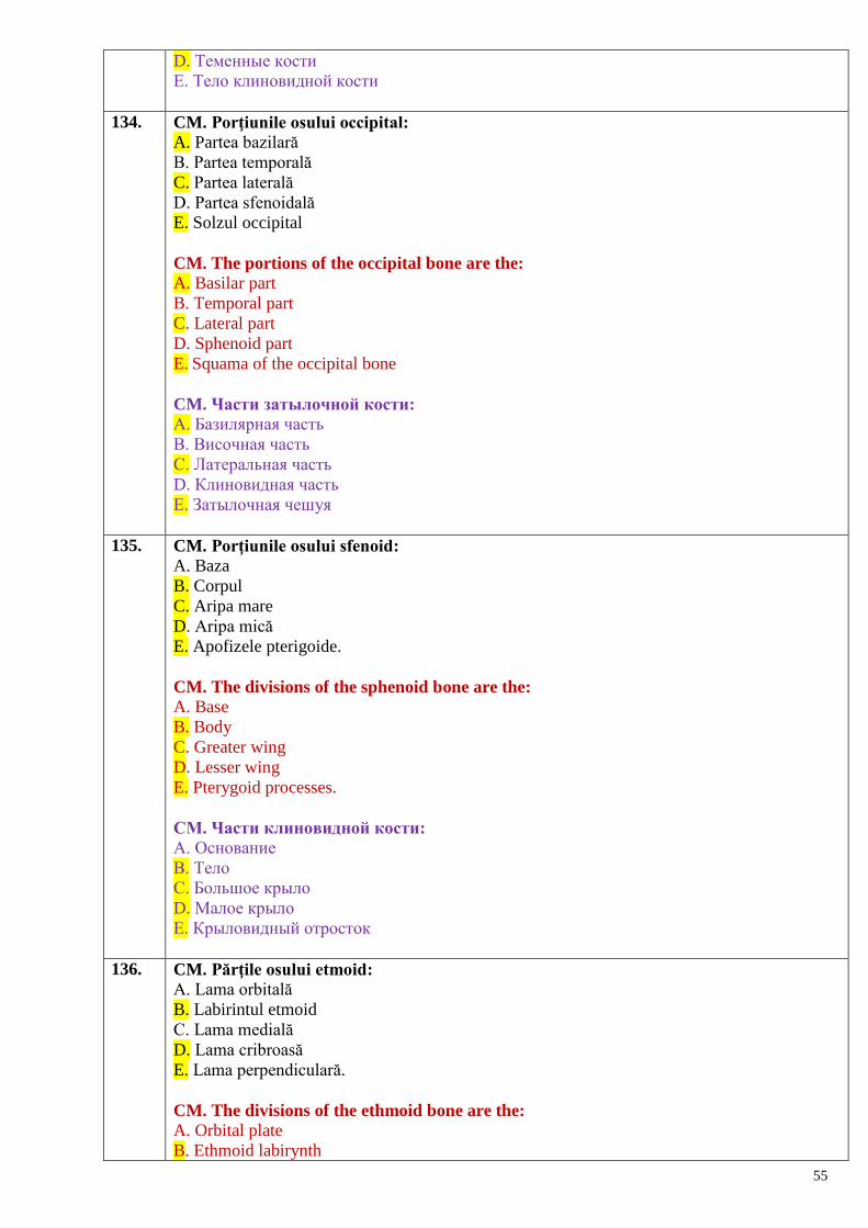
55
D. Теменные кости
Е. Тело клиновидной кости
134. CM. Porţiunile osului occipital:
A. Partea bazilară
B. Partea temporală
C. Partea laterală
D. Partea sfenoidală
E. Solzul occipital
CM. The portions of the occipital bone are the:
A. Basilar part
B. Temporal part
C. Lateral part
D. Sphenoid part
E. Squama of the occipital bone
CM. Части затылочной кости:
А. Базилярная часть
В. Височная часть
С. Латеральная часть
D. Клиновидная часть
Е. Затылочная чешуя
135. CM. Porţiunile osului sfenoid:
A. Baza
B. Corpul
C. Aripa mare
D. Aripa mică
E. Apofizele pterigoide.
CM. The divisions of the sphenoid bone are the:
A. Base
B. Body
C. Greater wing
D. Lesser wing
E. Pterygoid processes.
СМ. Части клиновидной кости:
А. Основание
В. Тело
С. Большое крыло
D. Малое крыло
Е. Крыловидный отросток
136. CM. Părţile osului etmoid:
A. Lama orbitală
B. Labirintul etmoid
C. Lama medială
D. Lama cribroasă
E. Lama perpendiculară.
CM. The divisions of the ethmoid bone are the:
A. Orbital plate
B. Ethmoid labirynth
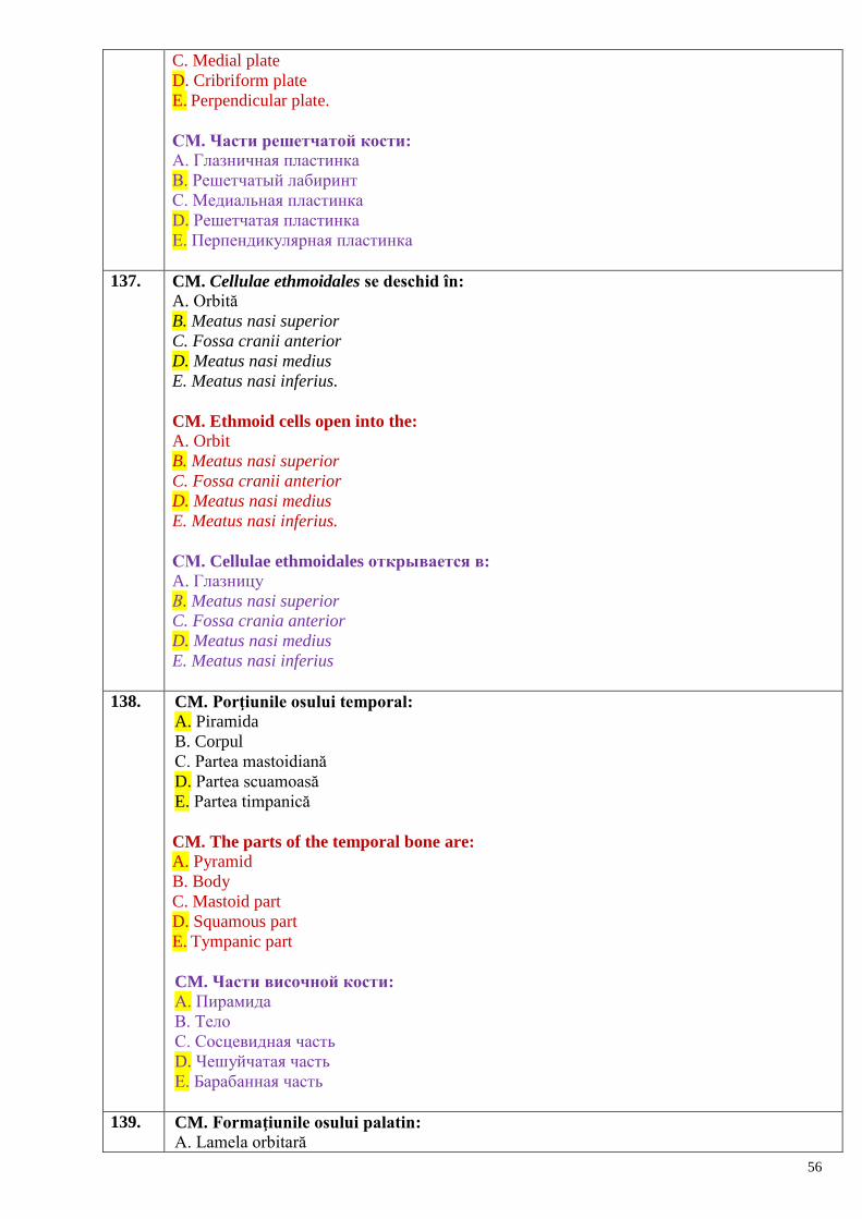
56
C. Medial plate
D. Cribriform plate
E. Perpendicular plate.
СМ. Части решетчатой кости:
А. Глазничная пластинка
В. Решетчатый лабиринт
С. Медиальная пластинка
D. Решетчатая пластинка
Е. Перпендикулярная пластинка
137. CM. Cellulae ethmoidales se deschid în:
A. Orbită
B. Meatus nasi superior
C. Fossa cranii anterior
D. Meatus nasi medius
E. Meatus nasi inferius.
CM. Ethmoid cells open into the:
A. Orbit
B. Meatus nasi superior
C. Fossa cranii anterior
D. Meatus nasi medius
E. Meatus nasi inferius.
СМ. Cellulae ethmoidales открывается в:
A. Глазницу
В. Meatus nasi superior
C. Fossa crania anterior
D. Meatus nasi medius
E. Meatus nasi inferius
138. CM. Porţiunile osului temporal:
A. Piramida
B. Corpul
C. Partea mastoidiană
D. Partea scuamoasă
E. Partea timpanică
CM. The parts of the temporal bone are:
A. Pyramid
B. Body
C. Mastoid part
D. Squamous part
E. Tympanic part
CM. Части височной кости:
А. Пирамида
В. Тело
С. Сосцевидная часть
D. Чешуйчатая часть
Е. Барабанная часть
139. CM. Formaţiunile osului palatin:
A. Lamela orbitară
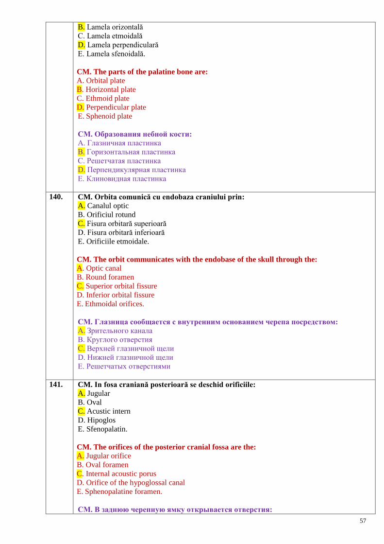
57
B. Lamela orizontală
C. Lamela etmoidală
D. Lamela perpendiculară
E. Lamela sfenoidală.
CM. The parts of the palatine bone are:
A. Orbital plate
B. Horizontal plate
C. Ethmoid plate
D. Perpendicular plate
E. Sphenoid plate
СМ. Образования небной кости:
А. Глазничная пластинка
В. Горизонтальная пластинка
С. Решетчатая пластинка
D. Перпендикулярная пластинка
Е. Клиновидная пластинка
140. CM. Orbita comunică cu endobaza craniului prin:
A. Canalul optic
B. Orificiul rotund
C. Fisura orbitară superioară
D. Fisura orbitară inferioară
E. Orificiile etmoidale.
CM. The orbit communicates with the endobase of the skull through the:
A. Optic canal
B. Round foramen
C. Superior orbital fissure
D. Inferior orbital fissure
E. Ethmoidal orifices.
СМ. Глазница сообщается с внутренним основанием черепа посредством:
А. Зрительного канала
В. Круглого отверстия
С. Верхней глазничной щели
D. Нижней глазничной щели
Е. Решетчатых отверстиями
141. CM. In fosa craniană posterioară se deschid orificiile:
A. Jugular
B. Oval
C. Acustic intern
D. Hipoglos
E. Sfenopalatin.
CM. The orifices of the posterior cranial fossa are the:
A. Jugular orifice
B. Oval foramen
C. Internal acoustic porus
D. Orifice of the hypoglossal canal
E. Sphenopalatine foramen.
СМ. В заднюю черепную ямку открывается отверстия:
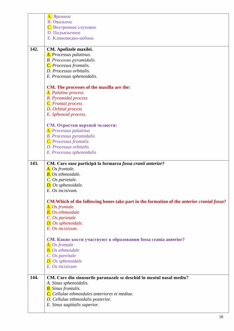
58
А. Яремное
В. Овальное
С. Внутреннее слуховое
D. Подъязычное
Е. Kлиновидно-небное
142. CM. Apofizele maxilei.
A. Processus palatinus.
B. Processus рyramidalis.
C. Processus frontalis.
D. Processus orbitalis.
E. Processus sphenoidalis.
CM. The processes of the maxilla are the:
A. Palatine process
B. Pyramidal process
C. Frontal process
D. Orbital process
E. Sphenoid process.
CM. Отростки верхней челюсти:
A. Processus palatinus
B. Processus pyramidalis
C. Processus frontalis
D. Processus orbitalis
E. Processus sphenoidalis
143. CM. Care oase participă la formarea fossa cranii anterior?
A. Os frontale.
B. Os ethmoidale.
C. Os parietale.
D. Os sphenoidale.
E. Os incisivum.
CM.Which of the following bones take part in the formation of the anterior cranial fossa?
A. Os frontale.
B. Os ethmoidale
C. Os parietale
D. Os sphenoidale.
E. Os incisivum.
CM. Какие кости участвуют в образовании fossa crania anterior?
A. Os frontale
B. Os ethmoidale
C. Os pareitale
D. Os sphenoidale
E. Os incisivum
144. CM. Care din sinusurile paranazale se deschid în meatul nasal mediu? A. Sinus sphenoidalis.
B. Sinus frontalis.
C. Cellulae ethmoidales anteriores et mediae.
D. Cellulae ethmoidalis posterior.
E. Sinus sagittalis superior.
59
CM. The paranasal sinuses that open into the middle nasal meatus are the: A. Sinus sphenoidalis.
B. Sinus frontalis.
C. Cellulae ethmoidales anteriores et mediae
D. Cellulae ethmoidalis posterior.
E. Sinus sagittalis superior.
CM. Какие околоносовые пазухи открываются в средний носовой ход?
A. Sinus sphenoidalis
B. Sinus frontalis
C. Celullae ethmoidalis anteriores et mediae
D. Celullae ethmoidalis posterior
E. Sinus sagittalis superior
145. CM. Care oase formează palatul osos? A. Vomer.
B. Os palatinum.
C. Os hyoideum.
D. Maxilla.
E. Os incisivum.
CM. The bones forming the hard palate are the: A. Vomer.
B. Os palatinum.
C. Os hyoideum.
D. Maxilla.
E. Os incisivum.
CM. Какие кости образуют костное нѐбо: A. Vomer
B. Os palatinum
C. Os hyoideum
D. Maxilla
E. Os incisivum
146. CS. Unirile oaselor prin membrane se numesc:
A. Sincondroze
B. Sinelastoze
C. Sinsarcoze
D. Sinfibroze
E. Sinostoze
CS. Articulation of bones by means of membranes is called:
A. Synchondrosis
B. Synelastosis
C. Synsarcosis
D. Synfibrosis
E. Synostosis
CS. Соединения костей при помощи мембран называются:
A. Синхондрозы.
B. Синеластозы.
C. Синсаркозы.
D. Синфиброзы.
E. Синостозы.
60
147. CS. Cum se numesc mișcările realizate în jurul axei frontale?
A. Flexio et extensio
B. Adductio et abductio.
C. Rotatio.
D. Circumductio.
E. Pronatio et supinatio.
CS. How are movements around the frontal axis called?
A. Flexio et extensio
B. Adductio et abductio
C. Rotatio
D. Circumductio
E. Pronatio et supinatio
CS. Как называются движения вокруг фронтальной оси?
A. Flexio et extensio.
B. Adductio et abductio.
C. Rotatio.
D. Circumductio.
E. Pronatio et supinatio.
148. CS. Cum se numesc mișcările realizate în jurul axei sagitale?
А. Flexio et extensio.
B. Adductio et abductio.
C. Rotatio.
D. Circumductio.
E. Pronatio et supinatio.
CS. How are movements around the sagittal axis called?
А. Flexio et extensio
B. Adductio et abductio
C. Rotatio
D. Circumductio
E. Pronatio et supinatio.
CS. Как называются движения вокруг cагиттальной оси?
A. Flexio et extensio.
B. Adductio et abductio.
C. Rotatio.
D. Circumductio.
E. Pronatio et supinatio.
149. CS. Suturile fac parte din:
A. Sinsarcoze
B. Sinelastoze
C. Sincondroze
D. Sinfibroze
C. Sinostoze.
CS. The sutures belong to:
A. Synsarcoses
B. Synelastoses
C. Synchondroses
D. Synfibroses
61
C. Synostoses
CS. Швы относятся к:
A. Синсаркозам.
B. Синеластозам.
C. Синхондрозам.
D. Синфиброзам.
E. Синостозам.
150. CS. Gomfozele (articulațiile dento-alveolare) ţin de:
A. Ligamente
B. Sincondroze
C. Sinfibroze
D. Sinelastoze
E. Membrane
CS. The gomphoses (dentoalveolar joint) belong to:
A. Ligaments
B. Synchondroses
C. Synfibroses
D. Synelastoses
E. Membranes.
CS. Гомфозы (вколачивание) относятся к:
A. Связкам.
B. Синхондрозам.
C. Синфиброзам.
D. Синеластозам.
E. Мембранам.
151. CS. Mişcarea prin care două segmente ale unui membru se apropie unul de altul:
A. Circumducţia
B. Adducţia
C. Abducţia
D. Extensia
E. Flexia
CS. How is the movement called when two segments of a limb get closer to each other:
A. Circumduction
B. Adduction
C. Abduction
D. Extension
F. Flexion.
CS. Движение каких двух сегментов одной конечности приближают их друг к
другу:
A. Круговое.
B. Приведение.
C. Отведение.
D. Разгибание.
E. Сгибание.
152. CS. Mişcarea datorită căreia un membru sau un segment de membru se apropie de
planul sagital al corpului:
A. Circumducţia
62
B. Adducţia
C. Abducţia
D. Extensia
E. Flexia
CS. Name the movement due to which a limb or one of its segments gets closer to the
sagittal plan of the body:
A. Circumduction
B. Adduction
C. Abduction
D. Extension
F. Flexion.
CS. Движение, благодаря которому конечность или сегмент конечности
приближается к сагиттальному плану тела:
A. Круговое.
B. Приведение.
C. Отведение.
D. Разгибание.
E. Сгибание.
153. CS. Mişcarea ce rezultă din realizarea succesivă a abducţiei, extensiei, adducţiei şi flexiei:
A. Circumducţia
B. Adducţia
C. Abducţia
D. Extensia
E. Flexia.
CS. Name the movement that results from successive abduction, extension, adduction
and flexion:
A. Circumduction
B. Adduction
C. Abduction
D. Extension
E. Flexion.
CS. Движение, которое является результатом последовательного перехода от
отведения, разгибания к приведению и сгибанию:
A. Круговое.
B. Приведение.
C. Отведение.
D. Разгибание.
E. Сгибание.
154. CS. Posedă feţe articulare identice:
A. Articulaţia elipsoidă
B. Articulaţia plană
C. Articulaţia trohoidă
D. Articulaţia condilară
E. Articulaţia sferoidă.
CS. Which of the following joints has identical articular surfaces:
A. Ellipsoid joint
B. Plane joint
C. Pivot joint
63
D. Condylar joint
E. Ball-and-socket joint.
CS. Имеют одинаковые (сопоставляющиеся) суставные поверхности:
A. Эллипсоидный сустав.
B. Плоский сустав.
C. Цилиндрический сустав.
D. Мыщелковый сустав.
E. Шаровидный сустав.
155. CS. Se formează prin unirea mai multor oase:
A. Diartroza simplă
B. Diartroza compusă
C. Diartroza combinată
D. Diartroza complexă
E. Enartroza
CS. Is formed by the connection of more than two articular surfaces:
A. Simple diarthrosis
B. Compound diarthrosis
C. Combined diarthrosis
D. Complex diarthrosis
E. Enarthrosis
CS. Образуется при соединении трѐх и более суставных поверхностей:
A. Простой сустав.
B. Сложный сустав.
C. Комбинированный сустав.
D. Комплексный сустав.
E. Чашеобразный сустав (энартроз).
156. CS. Conţine cartilaj intraarticular:
A. Diartroza simplă
B. Diartroza compusă
C. Diartroza combinată
D. Diartroza complexă
E. Amfiartroza
CS. Itraarticular cartilage contains:
A. Simple diarthrosis
B. Compound diarthrosis
C. Combined diarthrosis
D. Complex diarthrosis
E. Amphyarthrosis
CS. Содержит внутрисуставной хрящ:
A. Простой сустав.
B. Сложный сустав.
C. Комбинированный сустав.
D. Комплексный сустав.
E. Плоский сустав (амфиартроз).
157. CS. Este compusă prin unirea a două oase:
A. Diartroza simplă
B. Diartroza compusă
64
C. Diartroza combinată
D. Diartroza complexă
E. Enartroza.
CS. Is formed by the connection of two bones:
A. Simple diarthrosis
B. Compound diarthrosis
C. Combined diarthrosis
D. Complex diarthrosis
E. Enarthrosis
CS. Образуется при соединении двух суставных поверхностей:
A. Простой сустав.
B. Сложный сустав.
C. Комбинированный сустав.
D. Комплексный сустав.
E. Чашеобразный сустав (энартроз).
158. CS. La care indivizi toracele e de forma conică?
A. La femei
B. La cei de tip brahimorf
C. La cei de tip dolicomorf
D. La cei de tip mezomorf
E. La persoanele senile.
CS. In which individuals the thorax is of conical shape?
A. In women
B. In brachymorphic type
C. In dolicomorphic type
D. In mesomorphic type
E. In senile persons
CS. У каких личностей грудная клетка конической формы?
A. У женщин.
B. У брохиморфного типа.
C. У долихоморфного типа.
D. У мезоморфного типа.
E. У стариков.
159. CS. Joncţiunile apofizelor articulare ale vertebrelor sunt:
A. Diartroze
B. Sinelastoze
C. Sincondroze
D. Sinostoze
E. Sinfibroze
CM. Union between the articular processes belong to:
A. Diarthroses
B. Synelastoses
C. Synchondroses
D. Synostoses
E. Synfibroses
CS. Соединения суставных отростков позвонков это:
A. Диартрозы.
65
B. Синеластозы.
C. Синхондрозы.
D. Синостозы.
E. Синфиброзы.
160. CS. Unirile vertebrelor sacrale sunt:
A. Diartroze
B. Sinelastoze
C. Sincondroze
D. Sinostoze
E. Simfize
CS. Joints between the sacral vertebrae are:
A. Diarthroses
B. Synelastoses
C. Synchondroses
D. Synostoses
E. Symphyses.
CS. Соединения крестцовых позвонков это:
A. Диартрозы.
B. Синеластозы.
C. Синхондрозы.
D. Синостозы.
E. Симфизы.
161. CM. Care tipuri de uniri osoase se referă la articulații fibroase/sinfibroze?
A. Sutura.
B. Gomphosis.
C. Membrana interossea.
D. Synostosis.
E. Symphysis.
CM. Which of the following types of junctions belong to the synfibroses?
A. Sutura.
B. Gomphosis.
C. Membrana interossea.
D. Synostosis.
E. Symphysis.
CM. Какие виды соединений костей относятся к фиброзным соединениям
синфиброзы?
A. Sutura.
B. Gomphosis.
C. Membrana interossea.
D. Synostosis.
E. Symphysis.
162. CM. Indicați elementele principale ale unei diartroze (articulații sinoviale).
A. Discus articularis.
B. Capsula articularis.
C. Cavitas articularis.
D. Labrum articulare.
E. Facies articulares.
66
CM. The main elements of diarthrosis (synovial joint) are:
A. Discus articularis
B. Capsula articularis
C. Cavitas articularis
D. Labrum articulare
E. Facies articulares.
CМ. Назовите главные элементы диартроза (синовиальные соединения): A. Discus articularis.
B. Capsula articularis.
C. Cavitas articularis.
D. Labrum articulare.
E. Facies articulares.
163. CM. Indicați elementele auxiliare ale unei diartroze (articulații sinoviale).
А. Ligamentum.
B. Cartilago articularis (disci et menisci articulares).
C. Capsula articularis.
D. Bursae sinoviales.
E. Labrum articulare
CM. The auxilliary elements of diarthrosis (synovial joint) are:
А. Ligamentum
B. Cartilago articularis (disci et menisci articulares)
C. Capsula articularis
D. Bursae synoviales
E. Labrum articulare
CМ. Укажите добавочные элементы сустава (синовиальные соединения): A. Ligamentum.
B. Cartilago articularis (disci et menisci articulares).
C. Capsula articularis.
D. Bursae sinoviales.
E. Labrum articulare.
164. CM. În ce tipuri se împart articulațiile după complexitate?
А. Combinate
B. Condilare/bicondiliene
C. Compuse.
D. Complexe.
E. Simple.
CM. Joints are classified by their complexity into:
А. Combined joint
B. Condylar joint
C. Compound joint
D. Complex joint
E. Simple joint.
CМ. На какие виды делятся суставы по сложности строения?
A. Комбинированные.
B. Мыщелковые.
C. Сложные.
D. Комплексные.
E. Простые.
67
165. CM. În ce grupe se impart articulațiile după forma fețelor de articulare?
А. Compuse.
B. Sferoide.
C. Combinate.
D. Articulații în șa/seliforme/selare.
E. Elipsoidale.
CM. Joints are divided according to the shape of the articular surfaces into:
А. Compound joint
B. Ball-and-socket joint
C. Combined joint
D. Saddle joint
E. Ellipsoid joint
CМ. На какие группы делятся суставы по форме суставных поверхностей?
A. Сложные.
B. Шаровидные.
C. Комбинированные.
D. Седловидные.
E. Эллипсовидные.
166. CM. În ce grupe se împart articulațiile după numărul axelor în jurul cărora se execută
mișcări?
А. Simple.
B. Pluriaxiale
C. Compuse.
D. Uniaxiale
E. Biaxiale
CM. According to the number of axes on which movements are performed joints are
classified into:
А. Simple joints
B. Multiaxial joints
C. Compound joints
D. Uniaxial joints
E. Biaxial joints.
CМ. На какие группы делятся суставы в зависимости от количества осей, вокруг
которых осуществляются движения?
A. Простые.
B. Многоосные.
C. Сложные.
D. Одноосные.
E. Двуосные.
167. CM. Cum se numesc mișcările executate în jurul axei verticale?
А. Flexio et extensio.
B. Adductio et abductio.
C. Rotatio.
D. Circumductio.
E. Pronatio et supinatio.
CM. How are called movements around the vertical axis?
А. Flexio et extensio
68
B. Adductio et abductio
C. Rotatio
D. Circumductio
E. Pronatio et supinatio
CМ. Как называются движения, осуществляемые вокруг вертикальной оси?
A. Flexio et extensio.
B. Adductio et abductio.
C. Rotatio.
D. Circumductio.
E. Pronatio et supinatio.
168. CM. Care din articulațiile enumerate sunt biaxiale?
А. Articulatio ellipsoidea.
B. Articulatio trochoidea.
C. Articulatio sellaris.
D. Articulatio plana.
E. Articulatio bicondylaris.
CM. Which of the following joints are biaxial?
А. Articulatio ellipsoidea
B. Articulatio trochoidea
C. Articulatio sellaris
D. Articulatio plana
E. Articulatio bicondylaris.
CМ. Какие из перечисленных суставов являются двуосными?
A. Articulatio ellipsoidea.
B. Articulatio trochoidea.
C. Articulatio sellaris.
D. Articulatio plana.
E. Articulatio bicondylaris.
169. CM. Straturile capsulei articulare:
A. Adventiceal
B. Epitelial
C. Fibros
D. Seros
E. Sinovial
CM. The layers of the articular capsule are:
A. Adventitial
B. Epithelial
C. Fibrous
D. Serous
E. Synovial
CМ. Слои суставной капсулы:
A. Адвентициальный.
B. Эпителиальный.
C. Фиброзный.
D. Серозный.
E. Синовиальный.
170. CM. Lichidul sinovial are rol de:
69
A. Dirijare a mişcărilor
B. Metabolism
C. Amortizare
D. Lubrifiere a feţelor articulare
E. Creştere a oaselor
CM. The functions of the synovial fluid are:
A. Guiding the movements
B. Metabolism
C. Amortization
D. Lubrification of the articular surfaces
E. Growth of bones.
CМ. Синовиальная жидкость играет роль:
A. В регуляции движений.
B. В обмене веществ.
C. В амортизации.
D. В уменьшении трения суставных поверхностей.
E. В росте костей.
171. CM. Funcţiile capsulei articulare sunt de:
A. Protecţie
B. Sprijin
C. Consolidare a oaselor
D. Reglementare a mişcărilor
E. Secreţie
CM. The functions of the articular capsule are:
A. Protection
B. Support
C. Keeping in contact the articular surfaces
D. Guiding the movements
E. Secretion.
CМ. Функции суставной капсулы:
A. Защитная.
B. Опорная.
C. Соединение костей.
D. Направление движений.
E. Секреторная.
172. CM. Funcţiile ligamentelor sunt cele de:
A. Consolidare a oaselor
B. Fortificare a capsulei articulare
C. Frânare a mişcărilor
D. Dirijare a mişcărilor
E. Protecţie
CM. The functions of the ligaments are:
A. Joining (union) of bones
B. Strengthening of the articular capsule
C. Braking of movements
D. Guiding of movements
E. Secretion.
70
CМ. Функциями связок являются:
A. Соединение костей.
B. Укрепление суставной капсулы.
C. Ограничение движений.
D. Направление движений.
E. Защитная.
173. CM. Cartilajul articular:
A. Nu conţine terminaţii nervoase.
B. Are proprietăţi de compresibilitate şi elasticitate.
C. Joacă rol de amortizator.
D. Devine mai gros pe măsura înaintării în vârstă.
E. În caz de imobilizare îndelungată poate fi invadat de vase sangvine.
CM. Articular cartilage: A. Does not contain nervous endings
B. Has properties of compressibility and elasticity
C. Plays an amortization role
D. Becomes thicker with age
E.Can be invaded by blood vessels in case of long immobilization
CМ. Суставной хрящ:
A. Не содержит нервные окончания.
B. Имеет свойства компрессии и эластические.
C. Играет роль амортизатора.
D. С возрастом становится толще.
E. В случае долгой иммобилизации может прорасти сосудами.
174. CS. Mişcarea prin care două segmente ale unui membru se îndepărtează unul de altul:
A. Circumducţia
B. Adducţia
C. Abducţia
D. Extensia
E. Flexia.
CS. Name the movement when two segments of a limb get further from each other:
A. Circumduction
B. Adduction
C. Abdduction
D. Extension
E. Flexion.
CS. Движение, при котором два сегмента одной конечности отдаляются друг от
друга:
A. Круговое движение.
B. Приведение.
C. Отведение.
D. Разгибание.
E. Сгибание.
175. CM. Sutura serrata între:
A. Parietal şi occipital
B. Frontal şi nazal
C. Maxila dreaptă şi stângă
D. Temporal şi sfenoid
71
E. Frontal şi parietal.
CM. Serrate sutures are located between the:
A. Parietal and occipital bones
B. Frontal and nasal bones
C. Right and left maxilla
D. Temporal and sphenoid bones
E. Frontal and parietal bones.
CМ. Sutura serrata между: A. Теменной и затылочной.
B. Лобной и носовой.
C. Правой и левой верхними челюстями.
D. Височной и клиновидной.
E. Лобной и теменной.
176. CM. Suturae squamosa între:
A. Parietal şi occipital
B. Frontal şi nazal
C. Maxila dreaptă şi stângă
D. Temporal şi parietal
E. Temporal şi sfenoid.
CM. Suturae squamosa are located between the:
A. Parietal and occipital bones
B. Frontal and nasal bones
C. Right and left maxilla
D. Temporal and parietal bones
E. Temporal and sphenoid bones.
CМ. Sutura squamosa между: A. Теменной и затылочной.
B. Лобной и носовой.
C. Правой и левой верхней челюстью.
D. Височной и теменной.
E. Височной и клиновидной.
177. CM. Căror segmente ale coloanei vertebrale le este caracteristică lordoza fiziologică?
А. Porțiunii toracale.
B. Porțiunii cervicale.
C. Porțiunii lombare.
D. Porțiunii sacrale.
E. Porțiunii coccigiene.
CM. For which segments (parts, regions) of the vertebral column is characteristic
physiological lordosis?
А. Thoracic part
B. Cervical part
C. Lumbar part
D. Sacral part
E. Coccygeal part.
CМ. Для каких частей (сегментов) позвоночного столба характерен
физиологический лордоз?
A. Грудной части.
72
B. Шейной части.
C. Поясничной части.
D. Крестцовой части.
E. Копчиковой части.
178. CM. Căror segmente ale coloanei vertebrale le este caracteristică cifoza fiziologică?
А. Porțiunii toracale.
C. Porțiunii lombare.
B. Porțiunii cervicale.
D. Porțiunii sacrale.
E. Porțiunii coccigiene.
CM. For which segments (parts) of the vertebral column is characteristic physiological
kyphosis?
А. Thoracic part
B. Lumbar part
C. Cervical part
D. Sacral part
E. Coccygeal part
CМ. Какие отделы (сегменты) позвоночного столба имеют физиологический
кифоз?
A. Грудной отдел.
B. Шейный отдел.
C. Поясничный отдел.
D. Крестцовый отдел.
E. Копчиковый отдел.
179. CM. Coloana vertebrala prezintă mobilitate sporită la nivelul:
A. Lordozelor
B. Segmentului toracic
C. Cifozelor
D. Segmentului cervical
E. Segmentului lombar.
CM. A higher mobility of the vertebral column is characteristic for:
A. Lordoses
B. Thoracic part
C. Kyphoses
D. Cervical part
E. Lumbar part
CМ. Позвоночный столб наиболее подвижен на уровне:
A. Лордозов.
B. Грудного отдела.
C. Кифозов.
D. Шейного отдела.
E. Поясничного отдела.
Oasele şi articulaţiile
membrelor superioare şi inferioare,
rolul lor funcţional
180. CM. Oasele centurii scapulare:
A. Humerusul
B. Clavicula
73
C. Sternul
D. Omoplatul
E. I coastă
CM. The bones of the shoulder girdle are the:
A. Humerus
B. Clavicle
C. Sternum
D. Scapula
E. I-st rib.
CM. Кости пояса верхней конечности:
А. Плечевая кость
В. Ключица
С. Грудина
D. Лопатка
Е. I ребро
181. CM. Formaţiunile oaselor centurii scapulare palpabile pe viu:
A. Extremitatea sternală a claviculei
B. Tuberculul supraglenoidal
C. Extremitatea acromială a claviculei
D. Acromionul
E. Corpul claviculei
CM. The structural elements of the shoulder girdle bone palpable on alive person are
the:
A. Sternal end of the clavicle
B. Supraglenoid tubercle
C. Acromial end of the clavicle
D. Acromion
E. Body of the clavicle
CM. Костные образования пояса верхней конечности, пальпирующиеся на живом:
А. Грудинный конец ключицы
В. Надсуставной бугорок
С. Акромиальный конец лопатки
D. Акромион
Е. Тело ключицы
182. CM. Elementele humerusului ce pot fi palpate pe viu:
A. Colul anatomic
B. Epicondilul medial
C. Fosa coronoidă
D. Epicondilul lateral
E. Colul chirurgical
CM. The elements of the humerus that may be palpated on alive person are the:
A. Anatomical neck
B. Medial epicondyle
C. Coronoid fossa
D. Lateral epicondyle
E. Surgical neck.
CM. Элементы плечевой кости, прощупывающиеся на живом:
74
А. Анатомическая шейка
В. Медиальный мыщелок
С. Венечная ямка
D. Латеральный мыщелок
Е. Хирургическая шейка
183. CM. Care oase formează scheletul antebraţului?
A. Radiusul.
B. Humerusul.
C. Fibula.
D. Ulna.
E. Olecranonul.
CM. Which of the following bones form the skeleton of the forearm?
A. Radius
B. Humerus
C. Fibula
D. Ulna
E. Olecranon.
CM. Какие кости образуют скелет предплечья?
А. Лучевая кость
В. Плечевая кость
С. Малоберцовая
D. Локтевая кость
Е. Локтевой отросток
184. CM. Care dintre oasele membrului superior comportă circumferinţă articulară?
A. Humerusul.
B. Ulna.
C. Clavicula.
D. Radiusul.
E. Toate corecte.
CM. Which bones of upper limbs have the articular circumference?
A. Humerus
B. Ulna
C. Clavicle
D. Radius
E. All mentioned above are correct.
CM. Какие кости верхней конечности имеют суставную окружность?
А. Плечевая кость
В. Локтевая кость
С. Ключица
D. Лучевая кость
Е. Все правильные
185. CM. Care dintre oasele membrului superior comportă apofiză stiloidă?
A. Os hamatum.
B. Humerusul.
C. Ulna.
D. Radiusul.
E. Scapula.
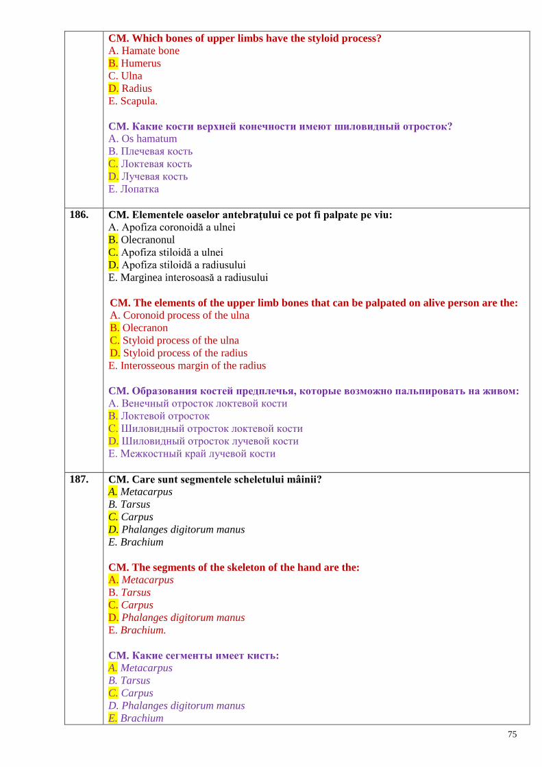
75
CM. Which bones of upper limbs have the styloid process?
A. Hamate bone
B. Humerus
C. Ulna
D. Radius
E. Scapula.
CM. Какие кости верхней конечности имеют шиловидный отросток?
A. Os hamatum
B. Плечевая кость
C. Локтевая кость
D. Лучевая кость
E. Лопатка
186. CM. Elementele oaselor antebraţului ce pot fi palpate pe viu:
A. Apofiza coronoidă a ulnei
B. Olecranonul
C. Apofiza stiloidă a ulnei
D. Apofiza stiloidă a radiusului
E. Marginea interosoasă a radiusului
CM. The elements of the upper limb bones that can be palpated on alive person are the:
A. Coronoid process of the ulna
B. Olecranon
C. Styloid process of the ulna
D. Styloid process of the radius
E. Interosseous margin of the radius
CM. Образования костей предплечья, которые возможно пальпировать на живом:
А. Венечный отросток локтевой кости
В. Локтевой отросток
С. Шиловидный отросток локтевой кости
D. Шиловидный отросток лучевой кости
Е. Межкостный край лучевой кости
187. CM. Care sunt segmentele scheletului mâinii?
A. Metacarpus
B. Tarsus
C. Carpus
D. Phalanges digitorum manus
E. Brachium
CM. The segments of the skeleton of the hand are the:
A. Metacarpus
B. Tarsus
C. Carpus
D. Phalanges digitorum manus
E. Brachium.
CM. Какие сегменты имеет кисть:
A. Metacarpus
B. Tarsus
C. Carpus
D. Phalanges digitorum manus
E. Brachium
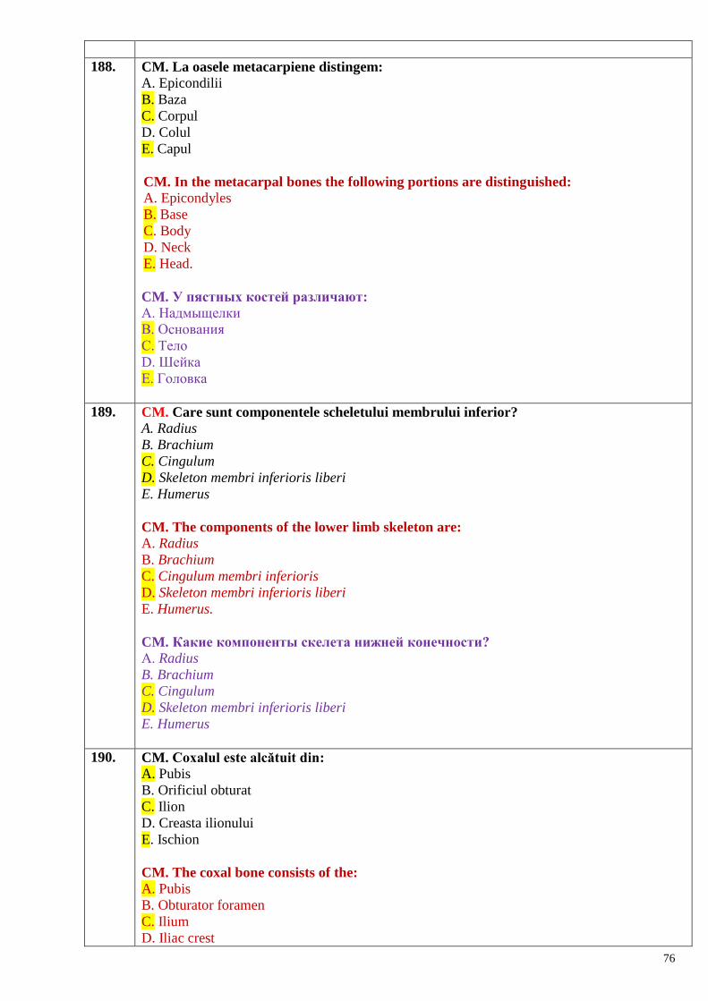
76
188. CM. La oasele metacarpiene distingem:
A. Epicondilii
B. Baza
C. Corpul
D. Colul
E. Capul
CM. In the metacarpal bones the following portions are distinguished:
A. Epicondyles
B. Base
C. Body
D. Neck
E. Head.
CM. У пястных костей различают:
А. Надмыщелки
В. Основания
С. Тело
D. Шейка
Е. Головка
189. CM. Care sunt componentele scheletului membrului inferior?
A. Radius
B. Brachium
C. Cingulum
D. Skeleton membri inferioris liberi
E. Humerus
CM. The components of the lower limb skeleton are:
A. Radius
B. Brachium
C. Cingulum membri inferioris
D. Skeleton membri inferioris liberi
E. Humerus.
CM. Какие компоненты скелета нижней конечности?
А. Radius
B. Brachium
C. Cingulum
D. Skeleton membri inferioris liberi
E. Humerus
190. CM. Coxalul este alcătuit din:
A. Pubis
B. Orificiul obturat
C. Ilion
D. Creasta ilionului
E. Ischion
CM. The coxal bone consists of the: A. Pubis
B. Obturator foramen
C. Ilium
D. Iliac crest
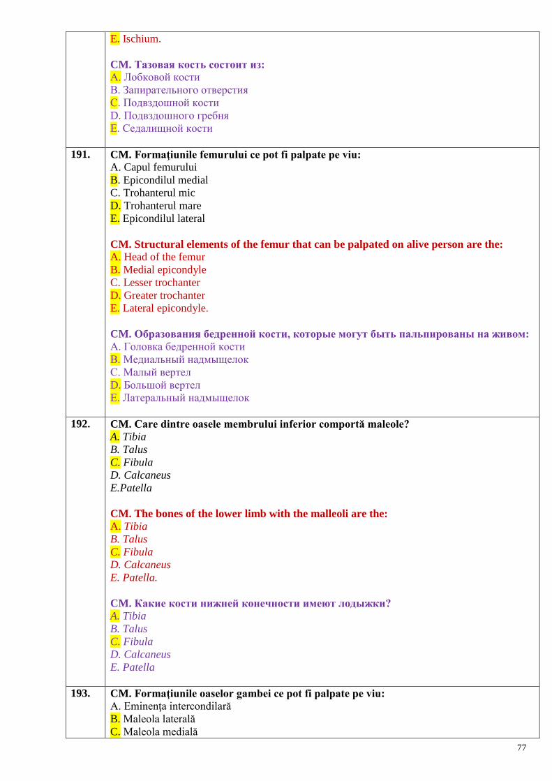
77
E. Ischium.
CM. Тазовая кость состоит из:
А. Лобковой кости
В. Запирательного отверстия
С. Подвздошной кости
D. Подвздошного гребня
Е. Седалищной кости
191. CM. Formaţiunile femurului ce pot fi palpate pe viu:
A. Capul femurului
B. Epicondilul medial
C. Trohanterul mic
D. Trohanterul mare
E. Epicondilul lateral
CM. Structural elements of the femur that can be palpated on alive person are the:
A. Head of the femur
B. Medial epicondyle
C. Lesser trochanter
D. Greater trochanter
E. Lateral epicondyle.
CM. Образования бедренной кости, которые могут быть пальпированы на живом:
А. Головка бедренной кости
В. Медиальный надмыщелок
С. Малый вертел
D. Большой вертел
Е. Латеральный надмыщелок
192. CM. Care dintre oasele membrului inferior comportă maleole?
A. Tibia
B. Talus
C. Fibula
D. Calcaneus
E.Patella
CM. The bones of the lower limb with the malleoli are the: A. Tibia
B. Talus
C. Fibula
D. Calcaneus
E. Patella.
CM. Какие кости нижней конечности имеют лодыжки? A. Tibia
B. Talus
C. Fibula
D. Calcaneus
E. Patella
193. CM. Formaţiunile oaselor gambei ce pot fi palpate pe viu:
A. Eminenţa intercondilară
B. Maleola laterală
C. Maleola medială
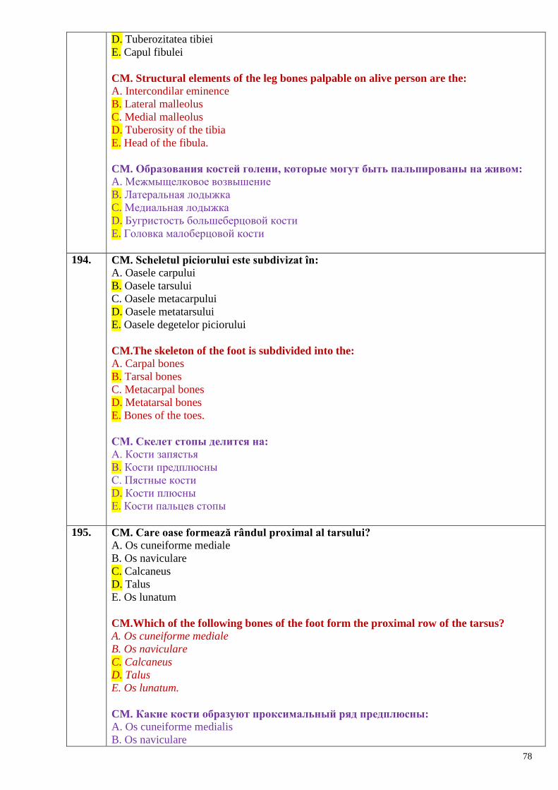
78
D. Tuberozitatea tibiei
E. Capul fibulei
CM. Structural elements of the leg bones palpable on alive person are the:
A. Intercondilar eminence
B. Lateral malleolus
C. Medial malleolus
D. Tuberosity of the tibia
E. Head of the fibula.
CM. Образования костей голени, которые могут быть пальпированы на живом:
A. Межмыщелковое возвышение
B. Латеральная лодыжка
C. Медиальная лодыжка
D. Бугристость большеберцовой кости
E. Головка малоберцовой кости
194. CM. Scheletul piciorului este subdivizat în:
A. Oasele carpului
B. Oasele tarsului
C. Oasele metacarpului
D. Oasele metatarsului
E. Oasele degetelor piciorului
CM.The skeleton of the foot is subdivided into the:
A. Carpal bones
B. Tarsal bones
C. Metacarpal bones
D. Metatarsal bones
E. Bones of the toes.
CM. Скелет стопы делится на:
А. Кости запястья
В. Кости предплюсны
С. Пястные кости
D. Кости плюсны
Е. Кости пальцев стопы
195. CM. Care oase formează rândul proximal al tarsului?
A. Os cuneiforme mediale
B. Os naviculare
C. Calcaneus
D. Talus
E. Os lunatum
CM.Which of the following bones of the foot form the proximal row of the tarsus?
A. Os cuneiforme mediale
B. Os naviculare
C. Calcaneus
D. Talus
E. Os lunatum.
CM. Какие кости образуют проксимальный ряд предплюсны:
A. Os cuneiforme medialis
B. Os naviculare
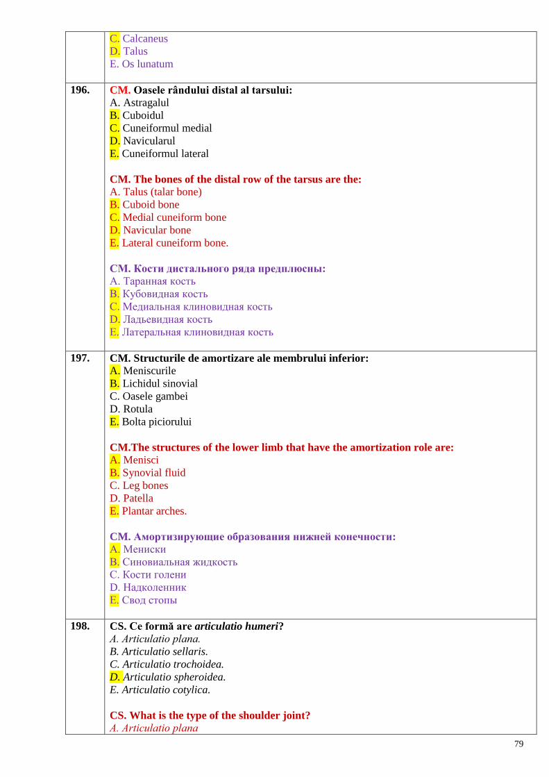
79
C. Calcaneus
D. Talus
E. Os lunatum
196. CM. Oasele rândului distal al tarsului:
A. Astragalul
B. Cuboidul
C. Cuneiformul medial
D. Navicularul
E. Cuneiformul lateral
CM. The bones of the distal row of the tarsus are the:
A. Talus (talar bone)
B. Cuboid bone
C. Medial cuneiform bone
D. Navicular bone
E. Lateral cuneiform bone.
CM. Кости дистального ряда предплюсны:
А. Таранная кость
В. Кубовидная кость
С. Медиальная клиновидная кость
D. Ладьевидная кость
Е. Латеральная клиновидная кость
197. CM. Structurile de amortizare ale membrului inferior:
A. Meniscurile
B. Lichidul sinovial
C. Oasele gambei
D. Rotula
E. Bolta piciorului
CM.The structures of the lower limb that have the amortization role are:
A. Menisci
B. Synovial fluid
C. Leg bones
D. Patella
E. Plantar arches.
CM. Амортизирующие образования нижней конечности:
А. Мениски
В. Синовиальная жидкость
С. Кости голени
D. Надколенник
Е. Свод стопы
198. CS. Ce formă are articulatio humeri?
А. Articulatio plana.
B. Articulatio sellaris.
C. Articulatio trochoidea.
D. Articulatio spheroidea.
E. Articulatio cotylica.
CS. What is the type of the shoulder joint?
А. Articulatio plana
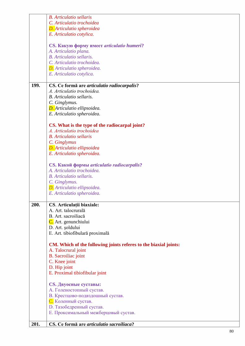
80
B. Articulatio sellaris
C. Articulatio trochoidea
D. Articulatio spheroidea
E. Articulatio cotylica.
CS. Какую форму имеет аrticulatio humeri?
A. Articulatio plana.
B. Articulatio sellaris.
C. Articulatio trochoidea.
D. Articulatio spheroidea.
E. Articulatio cotylica.
199. CS. Ce formă are articulatio radiocarpalis?
А. Articulatio trochoidea.
B. Articulatio sellaris.
C. Ginglymus.
D. Articulatio ellipsoidea.
E. Articulatio spheroidea.
CS. What is the type of the radiocarpal joint?
А. Articulatio trochoidea
B. Articulatio sellaris
C. Ginglymus
D. Articulatio ellipsoidea
E. Articulatio spheroidea.
CS. Какой формы аrticulatio radiocarpalis?
A. Articulatio trochoidea.
B. Articulatio sellaris.
C. Ginglymus.
D. Articulatio ellipsoidea.
E. Articulatio spheroidea.
200. CS. Articulații biaxiale:
A. Art. talocrurală
B. Art. sacroiliacă
C. Art. genunchiului
D. Art. şoldului
E. Art. tibiofibulară proximală
CM. Which of the following joints referes to the biaxial joints:
A. Talocrural joint
B. Sacroiliac joint
C. Knee joint
D. Hip joint
E. Proximal tibiofibular joint
CS. Двуосные суставы:
A. Голеностопный сустав.
B. Крестцово-подвздошный сустав.
C. Коленный сустав.
D. Тазобедренный сустав.
E. Проксимальный межберцовый сустав.
201. CS. Ce formă are articulatio sacroiliaca?
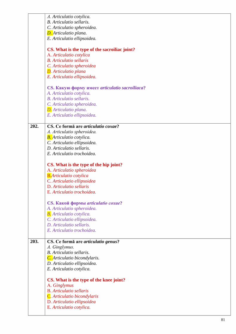
81
А. Articulatio cotylica.
B. Articulatio sellaris.
C. Articulatio spheroidea.
D. Articulatio plana.
E. Articulatio ellipsoidea.
CS. What is the type of the sacroiliac joint? А. Articulatio cotylica
B. Articulatio sellaris
C. Articulatio spheroidea
D. Articulatio plana
E. Articulatio ellipsoidea.
CS. Какую форму имеет articulatio sacroiliaca?
A. Articulatio cotylica.
B. Articulatio sellaris.
C. Articulatio spheroidea.
D. Articulatio plana.
E. Articulatio ellipsoidea.
202. CS. Ce formă are articulatio coxae?
А. Articulatio spheroidea.
B. Articulatio cotylica.
C. Articulatio ellipsoidea.
D. Articulatio sellaris.
E. Articulatio trochoidea.
CS. What is the type of the hip joint?
А. Articulatio spheroidea
B.Articulatio cotylica
C. Articulatio ellipsoidea
D. Articulatio sellaris
E. Articulatio trochoidea.
CS. Какой формы аrticulatio сoxae?
A. Articulatio spheroidea.
B. Articulatio cotylica.
C. Articulatio ellipsoidea.
D. Articulatio sellaris.
E. Articulatio trochoidea.
203. CS. Ce formă are articulatio genus?
А. Ginglymus.
B. Articulatio sellaris.
C. Articulatio bicondylaris.
D. Articulatio ellipsoidea.
E. Articulatio cotylica.
CS. What is the type of the knee joint?
А. Ginglymus
B. Articulatio sellaris
C. Articulatio bicondylaris
D. Articulatio ellipsoidea
E. Articulatio cotylica.

82
CS. Какую форму имеет аrticulatio genus?
A. Ginglymus.
B. Articulatio sellaris.
C. Articulatio bicondylaris.
D. Articulatio ellipsoidea.
E. Articulatio cotylica.
204. CM. Ce mișcări sunt posibile în articulația umărului?
А. Flexio et extensio.
B. Adductio et abductio.
C. Rotatio.
D. Circumductio.
E. Oppositio et reppositio.
CM. What kinds of movements are in the shoulder joint possible? А. Flexio et extensio
B. Adductio et abductio
C. Rotatio
D. Circumductio
E. Oppositio et reppositio.
СМ. В плечевом суставе возможны движения:
A. Сгибание и разгибание
B. Отведение и приведение
C. Вращение
D. Круговые
E. Сопоставление и противосопоставление
205. CS. Element cheie al art. Chopart:
A. Lig.deltoid
B. Ligg. colaterale
C. Lig. lateral
D. Lig. cuneometatarsian interosos medial
E. Lig. bifurcat
CS. Which of the following ligaments is the `key` of the Chopart's joint: A. Deltoid ligament
B. Collateral ligament
C. Lateral ligament
D. Medial interosseous cuneometatarsal ligament
E. Bifurcate ligament.
CS. Ключ от Шопаровского сустава является:
A. Дельтовидная связка
B. Окольные связки
C. Латеральные связки
D. Медиальная межкостная клиновидная связка
E. Раздвоенная связка
206. CS. Ce mișcări sunt posibile în articulationes interphalangeae pedis?
А. Flexio et extensio.
B. Adductio et abductio.
C. Rotatio.
D. Circumductio.
E. Oppositio et reppositio.
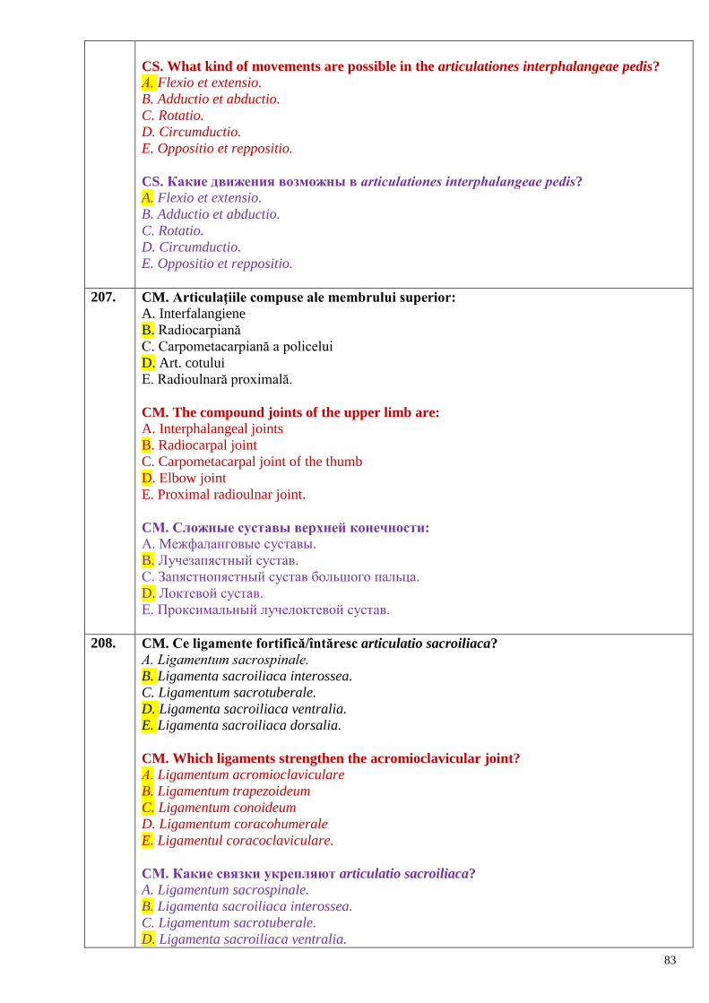
83
CS. What kind of movements are possible in the articulationes interphalangeae pedis?
А. Flexio et extensio.
B. Adductio et abductio.
C. Rotatio.
D. Circumductio.
E. Oppositio et reppositio.
CS. Какие движения возможны в аrticulationes interphalangeae pedis?
A. Flexio et extensio.
B. Adductio et abductio.
C. Rotatio.
D. Circumductio.
E. Oppositio et reppositio.
207. CM. Articulaţiile compuse ale membrului superior:
A. Interfalangiene
B. Radiocarpiană
C. Carpometacarpiană a policelui
D. Art. cotului
E. Radioulnară proximală.
CM. The compound joints of the upper limb are: A. Interphalangeal joints
B. Radiocarpal joint
C. Carpometacarpal joint of the thumb
D. Elbow joint
E. Proximal radioulnar joint.
CМ. Сложные суставы верхней конечности:
A. Межфаланговые суставы.
B. Лучезапястный сустав.
C. Запястнопястный сустав большого пальца.
D. Локтевой сустав.
E. Проксимальный лучелоктевой сустав.
208. CM. Ce ligamente fortifică/întăresc articulatio sacroiliaca?
А. Ligamentum sacrospinale.
B. Ligamenta sacroiliaca interossea.
C. Ligamentum sacrotuberale.
D. Ligamenta sacroiliaca ventralia.
E. Ligamenta sacroiliaca dorsalia.
CM. Which ligaments strengthen the acromioclavicular joint?
А. Ligamentum acromioclaviculare
B. Ligamentum trapezoideum
C. Ligamentum conoideum
D. Ligamentum coracohumerale
E. Ligamentul coracoclaviculare.
CМ. Какие связки укрепляют articulatio sacroiliaca?
A. Ligamentum sacrospinale.
B. Ligamenta sacroiliaca interossea.
C. Ligamentum sacrotuberale.
D. Ligamenta sacroiliaca ventralia.
84
E. Ligamenta sacroiliaca dorsalia.
209. CM. Simfiza pubiană:
A. Este o hemiartroză formată între oasele pubiene.
B. Discul fibrocartilaginos interpubian este mai masiv la bărbaţi.
C. Înălţimea simfizei este mai mare la femei.
D. Mişcările sunt posibile la femei în timpul naşterii.
E. Este fixată de ligamentul pubian superior şi ligamentul arcuat al pubisului.
CM. Which of the following statements about the pubic symphysis are true:
A. It is a hemiarthrosis (half-a-joint) located between the pubic bones
B. The interpubic fibrocartilaginous disc is more massive (bigger) in male than in female
C. The pubic symphysis is higher in females
D. Movements of the symphysis are possible in females during labour
E. It is fixed by the superior pubic ligament and the arcuatepubic ligament.
CМ. Лобковый симфиз:
A. Это полусустав, соединяющий лобковые кости.
B. Межлобковый волокнисто-хрящевой диск толще у мужчин.
C. Высота симфиза выше у женщин.
D. Возможны движения у женщин во время родов.
E. Укреплѐн верхней лобковой связкой и дугообразной связкой лобка.
210. CM. Bazinul în ansamblu:
A. Constituie un inel osos.
B. Bazinul mare alcătuieşte partea inferioară a cavităţii abdominale.
C. Orificiul sciatic mare este delimitat de incizura schiatică mare şi ligamentul sacrotuberal.
D. Orificiul sciatic mic este format de incizura schiatică mică şi ligamentul sacrospinal.
E. În poziţie anatomică apertura superioară a bazinului la femei formează cu planul orizontal
un unghi (50-550).
CM. Which of the following statements about the pelvis are true: A. It is a bony ring
B. The greater pelvis forms the inferior part of the abdominal cavity
C. The greater sciatic foramen is bounded by the greater sciatic notch and by the sacrotuberal
ligament
D. The lesser sciatic foramen is formed by the lesser sciatic notch and sacrospinal ligament
E. In normal anatomical position the superior pelvic aperture in female forms with the
horizontal plan an angle about (50-550)
CМ. Таз в целом:
A. Представляет собой костное кольцо.
B. Полость большого таза является нижней частью брюшной полости.
C. Большое седалищное отверстие ограничено большой седалищной вырезкой и
крестцово-бугорной связкой.
D. Малое седалищное отверстие образовано малой седалищной вырезкой и крестцово-
остистой связкой.
E. В нормальном анатомическом положении верхняя апертура таза образует у женщин с
горизонтальной плоскостью угол (500 – 55
0).
211. CM. Indicați ligamentele intraarticulare ale articulatio coxae.
А. Zona orbicularis.
B. Ligamentum iliofemorale.
C. Ligamentum transversum acetabuli.
D. Ligamentum capitis femoris.
85
E. Ligamentum pubofemorale.
CM. The intraarticular ligaments of the hip joint are: А. Zona orbicularis
B. Ligamentum iliofemorale
C. Ligamentum transversum acetabuli
D. Ligamentum capitis femoris
E. Ligamentum pubofemorale.
CМ. Назовите внутрисуставные связки articulatio coxae:
A. Zona orbicularis.
B. Ligamentum iliofemоrale.
C. Ligamentum transversum acetabuli.
D. Ligamentum capitis femoris.
E. Ligamentum pubofemоrale.
212. CM. Ce mișcări sunt posibile în articulatio coxae?
А. Flexio et extensio.
B. Adductio et abductio.
C. Rotatio.
D. Circumductio.
E. Oppositio et reppositio.
CM. What kind of movements are possible in the articulatio coxae?
А. Flexio et extensio.
B. Adductio et abductio.
C. Rotatio.
D. Circumductio.
E. Oppositio et reppositio.
CМ. Какие движения возможны в articulatio coxae?
А. Flexio et extensio.
B. Adductio et abductio.
C. Rotatio.
D. Circumductio.
E. Oppositio et reppositio.
213. CM. Indicați oasele care participă la formarea articulației genunchiului.
А. Fibula.
B. Tibia.
C. Patella.
D. Femur.
E. Oasele gambei.
CM. The bones that take part in formation of the knee join are:
А. Fibula.
B. Tibia.
C. Patella.
D. Femur.
E. Bones of the leg.
CМ. Назовите кости, которые участвуют в образовании коленного сустава:
А. Fibula.
B. Tibia.
C. Patella.
86
D. Femur.
E. Кости голени.
214. CM. Articulaţia talocrurală:
A. Este formată de 6 feţe articulare.
B. Capsula articulară a ei este fină, întărită de ligamente puternice
C. Capsula se inseră pe marginea feţelor articulare.
D. Membrana sinovială este laxă.
E. Cavitatea sinovială se extinde deseori superior între ligamentele tibiofibulare.
CM. Which of the following statements about the talocrural joint are true: A. It is formed by the articular surfaces of three bones
B. The articular capsule is thin, strengthened by strong ligaments
C. The capsule is inserted to the edges of the articular surfaces
D. The synovial membrane is weak
E. The synovial cavity often extends superiorly between the tibiofibular ligaments.
CМ. Голеностопный сустав:
A. Образован шестью суставными поверхностями.
B. Суставная капсула тонкая, укреплена прочными связками.
C. Капсула фиксируется по краям суставных поверхностей.
D. Синовиальная мембрана слабая.
E. Суставная полость часто впячивается выше между межберцовыми связками.
Noţiuni generale privind sistemul muscular.
Clasificarea muşchilor, structura şi conformaţia lor exterioară, muşchiul ca organ.
Muşchii, fasciile şi topografia
capului, gâtului şi trunchiului
215. CS. Dispozitive auxiliare ale muşchilor sunt:
A. Aponevrozele
B. Tendoanele
C. Plicele sinoviale
D. Venterele musculare
E. Tecile sinoviale
CS. The auxiliary devices of muscles are: A. Aponeurosiss
B. Tendons
C. Synovial folds
D. Muscular bellies
E. Synovial sheaths.
CS. Вспомогательными приспособлениями мышц являются:
А. Апоневрозы.
B. Сухожилия.
C. Синовиальные складки.
D. Мышечные брюшки.
E. Синовиальные влагалища.
216. CM. Indicați părțile principale ale unui mușchi scheletal:
А. Caput.
B. Venter.
C. Tendo.
D. Aponeurosis.
E. Fascia.
87
CM. The main parts of each skeletal muscle are: А. Caput
B. Venter
C. Tendo
D. Anulus tendineus
E. Fascia.
CМ. Укажите основные части скелетной мышцы:
А. Caput.
B. Venter.
C. Tendo.
D. Aponeurosis.
E. Fascia.
217. CM. Indicați elementele aparatului auxiliar al mușchilor.
А. Tecile sinoviale ale tendoanelor.
B. Tecile fibroase ale tendoanelor.
C. Fasciile.
D. Oasele sesamoide.
E. Ligamentele
CM. The elements of the muscle auxiliary apparatus are: А. The synovial sheath of the tendons
B. The fibrous sheath of the tendons
C. The fasciae
D. The sesamoid bones
E. The ligaments.
CМ. Укажите элементы вспомогательного аппарата мышц.
А. Синовиальные влагалища сухожилий.
B. Фиброзные влагалища сухожилий.
C. Фасции.
D. Сесамовидные кости.
E. Связки.
218. CM. Indicați, care este rolul fasciilor.
А. Servesc drept punct de origine pentru unii mușchi.
B. Servesc pentru inserția unor mușchi.
C. Determină direcția tracțiunii musculare.
D. Determină gradul de contracție a venterului (corpului) muscular.
E. Reprezintă surse de inervaţie şi vascularizare ale muşchilor.
CM. The functions of fascia are:
А. To be a point of origin of some muscles
B. To be a site for muscle insertion
C. To determine the direction of muscle traction
D. To determine the grade of contraction of the muscular belly (venter)
E. Represent the sources of blood and nerve supply of the muscles.
CМ. Укажите роль фасций как вспомогательного аппарата мышц.
А. Являются местом начала некоторых мышц.
B. Являются местом прикрепления некоторых мышц.
C. Определяют направление мышечной тяги.
D. Определяют объем сокращения мышечного брюшка.
88
E. Являются источником иннервации и кровоснабжения мышц.
219. CM. Fasciile:
A. Unesc pielea cu muşchii
B. Asigură integritatea morfofuncţională a muşchilor
C. Măresc randamentul muscular
D. Formează teci pentru fasciculele vasculonervoase
E. Separă periostul de muşchi.
CM. The fasciae: A. Connect the skin to muscles.
B. Insure the morphofunctional integrities of the muscles
C. Increase the muscular output.
D. Form the sheaths for vessels and nerves
E. Separate the periosteum from muscles.
CМ. Фасции:
A. Соединяют кожу с мышцами.
B. Обеспечивают морфо-функциональную целостность мышц.
C. Повышают эффективность мышечной работы.
D. Образуют влагалища для сосудисто-нервных пучков.
E. Отделяют мышцы от надкостницы.
220. CS. Care este termenul anatomic ce denumește tendonul lat?
А. Fascia.
B. Aponeurosis.
C. Tendo.
D. Intersectio tendinea.
E. Caput.
CS. The expanded tendon of the broad muscle is named:
А. Fascia.
B. Aponeurosis.
C. Tendo.
D. Intersectio tendinea.
E. Caput.
CS. Какой анатомический термин обозначает широкое сухожилие мышцы?
А. Fascia.
B. Aponeurosis.
C. Tendo.
D. Intersectio tendinea.
E. Caput.
221. CM. Bursele sinoviale:
A. Se formează la nivelul tendoanelor şi muşchilor în locurile unde aceştea sunt expuşi
presiunii.
B. Sunt subcutanate, subfasciale, subtendinoase şi submusculare.
C. În unele cazuri comunică cu cavitatea articulară.
D. La copii şi bătrâni lipsesc
E. Întotdeauna comunică cu cavitatea articulaţiei apropiate
CM. The synovial bursae: A. Are formed at the level of tendons and muscles with excessive load power
B. There are subcutaneous, subfascial, subtendinous and submuscular synovial burses
89
C. Communicate with the cavity of joint in some cases
D. Are absent in children and old men
E. Always communicate with the cavity of adjacent joints.
CМ. Синовиальные сумки:
A. Располагаются на уровне сухожилий и мышц в местах, где эти структуры
подвержены давлению.
B. Могут быть подкожными, подфасциальными, подсухожильными и подмышечными.
C. Иногда сообщаются с полостью суставов.
D. Не наблюдаются у детей и стариков.
E. Всегда сообщаются с полостью ближайших суставов.
222. CM. Proprietăţile fizice ale muşchilor scheletici:
A. Elasticitatea
B. Extensibilitatea
C. Conductibilitatea
D. Contractilitatea
E. Tonicitatea
CM. The physical properties of the skeletal muscles are:
A. Elasticity
B. Extensibility
C. Conductivity
D. Contractility
E. Tonicity
CМ. Физические свойства скелетных мышц:
A. Эластичность.
B. Растяжимость.
C. Проводимость.
D. Сократимость.
E. Наличие тонуса.
223. CS. Centrul tendinos al diafragmului prezintă:
A. Hiatul aortic
B. Spaţiul sternocostal
C. Orificiul venei cave inferioare
D. Hiatul esofagian
E. Trigonul lombocostal
CS. The central tendon of the diaphragm contains: A. Aortic hiatus
B. Sternocostal space
C. Caval opening
D. Esophageal hiatus
E. Lumbocostal triangle.
CS. В сухожильном центре диафрагмы расположено:
A. Аортальная щель
B. Грудиннореберная щель
C. Отверстие нижней полой вены
D. Отверстие пищевода
E. Пояснично-реберный треугольник
224. CS. Inserţia muşchilor mimici:
90
A. În fascia superficială
B. În aponeuroze
C. În piele
D. Pe ligamente
E. Pe tendoanele muşchilor masticatori
CS. The site of insertion of the muscles of facial expression is: A. Superficial fascia
B. Aponeuroses
C. Skin
D. Ligaments
E. Tendons of the muscles of mastocation.
CS. Мимические мышцы прикрепляются:
А. К поверхностной фасции.
B. На апоневрозе.
C. В коже.
D. На связках.
E. На сухожилиях жевательных мышц.
225. CS. Mușchi pielos a gâtului:
А. Musculus digastricus.
B. Musculus scalenus anterior.
C. Musculus omohyoideus.
D. Musculus sternothyroideus.
E. Musculus platysma.
CS. Muscle of the neck located immediately under the skin is:
А. Musculus digastricus.
B. Musculus scalenus anterior.
C. Musculus omohyoideus.
D. Musculus sternothyroideus.
E. Musculus platysma.
CS. Подкожная мышца шеи.
А. Musculus digastricus.
B. Musculus scalenus anterior.
C. Musculus omohyoideus.
D. Musculus sternothyroideus.
E. Musculus platysma.
226. CM. Indicați grupele de mușchi ai capului:
А. Mușchii mimici.
B. Mușchii masticatori.
C. Mușchii submandibulari.
D. Mușchii organelor de simț.
E. Mușchii orbiculari.
CM. Indicate the groups of the muscles of the head: А. Muscles of facial expression
B. Muscles of mastication.
C. Submandibular muscles.
D. Muscles of sense organs.
E. Orbicular muscles.
91
CМ. Укажите группы мышц головы:
А. Мимические мышцы.
B. Жевательные мышцы.
C. Поднижнечелюстные мышцы.
D. Мышцы органов чувств
E. Круговые мышцы.
227. CM. Indicați porțiunile mușchiului epicranian:
А. Venter frontalis.
B. Venter parietalis.
C. Venter occipitalis.
D. Galea aponeurotica.
E. Venter temporoparietalis.
CM. Indicate the portions of the epicranius muscle: А. Venter frontalis.
B. Venter parietalis.
C. Venter occipitalis.
D. Galea aponeurotica.
E. Venter temporoparietalis.
CМ. Укажите части надчерепной мышцы:
А. Venter frontalis.
B. Venter parietalis.
C. Venter occipitalis.
D. Galea aponeurotica.
E. Venter temporoparietalis.
228. CM. Indicați mușchii din jurul orbitei:
А. Мusculus orbicularis oculi.
B. Мusculus corrugator supercilii.
C. Мusculus levator supercilii.
D. Мusculus procerus.
E. Musculus depressor supercilii.
CM. Indicate the muscles around the orbit: А. Мusculus orbicularis oculi.
B. Мusculus corrugator supercilii.
C. Мusculus levator supercilii.
D. Мusculus procerus.
E. Musculus depressor supercilii.
CМ. Укажите мышцы, окружающие глазную щель:
А. Мusculus orbicularis oculi.
B. Мusculus corrugator supercilii.
C. Мusculus levator supercilii.
D. Мusculus procerus.
E. Musculus depressor supercilii.
229. CM. Indicați mușchii din jurul orificiului bucal:
А. Musculus levator anguli oris.
B. Musculus depressor labii inferioris.
C. Musculus orbicularis oris.
D. Musculus masseter.
E. Musculus levator labii superioris.
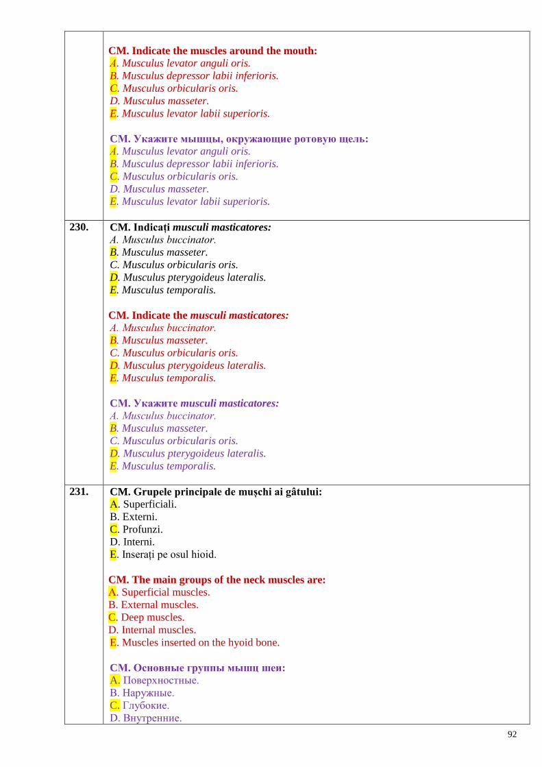
92
CM. Indicate the muscles around the mouth: А. Musculus levator anguli oris.
B. Musculus depressor labii inferioris.
C. Musculus orbicularis oris.
D. Musculus masseter.
E. Musculus levator labii superioris.
CМ. Укажите мышцы, окружающие ротовую щель:
А. Musculus levator anguli oris.
B. Musculus depressor labii inferioris.
C. Musculus orbicularis oris.
D. Musculus masseter.
E. Musculus levator labii superioris.
230. CM. Indicați musculi masticatores:
А. Musculus buccinator.
B. Musculus masseter.
C. Musculus orbicularis oris.
D. Musculus pterygoideus lateralis.
E. Musculus temporalis.
CM. Indicate the musculi masticatores: А. Musculus buccinator.
B. Musculus masseter.
C. Musculus orbicularis oris.
D. Musculus pterygoideus lateralis.
E. Musculus temporalis.
CМ. Укажите musculi masticatores:
А. Musculus buccinator.
B. Musculus masseter.
C. Musculus orbicularis oris.
D. Musculus pterygoideus lateralis.
E. Musculus temporalis.
231. CM. Grupele principale de mușchi ai gâtului:
А. Superficiali.
B. Externi.
C. Profunzi.
D. Interni.
E. Inserați pe osul hioid.
CM. The main groups of the neck muscles are: А. Superficial muscles.
B. External muscles.
C. Deep muscles.
D. Internal muscles.
E. Muscles inserted on the hyoid bone.
CМ. Основные группы мышц шеи:
A. Поверхностные.
B. Наружные.
C. Глубокие.
D. Внутренние.
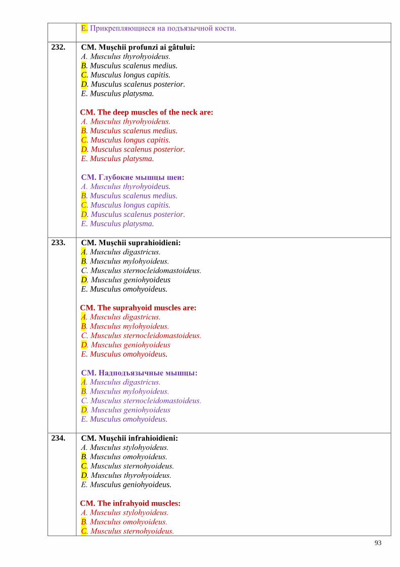
93
E. Прикрепляющиеся на подъязычной кости.
232. CM. Mușchii profunzi ai gâtului:
А. Musculus thyrohyoideus.
B. Musculus scalenus medius.
C. Musculus longus capitis.
D. Musculus scalenus posterior.
E. Musculus platysma.
CM. The deep muscles of the neck are: А. Musculus thyrohyoideus.
B. Musculus scalenus medius.
C. Musculus longus capitis.
D. Musculus scalenus posterior.
E. Musculus platysma.
CМ. Глубокие мышцы шеи:
А. Musculus thyrohyoideus.
B. Musculus scalenus medius.
C. Musculus longus capitis.
D. Musculus scalenus posterior.
E. Musculus platysma.
233. CM. Mușchii suprahioidieni:
А. Мusculus digastricus.
B. Мusculus mylohyoideus.
C. Мusculus sternocleidomastoideus.
D. Мusculus geniohyoideus
E. Musculus omohyoideus.
CM. The suprahyoid muscles are: А. Мusculus digastricus.
B. Мusculus mylohyoideus.
C. Мusculus sternocleidomastoideus.
D. Мusculus geniohyoideus
E. Musculus omohyoideus.
CМ. Надподъязычные мышцы:
А. Мusculus digastricus.
B. Мusculus mylohyoideus.
C. Мusculus sternocleidomastoideus.
D. Мusculus geniohyoideus
E. Musculus omohyoideus.
234. CM. Mușchii infrahioidieni:
А. Мusculus stylohyoideus.
B. Мusculus omohyoideus.
C. Мusculus sternohyoideus.
D. Мusculus thyrohyoideus.
E. Мusculus geniohyoideus.
CM. The infrahyoid muscles: А. Мusculus stylohyoideus.
B. Мusculus omohyoideus.
C. Мusculus sternohyoideus.
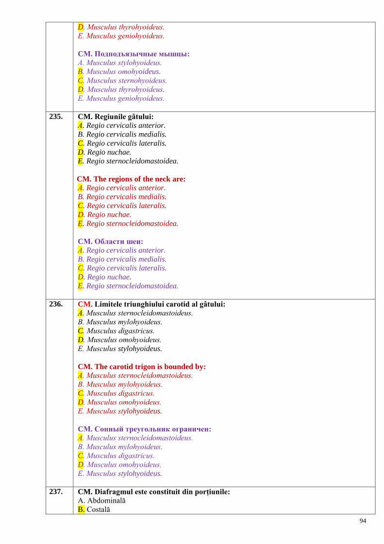
94
D. Мusculus thyrohyoideus.
E. Мusculus geniohyoideus.
CМ. Подподъязычные мышцы:
А. Мusculus stylohyoideus.
B. Мusculus omohyoideus.
C. Мusculus sternohyoideus.
D. Мusculus thyrohyoideus.
E. Мusculus geniohyoideus.
235. CM. Regiunile gâtului:
А. Regio cervicalis anterior.
B. Regio cervicalis medialis.
C. Regio cervicalis lateralis.
D. Regio nuchae.
E. Regio sternocleidomastoidea.
CM. The regions of the neck are: А. Regio cervicalis anterior.
B. Regio cervicalis medialis.
C. Regio cervicalis lateralis.
D. Regio nuchae.
E. Regio sternocleidomastoidea.
CМ. Области шеи:
А. Regio cervicalis anterior.
B. Regio cervicalis medialis.
C. Regio cervicalis lateralis.
D. Regio nuchae.
E. Regio sternocleidomastoidea.
236. CM. Limitele triunghiului carotid al gâtului:
А. Мusculus sternocleidomastoideus.
B. Мusculus mylohyoideus.
C. Мusculus digastricus.
D. Мusculus omohyoideus.
E. Мusculus stylohyoideus.
CM. The carotid trigon is bounded by:
А. Мusculus sternocleidomastoideus.
B. Мusculus mylohyoideus.
C. Мusculus digastricus.
D. Мusculus omohyoideus.
E. Мusculus stylohyoideus.
CМ. Сонный треугольник ограничен:
А. Мusculus sternocleidomastoideus.
B. Мusculus mylohyoideus.
C. Мusculus digastricus.
D. Мusculus omohyoideus.
E. Мusculus stylohyoideus.
237. CM. Diafragmul este constituit din porţiunile:
A. Abdominală
B. Costală
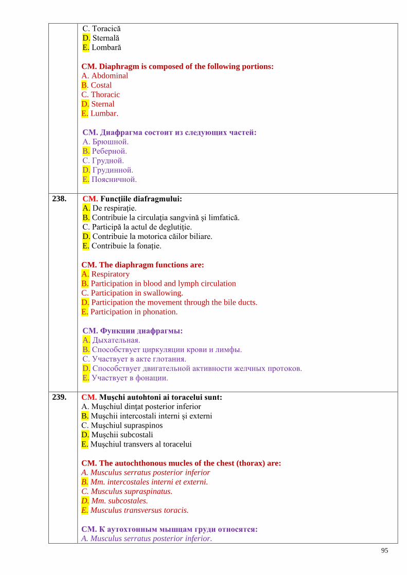
95
C. Toracică
D. Sternală
E. Lombară
CM. Diaphragm is composed of the following portions: A. Abdominal
B. Costal
C. Thoracic
D. Sternal
E. Lumbar.
CМ. Диафрагма состоит из следующих частей:
A. Брюшной.
B. Реберной.
C. Грудной.
D. Грудинной.
E. Поясничной.
238. CM. Funcţiile diafragmului: A. De respiraţie.
B. Contribuie la circulaţia sangvină şi limfatică.
C. Participă la actul de deglutiţie.
D. Contribuie la motorica căilor biliare.
E. Contribuie la fonație.
CM. The diaphragm functions are: A. Respiratory
B. Participation in blood and lymph circulation
C. Participation in swallowing.
D. Participation the movement through the bile ducts.
E. Participation in phonation.
CМ. Функции диафрагмы:
A. Дыхательная.
B. Способствует циркуляции крови и лимфы.
C. Участвует в акте глотания.
D. Способствует двигательной активности желчных протоков.
E. Участвует в фонации.
239. CM. Muşchi autohtoni ai toracelui sunt:
A. Mușchiul dinţat posterior inferior
B. Mușchii intercostali interni și externi
C. Mușchiul supraspinos
D. Mușchii subcostali
E. Mușchiul transvers al toracelui
CM. The autochthonous mucles of the chest (thorax) are:
A. Musculus serratus posterior inferior
B. Mm. intercostales interni et externi.
C. Musculus supraspinatus.
D. Mm. subcostales.
E. Musculus transversus toracis.
CМ. К аутохтонным мышцам груди относятся:
A. Musculus serratus posterior inferior.
96
B. Mm. intercostales interni et externi.
C. Musculus supraspinatus.
D. Mm. subcostales.
E. Musculus transversus toracis.
240. CM. Indicați, mușchii superficiali ai spatelui.
А. Musculus rhomboideus major.
B. Musculus serratus posterior inferior.
C. Musculus spinalis.
D. Musculus serratus posterior superior.
E. Musculus erector spinae.
CM. The superficial muscles of the back are:
А. Musculus rhomboideus major.
B. Musculus serratus posterior inferior.
C. Musculus spinalis.
D. Musculus serratus posterior superior.
E. Musculus erector spinae.
CМ. Назовите поверхностные мышцы спины:
А. Musculus rhomboideus major.
B. Musculus serratus posterior inferior.
C. Musculus spinalis.
D. Musculus serratus posterior superior.
E. Musculus erector spinae.
241. CM. Indicați mușchii profunzi ai spatelui.
А. Мusculus erector spinae.
B. Мusculus longissimus.
C. Мusculi transversospinales.
D. Мusculi multifidi.
E. Mușchii romboizi.
CM. The deep muscles of the back are:
А. Мusculus erector spinae
B. Мusculus longissimus
C. Мusculi transversospinales
D. Мusculi multifidi
E. Rhomboid muscles.
CМ. Назовите глубокие мышцы спины:
А. Мusculus erector spinae.
B. Мusculus longissimus.
C. Мusculi transversospinales.
D. Мusculi multifidi.
E. Ромбовидные мышцы.
242. CM. Muşchii abdomenului: A. Peretele anterolateral este format din muşchi autohtoni.
B. Fiecare muşchi lat are porţiune musculară şi aponeuroză.
C. Participă la inspiraţie.
D. Contribuie la fixarea organelor interne.
E. Au acţiune asupra coloanei vertebrale.
CM. Which of the following statements about the abdominal muscles is true:
97
A. The anterolateral wall is formed by autochthon musculature
B. Each broad muscle has the muscular and aponeurotic portions
C. They take part in inspiration.
D. They contribute to uphold the internal organs.
E. They possess action upon spine.
CМ. Мышцы живота:
A. Аутохтонные мышцы образуют переднебоковую стенку.
B. Каждая широкая мышца имеет мышечную часть и апоневроз.
C. Участвуют в акте вдоха.
D. Способствуют фиксации внутренних органов.
E. Действуют на позвоночник.
243. CM. Locuri slabe ale diafragmului:
A. Triunghiul lombocostal
B. Spaţiul dintre fasciculele sternale ale diafragmului
C. Triunghiul sternocostal
D. Triunghiul Petit
E. Fascia transversală dintre lig. Henle şi lig. interfoveolar
CM. The weak places of the diaphragm are: A. The lumbocostal triangle
B. The space between the sternal bundles of the diaphragm
C. The sternocostal triangle
D. The Petit’s triangle
E. The fascia transversalis between the ligament of Henle and lig. interfoveolaris.
CМ. Слабые места диафрагмы:
A. Реберно-поясничный треугольник.
B. Щель между грудинными мышечными пучками диафрагмы.
C. Грудинно-реберный треугольник.
D. Поясничный треугольник (Petit).
E. Поперечная фасция между lig. Henle и lig. interfoveolare.
244. CM. Locuri slabe ale peretelui abdominal anterior:
A. Tetragonul Grynfelt
B. Triunghiul sternocostal
C. Linia semilunară
D. Triunghiul Volânski
E. Linia albă
CM. The weak places of the anterior abdominal wall are: A. The tetragon of Grynfelt
B. The sternocostal triangle
C. The semilunar line
D. The triangle of Volânski
E.The linia alba.
CМ. Слабые места передней стенки живота:
A. Четырехугольник Grynfelt.
B. Грудинно-реберный треугольник.
C. Linia semilunaris.
D. Треугольник Волынского.
E. Белая линия.
98
245. CM. Locuri slabe ale peretelui abdominal posterior:
A. Triunghiul lombocostal
B. Tetragonul Grynfelt
C. Linia semilunară
D. Triunghiul Petit
E. Triunghiul Volânski
CM. The weak places of the posterior abdominal wall are: A. The lumbocostal triangle
B. The tetragon of Grynfelt
C. The semilunar line
D. The triangle of Petit
E.The linia alba.
CМ. Слабые места задней стенки живота:
A. Пояснично-реберный треугольник.
B. Четырехугольник Grynfelt.
С. Полулунная линия.
D. Поясничный треугольник.
E. Треугольник Волынского.
246. CM. Clasificarea muşchilor capului:
A. Muşchii penaţi
B. Muşchii mimici
C. Muşchii biventeri
D. Muşchii masticatori
E. Muşchii antagonişti
CM. The muscles of the head can be classified in:
A. Pennate (or pinnate) muscles
B. Muscles of facial expression
C. Biventer (two-bellied) muscles
D. Muscles of mastication
E. Antagonist muscles.
CМ. Классификация мышц головы:
A. Перистые.
B. Мимические.
C. Двубрюшные.
D. Жевательные.
E. Мышцы-антагонисты.
247. CM. Clasificarea muşchilor gâtului:
A. Muşchi anteriori
B. Muşchii superficiali
C. Muşchi scurţi
D. Muşchi profunzi
E. Muşchi fusiformi
CM. The muscles of the neck can be classified in:
A. Anterior muscles
B. Superficial muscles
C. Short muscles
D. Deep muscles
E. Fusiform muscles.
99
CМ. Классификация мышц шеи:
A. Передние.
B. Поверхностные.
C. Короткие.
D. Глубокие.
E. Веретенообразные.
248. CM. Muşchi superficiali ai gâtului sunt:
A. Suprahioidienii
B. Scalenii
C. Sternocleidomastoidianul
D. Mușchiul lung al gâtului
E. Mușchiul pielos al gâtului
CM. The superficial muscles of the neck are:
A. Suprahyoid muscles
B. Scalene muscles
C. Sternocleidomastoid muscle
D. Longus colli muscle
E. Platysma muscle.
CМ. Поверхностные м-цы шеи:
A. Надподъязычные.
B. Лестничные.
C. Грудинноключичнососцевидная
D. Длинная мышца шеи.
E. Подкожная мышца шеи.
249. CM. Grupul muşchilor profunzi ai gâtului include:
A. Muschii infrahioidieni
B. Muschii lungi al capului şi al gâtului
C. Muschii scaleni
D. Muschiul digastric
E. Muschii drepţi ai capului anterior și lateral.
CM. The deep muscles of the neck are:
A. Infrahyoid muscles
B. Longus capitis and longus colli muscles
C. Scalene muscles
D. Digastric muscle
E. Rectus capitis anterior and lateralis muscles.
CМ. К глубоким мышцам шеи относятся:
A. Подподъязычные мышцы.
B. Длинные мышцы головы и шеи.
C Лестничные мышцы.
D. Двубрюшная мышца.
E. Передняя и латеральная прямые мышцы головы.
Muşchii,
fasciile şi topografia
membrelor superioare şi inferioare
250. CM. Noțiunea de membru superior include:
A. Coapsa
100
B. Centura scapulară
C. Membrul superior liber
D. Antebraț
E. Braț
CM.The main components of the upper limb are the:
A. Thigh
B. Shoulder girdle
C. Free upper limb
D. Forearm
E. Arm
СМ. Верхняя конечность в себе включает:
A. Бедро
B. Пояс верхней конечности
C. Свободную верхнюю конечность
D. Предплечье
E. Область плеча
251. CM. Noțiunea de membru inferior liber include:
A. Coapsa
B. Centura pelviană
C. Gamba
D. Piciorul
E. Tibia
CM. The main components of the lower limb are the:
A. Thigh
B. Pelvic girdle
C. Leg
D. Foot
E. Tibia.
СМ. Свободная нижняя конечность в себе включает:
A. Бедро
B. Тазовый пояс
C. Голень
D. Стопу
E. Большеберцовцую кость
252. CM. Sunt muşchi ai centurii scapulare:
A. M. levator al scapulei
B. M. supraspinos
C. M. deltoid
D. M. pectoral mic
E. M. infraspinos
CM. The muscles of shoulder girdle are:
A. Levator scapulae muscle
B. Supraspinatus muscle
C. Deltoid muscle
D. Pectoralis minor muscle
E. Infraspinatus muscle.
CМ. К мышцам плечевого пояса относятся:
101
A. Мышца, поднимающая лопатку.
В. Надостная мышца.
С. Дельтовидная мышца.
D. Малая грудная мышца.
E. Подостная мышца.
253. CM. Numiți șanțurile feței anterioare a antebrațului А. Median
B. Radioulnar
C. Cubital
D. Radial
E. Ulnar
CM.The grooves of the anterior surface of the forearm are:
A. Median
B. Radioulnar
C. Cubital
D. Radial
E. Ulnar.
CМ. Назовите борозды расположенные на передней поверхности предплечья: А. Срединная.
B. Лучелоктевая.
С. Кубитальная.
D. Лучевая.
E. Локтевая.
254. CM. Funcţiile muşchilor anteriori ai braţului se rezumă la:
A. Extensia în articulaţia umărului
B. Flexia în articulaţia cotului
C. Abducţia în articulaţia umărului
D. Flexia anterioară a braţului
E. Rotirea braţului
CM. The functions of the anterior group of muscles of the arm are:
A. Extension in the shoulder joint
B. Flexion in the elbow joint
C. Abduction in the shoulder joint
D. Anterior flexion of the arm
E. Rotation of the arm.
CМ. Мышцы передней группы плеча осуществляют:
A. Разгибание в плечевом суставе
B. Сгибание в локтевом суставе
С. Отведение в плечевом суставе
D. Переднее сгибание плеча
E. Вращение плеча
255. CS. Determină configurația umărului:
A. Mușchiul deltoid
B. Mușchiulsubscapular
C. Mușchiul supaspinos
D. Mușchiul rotund mare
E. Mușchiul rotund mic
102
CS. Which of the following muscles determines the shoulder shape: A. M. deltoideus
B. M. subscapularis
C. M. supaspinosus
D. M. teres major
E. M. teres minor.
CS. Формирует внешний контур плеча:
А. Дельтовидная мышца.
B. Подлопаточная мышца.
C. Надостная мышца.
D. Большая круглая мышца
E. Малая круглая мышца
256. CS. Muşchiul dorsal mare realizează:
A. Adducţia braţului
B. Flexia braţului
C. Supinaţia braţului
D. Rotirea braţului spre exterior
E. Extensia in articulaţia umărului.
CS. The latissimus dorsi muscle realizes: A. Adduction of the arm
B. Flexion of the arm
C. Supination of the arm
D. Lateral rotation of the arm
E. Extension in the shoulder joint.
CS. Широчайшая мышца спины производит:
А. Приведение плеча.
B. Сгибание плеча.
C. Супинацию плеча.
D. Вращение плеча кнаружи.
E. Разгибание в плечевом суставе.
257. CS. Sub retinaculul extensorilor in regiunea carpiană se află:
A. Canale pentru nervi
B. Spaţii pentru artere
C. Teci sinoviale pentru tendoanele muşchilor extensori ai mâinii
D. Burse sinoviale
E. Teci fibroase pentru vase limfatice şi vene
CS. Which of the following lodges under the extensor retinaculum in the carpal region: A. Canals for nerves
B. Spaces for arteries
C. Synovial sheaths for tendons of extensor muscles of the hand.
D. Synovial bursae.
E. Synovial sheaths for lymph vessels and veins.
CS. Под удерживателем разгибателей и области запястья располагаются:
А. Каналы для нервов
B. Пространства для артерий
C. Синовиальные влагалища для сухожилий мышц разгибателей кисти
D. Синовиальные сумки
E. Фиброзные влагалища для лимфатических сосудов и вен
103
258. CM. Indicați care mușchi participă la extensia brațului: А. Мusculus suprascapularis.
B. Мusculus teres major.
C. Мusculus deltoideus.
D. Мusculus pectoralis minor.
E. Мusculus subscapularis.
CM. The muscle that flex the arm is:
А. Мusculus suprascapularis.
B. Мusculus teres major.
C. Мusculus deltoideus.
D. Мusculus pectoralis minor.
E. Мusculus subscapularis.
CM. Укажите мышцу, участвующую в разгибании плеча:
А. Мusculus suprascapularis
B. Мusculus teres major
C. Мusculus deltoideus
D. Мusculus pectoralis minor
E. Мusculus subascapularis
259. CS. Acţiunea muşchiului brahial: А. Extinde braţul
B. Flexează mâna
C. Extinde antebraţul
D. Flexează antebraţul
E. Pronează braţul
CS. The function of the brachialis muscle is: А. Extension of the arm
B. Flexion of the hand
C. Extension of the forearm
D.Flexion of the forearm
E. Pronation of the arm.
CS. Функция musculus brachialis:
А. Разгибает плечо.
B. Сгибает плечо.
C. Разгибает предплечье.
D. Сгибает предплечье.
E. Пронирует плечо
260. CS. Prin orificiul sciatic mic trece: A. M. piriform
B. M. obturator intern
C. M. obturator extern
D. Mm. gemeni
E. M. psoas mic
CS. Which of the following muscles passes through the lesser sciatic orifice: A. M. piriformis
B. M. obturatorius internus
C. M. obturatorius externus
D. Mm. gemeli
104
E. M. psoas minor.
CS. Через малое седалищное отверстие проходит:
А. Грушевидная мышца
B. Внутренняя запирательная мышца
C. Наружная запирательная мышца
D. Близнецовые мышцы
E. Малая поясничная мышца
261. CS. Lacuna vasculară se separă de cea musculară prin:
A. Lig. lacunar
B. Lig. inghinal
C. Lig. reflex
D. Arcul iliopectineu
E. Fascia transversă
CS. The vascular space is separated from the muscular one by: A. Lig. lacunare
B. Lig. inguinale
C. Lig. reflexus
D. Arcus iliopectineus
E. Fascia transversalis.
CS. Сосудистую и мышечную лакуны разделяет:
А. Лакунарная связка
B. Паховая связка
C. Возвратная связка
D. Подвздошно-гребенчатая дуга
E. Поперечная фасция
262. CS. Peretele anterior al canalului inghinal se formează din: А. Мusculus transversus abdominis.
B. Fascia transversalis.
C. Мusculus obliquus externus abdominis (aponeuroza).
D. Мusculus obliquus internus abdominis.
E. Lig. inguinale
CS. The anterior wall of the inguinal canal is formed by:
А. Мusculus transversus abdominis
B. Fascia transversalis
C. Мusculus obliquus externus abdominis (aponeurosis)
D. Мusculus obliquus internus abdominis
E. Lig. inguinale.
CS. Передняя стенка пахового канала образована:
А. Мusculus transversus abdominis.
B. Fascia transversalis.
C. Мusculus obliquus externus abdominis (апоневроз).
D. Мusculus obliquus internus abdominis.
E. Lig. inguinale.
263. CS. Peretele posterior al canalului inghinal e dat de: А. Мusculus transversus abdominis.
B. Fascia transversalis.
C. Мusculus obliquus externus abdominis.
105
D. Мusculus obliquus internus abdominis.
E. Lig. inguinale
CS. The posterior wall of the inguinal canal is formed by: А. Мusculus transversus abdominis
B. Fascia transversalis
C. Мusculus obliquus externus abdominis
D. Мusculus obliquus internus abdominis
E. Lig. inguinale.
CS. Заднюю стенку пахового канала образуют:
А. Мusculus transversus abdominis.
B. Fascia transversalis.
C. Мusculus obliquus externus abdominis.
D. Мusculus obliquus internus abdominis.
E. Lig. inguinale.
264. CS. Limita superioară a trigonum femorale e formată de: А. Мusculus iliopsoas.
B. Мusculus sartorius.
C. Мusculus vastus medialis.
D. Ligamentum inguinale.
E. Мusculus obliquus externus abdominis.
CM. Superiorly the femoral triangle is bounded by:
А. Мusculus iliopsoas.
B. Мusculus sartorius.
C. Мusculus vastus medialis.
D. Ligamentum inguinale.
E. Мusculus obliquus externus abdominis.
CS. Сверху trigonum femorale ограничен:
А. Мusculus iliopsoas.
B. Мusculus sartorius.
C. Мusculus vastus medialis.
D. Ligamentum inguinale.
E. Мusculus obliquus externus abdominis.
265. CS. Limita medială a trigonum femorale o constituie: А. Мusculus adductor longus.
B. Мusculus vastus medialis.
C. Мusculus rectus femoris.
D. Ligamentum inguinale.
E. Мusculus obliquus externus abdominis.
CS. The medial border of the trigonum femorale is formed by: А. Мusculus adductor longus.
B. Мusculus vastus medialis.
C. Мusculus rectus femoris.
D. Ligamentum inguinale.
E. Мusculus obliquus externus abdominis.
CS. С медиальной стороны trigonum femorale ограничен:
А. Мusculus adductor longus.
B. Мusculus vastus medialis.
106
C. Мusculus rectus femoris.
D. Ligamentum inguinale.
E. Мusculus obliquus externus abdominis.
266. CS. Limita laterală a trigonum femorale e dată de: А. Мusculus rectus femoris.
B. Мusculus sartorius.
C. Мusculus vastus medialis.
D. Мusculus vastus lateralis.
E. Мusculus obliquus externus abdominis.
CS. The lateral border of the trigonum femorale is formed by: А. Мusculus rectus femoris.
B. Мusculus sartorius.
C. Мusculus vastus medialis.
D. Мusculus vastus lateralis.
E. Мusculus obliquus externus abdominis.
CS. С латеральной стороны trigonum femorale ограничен:
А. Мusculus rectus femoris.
B. Мusculus sartorius.
C. Мusculus vastus medialis.
D. Мusculus vastus lateralis.
E. Мusculus obliquus externus abdominis.
267. CS. În triunghiul femural se deschid:
A. Canalul inghinal
B. Canalul femural
C. Canalul obturator
D. Canalul adductor
E. Canalul cruropopliteu
CS. In the femoral triangle opens the: A. Inguinal canal
B. Femoral canal
C. Obturator canal
D. Adductor canal
E. Cruropopliteal canal.
CS. В бедренный треугольник открываются:
А. Паховый канал.
B. Бедренный канал.
C. Запирательный канал.
D. Приводящий канал.
E. Голеноподколенный канал.
268. CS. Muşchii coapsei. Grup muscular medial:
A. Semimembranos
B. Biceps femural
C. Semitendinos
D. Graţios
E. Croitor
CS. The medial muscles of the thigh are:
A. Semimembranous muscle
107
B. Biceps femuris muscle
C. Semitendinous muscle
D. Gracilis muscle
E. Sartorius muscle.
CS. Мышцы бедра. Медиальная группа:
A. Полуперепончатая мышца
B. Двуглавая мышца бедра
C. Полусухожильная мышца
D. Тонкая мышца
E. Портняжная мышца
269. CM. Fosa cubitală este delimitată de:
A. M. coracobrahial
B. M. brahial
C. M. brahioradial
D. M. supinator
E. M. pronator patrat.
CM. The cubital fossa is delimited by: A. M. coracobrahialis
B. M. brachialis
C. M. brachioradialis
D. M. supinator
E. M. pronator qudratus.
CМ. Локтевая ямка ограничена:
A. Клювовидно-плечевой м-цей
B. Плечевой м-цей
C. Плечелучевой м-цей
D. Супинирующей м-цей
E. Квадратным пронатором
270. CM. Canalul obturator este delimitat de: M. obturator extern
B. M. pectineu
C. Orificiul obturator
D. Şantul obturator
E. M. obturator intern.
CM. The obturator canal is delimitated by: A. M. obturatorius externus
B. M. pectineus
C. Obturator foramen
D. Obturator groove
E. M. obturatorius internus.
CМ. Запирательный канал ограничен: А. Наружной запирательной мышцей
B. Гребенчатой мышцей
C. Запирательным отверстием
D. Запирательной бороздой
E. Внутренней запирательной мышцей
271. CM. Muşchii mediali ai coapsei:
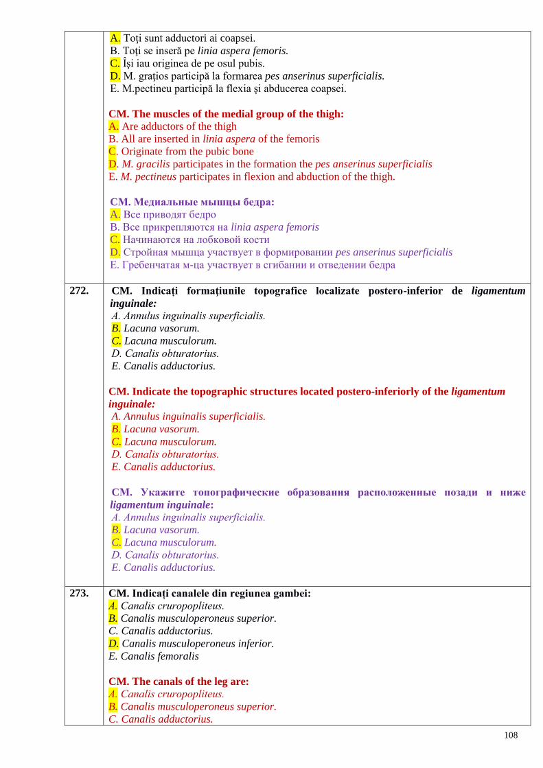
108
A. Toţi sunt adductori ai coapsei.
B. Toţi se inseră pe linia aspera femoris.
C. Îşi iau originea de pe osul pubis.
D. M. graţios participă la formarea pes anserinus superficialis.
E. M.pectineu participă la flexia şi abducerea coapsei.
CM. The muscles of the medial group of the thigh:
A. Are adductors of the thigh
B. All are inserted in linia aspera of the femoris
C. Originate from the pubic bone
D. M. gracilis participates in the formation the pes anserinus superficialis
E. M. pectineus participates in flexion and abduction of the thigh.
CМ. Медиальные мышцы бедра: A. Все приводят бедро
B. Все прикрепляются на linia aspera femoris
C. Начинаются на лобковой кости
D. Стройная мышца участвует в формировании pes anserinus superficialis
E. Гребенчатая м-ца участвует в сгибании и отведении бедра
272. CM. Indicați formaţiunile topografice localizate postero-inferior de ligamentum
inguinale: А. Annulus inguinalis superficialis.
B. Lacuna vasorum.
C. Lacuna musculorum.
D. Canalis оbturatorius.
E. Canalis adductorius.
CM. Indicate the topographic structures located postero-inferiorly of the ligamentum
inguinale: A. Annulus inguinalis superficialis.
B. Lacuna vasorum.
C. Lacuna musculorum.
D. Canalis оbturatorius.
E. Canalis adductorius.
CМ. Укажите топографические образования расположенные позади и ниже
ligamentum inguinale: А. Annulus inguinalis superficialis.
B. Lacuna vasorum.
C. Lacuna musculorum.
D. Canalis оbturatorius.
E. Canalis adductorius.
273. CM. Indicați canalele din regiunea gambei: А. Canalis cruropоpliteus.
B. Canalis musculoperoneus superior.
C. Canalis adductorius.
D. Canalis musculoperoneus inferior.
E. Canalis femoralis
CM. The canals of the leg are:
А. Canalis cruropоpliteus.
B. Canalis musculoperoneus superior.
C. Canalis adductorius.
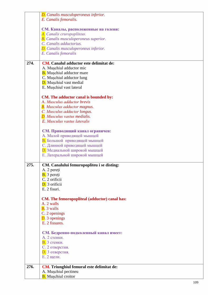
109
D. Canalis musculoperoneus inferior.
E. Canalis femoralis.
CМ. Каналы, расположенные на голени: А. Canalis cruropоpliteus.
B. Canalis musculoperoneus superior.
C. Canalis adductorius.
D. Canalis musculoperoneus inferior.
E. Canalis femoralis
274. CM. Canalul adductor este delimitat de:
A. Mușchiul adductor mic
B. Mușchiul adductor mare
C. Mușchiul adductor lung
D. Mușchiul vast medial
E. Mușchiul vast lateral
CM. The adductor canal is bounded by:
А. Мusculus adductor brevis
B. Мusculus adductor magnus.
C. Мusculus adductor longus.
D. Мusculus vastus medialis.
E. Мusculus vastus lateralis
CМ. Приводящий канал ограничен:
A. Малой приводящей мышцей
B. Большой приводящей мышцей
C. Длинной приводящей мышцей
D. Медиальной широкой мышцей
E. Латеральной широкой мышцей
275. CM. Canalului femuropopliteu i se disting:
A. 2 pereţi
B. 3 pereţi
C. 2 orificii
D. 3 orificii
E. 2 fisuri.
CM. The femoropopliteal (adductor) canal has: A. 2 walls
B. 3 walls
C. 2 openings
D. 3 openings
E. 2 fissures.
CМ. Бедренно-подколенный канал имеет:
A. 2 стенки.
B. 3 стенки.
C. 2 отверстия.
D. 3 отверстия.
E. 2 щели.
276. CM. Triunghiul femural este delimitat de: A. Mușchiul pectineu
B. Mușchiul croitor
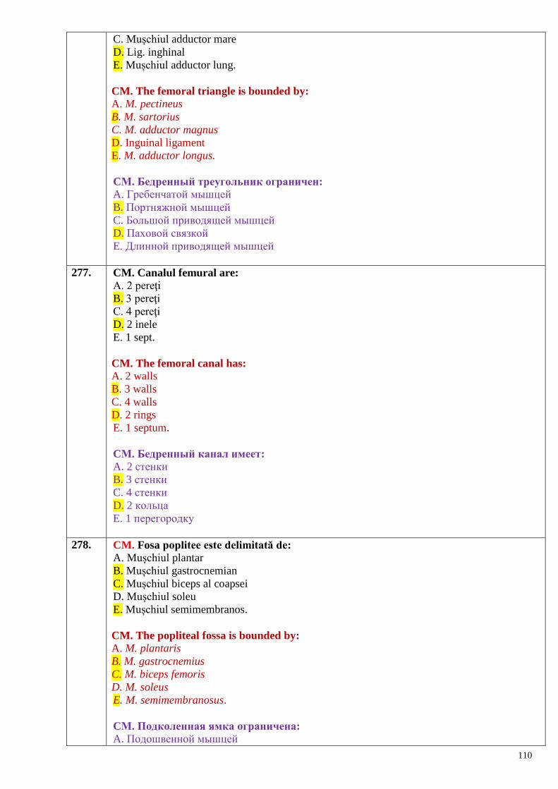
110
C. Mușchiul adductor mare
D. Lig. inghinal
E. Mușchiul adductor lung.
CM. The femoral triangle is bounded by: A. M. pectineus
B. M. sartorius
C. M. adductor magnus
D. Inguinal ligament
E. M. adductor longus.
CМ. Бедренный треугольник ограничен: A. Гребенчатой мышцей
B. Портняжной мышцей
C. Большой приводящей мышцей
D. Паховой связкой
E. Длинной приводящей мышцей
277. CM. Canalul femural are:
A. 2 pereţi
B. 3 pereţi
C. 4 pereţi
D. 2 inele
E. 1 sept.
CM. The femoral canal has: A. 2 walls
B. 3 walls
C. 4 walls
D. 2 rings
E. 1 septum.
CМ. Бедренный канал имеет:
A. 2 стенки
B. 3 стенки
C. 4 стенки
D. 2 кольца
E. 1 перегородку
278. CM. Fosa poplitee este delimitată de:
A. Mușchiul plantar
B. Mușchiul gastrocnemian
C. Mușchiul biceps al coapsei
D. Mușchiul soleu
E. Mușchiul semimembranos.
CM. The popliteal fossa is bounded by: A. M. plantaris
B. M. gastrocnemius
C. M. biceps femoris
D. M. soleus
E. M. semimembranosus.
CМ. Подколенная ямка ограничена:
A. Подошвенной мышцей
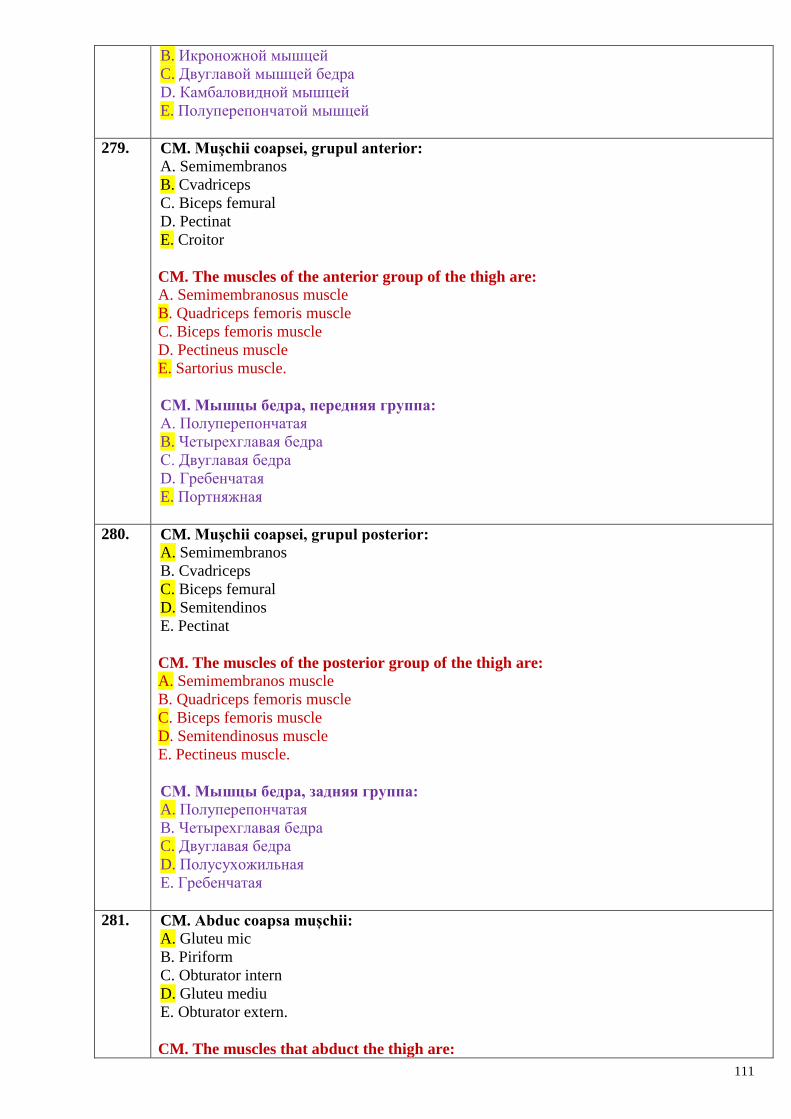
111
B. Икроножной мышцей
C. Двуглавой мышцей бедра
D. Камбаловидной мышцей
E. Полуперепончатой мышцей
279. CM. Muşchii coapsei, grupul anterior:
A. Semimembranos
B. Cvadriceps
C. Biceps femural
D. Pectinat
E. Croitor
CM. The muscles of the anterior group of the thigh are: A. Semimembranosus muscle
B. Quadriceps femoris muscle
C. Biceps femoris muscle
D. Pectineus muscle
E. Sartorius muscle.
CМ. Мышцы бедра, передняя группа:
A. Полуперепончатая
B. Четырехглавая бедра
C. Двуглавая бедра
D. Гребенчатая
E. Портняжная
280. CM. Muşchii coapsei, grupul posterior:
A. Semimembranos
B. Cvadriceps
C. Biceps femural
D. Semitendinos
E. Pectinat
CM. The muscles of the posterior group of the thigh are: A. Semimembranos muscle
B. Quadriceps femoris muscle
C. Biceps femoris muscle
D. Semitendinosus muscle
E. Pectineus muscle.
CМ. Мышцы бедра, задняя группа:
A. Полуперепончатая
B. Четырехглавая бедра
C. Двуглавая бедра
D. Полусухожильная
E. Гребенчатая
281. CM. Abduc coapsa mușchii:
A. Gluteu mic
B. Piriform
C. Obturator intern
D. Gluteu mediu
E. Obturator extern.
CM. The muscles that abduct the thigh are:
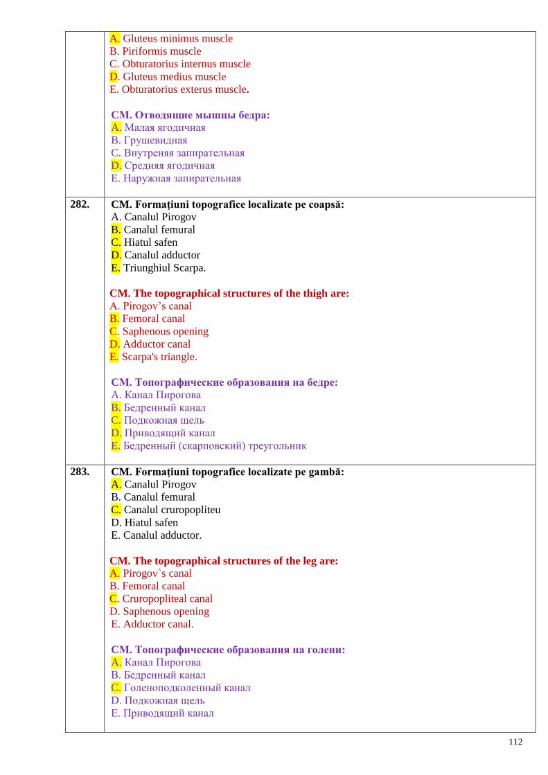
112
A. Gluteus minimus muscle
B. Piriformis muscle
C. Obturatorius internus muscle
D. Gluteus medius muscle
E. Obturatorius exterus muscle.
CМ. Отводящие мышцы бедра:
A. Малая ягодичная
B. Грушевидная
C. Внутреняя запирательная
D. Средняя ягодичная
E. Наружная запирательная
282. CM. Formaţiuni topografice localizate pe coapsă:
A. Canalul Pirogov
B. Canalul femural
C. Hiatul safen
D. Canalul adductor
E. Triunghiul Scarpa.
CM. The topographical structures of the thigh are: A. Pirogov’s canal
B. Femoral canal
C. Saphenous opening
D. Adductor canal
E. Scarpa's triangle.
CМ. Топографические образования на бедре:
A. Канал Пирогова
B. Бедренный канал
C. Подкожная щель
D. Приводящий канал
E. Бедренный (скарповский) треугольник
283. CM. Formaţiuni topografice localizate pe gambă:
A. Canalul Pirogov
B. Canalul femural
C. Canalul cruropopliteu
D. Hiatul safen
E. Canalul adductor.
CM. The topographical structures of the leg are: A. Pirogov`s canal
B. Femoral canal
C. Cruropopliteal canal
D. Saphenous opening
E. Adductor canal.
CМ. Топографические образования на голени:
A. Канал Пирогова
B. Бедренный канал
C. Голеноподколенный канал
D. Подкожная щель
E. Приводящий канал
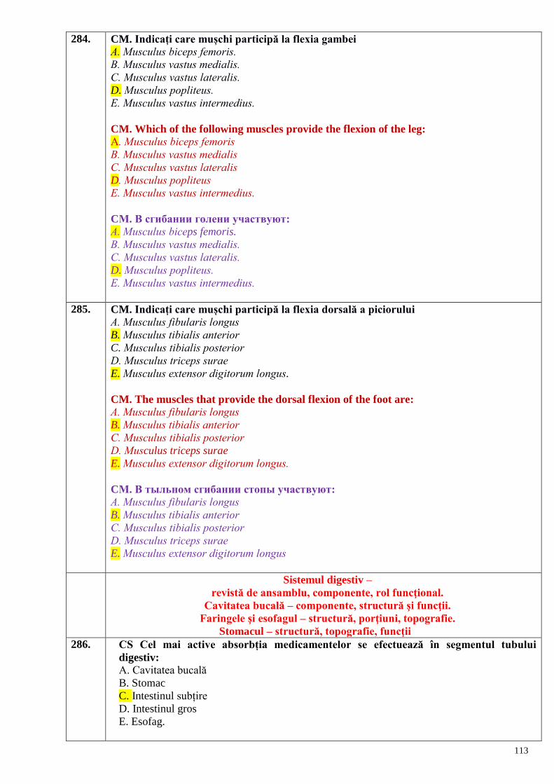
113
284. CM. Indicați care mușchi participă la flexia gambei А. Мusculus biceps femoris.
B. Мusculus vastus medialis.
C. Мusculus vastus lateralis.
D. Мusculus popliteus.
E. Мusculus vastus intermedius.
CM. Which of the following muscles provide the flexion of the leg:
А. Мusculus biceps femoris
B. Мusculus vastus medialis
C. Мusculus vastus lateralis
D. Мusculus popliteus
E. Мusculus vastus intermedius.
CМ. В сгибании голени участвуют: А. Мusculus biceps femoris.
B. Мusculus vastus medialis.
C. Мusculus vastus lateralis.
D. Мusculus popliteus.
E. Мusculus vastus intermedius.
285. CM. Indicați care mușchi participă la flexia dorsală a piciorului А. Мusculus fibularis longus
B. Мusculus tibialis anterior
C. Мusculus tibialis posterior
D. Мusculus triceps surae
E. Мusculus extensor digitorum longus.
CM. The muscles that provide the dorsal flexion of the foot are: А. Мusculus fibularis longus
B. Мusculus tibialis anterior
C. Мusculus tibialis posterior
D. Мusculus triceps surae
E. Мusculus extensor digitorum longus.
CМ. В тыльном сгибании стопы участвуют: А. Мusculus fibularis longus
B. Мusculus tibialis anterior
C. Мusculus tibialis posterior
D. Мusculus triceps surae
E. Мusculus extensor digitorum longus
Sistemul digestiv –
revistă de ansamblu, componente, rol funcţional.
Cavitatea bucală – componente, structură şi funcţii.
Faringele şi esofagul – structură, porţiuni, topografie.
Stomacul – structură, topografie, funcţii
286. CS Cel mai active absorbția medicamentelor se efectuează în segmentul tubului
digestiv:
A. Cavitatea bucală
B. Stomac
C. Intestinul subțire
D. Intestinul gros
E. Esofag.
114
CS. The most active absorbtion of the medicaments occurs in the following segment of
the alimentary canal:
A. Oral cavitaty
B. Stomac
C. Small intestine
D. Large intestine
E. Esophagus.
CS. Наиболее активно всасывание лекарственных веществ происходит:
A. В ротовой полости
B. В желудке
C. В тонком кишечнике
D. В толстом кишечнике
E. В пищеводе
287. CM. Capacitatea de absorbție a mucoasei bucale depinde de aplicarea comprimatelor:
A. Sublingvale
B. Mucoadezive
C. De mestecat
D. Orodis persabile
E. Nici unul nu e corect.
CM. The capacity of absorbtion of the mucous membrane of the oral cavity depends on
the application of tablets:
A. Sublingual
B. Mucoadhesive
C. Chewing
D. Orally dispersible
E. All are wrong
CМ. Всасывание в ротовой полости усиливается при приѐме таблеток:
A. Подъязычно
B. Мукоадезивных
C. Жевательных
D. Орально диспергируемых
E. Все неправильные
288. CM. Pe cale sublingvală se administrează:
A. Hormoni
B. Preparate cardiace (nitroglicerina)
C. Preparate hipotensive (captopril)
D. Validol
E. Mezim-forte.
CM. Medicaments that are administrated by the sublingual way are:
A. Hormons
B. Cardiac drugs (nitroglycerine)
C. Hipotensive drugs (captopril)
D. Validol
E. Mezim-forte.
CМ. Подъязычный путь администрации медикаментов используется для:
A. Гормонов
B. Сердечных препаратов (нитроглицерин)
C. Гипотензивных препаратов (каптоприл)
115
D. Валидол
E. Мезим
289. CM. Administrarea pe cale sublingvală poate fi argumentată prin:
A. Vascularizarea bogată a mucoasei sublingvale
B. Plexul venos sublingval, așezat superficial
C. Abundența salivei, în care se dizolvă aceste medicamente
D. Se dizolvă rapid
E. Se dizolvă lent.
CM. The sublingual administration may be argumented by:
A. Reach blood supply of the sublingual mucosa
B. Superficial location of the sublingual venous plexus
C.The presence of saliva, dissolving medicines
D. Rapid dissolving
E. Slow dissolving
CМ. Чем аргументировано использование медикаментов под языком:
A. Богатой васкуляризации слизистой оболочки
B. Поверхностное расположение подъязычной венозной сети
C. Наличием слюны, растворяющей медикаменты
D. Быстрым растворением
E. Медленным растворением
290. CM. În stomac se absorb:
A. Apa
B. Alcoolul
C. Cofeina
D. Unele medicamente
E. Bila.
CM. In the stomac are absorbed:
A. Water
B. Alcohol
C. Caffeine
D. Some drugs
E. Bile.
CМ. В желудке всасываются:
A. Вода
B. Алкоголь
C. Кофеин
D. Некоторые медикаменты
E. Жѐлчь
291. CM. Absorbția medicamentelor de către mucoasa gastrică depinde de:
A. Mediul acid în stomac
B. Calitatea hranei
C. Cantitatea hranei
D. Evacuarea gastrică
E. Nici una nu e corectă.
CM. Absorbtion of the medicaments by the gastric mucosa depends on:
A. Acid medium in the stomach
116
B. Quality of the food
C. Quantity of the food
D. Gastric evacuation
E. All are wrong
CМ. Всасывание лекарственных веществ в желудке зависит от:
A. Кислотной среды желудка
B. Качества пищи
C. Количества пищи
D. Эвакуаторной функции желудка
E. Нет правильных ответов
292. CM. Intensitatea absorbției substanțelor medicamentoase în intestinul subțire este
favorizată de:
A. Suprafața de contact cu mucoasa intestinală este mare (5 m2)
B. Timpul de staționare a medicamentelor este îndelungată (5-6 ore)
C. Vascularizația abundentă permite trecerea rapidă a medicamentelor
D. Ph-ul este neutru spre acid
E. În majoritatea cazurilor substanțele active sunt eliberate de forma farmaceutică și
disponibile pentru dizolvare și absorbție.
CM. Intensivity of medicaments absorbtion in the small intestine is increased by:
A. Big surface of contact with intestinal mucosa (5 m2)
B. Long time of retention of the drugs in this segment of the intestine (5-6 hours)
C. Reach vascularization that permits rapid passage of the medicaments
D. Neutral-acidic intestinal medium
E. Production of drugs with a high absorptive ability
CМ. Интенсивность абсорбции лекарственных веществ в тонком кишечнике
зависит от:
A. Большой поверхности соприкосновения со слизистой (5 м2)
B. Продолжительности нахождения лекарств в этом отрезке кишечника (5-6 часов)
C. Интенсивное кровоснабжение кишечника способствует быстрому прохождению
содержимого
D. От нейтрально-кислой среды кишечника
E. Фармацевтические фирмы производят лекарственные вещества с большой
всасывательной (всасывающей) способностью
293. CS. La nivelul intestinului gros cel mai activ absorbția medicamentelor are loc în:
A. Cec
B. Colonul transvers
C. Colonul sigmoid
D. Rect
E. Apendicele vermiform.
CS. The most active absorbtion of the medicaments in the large intestine occurs at
the level of the:
A. Cecum
B. Transverse colon
C. Sigmoid colon
D. Rectum
E. Vermiform appendix
CS. В толстом кишечнике лекарственные вещества лучше всасываются в:
A. Слепой кишке
117
B. Поперечной ободочной кишке
C. Сигмовидной кишке
D. Прямой кишке
E. Червеобразном отростке
294. CM. Calea rectală de administrare a medicamentelor se utilizează la:
A. Copii
B. Bolnavii cu leziuni în regiunea bucofaringiană
C. Bolnavii inconștienți (în comă)
D. Femeile gravide
E. Bolnavii, care vometează.
CM. Rectal way of drug administration is used for:
A. Children
B. Pacients with damaged oropharyngeal region
C. In patients who are in coma
D. In pregnant women
E. In patients with vomiting
CМ. Введение лекарств „per rectum” применяется (практикуется):
A. У детей
B. У больных с повреждениями в области ротоглотки
C. У больных, находящихся в коме
D. У беременных женщин
E. У больных с рвотой
295. CS. Metabolizarea medicamentelor are loc în:
A. Pancreas
B. Splină
C. Ficat
D. Glanda parotidă
E. Esofag.
CS. Metabolism of medicaments takes part in the:
A. Pancreas
B. Spleen
C. Liver
D. Parotid gland
E. Esophagus.
CS. Метаболизация лекарственных препаратов происходит в:
A. Поджелудочной железе
B. Селезѐнке
C. Печени
D. Околоушной железе
E. Пищеводе
296. CM. Eliminarea medicamentelor pe cale digestivă are loc prin:
A. Salivă
B. Bilă
C. Masele fecale
D. Sucul pancreatic
E. Uree.
CM. Elimination of the medicaments from digestive tube occurs by:
118
A. Salive
B. Bile
C. Fecal masses
D. Pancreatic juice
E. Ureea.
CМ. Выведение лекарственных веществ из организма происходит следующими
путями:
A. Через слюну
B. Через жѐлчь
C. Фекалиями
D. Поджелудочным соком
E. Мочевиной
297. CS. Organul reprezintă:
A. O formațiune constituita din trei tunici
B. Un element cavitar
C. Parte a organismului constituită dintr-un complex de țesuturi integrate in realizarea
anumitor funcții
D. O formațiune parenchimatoasă așezată in cavitatea abdominală
E. O formațiune constituită din epiteliu, vase sangvine şi nervi
SC. The organ represents: A. A structure made up by three layers
B. A hollow element
C. A part of the body, which consists of a complex of tissues integrated to realize some
functions
D. A parenchymatous structure located in the abdominal cavity
E. A formation constituted by the epithelium, blood vessels and nerves.
CS. Орган представляет:
A. Oбразование, состоящее из трѐх оболочек
B. Oтдельную полость
C. Часть организма, состоящую из комплекса интегрированных тканей для выполнения
определѐнных функций
D. Паренхиматозные органы, расположенные в брюшной полости
E. Образования, состоящие из эпителия, кровеносных сосудов и нервов
298. CS. Intestinul primitiv se dezvoltă din:
A. Ectoderm
B. Mezoderm
C. Endoderm
D. Dermatom
E. Miotom
SC. The primary gut develops from: A. Ectoderm
B. Mesoderm
C. Endoderm
D. Dermatome
E. Myotome.
CS. Первичная кишка развивается из:
A. Эктодермы
B. Мезодермы
119
C. Энтодермы
D. Дерматома
E. Миотома
299. CS. Din care foiţă embrionară se dezvoltă intestinul primar:
A. Din entodermă
B. Din ectodermă
C. Din sclerotomi
D. Din mezodermă
E. Din splanhnopleură
CS. The primary gut develops from:
A. Entoderm
B. Ectoderm
C. Sclerotome
D. Mesoderm
E. Splanchnopleura
CS. Из какого зародышевого листка развивается первичная кишка:
A. Из энтодермы
B. Из эктодермы
C. Из склеротомов
D. Из мезодермы
E. Из спланхноплевры
300. CM. Viscerele reprezintă:
A. Organe localizate în cavitatea abdominală
B. Sisteme de organe ce efectuează legătura organismului cu mediul ambiant
C. Organe sau complexe de organe localizate in cavitățile corpului și realizând funcții
necesare pentru menținerea vieții
D. Un complex de organe din cavitatea abdominală şi cea a bazinului mic
E. Complexul de organe din cavitatea toracică
SC. The viscera represent: A. Organs located in the abdominal cavity
B. Systems of organs that realize the connection of the body with the external environment
C. Organs or systems of organs located in the body cavities, which realize the metabolic
functions necessary to sustain the life
D. A complex of organs located in the thoracic, abdominal and pelvic cavities
E. A complex of organs in the thoracic cavity.
CM. Внутренности представлены:
A. Органами, расположенными в брюшной полости
B. Системой органов, обеспечивающих связь организма со средой обитания
C. Органами или комплексом органов, расположенных в полостях тела, выполняющие
функцию сохранения жизни
D. Комплекс органов грудной полости, брюшной полости и малого таза
E. Комплекс органов грудной полости
301. CM. Morfologic organele se împart in:
A. Seroase
B. Parenchimatoase
C. Glandulare
D. Epiteliale
E. Cavitare
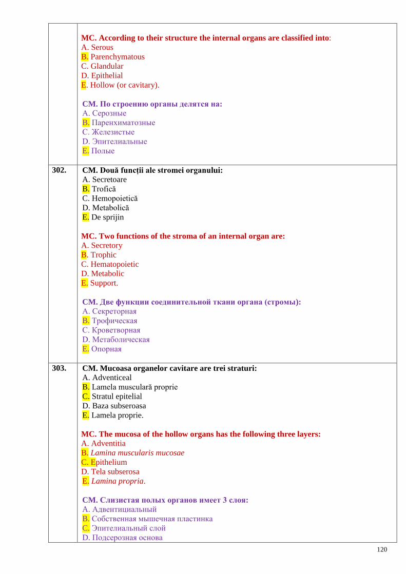
120
MC. According to their structure the internal organs are classified into:
A. Serous
B. Parenchymatous
C. Glandular
D. Epithelial
E. Hollow (or cavitary).
CM. По строению органы делятся на:
A. Серозные
B. Паренхиматозные
C. Железистые
D. Эпителиальные
E. Полые
302. CM. Două funcții ale stromei organului:
A. Secretoare
B. Trofică
C. Hemopoietică
D. Metabolică
E. De sprijin
MC. Two functions of the stroma of an internal organ are: A. Secretory
B. Trophic
C. Hematopoietic
D. Metabolic
E. Support.
CM. Две функции соединительной ткани органа (стромы):
A. Секреторная
B. Трофическая
C. Кроветворная
D. Метаболическая
E. Опорная
303. CM. Mucoasa organelor cavitare are trei straturi:
A. Adventiceal
B. Lamela musculară proprie
C. Stratul epitelial
D. Baza subseroasa
E. Lamela proprie.
MC. The mucosa of the hollow organs has the following three layers: A. Adventitia
B. Lamina muscularis mucosae
C. Epithelium
D. Tela subserosa
E. Lamina propria.
CM. Слизистая полых органов имеет 3 слоя:
A. Адвентициальный
B. Собственная мышечная пластинка
C. Эпителиальный слой
D. Подсерозная основа
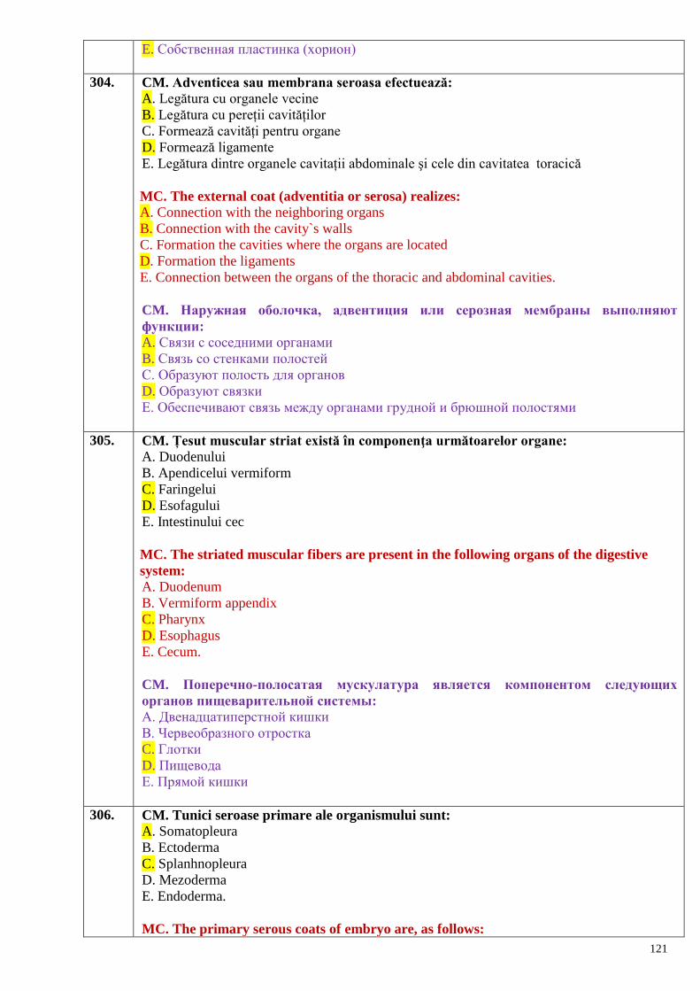
121
E. Собственная пластинка (хорион)
304. CM. Adventicea sau membrana seroasa efectuează:
A. Legătura cu organele vecine
B. Legătura cu pereții cavităților
C. Formează cavități pentru organe
D. Formează ligamente
E. Legătura dintre organele cavitații abdominale şi cele din cavitatea toracică
MC. The external coat (adventitia or serosa) realizes: A. Connection with the neighboring organs
B. Connection with the cavity`s walls
C. Formation the cavities where the organs are located
D. Formation the ligaments
E. Connection between the organs of the thoracic and abdominal cavities.
CM. Наружная оболочка, адвентиция или серозная мембраны выполняют
функции:
A. Связи с соседними органами
B. Связь со стенками полостей
C. Образуют полость для органов
D. Образуют связки
E. Обеспечивают связь между органами грудной и брюшной полостями
305. CM. Țesut muscular striat există în componenţa următoarelor organe:
A. Duodenului
B. Apendicelui vermiform
C. Faringelui
D. Esofagului
E. Intestinului cec
MC. The striated muscular fibers are present in the following organs of the digestive
system: A. Duodenum
B. Vermiform appendix
C. Pharynx
D. Esophagus
E. Cecum.
CM. Поперечно-полосатая мускулатура является компонентом следующих
органов пищеварительной системы:
A. Двенадцатиперстной кишки
B. Червеобразного отростка
C. Глотки
D. Пищевода
E. Прямой кишки
306. CM. Tunici seroase primare ale organismului sunt:
A. Somatopleura
B. Ectoderma
C. Splanhnopleura
D. Mezoderma
E. Endoderma.
MC. The primary serous coats of embryo are, as follows:
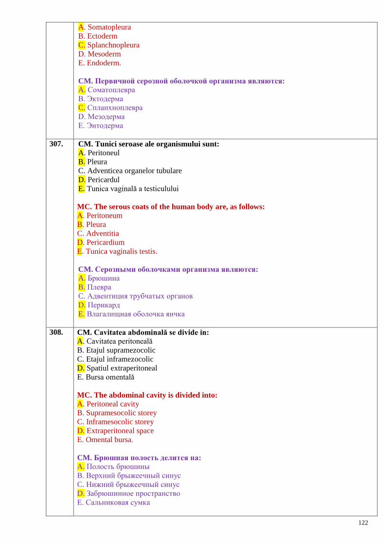
122
A. Somatopleura
B. Ectoderm
C. Splanchnopleura
D. Mesoderm
E. Endoderm.
CM. Первичной серозной оболочкой организма являются:
A. Соматоплевра
B. Эктодерма
C. Спланхноплевра
D. Мезодерма
E. Энтодерма
307. CM. Tunici seroase ale organismului sunt:
A. Peritoneul
B. Pleura
C. Adventicea organelor tubulare
D. Pericardul
E. Tunica vaginală a testiculului
MC. The serous coats of the human body are, as follows: A. Peritoneum
B. Pleura
C. Adventitia
D. Pericardium
E. Tunica vaginalis testis.
CM. Серозными оболочками организма являются:
A. Брюшина
B. Плевра
C. Адвентиция трубчатых органов
D. Перикард
E. Влагалищная оболочка яичка
308. CM. Cavitatea abdominală se divide in:
A. Cavitatea peritoneală
B. Etajul supramezocolic
C. Etajul inframezocolic
D. Spatiul extraperitoneal
E. Bursa omentală
MC. The abdominal cavity is divided into: A. Peritoneal cavity
B. Supramesocolic storey
C. Inframesocolic storey
D. Extraperitoneal space
E. Omental bursa.
CM. Брюшная полость делится на:
A. Полость брюшины
B. Верхний брыжеечный синус
C. Нижний брыжеечный синус
D. Забрюшинное пространство
E. Сальниковая сумка
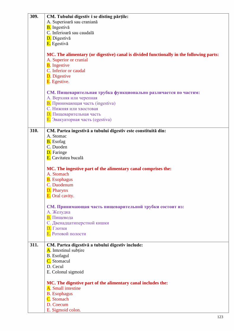
123
309. CM. Tubului digestiv i se disting părțile:
A. Superioară sau craniană
B. Ingestivă
C. Inferioară sau caudală
D. Digestivă
E. Egestivă
MC. The alimentary (or digestive) canal is divided functionally in the following parts: A. Superior or cranial
B. Ingestive
C. Inferior or caudal
D. Digestive
E. Egestive.
CM. Пищеварительная трубка функционально различается по частям:
A. Верхняя или черепная
B. Принимающая часть (ingestiva)
C. Нижняя или хвостовая
D. Пищеварительная часть
E. Эвакуаторная часть (еgestiva)
310. CM. Partea ingestivă a tubului digestiv este constituită din: A. Stomac
B. Esofag
C. Duoden
D. Faringe
E. Cavitatea bucală
MC. The ingestive part of the alimentary canal comprises the: A. Stomach
B. Esophagus
C. Duodenum
D. Pharynx
E. Oral cavity.
CM. Принимающая часть пищеварительной трубки состоит из:
A. Желудка
B. Пищевода
C. Двенадцатиперстной кишки
D. Глотки
E. Ротовой полости
311. CM. Partea digestivă a tubului digestiv include:
A. Intestinul subțire
B. Esofagul
C. Stomacul
D. Cecul
E. Colonul sigmoid
MC. The digestive part of the alimentary canal includes the: A. Small intestine
B. Esophagus
C. Stomach
D. Coecum
E. Sigmoid colon.
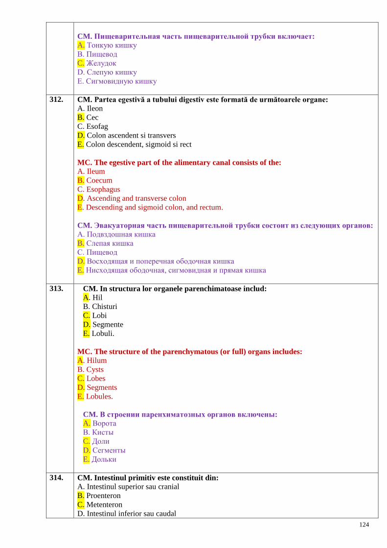
124
CM. Пищеварительная часть пищеварительной трубки включает:
A. Тонкую кишку
B. Пищевод
C. Желудок
D. Слепую кишку
E. Сигмовидную кишку
312. CM. Partea egestivă a tubului digestiv este formată de următoarele organe:
A. Ileon
B. Cec
C. Esofag
D. Colon ascendent si transvers
E. Colon descendent, sigmoid si rect
MC. The egestive part of the alimentary canal consists of the: A. Ileum
B. Coecum
C. Esophagus
D. Ascending and transverse colon
E. Descending and sigmoid colon, and rectum.
CM. Эвакуаторная часть пищеварительной трубки состоит из следующих органов:
A. Подвздошная кишка
B. Слепая кишка
C. Пищевод
D. Восходящая и поперечная ободочная кишка
E. Нисходящая ободочная, сигмовидная и прямая кишка
313. CM. In structura lor organele parenchimatoase includ:
A. Hil
B. Chisturi
C. Lobi
D. Segmente
E. Lobuli.
MC. The structure of the parenchymatous (or full) organs includes: A. Hilum
B. Cysts
C. Lobes
D. Segments
E. Lobules.
CM. В строении паренхиматозных органов включены:
A. Ворота
B. Кисты
C. Доли
D. Сегменты
E. Дольки
314. CM. Intestinul primitiv este constituit din:
A. Intestinul superior sau cranial
B. Proenteron
C. Metenteron
D. Intestinul inferior sau caudal
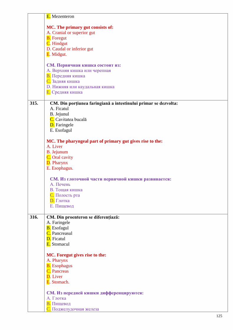
125
E. Mezenteron
MC. The primary gut consists of: A. Cranial or superior gut
B. Foregut
C. Hindgut
D. Caudal or inferior gut
E. Midgut.
CM. Первичная кишка состоит из:
A. Верхняя кишка или черепная
B. Передняя кишка
C. Задняя кишка
D. Нижняя или каудальная кишка
E. Средняя кишка
315. CM. Din porțiunea faringiană a intestinului primar se dezvolta:
A. Ficatul
B. Jejunul
C. Cavitatea bucală
D. Faringele
E. Esofagul
MC. The pharyngeal part of primary gut gives rise to the: A. Liver
B. Jejunum
C. Oral cavity
D. Pharynx
E. Esophagus.
CM. Из глоточной части первичной кишки развивается:
A. Печень
B. Тощая кишка
C. Полость рта
D. Глотка
E. Пищевод
316. CM. Din proenteron se diferențiază:
A. Faringele
B. Esofagul
C. Pancreasul
D. Ficatul
E. Stomacul
MC. Foregut gives rise to the: A. Pharynx
B. Esophagus
C. Pancreas
D. Liver
E. Stomach.
CM. Из передней кишки дифференцируются:
A. Глотка
B. Пищевод
C. Поджелудочная железа
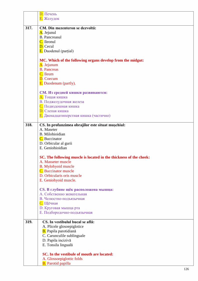
126
D. Печень
E. Желудок
317. CM. Din mezenteron se dezvoltă:
A. Jejunul
B. Pancreasul
C. Ileonul
D. Cecul
E. Duodenul (parțial)
MC. Which of the following organs develop from the midgut: A. Jejunum
B. Pancreas
C. Ileum
D. Coecum
E. Duodenum (partly).
CM. Из средней кишки развиваются:
A. Тощая кишка
B. Поджелудочная железа
C. Подвздошная кишка
D. Слепая кишка
E. Двенадцатиперстная кишка (частично)
318. CS. In profunzimea obrajilor este situat mușchiul:
A. Maseter
B. Milohioidian
C. Buccinator
D. Orbicular al gurii
E. Geniohioidian
SC. The following muscle is located in the thickness of the cheek:
A. Masseter muscle
B. Mylohyoid muscle
C. Buccinator muscle
D. Orbicularis oris muscle
E. Geniohyoid muscle.
CS. В глубине щѐк расположена мышца:
A. Собственно жевательная
B. Челюстно-подъязычная
C. Щѐчная
D. Круговая мышца рта
E. Подбородочно-подъязычная
319. CS. In vestibulul bucal se află:
A. Plicele glosoepiglotice
B. Papila parotidiană
C. Carunculile sublinguale
D. Papila incizivă
E. Tonsila linguală
SC. In the vestibule of mouth are located:
A. Glossoepiglottic folds
B. Parotid papilla
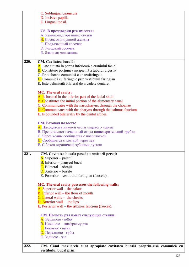
127
C. Sublingual caruncule
D. Incisive papilla
E. Lingual tonsil.
CS. В преддверии рта имеется:
A. Язычнонадгортанные связки
B. Сосок околоушной железы
C. Подъязычный сосочек
D. Резцовый сосочек
E. Язычная миндалина
320. CM. Cavitatea bucală:
A. Este situată în partea inferioară a craniului facial
B. Constituie porțiunea incipientă a tubului digestiv
C. Prin choane comunică cu nazofaringele
D. Comunică cu faringele prin vestibulul faringian
E. Este delimitată bilateral de arcadele dentare.
MC. The oral cavity: A. Is located in the inferior part of the facial skull
B. Constitutes the initial portion of the alimentary canal
C. Communicates with the nasopharynx through the choanae
D. Communicates with the pharynx through the isthmus faucium
E. Is bounded bilaterally by the dental arches.
CM. Ротовая полость:
A. Находится в нижней части лицевого черепа
B. Представляет начальный отдел пищеварительной трубки
C. Через хоаны сообщается с носоглоткой
D. Сообщается с глоткой через зев
E. С боков ограничена зубными дугами
321. CM. Cavitatea bucala poseda următorii pereți:
A. Superior – palatul
B. Inferior – planșeul bucal
C. Bilateral – obrajii
D. Anterior – buzele
E. Posterior – vestibulul faringian (faucele).
MC. The oral cavity possesses the following walls:
A. Superior wall – the palate
B. Inferior wall – the floor of mouth
C. Lateral walls – the cheeks
D. Anterior wall – the lips
E. Posterior wall – the isthmus faucium (fauces).
CM. Полость рта имеет следующие стенки:
A. Верхнюю - нѐбо
B. Нижнюю – диафрагму рта
C. Боковые - щѐки
D. Переднюю - губы
E. Заднюю - зев
322. CM. Când maxilarele sunt apropiate cavitatea bucală propriu-zisă comunică cu
vestibulul bucal prin:
128
A. Vestibulul faringian
B. Spaţiile interdentare
C. Rima oris
D. Spațiul retromolar
E. Nu comunică.
MC. When the jaws are closed the proper oral cavity communicates with the vestibule of
mouth by: A. Isthmus faucium
B. Interdental spaces
C. Rima oris
D. Retromolar space
E. It does not communicate.
CM. Каким образом сообщаются преддверие и собственно полость рта:
A. Через зев
B. Межзубные промежутки
C. Ротовую щель
D. Пространство позади последнего моляра
E. Не сообщаются
323. CM. Intre buze şi formațiunile vecine distingem șanțuri:
A. Nazolabial
B. Geniolabial
C. Palatoglos
D. Palatofaringian
E. Mentolabial.
MC. Between the lips and the neighboring formations the next grooves (or sulci) could be
distinguished: A. Nasolabial sulcus
B. Geniolabial sulcus
C. Palatoglossus sulcus
D. Palatopharyngeus sulcus
E. Mentolabial sulcus.
CM. Между губами и соседними образованиями различают борозды:
A. Носогубная
B. Щѐчно-губная
C. Нѐбноглоточная
D. Нѐбноязычная
E. Подбородочногубная
324. CM. În vestibulul bucal se deschid:
A. Canalele excretoare ale glandelor bucale
B. Canalele excretoare ale glandelor labiale
C. Canalul excretor al glandei parotide
D. Canalele excretoare mici ale glandei sublinguale
E. Canalele excretoare ale glandelor incisive.
MC. In the oral vestibule open: A. Excretory ducts of the buccal glands
B. Excretory ducts of the labial glands
C. Excretory duct of the parotid gland
D. Small excretory ducts of the sublingual glands
129
E. Excretory ducts of the incisive glands.
CM. В преддверие полости рта открываются:
A. Выводные протоки щѐчных желѐз
B. Выводные протоки губных желѐз
C. Выводной проток околоушной железы
D. Мелкие протоки подъязычных желѐз
E. Выводные протоки резцовых желѐз
325. CM. In vestibulul bucal prin inspecție se pot examina:
A. Frenul buzei superioare
B. Frenul buzei inferioare
C. Șanțurile vestibulare superior si inferior
D. Plicele glosoepiglotice
E. Papila parotidiană.
MC. Using the inspection in the oral vestibule could be examined: A. Frenulum of the upper lip
B. Frenulum of the lower lip
C. Superior and inferior vestibular grooves
D. Glossoepiglottic folds
E. Parotid papilla.
CM. В преддверии рта при осмотре можно заметить:
A. Уздечку верхней губы
B. Уздечку нижней губы
C. Верхнюю и нижнюю преддверные борозды
D. Язычно-надгортанные складки
E. Сосочек околоушной железы
326. CM. Cu privire la conformația exterioară a limbii:
A. Constă din vârf, corp şi rădăcină
B. Corpul este separat de rădăcină prin V-ul lingual
C. Gaura oarbă se află posterior de șanțul terminal
D. Șanțul median de pe corpul limbii se prelungește pe rădăcina ei
E. Caruncula sublinguală se află pe dorsul limbii
CM. Regarding the external structure of the heart:
A. May have oval or round shape
B. More often it is conic in shape
C. Shaped as a cut oval
D. Conic in shape, flattened antero-posteriorly
E. Shaped as a cut con.
CM. Наружное строение языка:
A. Состоит из кончика, тела и корня
B. Тело отделено от корня V-образной бороздой
C. Слепое отверстие находится позади терминальной борозды
D. Срединная борозда с тела продолжается на корень
E. Подъязычный сосочек находится на спинке языка
327. CM. Mușchii extrinseci ai limbii sunt:
A. M. longitudinal superior şi inferior
B. M. stiloglos
C. M. hioglos
130
D. M. genioglos
E. M. palatoglos.
MC. The extrinsic muscles of the tongue are, as follows: A. M. longitudinalis superior et inferior
B. M. styloglossus
C. M. hyoglossus
D. M. genioglossus
E. M. palatoglossus.
CM. Наружные мышцы языка:
A. Верхняя продольная и нижняя продольные мышцы
B. Шилоязычная мышца
C. Подъязычноязычная мышца
D. Подбородочноязычная мышца
E. Нѐбноязычная мышца
328. CM. Mușchii intrinseci ai limbii sunt:
A. M. vertical
B. M. transvers
C. M. stilofaringian
D. M. palatofaringian
E. M. longitudinal superior şi inferior.
MC. The intrinsic muscles of the tongue are, as follows: A. M. verticalis
B. M. transversus
C. M. stylopharyngeus
D. M. palatopharyngeus
E. M. longitudinalis superior et inferior.
CM. Внутренние мышцы языка:
A. Вертикальная мышца
B. Поперечная мышца
C. Шилоглоточная мышца
D. Нѐбноглоточная мышца
E. Верхняя и нижняя продольные мышцы
329. CM. La inspectarea feţei inferioare a limbii putem observa:
A. Plicele glosoepiglotice
B. Frâul limbii
C. Plicele sublinguale
D. Carunculele sublinguale
E. Papilele valate.
MC. During the inspection of the inferior surface of the tongue can be observed: A. Glossoepiglottic folds
B. Lingual frenumlum
C. Sublingual folds
D. Sublingual caruncles
E. Vallate papillae.
CM. При исследованиии нижней части лица и языка можем наблюдать:
A. Язычнонадгортанные складки
B. Уздечку языка
131
C. Подъязычные складки
D. Подъязычные сосочки
E. Желобоватые сосочки
330. CM. Care din papilele linguale conțin muguri gustativi:
A. Filiforme
B. Conice
C. Fungiforme
D. Valate
E. Foliate
MC. Which of the lingual papillae contain the taste buds: A. Filiform
B. Conical
C. Fungiform
D. Vallate
E. Foliate.
CM. Какие из язычных сосочков содержат вкусовые почки:
A. Нитевидные
B. Конические
C. Грибовидные
D. Желобоватые
E. Листовидные
331. CS. Masa de fond a dintelui o constituie:
A. Cementul
B. Dentina
C. Smalţul dentar (adamantina)
D. Pulpa dentară
E. Cavitatea dintelui .
SC. Most part of the tooth consists of: A. Cementum
B. Dentin
C. Enamel
D. Dental pulp
E. Tooth cavity.
CS. Основную массу зуба составляют:
A. Цемент
B. Dentin
C. Зубная эмаль
D. Зубная пульпа
E. Полость зуба
332. CS. La copiii de 2,5 ani lipsesc:
A. Dinții incizivi
B. Dinții canini
C. Dinții molari
D. Dinții premolari
E. Toți sunt prezenți
SC. At 2,5 year-old children the following teeth are absent: A. Incisors
132
B. Canines
C. Molars
D. Premolars
E. All are present.
CS. У ребѐнка 2,5 лет отсутствуют:
A. Резцы
B. Клыки
C. Большие коренные зубы
D. Малые коренные зубы
E. Все присутствуют
333. CS. Care din afirmațiile referitoare la eruperea dinților deciduali este corectă:
A. Primii erup incizivii superiori mediali
B. Primii erup incizivii inferiori mediali
C. Primii erup caninii superiori
D. Primii erup molarii inferiori
E. Primii erup molarii superiori.
SC. Which affirmation regarding the deciduous teeth eruption is correct: A. The first teeth to erupt are the medial (or central) superior incisors
B. The first teeth to erupt are the medial (or central) inferior incisors
C. The first teeth to erupt are the superior canines
D. The first teeth to erupt are the inferior molars
E. The first teeth to erupt are the superior molars.
CS. Какие из перечисленных утверждений относительно прорезывания зубов
являются правильными:
A. Первыми прорезываются верхние медиальные резцы
B. Первыми прорезываются нижние медиальные резцы
C. Первыми прорезываются верхние клыки
D. Первыми прорезываются нижние моляры
E. Первыми прорезываются верхние моляры
334. CS. Una din afirmaţiile ce urmează nu se referă la glanda sublingvală:
A. Elaborează un secret de tip mucos
B. Este situată pe faţa superioară a mușchiului milohioidian
C. Elimină secret de tip seros
D. Ductul glandei se deschide în papila sublingvală
E. Proeminarea ei formează plica sublingvală.
SC. One of the following affirmation is not related to the sublingual gland: A. It elaborates the mucous secret
B. It is located on the superior surface of the mylohyoid muscle
C. It produces the serous secret
C. Its duct opens at the sublingual papilla
E. Its prominence forms the sublingual fold.
CS. Одно из перечисленных ниже утверждений не относится к подъязычной
железе:
A. Выделяет секрет слизистого типа
B. Располагается на верхней поверхности челюстноподъязычной мышцы
C. Выделяет секрет серозного типа
D. Выводной проток железы открывается на подъязычном сосочке
E. Возвышение от железы формирует подъязычную складку
133
335. CM. Coroana dentară are următoarele feţe:
A. Linguală
B. Palatină
C. Vestibulară
D. Ocluzală
E. De contact.
MC. The tooth crown has the following surfaces: A. Lingual
B. Palatine
C. Vestibular
D. Occlusal
E. Contact.
CM. Зубная коронка имеет следующие поверхности:
A. Язычная
B. Нѐбная
C. Вестибулярная
D. Смыкания (жевательная)
E. Контактная (апроксимальная)
336. CM. Se disting următoarele forme de dinți:
A. Deciduali
B. Permanenți
C. Incizivi
D. Molari
E. Premolari.
MC. According to the shape the following types of teeth can be distinguished: A. Deciduous teeth
B. Permanent teeth
C. Incisors
D. Molars
E. Premolars.
CM. Различаются следующие формы зубов:
A. Молочные
B. Постоянные
C. Резцы
D. Большие коренные зубы
E. Малые коренные зубы
337. CM. Cu referinţă la glandele cavității bucale:
A. Se împart in glande salivare mari si mici
B. Cele mici sunt situate in profunzimea mucoasei sau în baza submucoasă
C. Glandele mici sunt lipsite de ducturi şi elimină secretul în sânge
D. Cele mai numeroase sunt glandele labiale si palatine
E. Elimină secret seros, mucos şi mixt
MC. Regarding the salivary glands: A. They are divided into the major and minor salivary glands
B. The minor salivary glands are embedded in the thickness of the mucosa or submucosa
C. The minor salivary glands have no ducts and discharge the secret into blood
D. The most numerous are the palatine and labial glands
134
E. They produce the serous, mucous and mixed secret.
CM. Слюнные железы полости рта:
A. Делятся на большие и малые
B. Малые железы расположены в глубине слизистой или подслизистой основы
C. Малые железы не имеют протоков и выделяют секрет в кровь
D. Самые многочисленные – это губные и нѐбные железы
E. Выделяют серозный, слизистый и смешанный секрет
338. CM. Care din caracterele următoare au referinţă la glandele salivare mari:
A. Sunt pare
B. Sunt situate în cavitatea bucală
C. Poseda ducturi ce se deschid în cavitatea bucală
D. După secretul eliminat sunt: seroase, mucoase şi mixte
E. Din ele fac parte glandele labiale, palatine şi linguale.
MC. Which of the following characteristics are related to the major salivary glands: A. They are paired
B. They are located in the oral cavity
C. They possess ducts that open in the oral cavity
D. According to the type of secretion they are serous, mucous and mixed
E. Among them there are labial, palatine and lingual glands.
CM. Крупные слюнные железы полости рта:
A. Парные
B. Расположены в полости рта
C. Их протоки открываются в полости рта
D. Выделяют серозный, слизистый и смешанный секрет
E. К ним относятся губные, нѐбные и язычные железы
339. CM. Pentru glanda parotidă sunt caracteristice 3 din criteriile enumerate mai jos:
A. Are o formă regulată şi este de tip mucos
B. Este amplasată in fosa submandibulară
C. Are consistenţă moale şi structură lobulară
D. Ductul glandei (Stenon) se deschide in vestibulul bucal
E. La exterior este acoperită cu o capsula de țesut conjunctiv.
MC. Three of the next criteria are characteristic for the parotid gland: A. It has a regular shape and it is a mucous gland
B. It is located in the submandibular fossa
C. It has a soft consistency and a lobular structure
D. Its duct (of Stenon) opens in the oral vestibule
E. Externally it is covered by the connective tissue capsule.
CM. Околоушную железу характеризуют три нижеперечисленных критерия:
A. Имеет правильную форму, слизистого типа
B. Расположена в поднижнечелюстной ямке
C. Имеет мягкую консистенцию и дольчатое строение
D. Еѐ проток (Стенонов) открывается в преддверие полости рта
E. Снаружи покрыта капсулой из соединительной ткани
340. CM. Mușchii faringelui:
A. Sunt alcătuiți din fibre musculare striate
B. Conțin fibre musculare striate si netede reprezentate uniform
C. Sunt in număr de 5, dintre care 3 sunt circulari si 2 longitudinali
135
D. Mușchii constrictori formează pe linia mediană posterioară a faringelui raphe pharyngis
E. Mușchii longitudinali ai faringelui sunt numiți si ridicători.
MC. The pharyngeal muscles: A. They consist of the striated muscular fibers
B. They contain the uniformly arranged striated and smooth muscles
C. There are five muscles of the pharynx, three of them are circular muscles and the other two
are longitudinal muscles
D. The constrictor muscles form along the posterior median line of pharynx the pharyngeal
raphe (raphe pharyngis)
E. The longitudinal muscles of the pharynx are named also the levator muscles.
CM. Мышцы глотки:
A. Представлены поперечно-полосатой мускулатурой
B. Стенка глотки состоит из поперечно-полосатой мускулатуры гладкомышечной,
распределѐнной равномерно
C. Их может быть до 5-ти, а между ними 3 круговых и 2 продольных
D. Мышцы-констрикторы образуют по задней средней линии глотки шов глотки
(raphe pharyngis)
E. Продольные мышцы глотки называются поднимателями
341. CS. Indicați afirmațiile corecte referitoare la esofag:
A. Are porțiunea cervicală, toracică şi abdominală
B. Tunica musculară a esofagului conține fibre musculare striate şi netede repartizate uniform
C. Musculatura striată este situată numai în partea inferioară a esofagului
D. Musculatura netedă este numai în partea superioară
E. Toate afirmațiile sunt corecte
SC. Choose the true statements regarding the esophagus: A. It has cervical, thoracic and abdominal portions
B. Its muscular coat (or tunic) contains the uniformly arranged striated and smooth muscular
fibers
C. The striated musculature is located only in the lower part of the esophagus
D. The smooth musculature is located only in the upper part of the esophagus
E. All statements are correct.
CS. Укажите правильные утверждения относительно пищевода:
A. Имеет следующие части: шейную, грудную и брюшную
B. В мышечной оболочке равномерно распределены гладкие и поперечно-полосатые
мышечные волокна
C. Поперечно-полосатая мускулатура расположена в нижней части пищевода
D. Гладкая мускулатура есть только в верхней части пищевода
E. Все утверждения правильные
342. CS. Care din straturile enumerate nu este caracteristic porţiunii cervicale a esofagului:
A. Tunica mucoasă
B. Baza submucoasă
C. Tunica musculară
D. Tunica seroasă
E. Adventicea
SC. Which of the coats listed below is not characteristic of the cervical portion of the
esophagus:
A. Mucosa
B. Submucosa
136
C. Muscular coat
D. Serous coat
E. Adventitia.
CS. Какие из перечисленных слоѐв не характерны для шейной части пищевода:
A. Слизистая оболочка
B. Подслизистая основа
C. Мышечная оболочка
D. Серозная оболочка
E. Соединительнотканная оболочка
343. CM. Endofaringele comunică cu:
A. Urechea inrernă
B. Traheea.
C. Cavitatea nazală.
D. Cavitatea bucală.
E. Cavitatea timpanică.
MC. Endopharynx (or cavity of pharynx) communicates with:
A. Auditory tube.
B. Trachea.
C. Nasal cavity.
D. Oral cavity.
E. Tympanic cavity.
CM. Полость глотки сообщается:
A. Посредством слуховой трубы
B. С трахеей
C. С полостью носа
D. С ротовой полостью
E. С барабанной полостью
344. CM. Selectați si notați afirmațiile corecte, referitoare la faringe:
A. Începe la baza craniului
B. La nivelul vertebrelor C6 - C7 trece in esofag
C. Trece in esofag la nivelul vertebrelor C6 - Th1
D. Lateral de el sunt situate fasciculele vasculo-nervoase ale gâtului
E. Faţa posterioară a faringelui aderă nemijlocit la corpurile vertebrelor.
MC. Select and mark the correct affirmations regarding the pharynx: A. It starts from the base of skull.
B. At the level of the CVI - CVII vertebrae it continuous with the esophagus.
C. At the level of the CVI – TI vertebrae it continuous with the esophagus.
D. Laterally of it the neurovascular bundle of the neck is located
E. Its posterior wall adheres directly to the vertebral bodies.
CM. Выберите правильные утверждения относительно глотки:
A. Начинается на основании черепа
B. На уровне 6-го и 7-го шейных позвонков переходит в пищевод
C. Переходит в пищевод на уровне 6-го шейного позвонка
D. Латерально расположен сосудисто-нервный пучок шеи
E. Задняя поверхность глотки непосредственно прилежит к телам позвонков
345. CM. Faringele comunică cu:
A. Esofagul
137
B. Traheea
C. Cavitatea nazală
D. Cavitatea bucală
E. Cavitatea timpanică.
MC. Pharynx communicates with: A. Oesophagus
B. Trachea
C. Nasal cavity
D. Oral cavity
E. Tympanic cavity.
CM. Глотка сообщается с:
A. Пищеводом
B. Трахеей
C. Полостью носа
D. Полостью рта
E. Барабанной полостью
346. CM. Îngustările anatomice ale esofagului sunt localizate:
A. La trecerea faringelui in esofag
B. La intersecția esofagului cu arcul aortei
C. La intersecția cu bronhia stângă
D. La trecerea prin diafragmă
E. La nivelul trecerii in stomac
MC. The anatomical constrictions of the esophagus are located:
A. Where the pharynx continues with the esophagus
B. Where the esophagus is crossed by the aortic arch
C. Where the esophagus is crossed by the left main bronchus
D. Where the esophagus passes through the diaphragm
E. Where the esophagus continues with the stomach.
CM. Анатомические сужения пищевода расположены:
A. В месте перехода глотки в пищевод
B. В месте пересечения пищевода с дугой аорты
C. В месте перекреста с левым бронхом
D. При прохождении диафрагмы
E. На уровне впадения в желудок
347. CM. Indicați îngustările fiziologice ale esofagului:
A. La trecerea faringelui in esofag
B. La intersecția esofagului cu arcul aortei
C. La intersecția cu bronhia principală stângă
D. La trecerea prin diafragmă
E. La nivelul trecerii în stomac
MC. Where are the physiological constrictions of the esophagus located:
A. Where the pharynx continues with the esophagus
B. Where the esophagus is crossed by the aortic arch
C. Where the esophagus is crossed by the left main bronchus
D. Where the esophagus passes through the diaphragm
E. Where the esophagus continues with the stomach.
CM. Укажите физиологические сужения пищевода:
138
A. В месте перехода глотки в пищевод
B. В месте перекрѐста пищевода и дуги аорты
C. В месте перекрѐста с главным левым бронхом
D. При прохождении через диафрагму
E. На уровне перехода в желудок
348. CM. Inelul limfoepitelial Pirogov-Waldeyer include amigdalele:
A. Palatine
B. Lingvală
C. Tubare
D. Laringiană
E. Faringiană.
MC. The lymphoepithelial (or pharyngeal) ring (of Pirogov-Waldeyer) includes the next
tonsils: A. Palatine
B. Lingval
C. Tube
D. Laryngeal
E. Pharyngeal.
CM. Лимфоэпителиальное кольцо Пирогова-Вальдейера включает миндалины
A. Нѐбную
B. Язычную
C. Трубную
D. Гортанную
E. Глоточную
349. CS. In raport cu peritoneul stomacul are o poziție:
A. Intraperitoneală
B. Extraperitoneală
C. Mezoperitoneală
D. Retroperitoneală
E. Nu are nici un fel de raporturi cu peritoneul
SC. The position of the stomach in relation to peritoneum is:
A. Intraperitoneal
B. Extraperitoneal
C. Mesoperitoneal
D. Retroperitoneal
E. It has no relations tothe peritoneum.
CS. По отношению к брюшине положение желудка:
A. Интраперитонеальное
B. Экстраперитонеальное
C. Мезоперитонеальное
D. Ретроперитонеальное
E. Ни одна сторона не покрыта брюшиной
350. CS. Stomacul este legat de organele vecine prin ligamentele enumerate, cu excepția celui:
A. Hepatogastric
B. Gastroduodenal
C. Gastrocolic
D. Gastrolienal
E. Gastrofrenic.
139
SC. The stomach is connected to the neighboring organs by the next ligaments, except: A. Hepatogastric ligament
B. Gastroduodenal ligament
C. Gastrocolic ligament
D. Gastrolienal ligament
E. Gastrophrenic ligament.
CS. Желудок связан с соседними органами перечисленными связками, кроме:
A. Печѐночно-желудочной
B. Желудочно-двенадцатиперстной
C. Желудочно-ободочной
D. Желудочно-селезѐночной
E. Желудочно-диафрагмальной
351. CS. La stomac se disting următoarele părți:
A. Curbura mică
B. Curbura mare
C. Cardiacă
D. Ostiul piloric
E. Ostiul cardiac
SC. The stomach has the following parts:
A. Lesser curvature
B. Grater curvature
C. Cardiac part
D. Pyloric orifice (or ostium)
E. Cardiac orifice (or ostium).
CS. В желудке различаются следующие части:
A. Малая кривизна
B. Большая кривизна
C. Кардиальная часть
D. Пилорическое отверстие
E. Кардиальное отверстие
352. CM. Amplasarea spațială a unui organ este caracterizată prin:
A. Sintopie
B. Stereotopie
C. Scheletotopie
D. Ortotopie
E. Holotopie
MC. The topography of an organ includes:
A. Syntopy
B. Stereotopy
C. Skeletotopy
D. Orthotopy
E. Holotopy.
CM. Пространственное расположение органа характеризуется через:
A. Синтопию
B. Стереотопию
C. Скелетотопию
D. Ортотопию
140
E. Голотопию
353. CM. Dimensiunile stomacului oricărui subiect sunt determinate de:
A. Vârstă
B. Gen
C. Deprinderile alimentare
D. Tipul constituțional
E. Factori ereditari
MC. The dimensions of the stomach are determined by:
A. Age
B. Gender
C. Alimentary behavior
D. Constitutional type
E. Hereditary factors.
CM. Размеры желудка каждого человека обусловлены:
A. Возрастом
B. Полом
C. Привычками приѐма пищи
D. Типом телосложения
E. Наследственными факторами
354. CM. Stomacul este în adiacenţă cu:
A. Colonul transvers
B. Splina
C. Diafragma
D. Rinichiul si suprarenala din dreapta
E. Lobul stâng al ficatului
MC. The stomach comes in contact with the:
A. Transverse colon
B. Spleen
C. Diaphragm
D. Right kidney and right adrenal gland
E. Left lobe of liver.
CM. Желудок соприкасается с:
A. Поперечной ободочной кишкой
B. Селезѐнкой
C. Диафрагмой
D. Правой почкой и правым надпочечником
E. Левой долей печени
355. CM. Pentru stomac sunt caracteristice următoarele părți:
A. Cardiacă
B. Pilorică
C. Corp
D. Fundul
E. Duodenală
MC. The stomach has the following parts:
A. Cardiac part
B. Pyloric part
C. Body
141
D. Fundus or fornix
E. Duodenal part.
CM. Для желудка характерны следующие части:
A. Кардиальная
B. Пилорическая
C. Тело
D. Дно
E. Двенадцатиперстная
356. CM. Peretele anterior al stomacului contactează cu:
A. Lobul stâng al ficatului
B. Diafragma
C. Colonul transvers
D. Lobul caudat al ficatului
E. Peretele abdominal anterior
MC. The anterior wall of stomach comes in contact with the:
A. Left lobe of liver
B. Diaphragm
C. Transverse colon
D. Caudate lobe of liver
E. Anterior abdominal wall.
CM. Передняя стенка желудка соприкасается с:
A. Левой долей печени
B. Диафрагмой
C. Поперечной ободочной кишкой
D. Хвостатой долей печени
E. Передней брюшной стенкой
357. CM. In peretele stomacului se disting următoarele straturi:
A. Adventiceal
B. Seros
C. Subseros
D. Muscular
E. Mucos
MC. The gastric wall consists of the following coats (or tunics):
A. Adventitia
B. Serous coat (or serosa)
C. Tela subserosa
D. Muscular coat
E. Mucosa.
CM. В стенке желудка различают следующие слои:
A. Соединительнотканный
B. Серозный
C. Субсерозный
D. Мышечный
E. Слизистый
358. CM. Care din termeni se utilizează la descrierea radioanatomică a stomacului
A. Sacul digestor
B. Corpul
142
C. Fundul
D. Canalul egestor
E. Toți termenii enumerați
CM. Which of the following terms is used to describe the X-ray examination of the
stomach:
A. Digestive sac
B. Sinusul
C. Fundus
D. Evacuation canal
E. All statements are true.
CM. Какие термины используются при описании радиоанатомии желудка:
A. Пищеварительный мешок
B. Тело
C. Дно
D. Эвакуаторный канал
E. Все перечисленные термины
Intestinul subţire şi gros –
structură, părţi componente, topografie,
particularităţi distinctive, rol funcţional
359. CS. Selectați afirmațiile corecte referitoare la duoden:
A. Este situat intraperitoneal
B. În porțiunea terminală are o dilatare numită bulbul duodenului
C. Porțiunea descendentă este in raport cu rinichiul stâng
D. Partea superioară este aderentă la stomac
E. În el se deschide ampula hepato-pancreatică
SC. Choose the correct statements regarding the duodenum:
A. It is located intraperitoneally
B. Its terminal portion forms a dilatation called the duodenal bulb
C. The descending portion comes in contact with the left kidney
D. The superior portion adheres to the stomach
E. The hepatopancreatic ampulla (of Vater) empties into the lumen of the duodenum.
CS. Выберите правильные утверждения относительно двенадцатиперстной кишки:
A. Расположена интраперитонеально
B. Конечный отдел имеет расширение, называемое луковицей
C. Нисходящая часть покрыта левой почкой
D. Верхняя часть прилежит к желудку
E. В неѐ открывается печѐночно-панкреатическая ампула
360. CS. Pentru duoden sunt caracteristice toate afirmațiile cu excepția:
A. Nu are mezou si este situat retroperitoneal
B. Trece in jejun la nivelul vertebrei L3 din dreapta
C. În porțiunea descendentă se deschide ampula hepato-pancreatică
D. Papila lui mare se află la partea inferioară a pliului longitudinal
E. Superior de papila duodenală mare este situată papila duodenală mică
SC. All the following statements are characteristic of the duodenum, except: A. It has no mesentery and it is located retroperitoneally
B. It continues with the jejunum at the level of the LIII vertebra (its right side)
C. The hepatopancreatic ampulla empties into its descending portion
D. The major duodenal papilla is located on the inferior part of the longitudinal fold
143
E. The minor duodenal papilla is located above the major one.
CS. Для двенадцатиперстной кишки все характеристики верны, за исключением:
A. Не имеет брыжейки и расположена ретроперитонеально
B. Переходит в тощую кишку на уровне 3-го поясничного позвонка
C. В нисходящий отдел открывается печѐночно-панкреатическая ампула
D. Еѐ большой сосок находится в нижней части продольной складки
E. Выше большого дуоденального соска находится малый сосок
361. CS. Care din formele indicate ale duodenului este mai frecvent întâlnită:
A. Inelară
B. De ansa dispusă vertical
C. De ansa dispusă orizontal
D. De potcoavă
E. Duoden mobil.
SC. Which of the indicated shapes of duodenum is more frequently met:
A. Shaped as a ring (or O-shaped)
B. Shaped as a vertical loop (or U-shaped)
C. Shaped as a horizontal loop
D. Horseshoe-shaped duodenum (or C-shaped)
E. Mobile duodenum.
CS. Какие из указанных форм двенадцатиперстной кишки характерны для
пожилых:
A. Кольцеобразная
B. В виде вертикально направленной петли
C. В виде горизонтально направленной петли
D. Подковы
E. Подвижная двенадцатиперстная кишка
362. CS. Prin care duct bila se scurge in duoden:
A. Hepatic comun
B. Cistic
C. Ducturile segmentare
D. Coledoc
E. Ducturile interlobulare
SC. Through which duct does bile flow into the duodenum:
A. Common hepatic duct
B. Cystic duct
C. Segmental ducts
D. Common bile duct
E. Interlobular ducts.
CS. Какой жѐлчный проток открывается в двенадцатиперстную кишку:
A. Общий жѐлчный
B. Проток жѐлчного пузыря
C. Сегментарные протоки
D. Общий жѐлчевыводящий проток
E. Междольковые протоки
363. CS. Selectați afirmația corectă referitoare la jejun - ileon:
A. Ileonul are o culoare roz-pronunțată
B. Ansele jejunului sunt situate superior in stânga
144
C. Peretele ileonului este mai gros
D. Jejunul are un diametru mai mic
E. Jejunul este de culoare gri-roz.
SC. Select the correct affirmation regarding the jejunum - ileum: A. The ileum is bright pink colored
B. The loops of jejunum are located upward and to the left
C. The wall of ileum is thicker
D. The jejunum has a smaller diameter
E. The jejunum is pinkish-grey colored.
CS. Выберите верные утверждения относительно тощей и подвздошной кишок:
A. Подвздошня кишка имеет ярко выраженный розовый цвет
B. Петли тощей кишки расположены выше и слева
C. Стенки подвздошной кишки более толстые
D. Тощая кишка имеет меньший диаметр
E. Тощая кишка имеет серо-розовый цвет
364. CM. La intestinul subțire se disting următoarele porțiuni:
A. Duodenul
B. Colonul
C. Jejunul
D. Ileonul
E. Cecul.
MC. The small intestine has the following portions: A. Duodenum
B. Colon
C. Jejunum
D. Ileum
E. Coecum.
CM. В тонкой кишке различают следующие части:
A. Двенадцатиперстная кишка
B. Ободочная кишка
C. Тощая кишка
D. Подвздошная кишка
E. Слепая кишка
365. CM. Duodenul:
A. Reprezintă prima porțiune a intestinului subțire
B. Are o lungime de aproximativ 25 cm
C. Cuprinde ca într-o potcoavă capul pancreasului
D. Constă din trei segmente
E. În fiecare segment se află papile duodenale
MC. Duodenum:
A. Is the first portion of the small intestine
B. Its length is about 25 cm
C. Is horseshoe-shaped and it surrounds the head of the pancreas
D. Consists of three parts
E. Each segment of the duodenum has the duodenal papillae.
CM. Двенадцатиперстная кишка:
A. Представляет первую часть тонкой кишки
145
B. Имеет длину примерно 25 см
C. Окружает в виде подковы головку поджелудочной железы
D. Состоит из 3-х отделов
E. В каждом отделе находятся дуоденальные сосочки
366. CM. Spre deosebire de tunica mucoasă a ileonului tunica mucoasă a jejunului:
A. Formează plice circulare mai înalte si situate mai aproape una de alta
B. E de culoare roz
C. Conține foliculi limfatici agregaţi (Peyer)
D. Nu conține foliculi limfatici solitari
E. Este mai groasă
MC. The mucosa of jejunum differs from the mucosa of ileum, as follows:
A. The mucosa of jejunum forms the higher and more numerous circular folds
B. It is a pink color
C. It contains the aggregate lymphatic follicles (Peyer`s patches)
D. It does not contain solitary lymphatic follicles (or nodules)
E. It is thicker than the mucosa of the ileum.
CM. Особенности слизистой оболочки подвздошной кишки в отличии от тощей:
A. Образует более высокие циркулярные складки, расположенные близко друг к другу
B. Имеет розовый цвет
C. Содержит групповые лимфоидные узелки (Пейровы бляшки)
D. Не содержит одиночных лимфоидных узелков
E. Толще
367. CM. Jejun-ileonul:
A. Reprezintă porțiunea mezenterială a intestinului subțire
B. Nu este separat de duoden printr-o zona de limitare precisă
C. Are o lungime medie de 5 - 6 m
D. Formează mai multe anse (14 - 16)
E. Începe la flexura duodeno-jejunală si se termină in unghiul ileo-cecal
MC. The jejuno-ileum:
A. Is the mesenteric part of small intestine
B. Is not separated from the duodenum by a clear delimitation
C. Is about 5 - 6 m in length
D. Forms several loops (14 - 16)
E. Starts at the duodenojejunal flexure and ends at the ileocecal angle.
CM. Тощая и подвздошная кишка:
A. Представляет брыжеечную часть тонкой кишки
B. Не отделена от двенадцатиперстной кишки выраженной пограничной зоной
C. Средняя длина 5 – 6 м
D. Образует множество петель
E. Начинается от двенадцатиперстнотощего изгиба и заканчивается в илеоцекальном
углу
368. CM. Care din formațiunile mucoasei intestinului asigură funcție imună:
A. Celulele endocrine
B. Celulele glandulare
C. Celulele caliciforme
D. Foliculii limfoizi agregaţi (Peyer)
E. Foliculii limfoizi solitari
146
MC. Which of the following elements of intestinal mucosa assure the immune function:
A. Endocrine cells
B. Glandular cells
C. Goblet cells
D. Aggregate lymphoid follicles (Peyer`s patches)
E. Solitary lymphoid follicles.
CM. Какие образования слизистой кишки обеспечивают иммунную функцию:
A. Эндокринные клетки
B. Железистые клетки
C. Чашеобразные клетки
D. Скопления лимфоидных узелков (Пейровы бляшки)
E. Одиночные лимфоидные узелки
369. CS. Alegeți varianta in care se succed segmentele intestinului gros:
A. Apendicele vermiform, cecul, colonul ascendent, colonul descendent, colonul transvers,
colonul sigmoid, rectul
B. Cecul, colonul ascendent, colonul transvers, colonul descendent, colonul sigmoid, rectul
C. Colonul ascendent, colonul descendent, colonul transvers, colonul sigmoid, rectul
D. Colonul descendent, colonul transvers, colonul ascendent, colonul sigmoid, rectul
E. Colonul sigmoid, colonul ascendent, colonul transvers, colonul descendent, rectul
SC. Choose the variant which shows the consecutive parts of the large intestine:
A. Vermiform appendices, coecum, ascending colon, descending colon, transverse colon,
sigmoid colon, rectum
B. Coecum, ascending colon, transverse colon, descending colon, sigmoid colon, rectum
C. Ascending colon, descending colon, transverse colon, sigmoid colon, rectum
D. Descending colon, transverse colon, ascending colon, sigmoid colon, rectum
E. Sigmoid colon, ascending colon, transverse colon, descending colon, rectum.
CS. Выберите варианты следования друг за другом отделов толстой кишки:
A. Червеобразный отросток, слепая кишка, восходящая ободочная кишка, нисходящая
ободочная кишка, поперечная ободочная кишка, сигмовидная кишка, прямая кишка
B. Слепая кишка, восходящая ободочная кишка, поперечная ободочная кишка,
нисходящая ободочная кишка, сигмовидная кишка и прямая
C. Восходящая ободочная кишка, нисходящая ободочная кишка, поперечная ободочная
кишка, сигмовидная кишка и прямая
D. Нисходящая ободочная кишка, поперечная ободочная кишка
E. Сигмовидная кишка, восходящая ободочная, поперечная ободочная и прямая
370. CS. Apendicele vermiform – afirmații incorecte:
A. Pornește de la cec
B. Are o lungime medie de 8,6 cm
C. Din exterior este acoperit de peritoneu
D. Nu posedă mezou
E. Poate avea diferite variante de pozițieю
SC. The vermiform appendix, mark the incorrect affirmations: A. It starts from the coecum
B. It has 8,6 cm in length
C. It is covered by peritoneum
D. It has no mesentery
E. It could possess different variants of position.
CS. Червеобразный отросток; неверные утверждения:
147
A. Направляется из слепой кишки
B. Имеет среднюю длину 8,6 см
C. Снаружи покрыт брюшиной
D. Не имеет брыжейки
E. Может иметь различные варианты положения
371. CS. Alegeți varianta corectă de răspuns:
A. Cecul - mezoperitoneal
B. Colonul ascendent - intraperitoneal
C. Colonul descendent - mezoperitoneal
D. Colonul sigmoid - extraperitoneal
E. Rectul in integritate - extraperitoneal
SC. Choose the correct affirmation:
A. Coecum – mesoperitoneal position
B. Ascending colon – intraperitoneal position
C. Descending colon – mesoperitoneal position
D. Sigmoid colon – extraperitoneal position
E. Entire rectum - extraperitoneal position.
CS. Выберите правильные варианты ответов:
A. Слепая кишка покрыта брюшиной мезоперитонеально
B. Восходящая ободочная кишка занимает интраперитонеальное положение
C. Нисходящая ободочная кишка занимает мезоперитонеальное положение
D. Сигмовидная кишка – экстраперитонеальный орган
E. Все отделы прямой кишки занимают экстраперитонеальное положение
372. CM. Intestinul gros se deosebește de cel subțire prin:
A. Lumenul mai larg
B. Lungimea mai mică
C. Prezenţa la exterior a teniilor, a haustrelor şi a apendicelor epiploice
D. Existenţa unor segmente fixate
E. Prezenţa plicelor circulare
MC. The large intestine differs from the small one, as follows:
A. Its lumen is larger
B. Its length is shorter
C. It has the teniae coli, haustrae coli and epiploic appendices
D. It has fixed segments
E. Its mucosa forms the circular folds.
CM. Толстая кишка отличается от тонкой:
A. Имеет больший просвет
B. Имеет меньшую длину
C. Снаружи представлены ленты, вздутия и жировые отростки
D. Имеет один фиксированный отдел
E. Имеет круговые складки
373. CM. Intestinul gros:
A. Are o lungime de 1,5 - 2 m
B. Diametrul lui transversal măsoară 5 - 8 cm
C. Conține plice semilunare
D. Mucoasa lui este prevăzută cu vili intestinali
E. Nu posedă pătură submucoasă
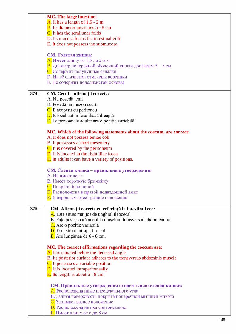
148
MC. The large intestine:
A. It has a length of 1,5 - 2 m
B. Its diameter measures 5 - 8 cm
C. It has the semilunar folds
D. Its mucosa forms the intestinal villi
E. It does not possess the submucosa.
CM. Толстая кишка:
A. Имеет длину от 1,5 до 2-х м
B. Диаметр поперечной ободочной кишки достигает 5 – 8 см
C. Содержит полулунные складки
D. На еѐ слизистой отмечены ворсинки
E. Не содержит подслизистой основы
374. CM. Cecul – afirmații corecte:
A. Nu posedă tenii
B. Posedă un mezou scurt
C. E acoperit cu peritoneu
D. E localizat in fosa iliacă dreaptă
E. La persoanele adulte are o poziție variabilă
MC. Which of the following statements about the coecum, are correct:
A. It does not possess teniae coli
B. It possesses a short mesentery
C. It is covered by the peritoneum
D. It is located in the right iliac fossa
E. In adults it can have a variety of positions.
CM. Слепая кишка – правильные утверждения:
A. Не имеет лент
B. Имеет короткую брыжейку
C. Покрыта брюшиной
D. Расположена в правой подвздошной ямке
E. У взрослых имеет разное положение
375. CM. Afirmații corecte cu referință la intestinul cec:
A. Este situat mai jos de unghiul ileocecal
B. Faţa posterioară aderă la mușchiul transvers al abdomenului
C. Are o poziție variabilă
D. Este situat intraperitoneal
E. Are lungimea de 6 - 8 cm.
MC. The correct affirmations regarding the coecum are: A. It is situated below the ileocecal angle
B. Its posterior surface adheres to the transversus abdominis muscle
C. It possesses a variable position
D. It is located intraperitoneally
E. Its length is about 6 - 8 cm.
CM. Правильные утверждения относительно слепой кишки:
A. Расположена ниже илеоцекального угла
B. Задняя поверхность покрыта поперечной мышцей живота
C. Занимает разное положение
D. Расположена интраперитонеально
E. Имеет длину от 6 до 8 см
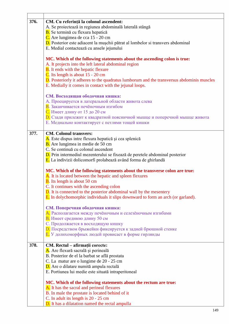
149
376. CM. Cu referință la colonul ascendent:
A. Se proiectează in regiunea abdominală laterală stângă
B. Se termină cu flexura hepatică
C. Are lungimea de cca 15 - 20 cm
D. Posterior este adiacent la mușchii pătrat al lombelor si transvers abdominal
E. Medial contactează cu ansele jejunului
MC. Which of the following statements about the ascending colon is true:
A. It projects into the left lateral abdominal region
B. It ends with the hepatic flexure
C. Its length is about 15 - 20 cm
D. Posteriorly it adheres to the quadratus lumborum and the transversus abdominis muscles
E. Medially it comes in contact with the jejunal loops.
CM. Восходящая ободочная кишка:
A. Проецируется в латеральной области живота слева
B. Заканчивается печѐночным изгибом
C. Имеет длину от 15 до 20 см
D. Сзади прилежит к квадратной поясничной мышце и поперечной мышце живота
E. Медиально контактирует с петлями тощей кишки
377. CM. Colonul transvers:
A. Este dispus intre flexura hepatică şi cea splenică
B. Are lungimea in medie de 50 cm
C. Se continuă cu colonul ascendent
D. Prin intermediul mezenterului se fixează de peretele abdominal posterior
E. La indivizii dolicomorfi prolabează având forma de ghirlandă
MC. Which of the following statements about the transverse colon are true:
A. It is located between the hepatic and spleen flexures
B. Its length is about 50 cm
C. It continues with the ascending colon
D. It is connected to the posterior abdominal wall by the mesentery
E. In dolychomorphic individuals it slips downward to form an arch (or garland).
CM. Поперечная ободочная кишка:
A. Располагается между печѐночным и селезѐночным изгибами
B. Имеет среднюю длину 50 см
C. Продолжается в восходящую кишку
D. Посредством брыжейки фиксируется к задней брюшной стенке
E. У долихоморфных людей провисает в форме гирлянды
378. CM. Rectul – afirmații corecte:
A. Are flexură sacrală şi perineală
B. Posterior de el la barbat se află prostata
C. La matur are o lungime de 20 - 25 cm
D. Are o dilatare numită ampula rectală
E. Portiunea lui medie este situată intraperitoneal
MC. Which of the following statements about the rectum are true:
A. It has the sacral and perineal flexures
B. In male the prostate is located behind of it
C. In adult its length is 20 - 25 cm
D. It has a dilatation named the rectal ampulla
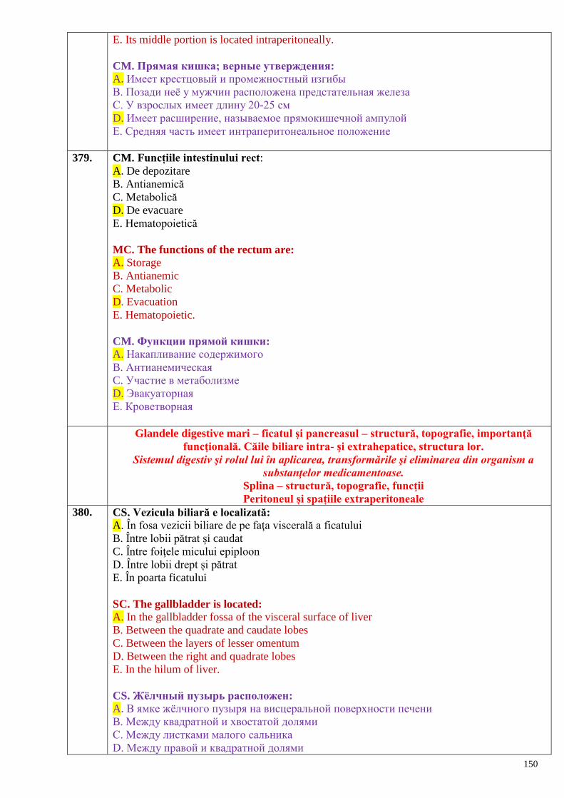
150
E. Its middle portion is located intraperitoneally.
CM. Прямая кишка; верные утверждения:
A. Имеет крестцовый и промежностный изгибы
B. Позади неѐ у мужчин расположена предстательная железа
C. У взрослых имеет длину 20-25 см
D. Имеет расширение, называемое прямокишечной ампулой
E. Средняя часть имеет интраперитонеальное положение
379. CM. Funcțiile intestinului rect:
A. De depozitare
B. Antianemică
C. Metabolică
D. De evacuare
E. Hematopoietică
MC. The functions of the rectum are:
A. Storage
B. Antianemic
C. Metabolic
D. Evacuation
E. Hematopoietic.
CM. Функции прямой кишки:
A. Накапливание содержимого
B. Антианемическая
C. Участие в метаболизме
D. Эвакуаторная
E. Кроветворная
Glandele digestive mari – ficatul şi pancreasul – structură, topografie, importanţă
funcţională. Căile biliare intra- şi extrahepatice, structura lor.
Sistemul digestiv şi rolul lui în aplicarea, transformările şi eliminarea din organism a
substanţelor medicamentoase.
Splina – structură, topografie, funcţii
Peritoneul și spațiile extraperitoneale
380. CS. Vezicula biliară e localizată:
A. În fosa vezicii biliare de pe faţa viscerală a ficatului
B. Între lobii pătrat și caudat
C. Între foiţele micului epiploon
D. Între lobii drept și pătrat
E. În poarta ficatului
SC. The gallbladder is located:
A. In the gallbladder fossa of the visceral surface of liver
B. Between the quadrate and caudate lobes
C. Between the layers of lesser omentum
D. Between the right and quadrate lobes
E. In the hilum of liver.
CS. Жѐлчный пузырь расположен:
A. В ямке жѐлчного пузыря на висцеральной поверхности печени
B. Между квадратной и хвостатой долями
C. Между листками малого сальника
D. Между правой и квадратной долями
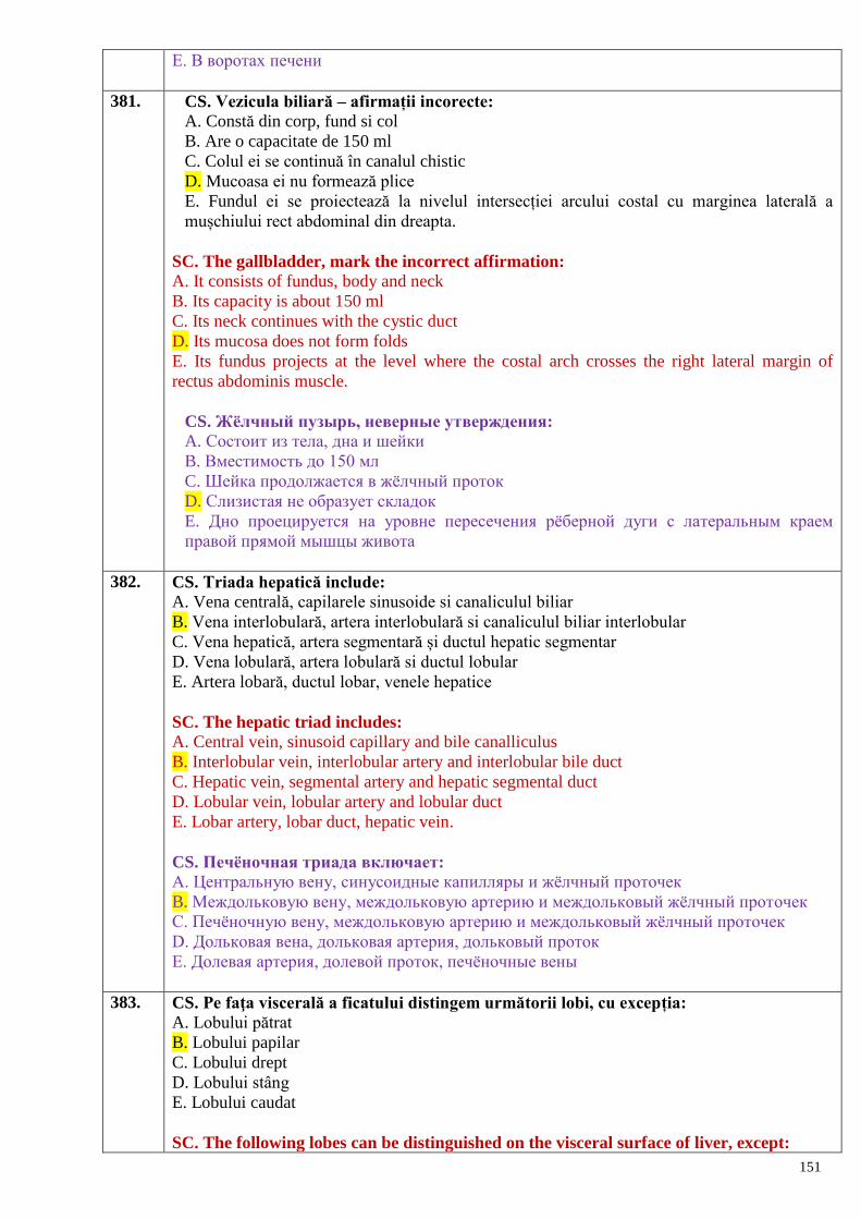
151
E. В воротах печени
381. CS. Vezicula biliară – afirmații incorecte:
A. Constă din corp, fund si col
B. Are o capacitate de 150 ml
C. Colul ei se continuă în canalul chistic
D. Mucoasa ei nu formează plice
E. Fundul ei se proiectează la nivelul intersecției arcului costal cu marginea laterală a
mușchiului rect abdominal din dreapta.
SC. The gallbladder, mark the incorrect affirmation: A. It consists of fundus, body and neck
B. Its capacity is about 150 ml
C. Its neck continues with the cystic duct
D. Its mucosa does not form folds
E. Its fundus projects at the level where the costal arch crosses the right lateral margin of
rectus abdominis muscle.
CS. Жѐлчный пузырь, неверные утверждения:
A. Состоит из тела, дна и шейки
B. Вместимость до 150 мл
C. Шейка продолжается в жѐлчный проток
D. Слизистая не образует складок
E. Дно проецируется на уровне пересечения рѐберной дуги с латеральным краем
правой прямой мышцы живота
382. CS. Triada hepatică include:
A. Vena centrală, capilarele sinusoide si canaliculul biliar
B. Vena interlobulară, artera interlobulară si canaliculul biliar interlobular
C. Vena hepatică, artera segmentară și ductul hepatic segmentar
D. Vena lobulară, artera lobulară si ductul lobular
E. Artera lobară, ductul lobar, venele hepatice
SC. The hepatic triad includes:
A. Central vein, sinusoid capillary and bile canalliculus
B. Interlobular vein, interlobular artery and interlobular bile duct
C. Hepatic vein, segmental artery and hepatic segmental duct
D. Lobular vein, lobular artery and lobular duct
E. Lobar artery, lobar duct, hepatic vein.
CS. Печѐночная триада включает:
A. Центральную вену, синусоидные капилляры и жѐлчный проточек
B. Междольковую вену, междольковую артерию и междольковый жѐлчный проточек
C. Печѐночную вену, междольковую артерию и междольковый жѐлчный проточек
D. Дольковая вена, дольковая артерия, дольковый проток
E. Долевая артерия, долевой проток, печѐночные вены
383. CS. Pe faţa viscerală a ficatului distingem următorii lobi, cu excepția:
A. Lobului pătrat
B. Lobului papilar
C. Lobului drept
D. Lobului stâng
E. Lobului caudat
SC. The following lobes can be distinguished on the visceral surface of liver, except:
152
A. Quadrate lobe
B. Papillary lobe
C. Right lobe
D. Left lobe
E. Caudate lobe.
CS. На висцеральной поверхности печени различаем следующие доли, за
исключением:
A. Квадратной доли
B. Сосцевидной доли
C. Правой доли
D. Левой доли
E. Хвостатой доли
384. CS. Pe faţa viscerală a ficatului distingem următoarele formațiuni, cu excepția:
A. Fisurii ligamentului rotund
B. Fisurii ductului venos
C. Șanțului venei cave inferioare
D. Hilului hepatic
E. Hilului biliar.
MC. The next structures can be distinguished on the visceral surface of liver, except:
A. Fissure of the round ligament
B. Fissure of the ductus venosus
C. Groove of the inferior vena cava
D. Hepatic hilum
E. Bile hilum.
CS. На висцеральной поверхности печени различаем следующие образования, за
исключением:
A. Щели круглой связки
B. Щели венозного протока
C. Борозды нижней полой вены
D. Ворот печени
E. Ворот жѐлчи
385. CS. Canalul coledoc – afirmații incorecte:
A. E situat intre foiţele ligamentului hepato-duodenal
B. Trece anterior de porțiunea superioară a duodenului
C. Se află in dreapta arterei hepatice comune
D. Confluează cu ductul pancreatic formând ampula hepatopancreatică
E. Se află anterior de orificiul epiploic
SC. Which of the following statements about the common bile duct is false:
A. It is located between the layers of the hepatoduodenal ligament
B. It passes in front of the superior part of the duodenum
C. It is situated to the right of common hepatic artery
D. It joins the pancreatic duct to form the hepatopancreatic ampulla
E. It is located in front of the epiploic foramen.
CS. Общий жѐлчевыводящий проток. Неверные утверждения:
A. Расположен между листками печѐночно-двенадцатиперстной связки
B. Проходит впереди верхней части двенадцатиперстной кишки
C. Находится справа от общей сонной артерии
D. В месте слияния с протоком поджелудочной железы, образуя печѐночно-
153
панкреатическую ампулу
E. Находится впереди сальникого отверстия
386. CM. Prin hilul aferent al ficatului trec:
A. Vena portă
B. Venele hepatice
C. Artera hepatică proprie
D. Canalul coledoc
E. Vase limfatice şi nervi
MC. The following structures pass through the afferent hilum of liver:
A. Portal vein
B. Hepatic veins
C. Proper hepatic artery
D. Common bile duct
E. Lymphatic vessels and nerves.
CM. В ворота печени входят:
A. Воротная вена
B. Печѐночные вены
C. Собственная печѐночная артерия
D. Общий жѐлчевыводящий проток
E. Лимфатические сосуды и нервы
387. CM. La nivelul hilului hepatic eferent se disting:
A. Venele hepatice
B. Vena portă
C. Artera hepatică comună
D. Nervi
E. Vase limfatice.
MC. The next structures pass through the efferent hilum of liver: A. Hepatic veins
B. Portal vein
C. Common hepatic artery
D. Nerves
E. Lymphatic vessels.
CM. Из ворот печени выходят:
A. Печѐночные вены
B. Воротная вена
C. Общая печѐночная артерия
D. Нервы
E. Лимфатические сосуды
388. CM. Lobulul hepatic:
A. Reprezintă unitatea morfo-functională a ficatului
B. Are formă prismatică
C. E separat de lobulii vecini prin capilare sinusoide
D. Are în centru o venă centrală
E. Constă din lame formate din hepatocite
MC. The hepatic lobule:
A. Is the morphofunctional unit of liver
B. It is prismatic in shape
154
C. It is separated from the other lobules by the sinusoid capillaries
D. It has the central vein in the middle
E. It consists of hepatocytes arranged in plates.
CM. Печѐночная долька:
A. Представляет морфофункциональную единицу печени
B. Имеет призматическую форму
C. Отделена от соседних долек синусоидными капиллярами
D. Имеет в центре центральную вену
E. Состоит из пластинок, образованных гепатоцитами
389. CM. Ficatul este fixat cu ajutorul ligamentelor:
A. Rotund al ficatului
B. Falciform
C. Coronar
D. Triunghiular drept şi stâng
E. Hepatofrenic
MC. The liver is fixed by the following ligaments:
A. Round ligament of liver
B. Falciform ligament
C. Coronary ligament
D. Right and left triangular ligaments
E. Hepatophrenic ligament.
CM. Печень фиксируется с помощью связок:
A. Круглой связки печени
B. Серповидной связки
C. Венечной связки
D. Правой и левой треугольных связок
E. Печѐночно-диафрагмальной связки
390. CM. Funcții ale parenchimului hepatic:
A. De dezintoxicare
B. Trofică
C. De sprijin
D. Digestie
E. Metabolică
MC. The functions of the hepatic parenchyma are:
A. Detoxification
B. Storage of glycogen
C. Support
D. Digestive
E. Metabolic.
CM. Четыре функции паренхимы печени:
A. Дезинтоксикационная
B. Трофическая
C. Поддерживающая
D. Пищеварительная
E. Метаболическая
391. CM. Căile biliare extrahepatice:
A. Canalul coledoc.
155
B. Canaliculul interlobular.
C. Ductul hepatic drept şi stâng.
D. Ductul hepatic comun.
E. Canaliculul biliar.
MC. The extrahepatic bile ducts are:
A. Common bile duct
B. Interlobular duct
C. Right and left hepatic ducts
D. Common hepatic duct
E. Bile canalliculus.
CM. Внепечѐночные жѐлчные пути:
A. Общий жѐлчевыводящий проток
B. Междольковый каналец
C. Правый и левый печѐночные протоки
D. Общий печѐночный проток
E. Жѐлчный проток
392. CM. Pancreasul:
A. E situat posterior de stomac
B. Se dezvoltă din intestinul primar
C. Se află mezoperitoneal
D. Capul lui e încadrat in potcoava duodenului
E. Are o lungime de 14 - 18 cm
MC. Choose the true statements about the pancreas:
A. Is located behind of the stomach
B. It develops from the primary gut
C. It is located mesoperitoneally
D. Its head is enclosed in the duodenal loop
E. Its length is 14 - 18 cm.
CM. Поджелудочная железа:
A. Расположена позади желудка
B. Развивается из первичной кишки
C. Покрыта брюшиной мезоперитонеально
D. Еѐ головка окружена «подковой» двенадцатиперстной кишки
E. Имеет длину от 14 до 18 см
393. CM. Pancreasul endocrin:
A. Este reprezentat de insulele lui Langerhans
B. Se află la periferia organului
C. Conține celule alfa şi beta
D. Celulele beta secretă insulina
E. De la insulele Langerhans pornesc canale excretoare secundare ale pancreasului
MC. As a endocrine gland the pancreas is:
A. It is represented by the islets of Langerhans
B. It is located on the periphery of the organ
C. It contains alpha and betacells
D. Betacells produce insulin
E. The secondary excretory ducts of the pancreas originate from the islets of Langerhans
CM. Поджелудочная железа как эндокринный орган:
156
A. Представлена островками Лангерганса
B. Находится на периферии органа
C. Содержит альфа-, и бета-клетки
D. Бета-клетки секретируют инсулин
E. Из островков Лангерганса направляется вторичный выводной проток поджелудочной
железы
394. CM. Funcții ale pancreasului:
A. Exocrină
B. Hemopoietică
C. De protecţie
D. Antitoxică
E. Endocrină
C. Two functions of the pancreas are:
A. Exocrine function
B. Hematopoietic function
C. Protection function
D. Antitoxic function
E. Endocrine function.
CM. Две функции поджелудочной железы:
A. Экзокринная
B. Кроветворная
C. Защитная
D. Антитоксическая
E. Эндокринная
395. CM. Celulele alfa şi beta ale pancreasului produc:
A. Suc pancreatic
B. Bilă
C. Insulină
D. Mucus
E. Glucagon
MC. The pancreatic alpha and beta cells produce:
A. Pancreatic juice
B. Bile
C. Insulin
D. Mucus
E. Glucagon.
CM. Находящиеся в поджелудочной железе клетки и бета-клетки продуцируют:
A. Панкреатический сок
B. Жѐлчь
C. Инсулин
D. Слизь
E. Глюкагон
396. CM. Splina – afirmații corecte:
A. Este un organ al sistemului imunitar
B. Se află în hipocondrul drept
C. Parenchimul ei constă din pulpa roșie si pulpa albă
D. De capsula ei fibroasă sunt legate trabeculele splenice
E. La nou-născut are o structură lobulară
157
MC. Which of the following statements about the spleen are true:
A. It is an organ which belongs to the limfoid system
B. It is located in the right hypochondrium
C. Its parenchyma contains red and white pulps
D. The splenic trabeculae are connected to the fibrous capsule
E. It has a lobular structure in newborn.
CM. Селезѐнка, верные утверждения:
A. Это орган иммунной системы
B. Расположен в правом подреберье
C. Паренхима состоит из красной и белой пульпы
D. Фиброзная капсула соединяется с селезѐночными перегородками
E. У новорождѐнных имеет дольчатое строение
397. CS. Determinați afirmația corectă referitoare la cavitatea peritoneală:
A. Este limitată de pereții cavității abdominale
B. Prezintă un spațiu lamelar situat intre foita viscerală si parietală a peritoneului
C. Conține toate organele cavității abdominale
D. La bărbat comunică cu mediul ambiant
E. La femeie prezintă un sac închis.
SC. Determine the correct affirmation regarding the peritoneal cavity: A. It is limited by the abdominal cavity walls
B. It is a capillary space situated between the visceral and parietal layers of peritoneum
C. It contains all the organs of abdominal cavity
D. In male it communicates with the external environment
E. In female it is a closed sac.
CS. Обозначьте правильные утверждения относительно полости брюшины:
A. Ограничена стенками брюшной полости
B. Представляет пространство между висцеральным и париетальным листками
брюшины
C. Содержит все органы брюшной полости
D. У мужчин сообщается с внешней средой
E. У женщин представляет закрытый мешок
398. CS. Organe care nu se află în etajul supramezocolic al cavității peritoneale:
A. Ficatul
B. Stomacul
C. Rinichii
D. Splina
E. Porțiunea abdominală a esofagului.
SC. Which organs listed below are not located in the supramesocolic storey of peritoneal
cavity: A. Liver
B. Stomach
C. Kidney
D. Spleen
E. Abdominal portion of the esophagus.
CS. Какие из перечисленных органов не находятся в верхнем этаже полости
брюшины:
A. Печень
158
B. Желудок
C. Почки
D. Селезѐнка
E. Брюшной отдел пищевода
399. CS. Mai mobile în raport cu peritoneul sunt organele:
A. Mezoperitoneale
B. Retroperitoneale
C. Extraperitoneale
D. Intraperitoneale
E. Toate posedă același grad de mobilitate
SC. The most mobile organs in relation to the peritoneum are located:
A. Mesoperitoneally
B. Retroperitoneally
C. Extraperitoneally
D. Intraperitoneally
E. All of them possess the same degree of mobility.
CS. Какие органы, покрытые брюшиной более подвижны:
A. Мезоперитонеальные
B. Ретроперитонеальные
C. Экстраперитонеальные
D. Интраперитонеальные
E. Все органы имеют одинаковую степень подвижности
400. CM. Funcțiile peritoneului:
A. De absorbție
B. De transudare (secretoare)
C. De protecție biologică (barieră)
D. De digestie
E. De depozitare a sângelui şi a grăsimilor
MC. The functions of the peritoneum are: A. Absorption
B. Secretion
C. Biologic protection (barrier)
D. Digestive
E. Depositing of blood and fat.
CM. Функции брюшины:
A. Всасывание
B. Секреция (транссудация)
C. Барьерная
D. Пищеварительная
E. Депонирования крови и жира
401. CM. Care sunt limitele etajului supramezocolic:
A. Omentul mic
B. Ligamentul coronar
C. Diafragma
D. Colonul transvers şi mezocolonul său
E. Ligamentul gastrocolic
MC. The limits of the supramesocolic storey are:
159
A. Lesser omentum
B. Coronary ligament
C. Diaphragm
D. Transverse colon and its mesentery
E. Gastrocolic ligament.
CM. Какие границы имеет верхний этаж брюшной полости:
A. Малый сальник
B. Венечная связка
C. Диафрагма
D. Поперечная ободочная кишка и еѐ брыжейка
E. Желудочноободочная связка
402. CM. În etajul supramezocolic sunt prezente bursele:
A. Lienală
B. Hepatică
C. Renală
D. Pregastrică
E. Omentală.
MC. In the supramesocolic storey there are three bursae: A. Lineal bursa
B. Hepatic bursa
C. Renal bursa
D. Pregastric bursa
E. Omental bursa
CM. В верхнем этаже представлены 3 сумки:
A. Селезѐночная
B. Печѐночная
C. Почечная
D. Преджелудочная
E. Сальниковая
403. CM. Funcțiile omentului mare, graţie cărora el poate localiza procesul inflamator:
A. De absorbție
B. De transudare
C. Capacitatea de a se concrește cu suprafața inflamata
D. Acțiunea macrofagilor din căile limfatice
E. Capacitatea de hemostază.
MC. The properties of the greater omentum that allow to stop the spread of
inflammation are: A. Absorbtion
B. Secretion
C. Capacity to adhere to the inflammatory surface
D. Macrophage action of lymphatic ways
E. Capacity of homeostasis.
CM. Три функции большого сальника, благодаря которым возможна
локализация воспалительного процесса:
A. Всасывание
B. Выделение серозной жидкости
C. Способность ограничить поверхность воспаления
D. Действия макрофагов лимфатических путей
160
E. Кровоостанавливающая способность
404. CM. 4 porțiuni ale intestinului care posedă mezou:
A. Duodenul
B. Jejunul
C. Ileonul
D. Colonul transvers
E. Porțiunea superioară a rectului
MC. The four portions of the intestine that possess a mesentery are:
A. Duodenum
B. Jejunum
C. Ileum
D. Transverse colon
E. Superior portion of rectum.
CM. Четыре части кишки, которые имеют брыжейку:
A. Двенадцатиперстная
B. Тощая
C. Подвздошная
D. Поперечная ободочная
E. Верхний отдел прямой кишки
405. CM. Tunica seroasă a organelor este data de:
A. Foița viscerală a pericardului
B. Pleura viscerală
C. Pleura parietală
D. Peritoneul visceral
E. Foița viscerală a tunicii vaginale a testiculului
CM. The serous coats of viscera are represented by:
A.The visceral layer of serous pericardium
B. The visceral pleura
C. The parietal pleura
D. The visceral peritoneum
E. The visceral layer of tunica vaginalis testis.
СМ. Серозная оболочка, покрывающая органы, представлена:
A. Висцеральным листком перикарда.
B. Висцеральной плеврой.
C. Париетальной плеврой.
D. Висцеральным листком брюшины.
E. Висцеральным листком влагалищной оболочки яичка.
406. CM. Organe aflate mezoperitoneal:
A. Ficatul
B. Splina
C. Colonul ascendent si descendent
D. Treimea medie a rectului
E. Vezicula biliară umplută cu bilă
MC. The organs located mesoperitoneally are:
A. Liver
B. Spleen
C. Ascending and descending colon
161
D. Middle third of rectum
E. Full gallbladder.
CM. Органы, покрытые брюшиной мезоперитонеально:
A. Печень
B. Селезѐнка
C. Восходящая и нисходящая ободочная кишка
D. Средняя треть прямой кишки
E. Жѐлчный пузырь, заполненный жѐлчью
407. CM. Organe aflate extraperitoneal:
A. Ficatul
B. Rinichii
C. Duodenul
D. Ureterele
E. Vezicula biliară în vacuitate
MC. The organs located extraperitoneally are:
A. Liver
B. Kidney
C. Duodenum
D. Ureters
E. Empty gallbladder.
CM. Органы, расположенные экстраперитонеально:
A. Печень
B. Почки
C. Двенадцатиперстная кишка
D. Мочеточники
E. Жѐлчный пузырь в опорожнѐнном состоянии
408. CS. Corpusculii Hassall se află în:
A. Insulele Langerhans
B. Substanța medulară a timusului
C. Substanța medulară a suprarenalei
D. Neurohipofiză
E. Substanța corticală a timusului
SC. The Hassall's corpuscules are located in:
A. Islets of Langerhans
B. Medulla of the thymus
C. Medulla of the suprarenal glands
D. Neurohypophysis
E. Cortex of the thymus.
CS. Тельца Hassall находятся в:
A. Островках Langerhans
B. Мозговом веществе тимуса
C. Мозговом веществе надпочечников
D. Нейрогипофизе
E. Корковом веществе тимуса
409. CS. Hormonii insulelor pancreatice (Langerhans) reglează:
A. Metabolismul lipidic
B. Metabolismul proteic
162
C. Metabolismul glucidic
D. Metabolismul hidro-salin
E. Metabolismul substanțelor minerale
SC. The hormones of the islets of Langerhans regulate:
A. Lipid metabolism
B. Protein metabolism
C. Carbohydrate metabolism
D. Water-salt metabolism
E. Metabolism of the mineral substances
CS. Гормоны панкреатических островков (Langerhans) регулируют:
A. Жировой (липидный) обмен
B. Белковый обмен
C. Углеводный обмен
D. Водно-солевой обмен
E. Обмен минеральных веществ
410. CS. Glucagonul este secretat de:
A. Celulele acinilor glandulari
B. Celulele B ale insulelor Langerhans
C. Celulele A ale insulelor Langerhans
D. Celulele glandelor gastrice care secretă şi HCL
E. Celulele glandelor duodenale care secretă şi secretina
SC. The glucagon is produced by:
A. Cells of the pancreatic acini
B. B-cells of the islets of Langerhans
C. A-cells of the islets of Langerhans
D. Cells of the gastric glands, which also produce HCL
E. Cells of the duodenal glands, which also produce secretin
CS. Глюкагон вырабатывают:
A. клетки железистых ацинусов
B. β - бэта клетки островков Langerhans)
C. α – альфа клетки островков Langerhans
D. клетки желудочных желѐз, выделяющие и HCl
E. клетки двенадцатиперстных желѐз, выделяющие и секретин
411. CS. Pancreasul este o glandă:
A. Exocrină
B. Endocrină
C. Mixtă
D. Alveolară
E. Alveolo-tubulară
SC. What kind of gland is the pancreas?
A. Exocrine
B. Endocrine
C. Mixed
D. Alveolar
E. Alveolar-tubular
CS. Поджелудочная железа:
A. Экзокринная
163
B. Эндокринная
C. Смешанная
D. Альвеолярная
E. Альвеолярно-трубчатая
Sistemul respirator –
componente, structură, rol funcţional.
Glanda tiroidă şi timusul – structură, topografie, funcţii.
Sistemul respirator ca obiect al influenţei substanţelor medicamentoase
Mediastinul – generalități
412. CM. Administrarea aerogenă a medicamentelor este favorizată de:
A. Pereții subțiri ai căilor respiratorii
B. Umiditatea sporită a tunicii mucoase
C. Suprafața mare a alveolelor pulmonare
D. Vascularizația imensă a căilor respiratorii
E. Toate false.
MC. Drug administration through inhalation is favored by:
A. The thin walls of respiratory ways
B. The high umidity of mucous coat
C. The large surface of pulmonary alveoli
D. The intense vascularization of respiratory ways
E. All false.
CМ. Эффективность введения лекарственных веществ аэрогенным путѐм зависит
от:
A. Тонких стенок дыхательных путей
B. Повышенной влажности слизистой оболочки
C. Большой поверхности легочных альвеол
D. Интенсивной васкуляризации дыхательных путей
E. Все ответы неверны
413. CM. Administrarea medicamentelor la sistemul respirator se realizează pe următoarele
căi:
A. Intraseroasă
B. Nazală
C. Pulmonară /inhalatorie/
D. Intravaginală
E. Rectală.
MC. In relation to the respiratory system the following routes of drug administration are
used:
A. Intraserous
B. Nasal
C. Pulmonary/inhalational
D. Intravaginal
E. Rectal
CМ. Введение лекарственных веществ через дыхательные пути производят
следующими методами:
A. Внутрисерозным
B. Носовым (назальным)
C. Легочным (ингаляционным)
D. Внутривлагалищным
E. Ректальным
164
414. CM. Administrarea medicamentelor pe cale nazală este favorizată de:
A. Permiabilitatea mare a mucoasei
B. Structura epiteliului
C. Vascularizația imensă a mucoasei
D. Suprafața mare a mucoasei
E. Toate false.
MC. Nasal drug administration (through the nose) is favored by:
A. The high permeability of mucosa
B. The structure of the epithelium
C. The intense vascularization of mucosa
D. The large surface of mucosa
E. All false.
CМ. Эффективность введения лекарственных веществ назальным путѐм зависит
от:
A. Повышенной проницаемости слизистой оболочки
B. Строения эпителия
C. Васкуляризации слизистой оболочки
D. Большой поверхности соприкосновения слизистой оболочки
E. Все ответы неверны
415. CM. Calea pulmonară (inhalatorie) se utilizează pentru:
A. Anesteticele gazoase
B. Bronhodilatatoare
C. Expectorante
D. Antibiotice (pulbere)
E. Toate false.
MC. Pulmonary (inhalational) drug administration is used for:
A. Inhalational anesthetics
B. Bronchodilators
C. Expectorants
D. Antibiotics (dust)
E. All false.
CМ. Ингаляторный путь применяется для:
A. Газовых анестетиков
B. Бронхорасширения
C. Отхаркивающих средств
D. Антибиотиков (порошки)
E. Все ответы неверны
416. CM. Preparatele medicamentoase care se elimină pe cale respiratorie sunt substanțele:
A. Volatile
B. Gazoase
C. Uneori în stare lichidă
D. Stare solidă
E. Pulbere.
MC. Pharmaceutical drugs (or medicines) eliminated from an organism through the
respiratory ways are:
A. Volatile substances
165
B. Gases
C. Sometimes liquid (fluid) substances
D. Solid substances
E. Dust.
CМ. Лекарственные препараты, выделяющиеся дыхательным путѐм:
A. Легко испаряющиеся
B. Газообразные
C. В редких случаях жидкие
D. Твѐрдые
E. Порошки
417. CM. Hormonii tiroidieni stimulează:
A. Metabolizmul energic și oxidativ
B. Formarea oaselor
C. Funcțiile ficatului
D. Funcția inimii
E. Activitatea glandelor sexuale.
CM. The hormones of the thyroid gland control:
A. Energy metabolism
B. Bone formation
C. Functions of the liver
D. Heart rate
A. Activity of the sexual glands.
CМ. Гормоны щитовидной железы стимулируют:
A. Энергетический и окислительный обмен
B. Образование костей
C. Функции печени
D. Функции сердца
E. Активность половых желез
418. CS. La copii hiposecreția horminilor tiroidieni provoacă:
A. Mixedem (edem mucos)
B. Apariția premature a nucleelor de osificare
C. Cretinismul
D. Exoftalmia
E. Tahicardia.
SC. The hypofunction of the thyroid gland in children provokes:
A. Myxedema
B. Premature apparition of the nuclei of the ossification
C. Cretinism
D. Exophthalmia
E. Tachycardia
CS. Гипосекреция гормонов щитовидной железы у детей провоцирует:
A. Микседему (серозный отѐк)
B. Преждевременное происхождение ядер окостенения
C. Кретинизм
D. Экзофталмию
E. Тахикардию
419. CS. Parathormonul este produs de:
166
A. Adenohipofiză
B. Tiroidă
C. Timus
D. Paratiroide
E. Suprarenale.
SC. The parathormone is produced by:
A. Adenohypophysis
B. Thyroid gland
C. Thymus
D. Parathyroid glands
E. Suprarenal (adrenal) glands.
CS. Парат гормон выделяется:
A. Аденогипофизом
B. Щитовидной железой
C. Вилочковой железой
D. Околощитовидными железами
E. Надпочечниками
420. CM. În practica medicală paratiroidina se utilizează în:
A. Hipoparatiroidism cronic
B. Tetanie (hipoparatiroidism acut)
C. Spasmofilie
D. Tirotoxicoză
E. Osteoporoză
MC. In the medicine the parathormone is used (to treat) in case of:
A. Chronic hypoparathyroidism
B. Tetany (acute hypoparathyroidism)
C. Spasmophily
D. Thyrotoxicosis
E. Osteoporosis
CМ. Паратироидин в медицинской практике употребляется при:
A. Хроническом гипопаратироидисме
B. Тетании (острый гипопаратироидий)
C. Спазмофилии
D. Тиротоксикозе
E. Остеопорозе
421. CS. Organele respiratorii se dezvoltă din:
A. Intestinul mijlociu
B. Peretele ventral al intestinului anterior
C. Intestinul posterior
D. Ectoderm
E. Mezoderm
SC. The respiratory organs develop from:
A. Middle gut
B. Ventral wall of the foregut
C. Hindgut
D. Ectoderm
E. Mesoderm .
167
CS. Органы дыхания развиваются из:
А. Cредней кишки
В. Вентральной стенки передней кишки
C. Задней кишки
D. Эктодермы
E. Мезодермы
422. CS. Căile respiratorii se intersectează cu cele digestive în:
A. Nazofaringe
B. Bucofaringe
C. Laringofaringe
D. Fauce
E. Aditusul laringian .
MC. The airways intersects the digestive ones within the:
A. Nasopharynx
B. Oropharynx
C. Laryngopharynx
D. The oral cavity
E. Aditus laryngis (inlet into the larynx)
CS. Дыхательные и пищеварительные пути пересекаются в:
А. Носоглотке
B. Ротовой части глотки
C. Гортанной части глотки
D. Зеве
E. У входа в гортань
423. CM. Respirația, ca totalitate a proceselor fiziologice cuprinde:
A. Respirația pulmonară (sau externă)
B. Respirația cutanată
C. Respirația bronhoveziculară
D. Respirația tisulară (internă)
E. Respirația abdominală.
MC. Breathing as a physiological process includes:
A. Pulmonary respiration (or external one)
B. Cutaneous respiration
C. Bronchovesicular respiration
D. Tissue respiration (or internal one)
E. Abdominal respiration.
CМ. Дыхание – это общий физиологический процесс, который охватывает:
A. Лѐгочное дыхание (или внешнее)
B. Кожное дыхание
C. Бронховезикулярное дыхание
D. Тканевое дыхание
E. Брюшное дыхание
424. CM. Referitor la căile respiratorii:
A. Sunt clasificate in supraglotice si infraglotice
B. Au pereții rigizi graţie scheletului osos sau fibrocartilaginos
C. Din exterior sunt tapetate cu peribronhie
D. Conțin tunica mucoasă care învelește pereții lor din interior
E. În pătura lor submucoasă se află rețele bogate de vase sangvine menite să încălzească aerul
168
inspirat
MC. Regarding the respiratory ways:
A. Are divided into supra- and infraglottic ones
B. Their wall are rigid due to the bony or fibro-cartilaginous skeleton
C. Outside they are covered by the peribronchial tissue
D. Inside their walls are lined by the mucous coat
E. Their submucosa is rich in blood vessels networks that assure the warming of the inspired
air
CМ. Дыхательные пути:
A. Подразделяются на верхние и нижние
B. Имеют регидные стенки, характерные для костного скелета или соединительно-
хрящевого
C. Снаружи покрыты перибронхиальной тканью
D. Слизистая оболочка покрывает стенки изнутри
E. В подслизистой оболочке имеются выраженные кровеносные сети, согревающие
вдыхаемый воздух
425. CM. Referitor la mucoasa cailor respiratorii:
A. E tapetată cu epiteliu ciliat
B. Dedesubtul ei se află o patură submucoasă
C. Nu conține rețele de vase sangvine
D. Nu conține rețele nervoase
E. În totalitatea sa se prezintă ca o zonă tusigen.
MC. Choose the true statements regarding mucous coat of the airways:
A. It is covered by ciliated epithelium
B. The submucous coat is located under it
C. It does not contain blood vessels networks
D. It does not contain nervous plexuses
E. Generally it is a coughing zone.
CМ. Слизистая оболочка дыхательных путей:
A. Покрыта цилиндрическим мерцательным эпителием
B. Содержит подслизистый слой
C. Не содержит сети кровеносных сосудов
D. Не содержит нервные сплетения
E. Представляет кашлевую зону
426. CM. Formațiunile limfoide ale cailor respiratorii sunt reprezentate prin:
A. Țesutul limfoid din corionul mucoasei lor
B. Aglomerări de foliculi limfoizi la nivelul epiglotei
C. Amigdala carinei traheale
D. Amigdala laringeană
E. Amigdala nazofaringeană.
MC. The lymphoid structures of the airways are formed by:
A. Lymph tissue of the mucous coat chorion
B. Agglomeration of lymph nodes at the level of the epiglottis
C. Tonsil of the tracheal carina (carina trachea)
D. Laryngeal tonsil
E. Nasopharyngeal tonsil
CМ. Лимфоидные образования дыхательных путей представлены:
169
A. Лимфоидная ткань ворсинчатой оболочки слизистой
B. Скопление лимфоидных фолликулов на уровне надгортанника
C. Миндалина киля трахеи
D. Гортанная миндалина
E. Носоглоточная миндалина
427. CM. Funcțiile căilor respiratorii sunt:
A. De umectare a aerului inspirat
B. Purificarea aerului inspirat
C. Schimbul de gaze
D. Participă in procesele metabolice
E. De încălzire a aerului
MC. The functions of the respiratory ways are:
A. Humidifying the inspired air
B. Purification of the inspired air
C. Exchange of gases
D. Participation in the metabolic processes
E. Warming the air.
CМ. Функциями дыхательных путей являются:
A. Увлажнение вдыхаемого воздуха
B. Очищение вдыхаемого воздуха
C. Обмен газами
D. Участие в обменных процессах
E. Согревание воздуха
428. CS. Zona hemoragică (pata vasculară Kisselbach) e situată în mucoasa:
A. Meatului superior
B. Meatului mijloci
C. Septului nazal la cca 1 cm deasupra narinelor
D. Cornetului superior
E. Cornetului mijlociu
SC. The hemorrhagic zone (vascular plate of Kiesselbach) is located in the mucous coat
of the:
A. Superior nasal meatus
B. Middle nasal meatus
C. Nasal septum at about 1 cm above the nostrils
D. Superior nasal concha
E. Middle nasal concha
CS. Зона густой сосудистой сети (сосудистое русло Киссельбаха) располагается в
слизистой:
A. Верхнего носового хода
B. Среднего носового хода
C. В перегородке носа на 1 см выше ноздрей
D. В верхней носовой раковине
E. В средней носовой раковине
429. CM. Principalele configurații de nas extern sunt:
A. Nasul drept (tip August)
B. Nasul grec (tip Venus de Millo)
C. Nasul bifurcat
D. Nasul acvilin (coroiat) (tip Dante)
170
E. Nasul scobit (tip Socrate).
MC. The main shapes of the external nose are:
A. Straight nose (Augustine type)
B. Greek nose (Venus de Millo type)
C. Bifurcated nose
D. Curved nose aquiline (Dantes type)
E. Hollow nose (Socrates type)
CМ. Основные формы наружного носа:
A. Прямой нос (тип Августа)
B. Греческий нос (тип Венуса де Мило)
C. Раздвоенный нос
D. Орлиный нос (тип Данте)
E. Горбатый нос (тип Сократа).
430. CM. Mucoasa nazală exercită funcțiile:
A. Respiratorie
B. Olfactivă
C. De protecție
D. Rezonatorie
E. De încălzire a aerului inspirat
MC. The nasal mucosa has the following functions:
A. Umedify the inhalated air
B. Olfactory
C. Protective
D. Resonance
E. Warming of inspired air
CМ. Слизистая оболочка носа выполняет функции:
A. Дыхательную
B. Обонятельную
C. Защитную
D. Резонаторную
E. Согревания вдыхаемого воздуха
431. CM. Pe viu cavitatea nazală poate fi explorată prin:
A. Inspecție
B. Palpație
C. Rinoscopie
D. Examen ultrasonic
E. Examen radiologic
MC. On alive the nasal cavity can be examined by:
A. Inspection
B. Palpation
C. Rhinoscopy
D. Ultrasound examination
E. X-ray examination
CМ. У живого полость носа может быть исследована через:
A. Осмотр
B. Пальпацию
C. Риноскопию
171
D. Исследование с помощью УЗИ
E. Радиологическое исследование
432. CM. In meatul nazal mediu se deschid:
A. Sinusul frontal
B. Celulele etmoidale posterioare
C. Sinusul maxilar
D. Canalul nazolacrimal
E. Celulele etmoidale anterioare
MC. Which of the following statements opens into the middle nasal meatus:
A. Frontal sinus
B. Posterior ethmoidal cells
C. Maxillary sinus
D. Nasolacrimal canal
E. Anterior ethmoidal cells
CМ. В средний носовой ход открываются:
A. Лобная пазуха
B. Задние ячейки решѐтчатой кости
C. Верхнечелюстная пазуха
D. Носослѐзный канал
E. Передние ячейки решѐтчатой кости
433. CM. In meatul nazal superior se deschid:
A. Sinusul sfenoid
B. Celulele etmoidale posterioare
C. Celulele etmoidale anterioare
D. Sinusul frontal
E. Sinusul maxilar
MC. Which of the following statements opens into the superior nasal meatus:
A. Sphenoid sinus
B. Posterior ethmoidal cells
C. Anterior ethmoidal cells
D. Frontal sinus
E. Maxillary sinus
CМ. В верхний носовой ход открываются:
A. Пазуха клиновидной кости
B. Задние ячейки решѐтчатой кости
C. Передние ячейки решѐтчатой кости
D. Лобная пазуха
E. Верхнечелюстная пазуха
434. CM. Referitor la funcțiile sinusurilor paranazale:
A. Micșorează greutatea craniului
B. Măresc rezistenta craniului
C. Au rol de izolatori termici
D. Sunt rezonatori ai sunetelor
E. Măresc greutatea craniului
MC. The functions of the paranasal sinuses are:
A. Diminishing the weight of the skull
B. Increase of the resistance of the skull
172
C. Thermal insulation
D. Are sounds' resonators
E. Increase the weight of the skull
CМ. Относительно функций параназальных синусов:
A. Уменьшают вес черепа
B. Способствуют укреплению черепа
C. Выполняют роль теплового изолятора
D. Участвуют в резонации звука
E. Увеличивают вес черепа
435. CM. Meaturile nazale sunt:
A. Superior, mediu, inferior
B. Nazal comun
C. Nazofaringian
D. Nazobucal
E. Nazolaringean
MC. The following nasal meatuses are distinguished:
A. Superior, middle, inferior nasal meatuses
B. Common nasal meatus
C. Nasopharyngeal meatus
D. Nasobuccal meatus
E. Nasolaryngeal meatus.
CМ. Носовые ходы:
A. Верхний, средний, нижний
B. Общий носовой ход
C. Носоглоточный ход
D. Носоротовой ход
E. Носогортанный ход
436. CS. Limita inferioara a laringelui la maturi este:
A. C5
B. C6
C. C4
D. Osul hioid
E. T1
SC. The inferior margin of the larynx is located at the level of the :
A. C5
B. C6
C. C4
D. Hyoid bone
E. T1
CS. Нижняя граница гортани расположена на уровне:
A. Пятого шейного позвонка
B. Шестого шейного позвонка
C. Четвѐртого шейного позвонка
D. Подъязычной кости
E. Первого грудного позвонка
437. CS. Referitor la scheletotopia laringelui:
A. Corespunde vertebrelor cervicale II - VII
173
B. Corespunde vertebrelor cervicale IV, V, VI
C. E plasat intre exobaza craniului si vertebra cervicală VI
D. Se află la nivelul vertebrelor cervicale II - V
E. Se află la nivelul vertebrelor cervicale III - VI
SC. The skeletotopy of the larynx:
A. Corresponds to the IInd-VIIth cervical vertebrae
B. Corresponds to the IVth, Vth, VIth cervical vertebrae
C. Is located between the exobase of the skull and VIth cervical vertebra
D. Is located at the level of the IInd-Vth cervical vertebrae
E. Is located at the level of the IIIrd-VIth cervical vertebrae
CS. Относительно скелетотопии гортани:
A. Соответствует уровню от второго до седьмого шейных позвонков
B. Соответствует уровню четвѐртого, пятого и шестого шейных позвонков
C. Лежит между наружным основанием черепа и шестым шейным позвонком
D. Находится на уровне от второго до пятого шейных позвонков
E. Находится на уровне от третьего до шестого шейных позвонков
438. CS. Doar din țesut cartilaginos elastic e constituit:
A. Cartilajul tiroid
B. Cartilajele aritenoide
C. Cartilajele corniculate
D. Cartilajele cuneiforme
E. Epiglota
SC. Which of the following consists of only elastic cartilaginous tissue:
A. Thyroid cartilage
B. Arytenoid cartilage
C. Corniculate cartilage
D. Cuneiform cartilage
E. Epiglottis
CS. Только из эластической хрящевой ткани состоит:
A. Щитовидный хрящ
B. Черпаловидные хрящи
C. Рожковидные хрящи
D. Клиновидные хрящи
E. Надгортанник
439. CM. Funcțiile laringelui sunt:
A. De protecție
B. De sprijin
C. De respirație
D. De locomoție
E. De fonație
MC. The functions of the larynx are:
A. Protection
B. Support
C. Respiration
D. Locomotion
E. Phonation
CМ. Функции гортани:
174
A. Защитная
B. Поддерживающая
C. Дыхательная
D. Двигательная
E. Голосообразования
440. CM. Cavității laringelui i se disting compartimentele:
A. Aditusul laringian
B. Vestibulul
C. Cavitatea infraglotică
D. Cavitatea intermediară (glota)
E. Fanta glotică
MC. The laryngeal cavity consists of the following parts:
A. Aditus laryngis
B. Vestibule of the larynx
C. Infraglottic cavity
D. Glottis
E. Rima glottidis
CМ. В полости гортани различаются отделы:
A. Вход в гортань
B. Преддверие
C. Подсвязочная полость
D. Межжелудочковая часть (glottis)
E. Голосовая щель
441. CM. In raport cu laringele (sintopia) vin urmatoarele organe:
A. Faringele
B. Glanda tiroidă
C. Cavitatea nazală
D. Mușchii suprahioidieni
E. Mușchii infrahioidieni
MC. According to its syntopy the larynx comes in contact with following organs:
A. Pharynx
B. Thyroid gland
C. Nasal cavity
D. Suprahyoid muscles
E. Infrahyoid muscles
CМ. К синтопии гортани относятся следующие органы:
A. Глотка
B. Щитовидная железа
C. Полость носа
D. Надподъязычные мышцы
E. Подподъязычные мышцы
442. CM. Din cartilajele laringiene impare fac parte:
A. Tiroidul
B. Cricoidul
C. Epiglota
D. Aritenoidele
E. Alarele
175
CM. The unpaired cartilages of the larynx are:
A.Thyroid cartilage
B. Cricoid cartilage
C. Epiglottis
D. Arytenoid cartilages
E. Alar cartilages
CS. К непарным хрящам гортани относятся:
A. Щитовидный
B. Перстневидный
C. Надгортанник
D. Черпаловидные хрящи
E. Крыловидные хрящи
443. CM. Pe viu laringele poate fi explorată prin:
A. Palpaţie
B. Auscultaţie
C. Laringoscopie
D. Examen radiologic
E. Examen ultrasonic
MC. On alive person the morphology of the larynx can be examined by:
A. Palpation
B. Auscultation
C. Laryngoscopy
D. X-rays examination
E. Ultrasonic examination
CМ. У живого строение гортани можно исследовать путѐм:
A. Пальпации
B. Аускультации (выслушивания)
C. Ларингоскопии
D. Радиологическим исследованием
E. Методом УЗИ (ультразвуковым исследованием)
444. CM. Plica (coarda) vocală include:
A. Ligamentul vocal
B. Muşchiul vocal
C. Tunica mucoasă, tapetată cu epiteliu cilindric ciliat
D. O pătură submucoasă
E. Ţesut celuloadipos
MC. The vocal fold contains the:
A. Vocal ligament
B. Vocal muscle
C. Mucous coat, lined with cylindrical ciliated epithelium
D. Submucous coat
E. Adipose tissue
CМ. Голосовая складка содержит:
A. Голосовую связку
B. Голосовую мышцу
C. Слизистую оболочку, покрытую цилиндрическим эпителием
D. Подслизистое русло
E. Жировую клетчатку
176
445. CS. Cea mai sensibilă zonă a arborelui traheobronhic (ultima linie de apărare) e situată la
nivelul:
A. Originii bronhiilor de ordinul I
B. Carinei traheale
C. Originii bronhiilor de ordinul III
D. Originii bronhiilor de ordinul IV
E. Originii bronhiilor de ordinul V
SC. The most sensitive zone of the tracheobronchial tree (the last defense line) is located
at the level of the:
A. Origin of the first order bronchi
B. Carina of the trachea
C. Origin of the third order bronchi
D. Origin of the fourth order bronchi
E. Origin of the fifth order bronchi.
CS. Самая чувствительная зона трахеобронхиального дерева (последняя линия
защиты) находится на уровне:
A. Начала бронхиол I порядка
B. Киля трахеи
C. Начала бронхиол III порядка (начала)
D. Начала бронхиол IV порядка
E. Начала бронхиол V порядка
446. CS. Unitatea morfofuncțională a plămânului este reprezentată de:
A. Lobulul pulmonar primar
B. Lobulul pulmonar secundar
C. Acinul pulmonar
D. Segmentul bronhopulmonar
E. Arborele alveolar
SC. The morphofunctional (or structural) unit of the lung is:
A. Primary pulmonary lobule
B. Secondary pulmonary lobule
C. Pulmonary acinus
D. Bronchopulmonary segment
E. Alveolar tree.
CS. Морфофункциональная единица лѐгких:
A. Первичная лѐгочная долька
B. Вторичная лѐгочная долька
C. Ацинус
D. Бронхолегочной сегмент
E. Альвеолярное дерево
447. CM. Particularitățile structurale ale peretelui traheal sunt:
A. Prezenţa musculaturii striate
B. Prezenţa semiinelelor cartilaginoase
C. Prezenţa epiteliului ciliat
D. Prezenţa epiteliului pavimentos stratificat
E. Prezenţa mușchiului traheal
MC. The structural features of the tracheal wall are:
177
A. Presence of stripped muscles
B. Presence of cartilaginous semirings
C. Presence of ciliated epithelium
D. Presence of stratified squamous epithelium
E. Presence of tracheal muscle.
CМ. Структурные особенности стенки трахеи:
A. Представлена поперечно-полосатой мускулатурой
B. Представлена хрящевыми полукольцами
C. Представлена цилиндрическим мерцательным эпителием
D. Представлена многослойным плоским эпителием
E. Представлена трахеальной мышцей
448. CM. Traheea vine in raport (sintopia) cu:
A. Esofagul
B. Timusul
C. Mușchii subhioidieni
D. Mușchii suprahioidieni
E. Pachetul neurovascular al gâtului
MC. The trachea comes in contact with (syntopy) the:
A. Esophagus
B. Thymus
C. Infrahyoid muscles
D. Suprahyoid muscles
E. Neurovascular patch (bundle) of the neck
CМ. Синтопия трахеи:
A. Пищевод
B. Вилочковая железа
C. Подподъязычные мышцы
D. Надподъязычные мышцы
E. Сосудисто-нервный пучок шеи
449. CM. Cu privire la bronhiile principale:
A. Ca grosime şi lungime sunt similare
B. Cea stângă e mai lungă şi mai îngustă
C. Cea dreaptă e mai scurtă şi mai largă
D. Cea dreaptă e mai lungă şi mai largă
E. Cea dreaptă reprezintă continuarea traheei
MC. Which of the following statements regarding the main bronchi is true:
A. They have the same length and width
B. The left bronchus is longer and narrower
C. The right bronchus is shorter and larger
D. The right bronchus is longer and larger
E. The right bronchus is a continuation of the trachea.
CМ. Главные бронхи:
A. Одинаковые по толщине и длине
B. Левый бронх длиннее и уже
C. Правый бронх короче и шире
D. Правый длиннее и шире
E. Правый является продолжением трахеи
178
450. CM. Formațiuni cartilaginoase se conțin in pereții:
A. Bronhiei lobare
B. Bronhiei segmentare
C. Bronhiolei terminale
D. Bronhiolei respiratorii
E. Ductului alveolar
MC. Cartilaginous structures are present in the walls of the:
A. Lobar bronchus
B. Segmental bronchus
C. Terminal bronchiole
D. Respiratory bronchiole
E. Alveolar duct.
CМ. Хрящевые образования содержатся в стенках:
A. Долевых бронхов
B. Сегментарных бронхов
C. Конечных бронхиолах
D. Дыхательных бронхиолах
E. Альвеолярных ходов
451. CM. Cu privire la plămânul stâng:
A. Posedă o fisură oblică şi una orizontală
B. Posedă doar o fisură oblică
C. Prin hilul lui artera pulmonară stângă pătrunde mai sus decât bronhia principală
D. Peste rădăcina lui trece cârja venei hemiazigos
E. Are 3 lobi
MC. The true statements regarding the left lung are:
A. It has a horizontal and an oblique fissure
B. It has only an oblique fissure
C. In the hilum the pulmonary artery is located above the main bronchus
D. The azygos vein passes above its root
E. It consists of three lobes
CМ. Левое лѐгкое:
A. Содержит косую и горизонтальную щели
B. Содержит только косую щель
C. Через ворота левая лѐгочная артерия проникает в лѐгкое выше главного бронха
D. Через корень проходит дуга полунепарной вены
E. Имеет 3 доли
452. CM. In componenţa acinului intră:
A. Bronhiolele respiratorii legate cu o bronhiolă terminală
B. Ducturile alveolare
C. Alveolele respiratorii și săculeții alveolari
D. Bronhiola terminală
E. Bronhiola lobulară secundară
MC The acinus is made up by the:
A. Respiratory bronchioles connected with a terminal bronchiole
B. Alveolar ducts
C. Respiratory alveoles and alveolar sacs
D. Terminal bronchiole
E. Secondary lobular bronchiole
179
CМ. В состав ацинуса входят:
A. Респираторные бронхиолы, связанные с конечными бронхиолами
B. Альвеолярные ходы
C. Респираторные альвеолы и альвеолярные мешочки
D. Конечная бронхиола
E. Вторичная дольковая бронхиола
453. CM. Foiţa viscerală a pleurei:
A. Căptușește plămânii din exterior
B. Pătrunde in scizuri si delimitează lobii pulmonari
C. Nu are legături cu stroma plămânului
D. Contribuie la producerea lichidului pleural
E. Nici o afirmație corectă
MC The visceral layer of the pleura:
A. Lines the outside the lungs
B. Enters the fissures and separates the pulmonary lobes
C. Does not have any connections with the stroma of the lungs
D. Contributes to the production (secretion) of the plural fluid
E. None of the above mentioned affirmations is true
CМ. Висцеральный листок плевры:
A. Срастается снаружи с лѐгкими
B. Проникает в щели и ограничивает доли лѐгких
C. Не связан со стромой лѐгкого
D. Участвует в продукции плевральной жидкости
E. Все утверждения неверны
454. CM. Pleura parietală:
A. Aderă la formațiunile subiacente de pe torace
B. Sub ea se află fascia endotoracică
C. Nu este vizibilă radiografic
D. Realizează recesuri pleurale
E. Nici o afirmație corectă
MC The parietal pleura:
A. Adheres to the subjacent thoracic structures
B. Under it the endothoracic fascia is located
C. Cannot be seen on radiography
D. Forms the pleural recessess
E. None of the above mentioned affirmations is true
CМ. Пристеночная плевра:
A. Срастается с образованиями грудной клетки
B. Под ней находится внутригрудная фасция
C. Не просматривается при радиографии
D. Образует плевральные карманы
E. Все утверждения неверны
455. CM. Recesurile pleurale:
A. Reprezintă spaţii suplimentare ale cavității pleurale
B. Există la nivelul trecerii a unei portiuni de pleură parietală în alta
C. La formarea lor participă pleura viscerală
D. Mai adânc e cel costodiafragmatic, mai ales pe linia medioaxilară
180
E. Nici o afirmatie corectă
MC. The pleural recessess:
A. They are additional spaces of the pleural cavity
B. Are formed when one part of the parietal pleura continues with another one
C. Are formed with the participation of the visceral pleura
D. The costodiaphragmatic recess is the deepest one, especially on the middle axillary line
E. None of the above mentioned affirmations is true
CМ. Плевральные карманы:
A. Представляют дополнительные пространства полости плевры
B. Образуются при переходе пристеночной плевры с одной части на другую
C. В их образовании участвует висцеральная плевра
D. Самый глубокий из них – рѐберно-диафрагмальный по среднеподмышечной линии
E. Все утверждения неверны
456. CM. Ce conține substanța coloidală din interiorul foliculelor glandei tiroide:
A. Testosteron
B. Aldosteron
C. Tiroxină
D. Triiodtironină
E. Calcitonină
MC. What does the colloid of the follicles of the thyroid gland contain:
A. Testosterone
B. Aldosterone
C. Thyroxine
D. Triiodothyronine
E. Calcitonin
CМ. Что содержит коллоидное вещество фолликулов щитовидной железы:
A. Тестостерон
B. Альдостерон
C. Тироксин
D. Трийодтиронин
E. Кальцитонин
457. CS. Tiroida are o greutate medie de:
A.25-35 g
B.30-40 g
C.45-50 g fiind cea mai voluminoasă glandă din organism
D.40-45 g
E.25-30 g
SC. Thyroid gland has the weight of:
A. 25-35 g
B. 30-40 g
C. 45-50 g beeing the largest gland of the body
D. 40-45 g
E. 25-30 g
CS. Средний вес щитовидной железы:
A.25 – 35 г
B.30 – 40 г
C.45 – 50 г (самая крупная железа в организме)
181
D.40 – 45 г
E.25 – 30 г
458. CS. Principala proteină din coloidul tiroidian este:
A. Tirozina
B. Tiroxina
C. Tiroglobulina
D. Triiodotironina
E. Tiroxina şi triiodotironina
SC. The main protein of the thyroid colloid is:
A. Tyrosine
B. Thyroxine
C. Thyroglobulin
D. Triiodothyronine
E. Thyroxine şi triiodothyronine
CS. Основной белок коллоида щитовидной железы:
A. Тирозин
B. Тироксин
C. Тироглобуллин
D. Трийодтиронин
E. Тироксин и трийодтиронин
459. CS. Epiteliul folicular al tiroidei are capacitatea de a acumula:
A. Seleniu
B. Calciu
C. Iod
D. Fosfor
E. Bariu
SC. The follicular epithelial cells of the thyroid gland have the ability to capture:
A. Selenium
B. Calcium
C. Iodine
D. Phosphorus
E. Barium
CS. Фолликулярный эпителий щитовидной железы обладает способностью к
накоплению:
A. Селена
B. Кальция
C. Йода
D. Фосфора
E. Бария
460. CM. Glanda tiroidă constă din:
A. Lobul anterior
B. Lobul posterior
C. Lobul drept
D. Lobul stâng
E. Lobul intermediar
MC. The thyroid gland consists of:
A.Anterior lobe
182
B. Posterior lobe
C. Right lobe
D. Left lobe
E. Intermediate lobe
CМ. Щитовидная железа состоит из:
A. Передней доли
B. Задней доли
C. Правой доли
D. Левой доли
E. Промежуточной доли
461. CS. Lobii glandei tiroide sunt uniți prin:
A. Bandeletă
B. Cordon
C. Disc cartilaginos
D. Tendon
E. Istm
SC. The lobes of the thyroid gland are connected by:
A. Strip
B. Cord
C. Cartilaginous disc
D. Tendon
E. Isthmus
CS. Доли щитовидной железы соединены:
A. Лентой
B. Поясом
C. Хрящевым диском
D. Сухожилием
E. Перешейком
462. CS. In 30% cazuri la glanda tiroidă se întâlnește:
A. Lobul caudat
B. Lobul piramidal
C. Lobul pătrat
D. Lobul romboid
E. Toate false
SC. In 30% of cases the thyroid gland has:
A. Caudate lobe
B. Pyramidal lobe
C. Quadrate lobe
D. Rhomboid lobe
E. Auricular lobe
CS. В 30% случаев у щитовидной железы встречается:
A. Хвостатая доля
B. Пирамидальная доля
C. Квадратная доля
D. Ромбовидная доля
E. Все ложные
463. CS. Extirparea căre-i glande endocrine provoacă tetanie şi moarte:

183
A. Suprarenalelor
B. Paratiroidelor
C. Tiroidei
D. Hipofizei
E. Epifizei
SC. The tetany and death could apear as a rezult of the extirpation of the following
glands:
E. Suprarenal glands
B. Parathyroid glands
C. Thyroid gland
D. Hypophysis
E. Epiphysis
CS. При экстирпации (удалении) каких эндокринных желѐз наблюдаются судороги
и приводит к смерти?
A. Надпочечников
B. Паращитовидных
C. Щитовидной
D. Гипофиза
E. Эпифиза
464. CS. Numărul glandelor paratiroide de regulă e de:
A. 1
B. 2 - 4
C. 4
D. 7 - 8
E. 12
SC. The number of the parathyroid glands is:
A. 1
B. 2-4
C. 4
D. 7-8
E. 12
CS. Количество паращитовидных желѐз в норме:
A. 1
B. 2 - 4
C. 4
D. 7 - 8
E. 12
465. CS. Cele mai mici glande endocrine sunt:
A. Hipofiza
B. Epifiza
C. Glanda tiroida
D. Glandele paratiroide
E. Glanda timus
SC. The smallest endocrine glands are:
A. Hypophysis
B. Epiphysis
C. Thyroid gland
D. Parathyroid glands
184
E. Thymus
CS. Самыми маленькими железами являются:
A. Гипофиз
B. Эпифиз
C. Щитовидная железа
D. Паращитовидные железы
E. Вилочковая железа
466. CM. Parenchimul tiroidei e reprezentat de:
A. Lobuli
B. Foliculi
C. Acini
D. Segmente
E. Lobi
MC. The parenchyma of the thyroid gland consists of:
A. Lobules
B. Follicles
C. Acini
D. Segments
E. Lobes.
CМ. Паренхима щитовидной железы представлена:
A. Дольками
B. Фолликулами
C. Ацинами
D. Сегментами
E. Долями
467. CM. Glanda tiroidă se află:
A. În partea posterioară a gâtului
B. În partea anterioară a gâtului
C. La nivelul laringelui
D. La nivelul porțiunii superioare a traheei
E. La nivelul aperturii superioare a toracelui
MC. The thyroid gland is located:
A. In the posterior region of the neck
B. In the anterior region of the neck
C. At the level of the larynx
D. At the level of the upper part of the trachea
E. At the level of the superior aperture of the thorax.
CМ. Щитовидная железа находится:
A. В задней части шеи
B. В передней части шеи
C. На уровне гортани
D. На уровне верхней части трахеи
E. На уровне верхней апертуры грудной клетки
468. CM. Glandele paratiroide se localizează:
A. Pe faţa anterioară a glandei tiroide
B. Pe faţa posterioară cate 2 pe fiecare lob al tiroidei
C. Pa faţa posterioară a timusului
185
D. Pe faţa posterioară a pericardului
E. În grosimea parenchimului glandei tiroide
MC. The parathyroid glands are located:
A. On the anterior surface of the thyroid gland
B. Two in number on the posterior surface of each lobe of the thyroid gland
C. On the posterior surface of the thymus
D. On the posterior surface of the pericardium
E.In the thickness of the thyroid gland
CМ. Околощитовидные железы локализуются на:
A. Передней поверхности щитовидной железы
B. Задней поверхности, по две на каждой доле щитовидной железы
C. Задней поверхности тимуса
D. Задней поверхности перикарда
E. В толще паренхимы щитовидной железы
A.
469. CM. Tiroida este glanda endocrină:
A. Mai mare la bărbaţi decât la femei
B. Cea mai voluminoasă
C. Pară
D. Impară
E. Cea mai mică
MC. The thyroid gland is:
A. Larger in male than in female
B. The largest endocrine gland
C. A paired gland
D. A unpaired gland
E. The smallest endocrine gland
CМ. Щитовидная железа:
A. Больше у мужчин, чем у женщин
B. Самая объѐмистая
C. Парная
D. Непарная
E. Самая маленькая.
470. CM. Cu privire la mediastin:
A. Reprezintă o cavitate, care apare la extirparea tuturor formaţiunilor anatomice, situate între
ambii plămâni.
B. Este spaţiul, cuprins între coloana vertebrală toracică, stern, diafragmă şi sacii pleurali.
C. Reprezintă complexul de organe, vase și nervi situate în regiunea dintre sacii pleurali.
D. După PNA este divizat în şase compartimente convenţionale.
E. Pe viu poate fi explorat prin mediastinoscopie.
MC. The mediastinum:
A. Is the cavity that appears when all the organs located between the lungs are removed
B. Is the region located between the thoracic segment of the vertebral column, sternum,
diaphragm and mediastinal pleura
C. Is a complex of anatomical structures located between the pleural sacs
D. Is divided into five conventional compartments after PNA
E. It can be examined on alive person by means of mediastinoscopy
CМ. Средостение:
186
A. Представляет полость, которая распространяется на все органы, расположенные
между лѐгкими
B. Пространство, находящуееся между грудным отделом позвоночного столба,
грудиной, диафрагмой и медиастинальной плеврой
C. Представляет комплекс анатомических образований расположенных между
плевральными мешками
D. Согласно PNA условно делится на шесть отделов
E. У живого можно исследовать с помощью медиастиноскопии
Aparatul uro-genital –
componente, structură, topografie, funcţii.
Rolul sistemului urinar privind aplicarea, transformările şi eliminarea din organism a
substanţelor medicamentoase.
Suprarenalele şi formaţiunile endocrine ale organelor genitale interne
471. CS. Din organele genitale masculine interne structură parenchimatoasă au:
A. Cordonul spermatic
B. Veziculele seminale
C. Glandele bulbouretrale
D. Testiculul
E. Prostata
SC. Which of the following male genitalia have the parenchymal structure:
A. Spermatic cord
B. Seminal vesicles
C. Bulbourethralglands
D.Testicle
E. Prostate
СS. Из мужских внутренних половых органов паренхиматозное строение имеет:
A. Семенной канатик
B. Семенные пузырьки
C. Бульбоуретральные железы
D. Яичко
E. Предстательная железа
472. CS. La exterior parenchimul testiculului este acoperit de:
A. Tunica seroasă
B. Fascia seprmatică internă
C. Tunica albuginee
D. Fascia spermatică externă
E. Fascia cremasterică
SC. Externally the testicular parenchyma is covered by the:
A. Serous tunic
B. Internal spermatic fascia
C. Tunica albuginea
D. External spermatic fascia
E. Cremasteric fascia
СS. Снаружи паренхима яичка покрыта:
A. Серозной оболочкой
B. Наружной семенной фасцией
C. Белочной оболочкой
D. Внутренней семенной фасцией
E. Фасцией мышцы, поднимающей яичко
187
473. CS. Parenchimul testiculului este format din:
A. Canaliculele eferente
B. Canaliculele aberante
C. Tunica albuginee
D. Lobulii epididimali
E. Canaliculele seminifere contorte
SC. The testicular parenchyma consists of the:
A. Efferent ductules
B. Aberrant ductules
C. Tunica albuginea
D. Lobules of epididymis
E. Convolutedseminiferoustubules
СS. Паренхима яичка состоит из:
A. Выносящих канальцев
B. Отклоняющихся проточков
C. Белочной оболочки
D. Долек придатка яичка
E. Извитых семенных канальцев
474. CS. Lobulul testiculului este alcătuit din:
A. Rete testis
B. 5 - 6 canalicule eferente
C. 10 canalicule aberante
D. 15 - 20 canalicule seminifere contorte
E. 2 - 3 canalicule seminifere contorte
SC. The testicular lobule consists of:
A. Rete testis
B. 5-6 efferent ductules
C. 10 aberrant ductules
D. 15-20 convoluted seminiferous tubules
E. 2-3 convolutedseminiferoustubules.
СS. Долька яичка состоит из:
A. Сети яичка
B. 5 – 6-ти выносящих канальцев
C. 10-ти отклоняющихся канальцев
D. 15 – 20-ти извитых семенных канальцев
E. 2 – 3-х извитых семенных канальцев
475. CS. Căile spermatice încep cu:
A. Tubii seminiferi contorţi
B. Tubii seminiferi recţi
C. Reţeaua testiculară
D. Ductele eferente
E. Ductul epididimar
SC.The pathway of sperm (semen-conveying ducts) starts with the:
A. Convoluted seminiferous tubules
B. Straight seminiferous tubules
C. Testicular network
D. Efferentducts
188
E. Ductofepididymis
СS. Семявыносящие пути начинаются с:
A. Извитых семенных канальцев
B. Прямых семенных канальцев
C. Сети яичка
D. Выносящих канальцев
E. Протока придатка яичка
476. CS. Din rețeaua testiculului pornesc:
A. Canaliculele seminifere rectilinii
B. Canaliculele testiculare eferente
C. Canaliculele seminifere aferente
D. Canaliculele seminifere contorte
E. Canalul epididimului
SC. Which of the following structures starts from the testicular network: A. Straight seminiferous tubules
B. Efferent ductules
C. Afferent seminiferous tubules
D. Convoluted seminiferous tubules
E. Duct of epididymis
СS. Из сети яичка начинаются:
A. Прямые семенные канальцы
B. Выносящие канальцы
C. Приносящие семенные канальцы
D. Извитые семенные канальцы
E. Канал придатка яичка
477. CS. Spermatozoizii sunt elaborați în:
A. Canaliculele seminifere recte
B. Rete testis
C. Canaliculele eferente
D. Canaliculele aferente
E. Canaliculele seminifere contorte
SC. The sperm cells (spermatozoa) are produced by the:
A. Straight seminiferous tubules
B. Rete testis
C. Efferent ductules
D. Afferent ductules
E. Convoluted seminiferous tubules
СS. Сперматозоиды вырабатываются в:
A. Прямых семенных канальцах
B. Сети яичка
C. Выносящих канальцыах
D. Приносящих канальцах
E. Извитых семенных канальцах
478. CM. Din funiculul spermatic fac parte formaţiunile:
A. Arterele testiculare
B. Mușchiul lacunar
C. Plexul pampiniform
189
D. Vasele limfatice
E. Nici una din cele menționate
MC. The spermatic cord consists of the following components:
A. Testicular artery
B. Lacunar muscle
C. Pampiniform plexus
D. Lymphaticvessels
E. None of those mentioned
СМ. Семенной канатик состоит из следующих образований:
A. Яичковой артерии
B. Лакунарной мышцы
C. Лозовидного сплетения
D. Лимфатических сосудов
E. Ни одно из вышеперечисленных
479. CS. Veziculele seminale sunt situate:
A. În fosa iliacă
B. Retrocecal
C. Retrorectal
D. În cavitatea bazinului mare
E. În cavitatea bazinului mic
SC. The seminal vesicles are located:
A. In the iliac fossa
B. In the retrocecal fossa
C. In the retrorectal fossa
D. In the cavity of greater pelvis
E. In the cavity of lesser pelvis
СS. Семенные пузырьки расположены:
A. В подвздошной ямке
B. Позади слепой кишки
C. Позади прямой кишки
D. В полости большого таза
E. В полости малого таза
480. CS. Canalul ejaculator se formează la confluierea:
A. Canalului epididimului cu canalul deferent
B. Canalului excretor cu cele eferente
C. Canalului excretor cu cele aferente
D. Canalului excretor cu canalul prostatei
E. Canalului excretor cu canalul deferent
SC. The ejaculatory duct is formed by the union of:
A. The duct of the epididymis with the ductus deferens
B. The excretory duct with the efferent ducts
C. The excretory duct with the afferent ducts
D. The excretory duct with the duct of the prostate
E. Theexcretoryductwiththeductusdeferens
СS. Семявыбрасывающий проток образуется при слиянии:
A. Канала придатка с семявыносящим протоком
B. Выделительного канала с выносящими
190
C. Выделительного канала с приносящими
D. Выделительного канала с каналом предстательной железы
E. Выделительного канала с семявыносящим протоком
481. CS. Canalul ejaculator se deschide in:
A. Prostată
B. Porțiunea membranoasă a uretrei
C. Veziculele seminale
D. Porțiunea prostatică a uretrei
E. Vezica urinară
SC. The ejaculatory duct opens into the:
A. Prostate
B. Membranous part of the urethra
C. Seminal vesicles
D. Prostatic part of the urethra
E. Urinary bladder
СS. Семявыбрасывающий проток открывается в:
A. Предстательную железу
B. Перепончатую часть мочеиспускательного канала
C. Семенных пузырьках
D. Предстательной части мочеиспускательного канала
E. Мочевом пузыре
482. CS. Prostata este un organ:
A. Glandular
B. Muscular
C. Cavitar
D. Tubular
E. Musculoglandular
SC. The prostate is an organ:
A. Glandular organ
B. Muscular organ
C. Cavitary organ
D. Tubular organ
E. Musculoglandular organ.
СS. Предстательная железа это орган:
A. Железистый
B. Мышечный
C. Полый
D. Трубчатый
E. Мышечно-железистый
483. CS. Prostata este situată:
A. În bazinul mare
B. În bazinul mic
C. Retrocecal
D. În fosa iliacă dreaptă
E. Retrovezical
SC. The prostate is located:
A. In the greater pelvis cavity
191
B. In the lesser pelvis cavity
C. Retrocecally
D. In the right iliac fossa
E. Retrovesically
СS. Предстательная железа расположена:
A. В полости большого таза
B. В полости малого таза
C. Позади слепой кишки
D. В правой подвздошной ямке
E. Позади мочевого пузыря
484. CS. Prin prostată trec:
A. Canalele epididimului
B. Canalele glandelor bulbouretrale
C. Canalul deferent
D. Porțiunea inițială a uretrei
E. Canalele excretoare
SC. Which of the following structures passes through the prostate:
A. Duct of the epididymis
B. Excretory ducts of the bulbourethral glands
C. Ductus deferens
D. Prostatic part of the urethra
E. Excretoryducts
СS. Через предстательную железу проходят:
A. Каналы придатка
B. Каналы бульбоуретральных желѐз
C. Семявыносящий проток
D. Предстательная часть мочеиспускательного канала
E. Выделительные каналы
485. CS. Palparea pe viu a prostatei se realizează:
A. Prin vezica urinară
B. Prin canalul inghinal
C. Prin intestinul rect
D. Prin scrot
E. Prin peretele anterior al cavității abdominale
SC. The palpation of the prostate on a living person is done through:
A. The urinary bladder
B. The inguinal canal
C. The rectum
D. The scrotum
E. The anterior wall of the abdominal cavity
СS. Исследование предстательной железы на живом осуществляется:
A. Через мочевой пузырь
B. Через паховый канал
C. Через прямую кишку
D. Через мошонку
E. Через переднюю стенку брюшной полости
486. CS. Ca organ muscular prostata funcționează pâna la perioada dezvoltării postnatale:
192
A. Mica copilărie
B. Maturare sexuală
C. Școlară inferioară
D. Pubertară
E. Maturitate
MC. The prostate activates as a muscular organ before the following period of the
postnatal ontogenesis:
A. Early childhood
B. Sexual maturation
C. Primary school
D. Puberty
E. Maturity
СS. Как мышечный орган предстательная железа функционирует в
постнатальном периоде развития до:
A. Раннего детского возраста
B. Периода полового созревания
C. Раннего школьного возраста
D. Пубертатного возраста
E. Зрелыого возраста
487. CS. Glandele bulbouretrale sunt situate:
A. În porțiunea prostatică a uretrei
B. În porțiunea spongioasă a uretrei
C. Lateral de uretra membranoasă
D. La baza prostatei
E. Posterior de porțiunea membranoasă a uretrei
SC. The bulbourethral glands are located:
A. In the prostatic part of the urethra
B. In the spongy part of the urethra
C. In the membranous part of the urethra
D. At the base of the prostate
E. Bilateral of the membranous part of the urethra
СS. Бульбоуретральные железы расположены:
A. В предстательной части мочеиспускательного канала
B. В губчатой части мочеиспускательного канала
C. Латеральнее перепончатой части мочеиспускательного канала
D. У основания предстательной железы
E. Позади перепончатой части мочеиспускательного канала
488. CM. Organele genitale masculine interne includ:
A. Scrotul
B. Glandele bulbouretrale
C. Penisul
D. Prostata
E. Testiculul
MC.The internal male genital organs are:
A. Scrotum
B. Bulbourethral glands
C. Penis
D. Prostate
193
E. Testicle
СМ. Внутренние мужские половые органы включают:
A. Мошонку
B. Бульбоуретральные железы
C. Мужской половой член
D. Предстательную железу
E. Яичко
489. CS. Maturarea spermatozoizilor are loc în:
A. Ductul deferent
B. Canaliculele seminifere recte
C. Glandele bulbouretrale
D. Veziculele seminale
E. Epididim
SC. Sperm maturation takes place in the:
A. Ductus deferens
B. Straight seminiferous tubules
C. Bulbourethral glands
D. Seminalvesicles
E. Epididymis
СS. Созревание сперматозоидов происходит в:
A. Семявыносящем протоке
B. Прямых семенных канальцах
C. Бульбоуретральных железах
D. Семенных пузырьках
E. Придатке яичка
490. CS. Bulbul penian este format de:
A. Corpul cavernos drept
B. Corpul spongios
C. Corpul cavernos stâng
D. Pedunculii penisului
E. Septul penian
SC. The bulb of the penis is formed by the:
A. Right corpus cavernosum
B. Spongy body
C. Left corpus cavernosum
D. Crura of penis
E. Septum of penis
СS. Луковица мужского полового члена образована:
A. Правым пещеристым телом
B. Губчатым телом
C. Левым пещеристым телом
D. Ножками полового члена
E. Перегородкой пениса
491. CS. Pedunculii penisului sunt formați de:
A. Tunica albuginee
B. Septul penian
C. Corpii cavernoși
194
D. Corpul spongios
E. Partea dorsală a penisului
SC. The crura of penis are formed by the:
A. Tunica albuginea
B. Septum penis
C. Corpora cavernosa
D. Spongy body
E. Dorsal part of penis
СS. Корни мужского полового члена образованы:
A. Белочной оболочкой
B. Перегородкой пениса
C. Пещеристыми телами
D. Губчатым телом
E. Дорзальной частью пениса
492. CS. Sfincterul voluntar al uretrei se află în:
A. Bulbul penian
B. Porțiunea membranoasă a uretrei
C. Porțiunea prostatică a uretrei
D. Porțiunea spongioasă a uretrei
E. Glandul penian
SC. The voluntary urethral sphincter is located: A. At the bulb of penis
B. At the level of the membranous part of urethra
C. At the level of the prostatic part of urethra
D. At the level of the spongy part of urethra
E. Attheglanspenis
СS. Произвольный сфинктер мочеиспускательного канала находится:
A. В луковице полового члена
B. В перепончатой части мочеиспускательного канала
C. Предстательной части мочеиспускательного канала
D. В губчатой части мочеиспускательного канала
E. В головке полового члена
493. CM. Feţele testiculului:
A. Anterioară
B. Posterioară
C. Medială
D. Superioară
E. Laterală
MC. The faces of the testis are:
A. Anterior
B. Posterior
C. Medial
D. Superior
E. Lateral.
СМ. Поверхности яичка:
A. Передняя
B. Задняя
195
C. Медиальная
D. Верхняя
E. Латеральная
494. CM. Marginile testiculului:
A. Posterioară
B. Medială
C. Laterală
D. Inferioară
E. Anterioară
MC. The borders of the testicle are:
A. Posterior
B. Medial
C. Lateral
D. Inferior
E. Anterior
СМ. Края яичка:
A. Задний
B. Медиальный
C. Латеральный
D. Нижний
E. Передний
495. CM. Funcțiile testiculului:
A. Hematopoietică
B. De regenerare
C. Ovulație
D. Exocrină
E. Endocrină
MC. The testicular functions are:
A. Hematopoiesis
B. Regeneration
C. Ovulation
D. Generative
E. Endocrine
СМ. Функции яичка:
A. Кроветворная
B. Регенеративная
C. Овуляции
D. Экзокринная
E. Эндокринная
496. CM. Sperma este constituită din:
A. Secretul prostatei
B. Spermatozoizi
C. Secretul epididimului
D. Secretul canaliculelor seminifere rectilinii
E. Secretul veziculelor seminale
MC. Sperm is composed of:
A. Prostate secretion
196
B. Spermatozoa
C. Epididymis secretion
D. Straight seminiferous ducts secretion
E. Seminal vesicle secretion
СМ. Сперма состоит из:
A. Секрета предстательной железы
B. Сперматозоидов
C. Секрета придатка
D. Секрета прямых семенных канальцев
E. Секрета семенных пузырьков
497. CM. Anomaliile procesului de descindere a testiculului:
A. Retenţia
B. Ectopia
C. Visceroptoza
D. Monorhie
E. Biorhie
MC. The abnormalities of the descent of testis are:
A. Retention (cryptorchism)
B. Ectopia
C. Visceroptosis
D. Monorhism
E. Biorchism.
СМ. Аномалии процесса опускания яичка:
A. Задержка яичка
B. Эктопия яичка
C. Висцероптоз
D. Монорхизм
E. Биорхизм
498. CM. Testiculul se explorează prin:
A. Diafanoscopie
B. Colpografie
C. Palpaţie
D. Pelviografie
E. Sonografie
MC. Testicle can be examined by:
A. Diaphanoscopy
B. Colpography
C. Palpation
D. Pelviography
E. Sonography
СМ. Яичко исследуется путѐм:
A. Диафаноскопии
B. Кольпографии
C. Пальпации
D. Пельвиографии
E. Сонографии
499. CM. Tunicile canalului deferent:
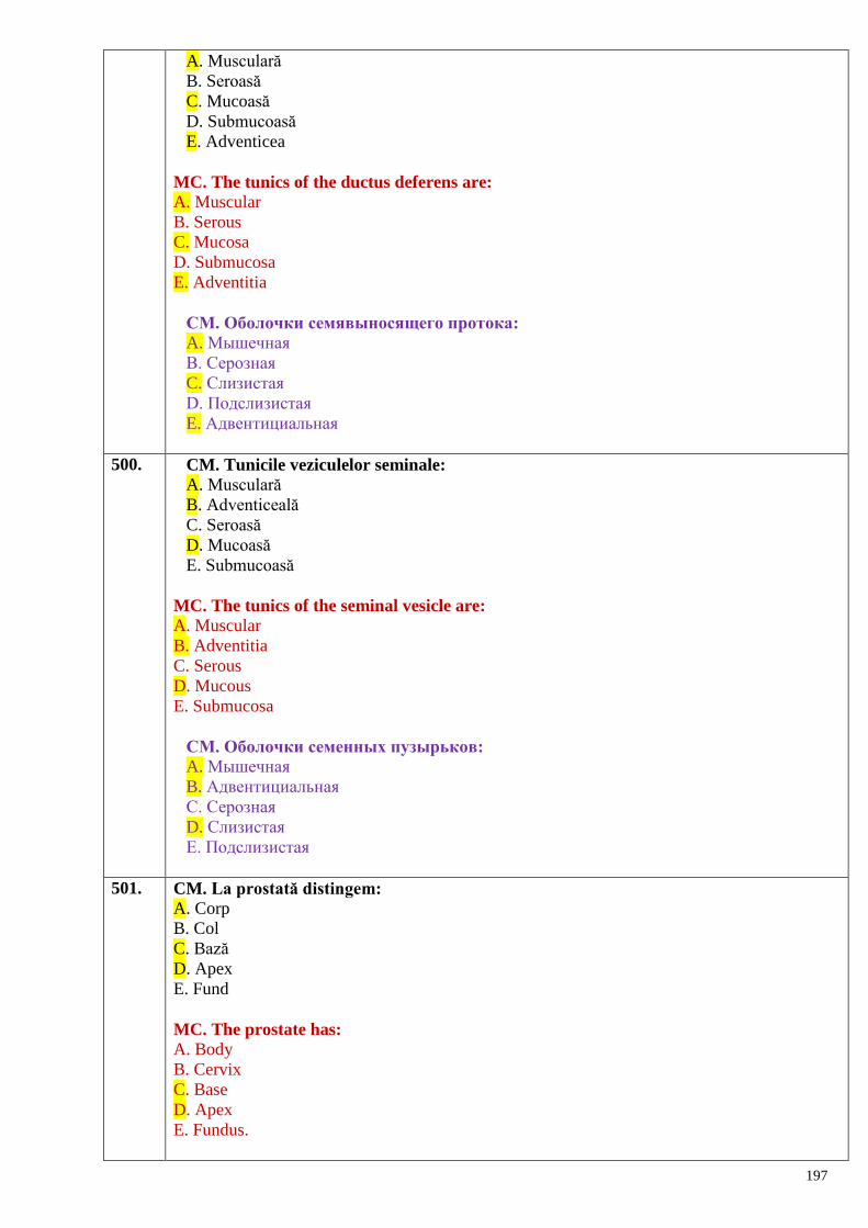
197
A. Musculară
B. Seroasă
C. Mucoasă
D. Submucoasă
E. Adventicea
MC. The tunics of the ductus deferens are:
A. Muscular
B. Serous
C. Mucosa
D. Submucosa
E. Adventitia
СМ. Оболочки семявыносящего протока:
A. Мышечная
B. Серозная
C. Слизистая
D. Подслизистая
E. Адвентициальная
500. CM. Tunicile veziculelor seminale:
A. Musculară
B. Adventiceală
C. Seroasă
D. Mucoasă
E. Submucoasă
MC. The tunics of the seminal vesicle are:
A. Muscular
B. Adventitia
C. Serous
D. Mucous
E. Submucosa
СМ. Оболочки семенных пузырьков:
A. Мышечная
B. Адвентициальная
C. Серозная
D. Слизистая
E. Подслизистая
501. CM. La prostată distingem:
A. Corp
B. Col
C. Bază
D. Apex
E. Fund
MC. The prostate has:
A. Body
B. Cervix
C. Base
D. Apex
E. Fundus.
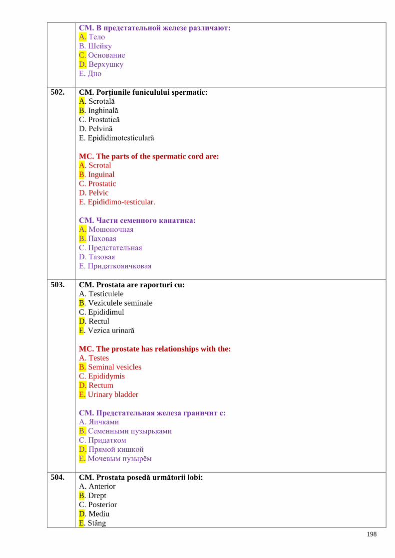
198
СМ. В предстательной железе различают:
A. Тело
B. Шейку
C. Основание
D. Верхушку
E. Дно
502. CM. Porțiunile funiculului spermatic:
A. Scrotală
B. Inghinală
C. Prostatică
D. Pelvină
E. Epididimotesticulară
MC. The parts of the spermatic cord are:
A. Scrotal
B. Inguinal
C. Prostatic
D. Pelvic
E. Epididimo-testicular.
СМ. Части семенного канатика:
A. Мошоночная
B. Паховая
C. Предстательная
D. Тазовая
E. Придаткояичковая
503. CM. Prostata are raporturi cu:
A. Testiculele
B. Veziculele seminale
C. Epididimul
D. Rectul
E. Vezica urinară
MC. The prostate has relationships with the:
A. Testes
B. Seminal vesicles
C. Epididymis
D. Rectum
E. Urinary bladder
СМ. Предстательная железа граничит с:
A. Яичками
B. Семенными пузырьками
C. Придатком
D. Прямой кишкой
E. Мочевым пузырѐм
504. CM. Prostata posedă următorii lobi:
A. Anterior
B. Drept
C. Posterior
D. Mediu
E. Stâng
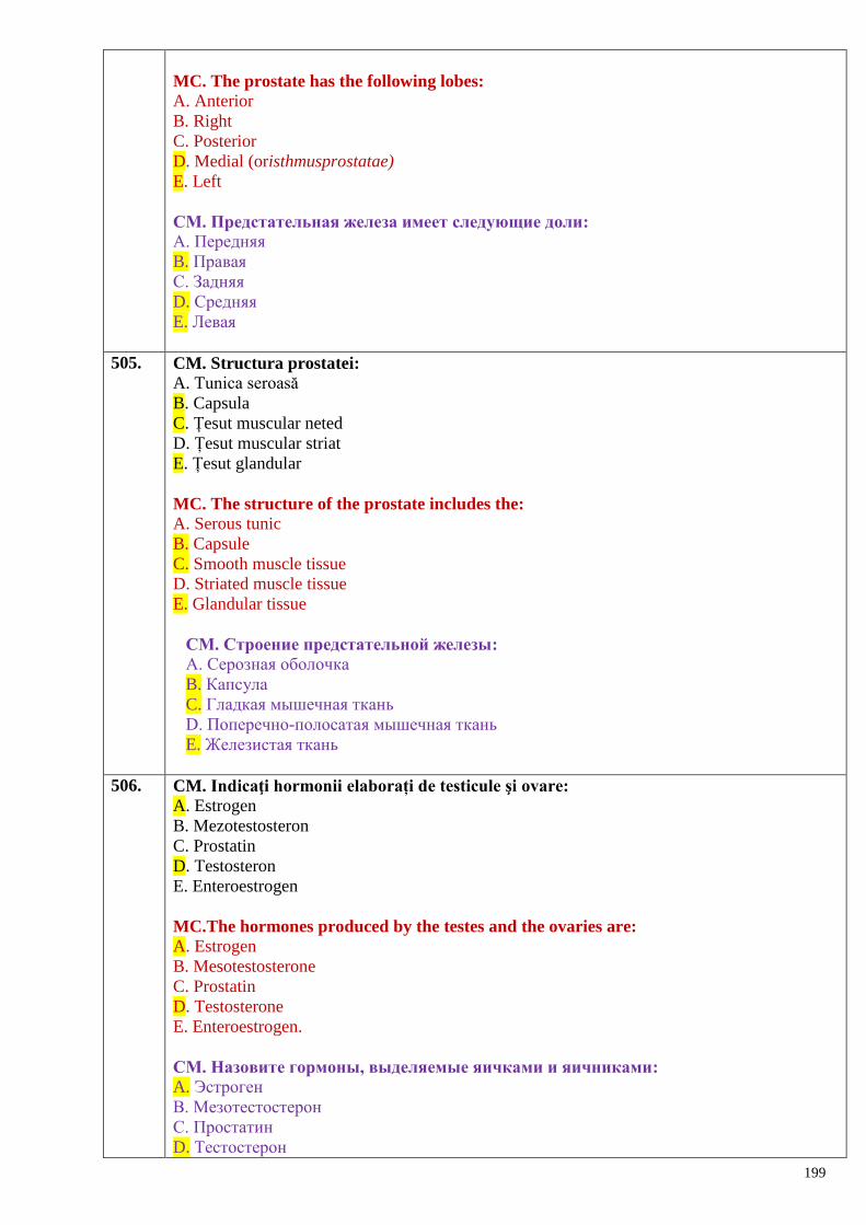
199
MC. The prostate has the following lobes:
A. Anterior
B. Right
C. Posterior
D. Medial (oristhmusprostatae)
E. Left
СМ. Предстательная железа имеет следующие доли:
A. Передняя
B. Правая
C. Задняя
D. Средняя
E. Левая
505. CM. Structura prostatei:
A. Tunica seroasă
B. Capsula
C. Țesut muscular neted
D. Țesut muscular striat
E. Țesut glandular
MC. The structure of the prostate includes the:
A. Serous tunic
B. Capsule
C. Smooth muscle tissue
D. Striated muscle tissue
E. Glandular tissue
СМ. Строение предстательной железы:
A. Серозная оболочка
B. Капсула
C. Гладкая мышечная ткань
D. Поперечно-полосатая мышечная ткань
E. Железистая ткань
506. CM. Indicaţi hormonii elaborați de testicule şi ovare:
A. Estrogen
B. Mezotestosteron
C. Prostatin
D. Testosteron
E. Enteroestrogen
MC.The hormones produced by the testes and the ovaries are: A. Estrogen
B. Mesotestosterone
C. Prostatin
D. Testosterone
E. Enteroestrogen.
СМ. Назовите гормоны, выделяемые яичками и яичниками:
A. Эстроген
B. Мезотестостерон
C. Простатин
D. Тестостерон
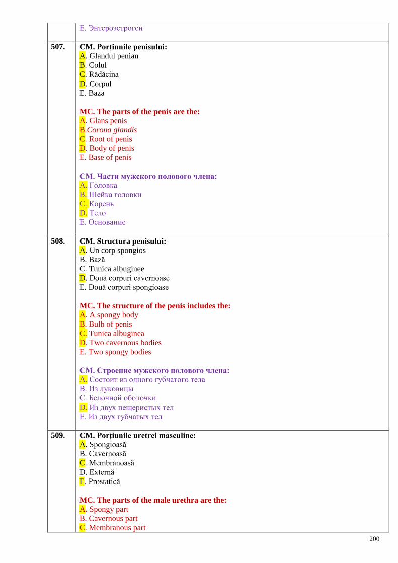
200
E. Энтероэстроген
507. CM. Porțiunile penisului:
A. Glandul penian
B. Colul
C. Rădăcina
D. Corpul
E. Baza
MC. The parts of the penis are the:
A. Glans penis
B.Corona glandis
C. Root of penis
D. Body of penis
E. Base of penis
СМ. Части мужского полового члена:
A. Головка
B. Шейка головки
C. Корень
D. Тело
E. Основание
508. CM. Structura penisului:
A. Un corp spongios
B. Bază
C. Tunica albuginee
D. Două corpuri cavernoase
E. Două corpuri spongioase
MC. The structure of the penis includes the: A. A spongy body
B. Bulb of penis
C. Tunica albuginea
D. Two cavernous bodies
E. Two spongy bodies
СМ. Строение мужского полового члена:
A. Состоит из одного губчатого тела
B. Из луковицы
C. Белочной оболочки
D. Из двух пещеристых тел
E. Из двух губчатых тел
509. CM. Porțiunile uretrei masculine:
A. Spongioasă
B. Cavernoasă
C. Membranoasă
D. Externă
E. Prostatică
MC. The parts of the male urethra are the: A. Spongy part
B. Cavernous part
C. Membranous part
201
D. External part
E. Prostatic part
СМ. Части мочеиспускательного канала:
A. Губчатая
B. Пещеристая
C. Перепончатая
D. Наружная
E. Предстательная
510. CM. Îngustările uretrei masculine se află la nivelul:
A. Bulbului penian
B. Diafragmului urogenital
C. Orificiului extern
D. Părții prostatice
E. Orificiului intern
MC. Narrowings (or constrictions) of the male urethra are located at the level of the:
A. Bulb of penis
B. Membranous part
C. External urethral orifice (or meatus)
D. Prostatic part
E. Internal uretheral orifice (or meatus)
СМ. Сужения мужского мочеиспускательного канала находятся на уровне:
A. Луковицы полового члена
B. Перепончатой части
C. Наружного отверстия
D. Предстательной части
E. Внутреннего отверстия
511. CM. Dilatările uretrei masculine se află în:
A. Partea membranoasă
B. Partea cavernoasă
C. Partea spongioasă
D. Partea prostatică
E. Regiunea bulbului penian
MC. Male urethra dilatations are located at the level of the:
A. Membranous part
B. Cavernous part
C. Glans penis
D. Prostatic part
E. Bulb of penis
СМ. Расширения мужского мочеиспускательного канала находятся в:
A. Перепончатой части
B. Пещеристой части
C. Области головки полового члена (fosea navicularis)
D. Предстательной части
E. Области луковицы полового члена
512. CM. Uretra masculină se explorează prin:
A. Cateterism
B. Colposcopie
202
C. Uretrografie
D. Uretroscopie
E. Colpografie
MC. The male urethra can be explored by:
A. Catheterization
B. Colposcopy
C. Urethrography
D. Urethrocystoscopy
E. Colpography
СМ. Мужской мочеиспускательный канал исследуется путѐм:
A. Катетеризации
B. Колпоскопии
C. Уретрографии
D. Уретроскопии
E. Колпографии
513. CM. Sunt tunici ale scrotului:
A. Pielea
B. Fascia cremasterică
C. Fascia spermatică internă
D. Mucoasa
E. Musculara
MC. The tunics of scrotum are the:
A. Skin
B. Cremasteric fascia
C. Internal spermatic fascia
D. Mucosa
E. Muscular coat
СМ. Оболочки мошонки:
A. Кожа
B. Фасция мышцы, поднимающей яичко
C. Внутренняя семенная фасция
D. Слизистая
E. Мышечная
514. CS. Care din organele genitale feminine sunt parenchimatoase:
A. Uterul
B. Ovarul
C. Trompele uterine
D. Vaginul
E. Clitorisul
SC. Which female genitalia are the parenchymal organs:
A. Uterus
B. Ovary
C. Fallopian tube
D. Vagina
E. Clitoris
СS. Какие из женских половых органов являются паренхиматозными:
A. Матка
203
B. Яичник
C. Маточные трубы
D. Влагалище
E. Клитор
515. CS. În raport cu peritoneul ovarul este situat:
A. Intraperitoneal
B. Mezoperitoneal
C. Extraperitoneal
D. În cavitatea peritoneală
E. Toate false
SC. In relation to the peritoneum the ovary is located:
A. Intraperitoneally
B. Mesoperitoneally
C. Extraperitoneally
D. In the peritoneal cavity
E. Retoperitoneally
СS. По отношению к брюшине яичник расположен:
A. Интраперитонеально
B. Мезоперитонеально
C. Экстраперитонеально
D. В полости брюшины
E. Все утверждения неверны
516. CS. Foliculii maturi ai ovarului se află în:
A. Substanţa medulară
B. Stroma ovarului
C. Tunica albuginee
D. Substanța corticală
E. Epiteliul embrionar
CS. Mature follicles of the ovaries are located in the:
A. Ovarian medulla
B. Ovarian stroma
C. Tunica albuginea
D. Ovarian cortex
E. Embryonic epithelium
СS. Зрелые фолликулы яичника расположены в:
A. Мозговом веществе
B. Строме яичника
C. Белочной оболочке
D. Корковом веществе
E. Эмбриональном эпителии
517. CM. Ovocitul din foliculul maturizat pătrunde în:
A. Trompa uterină
B. Cavitatea uterului
C. Parametrium
D. Vagin
E. Cavitatea peritoneală
CS. Oocyte from the matured follicle enters:
204
A. The fallopian tube
B. The cavity of the uterus
C. The parametrium
D. The vagina
E. The peritoneal cavity
СS. Овоцит из зрелых фолликулов поступает в:
A. Маточную трубу
B. Полость матки
C. Околоматочную клетчатку
D. Влагалище
E. Брюшинную полость
518. CS. Celulele sexuale feminine se maturizează în:
A. Foliculii ovarului
B. Trompele uterine
C. Hilul ovarului
D. Vagin
E. Uter
SC. The female sex cells mature in:
A. Ovarian follicles
B. Fallopian tubes
C. Ovarian hilum
D. Vagina
E. Uterus
СS. Женские половые клетки созревают в:
A. Фолликулах яичника
B. Маточной трубе
C. Воротах яичника
D. Влагалище
E. Матке
519. CS. Raportul trompei uterine cu peritoneul:
A. Extraperitoneal
B. Retroperitoneal
C. Mezoperitoneal
D. Intraperitoneal
E. In cavitatea peritoneului
SC. Therelation of the fallopian tube towards the peritoneum:
A. Extraperitoneal position
B. Retroperitoneal position
C. Mesoperitoneal position
D. Intraperitoneal position
E. In the peritoneal cavity
СS. Отношение брюшины к маточной трубе:
A. Экстраперитонеально
B. Ретроперитонеально
C. Мезоперитонеально
D. Интраперитонеально
E. В полости брюшины
205
520. CS. Ce formă are cavitatea uterului pe radiogramă:
A. De romb
B. Triunghiulară
C. Ovală
D. Pătrată
E. Neregulată
SC. What kind of shape does the cavity of the uterus have on a radiogram:
A. Rhombus
B. Triangular
C. Oval
D. Square
E. Irregular
СS. Какую форму на рентгенограмме имеет полость матки:
A. Ромба
B. Треугольную
C. Овальную
D. Квадратную
E. Неправильную
521. CM. Organele genitale feminine interne:
A. Uterul
B. Trompele uterine
C. Vaginul
D. Ovarele
E. Clitorisul
MC. The internal female genital organs are, as follows: A. Uterus
B. Fallopian tubes
C. Vagina
D. Ovaries
E. Clitoris
СМ. Внутренние женские половые органы:
A. Матка
B. Маточные трубы
C. Влагалище
D. Яичники
E. Клитор
522. CM. Organele genitale feminine externe:
A. Vaginul
B. Clitorisul
C. Labiile mici
D. Trompele uterine
E. Labiile mari
CM. The external female genital organs are, as follows:
A. Vagina
B. Clitoris
C. Labia minora
D. Fallopian tubes
E. Labia majora
206
СМ. Наружные женские половые органы:
A. Влагалище
B. Клитор
C. Малые половые губы
D. Маточные трубы
E. Большие половые губы
523. CM. La ovar distingem feţele:
A. Anterioară
B. Posterioară
C. Laterală
D. Superioară
E. Medială
CM. The ovarian faces are:
A. Anterior
B. Posterior
C. Lateral
D. Superior
E. Medial
СМ. Яичник имеет следующие поверхности:
A. Передняя
B. Задняя
C. Латеральная
D. Верхняя
E. Медиальная
524. CM. Marginile ovarului:
A. Liberă
B. Medială
C. Inferioară
D. Superioară
E. Mezovarică
CM. The ovarian borders are:
A. Free
B. Medial
C. Inferior
D. Superior
E. Mesovarian
СМ. Края яичника:
A. Свободный
B. Медиальный
C. Нижний
D. Верхний
E. Брыжеечный
525. CM. Extremitățile ovarului:
A. Uterină
B. Uretrală
C. Laterală
D. Tubară
207
E. Medială
CM. The ovarian ends are:
A. Uterine
B. Urethral
C. Lateral
D. Tubal
E. Medial
СМ. Концы яичника:
A. Маточный
B. Мочеиспускательный
C. Латеральный
D. Трубный
E. Медиальный
526. CM. Aparatul de fixare a ovarului include ligamentele:
A. Uteroovarian
B. Tuboovarian
C. Suspensor al ovarului
D. Rotund al ovarului
E. Propriu al ovarului
CM. The fixation apparatus of the ovary includes the following ligaments:
A. Uteroovarian
B. Tuboovarian
C. Suspensory ligament
D. Round ligament
E. Proper ligament of the ovary
СМ. Фиксирующий аппарат яичника включает связки:
A. Маточно-яичниковая
B. Трубно-яичниковая
C. Подвешивающая связка яичника
D. Круглая связка яичника
E. Собственная связка яичника
527. CM. Parenchimul ovarului este alcătuit din:
A. Substanță corticală
B. Substanță endocrină
C. Substanță canaliculară
D. Substanță glandulară
E. Substanță medulară
CM. The ovarian parenchyma is composed of: A. Cortical substance
B. Endocrine substance
C. Canalicular substance
D. Glandular substance
E. Medullar substance
СМ. Паренхима яичника состоит из:
A. Коркового вещества
B. Эндокринного вещества
C. Каналикулярного вещества
208
D. Железистого вещества
E. Мозговое вещество
528. CM. Fazele ciclului menstrual:
A. Premenstruală (secreție)
B. Intermediară
C. Menstruală
D. Retromenstruală
E. Postmenstruală
CM. The phases of the menstrual cycle are:
A. Premenstrual (secretion)
B. Intermediate
C. Menstrual
D. Retromenstrual
E. Postmenstrual
СМ. Фазы менструального цикла:
A. Предменструальная (секреции)
B. Промежуточная
C. Менструальная
D. Ретроменструальная
E. Постменструальная
529. CM. Metrosalpingografia reprezintă metoda de explorare radiologică a:
A. Uretrei
B. Uterului
C. Ureterelor
D. Trompelor uterine
E. Ovarelor
CM. Metrosalpingography is a radiological method of investigation for:
A. The urethra
B. The uterus
C. The ureters
D. The tubes
E. The ovaries
СМ. Метросалпингография представляет радиологический метод исследования:
A. Мочеиспускательного канала
B. Матки
C. Мочеточников
D. Маточных труб
E. Яичников
530. CM. Colposcopia reprezintă metoda de explorare a:
A. Ovarelor
B. Colului uterin
C. Uterului
D. Vaginului
E. Rectului
CM. Colposcopy is an investigation method for: A. The ovaries
B. The cervix
209
C. The uterus
D. The vagina
E. The rectum
СМ. Колпоскопия – это метод исследования:
A. Яичников
B. Шейки матки
C. Матки
D. Влагалища
E. Прямой кишки
531. CM. Porțiunile salpingelui:
A. Uterină
B. Ovarică
C. Ampula
D. Istmul
E. Infundibulul
CM. The following parts are distinguished in the salpinx:
A. Uterine part
B. Ovarian part
C. Ampulla
D. Isthmus
E. Infundibulum
СМ. Части маточной трубы:
A. Маточная
B. Яичниковая
C. Ампула
D. Перешеек
E. Воронка
532. CM. Porțiunile uterului:
A. Superioară
B. Fundul
C. Inferioară
D. Colul
E. Corpul
CM. The parts of the uterus are:
A. Superior
B. Fundus
C. Inferior
D. Neck
E. Body
СМ. Части матки:
A. Верхняя
B. Дно
C. Нижняя
D. Шейка
E. Тело
533. CM. Uterul este situat:
A. Anterior de rect
210
B. Anterior de vezica urinară
C. Posterior de rect
D. Posterior de vezica urinară
E. Între rect si vezica urinară
CM. The uterus is located:
A. In front of the rectum
B. In front of the urinary bladder
C. Behind of the rectum
D. Behind of the urinary bladder
E. Between the rectum and the urinary bladder
СМ. Матка расположена:
A. Впереди прямой кишки
B. Впереди мочевого пузыря
C. Позади прямой кишки
D. Позади мочевого пузыря
E. Между прямой кишкой и мочевым пузырѐм
534. CM. Porțiunile colului uterin:
A. Infravaginală
B. Istmul uterin
C. Vaginală
D. Extravaginală
E. Supravaginală
CM. The portions of uterine cervix are:
A. Infravaginal
B. Uterine isthmus
C. Vaginal
D. Extravaginal
E. Supravaginal
СМ. Части шейки матки:
A. Подвлагалищная
B. Перешеек матки
C. Влагалищная
D. Вневлагалищная
E. Надвлагалищная
535. CM. Tunicile peretelui uterin:
A. Endometrul
B. Mezometrul
C. Miometrul
D. Perimetrul
E. Parametrul
CM. The tunics of the uterine wall are:
A. Endometrium
B. Mesometrium
C. Myometrium
D. Perimetrium
E. Parametrium
СМ. Оболочки стенки матки:
211
A. Эндометрий
B. Мезометрий
C. Миометрий
D. Периметрий
E. Параметрий
536. CM. Peretele vaginal este constituit din tunicile:
A. Mucoasă
B. Musculară
C. Spongioasă
D. Adventice
E. Seroasă
CM. The vaginal wall consists of the follwing coats:
A. Mucous
B. Muscular
C. Spongious
D. Adventitia
E. Serous
СМ. Стенка влагалища состоит из оболочек:
A. Слизистой
B. Мышечной
C. Губчатой
D. Адвентициальной
E. Серозной
537. CS. Perineul reprezintă:
A. Foiţa ce acoperă viscerele cavității abdominale
B. Foiţa ce acoperă pereții cavității abdominale
C. Foiţa ce acoperă organele bazinului mic
D. Foiţa ce acoperă pereții cavității bazinului mic
E. Complex de țesuturi moi care închid ieșirea din cavitatea micului bazin
CS. The perineum is the:
A. Layer covering the abdominal viscera
B. Layer covering the walls of the abdominal cavity
C. Layer covering the lesser pelvic organs
D. Layer covering the lesser pelvic cavity walls
E. Complex of soft tissue that close the pelvic outlet
СS. Промежность представляет:
A. Листок, покрывающий внутренние органы брюшной полости
B. Листок, покрывающий стенки брюшной полости
C. Листок, покрывающий органы малого таза
D. Листок, покрывающий стенки малого таза
E. Комплекс мягких тканей, закрывающий выход из полости малого таза
538. CS. Posterior perineul este delimitat de:
A. Promontoriu
B. Vârful coccisului
C. Vertebra sacrală 5
D. Vertebra sacrală 2
E. Limita dintre vertebrele sacrale şi coccis
212
CS. Posteriorly the perineum is delimited by the:
A. Promontorium
B. Apex of the coccyx
C. The 5th
sacral vertebra
D. The 2nd
sacral vertebra
E. Borderline between the sacral and coccygeal vertebrae
СS. Сзади промежность ограничена:
A. Мысом крестца
B. Верхушкой копчика
C. 5-м крестцовым позвонком
D. 2-м крестцовым позвонком
E. Граница между крестцовыми позвонками и копчиком
539. CM. Diafragmul urogenital:
A. Ocupă partea posterioară a perineului
B. Prin el la bărbat trece uretra
C. Prin el la femeie trece uretra si vaginul
D. Este delimitat de ramurile superioare ale oaselor pubiene si ischiatice
E. Vârful este orientat spre centrul perineului
CM. The urogenital diaphragm:
A. Occupies the posterior part of perineum
B. The urethra passes through it in males
C. The urethra and vagina passes through it in females
D. It is delimited by superior rami of the pubic bones and rami of the ischium
E. Its tip is oriented to the center of the perineum
СМ. Мочеполовая диафрагма:
A. Занимает заднюю часть промежности
B. Через неѐ у мужчин проходит мочеиспускательный канал
C. Через неѐ у женщин проходит мочеиспускательный канал и отверстие влагалища
D. Ограничена верхними ветвями лобковых и ветвями седалищных костей
E. Верхушка направлена к сухожильному центру промежности
540. CS. Nefronul funcționează începând cu săptămâna:
A. A 8-a
B. A 9-a
C. A 4-a
D. A 7-ea
E. A 5-ea
SC. The nephron starts to function on:
A. 8th
week
B. 9th
week
C. 4th
week
D. 7th
week
E. 5th
week
CS. Нефрон начинает функционировать с:
A. 8-ой недели
B. 9-ой недели
C. 4-ой недели
D. 7-ой недели
E. 5-ой недели
213
541. CM. Etapele de dezvoltare a rinichilor:
A. Retronefros
B. Pronefros
C. Antinefros
D. Mezonefros
E. Metanefros
MC. The stages of the kidneys development are:
A. Retronephros
B. Pronephros
C. Antinephros
D. Mesonephros
E. Metanephros
CM. Этапы развития почек:
A. Ретронефрос
B. Предпочка (пронефрос)
C. Антинефрос
D. Первичная почка (мезонефрос)
E. Окончательная почка (метанефрос)
542. CS. Care proces stă la baza formării urinei primare:
A. Ultrafiltrația
B. Reabsorbția
C. Metabolic
D. Modificările presiunii arteriale
E. Modificările presiunii intraabdominale
SC. The process underlying the formation of the primary urine is:
A. Ultrafiltration (or glomerular filtration)
B. Reabsorbtion (or tubular reabsorbtion)
C. Metabolic processes
D. Changes of the blood pressure
E. Changes of the intra-abdominal pressure
CS. На основании какого процесса образуется первичная моча:
A. Ультрафильтрации
B. Реабсобции
C. Обмена веществ
D. Изменения артериального давления
E. Изменения внутрибрюшного давления
543. CS. Hilul rinichiului se află?
A. În porțiunea de mijloc a marginii laterale
B. Pe faţa anterioară
C. La polul superior şi cel inferior
D. Pe faţa posterioară
E. În porţiunea de mijloc a marginii mediale
SC.The renal hilum is located:
A. In the middle of the lateral margin
B.On the anterior surface
C. At the upper and lower poles
D. On the posterior surface
214
E. In the middle of the medial margin.
CS. Ворота почки находятся:
A. Посередине латерального края
B. На передней поверхности
C. На верхнем и нижнем полюсах
D. На задней поверхности
E. Посередине медиального края
544. CS. Capsula adipoasă prin hilul renal trece în:
A. Piramidele renale
B. Fascia renală
C. Substanța medulară
D. Substanța corticală
E. Sinusul renal
SC. The adipose capsule passes through the renal hilum into the:
A. Renal pyramids
B. Renal fascia
C. Renal medulla
D. Renal cortex
E. Renal sinus
CS. Жировая капсула через ворота почек переходит в:
A. Почечные пирамиды
B. Почечную фасцию
C. Мозговое вещество
D. Корковое вещество
E. Почечную лоханку
545. CS. Corpul adipos pararenal se află pe:
A. Faţa anterioară a rinichiului
B. Polul superior al rinichiului
C. În hilul renal
D. Faţa posterioară a rinichiului
E. Polul inferior al rinichiului
SC. The pararenal adipose body is located:
A. On the anterior surface of the kidney
B. On the upper pole of the kidney
C. In the renal hilum
D. On the posterior surface of the kidney
E. On the inferior pole of the kidney
CS. Околопочечное жировое тело находится на:
A. Передней поверхности почек
B. Верхнем полюсе почки
C. Почечных воротах
D. Задней поверхности почки
E. Нижнем полюсе почки
546. CS. Rinichiul este un organ:
A. Tubular
B. Glandular
C. Parenchimatos
215
D. Cavitar
E. Mixt
SC. The kidney is an organ:
A. Tubular
B. Glandular
C. Parenchymatous
D. Cavitary
E. Mixed
CS. Почка – это орган:
A. Трубчатый
B. Железистый
C. Паренхиматозный
D. Полый
E. Смешанный
547. CS. Stratul superficial al parenchimului renal este dat de:
A. Capsula adipoasă
B. Fascia renală
C. Substanța corticală
D. Tunica seroasă
E. Substanța medulară
SC. Superficial layer of the renal parenchyma is formed by the:
A. Adipose capsule
B. Renal fascia
C. Renal cortex
D. Serous tunic
E. Renal medulla
CS. Поверхностный слой паренхимы почек образован:
A. Жировой капсулой
B. Почечной фасцией
C. Корковым веществом
D. Серозной оболочкой
E. Мозговым веществом
548. CS. Unitatea morfo-funcţională a rinichiului este:
A.Lobul renal
B.Segmentul renal
C.Lobulul renal
D.Corpusculul renal
E.Nefronul
SC. The morphofunctional unit of the kidney is the:
A. Renal lobe
B.Renal segment
C.Renal lobule
D.Renal corpuscle
E. Nephron
CS. Морфо-функциональная единица почек это:
A. Почечная доля
B. Почечный сегмент
216
C. Почечная долька
D. Почечное тельце
E. Нефрон
549. CS. Aria cribroasă se află:
A. În porțiunea radiată
B. În coloanele renale
C. În piramidele renale
D. La baza piramidei renale
E. La vârful piramidei renale
SC. The cribriform area is located:
A. In the radiate portion
B. In the renal columns
C. In the renal pyramids
D. At the bottom of the renal pyramid
E. At the top of the renal pyramid
CS. Решѐтчатое поле находится:
A. В лучистой части
B. В почечных столбах
C. В почечных пирамидах
D. На основании почечных пирамид
E. На верхушке почечных пирамид
550. CS. Un lob renal include:
A. O piramidă renală şi caliciile mari
B. Piramida renală cu sinusul renal
C. Piramida renală cu porțiunea radiată a rinichiului
D. O piramidă renală cu caliciile mici
E. O piramidă renală cu substanța corticală adiacentă
SC. A renal lobe includes:
A. A renal pyramid and major calyces
B. A renal pyramid and renal sinus
C. A renal pyramid with radiate portion of the kidney
D. A renal pyramid and minor calyces
E. A renal pyramid with the adjacent renal cortex.
CS. В почечную долю входит:
A. Почечная пирамида с малыми чашечками
B. Почечная пирамида с почечной лоханкой
C. Почечная пирамида с лучистой частью почки
D. Почечная пирамида с большими чашечками
E. Почечная пирамида с прилежащим к ней корковым веществом
551. CS. Nefronii sunt situați în:
A. Piramidele renale
B. Profunzimea substanței corticale
C. Porțiunea radiată
D. Coloanele renale
E. Sinusul renal
SC. The nephrons are located:
A. In the renal pyramids
217
B. Only in the renal cortex
C. Only in the renal medulla
D. In the renal cortex and medulla
E. In the renal sinus
CS. Нефроны расположены:
A. В почечных пирамидах
B. В корковом веществе
C. В лучистой части
D. В почечных столбах
E. В почечной пазухе
552. CS. Bazinetul renal se formează la unirea:
A. A două calice renale mari cu una mică
B. La unirea a 5 - 6 calicii renale mici
C. A 2- calice renale mici cu 1 mare
D. A 2- 3 calice renale mici
E. A 2 - 3 calice mari
SC. The renal pelvis is formed by the union of:
A. Two major calyces with a minor one
B. 5 - 6 minor renal calyces
C. Two minor calyces with a major one
D. 2 - 3 minor renal calyces
E. 2 - 3 major renal calyces
CS. Почечная лоханка образуется при слиянии:
A. Двух больших чашек с одной малой
B. 5-ти – 6-ти малых чашек
C. Двух малых чашек с одной большой
D. Двух - трѐх малых чашек
E. Двух - трѐх больших чашек
553. CS. Structura lobulară externă a rinichilor se păstrează până la vârsta de:
a) nou-născut
b) 7 ani
c) pubertară
d) 22 - 25 ani
e) 2 - 3 ani
SC. The external lobular structure of the kidney is kept to the age:
A. In newborn
B. Until the age 7 years
C. Until puberty age
D. Until the age 22 - 25 years
E. Until the age 2 - 3 years
CS. Наружное дольчатое строение почек сохраняется до какого возраста?
a) у новорождѐнного
b) до 7-ми лет
c) до пубертатного
d) до 22 – 25 лет
e) до 2-3-х лет
554. CS. Sistola şi diatola căilor urinare (calicii-ureter) pot fi studiate prin:
218
a) angiografie
b) tomografie computerizată
c) sonografie
d) radioscopie
e) uretroscopie
SC. Systole and diastole of the urinary tract (calyces-ureter) may be examined by:
A. Angiography
B. CT
C. Sonography
D. Radioscopy
E. Ureteroscopy
CМ. Систола и диастола мочевыводящих путей (чашечки-мочеточник)
обследуется путѐм:
a) радиологического исследования сосудов
b) компьютерной томографии
c) сонографии
d) радиоскопии
e) уретроскопии
555. CS. Faţă de peritoneu ureterul este dispus:
a) extraperitoneal
b) în cavitatea peritoneală
c) mezoperitoneal
d) intraperitoneal
e) retroperitoneal
MC. In relation to the peritoneum the ureter is located:
A. Extraperitoneally
B. In the peritoneal cavity
C. Mesoperitoneally
D. Intraperitoneally
E. Retroperitoneally
CS. По отношению к брюшине мочеточник находится:
a) экстраперитонеально
b) в полости брюшины
c) мезоперитонеально
d) интраперитонеально
e) ретроперитонеально
556. CS. Vezica urinară se află în:
a) cavitatea bazinului mare
b) cavitatea bazinului mic
c) cavitatea abdominală
d) fosa iliacă dreaptă
e) regiunea pubiană
SC. The urinary bladder is located in:
A. The cavity of the greater pelvis
B. The cavity of the lesser pelvis
C. The abdominal cavity
D. The right iliac fossa
E. The pubic region
219
CS. Мочевой пузырь расположен:
a) в полости большого таза
b) в полости малого таза
c) в брюшной полости
d) в правой подвздошной ямке
e) в лобковой области
557. CS. Apexul vezicii urinare la bărbat limitrofează cu:
a) sigmoidul
b) porțiunea superioară a rectului
c) veziculele seminale
d) ansele intestinului subțire
e) prostata
SC. In males the apex of the urinary bladder comes in contact with: A. Sigmoid colon
B. Upper part of the rectum
C. Seminal vesicles
D. Loops of the small intestine
E. Anterior abdominal wall
CS. Верхушка мочевого пузыря у мужчин граничит с:
a) сигмовидной кишкой
b) верхней частью прямой кишки
c) семенными пузырьками
d) петлями тонкой кишки
e) передней стенкой брюшной полости
558. CS. Faţa superioară a vezicii urinare la femeie vine in adiacenţă cu:
a) porțiunea superioară a rectului
b) ovarele
c) cecul
d) sigmoidul
e) uterul
MC. In females the posterior surface of the urinary bladder comes in contact with: A. Upper part of the rectum
B. Ovaries
C. Coecum
D. Sigmoid colon
E. Uterus
CS. Верхняя поверхность мочевого пузыря у женщин соприкасается с:
a) верхней частью прямой кишки
b) яичниками
c) слепой кишкой
d) сигмовидной кишкой
e) маткой
559. CS. Faţă de peritoneu vezica urinară în plenitudine, este situată:
a) intraperitoneal
b) in cavitatea peritoneului
c) extraperitoneal
d) retroperitoneal
220
e) mezoperitoneal
SC. In relation to the peritoneum the full urinary bladder is located:
A. Intraperitoneally
B. In the peritoneal cavity
C. Extraperitoneally
D. Retroperitoneally
E. Mesoperitoneally
CS. По отношению к брюшине полный мочевой пузырь находится:
a) интраперитонеально
b) в полости брюшины
c) экстраперитонеально
d) ретроперитонеально
e) мезоперитонеально
560. CS. Vezica urinară goală în raport cu peritoneul este situată:
a) în cavitatea peritoneului
b) extraperitoneal
c) retroperitoneal
d) mezoperitoneal
e) intraperitoneal
SC. In relation to the peritoneum the empty urinary bladder is located:
A. In the peritoneal cavity
B. Extraperitoneally
C. Retroperitoneally
D. Mesoperitoneally
E. Intraperitoneally
CS. Пустой мочевой пузырь по отношению к брюшине находится:
a) в полости брюшины
b) экстраперитонеально
c) ретроперитонеально
d) мезоперитонеально
e) интраперитонеально
561. CS. Baza submucoasă a vezicii urinare lipsește în regiunea:
a) corpului
b) colului
c) fundului
d) apexului
e) triunghiului vezical
SC. Submucosa of the urinary bladder is absent in the following regions: A. Body
B. Neck
C. Fundus
D. Apex
E. Bladder trigone
CS. Подслизистая основа мочевого пузыря отсутствует:
a) в теле
b) в шейке
c) в области дна
221
d) в верхушке
e) в области пузырного треугольника
562. CS. Uretra:
A. are o lungime de cca 16 cm la bărbat şi la femeie – 4 cm
B. lumenul ei la bărbat e mai larg
C. la femeie prezintă 2 curburi şi trei porţiuni
D. la bărbat lungimea ei este variabilă şi depinde de starea penisului (flasc sau în erecţie)
E. la bărbat este rectilinie
SC. The urethra:
A. Has a length of about 16 cm in male and 4 cm in female
B. Its lumen is larger in male
C. Female urethra has two curvatures and three portions
D. Is of variable length in male; it depends on the condition of the penis (flaccid or erect)
E. Is straight males
CS. Мочеиспускательный канал:
a) имеет длину около 16 см у мужчин и 4 см – у женщин
b) просвет у мужчин больше
c) у женщин имеет 2 изгиба и 3 части
d) у мужчин длина варьирует в зависимости от состояния мужского полового члена
(расслаблен или в эрекции)
e) у мужчин прямой
563. CS. Voluntar e sfincterul uretrei:
A.Intermediar
B.intern
C.extern
D.glandopenian
E.intravezical
SC. The following urethral sphincter is voluntary one:
A. Intermediate
B. Internal
C. External
D. Glandopenile
E. Intravesical
CS. Какой сфинктер у мочеиспускательного канала произволен:
A. промежуточный
B. внутренний
C.наружный
D. на головке полового члена
E. в стенке мочевого пузыря
564. CS. La bărbat uretra are funcţie:
A.erectilă
B.de evacuare a urinei şi a spermei
C.de evacuare a urinei şi ejaculatoare
D.de evitare a refluxului urinar
E.de relaxare a corpilor cavernoşi
SC. The male urethra has the following functions:
A. Erectile
222
B. Discharge of urine and semen
C. Discharge of urine and ejaculatory function
D. Prevention of the urinary reflux
E. Relaxation of the corpora cavernosa
CS. Мочеиспускательный канал у мужчин выполняет функции:
a) эрекции
b) выведение мочи и спермы
c) выведение мочи и семяизвержение
d) выведение мочи
e) расслабление пещеристых тел
565. CM. Funcțiile rinichilor:
a) formarea urinei
b) evacuarea urinei
c) depou a urinei
d) termoreglare
e) endocrină
MC. The kidney`s functions are, as follows:
A. Formation of urine
B. Discharge of urine
C. Storage of urine
D. Thermoregulatory
E. Endocrine
CM. Функции почек:
a) образование мочи
b) выделение мочи
c) депо мочи
d) терморегуляция
e) эндокринная
566. CM. Rinichii prezintă:
A. două feţe – anterioară şi posterioară
B. două margini – laterală şi medială
C. două extremităţi – superioară şi inferioară
D. un hil care conduce în pelvisul renal
E. sunt de culoare roşie-brună
MC. The kidneys have:
A. Two surfaces - anterior and posterior
B. Two margins - lateral and medial
C. Two poles (or extremities) - superior and inferior
D. A renal hilum which continuous with renal pelvis
E. They are pinkish-grey
CM. Почки представляют:
a) две поверхности – передняя и задняя
b) два края – латеральный и медиальный
c) два полюса – верхний и нижний
d) почечные ворота, переходящие в почечную лоханку
e) орган тѐмно-красного цвета
567. CM. Rinichii sunt menţinuţi în poziţia lor prin:
223
A. Loja renală
B. Pediculul renal
C. Fascia renală
D. Ligamentele peritoneale
E. Presa abdominală
MC. The kidneys are maintained in their position by the:
A.Renal bed
B.Renal pedicle
C.Renal fascia
D.Peritoneal ligaments
E. Intra-abdominal pressure
CM. Топографическое расположение почек обеспечивается:
a) почечным ложем
b) почечной ножкой
c) почечной фасцией
d) брюшинными связками
e) внутрибрюшным давлением
568. CM. Capsulele rinichiului:
a) adventicea
b) fibroasă
c) adipoasă
d) fascia renală
e) mucoasă
MC. The kidney capsules are, as follows:
A. Adventitia
B. Fibrous
C. Adipose
D. Renal fascia
E. Mucous
CM. Оболочки почек:
a) адвентициальная
b) фиброзная
c) жировая
d) почечная фасция
e) слизистая
569. CM. In hilul rinichiului intră:
a) vena renală
b) artera renală
c) nervi
d) ureterul
e) vase limfatice
MC. The following elements enter into the kidney through the renal hilum:
A. Renal vein
B. Renal artery
C.Nerves
D. Ureter
E. Lymphatic vessels
224
CM. В почечные ворота входят:
a) почечная вена
b) почечная артерия
c) нервы
d) мочеточник
e) лимфатические сосуды
570. CM. Din rinichi prin hil ies:
a) vasele limfatice
b) artera renală
c) ureterul
d) nervi
e) vena renală
MC. The following elements leave the kidney through the renal hilum:
A. Lymphatic vessels
B. Renal artery
C. Ureter
D. Nerves
E. Renal vein
CM. Из ворот почек выходят:
a) лимфатические сосуды
b) почечная артерия
c) мочеточник
d) нервы
e) почечная вена
571. CM. Aparatul de fixare a rinichiului este constituit din:
a) loja renală
b) pedunculii diafragmei
c) pediculul renal
d) presiunea intraabdominală
e) capsulele renale
MC. Fixation apparatus of the kidney comprises:
A. Renal bed
B. Crura of the diaphragm
C. Renal pedicle
D. Intra-abdominal pressure
E. Renal capsules
CM. Фиксирующий аппарат почек состоит из:
a) почечного ложа
b) ножек диафрагмы
c) почечной ножки
d) внутрибрюшного давления
e) почечной фасции
572. CM. Elementele pediculului renal:
a) bazinetul renal
b) vena renală
c) artera renală
d) fascia renală
e) ureterul
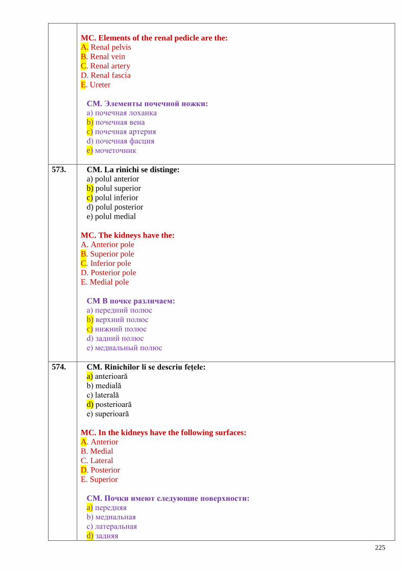
225
MC. Elements of the renal pedicle are the:
A. Renal pelvis
B. Renal vein
C. Renal artery
D. Renal fascia
E. Ureter
CM. Элементы почечной ножки:
a) почечная лоханка
b) почечная вена
c) почечная артерия
d) почечная фасция
e) мочеточник
573. CM. La rinichi se distinge:
a) polul anterior
b) polul superior
c) polul inferior
d) polul posterior
e) polul medial
MC. The kidneys have the:
A. Anterior pole
B. Superior pole
C. Inferior pole
D. Posterior pole
E. Medial pole
CM В почке различаем:
a) передний полюс
b) верхний полюс
c) нижний полюс
d) задний полюс
e) медиальный полюс
574. CM. Rinichilor li se descriu feţele:
a) anterioară
b) medială
c) laterală
d) posterioară
e) superioară
MC. In the kidneys have the following surfaces:
A. Anterior
B. Medial
C. Lateral
D. Posterior
E. Superior
CM. Почки имеют следующие поверхности:
a) передняя
b) медиальная
c) латеральная
d) задняя
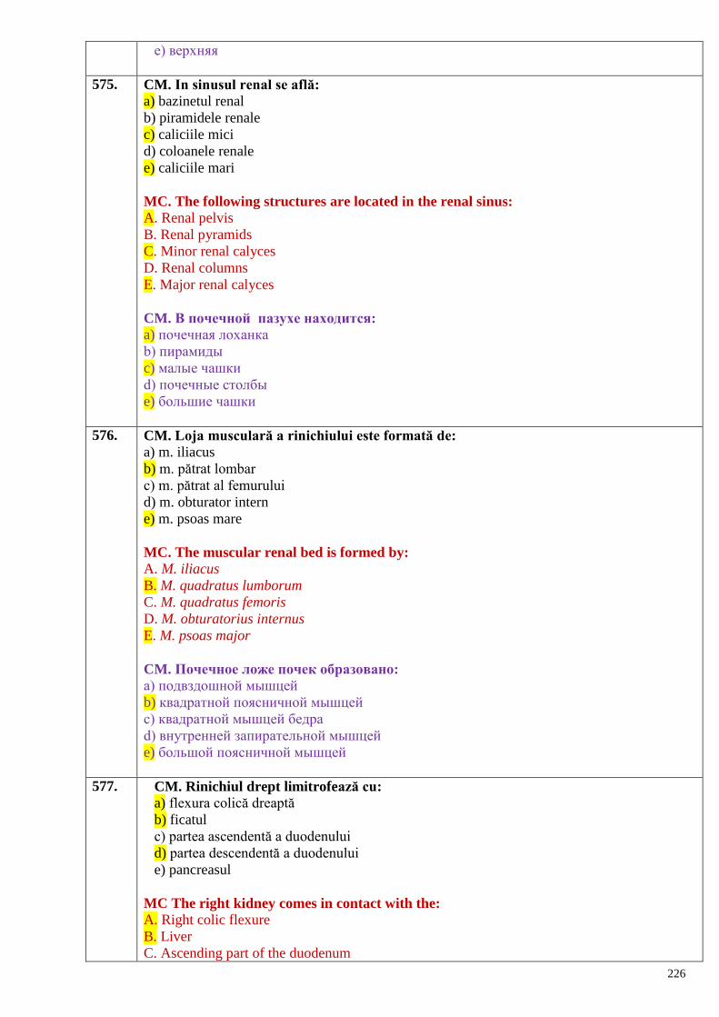
226
e) верхняя
575. CM. In sinusul renal se află:
a) bazinetul renal
b) piramidele renale
c) caliciile mici
d) coloanele renale
e) caliciile mari
MC. The following structures are located in the renal sinus:
A. Renal pelvis
B. Renal pyramids
C. Minor renal calyces
D. Renal columns
E. Major renal calyces
CM. В почечной пазухе находится:
a) почечная лоханка
b) пирамиды
c) малые чашки
d) почечные столбы
e) большие чашки
576. CM. Loja musculară a rinichiului este formată de:
a) m. iliacus
b) m. pătrat lombar
c) m. pătrat al femurului
d) m. obturator intern
e) m. psoas mare
MC. The muscular renal bed is formed by:
A. M. iliacus
B. M. quadratus lumborum
C. M. quadratus femoris
D. M. obturatorius internus
E. M. psoas major
CM. Почечное ложе почек образовано:
a) подвздошной мышцей
b) квадратной поясничной мышцей
c) квадратной мышцей бедра
d) внутренней запирательной мышцей
e) большой поясничной мышцей
577. CM. Rinichiul drept limitrofează cu:
a) flexura colică dreaptă
b) ficatul
c) partea ascendentă a duodenului
d) partea descendentă a duodenului
e) pancreasul
MC The right kidney comes in contact with the:
A. Right colic flexure
B. Liver
C. Ascending part of the duodenum
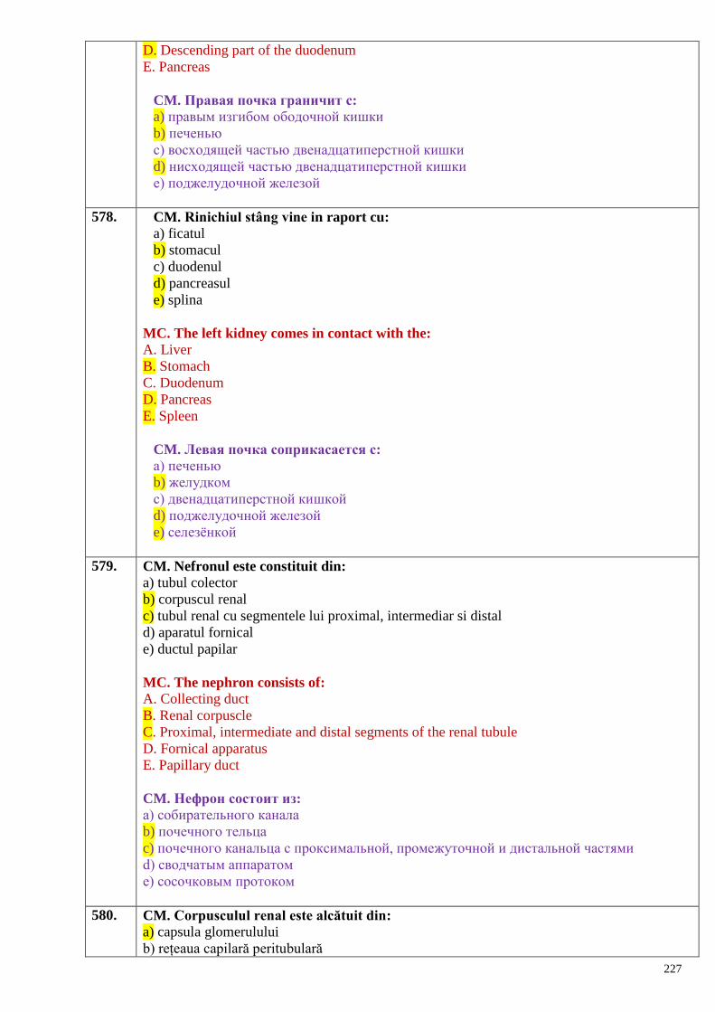
227
D. Descending part of the duodenum
E. Pancreas
CM. Правая почка граничит с:
a) правым изгибом ободочной кишки
b) печенью
c) восходящей частью двенадцатиперстной кишки
d) нисходящей частью двенадцатиперстной кишки
e) поджелудочной железой
578. CM. Rinichiul stâng vine in raport cu:
a) ficatul
b) stomacul
c) duodenul
d) pancreasul
e) splina
MC. The left kidney comes in contact with the:
A. Liver
B. Stomach
C. Duodenum
D. Pancreas
E. Spleen
CM. Левая почка соприкасается с:
a) печенью
b) желудком
c) двенадцатиперстной кишкой
d) поджелудочной железой
e) селезѐнкой
579. CM. Nefronul este constituit din:
a) tubul colector
b) corpuscul renal
c) tubul renal cu segmentele lui proximal, intermediar si distal
d) aparatul fornical
e) ductul papilar
MC. The nephron consists of:
A. Collecting duct
B. Renal corpuscle
C. Proximal, intermediate and distal segments of the renal tubule
D. Fornical apparatus
E. Papillary duct
CM. Нефрон состоит из:
a) собирательного канала
b) почечного тельца
c) почечного канальца с проксимальной, промежуточной и дистальной частями
d) сводчатым аппаратом
e) сосочковым протоком
580. CM. Corpusculul renal este alcătuit din:
a) capsula glomerulului
b) rețeaua capilară peritubulară
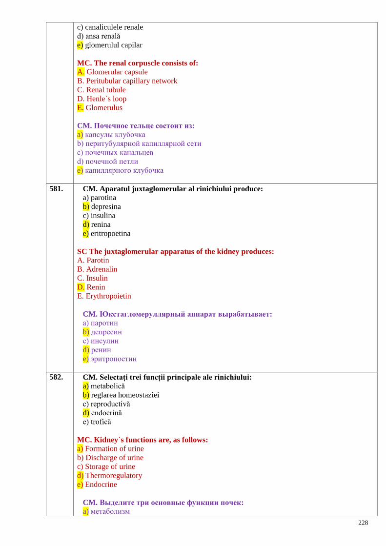
228
c) canaliculele renale
d) ansa renală
e) glomerulul capilar
MC. The renal corpuscle consists of:
A. Glomerular capsule
B. Peritubular capillary network
C. Renal tubule
D. Henle`s loop
E. Glomerulus
CM. Почечное тельце состоит из:
a) капсулы клубочка
b) перитубулярной капиллярной сети
c) почечных канальцев
d) почечной петли
e) капиллярного клубочка
581. CM. Aparatul juxtaglomerular al rinichiului produce:
a) parotina
b) depresina
c) insulina
d) renina
e) eritropoetina
SC The juxtaglomerular apparatus of the kidney produces:
A. Parotin
B. Adrenalin
C. Insulin
D. Renin
E. Erythropoietin
CM. Юкстагломеруллярный аппарат вырабатывает:
a) паротин
b) депресин
c) инсулин
d) ренин
e) эритропоетин
582. CM. Selectați trei funcții principale ale rinichiului:
a) metabolică
b) reglarea homeostaziei
c) reproductivă
d) endocrină
e) trofică
MC. Kidney`s functions are, as follows:
a) Formation of urine
b) Discharge of urine
c) Storage of urine
d) Thermoregulatory
e) Endocrine
CM. Выделите три основные функции почек:
a) метаболизм
229
b) регуляция гомеостаза
c) репродуктивная
d) эндокринная
e) трофическая
583. CM. Rolul aparatului fornical al rinichiului:
a) filtrația urinei
b) reglarea cantității de urină eliminată din canaliculele renale în caliciile mici
c) formarea urinei
d) reglarea presiunii din interiorul bazinetului
e) împiedicarea refluxul urinei
MC. The functions of the fornical apparatus of the kidney are: A. Filtration of urine
B. Regulation of the urine output from the renal tubules into the minor renal calyces
C. Formation of urine
D. Regulation of pressure inside the renal pelvis
E. Prevention of the urinary reflux
CM. Роль форникального аппарата почек:
a) фильтрация мочи
b) регулирование количества мочи, выводимой из почечных канальцев в малые
почечные чашки
c) образование мочи
d) регуляция давления внутри лоханки
e) препятствует обратному току мочи
584. CM. Holotopia rinichiului drept:
a) reg. abdominală laterală dreaptă
b) reg. inghinală dreaptă
c) reg. epigastrică
d) reg. hipocondriacă dreaptă
e) reg. ombilicală
MC. The holotopy of the right kidney is the:
A. Right lateral abdominal region
B. Right inguinal region
C. Epigastric region
D. Right hypochondriac region
E. Umbilical region
CM. Голотопия правой почки:
a) в правой латеральной области живота
b) в правой паховой области
c) в надчревной области
d) в правой подреберной области
e) в пупочной области
585. CM. Holotopia rinichiului stâng:
a) reg. ombilicală
b) reg. hipocondriacă stângă
c) reg. epigastrică
d) reg. inghinală stângă
e) reg. abdominală laterală stângă
230
MC. The holotopy of the left kidney is the:
A. Umbilical region
B. Left hypochondriac region
C. Epigastric region
D. Left inguinal region
E. Left lateral abdominal region
CM. Голотопия левой почки:
a) в пупочной области
b) в левой подрѐберной области
c) в надчревной области
d) в левой паховойобласти
e) в левой латеральной области живота
586. CM. Procesul de evacuare a urinei prin calicii, bazinet şi uretere are loc prin:
a) sistola
b) deplasarea ureterelor
c) contracţia musculaturii vezicii urinare
d) peristaltica
e) diastola
MC. Which of the following processes occurs during the evacuation of urine through the
calyces, the pelvis and the ureters:
A. Systole
B. Displacement of the ureters
C. Urinary bladder muscle contraction
D. Peristalsis
E. Diastole
CM. В процессе выделения мочи через чашечки, лоханки и мочеточник
происходит:
a) систола
b) перемещение мочеточников
c) сокращение мочевого пузыря
d) перистальтика
e) диастола
587. CM. Porțiunile ureterelor:
a) intramurală
b) abdominală
c) suprapelviană
d) pelviană
e) intravezicală
MC. The parts of the ureters are, as follows:
A. Intramural
B. Abdominal
C. Suprapelvic
D. Pelvic
E. Intravesical
CM. Части мочеточника:
a) внутристеночная
b) брюшная
c) надтазовая
231
d) тазовая
e) внутрипузырная
588. CM. Peretele ureterului este alcătuit din următoarele tunici:
a) seroasă
b) adventice
c) musculară
d) submucoasă
e) mucoasă
MC. The wall of the ureter consists of the following coats (or tunics):
A. Serous
B. Adventitia
C. Muscular
D. Submucosa
E. Mucous
CM. Стенка мочеточника состоит из следующих оболочек:
a) серозной
b) адвентиции
c) мышечной
d) подслизистой
e) слизистой
589. CM. Orificiile cu deschidere în vezica urinară:
A. al canalului ejaculator
B. al canalului excretor al veziculelor seminale
C. ale ureterelor
D. al canalului deferent
E. intern al uretrei
MC. The orifices that open into the urinary bladder are, as follows:
A. Ejaculatory duct orifice
B. Orifice of the excretory duct of the seminal vesicle
C. Orifices of the ureters
D. Orifice of the ductus deferens
E. Internal urethral orifice (or ostium)
CM. Отверстия, открывающиеся в мочевой пузырь:
A. семявыбрасывающего протока
B. выделительного протока семенного пузыря
C. мочеточников
D. семявыносящего протока
E. внутреннее мочеиспускательного канала
590. CM. Porțiunile vezicii urinare:
A. Fund
B. Col
C. Cap
D. Col vezical superior
E. Apex
MC. The parts of the urinary bladder are, as follows:
A. Fundus
B. Cervix
232
C. Head
D. Superior bladder neck
E. Apex
CM. Части мочевого пузыря:
A. Дно
B. Шейка
C. Головка
D. Верхняя шейка мочевого пузыря
E. Верхушка
591. CM. Sintopia vezicii urinare la bărbat:
A. Ampulele canalelor deferente
B. Rectul
C. Sigmoidul
D. Veziculele seminale
E. Prostata
MC. In males the sintopy of the urinary bladder is the: A. Ampullae of the ductus deferens
B. Rectum
C. Sigmoid colon
D. Seminal vesicles
E. Prostate
CM. Синтопия мочевого пузыря у мужчин:
A. Ампула семявыносящего протока
B. Прямая кишка
C. Сигмовидная кишка
D. Семенные пузырьки
E. Предстательная железа
592. CM. Sintopia vezicii urinare la femeie:
A. Colul uterin
B. Vaginul
C. Sigmoidul
D. Rectul
E. Diafragma urogenitală
MC. In females the sintopy of the urinary bladder is the: A. Uterine cervix
B. Vagina
C. Sigmoid colon
D. Rectum
E. Urogenital diaphragm
CM. Синтопия мочевого пузыря у женщин:
A. Шейка матки
B. Влагалище
C. Сигмовидная кишка
D. Прямая кишка
E. Мочеполовая диафрагма
593. CM Peretele vezicii urinare este alcătuit din:
A. Tunica mucoasă
233
B. Tunica musculară
C. Tunica adipoasă
D. Baza submucoasă
E. Tunica fibroseroasă
MC. The urinary bladder wall is composed of:
A. Mucosa
B. Muscular coat
C. Adipose coat
D. Submucosa
E. Fibroserous coat
CM Стенка мочевого пузыря состоит из:
A. Слизистой оболочки
B. Мышечной оболочки
C. Жировой оболочки
D. Подслизистой основы
E. Фибросерозной оболочки
594. CS. Sfincterul vezicii urinare este situat în regiunea:
A. Colului
B. Orificiului intern al uretrei
C. Fundului
D. Bazei triunghiului vezical
E. Corpului
MC. The urinary bladder sphincter is located in the region of the:
A. Neck (or cervix)
B. Internal urethral orifice (or ostium)
C.Fundus
D. Base of the bladder trigone
E.Body
CS. Сфинктер мочевого пузыря расположен в области:
A. Шейки
B. Внутреннего отверстия мочеиспускательного канала
C. Дна
D. Основания мочепузырного треугольника
E. Тела
595. CM. Metodele de explorare paraclinică a vezicii urinare:
A. Diafanoscopia
B. Cateterismul vezical
C. Pielografia
D. Cistoscopia
E. Laparoscopia
MC. Paraclinical methods of the examination of the urinary bladder are: A. Diaphanoscopy
B. Vesical catheterization
C. Pielography
D. Cystoscopy
E. Laparoscopy
CM. Параклинические методы исследования мочевого пузыря:
234
A. Диафаноскопия
B. Катетеризация мочевого пузыря
C. Пиелография
D. Цистоскопия
E. Лапароскопия
596. CM. Pe traiectul uretrei masculine se află sfincterele:
A. Intern
B. Intermediar
C. Bulbopenian
D. Extern
E. Spongios
MC. On the path of the male urethra there are the following sphincters:
A. Internal urethral sphincter
B. Intermediate urethral sphincter
C. Bulbopenile sphincter
D. External urethral sphincter
E. Spongy sphincter
CM. По ходу мочеиспускательного канала находится сфинктер:
A. Внутренний
B. Промежуточный
C. Луковично-ресничный
D. Наружный
E. Пещеристый
597. CM. Peretele uretrei conţine tunicile:
A. Mucoasă
B. Epitelială
C. Musculară
D. Submucoasă
E. Adventiceală
MC. The wall of the urethra contains the following coats:
A. Mucous
B. Epithelial
C. Muscular
D. Submucosa
E. Adventitia
CM. Стенка мочеиспускательного канала состоит из следующих оболочек:
A. Слизистой
B. Эпителиальной
C. Мышечной
D. Подслизистой
E. Адвентициальной
598. CS. Glandele suprarenale sunt situate:
A. Pe faţa anterioară a rinichiului
B. Pe faţa posterioară a rinichiului
C. De-a lungul marginii laterale
D. La nivelul hilului renal
E. La polul superior al rinichiului.
235
SC. The suprarenal glands are located:
A. On the anterior surface of the kidney
B. On the posterior surface of the kidney
C. Along the lateral border of the kidney
D. At the level of the renal hilum
E. Above the superior pole of the kidney
CS. Надпочечники расположены:
A. На передней поверхности почки
B. На задней поверхности почки
C. По длине латерального края
D. На уровне почечных ворот
E. На верхнем полюсе почки.
599. CS. Adrenalina este antagonistul:
A. Progesteronului
B. Aldosteronului
C. Insulinei
D. Tiroxinei
E. Melatoninei
SC. Adrenaline is the antagonist of:
A. Progesterone
B. Aldosterone
C. Insulin
D. Thyroxine
E. Melatonin
CS. Адреналин является антагонистом:
A. Прогестерона
B. Альдостерона
C. Инсулина
D. Тироксина
E. Мелатонина
600. CM Glucocorticoizii influențează metabolizmul:
A. Gazos
B. Proteic
C. Glucidic
D. Hidric
E. Lipidic.
MC. The glucocoticoids influence the:
A. Gas metabolism
B. Protein metabolism
C. Carbohydrate metabolism
D. Water metabolism
E. Lipid metabolism.
CM. Глюкокортикоиды влияют на обмен веществ:
А. Газовый
В. Белковый
С. Углеводный
D. Водный
Е. Жировой
236
601. CS. Hidrocortizonul posedă acțiune:
A. Analgetică
B. Antiinflamatoare și imunodepresivă
C. Inactivă
D. Vasodilatatoare
E. Vasoconstructoare
SC. The hydrocortisone has:
A. Pain killing action
B. Anti-inflammatory and immunosuppressive properties
C. No effects
D. Effects of vasodilatation
E. Effects of vasoconstriction
CS. Гидрокортизон обладает:
А. Противоболевым действием
В. Противовоспалительным и иммуносупрессивным действием
С. Неактивен
D. Сосудорасширяющим действием
Е. Сосудосуживающим действием
602. CM Adrenalina stimulează:
A. Peristaltica intestinală
B. Activitatea inimii
C. Construcția vaselor sangvine
D. Metabolismul mineral și hidric
E. Hematopoieza.
MC. The adrenalin boosts:
A. Intestinal peristaltic
B. Heart activity
C. Vasoconstriction
D. Water and salt metabolism
E. Hematopoiesis.
CM. Адреналин стимулирует:
А. Кишечную перистальтику
В. Сердечную деятельность
С. Сужение кровеносных сосудов
D. Водный и минеральный обмен
Е. Кроветворение (гемопоэз)
603. CM. Organele cu capacitatea înaltă de captare a substanțelor medicamentoase:
A. Oasele
B. Pielea
C. Ficatul
D. Mușchii
E. Rinichii
MC. Organs accumulating high concentrations of drugs are:
A. Bones
B. Skin
C. Liver
D. Muscles
237
E. Kidneys
СМ. Органы с высокой накопительной способностью лекарственных средств:
А. Кости
В. Кожа
С. Печень
D. Мышцы
Е. Почки
604. CM Organele cu capacitatea redusă de captare a substanțelor medicamentoase:
A. Oasele
B. Pielea
C. Ficatul
D. Mușchii
E. Rinichii
MC. Organs accumulating low concentrations of drugs are:
A. Bones
B. Skin
C. Liver
D. Muscles
E. Kidneys
CM. Органы с низкой накопительной способностью лекарственных средств:
А. Кости
В. Кожа
С. Печень
D Мышцы
Е. Почки
605. CS. Insulele pancreatice (Langerhans) se localizează preponderent în:
A. Regiunea capului pancreasului
B. Corpul pancreasului
C. Regiunea caudală
D. Toate porțiunile pancreasului
E. Regiunea tuberozității omentale a pancreasului
SC. Islets of Langerhans are prevalent located in:
A. The region of the head of the pancreas
B. The body of the pancreas
C. The region of the tail of the pancreas
D. All over the pancreas
E. In the region of the tuber omentale of the pancreas
CS. Панкреатические островки (Langerhans) локализуются с преобладанием в:
A. Области головки поджелудочной железы
B. Теле поджелудочной железы
C. Области хвоста
D. Во всех частях поджелудочной железы
E. Области сальниковой бугристости поджелудочной железы
606. CM. Insulele Langerhans elaborează hormonii:
A. Glucagonul
B. Insulina
C. Somatostatina
238
D. Oxitocina
E. Melatonina
MC. The islets of Langerhans produce the following hormones:
A. Glucagon
B. Insulin
C. Somatostatin
D. Oxytocin
E. Melatonin
CМ. Островки Langerhans вырабатывают гормоны:
A. Глюкагон
B. Инсулин
C. Соматостатин
D. Окситоцин
E. Мелатонин
607. CM. Glandele suprarenale au formă de:
A. Romb
B. Con
C. Ce-a dreaptă - de triunghi
D. Ce-a stângă - de semilună
E. Dreptunghi
MC. The suprarenal glands are:
A. Rhomboid in shape
B. Conic in shape
C. The right adrenal gland is triangular in shape
D. The left adrenal gland is semilunar in shape
E. Square in shape
CМ. Надпочечники имеют форму:
A. Ромбовидную
B. Коническую
C. Правая - треугольную
D. Левая - полулунную
E. Прямоугольную
608. CM. Corticosuprarenala constă din:
A. Substanța medulară
B. Insulele Langerhans
C. Zona glomerulară
D. Zona fasciculară
E. Zona reticulară
MC. The adrenal cortex consists of:
A. Medulla
B. Islets of Langerhans
C. Zona glomerulosa
D. Zona fasciculata
E.Zona reticularis
CМ. Корковое вещество надпочечников состоит из:
A. Мозгового вещества
B. Островков Langerhans
239
C. Клубочковой зоны
D. Пучковой зоны
E. Сетчатой зоны
609. CM. Medulosuprarenala secretă:
A. Corticosteron
B. Hormoni androgeni
C. Estrogen
D. Adrenalină
E. Noradrenalină
MC. The adrenal medulla produces:
A. Corticosterone
B. Androgen hormones
C. Estrogen
D. Adrenaline
E. Noradrenaline
CМ. Мозговое вещество надпочечников вырабатывает:
A. Кортикостерон
B. Адрогены
C. Эстроген (фолликулин)
D. Адреналин
E. Норадреналин
610. CM. Hipertensiunea arterială poate fi provocată de:
A. Glucagon
B. Adrenalină
C. Noradrenalină
D. Vasopresină
E. Oxitocină
MC. High blood pressure can be caused by:
A. Glucagon
B. Adrenaline
C. Noradrenaline
D. Vasopressin
E. Oxytocin
CМ. Артериальную гипертензию можно спровоцировать:
A. Глюкагоном
B. Адреналином
C. Норадреналином
D. Вазопрессином
E. Окситоцином
611. CS. Paraganglionii reprezintă glande endocrine mici, similare:
A. Porțiunii corticale a suprarenalei
B. Porțiunii medulare a suprarenalei
C. Ganglionilor lanțului simpatic
D. Glandelor paratiroide
E. Corpului pineal
SC. Paraganglia represent small endocrine glands, similar:
A. The cortex of the suprarenal gland
240
B. The medulla of the suprarenal gland
C. The ganglia of the sympathetic chain
D. The parathyroid glands
E. The pineal body
CS. Параганглии представляют малые эндокринные железы, подобные:
A. Корковой части надпочечника
B. Мозговой части надпочечника
C. Узлам симпатического ствола
D. Паратироидным железам
E. Шишковидному телу
Sistemul cardiovascular –
inima şi vasele sangvine –
structură, topografie, funcţii.
Sistemul cardiovascular ca obiect al aplicării substanţelor medicamentoase
612. CS. Circulaţia pulmonară:
A. Are rolul de irigare a plămânilor şi bronhiilor.
B. Începe în atriul drept şi se termină în atriul stâng.
C. Transportă sânge ce conține CO2 spre plămâni şi sânge îmbogăţit cu O2 spre cord.
D. La făt începe să funcţioneze cu 2 - 3 luni înainte de naştere.
E. Comparativ cu circulaţia mare prin vasele ei trece o cantitate de sânge mai mică.
CS. The lesser (pulmonary) circulation:
A. It supplies the lungs and bronchi
B. It starts from the right atrium and finishes in the left one
C. It carries blood rich in CO2 to the lungs and rich in O2 to the heart
D. It starts to function in the fetus 2-3 months before the delivery
E. It carries less blood in comparation with the greater circulation.
СS. Лѐгочный (малый) круг кровообращения:
A. Играет роль в кровоснабжении лѐгких и бронхов.
B. Начинается в правом предсердии и заканчивается в левом предсердии.
C. Транспортирует кровь, насыщенную СО2 к лѐгким, и кровь, обогащѐнную О2 сердцу.
D. У плодов начинает функционировать в течение 2-го-3-го месяцев перед рождением.
E. По сравнению с большим кругом кровообращения через его сосуды проходит
меньшее количество крови.
613. CS. Circulaţia corporală:
A. Are rolul de a transporta O2 şi substanţe nutritive spre toate organele şi ţesuturile.
B. Începe cu ventriculul drept.
C. Sfârşeşte cu atriul drept.
D. Începe să funcţioneze din momentul naşterii.
E. Include doar aorta şi venele cave .
CS. The greater (systemic) circulation:
A. It carries the O2 and nutritions to all organs and tissues
B. It starts from the right ventricle
C. It ends in the right atrium
D. It starts to function just after the birth
E. It includes the aorta and venae cavae only.
СS. Большой (телесный) круг кровообращения:
A. Выполняет роль транспорта О2 и питательных веществ ко всем органам и тканям.
B. Начинается в правом желудочке.
241
C. Заканчивается в правом предсердии.
D. Начинает функционировать с момента рождения.
E. Включает только аорту и полые вены.
614. CS. Vasele sangvine magistrale includ:
A. Cele prin care e realizat fluxul colateral al sângelui arterial sau venos.
B. Vasele sangvine mari.
C. Vasele sangvine principale ale unui segment de corp de la care pornesc ramuri laterale
asigurând vascularizaţia acestuia.
D. Vasele mai mici care unesc ramificaţiile vaselor vecine.
E. Ramificaţiile terminale.
CS. The magistral blood vessels include:
A. Collateral vessels that carry arterial or venous blood
B. Large blood vessels
C. The main blood vessels of a body segment which collateral branches supply the same
segment
D. Small blood vessels which anastomosis with the neighboring vessels
E. Terminal branches
СS. Магистральные кровеносные сосуды это:
A. Те, через которые происходит коллатеральный ток артериальной или венозной
крови.
B. Большие кровеносные сосуды.
C. Главные кровеносные сосуды определѐнного сегмента тела, от которых отходят
боковые ветви, обеспечивая его кровоснабжение.
D. Более мелкие сосуды, которые анастомозируют с соседними.
E. Конечные разветвления.
615. CM. Arterele pot fi grupate in:
A. Extraorganice sau intraorganice.
B. De tip elastic, muscular sau mixt.
C. Longitudinale sau circulare.
D. Magistrale sau colaterale.
E. Anastomozante sau terminale.
CM. The arteries can be classified into:
A. Extraorganic and intraorganic
B. Elastic, muscular or mixed
C. Longitudinal or circular
D. Magistral and collateral
E. Anastomosing and terminal.
СM. Артерии можно разделить на группы:
A. Экстраорганные или интраорганные.
B. Эластического, мышечного и смешанного типа.
C. Продольные или циркулярные.
D. Магистральные или окольные (коллатеральные).
E. Анастомозирующие или конечные.
616. CM. Pereţii arterelor sunt constituiţi din:
A. Tunica internă - intima.
B. Tunica intermediară.
C. Tunica medie - musculară.
D. Tunica perimusculară.
242
E. Tunica externă - adventicea.
CM. The walls of the arteries consist of:
A. Internal coat – intima
B. Intermediate coat
C. Middle coat – muscular
D. Perimuscular coat
E. External coat – adventitia
СМ. Стенки артерий состоят из:
A. Внутренней оболочки - интимы.
B. Межсрединной оболочки.
C. Средней оболочки - мышечной.
D. Околомышечной оболочки.
E. Наружной оболочки - адвентиции.
617. CS. Venele:
A. Transportă sângele de la cord spre periferie.
B. Au pereţii mai subţiri ca arterele.
C. Nu posedă tunică intimă.
D. Nu conţin structuri elastice şi musculare.
E. Nu colabează.
CS. The veins:
A. Carry blood from the heart to the periphery
B. Their walls are thinner than those of the arteries
C. They do not contain intima
D. They contain elastic and muscular structures
E. They do not collapse
СS. Вены:
A. Транспортируют кровь от сердца к периферии.
B. Имеют стенку тоньше артериальной.
C. Не имеют внутренней оболочки.
D. Не содержат в стенке эластической и мышечной ткани.
E. Не спадаются.
618. CM. Venele pot fi grupate în:
A. Mari, medii, mici, venule.
B. Superficiale şi profunde.
C. Magistrale şi colaterale.
D. Multiple şi solitare.
E. Plexuri şi sinusuri venoase.
CM. The viens can be classified into:
A. Large, middle, small, venules
B. Superficial and deep
C. Magistral and collateral
D. Multiple and solitary
E. Plexuses and venous sinuses.
СM. Вены подразделяются на группы:
A. Большие, средние, малые и венулы.
B. Поверхностные и глубокие
C. Магистральные и окольные (коллатеральные).
243
D. Множественные и одиночные.
E. Сплетения и венозные синусы.
619. CM. Nu posedă valve venele:
A. Membrului superior.
B. Membrului inferior.
C. Cave superioară şi inferioară.
D. Pulmonare.
E. Renale.
CM. The veins withuot valves are:
A. Veins of the superior limbs
B. Veins of the inferior limbs
C. Venae cavae
D. Pulmonary
E. Renal
СS. Вены, не имеющие клапанов:
A. Верхней конечности.
B. Нижней конечности.
C. Верхняя и нижняя полые вены.
D. Лѐгочные.
E. Почечные.
620. CM. De regulă venele:
A. Superficiale - au traiect individual şi sunt solitare.
B. Profunde - sunt pare (cu excepţia celor mari) şi însoţesc arterele.
C. Superficiale - nu se unesc cu cele profunde.
D. Profunde - poartă denumiri similare cu cele ale arterelor pe care le însoţesc.
E. Superficiale - nu posedă valve.
CM. The characteristic features of the veins:
A. Superficial veins have individual traject and are solitary
B. Deep veins are paired and accompany the arteries
C. Superficial veins do not anastomosis with the deep veins
D. Deep veins have the same names as the neighboring arteries
E. Superficial veins do not have valves.
СM. Закономерности распределения вен:
A. Поверхностные – имеют индивидуальный ход и могут быть одиночными.
B. Глубокие – как правило, парные (за исключением самых больших), сопровождают
одноимѐнные артерии.
C. Поверхностные не соединяются с глубокими.
D. Глубокие – называются одинаково с артериями, которые они сопровождают.
E. Поверхностные – не имеют клапанов.
621. CS. Microcirculaţia reprezintă:
A. Transportul lichidului tisular din ţesuturi în vasele sangvine şi limfatice.
B. Trecerea directă a sângelui din sistemul arterial în sistemul venos.
C. Zona intermediară dintre ramificaţiile distale ale sistemului arterial şi vasele iniţiale ale
sistemului venos.
D. Circulaţia sângelui şi a limfei prin segmentul microscopic al patului vascular.
E. Tranziţia transmembranală a componentei lichide a sângelui.
CS. The microcirculation is:
244
A. Transportation of the intercellular fluid into the blood and lymph vessels
B. Direct passage of the blood from the arteries into the veins
C. Iintermediate zone between the distal arterial branches and initial venous tributaries
D. Blood circulation through the microscopic segment of the vascular bed
E. Transmembranous transition of the fluid components of the blood
СS. Микроциркуляция обеспечивает:
A. Транспорт тканевой жидкости из тканей в кровеносные и лимфатические сосуды.
B. Прямой переход крови из артериальной в венозную систему.
C. Пограничную зону между дистальными разветвлениями артериальной системы и
начальными отделами венозной системы.
D. Циркуляцию крови и лимфы в микроскопическом поле видения.
E. Межмембранное проникновение компонентов жидкости в кровь.
622. CM. Din patul microcirculator fac parte:
A. Arteriolele.
B. Venulele.
C. Capilarele.
D. Vasele anastomotice.
E. Precapilarele.
CM. The microcirculatory bed comprises:
A. Arterioles
B. Venules
C.Capillaries
D. Anastomotic vessels
E. Precapillaries.
СM. Микроциркуляторное русло состоит из:
A. Артериол.
B. Венул.
C. Капилляров.
D. Анастоматических сосудов.
E. Прекапилляров.
623. CM. Anastomozele vaselor sangvine pot fi:
A. Arteriale.
B. Venoase.
C. Suprasistemice.
D. Arteriolo-venulare.
E. Intersistemice.
MC. The vascular anastomoses can be classified in:
A. Arterial
B. Venous
C. Suprasystemic
D. Arteriolo-venular
E. Intersystemic.
СM. Анастомозы кровеносных сосудов могут быть:
A. Артериальными.
B. Венозными.
C. Надсистемными.
D. Артериоло-венулярные.
245
E. Межсистемными.
624. CS. Anastomozele intrasistemice reprezintă conexiunile ramificaţiilor, provenite de la:
A. Artere.
B. Vene.
C. Unul şi acelaşi vas matern.
D. Vase magistrale.
E. Vase intraorganice.
CS. The intrasystemic anastomoses represent connexions between the branches that
originate from:
A. Arteries
B. Veins
C. From the same blood vessel
D. Magistral vessels
E. Intraorganic vessels.
СS. Внутрисистемные анастомозы представляют связи ветвей, происходящих от:
A. Артерий.
B. Вен.
C. Бассейна одного и того же сосуда.
D. Магистральных сосудов.
E. Интраорганных сосудов.
625. CS. Circulaţia colaterală reprezintă circulaţia sângelui prin:
A. Artere şi vene intraorganice.
B. Vene profunde.
C. Vase magistrale ale organelor sau segmentelor de corp.
D. Vase laterale, secundare, care constituie căi lăturalnice.
E. Anastomoze arterio-venoase.
CS. The collateral circulation represents circulation of the blood through:
A. Intraorganic arteries and veins
B. Deep veins
C. Magistral vessels of the organs and of the parts of the body
D. Lateral secondary vessels, that form the collateral ways
E. Arterio-venous anastomoses.
СS. Коллатеральное кровообращение представляет ток крови через:
A. Интраорганные артерии и вены.
B. Глубокие вены
C. Магистральные сосуды органов или отдельных сегментов тела.
D. Сосуды колатеральные, вторичные, которые составляют окольные (коллатеральные)
пути.
E. Артерио-венозные анастомозы.
626. CM. Distribuirea vaselor sangvine intraorganice depinde de:
A. Distanţa, la care se află organul de la vasul magistral.
B. Dimensiunile organului.
C. Rolul funcţional al organului.
D. Numărul surselor de irigare.
E. Structura organului.
CM. Distribution of the blood vessels depends on:
A. Distance between the organ and the magistral vessel
246
B. Size of the organ
C. Functional significance of the organ
D. Number of the sources of supplying
E. Structure of the organ.
СМ. Распределение интраорганных кровеносных сосудов зависит от:
A. Расстояния между органом и магистральным сосудом.
B. Размеров органа.
C. Функциональной роли органа.
D. Числа источников кровоснабжения.
E. Строения органа.
627. CM. Mica circulaţie include:
A. Patul microcirculator al plămânilor.
B. Venele cave.
C. Arterele pulmonare dreaptă şi stângă
D. Trunchiul pulmonar.
E. Două vene pulmonare drepte şi două vene pulmonare stângi.
CM. The lesser blood circulation includes:
A. Microcirculatory bed of the lungs
B. Venae cavae
C. Right and left pulmonary arteries
D. Pulmonary trunk
E. Two right pulmonary veins and two left pulmonary veins.
СM. Малый круг кровообращения включает:
A. Микроциркуляторное русло лѐгких.
B. Полые вены.
C. Правая и левая лѐгочные артерии.
D. Лѐгочный ствол.
E. Две правые и две левые лѐгочные вены
628. CS. Cu privire la trunchiul pulmonar:
A. Porneşte din ventriculul stâng.
B. Porneşte din ventriculul drept.
C. Se bifurcă în venele pulmonare dreaptă şi stângă.
D. Reprezintă vasul circulaţiei mari.
E. Transportă sânge îmbogăţit cu O2.
CS. The pulmonary trunk:
A. It starts from the left ventricle
B. It starts from the right ventricle
C. It divides into the right and left pulmonary veins
D. It is a blood vessel of the greater circulation
E. It carries blood rich in O2.
СS. Относительно лѐгочного ствола:
A. Выходит из левого желудочка.
B. Выходит из правого желудочка.
C. Делится на правую и левую лѐгочные вены.
D. Представляет сосуд большого круга кровообращения.
E. Транспортирует кровь, богатую О2.
629. CM. Cu privire la vasele sangvine ale circulaţiei mari:
247
A. Aorta porneşte din ventriculul drept.
B. Aorta porneşte din ventriculul stâng.
C. Venele cave superioară şi inferioară transportă sângele cu CO2 în atriul stâng.
D. Vena cavă superioară se varsă în atriul drept.
E. Vena cavă inferioară se varsă în atriul stâng.
CM. The blood vessels of the greater blood circulation:
A. Aorta starts from the right ventricle
B. Aorta starts from the left ventricle
C. Venae cavae carry blood rich in O2 to the left atrium
D.Vena cava superior opens into the right atrium
E. Vena cava inferior opens into the left atrium.
СM. Относительно сосудов большого круга кровообращения:
A. Аорта направляется из правого желудочка.
B. Аорта направляется из левого желудочка.
C. Верхняя и нижняя полые вены транспортируют кровь богатую СО2 в левое
предсердие.
D. Верхняя полая вена впадает в правое предсердие.
E. Нижняя полая вена впадает в левое предсердие.
630. CS. Inima se dezvoltă din:
A. Ectoderm
B. Tubul intestinal primitiv
C. Endoderm
D. Somite
E. Mezoderm
SC. The heart develops from:
A. Ectoderm
B. Primary gut
C. Endoderm
D. Somites
E. Mesoderm
CS. Сердце развивается из:
A. Эктодермы
B. Первичной кишки
C. Энтодермы
D. Сомитов
E. Мезодермы
631. CS. Cu privire la muşchii papilari:
A. Reprezintă formaţiuni rudimentare
B. Contribuie la închiderea valvelor atrioventriculare
C. Deschid valvele atrioventriculare în timpul diastolei
D. Menţin coardele tendinoase mereu încordate
E. Toate false
SC. The papillary muscles: A. Are rudimentary formations
B. Contribute to closing of the atrioventricular valves
C. Open the atrioventricular valves during the diastole
D. Always straine the tendinous threads (chordae tendineae)
E. All the statements are false
248
CS. Что такое сосочковые мышцы:
A. Представляют рудиментарные образования
B. Способствуют закрытию предсердно-желудочковых клапанов
C. Открывают предсердно-желудочковые клапаны во время диастолы
D. Поддерживают сухожильные нити в постоянном напряжении
E. Всѐ неправильно
632. CS. In stare normală apexul inimii se proiectează:
A. La nivelul coastei V pe linia medioclaviculară
B. La nivelul apofizei xifoide a sternului 1,5 lățimi de deget spre stânga
C. În spațiul intercostal III cu 1,5 cm medial de linia medioclaviculară stângă
D. În spațiul intercostal V, neajungând cu 1,0-1,5 cm la linia medioclaviculară stângă
E. Toate false
SC. Usually the apex of the heart projects:
A. At the level of the V rib on the midclavicular line
B. At the level of the xiphoid process of the sternum, 1.5 fingers widths to the left
C. In the III intercostal space, 1.5 cm medially of the left midclavicular line
D. In the V intercostal space on the left of the midclavicular line(1,5 cm medially)
E. All the statements are false
CS. В нормальном состоянии верхушка сердца проецируется:
A. На уровне V ребра по среднеключичной линии
B. На уровне основания мечевидного отростка грудины на расстоянии ширины 1,5
пальца слева
C. В III межрѐберном промежутке на расстоянии 1,5 см медиально от левой
среднеключичной линии
D. В V межреберье по среднеключичной линии слева, на 1,5 см медиальнее
E. Всѐ неправильно
633. CS. Zgomotul provocat de valva mitrală se aude mai deslușit:
A. In spațiul intercostal II din stânga, lângă stern
B. In spațiul intercostal V din stânga, lângă stern
C. In spațiul intercostal V pe linia medioclaviculară stângă
D. In spațiul intercostal V din dreapta, lângă stern
E. Toate false
SC. The mitral valve auscultation sounds are heard more distinctly:
A. In the II intercostal space to the left of the sternum
B. In the V intercostal space to the left of the sternum
C. In the V intercostal space on the left midclavicular line(1,5 cm medially)
D. In the V intercostal space to the right of the sternum
E. All false.
CS. Шумы сердца, провоцируемые митральным клапаном, выслушиваются:
A. Во втором межреберье слева, около грудины
B. В пятом межреберье слева, около грудины
C. В пятом межреберье, медиальнее левой среднеключичной линии
D. В пятом межреберье справа, около грудины
E. Всѐ неправильно
634. CM. Forma inimii depinde de:
A. Presiunea abdominală
B. Vârstă
249
C. Poziția inimii
D. Gen
E. Tip constituțional
MC. The heart shape depends on: A. Abdominal pressure
B. Age
C. Position of the heart
D. Gender
E. Constitutional type
CM. Форма сердца зависит от:
A. Брюшного пресса
B. Возраста
C. Положения сердца
D. Пола
E. Типа телосложения
635. CM. Cu privire la structura peretelui cardiac:
A. Cel mai gros strat al peretelui cardiac este miocardul
B. Miocardul atriilor e situat in trei straturi, al ventriculilor in două
C. Endocardul reprezintă stratul intern
D. Pericardul acoperă cordul din exterior
E. Cuspidele constituie duplicaturi ale epicardului
MC. Which of the following statements regarding the cardiac wall structure is true: A. The thickest layer of the cardiac wall is the myocardium
B. The myocardium of the atria consists of three layers, the myocardium of the
ventricles consists of two layers
C. The endocardium is the internal layer
D. The pericardium covers the heart from the exterior
E. The cusps are folds formed by the epicardium
CM. Структура стенки сердца:
A. Самым толстым слоем сердечной стенки является миокард
B. Миокард предсердий имеет 3 слоя, а желудочков - 2
C. Внутренний слой представлен эндокардом
D. Перикард покрывает сердце снаружи
E. Створки клапанов представляют дубликатуру эпикарда
636. CM. Valva atrioventriculară dreaptă:
A. Are trei cuspide - anterioară, posterioară și septală
B. E dotată cu trei mușchi papilari
C. E atașată orificiului atrioventricular drept
D. Cuspidele sunt formate în cea mai mare parte din duplicatura epicardului
E. Prin еа atriul drept comunică cu ventriculul drept
MC. The right atrioventricular valve: A. Has three cusps – anterior, posterior and septal
B. It is has three papillary muscles
C. It is attached to the right atrioventricular ostium
D. Cusps are formed mostly by the double layer of the epicardium
E. Right atrium communicates with the ventricle through it
CM. Правый предсердно-желудочковый клапан:
250
A. Имеет 3 створки – переднюю, заднюю и перегородочную
B. Имеет 3 сосочковые мышцы
C. Занимает правое предсердно-желудочковое отверстие
D. Створки образуются в большей степени дубликатурой эпикарда
E. Является сообщением между правым предсердием и правым желудочком
637. CM. Valva mitrală:
A. E dotată cu două valvule (cupside) - anterioară şi posterioară
B. Fiecare cuspidă a ei e legată cu ambii mușchi papilari - anterior si posterior
C. Se proiectează in spațiul intercostal III din stânga, in imediata apropiere de stern
D. Focarul de auscultație se află in spațiul intercostal V din stânga la 8-9 cm lateral de linia
mediosternală
E. Nimic din cele menționate
MC. Choose the true statements about the mitral valve: A. It has two cusps – anterior and posterior
B. Each cusp is related to the anterior and posterior papillary muscles
C. It projecs in the left III intercostal space, near the sternum
D. Mitral auscultation point is situated in the left V intercostal space, 8-9 cm laterally of
the midsternal line
E. None of the mentioned above is true
CM. Митральный клапан:
A. Имеет 2 створки – переднюю и заднюю
B. Каждая створка связана с двумя сосочковыми мышцами – передней и задней
C. Проецируется в третьем межреберье слева, близко к грудине
D. Шумы клапана выслушиваются в пятом межреберье слева, на расстоянии 8 – 9 см
латеральнее грудинной линии (у верхушки сердца)
E. Ничего из отмеченного
638. CM. Pe peretele posterior al atriului stâng se află:
A. Orificiul venei cave inferioare
B. Orificiile venelor pulmonare
C. Orificiul sinusului coronar
D. Fosa ovală
E. Orificiul atrioventricular stâng
CM. The structures located on the posterior wall of the left atrium are:
A. Orifice of the inferior vena cava
B. Four orifices of the pulmonary veins
C. Oval fossa
D. Orifice of the coronary sinus.
E. Left atrioventricular orifice
CM. На задней стенке левого предсердия находятся:
A. Отверстие нижней полой вены
B. Отверстия лѐгочных вен
C. Отверстие венечного синуса
D. Овальная ямка
E. Левое предсердно-желудочковое отверстие
639. CM. Pe faţa septului interatrial, orientată în cavitatea atriului drept se disting:
A. Fosa ovală
B. Limbul fosei ovale (Vieussens)
C. Tuberculul intervenos (Lower)
251
D. Foseta preseptală
E. Zona deprimată corespunzând fosei ovale
MC. Which of the following structurs are located on the interatrial septum surface,
facing the right atrium: A. Oval fossa
B. Limbus of the oval fossa (of Vieussens)
C. Intervenous tubercle (of Lower)
D. Preseptal fossa
E. A depressed area corresponding to the oval fossa
CM. На поверхности межпредсердной перегородки, ориентированной в полость
правого предсердия, наблюдаются следующие элементы:
A. Овальная ямка
B. Край овальной ямки (Вьессана)
C. Межвенозный бугорок (Ловера)
D. Предсептальная ямочка
E. Зона вдавления от овальной ямки
640. CM. Formele inimii sunt:
A. Rombică
B. Ovală
C. Sferoidă
D. Cor pendulum
E. Conică
MC. The shapes of the heart are: A. Rhomboid
B. Oval
C. Spheroid
D. Cor pendulum
E. Conical
CM. Сердце имеет следующие формы:
A. Ромбовидная
B. Овальная
C. Шаровидная
D. Маятникообразная (сor pendulum)
E. Коническая
641. CM. Cordul cu pericardul se află în:
A. Mediastinul inferior (PNA).
B. Mediastinul superior (PNA).
C. Mediastinul posterior (BNA).
D. Mediastinul mediu (PNA).
E. Mediastinul anterior (PNA).
CM. The heart with the pericardium is located in:
A. Inferior mediastinum (PNA)
B. Superior mediastinum (PNA)
C. Posterior mediastinum (PNA)
D. Middle mediastinum (PNA)
E. Anterior mediastinum (PNA).
СM. Сердце с перикардом расположено в:
252
A. В нижнем средостении (PNA).
B. В верхнем средостении (PNA).
C. В заднем средостении (BNA).
D. В среднем средостении (PNA).
E. В переднем средостении (PNA).
642. CM. Cu privire la conformaţia exterioară a cordului:
A. Poate avea formă ovală sau rotundă.
B. Mai des aminteşte forma unui con.
C. Îmbracă forma unui oval retezat.
D. E de forma unui con aplatizat in sens antero-posterior.
E. Are forma unui con retezat.
CM. Regarding the external structure of the heart:
A. May have oval or round shape
B. More often it is conic in shape
C. Shaped as a cut oval
D. Conic in shape, flattened antero-posteriorly
E. Shaped as a cut con.
СМ. Относительно внешнего строения сердца:
A. Может иметь овальную или круглую форму.
B. Чаще форма напоминает конус.
C. Приобретает форму усечѐнного овала.
D. Имеет форму конуса, сдавленного в передне-заднем направлении.
E. Имеет форму усечѐнного конуса.
643. CM. Cordului i se disting feţele:
A. Anterioară.
B. Posterioară.
C. Superioară.
D. Sternocostală.
E. Diafragmatică.
CM. The heart has the following surfaces:
A. Anterior
B. Posterior
C. Superior
D. Sternocostal
E. Diaphragmatic.
СM. На сердце различают поверхности:
A. Переднюю.
B. Заднюю.
C. Верхнюю.
D. Грудино-рѐберную.
E. Диафрагмальную.
644. CS. Pe o radiogramă anterioară cordului i se disting marginile:
A. Pulmonare.
B. Laterale.
C. Dreaptă şi stângă.
D. Mediastinale.
E. Pleurale.
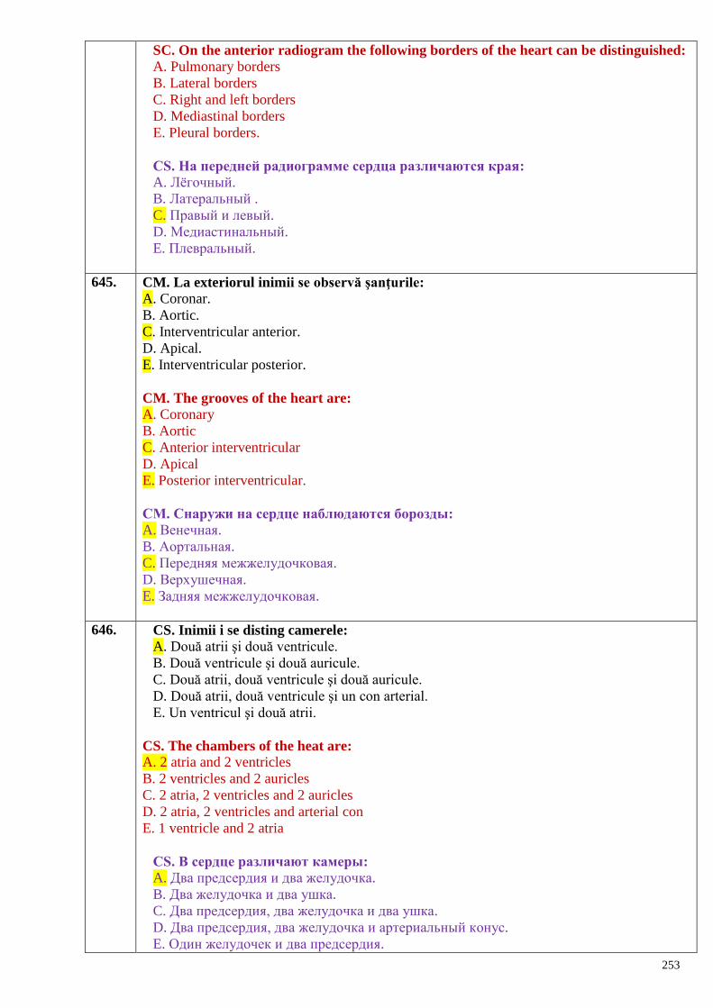
253
SC. On the anterior radiogram the following borders of the heart can be distinguished:
A. Pulmonary borders
B. Lateral borders
C. Right and left borders
D. Mediastinal borders
E. Pleural borders.
СS. На передней радиограмме сердца различаются края:
A. Лѐгочный.
B. Латеральный .
C. Правый и левый.
D. Медиастинальный.
E. Плевральный.
645. CM. La exteriorul inimii se observă şanţurile:
A. Coronar.
B. Aortic.
C. Interventricular anterior.
D. Apical.
E. Interventricular posterior.
CM. The grooves of the heart are:
A. Coronary
B. Aortic
C. Anterior interventricular
D. Apical
E. Posterior interventricular.
СM. Снаружи на сердце наблюдаются борозды:
A. Венечная.
B. Аортальная.
C. Передняя межжелудочковая.
D. Верхушечная.
E. Задняя межжелудочковая.
646. CS. Inimii i se disting camerele:
A. Două atrii şi două ventricule.
B. Două ventricule şi două auricule.
C. Două atrii, două ventricule şi două auricule.
D. Două atrii, două ventricule şi un con arterial.
E. Un ventricul şi două atrii.
CS. The chambers of the heat are:
A. 2 atria and 2 ventricles
B. 2 ventricles and 2 auricles
C. 2 atria, 2 ventricles and 2 auricles
D. 2 atria, 2 ventricles and arterial con
E. 1 ventricle and 2 atria
СS. В сердце различают камеры:
A. Два предсердия и два желудочка.
B. Два желудочка и два ушка.
C. Два предсердия, два желудочка и два ушка.
D. Два предсердия, два желудочка и артериальный конус.
E. Один желудочек и два предсердия.
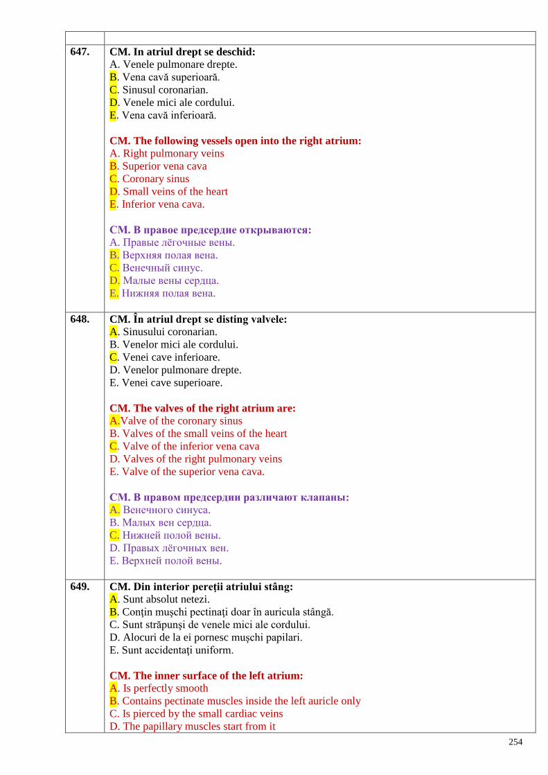
254
647. CM. In atriul drept se deschid:
A. Venele pulmonare drepte.
B. Vena cavă superioară.
C. Sinusul coronarian.
D. Venele mici ale cordului.
E. Vena cavă inferioară.
CM. The following vessels open into the right atrium:
A. Right pulmonary veins
B. Superior vena cava
C. Coronary sinus
D. Small veins of the heart
E. Inferior vena cava.
СМ. В правое предсердие открываются:
A. Правые лѐгочные вены.
B. Верхняя полая вена.
C. Венечный синус.
D. Малые вены сердца.
E. Нижняя полая вена.
648. CM. În atriul drept se disting valvele:
A. Sinusului coronarian.
B. Venelor mici ale cordului.
C. Venei cave inferioare.
D. Venelor pulmonare drepte.
E. Venei cave superioare.
CM. The valves of the right atrium are:
A.Valve of the coronary sinus
B. Valves of the small veins of the heart
C. Valve of the inferior vena cava
D. Valves of the right pulmonary veins
E. Valve of the superior vena cava.
СM. В правом предсердии различают клапаны:
A. Венечного синуса.
B. Малых вен сердца.
C. Нижней полой вены.
D. Правых лѐгочных вен.
E. Верхней полой вены.
649. CM. Din interior pereţii atriului stâng:
A. Sunt absolut netezi.
B. Conţin muşchi pectinaţi doar în auricula stângă.
C. Sunt străpunşi de venele mici ale cordului.
D. Alocuri de la ei pornesc muşchi papilari.
E. Sunt accidentaţi uniform.
CM. The inner surface of the left atrium:
A. Is perfectly smooth
B. Contains pectinate muscles inside the left auricle only
C. Is pierced by the small cardiac veins
D. The papillary muscles start from it
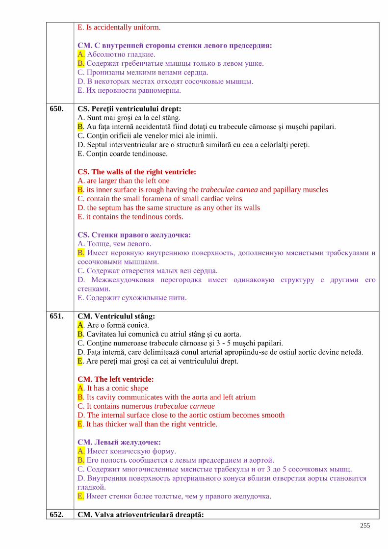
255
E. Is accidentally uniform.
СM. С внутренней стороны стенки левого предсердия:
A. Абсолютно гладкие.
B. Содержат гребенчатые мышцы только в левом ушке.
C. Пронизаны мелкими венами сердца.
D. В некоторых местах отходят сосочковые мышцы.
E. Их неровности равномерны.
650. CS. Pereţii ventriculului drept:
A. Sunt mai groşi ca la cel stâng.
B. Au faţa internă accidentată fiind dotaţi cu trabecule cărnoase şi muşchi papilari.
C. Conţin orificii ale venelor mici ale inimii.
D. Septul interventricular are o structură similară cu cea a celorlalţi pereţi.
E. Conţin coarde tendinoase.
CS. The walls of the right ventricle:
A. are larger than the left one
B. its inner surface is rough having the trabeculae carnea and papillary muscles
C. contain the small foramena of small cardiac veins
D. the septum has the same structure as any other its walls
E. it contains the tendinous cords.
СS. Стенки правого желудочка:
A. Толще, чем левого.
B. Имеет неровную внутреннюю поверхность, дополненную мясистыми трабекулами и
сосочковыми мышцами.
C. Содержат отверстия малых вен сердца.
D. Межжелудочковая перегородка имеет одинаковую структуру с другими его
стенками.
E. Содержит сухожильные нити.
651. CM. Ventriculul stâng:
A. Are o formă conică.
B. Cavitatea lui comunică cu atriul stâng şi cu aorta.
C. Conţine numeroase trabecule cărnoase şi 3 - 5 muşchi papilari.
D. Faţa internă, care delimitează conul arterial apropiindu-se de ostiul aortic devine netedă.
E. Are pereţi mai groşi ca cei ai ventriculului drept.
CM. The left ventricle:
A. It has a conic shape
B. Its cavity communicates with the aorta and left atrium
C. It contains numerous trabeculae carneae
D. The internal surface close to the aortic ostium becomes smooth
E. It has thicker wall than the right ventricle.
СM. Левый желудочек:
A. Имеет коническую форму.
B. Его полость сообщается с левым предсердием и аортой.
C. Содержит многочисленные мясистые трабекулы и от 3 до 5 сосочковых мышц.
D. Внутренняя поверхность артериального конуса вблизи отверстия аорты становится
гладкой.
E. Имеет стенки более толстые, чем у правого желудочка.
652. CM. Valva atrioventriculară dreaptă:
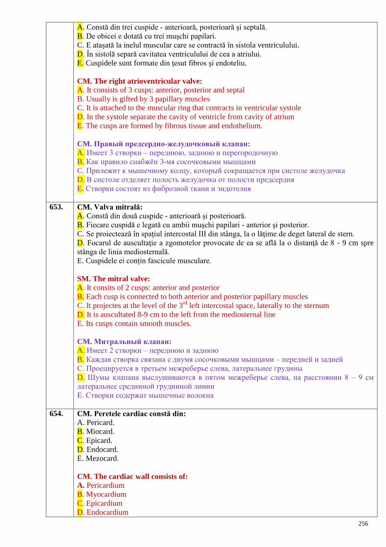
256
A. Constă din trei cuspide - anterioară, posterioară şi septală.
B. De obicei e dotată cu trei muşchi papilari.
C. E ataşată la inelul muscular care se contractă în sistola ventriculului.
D. În sistolă separă cavitatea ventriculului de cea a atriului.
E. Cuspidele sunt formate din ţesut fibros şi endoteliu.
CM. The right atrioventricular valve:
A. It consists of 3 cusps: anterior, posterior and septal
B. Usually is gifted by 3 papillary muscles
C. It is attached to the muscular ring that contracts in ventricular systole
D. In the systole separate the cavity of ventricle from cavity of atrium
E. The cusps are formed by fibrous tissue and endothelium.
CM. Правый предсердно-желудочковый клапан:
A. Имеет 3 створки – переднюю, заднюю и перегородочную
B. Как правило снабжѐн 3-мя сосочковыми мышцами
C. Прилежит к мышечному колцу, который сокращается при систоле желудочка
D. В систоле отделяет полость желудочка от полости предсердия
E. Створки состоят из фиброзной ткани и эндотелия
653. CM. Valva mitrală:
A. Constă din două cuspide - anterioară şi posterioară.
B. Fiecare cuspidă e legată cu ambii muşchi papilari - anterior şi posterior.
C. Se proiectează în spaţiul intercostal III din stânga, la o lăţime de deget lateral de stern.
D. Focarul de auscultaţie a zgomotelor provocate de ea se află la o distanţă de 8 - 9 cm spre
stânga de linia mediosternală.
E. Cuspidele ei conțin fascicule musculare.
SM. The mitral valve:
A. It consits of 2 cusps: anterior and posterior
B. Each cusp is connected to both anterior and posterior papillary muscles
C. It projectes at the level of the 3rd
left intercostal space, laterally to the sternum
D. It is auscultated 8-9 cm to the left from the mediosternal line
E. Its cusps contain smooth muscles.
CM. Митральный клапан:
A. Имеет 2 створки – переднюю и заднюю
B. Каждая створка связана с двумя сосочковыми мышцами – передней и задней
C. Проецируется в третьем межреберье слева, латеральнее грудины
D. Шумы клапана выслушиваются в пятом межреберье слева, на расстоянии 8 – 9 см
латеральнее срединной грудинной линии
E. Створки содержат мышечные волокна
654. CM. Peretele cardiac constă din:
A. Pericard.
B. Miocard.
C. Epicard.
D. Endocard.
E. Mezocard.
CM. The cardiac wall consists of:
A. Pericardium
B. Myocardium
C. Epicardium
D. Endocardium
257
E. Mesocardium.
СM. Стенки сердца состоят из:
A. Перикарда.
B. Миокарда.
C. Эпикарда.
D. Эндокарда.
E. Мезокарда.
655. CM. Endocardul:
A. Tapetează miocardul din interior.
B. Nu acoperă muşchii papilari şi coardele tendinoase.
C. Duplicaturi ale lui formează valvele atrioventriculare şi cele ale venei cave inferioare şi a
sinusului coronar.
D. Reprezintă o pătură fină şi transparentă.
E. Nu conţine vase sangvine şi nervi.
CM. The endocardium:
A. Covers the inner surface of the myocardium
B. Does not cover the papillary muscles and tendinous threads
C. Its duplicatures form the valves: atrioventricular valves, valve of the coronary sinus and
valve of the inferior vena cava
D. Is tiny and transparent
E. Does not contain blood vessels and nerves.
СМ. Эндокард:
A. Покрывает миокард изнутри.
B. Не покрывает сосочковые мышцы и сухожильные нити.
C. Его дубликатура образует предсердно-желудочковые клапаны, а также клапаны
нижней полой вены и венечного синуса.
D. Представляет тонкую и прозрачную ткань.
E. Не содержит сосуды и нервы.
656. CM. Septul interventricular:
A. Reprezintă un perete muscular.
B. I se disting porţiunile musculară şi membranoasă.
C. E tapetat cu endocard.
D. Separă ventriculele de atrii.
E. La copii constă numai din porţiunea musculară.
CM. The interventricular septum:
A. Is a muscular wall
B. Has the muscular and membranous parts
C. Is lined by the endocardium
D. Separates the atria from the ventricles
E. In childhood consists of the muscular part only.
СМ. Межжелудочковая перегородка:
A. Представлена мышечной стенкой.
B. Различают мышечную и перепончатую части.
C. Покрыта эндокардом.
D. Отделяет желудочки от предсердий.
E. У детей представлена только мышечной частью.
657. CM. Inima la copil:
258
A. Creşte mai intens la fetiţe.
B. Are linii limitrofe care diferă de cele ale adultului.
C. Are muşchii papilari atrofiaţi.
D. Nu posedă trabecule cărnoase.
E. În septurile interatrial şi interventricular există orificii.
CM. The heart of the child:
A. It grows faster in the girls
B. Its limitrophe lines differ from those of the adults
C. Its papillary muscles are atrophied
D. Does not have trabeculae
E. Its interatrial and interventriculat septa have orifices.
СМ. Сердце у детей:
A. Растѐт быстрее у девочек.
B. Его границы отличаются от таковых у взрослых.
C. Имеет атрофичные сосочковые мышцы.
D. Не имеет мышечных трабекул.
E. В межпредсердных и межжелудочковых перегородках есть отверстия.
658. CS. Apexul inimii se proiectează:
A. La nivelul coastei V pe linie medioclaviculară stângă.
B. La nivelul apofizei xifoide a sternului la două lăţimi de deget spre stânga.
C. În spaţiul intercostal V cu 1,5 cm medial de linia medioclaviculară din stânga.
D. În spaţiul intercostal V pe linie medioclaviculară stângă.
E. La copii nu se proiectează.
CS. The apex of the hear is projected:
A. At the level of the V rib, on the left medioclavicular line
B. At the level of the xyphoid process, 2-3 cm to the left of the sternum
C. In the V intercostal space, 1.5 cm medially from the left medioclavicular line
D. In the V intercostal space on the left medioclavicular line
E. In the children is not projected.
СS. Верхушка сердца проецируется:
A. На уровне V ребра по левой среднеключичной линии.
B. На уровне основания мечевидного отростка грудины, отступя на ширину 2-х пальцев
налево.
C. В V межреберье на 1,5 см медиальнее левой среднеключичной линии.
D. В V межреберье на левой среднеключичной линии.
E. У детей не проецируется.
659. CM. Pericardul seros:
A. Constă din doua foiţe - parietală şi viscerală.
B. Foiţa lui viscerală tapetează epicardul.
C. Produce lichid pericardic.
D. E fixat de stern prin ligg. sternopericardiace.
E. Conține lamina muscularis pericardii.
MC. Choose the true statements regarding the serous pericardium are: A.It consists of two layers - parietal and visceral
B. Its visceral layer covers the epicardium
C. Itproduces the pericardial fluid
D. It is fixed to the sternum by the sternopericardial ligaments
E. Contains lamina muscularis pericardii
259
CM. Серозный перикард:
A. Состоит из двух листков – париетального и висцерального
B. Его висцеральный листок покрывает эпикард
C. Продуктирует перикардиальную жидкость
D. Фиксируется к грудине грудино-перикардиальными связками
E. Coдержит lamina muscularis pericardii.
660. CM. Cavitatea pericardului:
A. E delimitată de foiţele parietală şi viscerală a pericardului fibros.
B. Reprezintă un spaţiu capilar.
C. Conţine o cantitate infimă de lichid seros.
D. I se disting sinusurile transvers, oblic şi interauricular.
E. Comunică cu cavitatea pleurei prin spaţiile perivasculare.
MC. Choose the true statements regarding the pericardial cavity are: A. It is bordered by the parietal and visceral layers of the fibrous pericardium
B. It is a capillary space
C. It contains a small amount of the serous fluid
D. It formes the transverse, oblique and interauricular sinuses
E. It communicates with the pleural cavity through the perivascular spaces
CM. Полость перикарда:
A. Ограничена париетальными и висцеральными листками фиброзного перикарда
B. Представляет капиллярное пространство
C. Содержит небольшое количество серозной жидкости
D. Различаются синусы: поперечный, косой и межушковый
E. Сообщается с полостью плевры через периваскулярные пространства
661. CM. Pe viu inima poate fi explorată prin:
A. Endoscopie cardiacă.
B. Percuţie.
C. Ecocardiografie.
D. Coronarografie.
E. Angiocardiografie.
CM. Methods of examination of the heart on alive person are:
A. Cardiac endoscopy
B. Percussion
C. Ecocardiography
D. Coronarography
E. Angiocardiography.
СМ. У живого сердце исследуют через:
A. Сердечную эндоскопию.
B. Перкуссию.
C. Эхокардиографию.
D. Коронарографию.
E. Ангиокардиографию.
662. CS. Apexul cordului este orientat:
A. În jos, anterior şi în dreapta.
B. În jos, posterior şi în stânga.
C. În jos, anterior şi în stânga.
D. Spre spaţiul intercostal IV.
260
E. În direcţia processus xiphoideus.
CS. The apex of the heart is directed:
A. Downward, foreward and to the right
B. Downward, backward and to the left
C. Downward, foreward and to the left
D. To the IV intercostal space
E. To the xyphoid process.
СS. Верхушка сердца обращена:
A. Вниз, вперѐд и вправо.
B. Вниз, назад и влево.
C. Вниз, вперѐд и влево.
D. К IV межрѐберному промежутку.
E. По направлению мечевидного отростка.
663. CM. Cu privire la baza cordului:
A. E formată de atrii, iar anterior de aortă şi trunchiul pulmonar.
B. Corespunde atriilor şi părţii superioare a ventriculului stâng.
C. E orientată posterior, în sus şi la dreapta.
D. Atinge spaţiul intercostal II din dreapta.
E. Formează peretele superior al atriilor.
CM. The base of the heart:
A. Is formed by the atria, aorta and pulmonary trunk (in front)
B. Corresponds to the atria and upper part of the left ventricle
C. Is directed backward, upward and to the right
D. Reaches the II intercostal space on the right
E. Forms the upper wall of the atria.
СМ. Основание сердца:
A. Образовано предсердиями, а впереди – аортой и лѐгочным стволом.
B. Соответствует предсердиям и верхней части левого желудочка.
C. Направлено назад, вверх и вправо.
D. Достигает 2-го межрѐберного промежутка справа.
E. Образует верхнюю стенку предсердий.
664. CM. Cu privire la dimensiunile cordului:
A. Sunt egale cu dimensiunile pumnului individului respectiv.
B. Diametrul longitudinal e de 12-13 cm.
C. Diametrul transversal măsoară 9-10 cm.
D. Diametrul antero-posterior e de 6-7 cm.
E. La femeie sunt mai mari.
CM. Regarding the dimensions of the heart:
A. Are equal with the fist of the respectiv individuum
B. Longitudinal diameter is 12-14 cm
C. Transverse diameter is 9-10 cm
D. Antero-posterior diameter is 6-7 cm
E. In females are bigger (wider)
СМ. Относительно размеров сердца:
A. Равны размерам кулака индивидуума.
B. Продольный размер равняется 12-13 см.
C. Поперечный размер равняется 9-10 см.
261
D. Переднезадний размер равняется 6-7 см.
E. У женщин они больше.
665. CM. Septul interatrial:
A. Separă atriile de ventricule.
B. Prezintă un perete comun pentru ambele atrii.
C. Pe faţa lui dreaptă este situată fossa ovalis.
D. La nivelul fosei ovale este mult mai subţire.
E. Constituie peretele comun al ventriculelor.
CM. The interatrial septum:
A. Separates the atria from the ventricles
B. Is common wall for both atria
C. The oval fossa is located on its right wall
D. Is more thinner in the region of the oval fossa
E. Forms common wall for both ventricles.
СМ. Межпредсердная перегородка:
A. Отделяет предсердия от желудочков.
B. Является общей стенкой для обоих предсердий.
C. На правой поверхности расположена овальная ямка.
D. В пределах овальной ямки перегородка тоньше.
E. Является общей стенкой желудочков.
666. CS. Aparatul valvular al inimii este constituit din:
A. Valvele atrioventriculare dreaptă şi stângă.
B. Valva trunchiului pulmonar şi valva aortică.
C. Valva atrioventriculară dreaptă, valva atrioventriculară stângă, valva trunchiului pulmonar
şi valva aortică.
D. Valvele semilunare ale trunchiului pulmonar şi ale aortei.
E. Valvele tricuspidă şi bicuspidă.
CS. The valvular apparatus of the heart consists of:
A. Right and left atrioventricular valves
B. Valves of the aorta and pulmonary trunk
C. Right and left atrioventricular valves, valve of the aorta and pulmonary trunk
D. Semilunar valves of the aorta and pulmonary trunk
E. Tricuspid and bicuspid valves.
СS. Клапанный аппарат сердца состоит из:
A. Правого и левого предсердно-желудочкового клапанов.
B. Клапанна лѐгочного ствола и аорты.
C. Правого предсердно-желудочкового клапана, левого предсердно-желудочкового
клапана, клапанов лѐгочного ствола и аорты.
D. Полулунных клапанов лѐгочного ствола и аорты.
E. Трѐхстворчатого и двухстворчатого клапанов.
667. CM. Sistemul conductil al inimii este constituit din:
A. Fibre musculare netede.
B. Fibre musculare atipice dispuse în miocard.
C. Fascicule musculare longitudinale.
D. Nodul sinoatrial, nodul atrioventricular şi fasciculul atrioventricular.
E. Doi pedunculi musculari.
CM. The conductive system of the heart includes:
262
A. Smooth muscular fibers
B. Atypical muscular fibers located inside the myocardium
C. Longitudinal muscular fibers
D. Sinoatrial node, atrioventricular node and atrioventricular fascicle of His
E. Two muscular pedicles.
СМ. Проводящая система сердца состоит из:
A. Гладких мышечных волокон.
B. Атипичных мышечных волокон, находящихся в миокарде.
C. Продольных мышечных пучков.
D. Синусно-предсердного узла, предсердно-желудочковго узла и предсердно-
желудочкового пучка.
E. Двух мышечных ножек.
668. CM. Cu privire la importanţa sistemului conductil al inimii:
A. Realizează activitatea ritmică a inimii.
B. Sincronizează contracţiile atriilor şi ventriculelor.
C. Cumulează rolul elementelor nervoase ale cordului.
D. Stimulează activitatea atriului şi ventriculului de pe dreapta.
E. Realizează automatismul inimii.
CM. Regarding the significans of the conducting system of the heart:
A. It coordinates the rhythmic activity of the heart.
B. It synchronizes the contractions of the atria and ventricles.
C. It cumulates the significans of the nervous elements of the heart.
D. It stimulates the activity of the right ventricle and right atrium.
E. It realizes (performs) the automatism of the heart.
СМ. Значение проводящей системы сердца:
A. Осуществляет ритмическую деятельность сердца.
B. Координирует сокращение предсердий и желудочков.
C. Выполняет роль нервных элементов сердца.
D. Стимулирует деятельность правого предсердия и желудочка.
E. Осуществляет автоматизм сердца.
669. CS. Miocardul:
A. Alcătuieşte pereţii atriilor şi ventriculelor independent de funcţiile lor.
B. La nivelul atriilor este mai gros, decât în ventricule.
C. E cel mai bine dezvoltat strat al cordului.
D. Constă din ţesut muscular scheletic.
E. Anatomic este comun pentru atrii şi ventricule.
CS. The myocardium:
A. It forms the walls of the atria and ventricles independent of their functions.
B. It is thicker at the level of the atria than at the level of the ventricles.
C. It is the most developed layer of the heart.
D. It consists of the skeletal muscular tissue.
E. Anatomically it is common for both atria and ventricles.
СS. Миокард:
A. Образует стенки предсердий и желудочков вне зависимости от их функций.
B. В предсердии он толще, чем в желудочках.
C. Самый развитый слой стенки сердца.
D. Состоит из поперечнополосатых мышц.
E. Анатомически он является общим для предсердий и желудочков.
263
670. CM. Prin examenul radiologic la adult se evidenţiază următoarele poziţii principale ale
cordului:
A. Transversală.
B. Oblică.
C. Orizontală.
D. Verticală.
E. Mediană.
CM. During the X-ray examination the folowing position of the heart can be observed:
A. Transverse.
B. Oblique.
C. Horizontal.
D. Vertical.
E. Median.
СМ. При рентгенологическом исследовании у взрослых наблюдаются следующие
основные положения сердца:
A. Поперечное.
B. Косое.
C. Горизонтальное.
D. Вертикальное.
E. Срединное.
671. CM. Circulaţia sangvină la făt:
A. Este pulmonară.
B. Este placentară.
C. Circulaţia sangvină mare comunică cu cea mică prin foramen ovale şi ductus arteriosus.
D. Are loc numai prin artere.
E. Aprovizionează fătul cu O2 şi substanţe nutritive.
CM. The blood circulation of the foetus:
A. It is the pulmonary circulation.
B. It is the placental circulation.
C.The greater blood circulation communicates with the lesser one through foramen ovale and
ductus arteriosus.
D. It takes part only through the arteries.
E. It supplies the foetus with oxygen and nutrients.
СМ. Кровообращение плода:
A. Лѐгочное.
B. Плацентарное.
C. Большой круг кровообращения сообщается с малым через овальное отверстие и
крупный артериальный (боталлов) проток.
D. Идѐт только через артерии.
E. Обеспечивает плод кислородом и питательными веществами.
672. CM. În activitatea inimii se disting următoarele faze:
A. Sistola generală.
B. Sistola atriilor.
C. Diastola generală.
D. Sistola ventriculelor.
E. Diastola ventriculelor.
CM. The heart activity consists of the following phases:
264
A. Total systole
B. Atrial systole
C. Total diastole
D. Ventricular systole
E. Ventricular diastole.
СМ. В деятельности сердца различают следующие фазы:
A. Общая систола.
B. Систола предсердий.
C. Общая диастола.
D. Систола желудочков.
E. Диастола желудочков.
673. CS. Şocul apexian se determină:
A. La apexul inimii.
B. În spaţiul intercostal III din stânga.
C. La nivelul cartilajului coastei IV din dreapta.
D. În spaţiul intercostal IV din dreapta.
E. Pe linia medioclaviculară stângă.
CS. The apex beat may be felt:
A. At the projection of the apex of the heart.
B. In the III left intercostal space.
C. At the level of the IV right costal cartilage.
D. In the IV right intercostal space.
E. On the left midclavicular line.
СS. Верхушечный толчок определяется:
A. На верхушке сердца.
B. В III левом межрѐберном промежутке.
C. На уровне IV рѐберного хряща справа.
D. В IV межреберье справа.
E. По левой среднеключичной линии.
674. CM. Sistemul conductil al cordului:
A. Constă din fibre musculare atipice.
B. Centrii lui sunt reprezentaţi de nodurile sinuatrial şi atrioventricular.
C. Nodul sinuatrial se află în peretele anterior al ventriculului drept.
D. Nodul atrioventricular e situat în septul interventricular.
E. Include noduri şi fascicule.
CM. The conducting system of the heart:
A. It consists of the atipical muscular cells.
B. Its centers are represented by the sinoatrial and atrioventricular nodes.
C. The sinoatrial node is located in the anterior wall of the right ventricle.
D. The atrioventricular node is located in the interventricular septum.
E. It includes nodes and bundles.
СМ. Проводящая система сердца:
A. Состоит из атипичных мышечных волокон.
B. Еѐ центры представлены синусно-предсердным и предсердно-желудочковым
узлами.
C. Синусно-предсердный узел расположен в передней стенке правого желудочка.
D. Предсердно-желудочковый узел расположен в межжелудочковой перегородке.
E. Включает узлы и пучки.
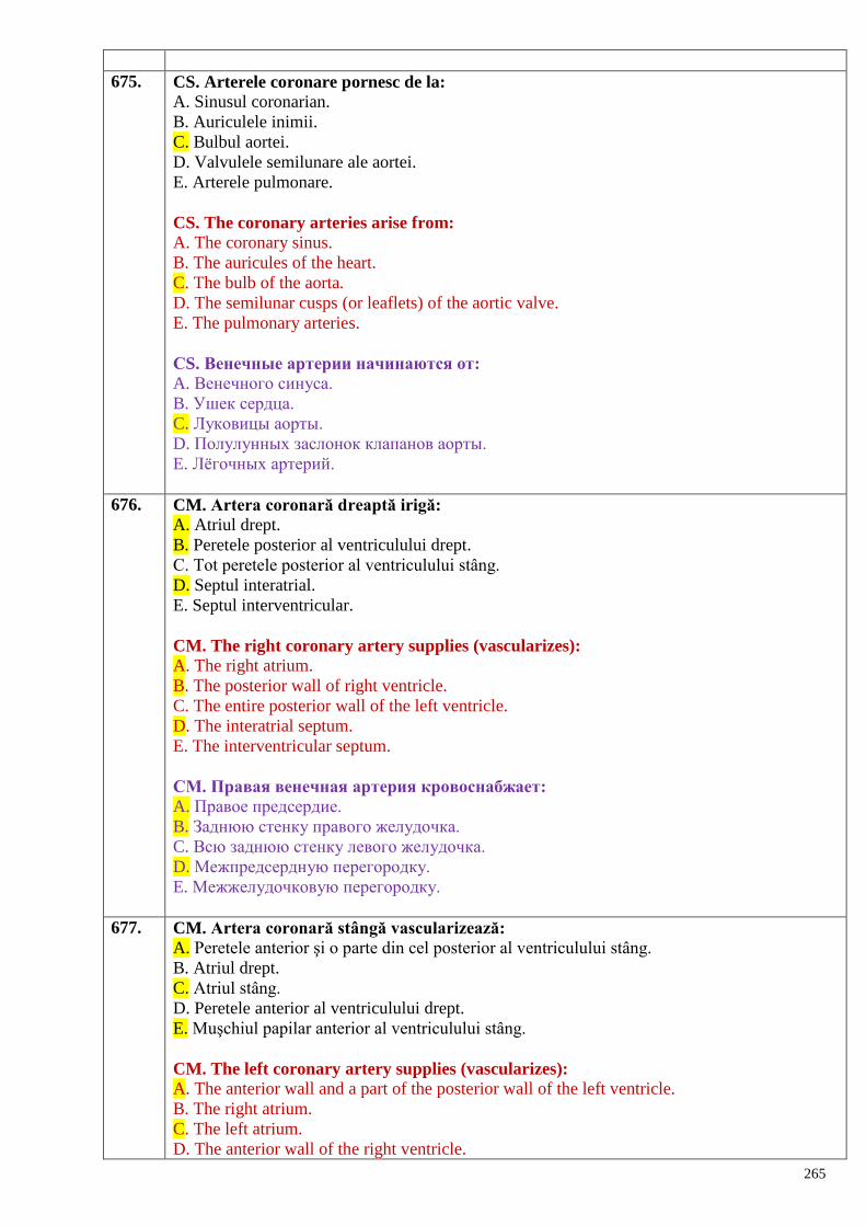
265
675. CS. Arterele coronare pornesc de la:
A. Sinusul coronarian.
B. Auriculele inimii.
C. Bulbul aortei.
D. Valvulele semilunare ale aortei.
E. Arterele pulmonare.
CS. The coronary arteries arise from:
A. The coronary sinus.
B. The auricules of the heart.
C. The bulb of the aorta.
D. The semilunar cusps (or leaflets) of the aortic valve.
E. The pulmonary arteries.
СS. Венечные артерии начинаются от:
A. Венечного синуса.
B. Ушек сердца.
C. Луковицы аорты.
D. Полулунных заслонок клапанов аорты.
E. Лѐгочных артерий.
676. CM. Artera coronară dreaptă irigă:
A. Atriul drept.
B. Peretele posterior al ventriculului drept.
C. Tot peretele posterior al ventriculului stâng.
D. Septul interatrial.
E. Septul interventricular.
CM. The right coronary artery supplies (vascularizes):
A. The right atrium.
B. The posterior wall of right ventricle.
C. The entire posterior wall of the left ventricle.
D. The interatrial septum.
E. The interventricular septum.
СМ. Правая венечная артерия кровоснабжает:
A. Правое предсердие.
B. Заднюю стенку правого желудочка.
C. Всю заднюю стенку левого желудочка.
D. Межпредсердную перегородку.
E. Межжелудочковую перегородку.
677. CM. Artera coronară stângă vascularizează:
A. Peretele anterior şi o parte din cel posterior al ventriculului stâng.
B. Atriul drept.
C. Atriul stâng.
D. Peretele anterior al ventriculului drept.
E. Muşchiul papilar anterior al ventriculului stâng.
CM. The left coronary artery supplies (vascularizes):
A. The anterior wall and a part of the posterior wall of the left ventricle.
B. The right atrium.
C. The left atrium.
D. The anterior wall of the right ventricle.
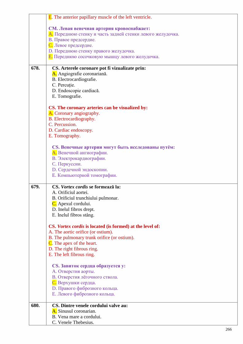
266
E. The anterior papillary muscle of the left ventricle.
СМ. Левая венечная артерия кровоснабжает:
A. Переднюю стенку и часть задней стенки левого желудочка.
B. Правое предсердие.
C. Левое предсердие.
D. Переднюю стенку правого желудочка.
E. Переднюю сосочковую мышцу левого желудочка.
678. CS. Arterele coronare pot fi vizualizate prin:
A. Angiografie coronariană.
B. Electrocardiografie.
C. Percuţie.
D. Endoscopie cardiacă.
E. Tomografie.
CS. The coronary arteries can be visualized by:
A. Coronary angiography.
B. Electrocardiography.
C. Percussion.
D. Cardiac endoscopy.
E. Tomography.
СS. Венечные артерии могут быть исследованы путѐм:
A. Венечной ангиографии.
B. Электрокардиографии.
C. Перкуссии.
D. Сердечной эндоскопии.
E. Компьютерной томографии.
679. CS. Vortex cordis se formează la:
A. Orificiul aortei.
B. Orificiul trunchiului pulmonar.
C. Apexul cordului.
D. Inelul fibros drept.
E. Inelul fibros stâng.
CS. Vortex cordis is located (is formed) at the level of:
A. The aortic orifice (or ostium).
B. The pulmonary trunk orifice (or ostium).
C. The apex of the heart.
D. The right fibrous ring.
E. The left fibrous ring.
СS. Завиток сердца образуется у:
A. Отверстия аорты.
B. Отверстия лѐгочного ствола.
C. Верхушки сердца.
D. Правого фиброзного кольца.
E. Левого фиброзного кольца.
680. CS. Dintre venele cordului valve au:
A. Sinusul coronarian.
B. Vena mare a cordului.
C. Venele Thebesius.
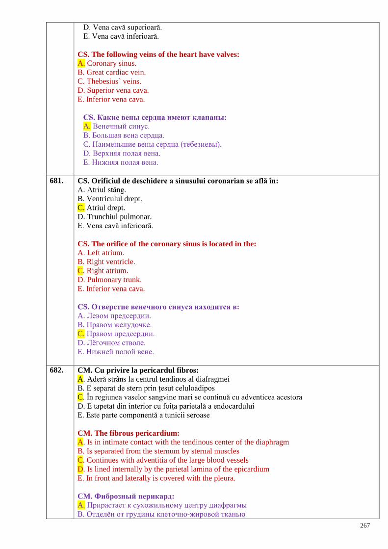
267
D. Vena cavă superioară.
E. Vena cavă inferioară.
CS. The following veins of the heart have valves:
A. Coronary sinus.
B. Great cardiac vein.
C. Thebesius` veins.
D. Superior vena cava.
E. Inferior vena cava.
СS. Какие вены сердца имеют клапаны:
A. Венечный синус.
B. Большая вена сердца.
C. Наименьшие вены сердца (тебезиевы).
D. Верхняя полая вена.
E. Нижняя полая вена.
681. CS. Orificiul de deschidere a sinusului coronarian se află în:
A. Atriul stâng.
B. Ventriculul drept.
C. Atriul drept.
D. Trunchiul pulmonar.
E. Vena cavă inferioară.
CS. The orifice of the coronary sinus is located in the:
A. Left atrium.
B. Right ventricle.
C. Right atrium.
D. Pulmonary trunk.
E. Inferior vena cava.
СS. Отверстие венечного синуса находится в:
A. Левом предсердии.
B. Правом желудочке.
C. Правом предсердии.
D. Лѐгочном стволе.
E. Нижней полой вене.
682. CM. Cu privire la pericardul fibros:
A. Aderă strâns la centrul tendinos al diafragmei
B. E separat de stern prin țesut celuloadipos
C. În regiunea vaselor sangvine mari se continuă cu adventicea acestora
D. E tapetat din interior cu foiţa parietală a endocardului
E. Este parte componentă a tunicii seroase
CM. The fibrous pericardium:
A. Is in intimate contact with the tendinous center of the diaphragm
B. Is separated from the sternum by sternal muscles
C. Continues with adventitia of the large blood vessels
D. Is lined internally by the parietal lamina of the epicardium
E. In front and laterally is covered with the pleura.
CM. Фиброзный перикард:
A. Прирастает к сухожильному центру диафрагмы
B. Отделѐн от грудины клеточно-жировой тканью
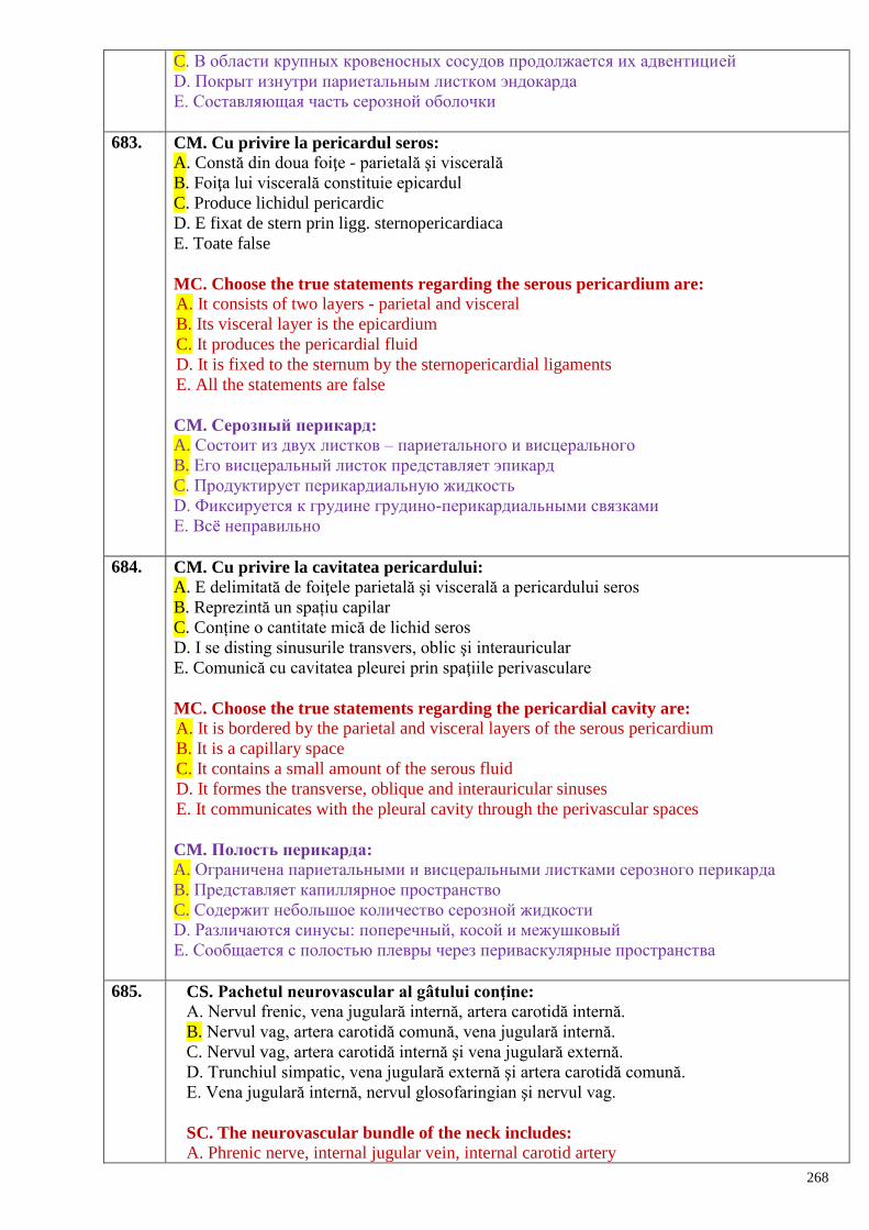
268
C. В области крупных кровеносных сосудов продолжается их адвентицией
D. Покрыт изнутри париетальным листком эндокарда
E. Составляющая часть серозной оболочки
683. CM. Cu privire la pericardul seros:
A. Constă din doua foiţe - parietală şi viscerală
B. Foiţa lui viscerală constituie epicardul
C. Produce lichidul pericardic
D. E fixat de stern prin ligg. sternopericardiaca
E. Toate false
MC. Choose the true statements regarding the serous pericardium are: A. It consists of two layers - parietal and visceral
B. Its visceral layer is the epicardium
C. It produces the pericardial fluid
D. It is fixed to the sternum by the sternopericardial ligaments
E. All the statements are false
CM. Серозный перикард:
A. Состоит из двух листков – париетального и висцерального
B. Его висцеральный листок представляет эпикард
C. Продуктирует перикардиальную жидкость
D. Фиксируется к грудине грудино-перикардиальными связками
E. Всѐ неправильно
684. CM. Cu privire la cavitatea pericardului:
A. E delimitată de foiţele parietală şi viscerală a pericardului seros
B. Reprezintă un spațiu capilar
C. Conține o cantitate mică de lichid seros
D. I se disting sinusurile transvers, oblic şi interauricular
E. Comunică cu cavitatea pleurei prin spaţiile perivasculare
MC. Choose the true statements regarding the pericardial cavity are: A. It is bordered by the parietal and visceral layers of the serous pericardium
B. It is a capillary space
C. It contains a small amount of the serous fluid
D. It formes the transverse, oblique and interauricular sinuses
E. It communicates with the pleural cavity through the perivascular spaces
CM. Полость перикарда:
A. Ограничена париетальными и висцеральными листками серозного перикарда
B. Представляет капиллярное пространство
C. Содержит небольшое количество серозной жидкости
D. Различаются синусы: поперечный, косой и межушковый
E. Сообщается с полостью плевры через периваскулярные пространства
685. CS. Pachetul neurovascular al gâtului conţine:
A. Nervul frenic, vena jugulară internă, artera carotidă internă.
B. Nervul vag, artera carotidă comună, vena jugulară internă.
C. Nervul vag, artera carotidă internă şi vena jugulară externă.
D. Trunchiul simpatic, vena jugulară externă şi artera carotidă comună.
E. Vena jugulară internă, nervul glosofaringian şi nervul vag.
SC. The neurovascular bundle of the neck includes:
A. Phrenic nerve, internal jugular vein, internal carotid artery
269
B. Vagus nerve, common carotid artery, internal jugular vein
C. Vagus nerve, internal carotid artery, internal jugular vein
D. Sympathetic trunk, external jugular vein, common carotid artery
E. Internal jugular vein, glossopharyngeal nerve, vagus nerve.
CS. Сосудисто-нервный пучок шеи содержит:
А. Диафрагмальный нерв, внутреннюю яремную вену, внутреннюю сонную артерию.
В. Блуждающий нерв, общую сонную артерию, внутреннюю яремную вену.
С. Блуждающий нерв, внутреннюю сонную артерию и наружную яремную вену.
D. Симпатический ствол, наружную сонную артерию и общую сонную артерию.
E. Внутреннюю яремную вену, языкоглоточный нерв и блуждающий нерв.
686. CS. Ramurile arcului aortic sunt dispuse de la dreapta spre stânga:
A. Artera subclaviculară dreaptă; artera subclaviculară stângă, trunchiul brahiocefalic.
B. Artera carotidă comună dreaptă; artera subclaviculară dreaptă, artera carotidă comună
stângă.
C. Trunchiul brahiocefalic, artera carotidă comună stângă, artera subclaviculară stângă.
D. Trunchiul brahiocefalic, artera subclaviculară stângă, artera vertebrală.
E. Artera vertebrală, trunchiul brahiocefalic, artera carotidă comună stângă.
CS. The arrangement of the branches of the aortic arch from the right side to the left one
is, as follows:
A. Right subclavian artery, left subclavian artery, brachiocephalic trunk.
B. Right common carotid artery, right subclavian artery, left common carotid artery.
C. Brachiocephalic trunk, left common carotid artery, left subclavian artery.
D. Brachiocephalic trunk, left subclavian artery, vertebral artery.
E. Vertebral artery, brachiocephalic trunk, left common carotid artery.
CS. Ветви дуги аорты расположены справа налево:
А. Правая подключичная артерия, левая подключичная артерия, плечеголовной ствол.
В. Правая общая сонная артерия, правая подключичная артерия, левая общая сонная
артерия.
С. Плечеголовной ствол, левая общая сонная артерия, левая подключичная артерия.
D. Плечеголовной ствол, левая подключичная артерия, позвоночная артерия.
E. Позвоночная артерия, плечеголовной ствол, левая общая сонная артерия.
687. CS. Pulsul pe artera carotidă comună poate fi luat:
A. În triunghiul omotrapezoid.
B. În triunghiul omoclavicular.
C. În şanţul jugular.
D. În triunghiul submandibular.
E. În triunghiul omotraheal.
SC. The pulse of the common carotid artery can be felt:
A. In the omotrapezoid triangle.
B. In the omoclavicular triangle.
C. In the jugular groove (sulcus).
D. In the submandibular triangle.
E. In the omotracheal triangle.
CS. Пульс на общей сонной артерии определяется:
А. В лопаточно-трапециевидном треугольнике.
В. В лопаточно-ключичном треугольнике.
С. В яремной борозде.
D. В поднижнечелюстном треугольнике.
270
E. В лопаточно-трахеальном (мышечном) треугольнике.
688. CS. În normă artera carotidă comună dreaptă:
A. Este ramură a arterei subclaviculare stângi.
B. Porneşte de la arcul aortei.
C. Este ramură a trunchiului brahiocefalic.
D. Este ramură a a. subclaviculare drepte.
E. Porneşte printr-un trunchi comun cu cea stângă.
SC. In norm the right common carotid artery:
A. Is a branch of the left subclavian artery.
B. Has its origin from the aortic arch.
C. Is a branch of the brachiocephalic trunk.
D. Is a branch of the right subclavian artery.
E. It originates from a common trunk together with the left common carotid artery.
CS. В норме правая общая сонная артерия:
А. Является ветвью левой подключичной артерии.
В. Начинается от дуги аорты.
С. Является ветвью плечеголовного ствола.
D. Является ветвью правой подключичной артерии.
E. Начинается от общего ствола с левой артерией.
689. CS. Artera carotidă comună se bifurcă:
A. La nivelul marginii superioare a cartilajului tiroid.
B. La nivelul articulaţiei sternoclaviculare.
C. La nivelul osului hioid.
D. La nivelul unghiului mandibulei.
E. Medial de apofiza mastoidiană.
SC. The common carotid artery bifurcates:
A. At the level of the superior margin of the thyroid cartilage.
B. At the level of the sternoclavicular joint.
C. At the level of the hyoid bone.
D. At the level of the mandibular angle.
E. Medially to the mastoid process.
CS. Общая сонная артерия делится:
А. На уровне верхнего края щитовидного хряща.
В. На уровне грудиноключичного сустава.
С. На уровне подъязычной кости.
D. На уровне угла нижней челюсти.
E. Медиальнее сосцевидного отростка.
690. CS. Artera carotidă comună se bifurcă:
A. În triunghiul omotrapezoid.
B. În triunghiul omoclavicular.
C. În triunghiul carotidian.
D. În triunghiul submandibular.
E. În triunghiul omotraheal.
SC. The common carotid artery bifurcates:
A. In the omotrapezoid triangle.
B. In the omoclavicular triangle.
C. In the carotid triangle.
271
D. In the submandibular triangle.
E. In the omotracheal triangle.
CS. Общая сонная артерия делится:
А. В лопаточно-трапециевидном треугольнике.
В. В лопаточно-ключичном треугольнике.
С. В сонном треугольнике.
D. В поднижнечелюстном треугольнике.
E. В лопаточно-трахеальном треугольнике.
691. CS. Ramuri terminale ale arterei carotide externe sunt:
A. Arterele maxilară şi facială.
B. Arterele temporală superficială şi maxilară.
C. Arterele facială şi temporală superficială.
D. Arterele lingvală şi occipitală.
E. Trunchiul lingvofacial.
SC. The terminal branches of the external carotid artery are:
A. Maxillary and facial arteries.
B. Superficial temporal and maxillary arteries.
C. Facial and superficial temporal arteries.
D. Lingual and occipital arteries.
E. The linguofacial trunk.
CS. Конечными ветвями наружной сонной артерии являются:
А. Верхнечелюстная и лицевая артерии.
В. Поверхностная височная и верхнечелюстная артерии.
С. Лицевая и поверхностная височная артерии.
D. Язычная и затылочная артерии.
E. Язычно-лицевой ствол.
692. CS. Artera tiroidiană superioară este ramură a:
A. Trunchiului tirocervical.
B. Arterei carotide externe.
C. Arterei laringiene superioare.
D. Trunchiului lingvofacial.
E. Arterei lingvale.
SC. The superior thyroid artery is a branch of the:
A. Thyrocervical trunk.
B. External carotid artery
C. Superior laryngeal artery
D. Linguofacial trunk.
E. Lingual artery.
CS. Верхняя щитовидная артерия является ветвью:
А. Щитошейного ствола.
В. Наружной сонной артерии.
С. Верхней гортанной артерии.
D. Язычно-лицевого ствола.
E. Язычной артерии.
693. CS. La vascularizarea limbii participă:
A. A. carotidă internă.
B. A. subclaviculară.
272
C. A. carotidă externă.
D. A. facială.
E. A. maxilară.
SC. The tongue is supplied by the:
A. Internal carotid artery.
B. Subclavian artery.
C. External carotid artery.
D. Facial artery.
E. Maxillary artery.
СМ. В васкуляризации языка участвуют:
A. Внутренняя сонная артерия.
B. Подключичная артерия.
C. Наружная сонная артерия.
D. Лицевая артерия.
E. Верхнечелюстная артерия.
694. CM. Localizarea punctelor de luare a pulsului de pe arterele capului şi gâtului:
A. Artera carotidă comună – în şanţul jugular.
B. Artera carotidă externă – lateral de laringe între cartilajul tiroid şi cornul mare al
hioidului.
C. Artera facială – marginea mandibulei, inaintea muşchiului maseter.
D. Artera angulară – marginea mandibulei pe limita anterioară a muşchiului maseter.
E. Artera temporală superficială – posterior de pavilionul urechii.
MC. Point out the points where can be taken the pulsation of the arteries of the head and
neck:
A. The common carotid artery – in the jugular sulcus.
B. External carotid artery – laterally to the larynx between the thyroid cartilage and greater
horn of the hyoid bone.
C. Facial artery – on the edge of the mandible, in front of the masseter muscle.
D. Angular artery – on the margin of the mandible on the anterior margin of the masseter
muscle.
E. Superficial temporal artery – behind the auricle.
СМ. Локализация точек определения пульса на артериях головы и шеи:
A. Общая сонная артерия – в яремной борозде.
B. Наружная сонная артерия – латеральнее гортани между щитовидным хрящом и
большим рогом подъязычной кости.
C. Лицевая артерия – нижний край нижней челюсти кпереди жевательной мышцы.
D. Угловая артерия – край нижней челюсти кпереди жевательной мышцы.
E. Поверхностная височная артерия – кзади ушной раковины.
695. CM. Din ramurile anterioare ale arterei carotide externe fac parte arterele:
A. Facială.
B. Lingvală.
C. Maxilară.
D. Tiroidiană superioră.
E. Occipitală.
MC. The anterior branches of the external carotid artery are:
A. The facial artery.
B. The lingual artery.
C. The maxillary artery.
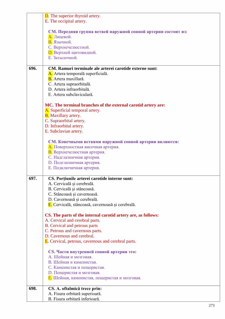
273
D. The superior thyroid artery.
E. The occipital artery.
СМ. Передняя группа ветвей наружной сонной артерии состоит из:
A. Лицевой.
B. Язычной.
C. Верхнечелюстной.
D. Верхней щитовидной.
E. Затылочной.
696. CM. Ramuri terminale ale arterei carotide externe sunt:
A. Artera temporală superficială.
B. Artera maxillară.
C. Artera supraorbitală.
D. Artera infraorbitală.
E. Artera subclaviculară.
MC. The terminal branches of the external carotid artery are:
A. Superficial temporal artery.
B. Maxillary artery.
C. Supraorbital artery.
D. Infraorbital artery.
E. Subclavian artery.
СМ. Конечными ветвями наружной сонной артерии являются:
A. Поверхностная височная артерия.
B. Верхнечелюстная артерия.
C. Надглазничная артерия.
D. Подглазничная артерия.
E. Подключичная артерия.
697. CS. Porţiunile arterei carotide interne sunt:
A. Cervicală şi cerebrală.
B. Cervicală şi stâncoasă.
C. Stâncoasă şi cavernoasă.
D. Cavernoasă şi cerebrală.
E. Cervicală, stâncoasă, cavernoasă şi cerebrală.
CS. The parts of the internal carotid artery are, as follows: A. Cervical and cerebral parts.
B. Cervical and petrous parts
C. Petrous and cavernous parts.
D. Cavernous and cerebral.
E. Cervical, petrous, cavernous and cerebral parts.
СS. Части внутренней сонной артерии это:
A. Шейная и мозговая.
B. Шейная и каменистая.
C. Каменистая и пещеристая.
D. Пещеристая и мозговая.
E. Шейная, каменистая, пещеристая и мозговая.
698. CS. A. oftalmică trece prin:
A. Fisura orbitară superioară.
B. Fisura orbitară inferioară.
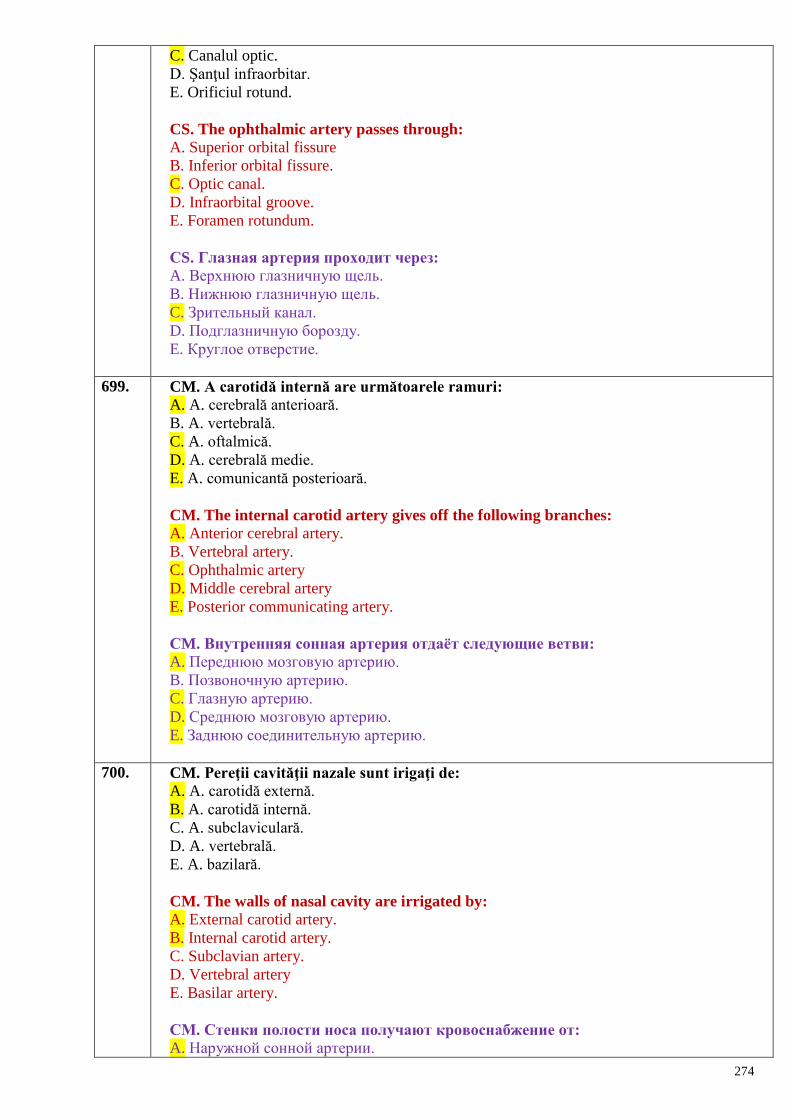
274
C. Canalul optic.
D. Şanţul infraorbitar.
E. Orificiul rotund.
CS. The ophthalmic artery passes through:
A. Superior orbital fissure
B. Inferior orbital fissure.
C. Optic canal.
D. Infraorbital groove.
E. Foramen rotundum.
СS. Глазная артерия проходит через:
A. Верхнюю глазничную щель.
B. Нижнюю глазничную щель.
C. Зрительный канал.
D. Подглазничную борозду.
E. Круглое отверстие.
699. CM. A carotidă internă are următoarele ramuri:
A. A. cerebrală anterioară.
B. A. vertebrală.
C. A. oftalmică.
D. A. cerebrală medie.
E. A. comunicantă posterioară.
CM. The internal carotid artery gives off the following branches:
A. Anterior cerebral artery.
B. Vertebral artery.
C. Ophthalmic artery
D. Middle cerebral artery
E. Posterior communicating artery.
СM. Внутренняя сонная артерия отдаѐт следующие ветви:
A. Переднюю мозговую артерию.
B. Позвоночную артерию.
C. Глазную артерию.
D. Среднюю мозговую артерию.
E. Заднюю соединительную артерию.
700. CM. Pereţii cavităţii nazale sunt irigaţi de:
A. A. carotidă externă.
B. A. carotidă internă.
C. A. subclaviculară.
D. A. vertebrală.
E. A. bazilară.
CM. The walls of nasal cavity are irrigated by:
A. External carotid artery.
B. Internal carotid artery.
C. Subclavian artery.
D. Vertebral artery
E. Basilar artery.
СМ. Стенки полости носа получают кровоснабжение от:
A. Наружной сонной артерии.
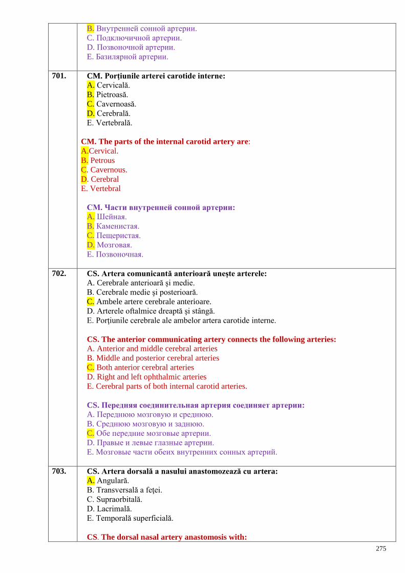
275
B. Внутренней сонной артерии.
C. Подключичной артерии.
D. Позвоночной артерии.
E. Базилярной артерии.
701. CM. Porţiunile arterei carotide interne:
A. Cervicală.
B. Pietroasă.
C. Cavernoasă.
D. Cerebrală.
E. Vertebrală.
CM. The parts of the internal carotid artery are:
A.Cervical.
B. Petrous
C. Cavernous.
D. Cerebral
E. Vertebral
СМ. Части внутренней сонной артерии:
A. Шейная.
B. Каменистая.
C. Пещеристая.
D. Мозговая.
E. Позвоночная.
702. CS. Artera comunicantă anterioară uneşte arterele:
A. Cerebrale anterioară şi medie.
B. Cerebrale medie şi posterioară.
C. Ambele artere cerebrale anterioare.
D. Arterele oftalmice dreaptă şi stângă.
E. Porţiunile cerebrale ale ambelor artera carotide interne.
CS. The anterior communicating artery connects the following arteries:
A. Anterior and middle cerebral arteries
B. Middle and posterior cerebral arteries
C. Both anterior cerebral arteries
D. Right and left ophthalmic arteries
E. Cerebral parts of both internal carotid arteries.
CS. Передняя соединительная артерия соединяет артерии:
A. Переднюю мозговую и среднюю.
B. Среднюю мозговую и заднюю.
C. Обе передние мозговые артерии.
D. Правые и левые глазные артерии.
E. Мозговые части обеих внутренних сонных артерий.
703. CS. Artera dorsală a nasului anastomozează cu artera:
A. Angulară.
B. Transversală a feţei.
C. Supraorbitală.
D. Lacrimală.
E. Temporală superficială.
CS. The dorsal nasal artery anastomosis with:
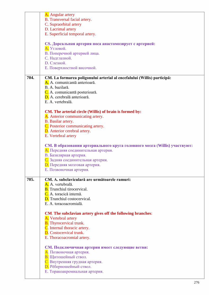
276
A. Angular artery
B. Transversal facial artery.
C. Supraorbital artery
D. Lacrimal artery
E. Superficial temporal artery.
CS. Дорсальная артерия носа анастомозирует с артерией:
A. Угловой.
B. Поперечной артерией лица.
C. Надглазной.
D. Слезной.
E. Поверхностной височной.
704. CM. La formarea poligonului arterial al encefalului (Willis) participă:
A. A. comunicantă anterioară.
B. A. bazilară.
C. A. comunicantă posterioară.
D. A. cerebrală anterioară.
E. A. vertebrală.
CM. The arterial circle (Willis) of brain is formed by:
A. Anterior communicating artery.
B. Basilar artery.
C. Posterior communicating artery.
D. Anterior cerebral artery.
E. Vertebral artery
СМ. В образовании артериального круга головного мозга (Willis) участвуют:
A. Передняя соединительная артерия.
B. Базилярная артерия.
C. Задняя соединительная артерия.
D. Передняя мозговая артерия.
E. Позвоночная артерия.
705. CM. A. subclaviculară are următoarele ramuri:
A. A. vertebrală.
B. Trunchiul tirocervical.
C. A. toracică internă.
D. Trunchiul costocervical.
E. A. toracoacromială.
CM. The subclavian artery gives off the following branches:
A. Vertebral artery
B. Thyrocervical trunk.
C. Internal thoracic artery.
D. Costocervical trunk.
E. Thoracoacromial artery.
CМ. Подключичная артерия имеет следующие ветви:
А. Позвоночная артерия.
В. Щитошейный ствол.
С. Внутренняя грудная артерия.
D. Рѐберношейный ствол.
E. Торакоакромиальная артерия.
277
706. CS. Artera subclaviculară trece prin:
A. Spaţiul antescalen.
B. Orificiul patrulater.
C. Orificiul trilater.
D. Spaţiul intercostal I.
E. Spaţiul interscalen.
CS. The subclavian artery passes through:
A. The antescalene space.
B. The quadrangular opening.
C. The triangular opening
D. The intercostal space.
E. The interscalene space.
CS. Подключичная артерия проходит через:
А. Предлестничный промежуток.
В. Четырѐхстороннее отверстие.
С. Трѐхстороннее отверстие.
D. Первый межрѐберный промежуток.
E. Межлестничное пространство.
707. CS. Pachetul neurovascular al gâtului conţine:
A. Nervul frenic, vena jugulară internă, artera carotidă internă.
B. Nervul vag, artera carotidă comună, vena jugulară internă.
C. Nervul vag, artera carotidă internă şi vena jugulară externă.
D. Trunchiul simpatic, vena jugulară externă şi artera carotidă comună.
E. Vena jugulară internă, nervul glosofaringian şi nervul vag.
SC. The neurovascular bundle of the neck contains:
A. Phrenic nerve, internal jugular vein, internal carotid artery.
B. Vagus nerve, common carotid artery, internal jugular vein.
C. Vagus nerve, internal carotid artery and external jugular vein.
D. Sympathetic trunk, external jugular vein and common carotid artery.
E. Internal jugular vein, glossopharyngeal and vagus nerves.
CS. Сосудисто-нервный пучок шеи содержит:
А. Диафрагмальный нерв, внутреннюю яремную вену, внутреннюю сонную артерию.
В. Блуждающий нерв, общую сонную артерию, внутреннюю яремную вену.
С. Блуждающий нерв, внутреннюю сонную артерию и наружную яремную вену.
D. Симпатический ствол, наружную яремную вену и общую сонную артерию.
E. Внутреннюю яремную вену, языкоглоточный нерв и блуждающий нерв.
708. CM. Vena jugulară internă:
A. Este o continuare a sinusului sagital superior.
B. Începe la nivelul orificiului jugular.
C. Are bulbul superior şi inferior.
D. Este continuarea sinusului sigmoid.
E. La început este situată posterior de a. carotidă internă.
MC. The internal jugular vein:
A. Is a continuation of the superior sagittal sinus.
B. Starts at the jugular orifice.
C. Has the upper and lower bulbs.
D. Is the continuation of the sigmoid sinus.
E. Formly is located posterior to the internal carotid artery.
278
CМ. Внутренняя яремная вена:
А. Является продолжением верхнего сагиттального синуса.
В. Начинается на уровне яремного отверстия.
С. Имеет верхнюю и нижнюю луковицы.
D. Является продолжением сигмовидного синуса.
E. Еѐ начало расположено позади внутренней сонной артерии.
709. CM. Cu referinţă la venele gâtului:
A. Cele mari conţin valve.
B. Din ele face parte vena retromandibulară.
C. Există venele jugulare internă, externă şi anterioară.
D. Reprezintă vase ale sistemului caval superior.
E. Sunt afluenţi ai sistemului caval inferior.
MC. Concerning the veins of the neck:
A. The larger veins contain valves.
B. The retromandibular vein is one of them.
C. There are internal, external and anterior jugular veins.
D. They belong to the superior caval system.
E. They are tributaries of the inferior caval system.
CМ. Относительно крупных вен шеи:
А. Крупные вены имеют клапаны.
В. К ним относится занижнечелюстная вена.
С. Существуют внутренние, наружные и передняя яремные вены.
D. Представляют сосуды системы верхней полой вены.
E. Являются притоками системы нижней полой вены.
710. CM. Ramurile parietale ale aortei:
A. Irigă pereţii cavităţilor trunchiului.
B. Vascularizează viscerele.
C. Sunt pare.
D. Sunt impare.
E. Vascularizează o anumită zonă a unui organ.
CM. The parietal branches of the aorta:
A. Supply the walls of the trunk cavities
B. Supply the viscera
C. Are paired
D. Are unpaired
E. Supply a certain area of the organ
СМ. Пристеночные ветви аорты:
A. Кровоснабжают стенки полостей туловища.
B. Кровоснабжают внутренности.
C. Они парные.
D. Они непарные.
E. Кровоснабжают определѐнные зоны одного органа.
711. CS. Ramurile viscerale ale aortei vascularizează:
A. Organele extracavitare.
B. Numai organele pare.
C. Numai organele impare.
D. Organele din cavităţile trunchiului.
279
E. Numai organele cavitare.
CS. The visceral branches of the aorta supply:
A. The extracavitary organs
B.The paired organs only
C.The unpaired organs only
D. The organs of the trunk cavities
E. The cavitary organs only.
СS. Висцеральные ветви аорты кровоснабжают:
A. Внеполостные органы.
B. Только парные органы.
C. Только непарные органы.
D. Органы полостей туловища
E. Только полые органы.
712. CM. Venele se caracterizează prin:
A. Viteza mică a fluxului sangvin.
B. Presiunea joasă a sângelui.
C. Prezenţa valvulelor.
D. Structura histologică variată.
E. Absenţa structurilor musculare.
CM. The characteristic features of the veins are:
A. Lower speed of the blood stream
B. Lower blood pressure
C. Presence of the valves
D. Variable histological structure
E. Absence of the muscular structures
СМ. К характеристике вен:
A. Малая скорость оттока крови.
B. Низкое давление крови.
C. Наличие клапанов.
D. Разнообразное гистологическое строение.
E. Отсутствие мышечных структур.
713. CS. Numărul venelor:
A. Depăşeşte numărul arterelor.
B. E identic cu cel al arterelor.
C. E mai redus decât al arterelor.
D. Este independent de numărul arterelor.
E. Depinde de numărul arterelor.
CS. The number of the veins:
A. Exceeds number the arteries
B. The same than that of arteries
C. Is less than that of arteries
D. It is not dependent on the number of arteries
E. It depends on the number of arteries.
СS. Число вен:
A. По количеству их больше, чем артерий.
B. Обозначаются как и артерии.
C. Их меньше, чем артерий.
280
D. Не зависит от числа артерий.
E. Зависит от числа артерий.
714. CM. Anastomozele venoase pot fi:
A. Venulo-arteriale.
B. Intrasistemice.
C. Intersistemice.
D. Extraorganice.
E. Intraorganice.
CM. The venous anastomoses may be:
A. Venulo-arterial
B. Intrasystemic
C. Intersystemic
D. Extraorganic
E. Intraorganic
СМ. Анастомозы вен могут быть:
A. Венуло-артериальные.
B. Внутрисистемные.
C. Межсистемные.
D. Внеорганные.
E. Внутриорганные.
715. CM. Sistemul vascular:
A. Constituie sistemul de tuburi, prin care circulă sângele şi limfa.
B. Realizează vehicularea substanţelor nutritive spre ţesuturi şi organe, şi eliminarea deşeurilor
metabolice.
C. Realizează schimbul de gaze.
D. Include artere, vene şi vase limfatice.
E. Transportă numai limfa.
CM. The vascular system:
A. It is a system of the tubes for circulation of the blood and lymph
B. Carries nutritions to the organs and tissues and metabolic substances from them
C. Performs change of the gazes
D. Includes arteries, veins and lymph vessels
E. Carries the lymph only.
СM. Сосудистая система:
A. Представляет систему трубок, по которым циркулирует кровь и лимфа.
B. Осуществляет доставку питательных веществ к тканям и органам и выведение
продуктов обмена.
C. Осуществляет газообмен.
D. Представлена артериями, венами и лимфатическими сосудами.
E. Транспортирует только лимфу.
716. CM. Vasele circulaţiei pulmonare includ:
A. Aorta ascendentă.
B. Venele pulmonare.
C. Vena cavă superioară.
D. Trunchiul pulmonar.
E. Arterele pulmonare.
CM. The vessels of the pulmonary blood circulation are, as follows:
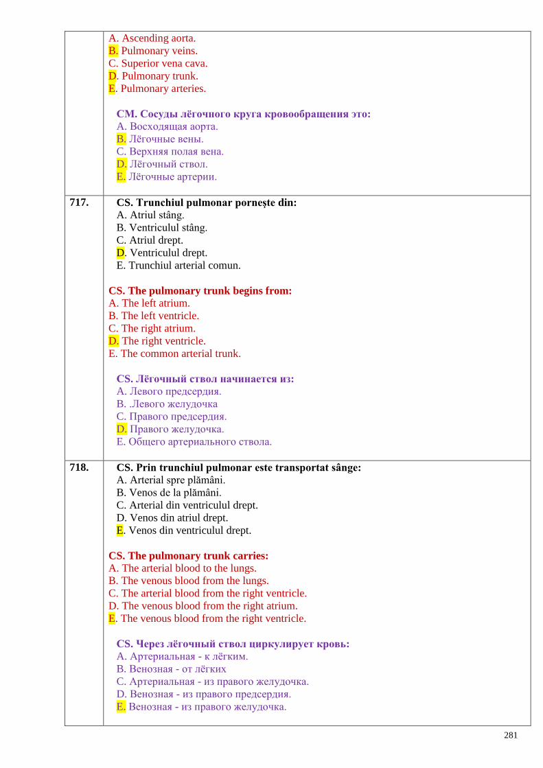
281
A. Ascending aorta.
B. Pulmonary veins.
C. Superior vena cava.
D. Pulmonary trunk.
E. Pulmonary arteries.
СМ. Сосуды лѐгочного круга кровообращения это:
A. Восходящая аорта.
B. Лѐгочные вены.
C. Верхняя полая вена.
D. Лѐгочный ствол.
E. Лѐгочные артерии.
717. CS. Trunchiul pulmonar porneşte din:
A. Atriul stâng.
B. Ventriculul stâng.
C. Atriul drept.
D. Ventriculul drept.
E. Trunchiul arterial comun.
CS. The pulmonary trunk begins from:
A. The left atrium.
B. The left ventricle.
C. The right atrium.
D. The right ventricle.
E. The common arterial trunk.
СS. Лѐгочный ствол начинается из:
A. Левого предсердия.
B. .Левого желудочка
C. Правого предсердия.
D. Правого желудочка.
E. Общего артериального ствола.
718. CS. Prin trunchiul pulmonar este transportat sânge:
A. Arterial spre plămâni.
B. Venos de la plămâni.
C. Arterial din ventriculul drept.
D. Venos din atriul drept.
E. Venos din ventriculul drept.
CS. The pulmonary trunk carries:
A. The arterial blood to the lungs.
B. The venous blood from the lungs.
C. The arterial blood from the right ventricle.
D. The venous blood from the right atrium.
E. The venous blood from the right ventricle.
СS. Через лѐгочный ствол циркулирует кровь:
A. Артериальная - к лѐгким.
B. Венозная - от лѐгких
C. Артериальная - из правого желудочка.
D. Венозная - из правого предсердия.
E. Венозная - из правого желудочка.
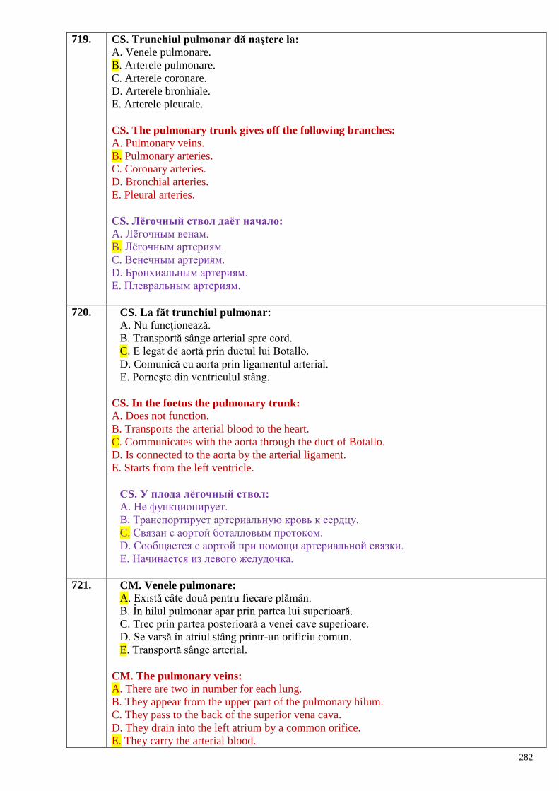
282
719. CS. Trunchiul pulmonar dă naştere la:
A. Venele pulmonare.
B. Arterele pulmonare.
C. Arterele coronare.
D. Arterele bronhiale.
E. Arterele pleurale.
CS. The pulmonary trunk gives off the following branches:
A. Pulmonary veins.
B. Pulmonary arteries.
C. Coronary arteries.
D. Bronchial arteries.
E. Pleural arteries.
СS. Лѐгочный ствол даѐт начало:
A. Лѐгочным венам.
B. Лѐгочным артериям.
C. Венечным артериям.
D. Бронхиальным артериям.
E. Плевральным артериям.
720. CS. La făt trunchiul pulmonar:
A. Nu funcţionează.
B. Transportă sânge arterial spre cord.
C. E legat de aortă prin ductul lui Botallo.
D. Comunică cu aorta prin ligamentul arterial.
E. Porneşte din ventriculul stâng.
CS. In the foetus the pulmonary trunk:
A. Does not function.
B. Transports the arterial blood to the heart.
C. Communicates with the aorta through the duct of Botallo.
D. Is connected to the aorta by the arterial ligament.
E. Starts from the left ventricle.
СS. У плода лѐгочный ствол:
A. Не функционирует.
B. Транспортирует артериальную кровь к сердцу.
C. Связан с аортой боталловым протоком.
D. Сообщается с аортой при помощи артериальной связки.
E. Начинается из левого желудочка.
721. CM. Venele pulmonare:
A. Există câte două pentru fiecare plămân.
B. În hilul pulmonar apar prin partea lui superioară.
C. Trec prin partea posterioară a venei cave superioare.
D. Se varsă în atriul stâng printr-un orificiu comun.
E. Transportă sânge arterial.
CM. The pulmonary veins:
A. There are two in number for each lung.
B. They appear from the upper part of the pulmonary hilum.
C. They pass to the back of the superior vena cava.
D. They drain into the left atrium by a common orifice.
E. They carry the arterial blood.
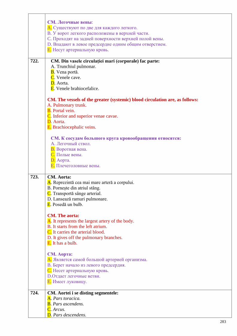
283
СМ. Легочные вены:
A. Существуют по две для каждого легкого.
B. У ворот легкого расположены в верхней части.
C. Проходят на задней поверхности верхней полой вены.
D. Впадают в левое предсердие одним общим отверстием.
E. Несут артериальную кровь.
722. CM. Din vasele circulaţiei mari (corporale) fac parte:
A. Trunchiul pulmonar.
B. Vena portă.
C. Venele cave.
D. Aorta.
E. Venele brahiocefalice.
CM. The vessels of the greater (systemic) blood circulation are, as follows:
A. Pulmonary trunk.
B. Portal vein.
C. Inferior and superior venae cavae.
D. Aorta.
E. Brachiocephalic veins.
СМ. К сосудам большого круга кровообращения относятся:
A. Легочный ствол.
B. Воротная вена.
C. Полые вены.
D. Аорта.
E. Плечеголовные вены.
723. CM. Aorta:
A. Reprezintă cea mai mare arteră a corpului.
B. Porneşte din atriul stâng.
C. Transportă sânge arterial.
D. Lansează ramuri pulmonare.
E. Posedă un bulb.
CM. The aorta:
A. It represents the largest artery of the body.
B. It starts from the left atrium.
C. It carries the arterial blood.
D. It gives off the pulmonary branches.
E. It has a bulb.
СМ. Аорта:
A. Является самой большой артерией организма.
B. Берет начало из левого предсердия.
C. Несет артериальную кровь.
D.Отдает легочные ветви.
E. Имеет луковицу.
724. CM. Aortei i se disting segmentele:
A. Pars toracica.
B. Pars ascendens.
C. Arcus.
D. Pars descendens.
284
E. Pars pelvina.
CM. The following parts of the aorta can be distinguished:
A. Pars toracica.
B. Pars ascendens
C. Arcus.
D. Pars descendens.
E. Pars pelvina.
СМ. Выделяют следующие сегменты аорты:
A. Pars toracica.
B. Pars ascendens.
C. Arcus.
D. Pars descendens.
E. Pars pelvina.
725. CS. Aorta toracică reprezintă:
A. Toate segmentele aortei, localizate în cavitatea toracică.
B. Doar porţiunea aortei descendente, localizate în mediastinul posterior.
C. Aorta ascendentă şi arcul aortic.
D. Porţiunea aortei cu cel mai mare diametru.
E. Segmentul aortei care lansează ramuri spre organele din cavitatea toracică.
CS. The thoracic aorta represents:
A. All the segments of the aorta located in the thoracic cavity.
B. Only the part of the descending aorta located in the posterior mediastinum.
C. The ascending aorta and aortic arch.
D. The part of the aorta which has the largest diameter.
E. The part of the aorta that gives off branches to the organs of the thoracic cavity.
CS. Грудная аорта представляет:
A. Все сегменты аорты, расположены в грудной полости.
B. Только часть нисходящей аорты расположена в заднем средостении.
C. Восходящая аорта и дуга аорты.
D. Часть аорты с самым большим диаметром.
E. Сегмент аорты, от которого отходят ветви к органам грудной полости.
726. CM. Sistemul venei cave superioare include venele:
A. Pulmonare drepte şi stângi.
B. Subclaviculare dreaptă şi stângă.
C. Brahiocefalice dreaptă şi stângă.
D. Impară.
E. Jugulare interne şi externe.
CM. The system of the superior vena cava includes:
A. The right and left pulmonary veins.
B. The right and left subclavian veins.
C. The right and left brachiocephalic veins.
D. The azygos vein.
E. The internal and external jugular veins.
СМ. Система верхней полой вены включает вены:
A. Правые и левые легочные.
B. Правые и левые подключичные.
C. Правые и левые плечеголовные.
285
D. Непарную.
E. Наружные и внутренние яремные.
727. CS. Venele brahiocefalice:
A. Se formează la confluerea venelor jugulare internă şi externă.
B. Au aceaşi lungime.
C. Vena brahiocefalică dreaptă este mai scurtă, decât cea stângă.
D. Vena brahiocefalică stângă este mai scurtă de două ori faţă de cea dreaptă.
E. Sunt afluenţi ai trunchiului pulmonar.
CS. The brachiocephalic veins:
A. Are formed by the union of the internal and external jugular veins.
B. They have the same length.
C. The right brachiocephalic vein is shorter than the left one.
D. The left brachiocephalic vein is twice shorter than the right one.
E. They are tributares of the pulmonary trunk.
СS. Плечеголовные вены:
A. Образуется при соединении внутренней и наружной яремных вен.
B. Имеют одинаковую длину.
C. Правая плечеголовная вена короче, чем левая.
D. Левая плечеголовная вена короче вдвойне, чем правая.
E. Являются притоками легочного ствола.
728. CS. Arterele pulmonare pornesc de la:
A. Atriul stâng.
B. Ventriculul drept.
C. Trunchiul pulmonar.
D. Vena cavă superioară.
E. Venele brahiocefalice.
CS. The pulmonary arteries start from:
A. The left atrium.
B. The right ventricle.
C. The pulmonary trunk.
D. The superior vena cava.
E. The brachiocephalic veins.
СS. Легочные артерии отходят от: A. Левого предсердия.
B. Правого желудочка.
C. Легочного ствола.
D. Верхней полой вены.
E. Плечеголовных вен.
729. CM. Referitor la venele pulmonare:
A. Există două vene pulmonare - anterioară şi posterioară.
B. Se deschid în atriul stâng.
C. Transportă sânge venos.
D. Transportă sânge arterial în atriul drept.
E. Sunt patru ca număr: două drepte şi două stângi.
CM. Concerning the pulmonary veins:
A. There are two pulmonary veins - anterior and posterior.
B. They drain into the left atrium.
286
C. They transport the venous blood.
D. They transport the arterial blood to the right atrium.
E. There are four pulmonary veins: two right and two left.
СМ. По отношению к легочным венам:
A. Существуют две легочные вены – передняя и задняя.
B. Впадают в левое предсердие.
C. Несут венозную кровь.
D. Несут артериальную кровь в правое предсердие.
E. Существуют четыре: две правые и две левые.
730. CM. Circulaţia mare:
A. Începe cu aorta din ventriculul stâng.
B. Se termină cu venele cave în atriul stâng.
C. Se termină în atriul drept.
D. Începe din ventriculul drept cu trunchiul pulmonar şi se termină cu patru vene pulmonare.
în atriul stâng.
E. Include: aorta, trunchiul pulmonar şi venele cave superioară şi inferioară.
CM. The greater blood circulation:
A. Starts with the aorta from the left ventricle.
B. Finishes by the caval veins into the left atrium.
C. Finishes into the right atrium.
D. Starts from the right ventricle with the pulmonary trunk and finishes with four pulmonary
veins into the left atrium.
E. Includes the aorta, pulmonary trunk and superior and inferior caval veins.
СМ. Большой круг кровообращения:
A. Начинается аортой из левого желудочка.
B. Заканчивается полыми венами в левом предсердии.
C. Заканчивается в правом предсердии.
D. Начинается из правого желудочка легочным стволом и заканчивается четырьмя
легочными венами в левом предсердии.
E. Включает: аорту, легочного ствола и верхними и нижними полыми венами.
731. CM. Segmentele aortei sunt:
A. Superior.
B. Cârja aortei.
C. Inferior.
D. Ascendent.
E. Descendent.
CM. The segments of the aorta are:
A. Superior part.
B. Arch of the aorta.
C. Inferior part.
D. Ascending part.
E. Descending part.
СМ. Сегменты аорты:
A. Верхний.
B. Дуга аорты.
C. Нижний.
D. Восходящий.
E. Нисходящий.
287
732. CM. Cu privire la porţiunea toracică a aortei:
A. Lansează artera toracică internă.
B. Vascularizează numai viscerele din cavitatea toracică.
C. Emite ramuri parietale şi viscerale.
D. Începe de la nivelul vertebrei toracice IV.
E. Lansează arterele frenice inferioare.
CM. Regarding the thoracic part of aorta:
A. Launches the internal thoracic artery.
B. Blood supply only viscera of thoracic cavity.
C. Emits the parietal and visceral branches.
D. Starts at the thoracic vertebrae IV level.
E. Launches the inferior phrenic arteries.
СМ. По отношению к грудной части аорты:
A. Отдает внутреннюю грудную аорту.
B. Кровоснабжает только органы грудной полости.
C. Отдает париетальные и висцеральные ветви.
D. Начинается на уровне IV грудного позвонка.
E. Отдает нижние диафрагмальные артерии.
733. CS. Cu privire la artera carotidă comună stângă:
A. Se află în mediastinul posterior.
B. Porneşte de la arcul aortic printr-un trunchi comun cu artera subclaviculară.
C. Se extinde de-a lungul regiunii cervicale a coloanei vertebrale.
D. Lansează ramuri esofagiene.
E. E mai lungă ca cea dreaptă.
CS. Regarding to the left common carotid artery:
A. It is located in the posterior mediastinum.
B. It starts from the aortic arch together with the subclavian artery as a common trunk.
C. It extends along the cervical region of the vertebral column.
D. It gives off the esophageal branches.
E. The left common carotid artery is longer than the right one.
СS. По отношению к левой общей сонной артерии: A. Находится в заднем средостении.
B. Отходит от дуги аорты общим стволом с подключичной артерией.
C. Расположена на уровне шейного отдела позвоночного столба.
D. Отдает пищеводные ветви.
E. Длиннее правой артерии.
734. CM. Artera subclaviculară dreapta:
A. Prezintă o ramură a trunchiului brahiocefalic.
B. Lansează artera axilară.
C. Abandonează cavitatea toracică prin apertura ei superioară.
D. Lansează ramuri intercostale anterioare superioare.
E. În regiunea cervicală trece împreună cu plexul cervical.
CM. The right subclavian artery:
A. Represents a branch of the brachiocephalic trunk.
B. Gives off the axillary artery.
C. Leaves the thoracic cavity through its superior aperture.
D. Gives rise to the superior anterior intercostal branches.
288
E. Passes in the cervical region together with the cervical plexus.
СМ. Правая подключичная артерия:
A. Представляет ветвь плечеголовного ствола.
B. Отдает подмышечную артерию.
C. Выходит из грудной полости через верхнюю грудную апертуру.
D. Отдает передние верхние межреберные ветви.
E. В шейном отделе проходит вместе с шейным сплетением.
735. CM. Artera subclaviculară stângă:
A. Are trunchi comun cu cea dreaptă.
B. Porneşte de la arcul aortei.
C. Se situează în şanţul arterei subclaviculare de pe coasta I.
D. Porneşte de la aorta toracică.
E. În regiunea cervicală trece împreună cu plexul brahial.
CM. The left subclavian artery:
A. Has a common trunk with right one.
B. Starts from the aortic arch.
C. Is located in the subclavian groove of the Ist rib.
D. Starts from the thoracic aorta.
E. Passes in the cervical region together with the brachial plexus.
СМ. Левая подключичная артерия:
A. Имеет общий ствол с правой артерией.
B. Отходит от дуги аорты.
C. Расположена в борозде подключичной артерии 1 ребра.
D. Отходит от грудной аорты.
E. В шейном отделе проходит вместе с плечевым сплетением
736. CM. Vena cavă superioară colectează sângele din venele:
A. Capului şi gâtului.
B. Membrelor superioare.
C. Parietale ale cavităţii toracice.
D. Viscerale ale cavităţii abdominale.
E. Mediastinale.
CM. The superior vena cava collects the blood from following veins:
A. Head and neck.
B. Upper limb.
C. The thoracic wall.
D. The abdominal viscera.
E. Mediastinal.
СМ. Верхняя полая вена собирает кровь от вен:
A. Головы и шеи.
B. Верхних конечностей.
C. Пристеночных вен грудной полости.
D. Висцеральных вен брюшной полости.
E. Средостенных.
737. CM. Vascularizaţia funcţională a plămânilor este realizată de:
A. Rr. pulmonales de la aorta toracică.
B. Aa. pulmonales.
C. Vv. pulmonales.
289
D. Rr. bronchiales.
E. Vv. bronchiales.
CM. The functional blood supply of the lungs is realized by:
A. Rr. pulmonales of the thoracic aorta
B. Aa. pulmonales
C. Vv. pulmonales
D. Rr. bronchiales
E. Vv. bronchiales
СМ. Функциональное кровоснабжение лѐгких осуществляется:
A. Rr. pulmonales de la aorta toracică.
B. Aa. pulmonales.
C. Vv. pulmonales.
D. Rr. bronchiales.
E. Vv. bronchiales.
738. CS. Pentru patul vascular al plămânilor e specifică prezenţa:
A. Reţelelor miraculoase arteriale.
B. Reţelelor miraculoase venoase.
C. Anastomozelor dintre vasele circulaţiilor mare şi mică.
D. Arterelor şi venelor magistrale.
E. Arterelor şi venelor colaterale.
CS. There is special feature of the lung blood supply by the presence of:
A. The arterial `rete mirabile`.
B. The venous `rete mirabile`.
C. The anastomosis between vessels of the greater and lesser blood circulations.
D. The magistral arteries and veins.
E. The collateral arteries and veins.
CS. Для микроциркуляторного русла легких характерно присутствие:
A. Чудесной артериальной сети.
B. Чудесной венозной сети.
C. Анастомозов между сосудов большого и малого круга кровообращения.
D. Магистральных артерий и вен.
E. Коллатеральных артерий и вен.
739. CS. Aorta toracică:
A. Lansează ramuri parietale şi viscerale.
B. De la ea pornesc arterele intercostale anterioare.
C. Trimite din partea sa posterioară ramuri spre măduva spinării.
D. Prin ramurile sale bronhiale anastomozează cu ramurile arterelor pulmonare.
E. Este situată în mediastinul mediu.
CS. The thoracic aorta:
A. Gives off the parietal and visceral branches.
B. Gives rise to the anterior intercostal arteries.
C. Transmits backward branches for the spinal cord.
D. Via its bronchial branches anastomosis with the pulmonary arteries.
E. Is located in the middle mediastinum.
CS. Грудная аорта: A. Отдает париетальные и висцеральные ветви.
B. От нее отходят передние межреберные артерии.
290
C. Отдает от задней еѐ части ветви к спинному мозгу.
D. Через бронхиальные ветви анастомозирует с ветвями легочных артерий.
E. Расположена в среднем средостении.
740. CM. Prin ramurile sale viscerale aorta toracică irigă:
A. Bronhiile.
B. Traheea.
C. Esofagul.
D. Pericardul.
E. Inima.
CM. Via its visceral branches the thoracic aorta supply:
A. Bronchi.
B. Trachea.
C. Esophagus.
D. Pericardium.
E. Heart.
СМ. Посредством своих висцеральных ветвей грудная аорта кровоснабжает:
A. Бронхи.
B. Трахею.
C. Пищевод.
D. Перикард.
E. Сердце.
741. CS. Vena azigos:
A. Reprezintă o continuare a venei lombare ascendente din dreapta.
B. Vine în raport cu pediculul plămânului stâng.
C. Nu posedă valve.
D. Are numai afluenţi viscerali.
E. Este un vas par.
CS. The azygos vein:
A. Represents a continuation of the right ascending lumbar vein.
B. Has relation with the left pulmonary pedicle.
C. Has no valves.
D. Possesses only visceral tributaries.
E. Is a paired blood vessel.
CS. Непарная вена: A. Представляет продолжение правой восходящей поясничной вены.
B. Проходит близко к ножке левого лѐгкого
C. Не содержит клапанов.
D. Имеет только висцеральные притоки.
E. Является парным сосудом.
742. CM. Vascularizaţia nutritivă a plămânilor este realizată de:
A. Arterele pulmonare.
B. Arterele bronhiale.
C. Venele pulmonare.
D. Venele bronhiale.
E. Ramuri pulmonare de la aorta ascendentă.
CM. The blood supply of the lungs is realized by:
A. The pulmonary arteries.
291
B. The bronchial arteries.
C. The pulmonary veins.
D. The bronchial veins.
E. The pulmonary branches of the ascendant aorta.
СМ. Сосуды, питающие ткань лѐгких:
A. Лѐгочные артерии.
B. Бронхиальные артерии.
C. Лѐгочные вены.
D. Бронхиальные вены.
E. Лѐгочные ветви восходящей аорты.
743. CM. Pentru plămâni este caracteristică:
A. Circulaţia sangvină funcţională.
B. Circulaţie sangvină mixtă.
C. Circulaţie sangvină nutritivă.
D. Amestecarea sângelui arterial cu cel venos.
E. Prezenţa anastomozelor dintre vasele circulaţiilor mare şi mică.
CM. The lungs have the following characteristic features:
A. Functional blood circulation.
B. Mixed blood circulation.
C. Nutritive blood circulation.
D. Mixture of arterial and venous blood.
E. Presence of anastomoses between the greater and lesser blood circulations.
СМ. В лѐгких существует:
A. Функциональное кровообращение.
B. Смешанное кровообращение.
C. Нутриционное кровообращение.
D. Смешивание артериальной и венозной крови.
E. Анастомозы между сосудами большого и малого круга кровообращения.
744. CM. Aorta toracică:
A. Este situată în mediastinul posterior.
B. Este în raport cu esofagul.
C. Are o lungime de 30 cm.
D. Pe flancul stâng al ei se află vena azigos şi canalul limfatic toracic.
E. Este continuarea aortei ascendente.
CM. The thoracic aorta:
A. Is located in the posterior mediastinum.
B. Has relation with the esophagus.
C. Has 30 cm in length.
D. On its left side the azygos vein and the lymphatic thoracic duct are located.
E. Is a continuation of the ascending aorta.
СМ. Грудная аорта:
A. Расположена в заднем средостении.
B. Граничит с пищеводом.
C. Длиной в 30 см.
D. По левому краю находится непарная вена и грудной лимфатический проток.
E. Является продолжением восходящей аорты.
745. CS. Vena azigos:
292
A. Este situată în mediastinul mediu (PNA).
B. Se află pe flancul stâng al coloanei vertebrale.
C. Este continuare a venei lombare ascendente din stânga.
D. Are afluenţi parietali şi viscerali.
E. Are numai afluenţi parietali.
CS. The azygos vein:
A. Is situated in the middle mediastinum (PNA).
B. Is located along the left side of the spine.
C. Is a continuation of the left ascending lumbar vein.
D. Has the parietal and visceral tributaries.
E. Has only the parietal tributaries.
СS. Vena azygos:
A. Расположена в среднем средостении (PNA).
B. Находится слева от позвоночника.
C. Является продолжением левой восходящей поясничной вены.
D. Получает пристеночные и висцеральные притоки.
E. Получает только пристеночные притоки.
746. CM. Artera axilară:
A. Reprezintă o continuare a arterei subclaviculare.
B. E înconjurată de trunchiurile nervoase ale plexului cervical.
C. La nivelul marginii inferioare a muşchiului pectoral mare se continuă cu artera brahială.
D. Se proiectează în şanţul deltoidopectoral.
E. E localizată în profunzimea fosei axilare.
MC. The axillary artery:
A. It is a continuation of the subclavian artery.
B. It is surrounded by the nervous trunks of the cervical plexus.
C. At the level of the inferior margin of the pectoralis major muscle it continues with the
brachial artery.
D. It projects within the deltoideopectoral groove (sulcus).
E. It is located in the deepness of the axillary fossa.
СM. Подмышечная артерия:
A. Является продолжением подключичной артерии.
B. Окружена нервными стволами шейного сплетения.
C. На уровне нижнего края большой грудной мышцы продолжается в плечевую
артерию.
D. Проецируется на уровне дельтогрудной борозды.
E. Располагается в глубине подмышечной ямки.
747. CS. Artera axilară:
A. Reprezintă o ramură a arterei subclaviculare.
B. E divizată condiţional în trei segmente conform topografiei peretelui posterior al cavităţii
axilare.
C. E însoţită de două vene axilare.
D. Poate fi palpată pe marginea medială a m. coracobrachialis.
E. Lansează ramuri spre muşchii scaleni.
MC. The axillary artery:
A. Is a branch of the subclavian artery.
B. According to the topography of the anterior thoracic wall it is divided into three segments.
C. Is accompanied by two axillary veins.
293
D. Can be palpated on the medial margin of the coracobrachialis muscle.
E. Gives off branches to the scalene muscles.
СS. Подмышечная артерия:
A. Является ветвью подключичной артерии.
B. Условно разделена на 3 сегмента, соответственно задней стенки подмышечной
ямки.
C. Еѐ сопровождают две подмышечные вены.
D. Еѐ можно прощупывать у медиального края m. coracobrachialis.
E. Отдаѐт ветви к лестничным мышцам
748. CM. Artera brahială:
A. Porneşte de la artera axilară.
B. Se desprinde de la artera subclaviculară.
C. Reprezintă o continuare a arterei axilare.
D. Este o componentă a pachetului neurovascular al braţului.
E. Se întinde de la nivelul marginii inferioare a muşchiului pectoral mare până la nivelul fosei
coronoide.
MC. The brachial artery:
A. It arises from the axillary artery.
B. It arises from the subclavian artery.
C. It is a continuation of the axillary artery.
D. It is a component of the neurovascular bundle (patch) of the arm.
E. It extends from the inferior margin of the pectoralis major muscle until the coronoid fossa.
СМ. Плечевая артерия:
A. Начинается от подмышечной артерии.
B. Ответвляется от подключичной артерии.
C. Является продолжением подмышечной артерии.
D. Является составной частью сосудисто-нервного пучка плеча.
E. Простирается от нижнего края большой грудной мышцы до венечной ямки.
749. CS. Artera brahială:
A. Trece prin canalul humeromuscular.
B. Se plasează în şanţul bicipital medial.
C. E însoţită de două vene brahiale şi nervul radial.
D. Poate fi palpată în fosa cubitală lateral de tendonul bicepsului brahial.
E. În scop de suspendare a hemoragiei poate fi comprimată pe epicondilul lateral al
humerusului.
SC. The brachial artery:
A. It passes through the humeromuscular canal.
B. It lodges within the medial bicipital groove.
C. It is accompanied by two brachial veins and by the radial nerve.
D. It can be palpated in the cubital fossa lateraly to the tendon of the biceps brachii muscle.
E. In order to stop the bleeding it can be compressed against the lateral epicondyle of the
humerus.
СS. Плечевая артерия:
A. Проходит через плечемышечный канал.
B. Располагается в медиальной борозде плеча.
C. Еѐ сопровождают две вены и лучевой нерв.
D. Еѐ можно прощупать в локтевой ямке, латеральнее сухожилия двуглавой мышцы
плеча.
294
E. Для остановки кровотечения еѐ можно прижать к латеральному надмыщелку
плечевой кости.
750. CS. Ramurile terminale ale arterei brahiale:
A. Iau naştere în fosa cubitală, la nivelul colului radial.
B. Sunt în număr de trei - arterele radială, ulnară şi brahială profundă.
C. Irigă toate formaţiunile anatomice de la nivelul braţului.
D. Nu anastomozează între ele.
E. Trec prin canalul humeromuscular şi şanţurile bicipitale anterioare medial şi lateral.
SC. The terminal branches of the brachial artery:
A. Have their origin in the cubital fossa, at the level of the neck of the radius.
B. There are three in number – the radial, ulnar and the deep brachial arteries.
C. They supply all the anatomical structures at the level of the arm.
D. They do not anastomosis to each other.
E. They pass through the humeromuscular canal and through the medial and lateral bicipital
grooves.
СS. Конечные ветви плечевой артерии:
A. Начинаются в локтевой ямке на уровне шейки лучевой кости.
B. Их три – лучевая, локтевая и глубокая артерии плеча.
C. Кровоснабжают все анатомические элементы на уровне плеча.
D. Не анастомозируют между собой.
E. Проходят через плечемышечный канал и переднюю латеральную (лучевую) и
медиальную (локтевую) борозды.
751. CM. La nivelul carpului există reţelele arteriale:
A. Carpiană comună.
B. Carpiană palmară.
C. Carpiană dorsală.
D. Carpiană medială.
E. Carpiană laterală.
MC. The following arterial networks are distinguished at the carpus:
A. Common carpal.
B. Palmar carpal.
C. Dorsal carpal.
D. Medial carpal.
E. Lateral carpal.
СМ. На уровне запястья имеются артериальные сети:
A. Общая артериальная сеть запястья.
B. Ладонная артериальная сеть запястья.
C. Тыльная артериальная сеть запястья.
D. Медиальная артериальная сеть запястья.
E. Латеральная артериальная сеть запястья.
752. CM. Ramurile arterei brahiale irigă:
A. Articulaţia scapulohumerală.
B. Muşchiul pectoral mare.
C. Muşchii anteriori ai braţului.
D. Articulaţia cotului.
E. Muşchiul triceps al braţului cu excepţia capului lui lateral.
MC. The branches of the brachial artery supply the:
295
A. Shoulder joint.
B. Pectoralis major muscle.
C. Anterior muscles of the arm.
D. The elbow joint.
E. The triceps brachii muscle excepting its lateral head.
СМ. Ветви плечевой артерии кровоснабжают:
A. Плечевой сустав.
B. Большую грудную мышцу.
C. Переднюю группу мышц плеча.
D. Локтевой сустав.
E. Трѐхглавую мышцу плеча, кроме латеральной головки
753. CM. Artera radială:
A. Reprezintă o continuare a arterei brahiale.
B. Continuă traiectul arterei brahiale.
C. Este una din cele două ramuri terminale ale arterei brahiale.
D. Reprezintă artera colaterală a antebraţului.
E. Formează arcada superficială palmară.
MC. The radial artery:
A. It is a continuation of the brachial artery.
B. It continues the course of the brachial artery.
C. It is one of those two terminal branches of the brachial artery.
D. It is a collateral artery of the forearm.
E. It forms the superficial palmar arch.
CM. Глубокая артерия плеча:
A. Является ветвью плечевой артерии
B. Продолжает ход плечевой артерии.
C. Является одной из двух конечных ветвей плечевой артерии
D. Представляет собой коллатеральную артерию предплечья
E. Образует поверхностную артериальную дугу
754. CM. Artera brahială profundă:
A. Reprezintă artera colaterală a braţului.
B. Trece prin canalul humeromuscular.
C. Este însoţită de venele omonime şi nervul median.
D. Irigă muşchii anteriori ai braţului.
E. Lansează două artere colaterale - medie şi laterală spre reţeaua arterială a cotului.
MC. The deep brachial artery:
A. Is a collateral artery of the arm.
B. Passes through the humeromuscular canal.
C. Is accompanied by the homonymous veins and by the median nerve.
D. Supplies the anterior group of muscles of the arm.
E. Gives off two collateral arteries – middle and lateral ones to the arterial network of the
elbow.
CM. Глубокая артерия плеча:
A. Является коллатеральной артерией плеча.
B. Проходит через плечемышечный канал.
C. Еѐ сопровождают одноимѐнные вены и срединный нерв.
D. Кровоснабжает переднюю группу мышц плеча.
E. Делится на две коллатеральные артерии – средняя и латеральная, для сосудистой
296
артериальной сети локтевого сустава.
755. CM. La nivelul membrului superior există anastomoze în sistemul:
A. Arterei axilare.
B. Arterei mediane.
C. Arterei subclaviculare.
D. Arterei humerale.
E. Arterelor radială şi ulnară.
MC. At the level of the upper limb are distinguished anastomoses in the following
arterial systems:
A. Axillary artery.
B. Median artery.
C. Subclavian artery.
D. Brachial artery.
E. Radial and ulnar arteries.
СМ. На уровне верхней конечности существуют анастомозы в системе:
A. Подмышечной артерии.
B. Срединной артерии.
C. Подключичной артерии.
D. Плечевой артерии.
E. Лучевой и локтевой артерий.
756. CM. Anastomozele arterelor membrului superior sunt localizate:
A. La nivelul articulaţiilor.
B. La nivelul treimii medii a braţului.
C. La nivelul mâinii.
D. Pe faţa anterioară a braţului.
E. În şanţul radial.
MC. The anastomoses of the upper limb are located:
A. At the level of the joints.
B. At the level of the middle third of the arm.
C. At the level of the hand.
D. On the anterior surface of the arm.
E. In the radial groove.
СМ. Анастомозы артерий верхней конечности локализуются:
A. На уровне суставов.
B. На уровне средней трети плеча.
C. На уровне кисти.
D. На наружной поверхности плеча.
E. В лучевой борозде.
757. CM. Venele membrului superior:
A. Sunt grupate în superficiale şi profunde.
B. Nu conţin valve.
C. Sunt legate prin anastomoze numeroase.
D. Cele profunde sunt mai dezvoltate ca cele superficiale.
E. Toate transportă sângele în vena axilară (sistemul venei cave superioare).
MC. The veins of the upper limb:
a. Are classified into the superficial and deep veins.
b. Do not contain valves.
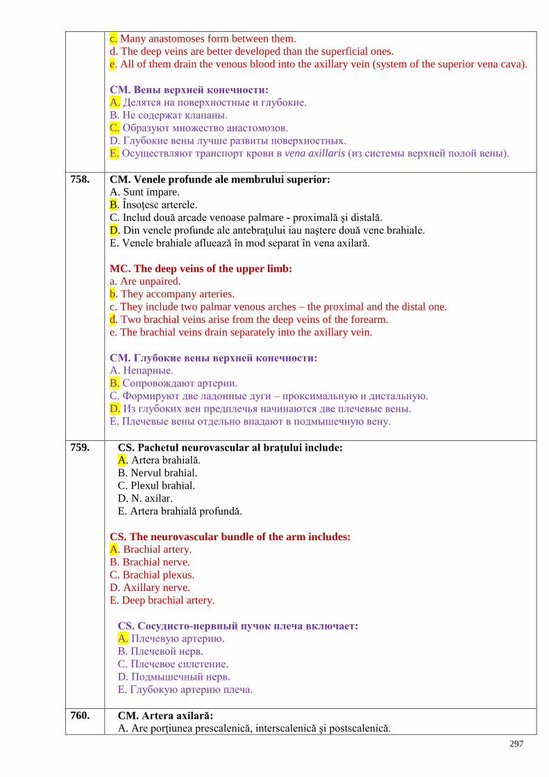
297
c. Many anastomoses form between them.
d. The deep veins are better developed than the superficial ones.
e. All of them drain the venous blood into the axillary vein (system of the superior vena cava).
СМ. Вены верхней конечности:
A. Делятся на поверхностные и глубокие.
B. Не содержат клапаны.
C. Образуют множество анастомозов.
D. Глубокие вены лучше развиты поверхностных.
E. Осуществляют транспорт крови в vena axillaris (из системы верхней полой вены).
758. CM. Venele profunde ale membrului superior:
A. Sunt impare.
B. Însoţesc arterele.
C. Includ două arcade venoase palmare - proximală şi distală.
D. Din venele profunde ale antebraţului iau naştere două vene brahiale.
E. Venele brahiale afluează în mod separat în vena axilară.
MC. The deep veins of the upper limb:
a. Are unpaired.
b. They accompany arteries.
c. They include two palmar venous arches – the proximal and the distal one.
d. Two brachial veins arise from the deep veins of the forearm.
e. The brachial veins drain separately into the axillary vein.
СМ. Глубокие вены верхней конечности:
A. Непарные.
B. Сопровождают артерии.
C. Формируют две ладонные дуги – проксимальную и дистальную.
D. Из глубоких вен предплечья начинаются две плечевые вены.
E. Плечевые вены отдельно впадают в подмышечную вену.
759. CS. Pachetul neurovascular al braţului include:
A. Artera brahială.
B. Nervul brahial.
C. Plexul brahial.
D. N. axilar.
E. Artera brahială profundă.
CS. The neurovascular bundle of the arm includes:
A. Brachial artery.
B. Brachial nerve.
C. Brachial plexus.
D. Axillary nerve.
E. Deep brachial artery.
СS. Сосудисто-нервный пучок плеча включает:
A. Плечевую артерию.
B. Плечевой нерв.
C. Плечевое сплетение.
D. Подмышечный нерв.
E. Глубокую артерию плеча.
760. CM. Artera axilară:
A. Are porţiunea prescalenică, interscalenică şi postscalenică.
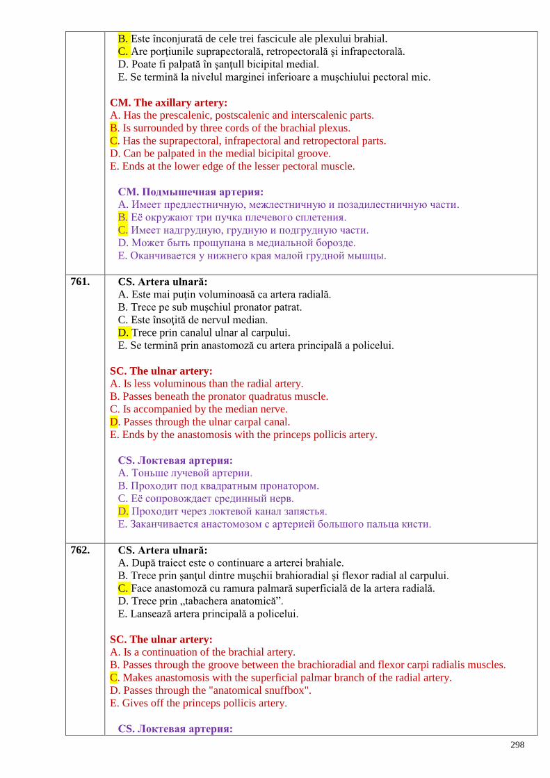
298
B. Este înconjurată de cele trei fascicule ale plexului brahial.
C. Are porţiunile suprapectorală, retropectorală şi infrapectorală.
D. Poate fi palpată în şanţull bicipital medial.
E. Se termină la nivelul marginei inferioare a muşchiului pectoral mic.
CM. The axillary artery:
A. Has the prescalenic, postscalenic and interscalenic parts.
B. Is surrounded by three cords of the brachial plexus.
C. Has the suprapectoral, infrapectoral and retropectoral parts.
D. Can be palpated in the medial bicipital groove.
E. Ends at the lower edge of the lesser pectoral muscle.
СМ. Подмышечная артерия: A. Имеет предлестничную, межлестничную и позадилестничную части.
B. Еѐ окружают три пучка плечевого сплетения.
C. Имеет надгрудную, грудную и подгрудную части.
D. Может быть прощупана в медиальной борозде.
E. Оканчивается у нижнего края малой грудной мышцы.
761. CS. Artera ulnară:
A. Este mai puţin voluminoasă ca artera radială.
B. Trece pe sub muşchiul pronator patrat.
C. Este însoţită de nervul median.
D. Trece prin canalul ulnar al carpului.
E. Se termină prin anastomoză cu artera principală a policelui.
SC. The ulnar artery:
A. Is less voluminous than the radial artery.
B. Passes beneath the pronator quadratus muscle.
C. Is accompanied by the median nerve.
D. Passes through the ulnar carpal canal.
E. Ends by the anastomosis with the princeps pollicis artery.
СS. Локтевая артерия:
A. Тоньше лучевой артерии.
B. Проходит под квадратным пронатором.
C. Еѐ сопровождает срединный нерв.
D. Проходит через локтевой канал запястья.
E. Заканчивается анастомозом с артерией большого пальца кисти.
762. CS. Artera ulnară:
A. După traiect este o continuare a arterei brahiale.
B. Trece prin şanţul dintre muşchii brahioradial şi flexor radial al carpului.
C. Face anastomoză cu ramura palmară superficială de la artera radială.
D. Trece prin „tabachera anatomică”.
E. Lansează artera principală a policelui.
SC. The ulnar artery:
A. Is a continuation of the brachial artery.
B. Passes through the groove between the brachioradial and flexor carpi radialis muscles.
C. Makes anastomosis with the superficial palmar branch of the radial artery.
D. Passes through the "anatomical snuffbox".
E. Gives off the princeps pollicis artery.
СS. Локтевая артерия:
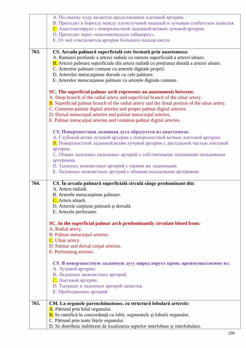
299
A. По своему ходу является продолжением плечевой артерии.
B. Проходит в борозду между плечелучевой мышцей и лучевым сгибателем запястья.
C. Анастомозирует с поверхностной ладонной ветвью лучевой артерии.
D. Проходит через «анатомическую табакерку» .
E. От неѐ ответвляется артерия большого пальца кисти.
763. CS. Arcada palmară superficială este formată prin anastomoza:
A. Ramurei profunde a arterei radiale cu ramura superficială a arterei ulnare.
B. Arterei palmare superficiale din artera radială cu porţiunea distală a arterei ulnare.
C. Arterelor palmare comune cu arterele digitale proprii.
D. Arterelor metacarpiene dorsale cu cele palmare.
E. Arterelor metacarpiene palmare cu arterele digitale comune.
SC. The superficial palmar arch represents an anastomosis between:
A. Deep branch of the radial artery and superficial branch of the ulnar artery.
B. Superficial palmar branch of the radial artery and the distal portion of the ulnar artery.
C. Common palmar digital arteries and proper palmar digital arteries.
D. Dorsal metacarpal arteries and palmar metacarpal arteries.
E. Palmar metacarpal arteries and common palmar digital arteries.
СS. Поверхностная ладонная дуга образуется из анастомоза:
A. Глубокой ветви лучевой артерии с поверхностной ветвью локтевой артерии.
B. Поверхностной ладонной ветви лучевой артерии с дистальной частью локтевой
артерии.
C. Общих ладонных пальцевых артерий с собственными ладонными пальцевыми
артериями.
D. Тыльных межпястных артерий с такими же ладонными.
E. Ладонных межпястных артерий с общими пальцевыми артериями.
764. CS. În arcada palmară superficială circulă sânge predominant din:
A. Artera radială.
B. Arterele metacarpiene palmare.
C. Artera ulnară.
D. Arterele carpiene palmară şi dorsală.
E. Arterele perforante.
SC. In the superficial palmar arch predominantly circulate blood from:
A. Radial artery.
B. Palmar metacarpal arteries.
C. Ulnar artery.
D. Palmar and dorsal carpal arteries.
E. Perforating arteries.
СS. В поверхностную ладонную дугу циркулирует кровь преимущественно из:
A. Лучевой артерии.
B. Ладонных межпястных артерий.
C. Локтевой артерии.
D. Тыльных и ладонных артерий запястья.
E. Прободающих артерий.
765. CM. La organele parenchimatoase, cu structură lobulară arterele:
A. Pătrund prin hilul organului.
B. Se ramifică în concordanţă cu lobii, segmentele şi lobulii organului.
C. Pătrund prin toate feţele organului.
D. Se distribuie indiferent de localizarea septelor interlobare şi interlobulare.
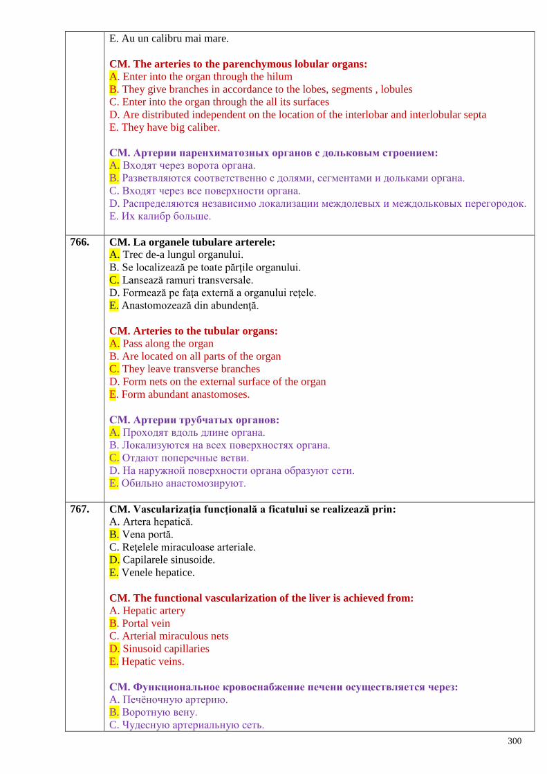
300
E. Au un calibru mai mare.
CM. The arteries to the parenchymous lobular organs:
A. Enter into the organ through the hilum
B. They give branches in accordance to the lobes, segments , lobules
C. Enter into the organ through the all its surfaces
D. Are distributed independent on the location of the interlobar and interlobular septa
E. They have big caliber.
СМ. Артерии паренхиматозных органов с дольковым строением:
A. Входят через ворота органа.
B. Разветвляются соответственно с долями, сегментами и дольками органа.
C. Входят через все поверхности органа.
D. Распределяются независимо локализации междолевых и междольковых перегородок.
E. Их калибр больше.
766. CM. La organele tubulare arterele:
A. Trec de-a lungul organului.
B. Se localizează pe toate părţile organului.
C. Lansează ramuri transversale.
D. Formează pe faţa externă a organului reţele.
E. Anastomozează din abundenţă.
CM. Arteries to the tubular organs:
A. Pass along the organ
B. Are located on all parts of the organ
C. They leave transverse branches
D. Form nets on the external surface of the organ
E. Form abundant anastomoses.
СМ. Артерии трубчатых органов:
A. Проходят вдоль длине органа.
B. Локализуются на всех поверхностях органа.
C. Отдают поперечные ветви.
D. На наружной поверхности органа образуют сети.
E. Обильно анастомозируют.
767. CM. Vascularizaţia funcţională a ficatului se realizează prin:
A. Artera hepatică.
B. Vena portă.
C. Reţelele miraculoase arteriale.
D. Capilarele sinusoide.
E. Venele hepatice.
CM. The functional vascularization of the liver is achieved from:
A. Hepatic artery
B. Portal vein
C. Arterial miraculous nets
D. Sinusoid capillaries
E. Hepatic veins.
СМ. Функциональное кровоснабжение печени осуществляется через:
A. Печѐночную артерию.
B. Воротную вену.
C. Чудесную артериальную сеть.
301
D. Синусоидные капилляры.
E. Печѐночные вены.
768. CM. Ramuri viscerale impare ale aortei abdominale sunt:
A. Artera lienală.
B. Trunchiul celiac.
C. Artera hepatică comună.
D. Artera mezenterică superioară.
E. Artera mezenterică inferioară.
CM. The unpaired visceral branches of the abdominal aorta:
A. Splenic artery
B. Celiac trunk
C. Common hepatic artery
D. Superior mesenteric artery
E. Inferior mesenteric artery.
СМ. Непарными висцеральными ветвями брюшной части аорты являются:
A. Селезѐночная артерия.
B. Чревный ствол.
C. Общая печѐночная артерия.
D. Верхняя брыжеечная артерия.
E. Нижняя брыжеечная артерия.
769. CM. Aorta abdominală:
A. Porneşte din atriul stâng al inimii.
B. Reprezintă un segment al aortei descendente.
C. Se întinde între ostiul aortic al diafragmei şi nivelul marginii superioare a vertebrei L5.
D. E situată în spaţiul retroperitoneal.
E. Trece în stânga de coloana vertebrală.
CM. The abdominal aorta:
A. Starts from the left atrium of the heart
B. It is a segment of the descending aorta
C. It extends between the aortic ostium of the diaphragm and upper margin of the L5 vertebra
D. Is located inside the retroperitoneal space
E. Is placed on the left of the vertebral column.
СМ. Брюшная часть аорты:
A. Выходит из левого предсердия сердца.
B. Представляет сегмент нисходящей части аорты.
C. Тянется между аортальным отверстием диафрагмы и уровнем верхнего края позвонка
L5.
D. Распoложена в забрюшинном пространстве.
E. Лежит слева от позвоночного столба.
770. CM. De la trunchiul celiac pornesc:
A. Artera gastrică dreaptă.
B. Artera hepatică comună.
C. Artera gastrică stângă.
D. Artera lienală.
E. Artera hepatică proprie.
CM. The branches of the celiac trunk are:
A. Right gastric artery
302
B. Common hepatic artery
C. Left gastric artery
D. Splenic artery
E. Proper hepatic artery.
СМ. От чревного ствола отходят:
A. Правая желудочная артерия.
B. Общая печѐночная артерия .
C. Левая желудочная артерия.
D. Селезѐночная артерия.
E. Собственная печѐночная артерия.
771. CM. Artera mezenterică superioară vascularizează:
A. Intestinul mezenterial.
B. Colonul ascendent.
C. Duodenul.
D. Colonul sigmoid.
E. Cecoapendicele.
CM. The regions of supplying of the superior mesenteric artery are:
A. Mesenterial intestine
B. Ascending colon
C. Duodenum
D. Sigmoid colon
E. Cecum and vermiform appendix.
СМ. Верхняя брыжеечная артерия кровоснабжает:
A. Брыжеечную кишку.
B. Восходящую ободочную кишку.
C. Двенадцатиперстную кишку.
D. Сигмовидную кишку.
E. Червеобразный отросток.
772. CM. Artera mezenterică inferioară:
A. Se termină cu a. rectalis sup.
B. De la ea pornesc artere sigmoidiene.
C. Vascularizează intestinul mezenterial.
D. Vascularizează colonul ascendent.
E. Irigă colonul descendent.
CM. The inferior mesenteric artery:
A. Finishes by the superior rectal artery
B. Sigmoid arteries start from it
C. Supplies the mesenterial intestine
D. Supplies the ascending colon
E. Irrigates the descending colon.
СМ. Нижняя брыжеечная артерия:
A. Заканчивается a. rectalis sup.
B. От неѐ отходят сигмовидные артерии.
C. Кровоснабжает брыжеечную кишку.
D. Кровоснабжает восходящую ободочную кишку.
E. Кровоснабжает нисходящую ободочную кишку.
773. CM. Ramuri viscerale pare ale aortei abdominale sunt:
303
A. Arterele frenice inferioare.
B. Artera suprarenală medie.
C. Artera suprarenală inferioară.
D. Artera renală.
E. Artera testiculară (ovariană).
CM. The paired visceral branches of the abdominal aorta are:
A. Inferior phrenic artery
B. Middle adrenal (suprarenal) artery
C. Inferior adrenal (suprarenal) artery
D. Renal artery
E. Testicular (ovarian) artery.
СМ. Парные висцеральные ветви брюшной части аорты:
A. Нижние диафрагмальные артерии.
B. Средняя надпочечная артерия.
C. Нижняя надпочечная артерия.
D. Почечная артерия.
E. Яичковая (яичниковая) артерия.
774. CM. Sunt ramuri parietale ale aortei abdominale:
A. Arterele frenice inferioare.
B. Artera iliolombară.
C. Arterele lombare.
D. Arterele testiculare (ovariene).
E. Artera sacrală mediană.
CM. The parietal branches of the abdominal aorta are:
A. Inferior phrenic artery
B. Iliolumbar artery
C. Lumbar arteries
D. Testicular (ovarian) artery
E. Median sacral artery.
СМ. Являются париетальными ветвями брюшной части аорты:
A. Нижние диафрагмальные артерии.
B. Подвздошно-поясничные артерии.
C. Поясничные артерии.
D. Яичковые (яичниковые) артерии.
E. Срединная крестцовая артерия.
775. CM. Cu privire la arterele iliace comune:
A. Constituie ramuri terminale ale aortei abdominale.
B. Îşi iau originea de la aorta abdominală la nivelul vertebrei lombare V.
C. Se împart în arterele iliace externă şi internă la nivelul articulaţiei sacroiliace.
D. Lansează ramuri parietale şi viscerale.
E. Se continuă cu artera femurală.
CM. The common iliac arteries:
A. These are terminal branches of the abdominal aorta
B. Have origin from the abdominal aorta at the level of the L5 vertebra
C. Are divided into the external and internal iliac arteries at the level of the sacroiliac joint
D. Give off the parietal and visceral branches
E. Continuous with the femoral artery.
304
СМ. Относительно общих подвздошных артерий:
A. Представляют конечные ветви брюшной части аорты.
B. От брюшной части аорты начинаются на уровне 4-го поясничного позвонка.
C. На уровне крестцово-подвздошного сустава делятся на наружную и внутреннюю
подвздошные артерии.
D. Отдают париетальные и висцеральные ветви.
E. Продолжаются бедренной артерией.
776. CM. Intestinul gros e vascularizat din arterele:
A. Iliacă externă.
B. Mezenterică superioară.
C. Lienală.
D. Mezenterică inferioară.
E. Iliacă internă.
CS. The arteries that supply the large intestine are:
A. External iliac artery
B. Superior mesenteric artery
C. Splenic artery
D. Inferior mesenteric artery
E. Internal iliac artery.
СМ. Толстая кишка кровоснабжается следующими артериями:
A. Внутренней подвздошной.
B. Верхней брыжеечной.
C. Селезѐночной.
D. Нижней брыжеечной.
E. Внутренней подвздошной.
777. CM. La vascularizaţia stomacului participă în totalitate:
A. Artera gastrică stângă.
B. Artera gastroduodenală.
C. Artera gastrică dreaptă.
D. Artera gastroomentală stângă.
E. Artera gastroomentală dreaptă.
CM. The arteries that supply the stomach are:
A. Left gastric artery
B. Superior mesenteric artery
C. Right gastric artery
D. Left gastroomental artery
E. Right gastroomental artery.
СМ. В кровоснабжении желудка в основном участвуют:
A. Левая желудочная артерия.
B. Гастродуаденальная артерия.
C. Правая желудочная артерия.
D. Левая желудочно-сальниковая артерия.
E. Правая желудочно-сальниковая артерия.
778. CM. Duodenul este vascularizat de ramuri ale arterelor:
A. Hepatică proprie.
B. Gastroduodenală.
C. Lienală.
D. Mezenterică superioară.
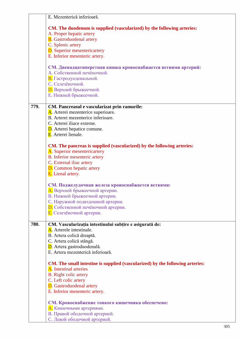
305
E. Mezenterică inferioară.
CM. The duodenum is supplied (vascularized) by the following arteries:
A. Proper hepatic artery
B. Gastroduodenal artery
C. Splenic artery
D. Superior mesentericartery
E. Inferior mesenteric artery.
СМ. Двенадцатиперстная кишка кровоснабжается ветвями артерий:
A. Собственной печѐночной.
B. Гастродуоденальной.
C. Селезѐночной.
D. Верхней брыжеечной.
E. Нижней брыжеечной.
779. CM. Pancreasul e vascularizat prin ramurile:
A. Arterei mezenterice superioare.
B. Arterei mezenterice inferioare.
C. Arterei iliace externe.
D. Arterei hepatice comune.
E. Arterei lienale.
CM. The pancreas is supplied (vascularized) by the following arteries:
A. Superior mesentericartery
B. Inferior mesenteric artery
C. External iliac artery
D. Common hepatic artery
E. Lienal artery.
СМ. Поджелудочная железа кровоснабжается ветвями:
A. Верхней брыжеечной артерии.
B. Нижней брыжеечной артерии.
C. Наружной подвздошной артерии.
D. Собственной печѐночной артерии.
E. Селезѐночной артерии.
780. CM. Vascularizaţia intestinului subţire e asigurată de:
A. Arterele intestinale.
B. Artera colică dreaptă.
C. Artera colică stângă.
D. Artera gastroduodenală.
E. Artera mezenterică inferioară.
CM. The small intestine is supplied (vascularized) by the following arteries:
A. Intestinal arteries
B. Right colic artery
C. Left colic artery
D. Gastroduodenal artery
E. Inferior mesenteric artery.
СМ. Кровоснабжение тонкого кишечника обеспечено:
A. Кишечными артериями.
B. Правой ободочной артерией.
C. Левой ободочной артерией.
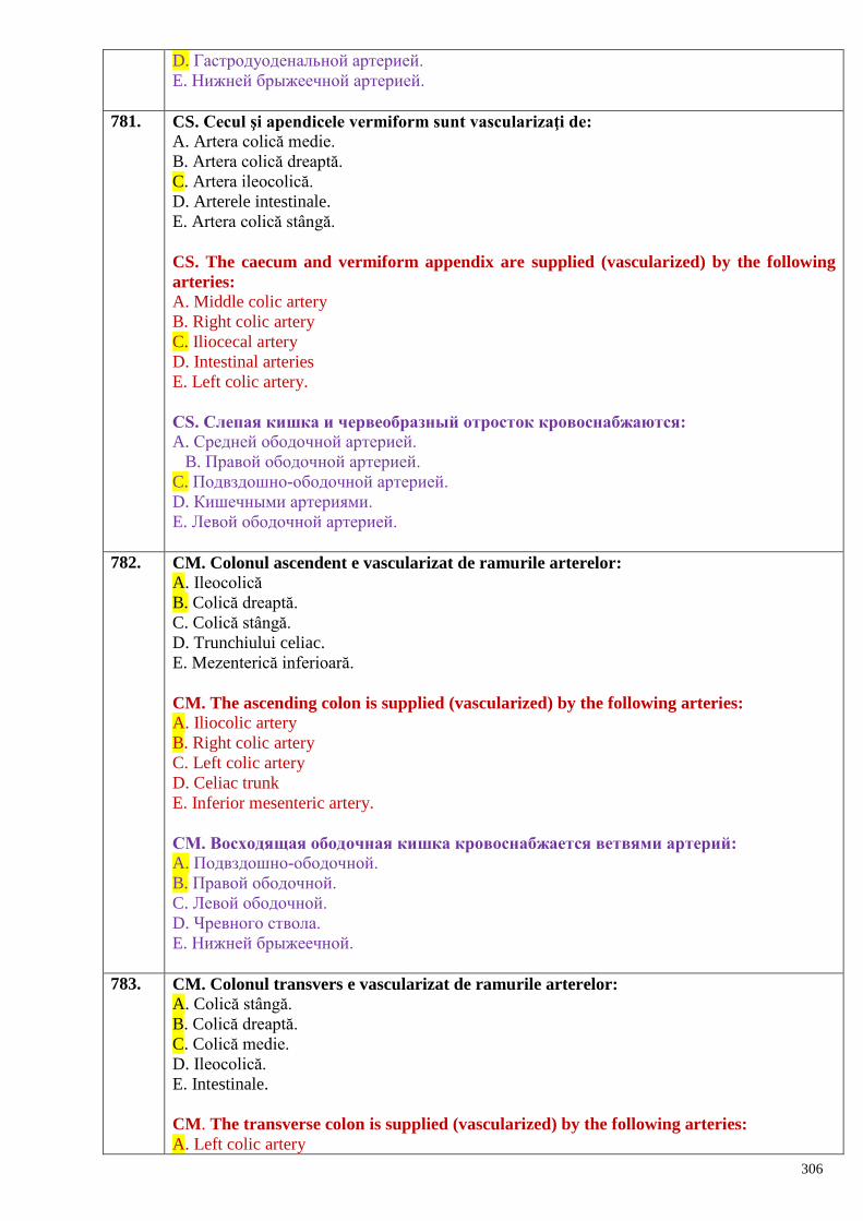
306
D. Гастродуоденальной артерией.
E. Нижней брыжеечной артерией.
781. CS. Cecul şi apendicele vermiform sunt vascularizaţi de:
A. Artera colică medie.
B. Artera colică dreaptă.
C. Artera ileocolică.
D. Arterele intestinale.
E. Artera colică stângă.
CS. The caecum and vermiform appendix are supplied (vascularized) by the following
arteries:
A. Middle colic artery
B. Right colic artery
C. Iliocecal artery
D. Intestinal arteries
E. Left colic artery.
СS. Слепая кишка и червеобразный отросток кровоснабжаются:
A. Средней ободочной артерией.
B. Правой ободочной артерией.
C. Подвздошно-ободочной артерией.
D. Кишечными артериями.
E. Левой ободочной артерией.
782. CM. Colonul ascendent e vascularizat de ramurile arterelor:
A. Ileocolică
B. Colică dreaptă.
C. Colică stângă.
D. Trunchiului celiac.
E. Mezenterică inferioară.
CM. The ascending colon is supplied (vascularized) by the following arteries:
A. Iliocolic artery
B. Right colic artery
C. Left colic artery
D. Celiac trunk
E. Inferior mesenteric artery.
СМ. Восходящая ободочная кишка кровоснабжается ветвями артерий:
A. Подвздошно-ободочной.
B. Правой ободочной.
C. Левой ободочной.
D. Чревного ствола.
E. Нижней брыжеечной.
783. CM. Colonul transvers e vascularizat de ramurile arterelor:
A. Colică stângă.
B. Colică dreaptă.
C. Colică medie.
D. Ileocolică.
E. Intestinale.
CM. The transverse colon is supplied (vascularized) by the following arteries:
A. Left colic artery
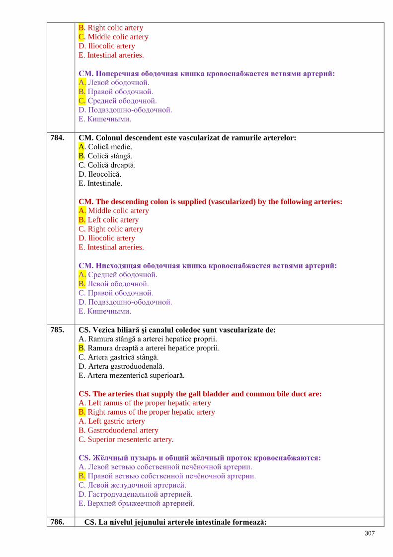
307
B. Right colic artery
C. Middle colic artery
D. Iliocolic artery
E. Intestinal arteries.
СМ. Поперечная ободочная кишка кровоснабжается ветвями артерий:
A. Левой ободочной.
B. Правой ободочной.
C. Средней ободочной.
D. Подвздошно-ободочной.
E. Кишечными.
784. CM. Colonul descendent este vascularizat de ramurile arterelor:
A. Colică medie.
B. Colică stângă.
C. Colică dreaptă.
D. Ileocolică.
E. Intestinale.
CM. The descending colon is supplied (vascularized) by the following arteries:
A. Middle colic artery
B. Left colic artery
C. Right colic artery
D. Iliocolic artery
E. Intestinal arteries.
СМ. Нисходящая ободочная кишка кровоснабжается ветвями артерий:
A. Средней ободочной.
B. Левой ободочной.
C. Правой ободочной.
D. Подвздошно-ободочной.
E. Кишечными.
785. CS. Vezica biliară şi canalul coledoc sunt vascularizate de:
A. Ramura stângă a arterei hepatice proprii.
B. Ramura dreaptă a arterei hepatice proprii.
C. Artera gastrică stângă.
D. Artera gastroduodenală.
E. Artera mezenterică superioară.
CS. The arteries that supply the gall bladder and common bile duct are:
A. Left ramus of the proper hepatic artery
B. Right ramus of the proper hepatic artery
A. Left gastric artery
B. Gastroduodenal artery
C. Superior mesenteric artery.
СS. Жѐлчный пузырь и общий жѐлчный проток кровоснабжаются:
A. Левой ветвью собственной печѐночной артерии.
B. Правой ветвью собственной печѐночной артерии.
C. Левой желудочной артерией.
D. Гастродуаденальной артерией.
E. Верхней брыжеечной артерией.
786. CS. La nivelul jejunului arterele intestinale formează:
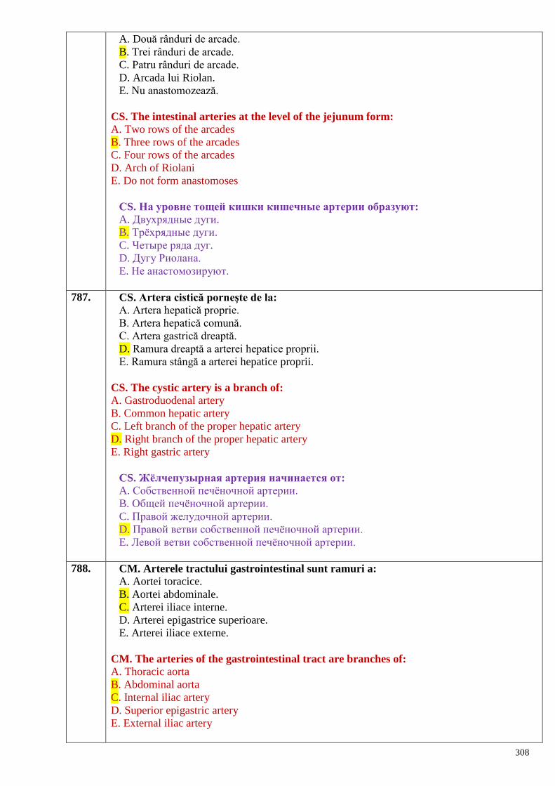
308
A. Două rânduri de arcade.
B. Trei rânduri de arcade.
C. Patru rânduri de arcade.
D. Arcada lui Riolan.
E. Nu anastomozează.
CS. The intestinal arteries at the level of the jejunum form:
A. Two rows of the arcades
B. Three rows of the arcades
C. Four rows of the arcades
D. Arch of Riolani
E. Do not form anastomoses
СS. На уровне тощей кишки кишечные артерии образуют:
A. Двухрядные дуги.
B. Трѐхрядные дуги.
C. Четыре ряда дуг.
D. Дугу Риолана.
E. Не анастомозируют.
787. CS. Artera cistică porneşte de la:
A. Artera hepatică proprie.
B. Artera hepatică comună.
C. Artera gastrică dreaptă.
D. Ramura dreaptă a arterei hepatice proprii.
E. Ramura stângă a arterei hepatice proprii.
CS. The cystic artery is a branch of:
A. Gastroduodenal artery
B. Common hepatic artery
C. Left branch of the proper hepatic artery
D. Right branch of the proper hepatic artery
E. Right gastric artery
СS. Жѐлчепузырная артерия начинается от:
A. Собственной печѐночной артерии.
B. Общей печѐночной артерии.
C. Правой желудочной артерии.
D. Правой ветви собственной печѐночной артерии.
E. Левой ветви собственной печѐночной артерии.
788. CM. Arterele tractului gastrointestinal sunt ramuri a:
A. Aortei toracice.
B. Aortei abdominale.
C. Arterei iliace interne.
D. Arterei epigastrice superioare.
E. Arterei iliace externe.
CM. The arteries of the gastrointestinal tract are branches of:
A. Thoracic aorta
B. Abdominal aorta
C. Internal iliac artery
D. Superior epigastric artery
E. External iliac artery
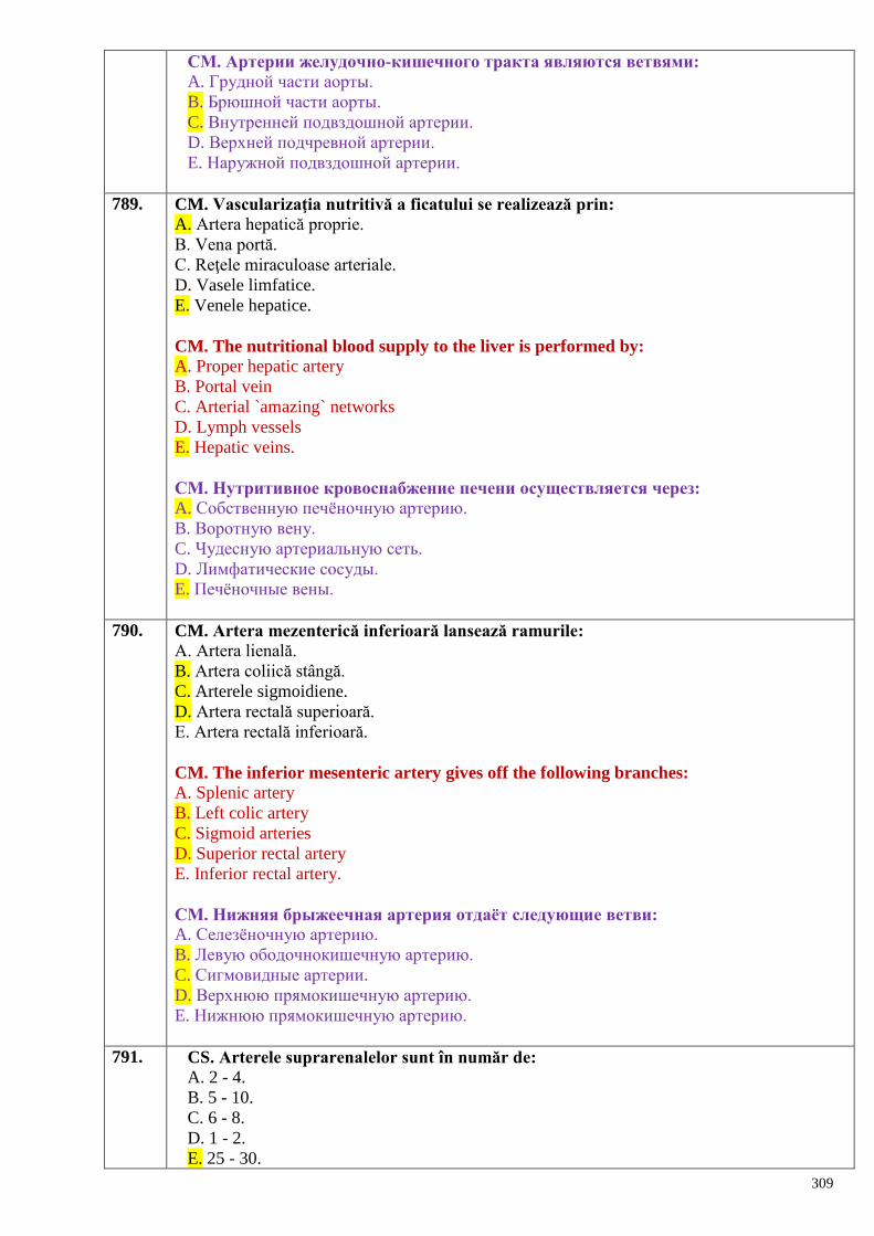
309
СМ. Артерии желудочно-кишечного тракта являются ветвями:
A. Грудной части аорты.
B. Брюшной части аорты.
C. Внутренней подвздошной артерии.
D. Верхней подчревной артерии.
E. Наружной подвздошной артерии.
789. CM. Vascularizaţia nutritivă a ficatului se realizează prin:
A. Artera hepatică proprie.
B. Vena portă.
C. Reţele miraculoase arteriale.
D. Vasele limfatice.
E. Venele hepatice.
CM. The nutritional blood supply to the liver is performed by:
A. Proper hepatic artery
B. Portal vein
C. Arterial `amazing` networks
D. Lymph vessels
E. Hepatic veins.
СМ. Нутритивное кровоснабжение печени осуществляется через:
A. Собственную печѐночную артерию.
B. Воротную вену.
C. Чудесную артериальную сеть.
D. Лимфатические сосуды.
E. Печѐночные вены.
790. CM. Artera mezenterică inferioară lansează ramurile:
A. Artera lienală.
B. Artera coliică stângă.
C. Arterele sigmoidiene.
D. Artera rectală superioară.
E. Artera rectală inferioară.
CM. The inferior mesenteric artery gives off the following branches:
A. Splenic artery
B. Left colic artery
C. Sigmoid arteries
D. Superior rectal artery
E. Inferior rectal artery.
СМ. Нижняя брыжеечная артерия отдаѐт следующие ветви:
A. Селезѐночную артерию.
B. Левую ободочнокишечную артерию.
C. Сигмовидные артерии.
D. Верхнюю прямокишечную артерию.
E. Нижнюю прямокишечную артерию.
791. CS. Arterele suprarenalelor sunt în număr de:
A. 2 - 4.
B. 5 - 10.
C. 6 - 8.
D. 1 - 2.
E. 25 - 30.
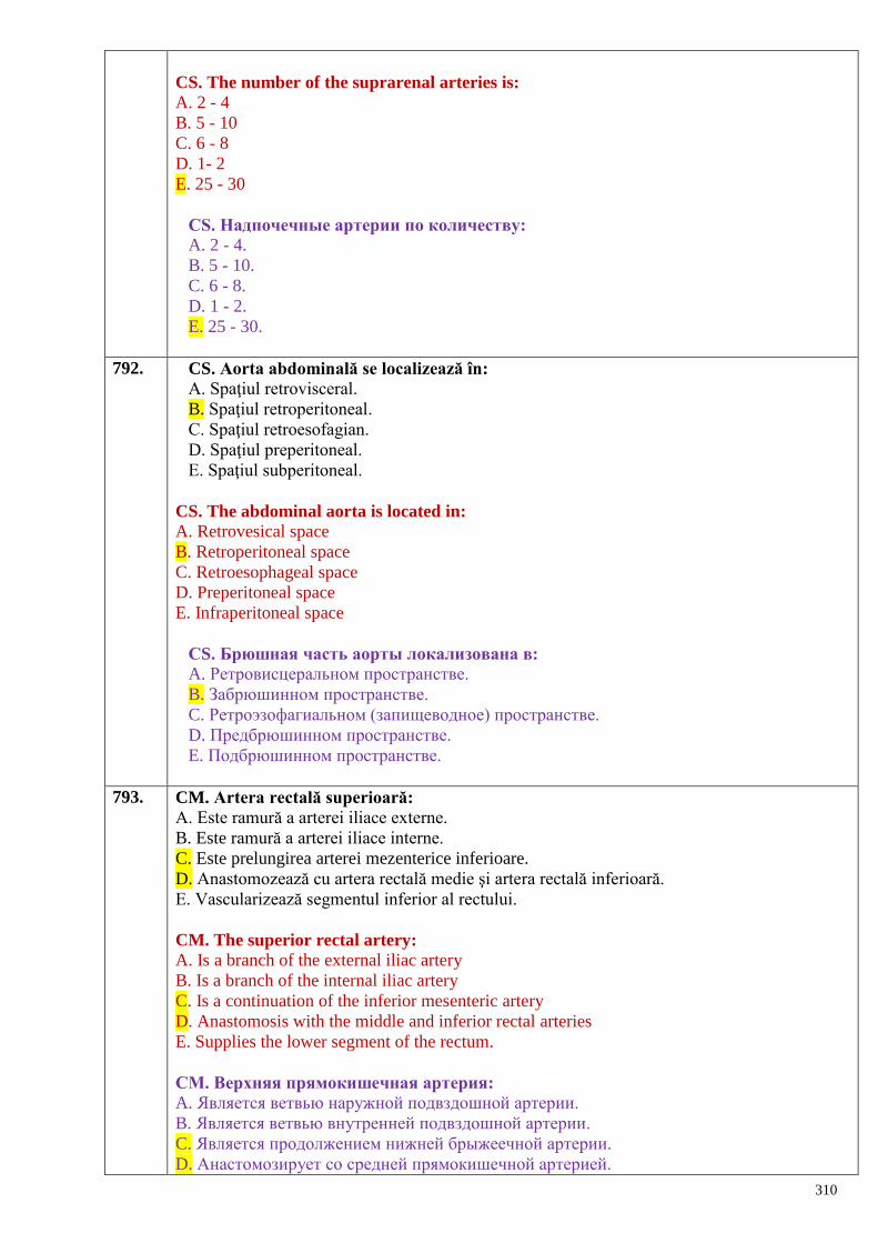
310
CS. The number of the suprarenal arteries is:
A. 2 - 4
B. 5 - 10
C. 6 - 8
D. 1- 2
E. 25 - 30
СS. Надпочечные артерии по количеству:
A. 2 - 4.
B. 5 - 10.
C. 6 - 8.
D. 1 - 2.
E. 25 - 30.
792. CS. Aorta abdominală se localizează în:
A. Spaţiul retrovisceral.
B. Spaţiul retroperitoneal.
C. Spaţiul retroesofagian.
D. Spaţiul preperitoneal.
E. Spaţiul subperitoneal.
CS. The abdominal aorta is located in:
A. Retrovesical space
B. Retroperitoneal space
C. Retroesophageal space
D. Preperitoneal space
E. Infraperitoneal space
СS. Брюшная часть аорты локализована в:
A. Ретровисцеральном пространстве.
B. Забрюшинном пространстве.
C. Ретроэзофагиальном (запищеводное) пространстве.
D. Предбрюшинном пространстве.
E. Подбрюшинном пространстве.
793. CM. Artera rectală superioară:
A. Este ramură a arterei iliace externe.
B. Este ramură a arterei iliace interne.
C. Este prelungirea arterei mezenterice inferioare.
D. Anastomozează cu artera rectală medie şi artera rectală inferioară.
E. Vascularizează segmentul inferior al rectului.
CM. The superior rectal artery:
A. Is a branch of the external iliac artery
B. Is a branch of the internal iliac artery
C. Is a continuation of the inferior mesenteric artery
D. Anastomosis with the middle and inferior rectal arteries
E. Supplies the lower segment of the rectum.
СМ. Верхняя прямокишечная артерия:
A. Является ветвью наружной подвздошной артерии.
B. Является ветвью внутренней подвздошной артерии.
C. Является продолжением нижней брыжеечной артерии.
D. Анастомозирует со средней прямокишечной артерией.
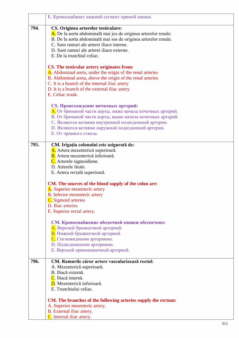
311
E. Кровоснабжает нижний сегмент прямой кишки.
794. CS. Originea arterelor testiculare:
A. De la aorta abdominală mai jos de originea arterelor renale.
B. De la aorta abdominală mai sus de originea arterelor renale.
C. Sunt ramuri ale arterei iliace interne.
D. Sunt ramuri ale arterei iliace externe.
E. De la trunchiul celiac.
CS. The testicular artery originates from:
A. Abdominal aorta, under the origin of the renal arteries
B. Abdominal aorta, above the origin of the renal arteries
C. It is a branch of the internal iliac artery
D. It is a branch of the external iliac artery
E. Celiac trunk.
СS. Происхождение яичковых артерий:
A. От брюшной части аорты, ниже начала почечных артерий.
B. От брюшной части аорты, выше начала почечных артерий.
C. Являются ветвями внутренней подвздошной артерии.
D. Являются ветвями наружной подвздошной артерии.
E. От чревного ствола.
795. CM. Irigaţia colonului este asigurată de:
A. Artera mezenterică superioară.
B. Artera mezenterică inferioară.
C. Arterele sigmoidiene.
D. Arterele ileale.
E. Artera rectală superioară.
CM. The sources of the blood supply of the colon are:
A. Superior mesenteric artery
B. Inferior mesenteric artery
C. Sigmoid arteries
D. Iliac arteries
E. Superior rectal artery.
СМ. Кровоснабжение ободочной кишки обеспечено:
A. Верхней брыжеечной артерией.
B. Нижней брыжеечной артерией.
C. Сигмовидными артериями.
D. Подвздошными артериями.
E. Верхней прямокишечной артерией.
796. CM. Ramurile căror artere vascularizează rectul:
A. Mezenterică superioară.
B. Iliacă externă.
C. Iliacă internă.
D. Mezenterică inferioară.
E. Trunchiului celiac.
CM. The branches of the following arteries supply the rectum:
A. Superior mesenteric artery.
B. External iliac artery.
C. Internal iliac artery.
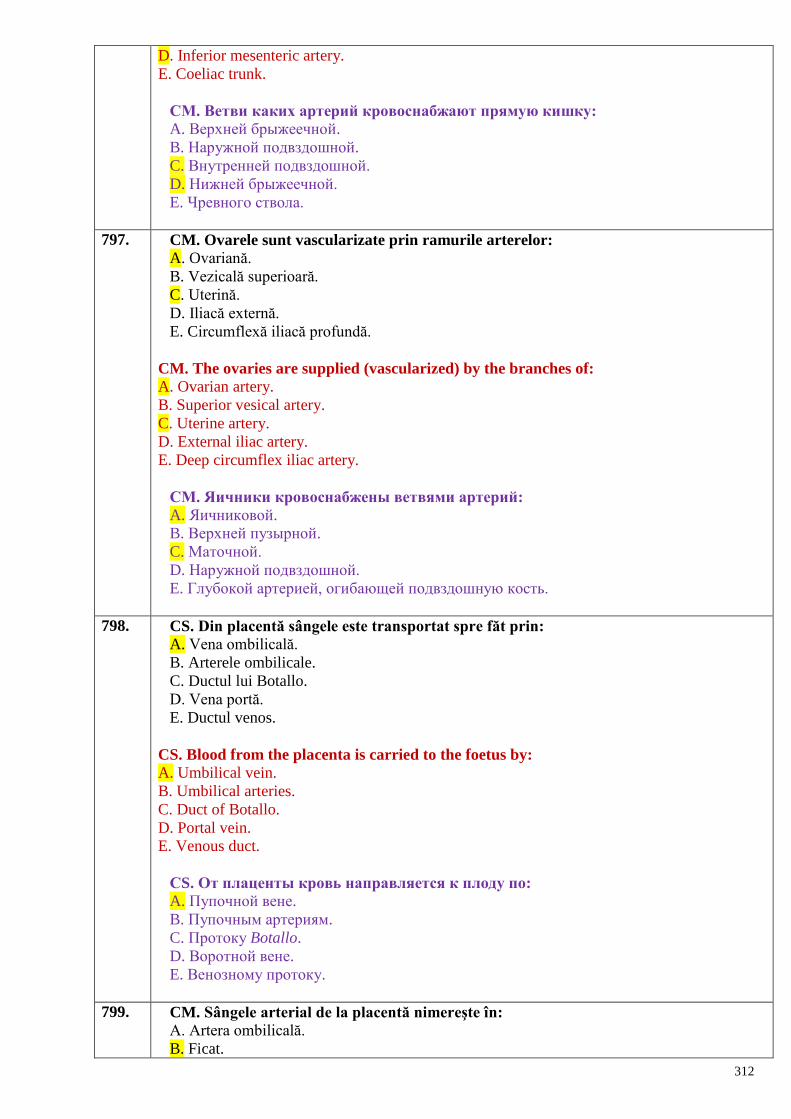
312
D. Inferior mesenteric artery.
E. Coeliac trunk.
СМ. Ветви каких артерий кровоснабжают прямую кишку:
A. Верхней брыжеечной.
B. Наружной подвздошной.
C. Внутренней подвздошной.
D. Нижней брыжеечной.
E. Чревного ствола.
797. CM. Ovarele sunt vascularizate prin ramurile arterelor:
A. Ovariană.
B. Vezicală superioară.
C. Uterină.
D. Iliacă externă.
E. Circumflexă iliacă profundă.
CM. The ovaries are supplied (vascularized) by the branches of:
A. Ovarian artery.
B. Superior vesical artery.
C. Uterine artery.
D. External iliac artery.
E. Deep circumflex iliac artery.
СМ. Яичники кровоснабжены ветвями артерий:
A. Яичниковой.
B. Верхней пузырной.
C. Маточной.
D. Наружной подвздошной.
E. Глубокой артерией, огибающей подвздошную кость.
798. CS. Din placentă sângele este transportat spre făt prin:
A. Vena ombilicală.
B. Arterele ombilicale.
C. Ductul lui Botallo.
D. Vena portă.
E. Ductul venos.
CS. Blood from the placenta is carried to the foetus by:
A. Umbilical vein.
B. Umbilical arteries.
C. Duct of Botallo.
D. Portal vein.
E. Venous duct.
СS. От плаценты кровь направляется к плоду по:
A. Пупочной вене.
B. Пупочным артериям.
C. Протоку Botallo.
D. Воротной вене.
E. Венозному протоку.
799. CM. Sângele arterial de la placentă nimereşte în:
A. Artera ombilicală.
B. Ficat.
313
C. Vena cavă inferioară.
D. Venele hepatice.
E. Atriul drept.
CM. The arterial blood from the placenta gets the:
A. Umbilical artery.
B. Liver.
C. Inferior vena cava.
D. Hepatic veins.
E. Right atrium.
СМ. Артериальная кровь от плаценты вливается в:
A. Пупочную артерию.
B. Печень.
C. Нижнюю полую вену.
D. Печѐночные вены.
E. Правое предсердие.
800. CM. Artera iliacă externă:
A. Este ramură a arterei iliace comune.
B. Trece pe marginea laterală a muşchiului psoas mare.
C. Prin inelul femural apare pe coapsă sub denumirea de arteră femurală.
D. În lacuna vasculară e însoţită de vena omonimă.
E. Lansează ramuri viscerale.
CM. The external iliac artery:
A. Is a ramus of the common iliac artery
B. Passes over the lateral border of the psoas major muscle
C. After passing through the femoral ring it appears in the region of the thigh as femoral artery
D. In the lacuna vasorum it is accompanied by the similarly named vein
E. Gives off the visceral branches.
СМ. Наружная подвздошная артерия: A. Является ветвью общей подвздошной артерии.
B. Проходит по латеральному краю большой поясничной мышцы.
C. Через бедренное кольцо проходит на бедро под названием бедренная артерия.
D. В сосудистой лакуне сопровождается одноимѐнной веной.
E. Отдаѐт висцеральные ветви.
801. CS. Vena iliacă externă reprezintă continuarea:
A. V. iliace interne.
B. V. sacrale mediane.
C. V. iliace comune.
D. V. femurale.
E. V. femurale profunde.
CS. The external iliac vein is the continuation of:
A. V. iliaca interna.
B. V. sacralis mediana.
C. V. iliaca communis.
D. V. femoralis.
E. V. profunda femoris.
СS. Наружная подвздошная вена является продолжением: A. V. iliace interne.
314
B. V. sacrale mediane.
C. V. iliace comune.
D. V. femurale.
E. V. femurale profunde.
802. CS. Artera iliacă externă se continuă pe faţa anterioară a coapsei cu artera:
A. Iliacă comună.
B. Femurală profundă.
C. Femurală
D. Femurală superficială.
E. Perforantă I.
CS. The external iliac artery continues on the anterior surface of the thigh as:
A. Common iliac artery.
B. Deep femoral artery.
C. Femoral artery.
D. Supericial femoral aartery.
E. The Ist perforating artery.
СS. Наружная подвздошная артерия продолжается на переднюю поверхность
бедра артерией:
A. Общей подвздошной.
B. Глубокой бедренной.
C. Бедренной.
D. Поверхностной бедренной.
E. Прободающей I.
803. CM. A. iliacă internă:
A. Trimite ramuri spre pereţii bazinului.
B. Irigă muşchii anteriori ai coapsei.
C. Trimite ramuri pentru organele din cavitatea bazinului.
D. Se ramifică în arterele femurală şi obturatorie.
E. Aprovizionează cu sânge toate organele genitale interne.
CM. A. iliaca interna:
A. It gives off branches for the walls of the pelvic cavity.
B. It supplies (vascularizes) the anterior muscles of the thigh.
C. It gives rise to the branches for the organs of the pelvic cavity.
D. It divides into the femoral and obturator arteries.
E. It supplies with the blood all the internal genital organs.
СМ. Внутренняя подвздошная артерия: A. Отдаѐт ветви к стенкам таза.
B. Кровоснабжает передние мышцы бедра.
C. Отдаѐт ветви для органов таза.
D. Разветвляется на бедренную и запирательную артерии.
E. Обеспечивает кровоснабжение всех внутренних половых органов.
804. CM. La vascularizația măduvei spinării participă ramurile spinale de la:
A. Arterele lombare.
B. Arterele renale.
C. Arterele iliace comune.
D. Arterele intercostale posterioare.
E. Arterele frenice inferioare.
315
CM. Blood supply of the spinal cord is performed by the spinal branches of:
A. Lumbar arteries.
B. Renal arteries.
C. Common iliac arteries.
D. Posterior intercostal arteries.
E. Inferior phrenic arteries.
СM. В кровоснабжении спинного мозга участвуют спинномозговые ветви от
артерий: A. Поясничных.
B. Почечных.
C. Общих подвздошных.
D. Задних межрѐберных.
E. Нижних диафрагмальных.
805. CM. Referitor la vena cavă inferioară:
A. Reprezintă cel mai voluminos vas venos.
B. E plasată la dreapta de aorta abdominală.
C. Colectează sânge de la organele abdominale pare.
D. Se formează la nivelul vertebrei lombare IV.
E. Colectează sângele de la organele abdominale impare.
CM. Concerning the inferior vena cava:
A. It represents the largest venous trunk.
B. It runs along the right side of the abdominal aorta.
C. It collects the blood from the paired organs of the abdominal cavity.
D. It is formed at the level of the L4 vertebra.
E. It collects the blood from the unpaired organs of the abdominal cavity.
СМ. Нижняя полая вена: A. Представляет собой самый крупный венозный сосуд.
B. Находится справа от брюшной аорты.
C. Собирает кровь от парных органов брюшной полости.
D. Образуется на уровне IV поясничного позвонка.
E. Собирает кровь от непарных органов брюшной полости.
806. CM. Afluenţi parietali ai venei cave inferioare sunt:
A. Venele lombare drepte.
B. Venele lombare stângi.
C. Venele frenice inferioare.
D. Vena testiculară (ovariană) dreaptă.
E. Venele suprarenale drepte.
CM. The parietal tributares (affluents) of the inferior vena cava are, as follows:
A. Right lumbar veins
B. Left lumbar veins
C. Inferior phrenic veins
D. Right testicular (ovarian) vein
E. Right suprarenal veins.
СМ. Пристеночные притоки нижней полой вены: A. Правые поясничные вены.
B. Левые поясничные вены.
C. Нижние диафрагмальные вены.
D. Правая яичковая (яичниковая) вена.
316
E. Правые надпочечниковые вены.
807. CM. Afluenţi viscerali ai venei cave inferioare sunt:
A. Vena testiculară (ovariană) stângă.
B. Vena testiculară (ovariană) dreaptă.
C. Venele renale.
D. Vena suprarenală dreaptă.
E. Venele hepatice.
CM. The visceral tributares (affluents) of the inferior vena cava are, as follows:
a. Left testicular (ovarian) vein.
b. Right testicular (ovarian) vein.
c. Renal veins
d. Right suprarenal vein
e. Hepatic veins.
СМ. Висцеральными притоками нижней полой вены являются: A. Левая яичковая (яичниковая) вена.
B. Правая яичковая (яичниковая) вена.
C. Почечные вены.
D. Правая надпочечниковая вена.
E. Печѐночные вены.
808. CM. Referitor la vena portă:
A. E situată între foiţele ligamentului hepatoduodenal.
B. Se formează posterior de capul pancreasului.
C. Colectează sângele de la organele abdominale impare.
D. Colectează sângele de la organele pare ale cavităţii abdominale.
E. Se formează din venele mezenterice superioară şi inferioară.
CM. Concerning the portal vein:
a. It is situated between the laminae of the hepatoduodenal ligament
b. It is formed behind of the head of the pancreas
c. It collects the blood from the unpaired organs of the abdominal cavity
d. It collects the blood from the paired organs of the abdominal cavity
e. It is formed by the confluence of the superior and inferior mesenteric veins.
СМ. Воротная вена: A. Находится между листками печѐночно-двенадцатиперстной связки.
B. Образуется позади головки поджелудочной железы.
C. Собирает кровь от непарных органов брюшной полости.
D. Собирает кровь от парных органов брюшной полости.
E. Образуется верхней и нижней брыжеечными венами.
809. CS. Cu ce vas începe refluxul venos de la ficat:
A. Venele subsegmentare.
B. Vena centrală.
C. Venele interlobulare.
D. Vena cavă inferioară.
E. Vena portă.
CS. The venous reflux from the liver begins with the following veins:
A. Subsegmentar veins.
B. Central veins.
C. Interlobular veins.
317
D. Inferior vena cava.
E. Portal vein.
СS. Какими сосудами начинается венозный рефлюкс от печени: A. Подсегментраными венами.
B. Центральной веной.
C. Междольковыми венами.
D. Нижней полойя веной.
E. Воротной веной.
810. CM. Vena portă:
A. Iese din ficat.
B. Intră în ficat.
C. Colectează sângele exclusiv de la organele abdominale pare.
D. Colectează sângele de la organele abdominale impare, cu excepția ficatului.
E. Se formează prin confluerea venelor mezenterică superioară, lienală şi gastrică stângă.
CM. The portal vein:
A. Leaves the liver.
B. Enters the liver.
C. Collects blood from the paired organs of the abdominal cavity.
D. Collects blood from the unpaired organs of the abdominal cavity, exept the liver
E. Is formed by the confluence of the superior mesenteric, lienal and left gastric veins.
СМ. Воротная вена: A. Выходит из печени.
B. Входит в печень.
C. Собирает кровь исключительно от парных органов брюшной полости.
D. Собирает кровь исключительно от непарных органов брюшной полостиб кроме
печени.
E. Образуется соединением верхней брыжеечной, селезѐночной и левой желудочной
вен.
811. CS. Afluenţi parietali ai venei cave inferioare sunt:
A. Venele lombare I – II.
B. Venele lombare II – V.
C. Venele frenice inferioare.
D. Venele intercostale posterioare.
E. Venele testiculare (ovariene).
CM. The parietal tributares (affluents) of the inferior vena cava are, as follows:
a. Lumbar veins I-II
b. Lumbar veins II-V
c. Inferior phrenic veins
d. Right testicular (ovarian) vein
e. Right suprarenal veins.
СS. Париетальные притоки нижней полой вены: A. Поясничные вены I-II.
B. Поясничные вены II-V.
C. Нижние диафрагмальные вены.
D. Задние межрѐберные вены.
E. Яичковые (яичниковые) вены.
812. CM. Vene care formează anastomoze porto-cave la nivelul rectului sunt:
318
A. Rectală superioară.
B. Rectală medie.
C. Mezenterică superioară.
D. Iliacă externă.
E. Mezenterică inferioară.
CM. The veins which form the portacaval anastomoses at the level of the rectum are, as
follows:
A. Superior rectal veins.
M. Middle rectal veins.
C. Superior mesenteric vein.
D. External iliac vein.
E. Inferior mesenteric vein.
СМ. Вены, которые образуют портокавальные анастомозы в прямой кишке: A. Верхняя прямокишечная.
B. Средняя прямокишечная.
C. Верхняя брыжеечная.
D. Наружная подвздошная.
E. Нижняя брыжеечная.
813. CM. Prin care vene sângele este colectat în vena portă:
A. Lienală.
B. Hepatice.
C. Mezenterică superioară.
D. Testiculare (ovariene).
E. Mezenterică inferioară.
CM. The blood flows to the portal vein through the following veins:
a. Lienal vein
b. Hepatic veins
c. Superior mesenteric vein
d. Testicular (ovarian) vein
e. Inferior mesenteric vein.
CM. От каких вен кровь собирается в воротную вену? A. Селезѐночной.
B. Печѐночной.
C. Верхней брыжеечной.
D. Яичковых (яичниковые).
E. Нижней брыжеечной.
814. CM. Pe peretele posterior al abdomenului anastomoze cavocave formează venele:
A. Impară.
B. Esofagiene.
C. Semiimpară.
D. Rectală inferioară.
E. Lombare.
CM. The following veins form the cavocaval anastomoses on the posterior wall of the
abdominal cavity:
A. Azygos vein.
B. Esophageal veins.
C. Hemiazygos vein.
D. Inferior rectal veins.
319
E. Lumbar veins.
СМ. На задней стенке брюшной полости каво-кавальные анастомозы образуют
вены: A. Непарная.
B. Пищеводные.
C. Полунепарная.
D. Нижняя прямокишечная.
E. Поясничные.
815. CM. Referitor la artera femurală:
A. Prezintă continuarea trunchiului arterei iliace externe.
B. Trece prin lacuna musculară.
C. Sub ligamentul inghinal e plasată medial de vena femurală.
D. Pătrunde în canalul adductor.
E. Se continuă cu artera poplitee.
CM. Regarding to the femoral artery:
A. It represents the continuation of the external iliac artery.
B. It passes through the muscular lacuna.
C. It is emplaced beneath the inguinal ligament medially to the femoral vein.
D. It enters the adductor canal.
E. It continuities as popliteal artery.
СМ. Относительно бедренной артерии: A. Представляет продолжение ствола наружной подвздошной артерии.
B. Проходит через мышечную лакуну.
C. Под паховой связкой располагается медиально от бедренной вены.
D. Проходит в приводящем канале.
E. Продолжается подколенной артерией.
816. CS. Artera tibială anterioară se termină:
A. Formând arcada dorsală a piciorului.
B. Continuându-se cu artera dorsală a piciorului.
C. Anastomozându-se cu artera tibială posterioară.
D. Anastomozându-se cu artera peronieră.
E. Anastomozându-se cu artera tarsiană laterală.
CS. The anterior tibial artery:
A. Forms the dorsal arch of the foot.
B. Continues with the dorsal pedis artery.
C. Anastomosis with the posterior tibial artery.
D. Anastomosis with the peroneal artery.
E. Anastomosis with the lateral tarsal artery.
СS. Передняя большеберцовая артерия заканчивается:
A. Образуя дорзальную сосудистую дугу стопы.
B. Продолжаясь в дорсальную артерию стопы.
C. Анастомозируя с задней большеберцовой артерией.
D. Анастомозируя с малоберцовой артерией.
E. Анастомозируя с латеральной плюсневой артерией.
817. CS. Artera tibială posterioară:
A. Continuă artera femurală.
B. Se termină cu arterele plantare.
320
C. Trece prin canalul musculoperoneu inferior.
D. Se anastomozează cu artera dorsală a piciorului.
E. Trece prin membrana interosoasă.
CS. The posterior tibial artery:
A. Is a continuation of the femoral artery.
B. Finishes with the plantar arteries.
C. Passes though the inferior musculoperoneal canal.
D. Anastomosis with the dorsal artery of the foot
E. Passes though the interosseous membrane.
СS. Задняя большеберцовая артерия:
A. Является продолжением бедренной артерии.
B. Заканчивается подошвенными артериями.
C. Проходит через нижний мышечно-малоберцовый канал.
D. Анастомозирует с тыльной артерией стопы.
E. Прободает межкостную мембрану.
818. CS. Raportul posteroanterior al componentelor fosei poplitee:
A. Nervul tibial, artera poplitee şi vena poplitee.
B. Nerval sciatic (sau tibial), vena şi artera poplitee.
C. Nervul peroneu comun, artera şi vena poplitee.
D. Artera poplitee, vena poplitee.
E. Vena poplitee, nervul sciatic, artera poplitee.
CS. The postero-anterior relation of the components of the popliteal fossa:
A. The tibial nerve, popliteal artery and popliteal vein.
B. The sciatic (or tibial) nerve, popliteal vein and popliteal artery.
C. The common peroneal nerve, popliteal artery and popliteal vein.
D. The popliteal artery, popliteal vein.
E. The popliteal vein, sciatic nerve, popliteal artery.
СS. Задне-переднее соотношение сосудисто-нервных компонентов подколенной
ямки:
A. Большеберцовый нерв, подколенная артерия, подколенная вена.
B. Седалищный нерв (или большеберцовый), подколенная вена и артерия.
C. Общий малоберцовый нерв, подколенная артерия и вена.
D. Подколенная артерия и подколенная вена.
E. Подколенная вена, седалищный нерв, подколенная артерия.
819. CM. Din venele superficiale ale membrului inferior fac parte:
A. Vena safenă mare.
B. Vena subcutanată laterală.
C. Vena safenă mică.
D. Vena femurală.
E. Vena subcutanată medială.
CM. The superficial veins of lower limb are:
A. The great saphenous vein.
B. The lateral subcutaneous vein.
C. The small saphenous vein.
D. The femoral vein.
E. The medial subcutaneous vein.
СМ. К поверхностным венам нижней конечности относятся:
321
A. Большая подкожная вена.
B. Латеральная подкожная вена.
C. Малая подкожная вена.
D. Бедренная вена.
E. Медиальная подкожная вена.
820. CS. Vena safenă mică se varsă în:
A. Vena femurală.
B. Vena poplitee.
C. Vena tibială posterioară.
D. Vena iliacă externă.
E. Vena tibială anterioară.
CS. The small saphenous vein drains in:
A. The femoral vein.
B. The popliteal vein.
C. The posterior tibial vein.
D. The external iliac vein.
E. The anterior tibial vein.
CS. Малая подкожная вена вливается в:
A. Бедренную вену.
B. Подколенную вену.
C. Заднюю большеберцовую вену.
D. Наружную подвздошную вену.
E. Переднюю большеберцовую вену.
821. CS. Artera femurală iese din bazin prin:
A. Canalul obturator.
B. Lacuna musculară.
C. Canalul femural.
D. Lacuna vasculară.
E. Canalul inghinal.
CS. The femoral artery leaves the pelvis through:
a. The obturator canal.
b. The muscular lacuna.
c. The femoral canal.
d. The vascular lacuna.
e. The inguinal canal.
СS. Бедренная артерия выходит из таза через:
A. Запирательный канал.
B. Мышечную лакуну.
C. Бедренный канал.
D. Сосудистую лакуну.
E. Паховый канал.
822. CM. Artera poplitee:
A. Reprezintă continuarea arterei femurale.
B. Se află în fosa poplitee.
C. Trece prin canalul femuropopliteu.
D. Ramurile ei colaterale irigă muşchii anteriori ai coapsei.
E. Se divide în arterele tibiale anterioară şi posterioară.
322
CM. The popliteal artery:
A. Represents the continuation of the femoral artery.
B. Is located in the popliteal fossa.
C. Passes through the femoropopliteal canal.
D. Its branches supply the anterior group of thigh muscles.
E. Is divided in the anterior and posterior tibial arteries.
СМ. Подколенная артерия:
A. Является продолжением бедренной артерии.
B. Находится в подколенной ямке.
C. Проходит через бедренно-подколенный канал.
D. Еѐ коллатеральные ветви кровоснабжают передние мышцы бедра.
E. Делится на переднюю и заднюю большеберцовую артерию.
823. CM. Venele membrului inferior pot fi explorate prin:
A. Angiografie.
B. Flebografie.
C. Palpaţie.
D. Inspecţie (venele superficiale).
E. Puncţie (venele profunde).
CM. The veins of lower limb could be examined by:
A. Angiography.
B. Phlebography.
C. Palpation.
D. Inspection (superficial veins).
E. Puncture (deep veins).
СМ. Вены нижней конечности могут быть обследованы посредством:
A. Ангиографии.
B. Флебографии.
C. Прощупывания.
D. Инспекции (поверхностные).
E. Пункции (глубокие).
824. CM. Vene profunde pare însoţesc:
A. A. tibialis anterior.
B. A. poplitea.
C. A. femoralis.
D. A. tibialis posterior.
E. A. profunda femoris.
CM. The paired deep veins accompany:
A. A. tibialis anterior.
B. A. poplitea.
C. A. femoralis.
D. A. tibialis posterior.
E. A. profunda femoris.
СМ. Парные глубокие вены сопровождают следующие артерии:
A. Переднюю большеберцовую артерию.
B. Подколенную артерию.
C. Бедренную артерию.
D. Заднюю большеберцовую артерию.
E. Глубокую артерию бедра.
323
Sistemele imunitar şi limfatic –
componente, structură,
rol funcţional
825. CS. Timusul atinge o dezvoltare maximă:
A. La naștere
B. La copii
C. La pubertate (10 - 15 ani)
D. La adulți
E. La vârsta senila
SC. The thymus achieves its maximum development:
a) in newborn
b) in child
c) at puberty (10 - 15 years)
d) in adult
e) in old person.
СS. Вилочковая железа (thymus) достигает максимального развития:
A. К моменту рождения.
B. У детей.
C. В возрасте полового созревания (10-15 лет).
D. У взрослых.
E. В старческом возрасте.
826. CM. Parenchimul timusului constă din:
A. Substanță compactă
B. Substanță corticală
C. Substanță medulară
D. Substanță spongioasă
E. Substanță cenușie
MC. The parenchyma of the thymus consists of:
a) compact substance
b) cortex
c) medulla
d) spongy substance
e) gray matter.
СМ. Паренхима вилочковой железы состоит из:
A. Компактного вещества.
B. Коркового вещества.
C. Мозгового вещества.
D. Губчатого вещества.
E. Серого вещества.
827. CM. Timusul e situat în mediastinul:
A. Anterior
B. Posterior
C. Superior
D. Inferior (porțiunea anterioară)
E. Inferior (porțiunea medie)
SC. In which part of the mediastinum (PNA) is the thymus located:
a) anterior
324
b) posterior
c) superior
d) inferior (anterior part)
e) inferior (middle part).
СМ. Вилочковая железа (thymus) расположена в средостении:
A. Переднем.
B. Заднем.
C. Верхнем.
D. Нижнем (переднем отделе).
E. Нижнем (среднем отделе).
828. CM. Timusul constă din lobii:
A. Superior
B. Inferior
C. Drept
D. Stâng
E. Intermediar
MC. The thymus consists of the following lobes:
a) superior
b) inferior
c) right
d) left
e) intermediate.
СМ. Вилочковая железа (thymus) состоит из долей:
A. Верхней.
B. Нижней.
C. Правой.
D. Левой.
E. Промежуточной.
829. CM. Timusul este un organ:
A. Limfoid
B. Imunitar periferic
C. Imunitar central
D. Cavitar
E. Digestiv
MC. What kind of organ is the thymus:
a) lymphoid organ
b) peripheral immune organ
c) central immune organ
d) cavitary organ
e) digestive organ.
СМ. Вилочковая железа является органом:
A. Лимфоидным.
B. Иммунным периферическим.
C. Иммунным центральным.
D. Полостным.
E. Пищеварительным.
830. CM. Sistemul limfatic include:
325
A. Vase limfocapilare.
B. Vase limfatice.
C. Vase sangvine speciale.
D. Trunchiuri şi canale limfatice.
E. Splina şi timusul.
CM. The lymphatic system includes:
A. Lymph capillaries
B. Lymph vessels
C. Special blood vessels
D. Lymph trunks and ducts
E. Spleen and thymus.
СМ. Лимфатическая система состоит из:
A. Лимфатических капилляров.
B. Лимфатических сосудов.
C. Особых кровеносных сосудов.
D. Лимфатических стволов и протоков.
E. Селезѐнки и вилочковой железы.
831. CM. Capilarele limfatice:
A. Reprezintă segmentul iniţial al sistemului limfatic.
B. Sunt concentrate mai ales în encefal şi măduva spinării.
C. Sunt mai subţiri ca capilarele sangvine.
D. Traiectul lor are aspect rectiliniu.
E. Formează reţele limfocapilare.
MC. Lymphatic capillaries:
A. It represents the initial segment of the lymphatic system.
B. They are concentrated mainly in the brain and spinal cord.
C. They are thinner that the blood capillaries.
D. Their path has the straight appearance.
E. Forms lymph capillary network.
СМ. Лимфатические капилляры:
A. Являются начальным звеном лимфатической системы.
B. Сконцентрированы в головном и спинном мозге.
C. Их диаметр меньше, чем у кровеносных капилляров.
D. Имеют прямолинейное направление.
E. Образуют лимфокапиллярные сети.
832. CM. Nodurile limfatice:
A. Au formă rotundă, ovală sau de bob.
B. Formează grupuri regionale.
C. Nu posedă capsule conjunctivale.
D. Pot fi superficiale şi profunde, parietale şi viscerale.
E. Li se disting sinusurile marginal, intermediare şi portal.
CM. The lymph nodes:
A. Are round, ovoid or bean-shaped
B. Form the regional groups
C. Do not possess connective tissue capsules
D. Can be superficial and deep, parietal and visceral
E. Have the marginal, intermediate and portal sinuses.
326
СМ. Лимфатические узлы:
A. Имеют круглую, овальную или бобовидную форму.
B. Образуют региональные группы.
C. Не имеют соединительнотканных оболочек.
D. Разделяются на поверхностные и глубокие, пристеночные и внутренностные.
E. В них различают маргинальные, промежуточные и воротные синусы.
833. CM. Ductul limfatic drept se formează din:
A. Ductul jugular stâng.
B. Ductul jugular drept.
C. Ductul subclavicular drept.
D. Ductul bronhomediastinal drept.
E. Ductul bronhomediastinal stâng.
MC. The right lymphatic duct is formed by confluence of:
A. Left jugular trunk
B. Right jugular trunk
C. Right subclavian trunk
D. Right bronchomediastinal trunk
E. Left bronchomediastinal trunk.
СМ. Правый лимфатический проток образуется из:
A. Левого яремного протока.
B. Правого яремного протока.
C. Правого подключичного протока.
D. Правого бронхосредостенного протока.
E. Левого бронхосредостенного протока.
834. CS. În ductul limfatic drept limfa se scurge de la:
A. Membrul superior din dreapta.
B. Membrul inferior din stânga.
C. Hemitoracele stâng.
D. Jumătatea stângă a capului şi gâtului.
E. Bazin şi cavitatea abdominală.
CS. The right lymphatic duct receives the lymph from:
A. Right upper limb
B. Left lower limb
C. Left hemithorax
D. Left half of the head and neck
E. Pelvis and abdominal cavity
CS. В правый лимфатический проток лимфа оттекает от: A. Правой верхней конечности.
B. Левой верхней конечности.
C. Левой половины грудной полости.
D. Левой половины головы и шеи.
E. Таза и брюшной полости.
835. CM. Canalul limfatic toracic:
A. Începe cu cisterna chyli.
B. I se descriu trei segmente - abdominal, toracic şi cervical.
C. La nivelul vertebrelor toracice superioare deviază spre stânga şi formează arcul ductului
toracic.
D. Afluează în vena cavă superioară.
327
E. Colectează limfa de la partea posterioară a corpului.
CM. The thoracic lyimphatic duct:
a. It starts at the level of the T XII - L II vertebrae.
b. It is made of by confluence of right and left lumbar trunks.
c. It appears in posterior mediastinum by esophageal hiatus of the diaphragm.
d. Its thoracic segment is the longest.
e. It contain a lot of valves.
СМ. Грудной лимфатический проток:
A. Начинается с cisterna chyli.
B. Имеет три части – брюшная, грудная и шейная.
C. На уровне верхних грудных позвонков отходит налево и образует дугу грудного
протока.
D. Впадает в верхнюю полую вену.
E. Собирает лимфу от задней поверхности туловища.
836. CS. Ductul limfatic toracic părăseşte cavitatea abdominală prin:
A. Hiatul esofagian.
B. Hiatul aortic.
C. Orificiul venei cave inferioare.
D. Spaţiul dintre stâlpul lateral şi mijlociu al diafragmei.
E. Spaţiul dintre stâlpul medial şi mijlociu al diafragmei.
CS. The thoracic lymphatic duct leaves the abdominal cavity through:
A. The esophageal hiatus.
B. The aortic hiatus.
C. The caval opening
D. The space between the lateral and middle crura of diaphragm.
E. The space between medial and middle crura of diaphragm.
CS. Грудной лимфатический проток покидает брюшную полость через: A. Пищеводное отверстие.
B. Аортальное отверстие.
C. Отверстие нижней полой вены.
D. Пространство между латеральным и средним столбом диафрагмы.
E. Пространство между медиальным и средним столбом диафрагмы.
837. CS. Canalul limfatic toracic:
A. Se formează prin confluerea a două ducturi lombare la nivelul L4.
B. Este situat pe faţa anterioară a coloanei vertebrale între partea toracică a aortei şi vena
impară.
C. Se plasează între partea toracică a aortei şi esofag.
D. Un afluent constant al canalului este ductul intestinal.
E. Se deosebesc trei variante de formă şi structură a canalului limfatic toracic.
CS. The thoracic lymphatic duct:
A. Is formed by confluence of two lumbar trunks at the level of L4 vertebra.
B. Is located in front of the spine between the thoracic aorta and azygos vein.
C. Is emplaced between the thoracic aorta and esophagus.
D. The intestinal trunk is a constant affluent of the thoracic lymphatic duct.
E. There are three main variants of the thoracic lymphatic duct depending on its shape and
structure.
СS. Грудной лимфатический проток:
328
A. Образуется слиянием 2-х поясничных стволов на уровне L4.
B. Расположен на передней поверхности позвоночника между грудной аортой и
непарной веной.
C. Располагается между грудной аортой и пищеводом.
D. Постоянным притоком его является кишечный ствол.
E. Различают три варианта формы и структуры грудного лимфатического протока.
838. CM. Ductul limfatic toracic prezintă porțiunile:
A. Sacrală.
B. Abdominală.
C. Partea toracică.
D. Partea cervicală.
E. Craniană.
CM. The thoracic lymphatic duct possesses the following parts:
A. Sacral part.
B. Abdominal part.
C. Thoracic part.
D. Cervical part.
E. Cranial part.
СМ. Части грудного лимфатического протока:
A. Крестцовая.
B. Брюшная.
C. Грудная часть.
D. Шейная часть.
E. Черепная.
839. CM. Noduri limfatice ale capului sunt:
A. Occipitale.
B. Mastoidiene.
C. Parotidiene.
D. Paratraheale.
E. Prelaringiene.
MC. The lymphatic nodes of the head are:
A. Occipital
B. Mastoid
C. Parotid
D. Paratracheal
E. Prelaryngeal
СМ. К лимфатическим узлам головы относятся:
A. Затылочные.
B. Сосцевидные.
C. Околоушные.
D. Паратрахеальные.
E. Надгортанные.
840. CM. Ganglionii limfatici axilari includ grupurile:
A. Lateral.
B. Medial (pectoral).
C. Central.
D. Subscapular (posterior).
E. Interpectoral.

329
CM. The groups of the axillary lymph nodes are: A. Lateral.
B. Medial (pectoral).
C. Central.
D. Subscapular (posterior).
E. Interpectoral.
СМ. Подмышечные лимфатические узлы образуют группы:
A. Латеральную.
B. Медиальную (грудную).
C. Центральную.
D. Подлопаточную (заднюю).
E. Межгрудную.
841. CM. Vasele limfatice ale membrului superior:
A. Sunt grupate în superficiale şi profunde.
B. Afluează în ganglionii limfatici cubitali şi axilari.
C. Cele superficiale sunt localizate în apropierea nervilor pieloşi.
D. Cele superficiale formează grupurile lateral, medial şi posterior.
E. Cele profunde însoţesc ramurile plexului brahial.
CM. The lymphatic vessels of the upper limb:
A. They are grouped into superficial and deep.
B. Confluence i cubital and axillary lymph nodes.
C. The superficial one are located near the skin nerves.
D. The superficial one formes lateral, medial and posterior groups.
E. The deep one accompanies brachial plexus branches.
СМ. Лимфатические сосуды верхней конечности:
A. Делятся на поверхностные и глубокие.
B. Впадают в локтевые и подмышечные лимфатические узлы.
C. Поверхностные - располагаются вблизи кожных нервов.
D. Поверхностные лимфатические сосуды образуют 3 группы: латеральную,
медиальную и заднюю.
E. Глубокие лимфатические сосуды сопровождают ветви плечевого сплетения.
842. CM. În regiunea antebraţului se află reţelele limfatice:
A. Anterioară.
B. Posterioară.
C. Nu exista reţele.
D. Superficială.
E. Profundă.
MC. In the forearm the following lymph networks are located:
A. Anterior.
B. Posterior.
C. There are no networks.
D. Superficial.
E. Deep.
СМ. Предплечье имеет следующие лимфатические сети:
A. Переднюю.
B. Заднюю.
C. Сети отсутствуют.
330
D. Поверхностную.
E. Глубокую.
843. CS. Vasele limfatice superficiale ale antebraţului însoţesc:
A. Arterele superficiale.
B. Nervii cutanaţi.
C. Venele superficiale.
D. Nervul median.
E. Nervul ulnar.
SC. The superficial lymphatic vessels of the forearm accompany:
A. Superficial arteries.
B. Cutaneous nerves
C. Superficial veins.
D. Median nerve.
E. Ulnar nerve.
СS. Поверхностные лимфатические сосуды предплечья сопровождают:
A. Поверхностные артерии.
B. Кожные нервы.
C. Поверхностные вены.
D. Срединный нерв.
E. Локтевой нерв.
844. CM. Ganglionii superficiali ai membrului superior se grupează în:
A. Mediali.
B. Cubitali.
C. Laterali.
D. Deltopectorali.
E. Axilari.
MC. The superficial lymph nodes of the upper limb are grouped into:
A. Medial.
B. Cubital.
C. Lateral.
D. Deltopectoral.
E. Axillary
СМ. Поверхностные лимфоузлы верхней конечности образуют следующие
группы:
A. Медиальные.
B. Локтевые.
C. Латеральные.
D. Дельтовидногрудные.
E. Подмышечные.
845. CM. Ganglionii limfatici axilari:
A. Sunt în număr de 15-40.
B. Colectează limfa din reţeaua limfatică medială şi laterală a membrului superior.
C. Colectează limfa de la membrul superior.
D. Colectează limfa de la pereţii cavităţii toracice.
E. Se divid în trei grupe.
MC. The axillary lymph nodes:
A. They are in numbers 15-40.
331
B. Collects the lymph from the lymphatic medial and lateral networks upper limb.
C. Collects the lymph from the upper limb.
D. Collects the lymph from thoracic cavity walls.
E. Divide into three groups.
СМ. Подмышечные лимфатические узлы:
A. Их количество 15-40.
B. Принимают лимфу от медиальной и латеральной сетей верхней конечности.
C. Принимают лимфу от верхней конечности.
D. Принимают лимфу от стенок грудной полости.
E. Образуют три группы.
846. CM. Se disting ganglioni axilari:
A. Superiori.
B. Apicali.
C. Inferiori.
D. Centrali.
E. Laterali.
MC. The following groups of the axillary lymph nodes are distinguished:
A. Superior.
B. Apical.
C. Inferior.
D. Central.
E. Lateral
СМ. Следующие группы подмышечных узлов:
A. Верхние.
B. Верхушечные (апикальные).
C. Нижние.
D. Центральные.
E. Латеральные.
847. CS. În trunchiul limfatic subclavicular se varsă vasele limfatice eferente ale
ganglionilor axilari:
A. Centrali.
B. Laterali.
C. Apicali.
D. Anteriori.
E. Posteriori.
SC. In the subclavicular lymphatic trunk empties the efferent lymph vessels from the
following axilary lymph nodes:
A. Central.
B. Lateral.
C. Apical.
D. Anterior.
E. Posterior
СS. В подключичный ствол впадают выносящие лимфатические сосуды
подмышечных узлов:
A. Центральных.
B. Латеральных.
C. Апикальных.
D. Передних.
332
E. Задних
848. CM. De la glanda mamară limfa e transportată spre :
A. Ganglionii axilari mediali.
B. Ganglionii axilari subscapulari.
C. Ganglionii axilari inferiori.
D. Ganglionii parasternali.
E. Ganglionii intercostali .
MC. From the mammary gland the lymph is transported to:
A. Medial axillary lymph nodes.
B. Subscapular axillary lymph nodes.
C. Inferior axillary lymph nodes.
D. Parasternal lymph nodes.
E. Intercostal lymph nodes.
СМ. Лимфа от грудных желѐз направляется к:
A. Медиальным подмышечным узлам.
B. Задним (подлопаточным) подмышечным узлам.
C. Нижним подмышечным узлам.
D. Окологрудинным узлам.
E. Межрѐберным узлам.
849. CS. Cisterna ductului toracic se află:
A. La nivelul vertebrelor T12 – L2.
B. Anterior de aorta abdominală.
C. Anterior de vena cavă inferioară.
D. La dreapta de vena cavă inferioară.
E. La stânga de porţiunea incipientă a aortei abdominale.
SC. The cisterna chyli is located:
A. At the level of the T12-L2
B. In front of the abdominal aorta
C. In front of the inferior vena cava
D.To the right of the inferior vena cava
E. To the left of the initial part of the abdominal aorta.
СS. Цистерна грудного протока находится:
A. На уровне T12-L2.
B. Впереди брюшной аорты.
C. Впереди нижней полой вены.
D. Справа от нижней полой вены.
E. Слева от начальной части брюшной аорты.
850. CS. În fosa cubitală se află ganglionii:
A. Ganglioni limfatici cubitali.
B. Nervul ulnar.
C. Nervul median.
D. Artera brahială.
E. Venele brahiale.
SC. In the cubital fossa the following lymph nodes are located:
A. Cubital lymph nodes
B. Ulnar nerve
C. Median nerve
333
D. Brachial artery
E. Brachial veins.
СS. В локтевой ямке находим лимфатические узлы:
A. Локтевые лимфатические узлы.
B. Локтевой нерв.
C. Срединный нерв.
D. Плечевую артерию.
E. Плечевые вены.
851. CM. În regiunea mâinii există reţelele limfatice:
A. Superficială.
B. Profundă.
C. Palmară.
D. Dorsală.
E. Nu există.
MC. In the region of the hand the following lymphatic networks are present:
A. Superficial
B. Deep
C. Palmar
D. Dorsal
E. Do not exist.
СМ. В области кисти существуют лимфатические сети:
A. Поверхностная.
B. Глубокая.
C. Ладонная.
D. Тыльная.
E. Не существуют
Sistemul nervos central – măduva spinării (structură, porţiuni)
şi encefalul (componente).
Meningele cerebral şi rahidian,
sistemul ventricular şi lichidul cerebrospinal, producţia şi circulaţia lui.
Importanţa aplicativă a spaţiului subarahnoidian
852. CS. Structural neuronul include:
A. Dendrite
B. Corp celular
C. Organite celulare specifice
D. Axon
E. Toate de mai sus
CS. The structure of the neuron includes:
A. Dendrit
B. Cell body
C. Specific celular organelles
D. Axon
E. All mentioned above
CS. Нейрон состоит из:
A. Дендритов
B. Клеточного тела
C. Специфических клеточных органитов
D. Аксона
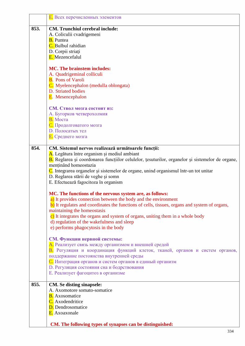
334
E. Всех перечисленных элементов
853. CM. Trunchiul cerebral include:
A. Coliculii cvadrigemeni
B. Puntea
C. Bulbul rahidian
D. Corpii striaţi
E. Mezencefalul
MС. The brainstem includes:
A. Quadrigeminal colliculi
B. Pons of Varoli
C. Myelencephalon (medulla oblongata)
D. Striated bodies
E. Mesencephalon
CM. Ствол мозга состоит из:
A. Бугорков четверохолмия
B. Моста
C. Продолговатого мозга
D. Полосатых тел
E. Среднего мозга
854. CM. Sistemul nervos realizează următoarele funcții:
A. Legătura între organism şi mediul ambiant
B. Reglarea şi coordonarea funcțiilor celulelor, ţesuturilor, organelor şi sistemelor de organe,
menținând homeostazia
C. Integrarea organelor şi sistemelor de organe, unind organismul într-un tot unitar
D. Reglarea stării de veghe şi somn
E. Efectuează fagocitoza în organism
MC. The functions of the nervous system are, as follows:
a) It provides connection between the body and the environment
b) It regulates and coordinates the functions of cells, tissues, organs and system of organs,
maintaining the homeostasis
c) It integrates the organs and system of organs, uniting them in a whole body
d) regulation of the wakefulness and sleep
e) performs phagocytosis in the body
CМ. Функции нервной системы:
A. Реализует связь между организмом и внешней средой
B. Регуляция и координация функций клеток, тканей, органов и систем органов,
поддержание постоянства внутренней среды
C. Интеграция органов и систем органов в единый организм
D. Регуляция состояния сна и бодрствования
E. Реализует фагоцитоз в организме
855. CM. Se disting sinapsele:
A. Axomotore somato-somatice
B. Axosomatice
C. Axodendritice
D. Dendrosomatice
E. Axoaxonale
CM. The following types of synapses can be distinguished:
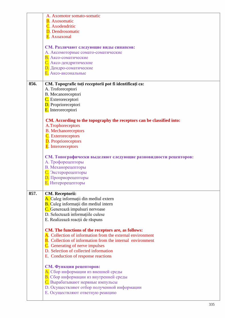
335
A. Axomotor somato-somatic
B. Axosomatic
C. Axodendritic
D. Dendrosomatic
E. Axoaxonal
CМ. Различают следующие виды синапсов:
A. Аксомоторные сомато-соматические
B. Аксо-соматические
C. Аксо-дендритические
D. Дендро-соматические
E. Аксо-аксональные
856. CM. Topografic toți receptorii pot fi identificaţi ca:
A. Troforeceptori
B. Mecanoreceptori
C. Exteroreceptori
D. Proprioreceptori
E. Interoreceptori
CM. According to the topography the receptors can be classified into:
A.Trophoreceptors
B. Mechanoreceptors
C. Exteroreceptors
D. Proprioreceptors
E. Interoreceptors
CМ. Топографически выделяют следующие разновидности рецепторов:
A. Трофорецепторы
B. Механорецепторы
C. Экстерорецепторы
D. Проприорецепторы
E. Интерорецепторы
857. CM. Receptorii:
A. Culeg informaţii din mediul extern
B. Culeg informaţii din mediul intern
C. Generează impulsuri nervoase
D. Selectează informaţiile culese
E. Realizează reacţii de răspuns
CM. The functions of the receptors are, as follows:
A. Collection of information from the external environment
B. Collection of information from the internal environment
C. Generating of nerve impulses
D. Selection of collected information
E. Conduction of response reactions
CМ. Функции рецепторов:
A. Сбор информации из внешней среды
B. Сбор информации из внутренней среды
C. Вырабатывают нервные импульсы
D. Осуществляют отбор полученной информации
E. Осуществляют ответную реакцию
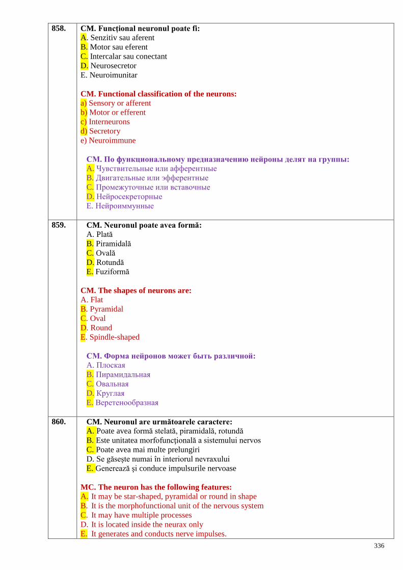
336
858. CM. Funcțional neuronul poate fi:
A. Senzitiv sau aferent
B. Motor sau eferent
C. Intercalar sau conectant
D. Neurosecretor
E. Neuroimunitar
CM. Functional classification of the neurons:
a) Sensory or afferent
b) Motor or efferent
c) Interneurons
d) Secretory
e) Neuroimmune
CМ. По функциональному предназначению нейроны делят на группы:
A. Чувствительные или афферентные
B. Двигательные или эфферентные
C. Промежуточные или вставочные
D. Нейросекреторные
E. Нейроиммунные
859. CM. Neuronul poate avea formă:
A. Plată
B. Piramidală
C. Ovală
D. Rotundă
E. Fuziformă
CM. The shapes of neurons are:
A. Flat
B. Pyramidal
C. Oval
D. Round
E. Spindle-shaped
CМ. Форма нейронов может быть различной:
A. Плоская
B. Пирамидальная
C. Овальная
D. Круглая
E. Веретенообразная
860. CM. Neuronul are următoarele caractere:
A. Poate avea formă stelată, piramidală, rotundă
B. Este unitatea morfofuncţională a sistemului nervos
C. Poate avea mai multe prelungiri
D. Se găseşte numai în interiorul nevraxului
E. Generează şi conduce impulsurile nervoase
MC. The neuron has the following features:
A. It may be star-shaped, pyramidal or round in shape
B. It is the morphofunctional unit of the nervous system
C. It may have multiple processes
D. It is located inside the neurax only
E. It generates and conducts nerve impulses.
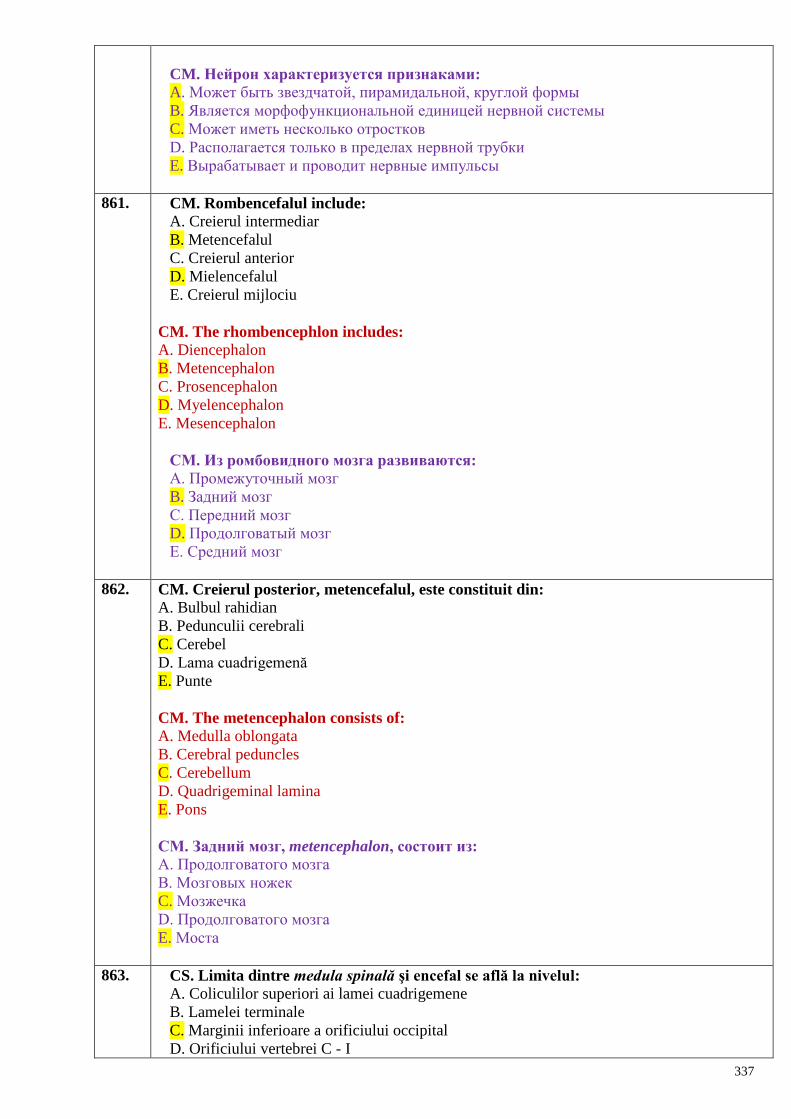
337
CM. Нейрон характеризуется признаками:
A. Может быть звездчатой, пирамидальной, круглой формы
B. Является морфофункциональной единицей нервной системы
C. Может иметь несколько отростков
D. Располагается только в пределах нервной трубки
E. Вырабатывает и проводит нервные импульсы
861. CM. Rombencefalul include:
A. Creierul intermediar
B. Metencefalul
C. Creierul anterior
D. Mielencefalul
E. Creierul mijlociu
CM. The rhombencephlon includes:
A. Diencephalon
B. Metencephalon
C. Prosencephalon
D. Myelencephalon
E. Mesencephalon
CМ. Из ромбовидного мозга развиваются: A. Промежуточный мозг
B. Задний мозг
C. Передний мозг
D. Продолговатый мозг
E. Средний мозг
862. CM. Creierul posterior, metencefalul, este constituit din:
A. Bulbul rahidian
B. Pedunculii cerebrali
C. Cerebel
D. Lama cuadrigemenă
E. Punte
CM. The metencephalon consists of:
A. Medulla oblongata
B. Cerebral peduncles
C. Cerebellum
D. Quadrigeminal lamina
E. Pons
CМ. Задний мозг, metencephalon, состоит из:
A. Продолговатого мозга
B. Мозговых ножек
C. Мозжечка
D. Продолговатого мозга
E. Моста
863. CS. Limita dintre medula spinală şi encefal se află la nivelul:
A. Coliculilor superiori ai lamei cuadrigemene
B. Lamelei terminale
C. Marginii inferioare a orificiului occipital
D. Orificiului vertebrei C - I
338
E. Punții Varolio
CS. The boundary between the brain and spinal medulla is located at the level of:
a) The superior colliculi of the quadrigeminal lamina
b) The terminal lamina
c) The inferior margin of the greater occipital foramen
d) The orifice of the first cervical vertebra
e) the pons of Varolio
CS. Граница между спинным мозгом, меdula spinalis, и головным мозгом
находится на уровне:
A. Верхних бугорков четверохолмия
B. Концевой пластинки
C. Нижнего края затылочного отверстия
D. Отверстия шейного позвонка С-1
E. Варолиева моста
864. CS. Limita inferioară a medulei spinale se află la nivelul vertebrelor:
A. CVII – CVIII
B. TXII – LI
C. LIII
D. LV – SI
E. SIV – SV
CS. The inferior limit of the spinal cord is located at the level of the vertebrae:
a) CVII – CVIII
b) TXII – LI
c) LII – LIII
d) LV – SI
e) SIV – SV
CS. Нижняя граница спинного мозга находится на уровне позвонков:
A. CVII- C VIII
B. ThXII –LI
C. LIII
D. LV –SI
E. SIV –SV
865. CM. La nivelul șanțurilor laterale ale medulei spinale se localizează:
A. Rădăcinile anterioare
B. Rădăcinile laterale
C. Rădăcinile dorsale
D. Septul median al măduvei
E. Ganglionii spinali
CM. Structures locatedat the level of the lateral grooves of the spinal cord are:
a) Ventral roots
b) Lateral roots
c) Dorsal roots
d) Median septum of the spinal cord
e) Spinal ganglia
CМ. На уровне боковых борозд спинного мозга располагаются:
A. Передние корешки
B. Боковые корешки
339
C. Дорсальные корешки
D. Срединная перегородка спинного мозга
E. Спинномозговые узлы
866. CS. Rădăcinile posterioare ale medulei spinale sunt formate de:
A. Dendritele celulelor cornului posterior
B. Dendritele neuronilor ganglionilor spinali
C. Axonii neuronilor pseudounipolari
D. Fibrele cordoanelor posterioare
E. Prelungirile neuronilor motori
CS. The posterior roots of the spinal nerves consist of:
a) Dendrites of the cells of posterior horn
b) Dendrites of the neurons of spinal ganglia
c) Axons of the pseudounipolar neurons
d) Fibers of the posterior funiculi of the spinal cord
e) Processes of the motor neurons.
CS. Задние корешки спинномозговых нервов состоят из:
A. Дендритов клеток заднего рога
B. Дендритов нервных клеток спинномозговых узлов
C. Аксонов ложно-униполярных нейронов
D. Волокон задних канатиков
E. Отростков двигательных нейронов
867. CS. Rădăcina anterioară a nervului spinal iese din măduvă prin:
A. Fisura mediană anterioară
B. Şanţurile laterale
C. Şanţul median posterior
D. Şanţul lateral anterior
E. Şanţul lateral posterior
CS. The anterior roots of the spinal nerves leave the spinal cord through the:
A. Anterior median fissure
B. Lateral grooves
C. Anterior median fissure
D. Anterolateral groove
E. Posterolateral groove
CS. Передние корешки покидают спинной мозг через:
A. Переднюю срединную щель
B. Боковые борозды
C. Заднюю срединную щель
D. Переднюю боковую борозду
E. Заднюю боковую борозду
868. CS. Coarnele laterale ale substanţei medulare cenuşii sunt mai pronunţate în regiunile:
A. Cervicală şi toracică
B. Cervicală şi lombară
C. Toracică şi lombară
D. Toracică şi sacrată
E. Cervicală şi sacrată.
CS. The lateral horns of the gray matter of spinal cord are pronounced better in the
following regions:

340
A. Cervical and thoracic regions
B. Cervical and lumbar regions
C. Thoracic and lumbar regions
D. Thoracic and sacral regions
E. Cervical and sacral regions.
CS. Боковые рога серого мозгового вещества лучше выражены в областях:
A. Шейной и грудной
B. Шейной и поясничной
C. Грудной и поясничной
D. Грудной и крестцовой
E. Шейной и крестцовой
869. CS. Corpii neuronilor somatomotori medulari se găsesc în:
A. Coarnele laterale
B. Coarnele posterioare
C. Comisura cenuşie
D. Coarnele anterioare
E. Substanța reticulară medulară
CS. The bodies of somatic motorneurons of the spinal cord are located in:
a) Lateral horns
b) Posterior horns
c) Grey commissure
d) Anterior horns
e) Medullary reticular substance.
CS. Тела двигательных нейронов спинного мозга располагаются в:
A. Боковых рогах
B. Задних рогах
C. Серой спайке
D. Передних рогах
E. Ретикулярном веществе спинного мозга
870. CS. Corpul neuronilor somatosenzitivi medulari se găseşte în:
A. Coarnele laterale
B. Comisura cenuşie
C. Coarnele anterioare
D. Cordonul posterior
E. Coarnele posterioare
CS. The bodies of somatic sensory neurons of the spinal cord are located in:
a) Lateral horns
b) Grey commissure
c) Anterior horns
d) Posterior funiculi
e) Posterior horns.
CS. Тела чувствительных нейронов заложены в:
A. Боковых рогах
B. Серой спайке
C. Передних рогах
D. Заднем канатике
E. Задних рогах
341
871. CS. Corpul neuronilor visceromotori este plasat în:
A. Coarnele anterioare
B. Cordonul lateral
C. Coarnele posterioare
D. Jumătatea posterioară a cornului lateral
E. Jumătatea anterioară a cornului lateral
CS. The bodies of visceromotor neurons of the spinal cord are located in:
a) Anterior horns
b) Lateral funiculi
c) Posterior horns
d) Posterior half of the lateral horn
e) Anterior half of the lateral horn.
CS. Тела висцеромоторных нейронов заложены в:
A. Передних рогах
B. Боковом канатике
C. Задних рогах
D. Задней половине бокового рога
E. Передней половине бокового рога
872. CS. Cele 31 perechi de nervi spinali sunt grupate astfel:
A. 8 cervicali, 10 toracali, 5 lombari, 5 sacraţi, 1 coccigian
B. 12 cervicali, 8 toracali, 5 lombari, 5 sacraţi, 1 coccigian
C. 8 cervicali, 12 toracali, 5 lombari, 5 sacraţi, 1 coccigian
D. 8 cervicali, 12 toracali, 4 lombari, 5 sacraţi, 1 coccigian
E. 8 cervicali, 12 toracali, 4 lombari, 4 sacraţi, 1 coccigian
CS. The 31 pairs of the spinal nerves are classified into:
a) 8 cervical, 10 thoracic, 5 lumbar, 5 sacral, 1 coccygeal
b) 12 cervical, 8 thoracic, 5 lumbar, 5 sacral, 1 coccygeal
c) 8 cervical, 12 thoracic, 5 lumbar, 5 sacral, 1 coccygeal
d) 8 cervical, 12 thoracic, 4 lumbar, 5 sacral, 1 coccygeal
e) 8 cervical, 12 thoracic, 4 lumbar, 4 sacral, 1 coccygeal
CS. 31 пара спинномозговых нервов группируются следующим образом:
A. 8 шейных, 10 грудных, 5 поясничных, 5 крестцовых, 1 копчиковый
B. 12 шейных, 8 грудных, 5 поясничных, 5 крестцовых, 1 копчиковый
C. 8 шейных, 12 грудных, 5 поясничных, 5 крестцовых,1 копчиковый
D. 8 шейных, 12 грудных, 4 поясничных, 5 крестцовых, 1 копчиковый
E. 8 шейных, 12 грудных, 4 поясничных, 4 крестцовых, 1 копчиковый
873. CS. Ganglionii spinali sunt situaţi:
A. În cornul medular posterior
B. Pe rădăcina posterioară a nervului spinal
C. Pe rădăcina anterioară a nervului spinal
D. În substanţa albă a măduvei
E. Pe trunchiul nervului spinal
CS. The spinal ganglia are located:
a) In the posterior horn
b) On the posterior root of the spinal nerve
c) On the anterior root of the spinal nerve
d) Inside the white matter of the spinal cord
e) On the trunk of the spinal nerve.
342
CS. Спинномозговые узлы располагаются:
A. В заднем роге серого вещества спинного мозга
B. На протяжении заднего корешка спинномозгового нерва
C. На протяжении переднего корешка спинномозгового нерва
D. В белом веществе спинного мозга
E. На стволе спинномозгового нерва
874. CS. Rădăcina anterioară a nervului spinal conţine:
A. Dendritele şi axonii neuronilor din ganglionul spinal
B. Axonii neuronilor somatomotori medulari
C. Axonii neuronilor visceromotori medulari
D. Axonii neuronilor motori somatici şi vegetativi medulari
E. Axonii neuronilor vegetativi medulari
CS. The anterior root of the spinal nerve consists of:
a) Dendrits and axons of the neurons of the spinal ganglion
b) Axons of somatic motor neurons of the spinal cord
c) Axons of visceral motor neurons of the spinal cord
d) Axons of motor somatic and vegetative neurons of the spinal cord
e) Axons of vegetative neurons of the spinal cord
CS. Передний корешок спинномозгового нерва содержит:
A. Дендриты и аксоны нейронов спинномозгового узла
B. Аксоны соматомоторных нейронов спинного мозга
C. Аксоны висцеромоторных нейронов спинного мозга
D. Аксоны двигательных соматических и вегетативных нейронов спинного мозга
E. Аксоны вегетативных нейронов спинного мозга
875. CS. La om ramurile nervilor spinali se distribuie metameric la nivelul:
A. Toracelui şi abdomenului
B. Abdomenului
C. Toracelui
D. Membrului inferior
E. Membrului superior
CS. In humans the branches of the spinal nerves are distributed metameric at the region
of: a) Thorax and abdomen
b) Abdomen
c) Thorax
d) Upper limb
e) Lower limb
CS. Ветви спинномозговых нервов распределяются метамерно на уровне:
A. Грудной клетки и живота
B. Живота
C. Грудной клетки
D. Нижней конечности
E. Верхней конечности
876. CS. Fundul de sac dural se termină la nivelul vertebrelor:
A. T12
B. L1
C. L2
343
D. S2
E. Vârful coccisului
CS. The bottom of dural sac ends at the level of the vertebra:
a) T12
b) L1
c) L2
d) S2
e) Apex of coccys
CS. Цистерна твердой оболочки спинного мозга заканчивается на уровне
позвонка
A. ThXII
B. LI
C. LII
D. SII
E. Верхушки копчика
877. CS. Nervul spinal se formează din rădăcinile:
A. Anterioare
B. Laterale
C. Anterioară şi posterioară
D. Posterioare
E. Vegetative
CS. The spinal nerve is formed by the roots:
a) Anterior
b) Lateral
c) Anterior and posterior
d) Posterior
e) Vegetative
CS. Спинномозговой нерв формируются из корешков: A. Передних
B. Боковых
C. Переднего и заднего
D. Задних
E. Вегетативных
878. CS. Substanța albă a măduvei spinării este organizată sub aspect de:
A. Cordoane de fibre nervoase
B. Ganglioni nervoși
C. Plexuri nervoase
D. Conglomerate de neuroni
E. Nuclei motori si senzitivi
CS. The white matter of the spinal cord is organized under aspects of:
a) Funiculi of nerve fibers
b) Nerve ganglia
c) Nerve plexuses
d) Clusters of neurons
e) Motor and sensory nuclei
CS. Белое вещество спинного мозга структурируется в виде:
A. Канатиков из нервных волокон
344
B. Нервных узлов
C. Нервных сплетений
D. Скоплений нервных клеток
E. Двигательных и чувствительных ядер
879. CS. Substanța cenușie a măduvei spinării este constituită din:
A. Axonii si dendritele celulelor nervoase
B. Ganglioni nervoși
C. Corpurile celulelor nervoase
D. Plexuri nervoase
E. Plexuri vasculare
CS. The gray matter of the spinal cord consists of:
a) Neuronal axons and dendrites
b) Nerve ganglia
c) Bodies of the neurons
d) Nerve plexuses
e) Vascular plexuses
CS. Серое вещество спинного мозга состоит из:
A. Аксонов и дендритов нервных клеток
B. Нервных узлов
C. Тел нервных клеток
D. Нервных сплетений
E. Сосудистых сплетений
880. CS. Substanța cenușie a coarnelor anterioare formează nuclei:
A. Senzitivi
B. Motori
C. Vegetativi
D. Visceromotori
E. Parasimpatici
CS. The gray matter of the anterior horns of the spinal cord forms:
a) Sensory nuclei
b) Motor nuclei
c) Vegetative nuclei
d) Visceromotor nuclei
e) Parasympathetic nuclei
CS. Серое вещество передних рогов спинного мозга образует ядра:
A. Чувствительные
B. Двигательные
C. Вегетативные
D. Висцеромоторные
E. Парасимпатические
881. CS. Dura mater a măduvei spinării:
A. Este ataşată la marginile foramen magnum
B. Se extinde de-a lungul canalului vertebral nu mai jos de nivelul vertebrei sacrate I
C. Înveleşte rădăcinile nervilor spinali
D. Pătrunde în fisura mediană anterioară a măduvei spinării
E. Este înconjurată din exterior de lichidul cerebrospinal din spaţiul epidural
CS. Which of thestatements regarding the spinal dura mater is true:
345
A. It is attached to the edges of the foramen magnum
B. It extends along the spinal canal not below the first sacral vertebra
C. It envelops the spinal nerve roots
D. It enters the spinal cord through the anterior median fissure
E.It is surrounded externally by the cerebrospinal fluid of the epidural space
CS. Твердая оболочка спинного мозга
A. Прикрепляется к краям затылочного отверстия
B. Располагается в позвоночном канале не ниже уровня первого крестцового позвонка
C. Покрывает корешки спинномозговых нервов
D. Проникают в переднюю срединную щель спинного мозга
E. Снаружи окружена спинномозговой жидкостью эпидурального пространства
882. CS. Coada de cal este formată din nervii:
A. Toracici şi filum terminale
B. Toracici, lombari şi filum terminale
C. Toracici, lombari şi sacrali
D. Lombari, sacrali şi filum terminale
E. Toracici, sacrali şi filum terminale
CS. The tail of horse (cauda equina) consists of the roots of the following nerves:
a) thoracic and filum terminale
b) thoracic, lumbar and filum terminale
c) thoracic, lumbar and sacral
d) lumbar, sacral and filum terminale
e) thoracic, sacral and filum terminale
CS. Корешки каких нервов образуют «конский хвост»: A. Грудные и filum terminale
B. Грудные, поясничные и filum terminale
C. Грудные, поясничные и крестцовые
D. Поясничные, крестцовые и filum terminale
E. Грудные, крестцовые и filum terminale
883. CS. Medula spinală superior continuă cu:
A. Cerebelul
B. Bulbul rahidian
C. Puntea Varolio
D. Pedunculii cerebrali
E. Mezencefalul
CS. The spinal cord continues upward with the:
a) Cerebellum
b) Medulla oblongata
c) Pons of Varolio
d) Cerebral peduncles
e) Mesencephalon
CS. Спинной мозг вверху продолжается:
A. Мозжечком
B. Продолговатым мозгом
C. Варолиевым мостом
D. Ножками мозга
E. Средним мозгом
346
884. CM. Medula spinală se termină în partea inferioară cu:
A. Lamela terminală
B. Coada de cal
C. Filamentul terminal
D. Conul medular
E. Striațiile medulare.
CS. The spinal cord ends inferiorly with the:
a) Terminal lamina
b) Cauda equina
c) Terminal filum
d) Medullary conus
e) Medullary striations.
CS. Спинной мозг внизу продолжается:
A. Терминальной пластинкой
B. Конским хвостом
C. Fillum terminalis
D. Мозговым конусом
E. Мозговыми полосками
885. CM. Medula spinală prezintă intumescenţele:
A. Occipitală
B. Cervicală
C. Toracică
D. Lombosacrală
E. Caudală
CM. The spinal medulla forms the following intumescences:
a) Occipital
b) Cervical
c) Thoracic
d) Lumbosacral
e) Caudal.
CМ. На протяжении спинного мозга различают утолщения:
A. Затылочное
B. Шейное
C. Грудное
D. Пояснично-крестцовое
E. Хвостовое
886. CM. Medula spinală prezintă la exterior:
A. Șanțul transversal
B. Fisura mediană anterioară
C. Șanțul limitrof
D. Șanțul median posterior
E. Șanțurile laterale anterior şi posterior
CM. Which of the following statements regarding the external structure of the spinal
cord is true:
a) Transverse groove
b) Anterior median fissure
c) Boundary groove
d) Posterior median groove
347
e) Anterior and posterior lateral grooves
CМ. На наружной поверхности спинного мозга описываются:
A. Поперечная борозда
B. Передняя срединная щель
C. Пограничная борозда
D. Задняя срединная борозда
E. Передняя и задняя боковые борозды
887. CM. Medula spinală prezintă la exterior cordoanele:
A. Superioare
B. Laterale
C. Inferioare
D. Anterioare
E. Posterioare
CM. The funiculi on the external surface of the spinal cord are:
a) Superior funiculi
b) Lateral funiculi
c) Inferior funiculi
d) Anterior funiculi
e) Posterior funiculi
CM. На внешней поверхности спинного мозга различают канатики:
A. Верхние
B. Боковые
C. Нижние
D. Передние
E. Задние
888. CM. Căile medulare ascendente specifice conţin:
A. Doi neuroni
B. Cinci neuroni
C. Trei neuroni
D. Patru neuroni
E. Toate false
CM. The specific ascending pathways of the spinal cord consist of:
a) Two neurons
b) Five neurons
c) Three neurons
d) Four neurons
e) All above mentioned are false
CМ. Восходящие специфические пути спинного мозга состоят из:
A. Двух нейронов
B. Пяти нейронов
C. Трех нейронов
D. Четырех нейронов
E. Все ответы ошибочные
889. CM. Substanţa cenuşie conţine:
A. Neuroni somatomotori
B. Neuroni vegetativi
C. Neuroni somatosenzitivi
348
D. Celule nervoase dispuse în reţea
E. Neuroni senzoriali.
CM. The gray matter of the spinal cord contains:
A. Somatomotor neurons
B. Vegetative neurons
C. Somatosensory neurons
D. Neurons forming networks (reticular system)
E. Special sensory neurons (of sense organs).
CМ. Серое вещество спинного мозга содержит: A. Соматомоторные нейроны
B. Вегетативные нейроны
C. Нейроны соматической чувствительности
D. Нервные клетки ретикулярной формации
E. Нейроны специальной (органов чувств) чувствительности
890. CM. În coarnele laterale ale substanţei medulare cenuşii se află neuroni:
A. Somatomotori
B. Visceromotori
C. Somatosenzitivi
D. Viscerosenzitivi
E. Intercalari
CM. The lateral horns of the spinal gray matter contain the neurons:
a) Somatomotor
b) Visceromotor
c) Somatosensory
d) Viscerosensory
e) Interneurons
CМ. Боковые рога серого вещества спинного мозга содержат нейроны:
A. Соматомоторные
B. Висцеромоторные
C. Соматической чувствительности
D. Висцеральной чувствительности
E. Вставочные
891. CM. Substanţa albă a măduvei conţine:
A. Neuroni somatomotori
B. Fibre nervoase mielinice
C. Fibre nervoase amielinice
D. Celule gliale
E. Fibre postganglionare
CM. The white matter of the spinal cord consists of:
A. Somatomotor neurons
B. Myelinated nerve fibers
C. Unmyelinated nerve fibers
D. Glial cells
E. Postganglionic fibers.
CМ. Белое вещество спинного мозга содержит: A. Соматомоторные нервные клетки
B. Миэлиновые нервные волокна
349
C. Безмякотные нервные волокна
D. Глиальные клетки
E. Постганглионарные нервные волокна
892. CM. Nervul spinal conține fibre nervoase:
A. Somatomotore
B. Senzitive
C. Asociative scurte
D. Asociative lungi
E. Comisurale
CM. The spinal nerve contains the following fibers:
a) Somatomotor
b) Sensory
c) Short associative
d) Long associative
e) Commissural
CМ. Спинномозговой нерв содержит нервные волокна:
А. Соматомоторные
B. Чувствительные
C. Короткие ассоцитативные
D. Длинные ассоцитативные
E. Спаечные
893. CM. În secțiune transversală substanța cenușie a măduvei spinării prezintă:
A. Coarnele posterioare
B. Coarnele inferioare
C. Ganglionii spinali
D. Coarnele ventrale
E. Plexul coroid
CM. On the cross-section of the spinal cord the gray matter presents:
a) Posterior horns
b) Inferior horns
c) Spinal ganglia
d) Anterior horns
e) Choroid plexus
CМ. На поперечном срезе спинного мозга выделляют:
А. Задние рога
B. Нижние рога
C. Спинальные ганглии
D. Вентральные рога
E. Сосудистое сплетение
894. CM. Rădăcinile nervilor spinali se localizează la nivelul:
A. Șanțului median posterior
B. Șanțului transversal
C. Șanțului lateral anterior
D. Șanțului lateral caudal
E. Șanțului lateral dorsal
CM. The roots of the spinal nerves are located at the level of the:
a) Posterior median groove
350
b) Transverse groove
c) Anterolateral groove
d) Lateral caudal groove
e) Dorsolateral groove.
CМ. Корешки спинномозговых нервов локализуются в: А. Задней срединной борозде
B. Поперечной борозде
C. Передней боковой борозде
D. Каудальной боковой борозде
E. Дорсальной боковой борозде
895. CM. Învelișurile măduvei spinării sunt:
A. Pahimeningele
B. Teaca Henle
C. Teaca mielinică
D. Arahnoida
E. Pia mater
CM. The spinal meninges are:
a) Pachimeninx
b) Henle’s membrane
c) Myelinated sheath
d) Arachnoid
e) Pia mater
CМ. Оболочки спинного мозга:
А. Твердая оболочка
B. Оболочка Генле
C. Мякотная оболочка
D. Паутинная оболочка
E. Сосудистая оболочка
896. CM. Leptomeningele include:
A. Dura mater
B. Arahnoida
C. Tunica fibroasă
D. Pia mater
E. Intima
CM. The leptomeninx includes:
a) Dura mater
b) Arachnoid
c) Fibrous tunic
d) Pia mater
e) Intima
CМ. Мягкие оболочки спинного мозга: А. Pachimеnings
B. Arahnoidea
C. Tunica fibrosa
D. Pia mater
E. Intima
897. CM. Lichidul cefalorahidian se conține în:
351
A. Ventriculul terminal al măduvei spinării
B. Spațiul epidural
C. Spațiul subarahnoidian
D. Canalul central
E. Fundul de sac subdural
CM. The cerebrospinal fluid is contained in:
a) The terminal cistern of the spinal cord
b) The epidural space
c) The subarachnoid space
d) The central canal
e) The subdural recess
CМ. Цереброспинальная жидкость содержится в:
А. Концевой цистерне спинного мозга
B. Эпидуральном пространстве
C. Подпаутинном пространстве
D. Центральном канале
E. Субдуральной цистерне
898. CM. Medula spinală este învelită de:
A. Tunica musculară
B. Dura mater
C. Tunica fibroasă
D. Pia mater
E. Arahnoidă
CM. The spinal cord is covered with:
a) The muscular tunic
b) The dura mater
c) The fibrous tunic
d) The pia mater
e) The arachnoid
CМ. Спинной мозг окружен:
А. Мышечной оболочкой
B. Pachimеnings
C. Фиброзной оболочкой
D. Pia mater
E. Arahnoidea
899. CM. Spaţiile intermeningiene ale măduvei spinării sunt:
A. Epicranian
B. Epidural
C. Subdural
D. Arahnoidian
E. Subarahnoidian
CM. The intermeningeal spaces of the spinal cord are:
a) Epicranian
b) Epidural
c) Subdural
d) Arachnoid
e) Subarachnoid
352
CМ. Межоболочечные пространства спинного мозга:
А. Эпикраниальное
B. Эпидуральное
C. Субдуральное
D. Паутинное
E. Подпаутинное
900. CM. Limitele bulbului rahidian se află la nivelul:
A. Coliculilor superiori ai lamei cuadrigemene
B. Marginii inferioare a punții
C. Adeziunii intertalamice
D. Orificiului occipital mare
E. Orificiului vertebrei CI
CM. The limits of the medulla oblongata are the following:
a) Superior colliculi of the lamina quadrigemina
b) Inferior margin of the pons
c) Interthalamic adhesion
d) Greater occipital foramen
e) Orifice of the vertebra CI
CМ. Границы продолговатого мозга находятся на уровне:
А. Верхних бугорков пластинки четверохолмия
B. Нижнего края моста
C. Межталамического сращения
D. Большого затылочного отверстия
E. Отверстия позвонка С1
901. CM. Bulbul rahidian prezintă următoarele formațiuni:
A. Pedunculi cerebrali
B. Piramidele bulbare
C. Tuberculi cuneaţi
D. Tuberculi mamilari
E. Olivele
CM. The structures associated with the medulla oblongata are:
a) Cerebral peduncles
b) Pyramids
c) Cuneate tubercles
d) Mamillary bodies
e) Olives
CМ. На продолговатом мозге различают:
А. Ножки мозга
B. Пирамиды
C. Клиновидные бугорки
D. Сосцевидные тела
E. Оливы
902. CM. In bulbul rahidian se localizează centrii:
A. Olfactiv
B. Vizual
C. Respirației
D. Acustic
E. Circulației sangvine
353
CM. The centers located inside the medulla oblongata are:
a) Olfactory
b) Visual
c) Respiratory
d) Acoustic
e) Cardiovascular
CМ. В продолговатом мозгу заложены центры: А. Обоняния
B. Зрения
C. Дыхательный
D. Слуха
E. Кровообращения
903. CM. În bulb se află nucleii motori de origine ai următorilor nervi cranieni:
A. XI
B. V
C. X
D. IX
E. XII
CM. The motor nuclei of the following cranial nerves are located inside the medulla
oblongata:
a) XI
b) V
c) X
d) IX
e) XII
CМ. В продолговатом мозгу заложены двигательные ядра черепных нервов
A. XI
B. V
C. X
D. IX
E. XII
904. CM. În bulb se află nucleii senzitivi ai nervilor cranieni:
A. X
B. IX
C. VII
D. V
E. XII
CM. The sensory nuclei of the following cranial nerves are located inside the medulla
oblongata:
a) X
b) IX
c) VII
d) V
e) XII
CМ. В продолговатом мозгу заложены чувствительные ядра черепных нервов
A. X
B. IX
354
C. VII
D. V
E. XII
905. CS. Care dintre nervii cranieni apare din encefal printre piramidă şi olivă?
A. Abducens.
B. Trigemen.
C. Accesor.
D. Hipoglos.
E. Glosofaringian.
CS. The cranial nerves passing between the pyramid and olive are:
A. Abducens nerve
B. Trigeminal nerve
C. Accessory nerve
D. Hypoglossal nerve
E. Glossopharyngeal nerve
CS. Какие черепные нервы выходят из мозга между пирамидой и оливой? А. Отводящий нерв
B. Тройничный
C. Добавочный
D. Подъязычный
E. Язычноглоточный
906. CS. Cerebelul este situat:
A. În etajul superior al cutiei craniene, înaintea trunchiului cerebral
B. În etajul superior al cutiei craniene, înaintea trunchiului cerebral
C. În etajul inferior al cutiei craniene, înaintea trunchiului cerebral
D. În etajul inferior al cutiei craniene, înapoia trunchiului cerebral
E. În etajul superior al cutiei craniene, înapoia trunchiului cerebral
CS. The cerebellum is located:
a) In the upper floor of the cranial cavity, in front of the brainstem
b) In the middle floor of the cranial cavity, in front of the brainstem
c) In the lower floor of the cranial cavity, in front of the brainstem
d) In the lower floor of the cranial cavity, behind the brainstem
e) In the upper floor of the cranial cavity, behind the brainstem
CS. Мозжечок расположен:
А. В верхнем этаже полости черепа впереди ствола мозга
B. В среднем этаже полости черепа впереди ствола мозга
C. В нижнем этаже полости черепа впереди ствола мозга
D. В нижнем этаже полости черепа позади ствола мозга
E. В верхнем этаже полости черепа позади ствола мозга
907. CS. Cerebelul este legat de bulb prin:
A. Fibre aferente
B. Fibre eferente
C. Pedunculii cerebeloşi mijlocii
D. Pedunculii cerebeloşi inferiori
E. Pedunculii cerebeloşi superiori
CS. The cerebellum is connected to the medulla oblongata by means of the:
a) Afferent fibers
355
b) Efferent fibers
c) Middle cerebellar peduncles
d) Inferior cerebellar peduncles
e) Superior cerebellar peduncles
CS. Мозжечок соединяется с продолговатым мозгом:
А. Афферентными волокнами
B. Эфферентными волокнами
C. Средними мозжечковыми ножками
D. Нижними мозжечковыми ножками
E. Верхними мозжечковыми ножками
908. CS. Cerebelul se unește cu segmentele adiacente ale encefalului prin:
A. Pedunculii cerebrali
B. Pedunculii cerebeloși
C. Emisferele cerebeloase
D. Viermisul cerebelului
E. Capsula internă
CS. The cerebellum is connected with the adjacent segments of the brain by means of
the:
a) Cerebral peduncles
b) Cerebellar peduncles
c) Cerebellar hemispheres
d) Cerebellar vermis
e) Internal capsule.
CS. Мозжечок связан с соседними частями мозга через:
А. Ножки мозга
B. Ножки мозжечка
C. Полушария мозжечка
D. Червь мозжечка
E. Внутреннюю капсулу
909. CS. Pedunculii cerebeloși superiori leagă cerebelul cu:
A. Diencefalul
B. Mielencefalul
C. Mezencefalul
D. Prozencefalul
E. Puntea
CS. The superior cerebellar peduncles connect the cerebellum to the:
a) Diencephalon
b) Myelencephalon
c) Mesencephalon
d) Prosencephalon
e) Pons
CS. Верхние ножки связывают мозжечок с:
А. Промежуточным мозгом
B. Продолговатым мозгом
C. Средним мозгом
D. Передним мозгом
E. Мостом
356
910. CS. Pedunculii mijlocii leagă cerebelul cu:
A. Substanța neagră
B. Nucleul Iacubovici
C. Bulbul rahidian
D. Puntea Varolio
E. Creierul intermediar
CS. The middle cerebellar peduncules connect the cerebellum to the:
a) Substantia nigra
b) Nucleus of Iacubovici
c) Medulla oblongata
d) Pons of Varolio
e) Diencephalon.
CS. Средние ножки связывают мозжечок с:
А. Черным веществом
B. Ядром Якубовича
C. Продолговатым мозгом
D. Варолиевым мостом
E. Промежуточным мозгом
911. CS. Ventriculul cerebral IV prezintă cavitatea:
A. Prozencefalului
B. Mezencefalului
C. Rombencefalului
D. Diencefalului
E. Emisferelor cerebrale
CS. The fourth cerebral ventricle is a cavity of the:
a) Prosencephalon
b) Mesencephalon
c) Rhombencephlon
d) Diencephalon
e) Cerebral hemispheres
CS. Четвертый мозговой желудочек является полостью:
А. Переднего мозга
B. Среднего мозга
C. Ромбовидного мозга
D. Промежуточного мозга
E. Полушарий мозга
912. CS. Ventriculul IV al encefalului comunică cu ventriculul III prin:
A. Canalul central
B. Apertura mediană
C. Aperturile laterale
D. Apeductul creierului
E. Orificiile interventriculare
CS. The fourth cerebral ventricle communicates with the third one by:
a) Central canal
b) Median aperture
c) Lateral apertures
d) Cerebral aqueduct
e) Interventricular foramina
357
CS. Четвертый мозговой желудочек сообщается с третьим желудочком через:
А. Центральный канал
B. Срединную апертуру
C. Боковые апертуры
D. Водопровод мозга
E. Межжелудочковые отверстия
913. CS. Din ventriculul IV LCR trece în spaţiul subarahnoidian prin:
A. Apeductul Sylvius
B. Orificiul Magendie
C. Orificiile Monro
D. Plexurile coroide ale ventriculului IV
E. Nimic din cele menţionate
CS. The cerebrospinal fluid flows from the IV cerebral ventricle into the subarachnoid
space through the:
a) Aqueduct of Sylvius
b) Foramen of Magendie
c) Foramen of Monro
d) Choroid plexus of the IVth
ventricle
e) All above mentioned are wrong
CS. Из четвертого мозгового желудочка ЦСЖ попадает в подпаутинное
пространство через:
A. Сильвиев водопровод
B. Отверстие Мажанди
C. Монроево отверстие
D. Сосудистые сплетения четвертого желудочка
E. Все перечисленные варианты ошибочны
914. CS. Apeductul Sylvius face legătura între:
A. Ventriculul IV şi canalul ependimar
B. Ventriculul IV şi spaţiul subarahnoidian
C. Ventriculii laterali şi ventriculul III
D. Ventriculii III şi IV
E. Ambii ventriculi laterali
CS. The aqueduct of Sylvius makes connections between the following structures:
a) the IVth
ventricle and ependimal canal
b) the IVth
ventricle and subarachnoid space
c) the lateral ventricles and the IIIrd
ventricle
d) the IIIrd
and IVth
ventricles
e) both lateral ventricles
CS. Через Сильвиев водопровод сообщаются:
A. IV желудочек с эпендимным каналом
B. IV желудочек с подпаутинным пространством
C. Боковые желудочки с III желудочком
D. III желудочек c IV желудочком
E. Боковые желудочки между собой
915. CM. Cerebelul prezintă următoarele formațiuni:
A. Emisfere cerebeloase
B. Pedunculi cerebeloși
358
C. Pedunculi cerebrali
D. Viermisul
E. Șanțul bazilar
CM. The following structures are related to the cerebellum:
a) Cerebellar hemisferae
b) Cerebellar peduncles
c) Cerebral peduncles
d) Vermis
e) Basilar groove
CМ. На мозжечке различают:
А. Полушария мозжечка
B. Ножки мозжечка
C. Ножки мозга
D. Червь мозжечка
E. Базилярную борозду
916. CM. Substanța cenușie a cerebelului formează:
A. Cortexul cerebral
B. Nuclei bazali
C. Cortexul cerebelos
D. Nucleul roşu
E. Nuclei cerebeloşi
CM. The gray matter of the cerebellum forms:
a) Cerebral cortex
b) Basal nuclei
c) Cerebellar cortex
d) Red nucleus
e) Cerebellar nuclei
CМ. Серое вещество мозжечка формирует:
А. Кору мозга
B. Базальные ядра
C. Кору мозжечка
D. Красное ядро
E. Ядра мозжечка
917. CM. Din nucleii cerebeloși fac parte:
A. Nucleul emboliform
B. Nucleul roșu
C. Nucleul dințat
D. Substanța neagră
E. Nucleul globos
CM. The cerebellar nuclei are:
a) Emboliform nucleus
b) Red nucleus
c) Dentate nucleus
d) Substantia nigra
e) Nucleus globosus
CМ. Ядра мозжечка:
А. Пробковидное
359
B. Красное
C. Зубчатое
D. Черное вещество
E. Шаровидное
918. CM. Substanţa cenuşie a cerebelului se dispune:
A. La suprafaţă
B. Reticular
C. În nucleii cerebeloşi
D. În pedunculii cerebeloşi
E. În vermis.
CM. The gray matter of the cerebellum is located:
A. Superficially
B. Forms a network
C. Forms the cerebellar nuclei
D. Inside the cerebellar peduncles
E. Inside the vermis.
CМ. Серое вещество мозжечка располагается:
А. На поверхности
B. Ретикулярно
C. В ядрах мозжечка
D. В ножках мозжечка
E. В черве
919. CM. Ventriculul IV al encefalului comunica cu:
A. Spațiul subdural
B. Spațiul subarahnoidian
C. Canalul central al medulei spinale
D. Ventriculele laterale
E. Ventriculul III
CM. The IV cerebral ventricle communicates with:
a) subdural space
b) subarachnoid space
c) central canal of the spinal cord
d) lateral ventricles
e) the III ventricle.
CМ. Четвертый мозговой желудочек сообщается с:
А. Субдуральным пространством
B. Субарахноидальным пространством
C. Центральным каналом спинного мозга
D. Боковыми желудочками
E. Третьим мозговым желудочком
920. CM. Ventriculul cerebral IV comunică cu spațiul subarahnoidian prin:
A. Apeductul Silvius
B. Orificiile Luschka
C. Orificiile Monro
D. Orificiul Magendie
E. Canalul central
CM. The IV cerebral ventricle communicates with the subarachnoid space through the:
360
a) Sylvian aqueduct
b) Orifices of Luschka
c) Orifice of Monro
d) Orifice of Magendie
e) Central canal
CМ. IV мозговой желудочек сообщается с подпаутинным пространством через:
A. Сильвиев водопровод
B. Отверстия Luschka
C. Монроевы отверстия
D. Отверстие Magendi
E. Центральный канал
921. CM. Ventriculul cerebral IV comunică cu spațiul subarahnoidian prin:
A. Apertura mediană
B. Apertura piriformă
C. Orificiile interventriculare
D. Aperturile laterale
E. Apeductul cerebral
CM. The IV cerebral ventricle communicates with the subarachnoid space through the:
A. Median aperture
B. Piriform aperture
C. Interventricular orifices
D. Lateral apertures
E. Cerebral aqueduct
CМ. IV мозговой желудочек сообщается с подпаутинным пространством через:
А. Срединную апертуру
B. Грушевидную апертуру
C. Межжелудочковые отверстия
D. Боковые апертуры
E. Водопровод мозга
922. CM. Ventriculul IV al encefalului conține:
A. Lichid seros
B. Lichid tisular
C. Plexul coroid
D. Plexuri venoase
E. Lichid cefalorahidian
CM. The IV cerebral ventricle contains:
a) Serous fluid
b) Tissular fluid
c) Choroid plexus
d) Venous plexuses
e) Cephalorachidian fluid
CМ. IV мозговой желудочек содержит:
А. Серозную жидкость
B. Тканевую жидкость
C. Сосудистое сплетение
D. Венозные сплетения
E. Спинномозговую жидкость
361
923. CM. Pereții ventriculului IV se constituie din:
A. Vălul medular superior
B. Fosa romboidă
C. Fastigiu
D. Vălul medular inferior
E. Pedunculii cerebeloși inferiori
CM. The walls of the IV cerebral ventricle are formed by:
a) Superior medullary velum
b) Rhomboid fossa
c) Fastigium of the fourth ventricle
d) Inferior medullary velum
e) Inferior cerebellar peduncles
CМ. Стенками IV мозгового желудочка являются:
А. Верхний мозговой парус
B. Ромбовидная ямка
C. Шатер
D. Нижний мозговой парус
E. Нижние ножки мозжечка
924. CM. Tavanul ventriculului IV se formează din:
A. Pedunculii cerebrali
B. Pedunculii cerebeloși superiori
C. Vălul medular inferior
D. Vălul medular superior
E. Piramidele bulbare
CM. The roof of the IV cerebral ventricle consists of:
a) Cerebral peduncles
b) Superior cerebellar peduncles
c) Inferior medullary velum
d) Superior medullary velum
e) Pyramids of the medulla oblongata
CM. Крышу IV мозгового желудочка образуют:
А. Ножки мозга
B. Верхние ножки мозжечка
C. Нижний мозговой парус
D. Верхний мозговой парус
E. Пирамиды продолговатого мoзга
925. CM. Fosa romboidă este constituită de:
A. Viermele cerebelos
B. Puntea Varolio
C. Pedunculii cerebrali
D. Faţa dorsală a bulbului rahidian
E. Faţa anterioară a medulei oblongate
CM. The rhomboid is formed by:
a) cerebellar vermis
b) pons of Varolio
c) cerebral peduncles
d) dorsal surface of the medulla oblongata
e) anterior surface of the medulla oblongata
362
CM. Ромбовидная ямка формируется из:
А. Червя мозжечка
B. Варолиева моста
C. Ножек мозга
D. Дорсальной поверхности продолговатого мозга
E. Передней поверхности продолговатого мозга
926. CM. Nucleii vegetativi ai fosei romboide sunt:
A. N. tractului mezencefalic
B. N. salivator superior
C. N. dorsal al nervului vag
D. N. ventrolateral
E. N. Lacrimal
CM. The vegetative nuclei of the rhomboid fossa are:
a) nucleus of the mesencephalic tract
b) superior salivatory nucleus
c) dorsal nucleus of the vagus nerve
d) ventrolateral nucleus
e) lacrimal nucleus.
CМ. Вегетативные ядра ромбовидной ямки:
А. Ядро мезенцефалического тракта
B. Верхнее слюноотделительное
C. Дорсальное ядро блуждающего нерва
D. Вентролатеральное
E. Слезоотделительное
927. CS. Tectul mezencefalului este format din:
A. Corpii geniculaţi laterali
B. Coliculii superiori
C. Lama cuadrigemena
D. Tuberculii mamilari
E. Coliculii inferiori
CS. The tectum of the mesencephalon is formed by:
a) lateral geniculate bodies
b) superior colliculi
c) quadrigeminal lamina
d) mamillary bodies
e) inferior colliculi
CS. Покрышкой среднего мозга является:
А. Латеральные коленчатые тела
B. Верхние бугорки четверохолмия
C. Пластинка четверохолмия
D. Сосцевидные тела
E. Нижние бугорки четверохолмия
928. CM. Mezencefalul este constituit din:
A. Mielencefal
B. Tect
C. Metencefal
D. Pedunculii cerebrali
363
E. Pedunculii cerebeloşi superiori
CM. The mesencephalon consists of:
a) Myelencephalon
b) Tectum
c) Metencephalon
d) Cerebral peduncles
e) Superior cerebellar peduncles
CМ. В состав среднего мозга входят:
А. Myelencephalon
B. Tectum
C. Methencephalon
D. Pedunculi cerebri
E. Pedunculi cerebelares superioris
929. CM. Pe o secțiune transversală pedunculii cerebrali prezintă:
A. Vârful pedunculului
B. Tegmentul mezencefalic
C. Baza pedunculului
D. Masele laterale
E. Substanța neagră
CM. The transverse section of the cerebral peduncles shows:
a) apex of peduncle
b) tegmentum of the mesencephalon
c) base of the cerebral peduncle
d) lateral masses
e) substantia nigra
CМ. На поперечном срезе ножек мозга наблюдают:
А. Верхушку ножки
B. Покрышку среднего мозга
C. Основание ножек мозга
D. Боковые массы
E. Черное вещество
930. CM. Substanța cenușie a mezencefalului este organizată sub aspect de:
A. Nucleu caudat
B. Nucleu roșu
C. Nucleu ambiguu
D. Nucleu vegetativ parasimpatic
E. Nuclei ai nervilor cranieni III şi IV
CM. The gray matter of the mesencephalon is organized to form the:
a) caudate nucleus
b) red nucleus
c) nucleus ambiguus
d) vegetative parasympathetic nuclei
e) nuclei of the cranial nerves III and IV
CМ. Серое вещество среднего мозга формирует:
А. Хвостатое ядро
B. Красное ядро
C. Двойное ядро
364
D. Вегетативные парасимпатические ядра
E. Ядра III и IV пар черепных нервов
931. CS. Diencefalul este situat:
A. Deasupra cerebelului şi sub emisferele cerebrale
B. Deasupra trunchiului cerebral şi sub cerebel
C. În continuarea trunchiului cerebral şi sub emisferele cerebrale
D. Deasupra măduvei spinării şi sub emisferele cerebrale
E. Sub emisferele cerebrale şi înaintea trunchiului cerebral
CS. The diencephalon is located:
A. Above the cerebellum and under the cerebral hemispheres
B. Above the brainstem and under the cerebellum
C. It continues the brainstem being located under the cerebral hemispheres
D. Above the spinal cord and under the cerebral hemispheres
E. Under the cerebral hemispheres and in front of the brainstem
CS. Промежуточный мозг расположен:
А. Над мозжечком, под полушариями мозга
B. Над стволом мозга, под мозжечком
C. В продолжение ствола мозга, под полушариями мозга
D. Над спинным мозгом, под полушариями мозга
E. Под полушариями мозга, впереди ствола
932. CS. Diencefalul este format din:
A. Talamus, metatalamus, hipotalamus
B. Talamus, epitalamus, hipotalamus, neurohipofiză
C. Talamus, metatalamus, hipofiză, hipotalamus
D. Talamus, metatalamus, epitalamus, hipotalamus
E. Talamus, corpi geniculaţi, epifiză, hipotalamus
CS. The diencephalon includes:
a) thalamus, metathalamus, hypothalamus
b) thalamus, epithalamus, hypothalamus, neurohypophysis
c) thalamus, metathalamus, hypophysis, hypothalamus
d) thalamus, metathalamus, epithalamus, hypothalamus
e) thalamus, geniculate bodies, epiphysis, hypothalamus.
CS. Промежуточный мозг состоит из:
А. Таламуса, метаталамуса, гипоталамуса
B. Таламуса, эпиталамуса, гипоталамуса, нейрогипофиза
C. Таламуса, метаталамуса, гипофиза, гипоталамуса
D. Таламуса, метаталамуса, эпиталамуса, гипоталамуса
E. Таламуса, коленчатых тел, эпифиза, гипоталамуса
933. CS. Hipotalamusul este controlat de:
A. Talamus
B. Trunchiul cerebral
C. Cerebel
D. Scoarţa emisferelor cerebraie
E. Nucleii bazali
CS. The hypothalamus is controled by:
a) thalamus
b) brainstem
365
c) cerebellum
d) cortex of cerebral hemispheres
e) basal nuclei.
CS. Гипоталамус контролируется:
А. Зрительным бугром
B. Стволом мозга
C. Мозжечком
D. Корой больших полушарий мозга
E. Базальными ядрами
934. CS. Hipotalamusul reprezintă partea diencefalului:
A. Ventrală
B. Dorsală
C. Antero-superioară
D. Postero-inferioară
E. Laterală
CS. Which part of the diencephalon is represented by hypothalamus?
a) ventral
b) dorsal
c) antero-superior
d) postero-inferior
e) lateral
CS. В промежуточном мозге гипоталамус расположен:
А. Вентрально
B. Дорсально
C. Впереди и сверху
D. Внизу и сзади
E. Латерально
935. CS. Prin orificiul Monro comunică:
A. Plexurile coroide cu ventriculul III
B. Ventriculele laterale cu ventriculul III
C. Ventriculele III şi V
D. Ventriculul IV cu spaţiul subarahnoidian
E. Ventriculele laterale cu ventriculul IV
CS. The orifice of Monro provides communication between:
a) choroid plexuses and III ventricle
b) lateral ventricles and III ventricle
c) the III and V ventricles
d) the IV ventricle and the subarachnoid space
e) lateral ventricles and the IV one
CS. Через Монроевы отверстия сообщаются:
А. Сосудистые сплетения с III желудочком
B. Боковые желудочки с III желудочком мозга
C. III и V желудочки
D. IV мозговой желудочек с подпаутинным пространством
E. Боковые желудочки с IV желудочком
936. CS. Plafonul ventriculului III este format de:
A. Fornix
366
B. Corpus callosum
C. Ependimă
D. Pânza coroidă
E. Toate de mai sus
CS. The roof of the III ventricle is formed by:
a) fornix
b) corpus callosum
c) ependima
d) tela choroidea
e) all mentioned above
CS. Крыша III желудочка образована: А. Сводом мозга
B. Мозолистым телом
C. Эпендимой
D. Сосудистым сплетением
E. Все варианты ошибочны
937. CM. Diencefalul este constituit din:
A. Lama cuadrigemenă
B. Regiunea talamică
C. Ventriculul IV
D. Hipotalamus
E. Ventriculul III
CM. The diencephalon consists of:
a) quadrigemnal lamina
b) thalamic region
c) the IV ventricle
d) hypothalamus
e) the IIIventricle
CМ. Промежуточный мозг состоит из:
А. Пластинки четверохолмия
B. Таламической области
C. IV мозгового желудочка
D. Гипоталамуса
E. III желудочка
938. CM. Regiunea talamică a creierului intermediar include:
A. Hipotalamusul
B. Metatalamusul
C. Metencefalul
D. Talamusul
E. Epitalamusul
CM. The component parts of the thalamic region are:
a) hypothalamus
b) metathalamus
c) metencephalon
d) thalamus
e) epithalamus
CМ. Таламическаяобласть промежуточного мозга состоит из:
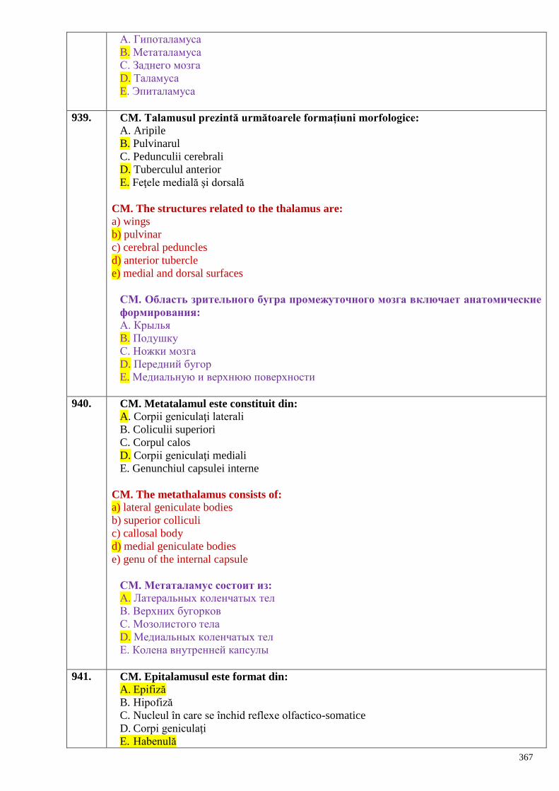
367
А. Гипоталамуса
B. Метаталамуса
C. Заднего мозга
D. Таламуса
E. Эпиталамуса
939. CM. Talamusul prezintă următoarele formațiuni morfologice:
A. Aripile
B. Pulvinarul
C. Pedunculii cerebrali
D. Tuberculul anterior
E. Feţele medială şi dorsală
CM. The structures related to the thalamus are:
a) wings
b) pulvinar
c) cerebral peduncles
d) anterior tubercle
e) medial and dorsal surfaces
CМ. Область зрительного бугра промежуточного мозга включает анатомические
формирования:
А. Крылья
B. Подушку
C. Ножки мозга
D. Передний бугор
E. Медиальную и верхнюю поверхности
940. CM. Metatalamul este constituit din:
A. Corpii geniculaţi laterali
B. Coliculii superiori
C. Corpul calos
D. Corpii geniculaţi mediali
E. Genunchiul capsulei interne
CM. The metathalamus consists of:
a) lateral geniculate bodies
b) superior colliculi
c) callosal body
d) medial geniculate bodies
e) genu of the internal capsule
CМ. Метаталамус состоит из: А. Латеральных коленчатых тел
B. Верхних бугорков
C. Мозолистого тела
D. Медиальных коленчатых тел
E. Колена внутренней капсулы
941. CM. Epitalamusul este format din:
A. Epifiză
B. Hipofiză
C. Nucleul în care se închid reflexe olfactico-somatice
D. Corpi geniculaţi
E. Habenulă
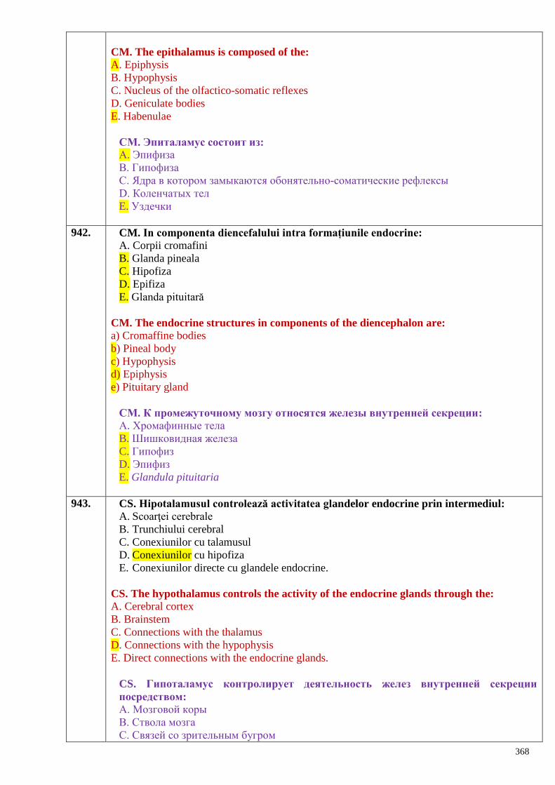
368
CM. The epithalamus is composed of the:
A. Epiphysis
B. Hypophysis
C. Nucleus of the olfactico-somatic reflexes
D. Geniculate bodies
E. Habenulae
CМ. Эпиталамус состоит из: А. Эпифиза
B. Гипофиза
C. Ядра в котором замыкаются обонятельно-соматические рефлексы
D. Коленчатых тел
E. Уздечки
942. CM. In componenta diencefalului intra formațiunile endocrine:
A. Corpii cromafini
B. Glanda pineala
C. Hipofiza
D. Epifiza
E. Glanda pituitară
CM. The endocrine structures in components of the diencephalon are:
a) Cromaffine bodies
b) Pineal body
c) Hypophysis
d) Epiphysis
e) Pituitary gland
CМ. К промежуточному мозгу относятся железы внутренней секреции:
А. Хромафинные тела
B. Шишковидная железа
C. Гипофиз
D. Эпифиз
E. Glandula pituitaria
943. CS. Hipotalamusul controlează activitatea glandelor endocrine prin intermediul:
A. Scoarţei cerebrale
B. Trunchiului cerebral
C. Conexiunilor cu talamusul
D. Conexiunilor cu hipofiza
E. Conexiunilor directe cu glandele endocrine.
CS. The hypothalamus controls the activity of the endocrine glands through the:
A. Cerebral cortex
B. Brainstem
C. Connections with the thalamus
D. Connections with the hypophysis
E. Direct connections with the endocrine glands.
CS. Гипоталамус контролирует деятельность желез внутренней секреции
посредством: А. Мозговой коры
B. Ствола мозга
C. Связей со зрительным бугром
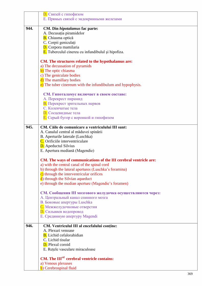
369
D. Связей с гипофизом
E. Прямых связей с эндокринными железами
944. CM. Din hipotalamus fac parte:
A. Decusația piramidelor
B. Chiasma optică
C. Corpii geniculaţi
D. Corpora mamilaria
E. Tuberculul cinereu cu infundibulul şi hipofiza.
CM. The structures related to the hypothalamus are:
a) The decussation of pyramids
b) The optic chiasma
c) The geniculate bodies
d) The mamillary bodies
e) The tuber cinereum with the infundibulum and hypophysis.
CМ. Гипоталамус включает в своем составе:
А. Перекрест пирамид
B. Перекрест зрительных нервов
C. Коленчатые тела
D. Сосцевидные тела
E. Серый бугор с воронкой и гипофизом
945. CM. Căile de comunicare a ventriculului III sunt:
A. Canalul central al măduvei spinării
B. Aperturile laterale (Luschka)
C. Orificiile interventriculare
D. Apeductul Silvius
E. Apertura mediană (Magendie)
CM. The ways of communications of the III cerebral ventricle are:
a) with the central canal of the spinal cord
b) through the lateral apertures (Luschka’s foramina)
c) through the interventricular orifices
d) through the Silvian aqueduct
e) through the median aperture (Magendie’s foramen)
CМ. Сообщения III мозгового желудочка осуществляются через:
А. Центральный канал спинного мозга
B. Боковые апертуры Luschka
C. Межжелудочковые отверстия
D. Сильвиев водопровод
E. Срединную апертуру Мagendi
946. CM. Ventriculul III al encefalului conține:
A. Plexuri venoase
B. Lichid cefalorahidian
C. Lichid tisular
D. Plexul coroid
E. Rețele vasculare miraculoase
CM. The IIIrd
cerebral ventricle contains:
a) Venous plexuses
b) Cerebrospinal fluid
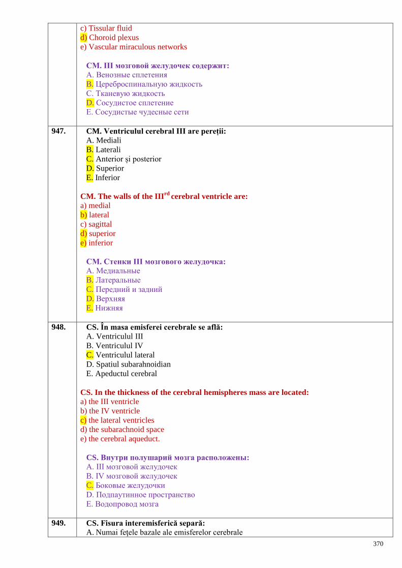
370
c) Tissular fluid
d) Choroid plexus
e) Vascular miraculous networks
CМ. III мозговой желудочек содержит:
А. Венозные сплетения
B. Цереброспинальную жидкость
C. Тканевую жидкость
D. Сосудистое сплетение
E. Сосудистые чудесные сети
947. CM. Ventriculul cerebral III are pereții:
A. Mediali
B. Laterali
C. Anterior și posterior
D. Superior
E. Inferior
CM. The walls of the IIIrd
cerebral ventricle are:
a) medial
b) lateral
c) sagittal
d) superior
e) inferior
CМ. Стенки III мозгового желудочка: А. Медиальные
B. Латеральные
C. Передний и задний
D. Верхняя
E. Нижняя
948. CS. În masa emisferei cerebrale se află:
A. Ventriculul III
B. Ventriculul IV
C. Ventriculul lateral
D. Spatiul subarahnoidian
E. Apeductul cerebral
CS. In the thickness of the cerebral hemispheres mass are located:
a) the III ventricle
b) the IV ventricle
c) the lateral ventricles
d) the subarachnoid space
e) the cerebral aqueduct.
CS. Внутри полушарий мозга расположены:
А. III мозговой желудочек
B. IV мозговой желудочек
C. Боковые желудочки
D. Подпаутинное пространство
E. Водопровод мозга
949. CS. Fisura interemisferică separă:
A. Numai feţele bazale ale emisferelor cerebrale
371
B. Lobul frontal de cel parietal
C. Lobul frontal de cel temporal
D. Cele 2 emisfere cerebrale
E. Numai feţele convexe ale emisferelor cerebrale
CS. The interhemispheric fissure separates:
a) only basal surfaces of the cerebral hemispheres
b) frontal lobe from the parietal one
c) frontal lobe from the temporal one
d) two cerebral hemispheres
e) only convex surfaces of the cerebral hemispheres
CS. Межполушарная щель разделяет:
А. Только базальные поверхности полушарий
B. Лобную долю от теменной доли
C. Лобную долю от височной доли
D. Два полушария мозга
E. Только выпуклые поверхности полушарий
950. CS. Cea mai voluminoasă parte a encefalului este reprezentată de:
A. Diencefal
B. Emisferele cerebeloase
C. Trunchiul cerebral
D. Emisferele cerebrale
E. Ganglionii bazali
CS. The most voluminous part of the brain is:
a) diencephalon
b) cerebellar hemispheres
c) brainstem
d) cerebral hemispheres
e) basal ganglia
CS. Самой объемистой частью мозга являются:
А. Промежуточный мозг
B. Полушария мозжечка
C. Ствол мозга
D. Полушария мозга
E. Базальные ядра
951. CM. Emisferele cerebrale sunt brăzdate de şanţuri care delimitează pe feţele lor lobii:
A. Hipocampic
B. Occipital
C. Precentral
D. Frontal
E. Lobus insularis
CM. The interlobar grooves of the cerebral hemispheres separate the following lobes:
a) hippocampic
b) occipital
c) precentral
d) frontal
e) insula (or insular lobe).
CМ. На поверхности мозга наблюдаются борозды разделяющие полушария на
372
доли:
А. Гипокампную
B. Затылочную
C. Предцентральную
D. Лобную
E. Островок
952. CM. Emisferele cerebrale sunt unite prin:
A. Epitalamus
B. Comisura albă anterioară
C. Meninge
D. Corpul calos
E. Hipotalamus
CM. The cerebral hemispheres are united by:
A. Epithalamus
B. Anterior white commissure
C. Meninges
D. Callosal body
E. Hypothalamus.
CМ. Полушария мозга связаны между собой:
А. Эпиталамусом
B. Передней белой спайкой
C. Оболочками
D. Мозолистым телом
E. Гипоталамусом
953. CM. Feţele emisferelor cerebrale prezintă:
A. Şanţul lateral Sylvius
B. Şanţul central Rolando
C. Girusul precentral
D. Şanţul calcarin
E. Girusul auricular
CM. The surfaces of the cerebral hemispheres show:
a) lateral (Sylvian) groove
b) central (Rolando) groove
c) precentral gyrus
d) calcarine groove
e) auricular gyrus
CМ. На поверхности полушарий мозга различают:
А. Боковую Сильвиеву борозду
B. Центральную Роландову борозду
C. Предцентральную извилину
D. Шпорную борозду
E. Ушковидную извилину
954. CM. Lobul frontal al emisferei cerebrale este delimitat de:
A. Șanțul frontal superior
B. Fisura longitudinală
C. Șanțul precentral
D. Șanțul lateral
E. Șanțul central
373
CM. The frontal lobe of the cerebral hemisphere is limited by:
a) superior frontal groove
b) longitudinal fissure
c) precentral groove
d) lateral groove
e) central groove.
CМ. Лобная доля большого полушария мозга ограничена: А. Верхней лобной бороздой
B. Продольной щелью
C. Предцентральной бороздой
D. Латеральной бороздой
E. Центральной бороздой
955. CM. Emisferele mari ale encefalului prezintă lobii:
A. Intraparietal
B. Insular
C. Temporal
D. Orbital
E. Occipital
CM. The lobes of the cerebral hemisphere are:
a) intraparietal
b) insular
c) temporal
d) orbital
e) occipital
CМ. Доли больших полушарий мозга:
А. Внутритеменная
B. Островковая
C. Височная
D. Глазничная
E. Затылочная
956. CM. Emisferele cerebrale prezintă lobii:
A. Superior
B. Lateral
C. Occipital
D. Parietal
E. Frontal
CM. The lobes of the cerebral hemisphere are:
A. Superior
B. Lateral
C. Occipital
D. Parietal
E. Frontal
CМ. Доли больших полушарий мозга:
А. Верхняя
B. Латеральная
C. Затылочная
D. Теменная
374
E. Лобная
957. CM. Emisfera mare a creierului prezintă polii:
A. Superior
B. Frontal
C. Terminal
D. Temporal
E. Occipital
CM. The poles of the cerebral hemisphere are:
a) superior
b) frontal
c) terminal
d) temporal
e) occipital
CМ. Полюса больших полушарий мозга:
А. Верхний
B. Лобный
C. Концевой
D. Височный
E. Затылочный
958. CM. Emisferele cerebrale prezintă:
A. Faţa dorsolaterală
B. Faţa posterioară
C. Polul occipital
D. Polul inferior
E. Faţa medială
CM. The cerebral hemispheres show:
a) dorsolateral surface
b) posterior surface
c) occipital pole
d) inferior pole
e) medial surface
CМ. Большие полушария мозга представляют:
А. Дорсолатеральную поверхность
B. Задняя поверхность
C. Затылочный полюс
D. Нижний полюс
E. Медиальную поверхность
959. CS. Centrul cortical al stereognoziei se localizează în:
A. Lobulul paracentral
B. Circumvoluțiunea centrală posterioară
C. Lobulul parietal superior
D. Lobulul parietal inferior
E. Circumvoluțiunea angulară
CS. The cortical center of stereognosis is located in:
a) paracentral lobule
b) postcentral gyrus
c) superior parietal lobule
375
d) inferior parietal lobule
e) angular gyrus.
CS. Корковый центр стереогнозии локализуется в:
А. Околоцентральной дольке
B. Постцентральной извилине
C. Верхней теменной дольке
D. Нижней теменной дольке
E. Угловой извилине
960. CS. Centrul cortical al analizatorului motor al vorbirii scrise se află în:
A. Circumvoluțiunea frontală superioară
B. Circumvoluțiunea centrală anterioară
C. Circumvoluțiunea frontală mijlocie
D. Lobulul paracentral
E. Lobulul parietal superior
CS. The cortical motor center of written speech is located in:
a) superior frontal gyrus
b) precentral gyrus
c) middle frontal gyrus
d) paracentral lobule
e) superior parietal lobule.
CS. Ядро двигательного анализатора письменной речи располагается в:
А. Верхней лобной извилине
B. Предцентральной извилине
С. Средней лобной извилине
D. Парацентральной дольке
E. Верхней лобной дольке
961. CS. Nucleul cortical al analizatorului auditiv al vorbirii articulate se localizează în:
A. Circumvoluțiunea centrală posterioară
B. Circumvoluțiunea frontală inferioară
C. Circumvoluțiunea temporală superioară
D. Circumvoluțiunea temporală inferioară
E. Corpii geniculați mediali
CS. The cortical center of the auditory analyzer of the spoken speech is located in:
a) postcentral gyrus
b) inferior frontal gyrus
c) superior temporal gyrus
d) inferior temporal gyrus
e) medial geniculate bodies.
CS. Ядро слухового анализатора устной речи расположен в:
А. Постцентральной извилине
B. Нижней лобной извилине
С. Верхней височной извилине
D. Нижней височной извилине
E. Медиальном коленчатом теле
962. CS. Nucleul analizatorului motor al vorbirii articulate se localizează în:
A. Circumvoluțiunea centrală anterioară
B. Circumvoluțiunea temporală superioară
376
C. Circumvoluțiunea frontală mijlocie
D. Circumvoluțiunea frontală inferioară
E. Circumvoluțiunea lingvală
CS. The cortical motor center of the spoken speech is located in:
a) precentral gyrus
b) superior temporal gyrus
c) middle frontalgyrus
d) inferior frontal gyrus
e) lingual gyrus.
CS. Ядро двигательного анализатора артикуляции речи расположен в:
А. Предцентральной извилине
B. Верхней височной извилине
С. Средней лобной извилине
D. Нижней лобной извилине
E. Язычной извилине
963. CS. Ariile auditive sunt situate:
A. În toată zona somestezică
B. În girusul precentral
C. În lobul occipital
D. În girusul temporal superior
E. În girusul hipocampic
CS. The cortical auditory areas are located in:
a) all somesthetic area
b) the precentralgyrus
c) the occipital lobe
d) the superior temporal gyrus
e) the parahippocampal gyrus.
CS. Слуховые поля расположены:
А. Во всей чувствительной области
B. Предцентральной извилине
С. Затылочной доле
D. Верхней височной извилине
E. Парагиппокампальной извилине
964. CS. Aria motorie principală se găseşte în lobul:
A. Temporal
B. Limbic
C. Parietal
D. Occipital
E. Frontal
CS. The primary motor area of the cerebral cortex is located in the following lobe:
a) temporal lobe
b) limbic lobe
c) parietal lobe
d) occipital lobe
e) frontal lobe.
CS. Главная двигательная область коры расположена в следующей доле:
А. Височной
377
B. Лимбической
С. Теменной
D. Затылочной
E. Лобной
965. CS. Centrul (aria) cortical verbomotor este localizat în:
A. Lobul frontal
B. Lobul temporal
C. Lobul occipital
D. Lobul parietal
E. Lobul limbic
CS. The cortical motor center of the spoken speech is located in:
A. Frontal lobe
B. Temporal lobe
C. Occipital lobe
D. Parietal lobe
E. Limbic lobe
СS. Корковое ядро речедвигательного анализатора расположено:
A. Лобной доле
B. Височной доле
C. Затылочной доле
D. Теменной доле
E. Лимбической доле
966. CM. Nucleul analizatorului motor se localizează în:
A. Lobulul parietal superior
B. Lobulul paracentral
C. Circumvoluțiunea frontală medie
D. Circumvoluțiunea precentrală
E. Circumvoluțiunea angulară
CM. The motor cortical center is located in:
a) superior parietal lobule
b) paracentral lobule
c) middle frontal gyrus
d) precentral gyrus
e) angular gyrus.
СМ. Ядро двигательного анализатора расположено в:
A. Верхней теменной дольке
B. Парацентральной дольке
C. Средней лобной извилине
D. Предцентральной извилине
E. Gyrus angularis
967. CM. Centrul cortical al analizatorului sensibilității generale şi proprioceptive se află în:
A. Circumvoluțiunea precentrală
B. Circumvoluțiunea frontală superioară
C. Lobulul parietal superior
D. Circumvoluțiunea postcentrală
E. Lobulul paracentral
CM. The cortical center of general and proprioceptive sensibility is located in:
378
a) precentral gyrus
b) superior frontal gyrus
c) superior parietal lobule
d) postcentral gyrus
e) paracentral lobule.
CM. Корковое ядро общей и проприоцептивной чувствительности расположено:
A. Предцентральной извилине
B. Верхней лобной извилине
C. Верхней теменной дольке
D. Постцентральной извилине
E. Парацентральной дольке
968. CM. Nucleul cortical al analizatorului vizual se află în:
A. Circumvoluțiunea frontală inferioară
B. Lobul occipital
C. Circumvoluțiunile orbitale
D. Zona șanțului calcarin
E. Pintenul de cocoş
CM. The cortical center of the visual analyzer:
a) inferior frontal gyrus
b) occipital lobe
c) orbital gyri
d) cortex around the calcarine groove
e) calcarine spurs (calcar avis).
СМ. Корковое ядро зрительного анализатора находится:
A. Нижней лобной извилине
B. Затылочной доле
C. Глазничных извилинах
D. Области шпорной борозды
E. Птичьей шпоре
969. CM. Sistemul limbic este implicat în funcţia:
A. Olfactivă
B. Stereognoză
C. Sexuală
D. Reglarea aportului alimentar
E. Toate de mai sus
CM. The limbic system is involved in the following functions:
a) olfactory
b) stereognosis
c) sexual
d) regulation of food intake
e) all above mentioned.
СМ. Лимбическая система участвует в функции:
A. Обонятельной
B. Стереогнозии
C. Сексуальной
D. Регуляция приема пищи
E. Все перечисленные
379
970. CM. Pe faţa medială a emisferelor cerebrale se găseşte aria:
A. Auditivă
B. Gustativă
C. De proiecţie a echilibrului
D. De proiecţie somestezică a feţei
E. Olfactivă
CM. The areas located on the medial surface of the cerebral hemisphere are are:
A. Auditory area
B. Taste area
C. Vestibular area
D. Somesthetic area of the face
E. Olfactory area
CM. На медиальной поверхности полушарий мозга находится поле:
A. Слуховое
B. Вкусовое
C. Проекции равновесия
D. Проекции чувствительности лица
E. Обонятельное
971. CS. Corpul calos este constituit din fibre nervoase:
A. De proiecție
B. Asociative
C. Comisurale
D. Senzitive
E. Vegetative
CS.The callosal body consists of the following nerve fibers:
a) projection
b) association
c) commissural
d) sensory
e) vegetative.
СS. Мозолистое тело состоит из следующих нервных волокон:
A. Проекционных
B. Ассоциативных
C. Комиссуральных
D. Чувствительных
E. Вегетативных
972. CS. Emisferele cerebrale sunt unite între ele prin:
A. Meninge
B. Hipocamp
C. Ganglionii bazali
D. Trigonul cerebral
E. Comisuri
CS. Cerebral hemispheres are joined to each other by the:
A. Meninges
B. Hippocampus
C. Basal ganglia
D. Cerebral triangle
E. Commissures
380
СS. Полушария мозга соединяются:
A. Мозговыми оболочками
B. Гипокампом
C. Базальными узлами
D. Треугольником мозга
E. Спайками
973. CS. Nucleul caudat se separă de globul palid prin:
A. Capsula extremă
B. Capsula externă
C. Capsula internă
D. Comisura mare a creierului
E. Comisura anterioară
CS. The caudate nucleus is separated from the globus pallidus by:
a) extreme capsule
b) external capsule
c) internal capsule
d) great cerebral commissure (callosal body)
e) anterior commissure
СS. Хвостатое ядро отделено от globus pallidus посредством:
A. Capsula extrema
B. Capsula externa
C. Capsula interna
D. Большой спайкой мозга
E. Передней спайкой
974. CS. Ventriculul cerebral lateral comunică cu:
A. Spațiul subarahnoidian
B. Spațiul subdural
C. Ventriculul III
D. Ventriculul IV
E. Apeductul Silvius
CS. The lateral cerebral ventricle communicates with:
a) The subarachnoid space
b) The subdural space
c) The IIIrd
ventricle
d) The IVth
ventricle
e) The Sylvian aqueduct
CS. Боковой желудочек мозга сообщается с:
A. Подпаутинным пространством
B. Субдуральным пространством
C. Третьим желудочком
D. Четвертым желудочком
E. Водопроводом мозга
975. CS. Ventriculele cerebrale laterale comunică cu ventriculul III prin:
A. Apeductul cerebral
B. Orificiile interventriculare
C. Apertura mediană
D. Aperturile laterale
381
E. Canalul central
CS The lateral cerebral ventricles communicate with the third one through the:
a) cerebral aqueduct
b) interventricular orifices
c) median aperture
d) lateral apertures
e) central canal
CS. Боковые желудочки мозга сообщаются с третьим желудочком через:
A. Водопровод мозга
B. Межжелудочковые отверстия
C. Срединную апертуру
D. Латеральные апертуры
E. Центральный канал
976. CM. Substanţa albă a emisferei cerebrale conţine următoarele tipuri de fibre:
A. De asociaţie
B. Cerebeloase
C. Comisurale
D. Reticulare
E. De proiecţie
CM. The white matter of the cerebral hemisphere contains the following types of fibers:
a) associative
b) cerebellar
c) commissural
d) reticular
e) projection.
СМ. Белое вещество полушария мозга содержит волокна:
A. Ассоциативные
B. Мозжечковые
C. Комисуральные
D. Ретикулярные
E. Проекционные
977. CM. Substanța cenușie a emisferelor cerebrale se organizează sub aspect de:
A. Ganglioni vegetativi
B. Nuclei bazali
C. Nuclei subcorticali
D. Nuclei cerebeloși
E. Cortex cerebral
CM. The gray matter of the cerebral hemispheres forms:
a) vegetative ganglia
b) basal ganglia (or nuclei)
c) subcortical nuclei
d) cerebellar nuclei
e) cerebral cortex.
CM. Серое вещество полушарий мозга образуют:
A. Вегетативные ганглии
B. Базальные ядра
C. Подкорковые ядра
382
D. Ядра мозжечка
E. Кору полушарий мозга
978. CM. Fornixul encefalului prezintă următoarele formațiuni:
A. Bolta
B. Corpul
C. Brațul anterior
D. Columnele (stâlpii anteriori)
E. Pedunculii (stâlpii posteriori)
CM. The fornix consists of the following structures:
a) vault
b) body
c) anterior limb
d) columns (anterior pillars)
e) crura (posterior pillars).
СМ. Свод мозга имеет:
A. Свод
B. Тело
C. Переднее плечо
D. Столбы (передние столбы)
E. Ножки (задние столбы)
979. CM. Fibrele nervoase comisurale din emisferele cerebrale se localizează în:
A. Capsula internă
B. Corpul calos
C. Cordonul lateral
D. Capsula externă
E. Comisura albă
CM. The commissural nerve fibers of the cerebral hemispheres are located in:
a) internal capsule
b) callosal body
c) lateral funiculus
d) external capsule
e) white commissure.
CM. Комиссуральные волокна в полушариях мозга находятся в:
A. Внутренней капсуле
B. Мозолистом теле
C. Боковом канатике
D. Наружной капсуле
E. Белой спайке
980. CM. Corpului calos i se disting următoarele porțiuni:
A. Ciocul
B. Brațul anterior
C. Trunchiul
D. Ramurile laterale
E. Lamela terminală
CM. The parts of the callosal body are:
a) rostrum
b) anterior limb
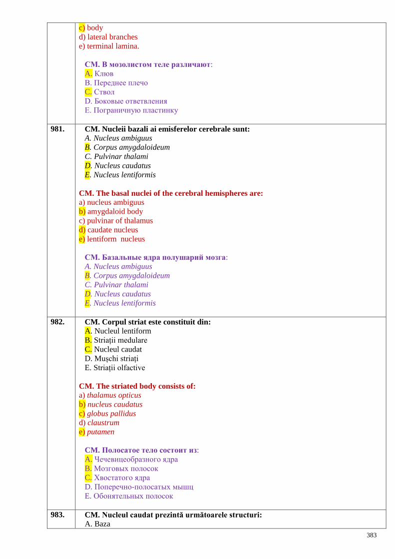
383
c) body
d) lateral branches
e) terminal lamina.
СМ. В мозолистом теле различают:
A. Клюв
B. Переднее плечо
C. Ствол
D. Боковые ответвления
E. Пограничную пластинку
981. CM. Nucleii bazali ai emisferelor cerebrale sunt:
A. Nucleus ambiguus
B. Corpus amygdaloideum
C. Pulvinar thalami
D. Nucleus caudatus
E. Nucleus lentiformis
CM. The basal nuclei of the cerebral hemispheres are:
a) nucleus ambiguus
b) amygdaloid body
c) pulvinar of thalamus
d) caudate nucleus
e) lentiform nucleus
СМ. Базальные ядра полушарий мозга:
A. Nucleus ambiguus
B. Corpus amygdaloideum
C. Pulvinar thalami
D. Nucleus caudatus
E. Nucleus lentiformis
982. CM. Corpul striat este constituit din:
A. Nucleul lentiform
B. Striații medulare
C. Nucleul caudat
D. Mușchi striați
E. Striații olfactive
CM. The striated body consists of:
a) thalamus opticus
b) nucleus caudatus
c) globus pallidus
d) claustrum
e) putamen
CM. Полосатое тело состоит из:
A. Чечевицеобразного ядра
B. Мозговых полосок
C. Хвостатого ядра
D. Поперечно-полосатых мышц
E. Обонятельных полосок
983. CM. Nucleul caudat prezintă următoarele structuri:
A. Baza
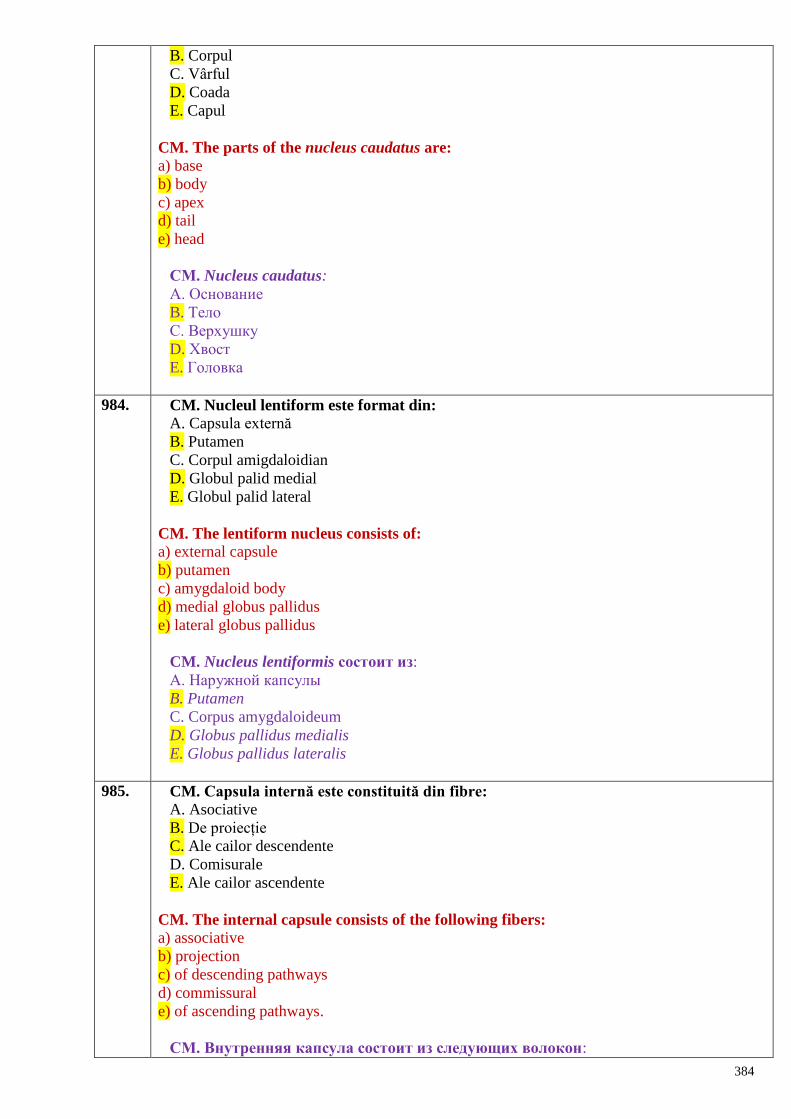
384
B. Corpul
C. Vârful
D. Coada
E. Capul
CM. The parts of the nucleus caudatus are:
a) base
b) body
c) apex
d) tail
e) head
СМ. Nucleus caudatus:
A. Основание
B. Тело
C. Верхушку
D. Хвост
E. Головка
984. CM. Nucleul lentiform este format din:
A. Capsula externă
B. Putamen
C. Corpul amigdaloidian
D. Globul palid medial
E. Globul palid lateral
CM. The lentiform nucleus consists of:
a) external capsule
b) putamen
c) amygdaloid body
d) medial globus pallidus
e) lateral globus pallidus
CM. Nucleus lentiformis состоит из:
A. Наружной капсулы
B. Putamen
C. Corpus amygdaloideum
D. Globus pallidus medialis
E. Globus pallidus lateralis
985. CM. Capsula internă este constituită din fibre:
A. Asociative
B. De proiecție
C. Ale cailor descendente
D. Comisurale
E. Ale cailor ascendente
CM. The internal capsule consists of the following fibers:
a) associative
b) projection
c) of descending pathways
d) commissural
e) of ascending pathways.
СМ. Внутренняя капсула состоит из следующих волокон:
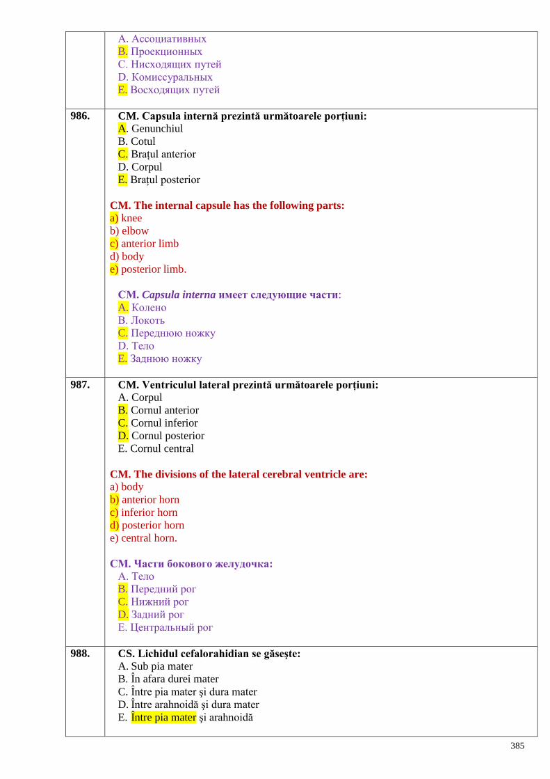
385
A. Ассоциативных
B. Проекционных
C. Нисходящих путей
D. Комиссуральных
E. Восходящих путей
986. CM. Capsula internă prezintă următoarele porțiuni:
A. Genunchiul
B. Cotul
C. Brațul anterior
D. Corpul
E. Brațul posterior
CM. The internal capsule has the following parts:
a) knee
b) elbow
c) anterior limb
d) body
e) posterior limb.
СМ. Capsula interna имеет следующие части:
A. Колено
B. Локоть
C. Переднюю ножку
D. Тело
E. Заднюю ножку
987. CM. Ventriculul lateral prezintă următoarele porțiuni:
A. Corpul
B. Cornul anterior
C. Cornul inferior
D. Cornul posterior
E. Cornul central
CM. The divisions of the lateral cerebral ventricle are:
a) body
b) anterior horn
c) inferior horn
d) posterior horn
e) central horn.
CМ. Части бокового желудочка:
А. Тело
B. Передний рог
C. Нижний рог
D. Задний рог
E. Центральный рог
988. CS. Lichidul cefalorahidian se găseşte:
A. Sub pia mater
B. În afara durei mater
C. Între pia mater şi dura mater
D. Între arahnoidă şi dura mater
E. Între pia mater şi arahnoidă
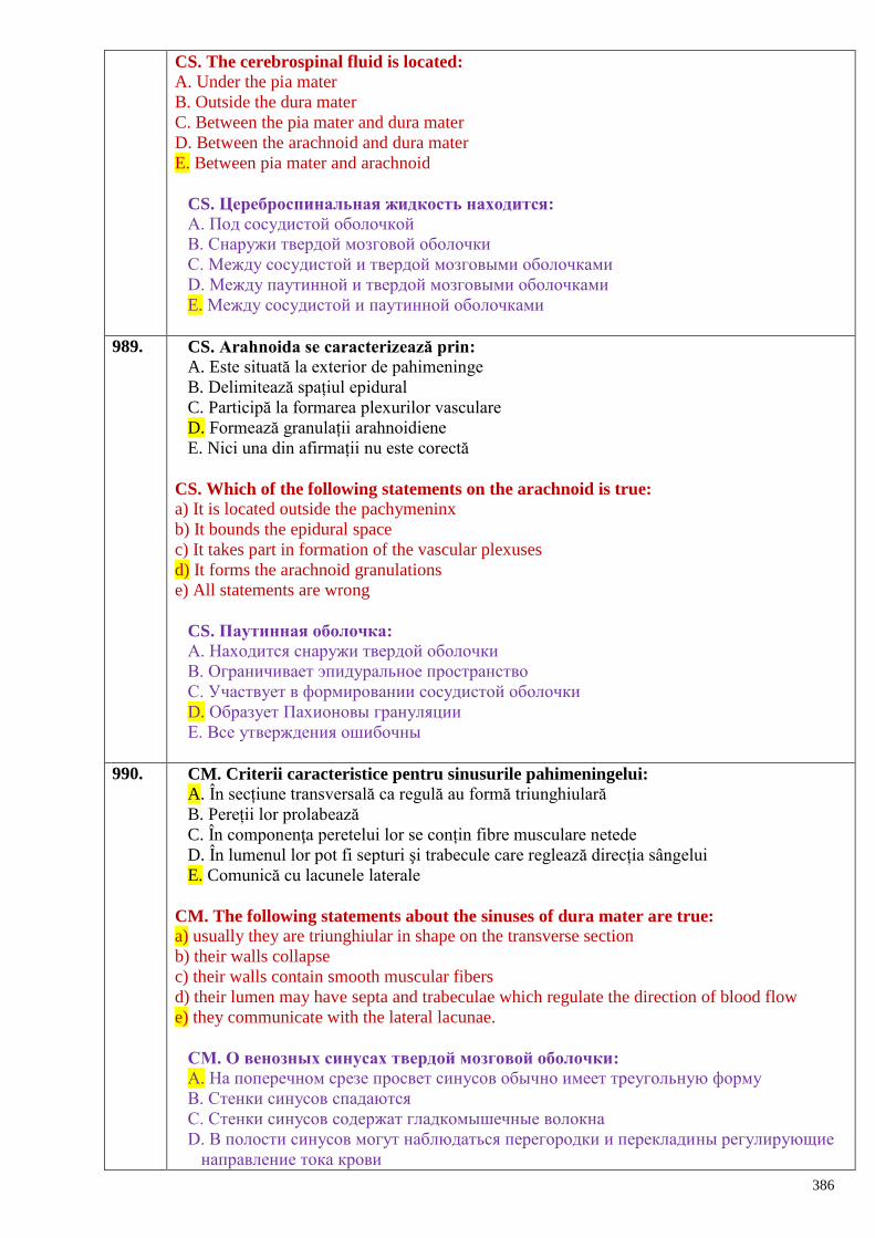
386
CS. The cerebrospinal fluid is located:
A. Under the pia mater
B. Outside the dura mater
C. Between the pia mater and dura mater
D. Between the arachnoid and dura mater
E. Between pia mater and arachnoid
CS. Цереброспинальная жидкость находится:
А. Под сосудистой оболочкой
B. Снаружи твердой мозговой оболочки
C. Между сосудистой и твердой мозговыми оболочками
D. Между паутинной и твердой мозговыми оболочками
E. Между сосудистой и паутинной оболочками
989. CS. Arahnoida se caracterizează prin:
A. Este situată la exterior de pahimeninge
B. Delimitează spațiul epidural
C. Participă la formarea plexurilor vasculare
D. Formează granulații arahnoidiene
E. Nici una din afirmații nu este corectă
CS. Which of the following statements on the arachnoid is true:
a) It is located outside the pachymeninx
b) It bounds the epidural space
c) It takes part in formation of the vascular plexuses
d) It forms the arachnoid granulations
e) All statements are wrong
CS. Паутинная оболочка:
А. Находится снаружи твердой оболочки
B. Ограничивает эпидуральное пространство
C. Участвует в формировании сосудистой оболочки
D. Образует Пахионовы грануляции
E. Все утверждения ошибочны
990. CM. Criterii caracteristice pentru sinusurile pahimeningelui:
A. În secțiune transversală ca regulă au formă triunghiulară
B. Pereții lor prolabează
C. În componenţa peretelui lor se conțin fibre musculare netede
D. În lumenul lor pot fi septuri şi trabecule care reglează direcția sângelui
E. Comunică cu lacunele laterale
CM. The following statements about the sinuses of dura mater are true:
a) usually they are triunghiular in shape on the transverse section
b) their walls collapse
c) their walls contain smooth muscular fibers
d) their lumen may have septa and trabeculae which regulate the direction of blood flow
e) they communicate with the lateral lacunae.
CМ. О венозных синусах твердой мозговой оболочки:
А. На поперечном срезе просвет синусов обычно имеет треугольную форму
B. Стенки синусов спадаются
C. Стенки синусов содержат гладкомышечные волокна
D. В полости синусов могут наблюдаться перегородки и перекладины регулирующие
направление тока крови
387
E. Сообщаются с боковыми лакунами
991. CM. Vasculara encefalului (pia mater) posedă următoarele particularități:
A. Pătrunde in șanțurile si scizurile creierului
B. In țesutul nervos delimitează spatiile perivasculare si pericelulare (Robin-Virchow)
C. Împreună cu vasele sangvine participă la formarea plexurilor coroide
D. Delimitează spațiul subdural
E. Toate afirmațiile sunt corecte
CM. The cerebral pia mater has the following specific features:
a) It enters the grooves and fissures of the brain
b) It delimits perivascular and pericellular spaces (Virchow-Robin) in the nervous tissue
c) It participates in formation of the choroid plexus together with blood vessels
d) It delimits the subdural space
e) It delimits the epidural space.
CМ. Сосудистая оболочка мозга (pia mater) А. Проникает в борозды и щели мозга
B. Внутри вещества мозга ограничивает околососудистые и и перицелюлярные
пространства (Robin-Virchow)
C. Вместе с кровеносными сосудами формирует сосудистые сплетения желудочков
мозга
D. Ограничивает субдуральное пространство
E. Верны все утверждения
992. CM. Care din afirmații caracterizează lichidul cefalorahidian: A. Se scurge in sinusurile pahimeningelui
B. Umple ventriculele creierului și spațiul subarahnoidian
C. Se produce in mod normal in cantitatea de 500 - 550 ml nictimeral
D. Circulă datorită presiunii hidrostatice
E. Din spațiul subarahnoidian prin orificiul Magendie şi Luschka se scurge în ventriculul IV
CM. Which of the following statements about the cerebrospinal fluid are true: a) It drains into the sinuses of dura mater
b) It fills the cerebebral ventricles and subarachnoid space
c) Normally it is produced in the amount of 500-550 ml daily
d) Its circulation is caused by the hydrostatic pressure
e) It flows from thesubarachnoid space through the orifices of Magendie and Luschka into the
IV ventricle.
CМ. О спинномозговой жидкости
А. Оттекает в венозные синусы твердой оболочки мозга
B. Заполняет желудочки мозга и подпаутинное пространство
C. Обычно продуцируется до 500-550 мл за 24 часа
D. Циркуляция СМЖ обеспечивается за счет гидростатического давления
E. Из подпаутинового пространства через отверстия Мagendi и Luschka оттекает в
четвертый мозговой желудочек
993. CS. Toate căile sensibilităţii exteroceptive au al treilea neuron în:
A. Măduva spinării
B. Bulb
C. Cerebel
D. Nucleii bazali
E. Talamus
388
CS. The third neuron of the exteroceptive conducting pathways is located in:
a) spinal cord
b) medulla oblongata (myelencephalon)
c) cerebellum
d) basal nuclei
e) thalamus.
CS. Третьи нейроны всех проводящих путей экстерорецептивной
чувcтвительности заложены в: А. Спинном мозге
B. Продолговатом мозге
C. Мозжечке
D. Базальных ядрах
E. Зрительном бугре
994. CM. Cerebelul primeşte informaţii de la receptorii situaţi în:
A. Sistemul circulator
B. Sistemul osteoarticular
C. Sistemul respirator
D. Sistemul muscular
E. Viscerele abdominale
CM. The cerebellum receives information from the receptors located in the:
a) Circulatory system
b) Osteoarticular system
c) Respiratory system
d) Muscular system
e) Abdominal viscera.
CМ. Мозжечок получает информацию от рецепторов заложенных в:
А. Системе кровообращения
B. Костно-суставной системе
C. Дыхательной системе
D. Мышечной системе
E. Брюшных органах
995. CS. Protoneuronul căii sensibilităţii exteroceptive este situat în:
A. Măduvă
B. Receptorii cutanaţi
C. Ganglionul spinal
D. Bulb
E. Punte
CS. The protoneuron of the exteroreceptive pathways is located in the:
A. Spinal cord
B. Cutaneous receptors
C. Spinal ganglion
D. Myelencephalon
E. Pons
CS. Протонейрон путей экстероцептивной чувствительности заложен в:
А. Спинном мозге
B. Кожных рецепторах
C. Спинальном ганглии
389
D. Продолговатом мозге
E. Мосту
996. CS. Calea cortico-spinală conduce:
A. Sensibilitatea tactilă, termică şi dureroasă
B. Sensibilitatea proprioceptivă inconştientă
C. Motilitatea involuntară
D. Motilitatea voluntară
E. Sensibilitatea epicritică
CS. The corticospinal pathway conducts:
A. The tactile, thermal and pain sensitivity
B. The unconscious proprioceptive sensibility
C. The involuntary motility
D. The voluntary motility
E. The epicritic sensibility
CS. Корково-спинномозговой путь проводит: А. Осязание, температурную и болевую чувствительность
B. Неосознанную проприоцептивную чувствительность
C. Обеспечивает непроизвольные движения
D. Обеспечивает произвольные движения
E. Эпикритическую чувствительность
997. CS. Care glande endocrine inhibă maturizarea sexuală precoce:
A. Hipofiza
B. Epifiza
C. Timusul
D. Tiroida
E. Suprarenalele
SC. Which endocrine glands inhibit the premature development of the sex glands:
A. Hypophysis
B. Epiphysis
C. Thymus
D. Thyroid gland
E. Suprarenal (adrenal) glands
CS. Какие эндокринные железы участвуют в преждевременном половом
созревании?
A. Гипофиз
B. Эпифиз
C. Вилочковая железа
D. Щитовидная железа
E. Надпочечники
998. CS. Hipofiza se localizează (loja hipofizei) in:
A. Șaua turcească
B. Orbită
C. Sinusul sfenoidal
D. Sinusul frontal
E. Meatul nazal inferior
SC. The hypophysis (hypophyseal fossa) is located in:
A. Turkish saddle (sella turcica)
390
B. Orbit
C. Sphenoidal sinus
D. Frontal sinus
E. Inferior nasal meatus
CS. Гипофиз расположен (гипофизарная ямка) в:
A. Турецком седле
B. Глазнице
C. Клиновидной пазухе.
D. Лобной пазухе
E. Среднем носовом ходе
999. CS. Șaua turceasca cu (fossa hypophysialis) aparține osului:
A. Frontal
B. Zigomatic
C. Sfenoid
D. Parietal
E. Temporal
SC. The Turkish saddle (with fossa hypophysialis) belongs to:
A. Frontal bone
B. Zygomatic bone
C. Sphenoid bone
D. Parietal bone
E. Temporal bone
CS. Турецкое седло с (fossa hypophysialis) принадлежит кости:
A. Лобной
B. Скуловой
C. Клиновидной
D. Теменной
E. Височной
1000. CS. Hipofiza e separată de spațiul subdural prin:
A. Diafragma șeii (turcești)
B. Arahnoidă
C. Cortul cerebelului
D. Septul nazal
E. Lamela cribroasă
SC. The hypophysis is separated from the subdural space by:
A. Diaphragm of Turkish saddle B. Arachnoid mater
C. Falx cerebelli
D. Nasal septum
E. Cribriform plate
CS. Гипофиз отделѐн от субдурального пространства посредством:
A. Диафрагмой (турецкого) седла
B. Паутинной оболочкой
C. Намѐтом мозжечка
D. Носовой перегородкой
E. Решѐтчатой пластинкой
1001. CS. Hipofiza are formă de:
391
A. Bob de mazăre
B. Bob de linte
C. Bob de grâu
D. Bob de cafea
E. Bob de fasole
SC. The hypophysis is:
A. Pea-shaped
B. Lentil-shaped
C. Shaped like a grain of the wheat
D. Coffee bean-shaped
E. Bean-shaped (ellipsoid in shape)
CS. Гипофиз имеет форму:
A. Гороха
B. Чечевицы
C. Пшеничного зерна
D. Кофейного зерна
E. Элипсоида
1002. CS. Hipofiza constă din:
A. Lobul anterior si posterior
B. Lobul anterior, porțiunea intermediară, lobul posterior
C. Lobul superior si inferior
D. Lobul superior, inferior si partea tuberală
E. Lobul stâng, lobul drept si partea intermediară
SC. The hypophysis consists of:
A. Anterior and posterior lobes
B. Anterior lobe, intermediate part, posterior lobe
C. Superior and inferior lobes
D. Superior lobe, inferior lobe and tuberal part
E. Left lobe, right lobe and intermediate part
CS. Гипофиз состоит из:
A. Передней и задней долей
B. Передней доли, промежуточной части, задней доли
C. Верхней и нижней долей
D. Верхней, нижней долей и бугорной части
E. Левой доли, правой доли и промежуточной части
1003. CS. 70% - 80% din masa hipofizei o constituie:
A. Lobul posterior
B. Porțiunea intermediară
C. Lobul anterior
D. Partea tuberală
E. Tuberul cenușiu
SC. The 70% - 80% of the weight of the hypophysis is represented by:
A. Posterior lobe
B. Intermediate part
C. Anterior lobe
D. Tuberal part
E. Tuber cinereum
392
CS. 70 - 80% из массы гипофиза составляет:
A. Задняя доля
B. Промежуточная часть
C. Передняя доля
D. Бугорная часть
E. Серый бугор
1004. CS. Adenohipofiza se mai numește:
A. Lob anterior
B. Lob posterior
C. Parte tuberală
D. Epifiză
E. Hipotalamus
SC. The adenohypophysis is also called:
A. Anterior lobe
B. Posterior lobe
C. Tuberal part
D. Epiphysis
E. Hypothalamus
CS. Аденогипофиз ещѐ называется:
A. Передняя доля
B. Задняя доля
C. Бугорная часть
D. Эпифиз
E. Гипоталамус
1005. CS. Hipofiza este legată cu:
A. Hipotalamusul
B. Epitalamusul
C. Corpul striat
D. Emisferele cerebrale
E. Măduva spinării
SC. The hypophysis is connected to:
A. Hypothalamus
B. Epithalamus
C. Striate body
D. Cerebral hemispheres
E. Spinal cord
CS. Гипофиз соединѐн с:
A. Гипоталамусом
B. Эпиталамусом
C. Полосатым телом
D. Полушарием большого мозга
E. Спинным мозгом
1006. CS. Neurohipofiza face legătura cu hipotalamusul prin:
A. Sistemul porthipofizar
B. Tractul hipotalamo-hipofizar
C. Tractul epitalamo-hipofizar
D. Tractul mamilotalamic (Vicq - d-Azyr)
E. Tractul olfactiv
393
SC. The neurohypophysis is connected to the hypothlamus by:
A. Hypophyseal portal system
B. Hypothalamo-hypophyseal tract
C. Epithalamo-hypophyseal tract
D. Mammillothalamic tract (Vicq d`Azyr)
E. Olfactory tract
CS. Нейрогипофиз образует связь с гипоталамусом через:
A. Портгипофизарную систему
B. Гипоталамогипофизарный тракт
C. Эпиталамогипофизарный тракт
D. Мамилоталамический тракт (Vicq-d-Azyr)
E. Обонятельный тракт
1007. CS. Neurohipofiza:
A. Produce hormoni
B. Acumulează hormoni
C. Secretă și acumulează hormoni
D. Nu produce și nu acumulează hormoni
E. Toate incorecte
SC. The neurohypophysis:
A. Produces hormones
B. Stores hormones
C. Stores and produces hormons
D. Does not store and not produce hormones
E. Does not store hormones
CS. Нейрогипофиз:
A. Продуцирует гормоны
B. Накапливает гормоны
C. Выделяет и накапливает гормоны
D. Не продуцирует и не накапливает гормоны
E. Всѐ ошибочно
1008. CS. Adenohipofiza face legătura cu hipotalamusul prin:
A. Tractul hipotalamohipofizar
B. Sistemul port hipotalamohipofizar
C. Tractul optic
D. Tractul piramidal
E. Tractul talamocortical
SC. The adenohypophysis is connected to the hypothalamus by:
A. Hypothalamo-hypophyseal tract
B. Hypophyseal portal system
C. Optic tract
D. Pyramidal tract
E. Thalamocortical tract
CS. Аденогипофиз образует связь с гипоталамусом через:
A. Гипоталамогипофизарный тракт
B. Портгипофизарную систему
C. Зрительный тракт
D. Пирамидальный путь
394
E. Таламокорковый тракт
1009. CS. Adenohipofiza secretă:
A. Hormoni glandulari tropi, prolactina, ADH
B. STH, prolactina, ocitocina
C. STH, ocitocina, hormonul melanocitostimulator
D. STH, tirotropina, gonadotropinele
E. STH, hormoni glandulari tropi, prolactina
SC. The adenohypophysis produces:
A. Tropic hormones, prolactin, ADH
B. STH, prolactin, oxytocin
C. STH, oxytocin, melanocyte-stimulating hormone
D. STH, thyrotropin, gonadotropic hormones
E. STH, tropic hormones, prolactin
CS. Аденогипофиз вырабатывает:
A. Железотропные гормоны, пролактин, АДГ
B. СТГ, пролактин, окситоцин
C. СТГ, окситоцин, меланоцитостимулирующий гормон
D. СТГ, тиротропин, гонадотропные гормоны
E. СТГ, железотропные гормоны, пролактин
1010. CS. In hiperfuncția hipofizei la adulţi poate să se dezvolte:
A. Boala lui Basedow
B. Acromegalia
C. Boala lui Addison
D. Gigantism
E. Mixedem
SC. In case of the hyperfunction of the hypophysis in adult could apear:
A. Basedow's disease
B. Acromegaly
C. Addison's disease
D. Gigantism
E. Myxedema
CS. При гиперфункции гипофиза в зрелом возрасте возможно развитие:
A. Болезни Basedow
B. Акромегалии
C. Болезни Addison
D. Гигантизма
E. Микседемы
1011. CS. Cum se mai numește corpul pineal:
A. Hipofiză
B. Epifiză
C. Glandă pituitară
D. Glandă tiroidă
E. Hipotalamus
SC. What is another name of the pineal body?
A. Hypophysis
B. Epiphysis
C. Pituitary gland
395
D. Thyroid gland
E. Hypothalamus
CS. Как ещѐ называется шишковидное тело?
A. Гипофиз
B. Эпифиз
C. Питуитарная железа
D. Щитовидная железа
E. Гипоталамус
1012. CS. Hormonul activ al epifizei este:
A. Vasopresina
B. Hormonul melanocitostimulator
C. Melanotonina
D. Aldosteronul
E. Calcitonina
SC. The active hormone of the epiphysis is:
A. Vasopressin
B. Melanocyte-stimulating hormone
C. Melatonin
D. Aldosterone
E. Calcitonin
CS. Активным гормоном эпифиза является:
A. Вазопрессин
B. Меланоцитостимулирующий гормон
C. Меланотонин
D. Альдостерон
E. Кальцитонин
1013. CS. Cel mai înalt grad de dezvoltare epifiza îl atinge:
A. La copii
B. La adulți
C. La bătrâni
D. La bărbat
E. La femeie
SC. The epiphysis achieves the highest level of development:
A. In child
B. In adult
C. In old person
D. In man
E. In woman
CS. Наибольшего развития эпифиз достигает:
A. У детей
B. У взрослых
C. У стариков
D. У мужчины
E. У женщины
1014. CS. Epifiza face parte:
A. Hipotalamusul
B. Epitalamusul
396
C. Metatalamusul
D. Măduva spinării
E. Cerebelul
SC. The epiphysis is a part of: A. Hypothalamus
B. Epithalamus
C. Metathalamus
D. Spinal cord
E. cerebellum
CS. Эпифиз относится к:
A. Гипоталамусу
B. Эпиталамусу
C. Метаталамусу
D. Спинному мозгу
E. Мозжечку
1015. CS. Prin care formațiuni epifiza e legată cu creierul:
A. Prin comisura cerebrală anterioară
B. Prin habenulă
C. Prin pedunculii cerebrali
D. Prin comisura fornixului
E. Prin comisura cerebrală posterioară
SC. The epiphysis is connected to the brain by:
A. Anterior cerebral commissure
B. Habenulae
C. Cerebral peduncles
D. Commissure of fornix
E. Posterior cerebral commissure
CS. Эпифиз соединѐн с головным мозгом через:
A. Переднюю мозговую спайку
B. Поводки
C. Ножки мозга
D. Спайки свода
E. Заднюю мозговую спайку
1016. CM. Ce formă are epifiza:
A. Ovală
B. Sferică
C. Conică
D. Infundibuliformă
E. Piriformă
MC. The epiphysis has the following shapes:
A. Oval
B. Spheroid
C. Conic
D. Infundibuliforme
E. Piriform
CM. Какую форму имеет эпифиз?
A. Овальную
397
B. Шаровидную
C. Коническую
D. Воронкообразную
E. Грушевидную
1017. CM. Glande endocrine centrale sunt:
A. Paraganglionii
B. Timusul
C. Hipotalamusul
D. Hipofiza
E. Epifiza
MC. The central endocrine glands are, as follows:
A.Paraganglia
B. Thymus
C. Hypothalamus
D. Hypophysis
E. Epiphysis
CМ. Центральными эндокринными железами являются:
A. Параганглии
B. Тимус
C. Гипоталамус
D. Гипофиз
E. Эпифиз
Sistemul nervos vegetativ – generalităţi.
SN simpatic şi parasimpatic – porţiuni centrale şi periferice
Plexurile somatice şi vegetative, ramurile lor principale.
Lanţul simpatic – componente, topografie, ramuri
1018. CS. Porţiunea centrală a sistemului nervos vegetativ simpatic se află în segmentele
medulare:
A. C3-T12.
B. C8-L3.
C. C1-T4.
D. L2-S3.
E. În toate segmentele.
CS. The central part of the sympathetic nervous system comprises the following spinal
segments:
A. C3-T12
B. C8-L2
C. C1-T4
D. L2-S3
E. All the segments.
СS. Центральный отдел симпатической части вегетативной нервной системы
расположен в следующих сегментах спинного мозга:
A. C3-T12.
B. C8-L3.
C. C1-T4.
D. L2-S3.
E. Во всех сегментах.
1019. CM. Ganglioni parasimpatici sunt:
398
A. Spinali.
B. Ciliar.
C. Pterigopalatin.
D. Otic.
E. Submandibular.
CM.There are the following parasympathetic ganglia:
A. Spinal
B. Ciliary
C. Pterygopalatine
D. Otic
E. Submandibular
СМ. Укажите парасимпатические узлы:
A. Спинномозговые.
B. Ресничный.
C. Крылонебный.
D. Ушной.
E. Подчелюстной.
1020. CM. Particularități caracteristice sistemului nervos somatic:
A. Inervaţia musculaturii netede.
B. Inervaţia musculaturii striate.
C. Amplasarea segmentară a centrilor.
D. Amplasarea centrilor sub formă de focare.
E. Neuronul efector este în afara sistemului nervos central.
CM. The specific features of the somatic nervous system are:
A. It supplies the smooth muscles
B. It supplies the striated muscles
C. It has segmental structure
D. It centers are placed as foci
E. Its effector neuron is located outside the central nervous system.
СМ. Укажите отличительные особенности соматической нервной системы:
A. Иннервация гладких мышц.
B. Иннервация поперечно-полосатой мускулатуры.
C. Сегментарность в расположении центров.
D. Очаговость расположения центров.
E. Эффекторный нейрон расположен вне центральной нервной системы.
1021. CM. Indicaţi criteriile de bază ale sistemului nervos vegetativ:
A. Inervaţia musculaturii netede.
B. Amplasarea centrilor în focare.
C. Amplasarea segmentară a centrilor.
D. Fibre exclusiv amielinice.
E. Neuronul efector situat la periferie.
CM. Choose the main criteria of the vegetative nervous system:
A. Innervation of the smooth muscles
B.Location of centers as foci
C. Segmental location of the centers
D. All fibers are myelinated
E. The effector neuron is located outside of central nervous system
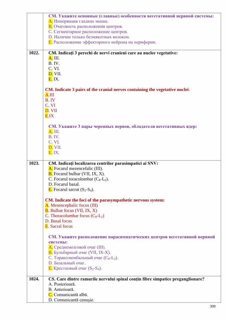
399
СМ. Укажите основные (главные) особенности вегетативной нервной системы:
A. Иннервация гладких мышц.
B. Очаговость расположения центров.
C. Сегментарное расположение центров.
D. Наличие только безмякотных волокон.
E. Расположение эффекторного нейрона на периферии.
1022. CM. Indicaţi 3 perechi de nervi cranieni care au nuclee vegetative:
A. III.
B. IV.
C. VI.
D. VII.
E. IX.
CM. Indicate 3 pairs of the cranial nerves containing the vegetative nuclei:
A.III
B. IV
C. VI
D. VII
E.IX
СМ. Укажите 3 пары черепных нервов, обладатели вегетативных ядер:
A. III.
B. IV.
C. VI.
D. VII.
E. IX.
1023. CM. Indicaţi localizarea centrilor parasimpatici ai SNV:
A. Focarul mezencefalic (III).
B. Focarul bulbar (VII, IX, X).
C. Focarul toracolombar (C8-L2).
D. Focarul bazal.
E. Focarul sacrat (S2-S4).
CM. Indicate the foci of the parasympathetic nervous system:
A. Mesencephalic focus (III)
B. Bulbar focus (VII, IX, X)
C. Thoracolumbar focus (C8-L2)
D. Basal focus
E. Sacral focus
СМ. Укажите расположение парасимпатических центров вегетативной нервной
системы:
A. Среднемозговой очаг (III).
B. Бульбарный очаг (VII, IX-X).
C. Тораколюмбальный очаг (C8-L2).
D. Базальный очаг.
E. Крестцовый очаг (S2-S4).
1024. CS. Care dintre ramurile nervului spinal conţin fibre simpatice preganglionare?
A. Posterioară.
B. Anterioară.
C. Comunicantă albă.
D. Comunicantă cenuşie.
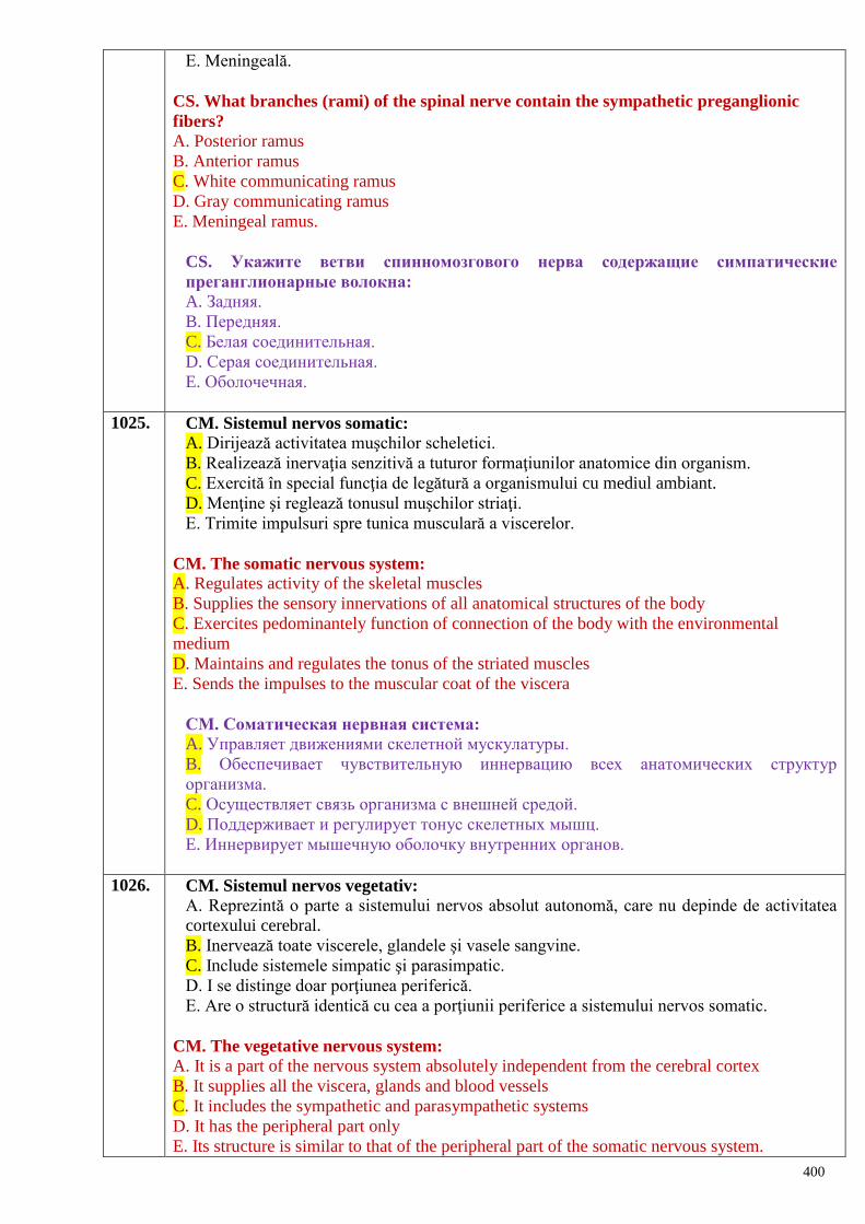
400
E. Meningeală.
CS. What branches (rami) of the spinal nerve contain the sympathetic preganglionic
fibers?
A. Posterior ramus
B. Anterior ramus
C. White communicating ramus
D. Gray communicating ramus
E. Meningeal ramus.
CS. Укажите ветви спинномозгового нерва содержащие симпатические
преганглионарные волокна:
A. Задняя.
B. Передняя.
C. Белая соединительная.
D. Серая соединительная.
E. Оболочечная.
1025. CM. Sistemul nervos somatic:
A. Dirijează activitatea muşchilor scheletici.
B. Realizează inervaţia senzitivă a tuturor formaţiunilor anatomice din organism.
C. Exercită în special funcţia de legătură a organismului cu mediul ambiant.
D. Menţine şi reglează tonusul muşchilor striaţi.
E. Trimite impulsuri spre tunica musculară a viscerelor.
CM. The somatic nervous system:
A. Regulates activity of the skeletal muscles
B. Supplies the sensory innervations of all anatomical structures of the body
C. Exercites pedominantely function of connection of the body with the environmental
medium
D. Maintains and regulates the tonus of the striated muscles
E. Sends the impulses to the muscular coat of the viscera
СМ. Соматическая нервная система:
A. Управляет движениями скелетной мускулатуры.
B. Обеспечивает чувствительную иннервацию всех анатомических структур
организма.
C. Осуществляет связь организма с внешней средой.
D. Поддерживает и регулирует тонус скелетных мышц.
E. Иннервирует мышечную оболочку внутренних органов.
1026. CM. Sistemul nervos vegetativ:
A. Reprezintă o parte a sistemului nervos absolut autonomă, care nu depinde de activitatea
cortexului cerebral.
B. Inervează toate viscerele, glandele şi vasele sangvine.
C. Include sistemele simpatic şi parasimpatic.
D. I se distinge doar porţiunea periferică.
E. Are o structură identică cu cea a porţiunii periferice a sistemului nervos somatic.
CM. The vegetative nervous system:
A. It is a part of the nervous system absolutely independent from the cerebral cortex
B. It supplies all the viscera, glands and blood vessels
C. It includes the sympathetic and parasympathetic systems
D. It has the peripheral part only
E. Its structure is similar to that of the peripheral part of the somatic nervous system.
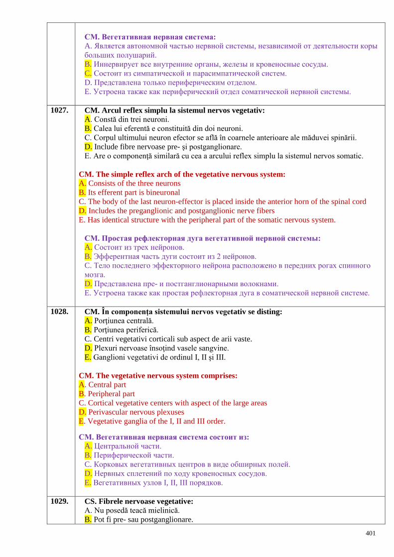
401
СМ. Вегетативная нервная система:
A. Является автономной частью нервной системы, независимой от деятельности коры
больших полушарий.
B. Иннервирует все внутренние органы, железы и кровеносные сосуды.
C. Состоит из симпатической и парасимпатической систем.
D. Представлена только периферическим отделом.
E. Устроена также как периферический отдел соматической нервной системы.
1027. CM. Arcul reflex simplu la sistemul nervos vegetativ:
A. Constă din trei neuroni.
B. Calea lui eferentă e constituită din doi neuroni.
C. Corpul ultimului neuron efector se află în coarnele anterioare ale măduvei spinării.
D. Include fibre nervoase pre- şi postganglionare.
E. Are o componenţă similară cu cea a arcului reflex simplu la sistemul nervos somatic.
CM. The simple reflex arch of the vegetative nervous system:
A. Consists of the three neurons
B. Its efferent part is bineuronal
C. The body of the last neuron-effector is placed inside the anterior horn of the spinal cord
D. Includes the preganglionic and postganglionic nerve fibers
E. Has identical structure with the peripheral part of the somatic nervous system.
СМ. Простая рефлекторная дуга вегетативной нервной системы:
A. Состоит из трех нейронов.
B. Эфферентная часть дуги состоит из 2 нейронов.
C. Тело последнего эффекторного нейрона расположено в передних рогах спинного
мозга.
D. Представлена пре- и постганглионарными волокнами.
E. Устроена также как простая рефлекторная дуга в соматической нервной системе.
1028. CM. În componenţa sistemului nervos vegetativ se disting:
A. Porţiunea centrală.
B. Porţiunea periferică.
C. Centri vegetativi corticali sub aspect de arii vaste.
D. Plexuri nervoase însoţind vasele sangvine.
E. Ganglioni vegetativi de ordinul I, II şi III.
CM. The vegetative nervous system comprises:
A. Central part
B. Peripheral part
C. Cortical vegetative centers with aspect of the large areas
D. Perivascular nervous plexuses
E. Vegetative ganglia of the I, II and III order.
СМ. Вегетативная нервная система состоит из:
A. Центральной части.
B. Периферической части.
C. Корковых вегетативных центров в виде обширных полей.
D. Нервных сплетений по ходу кровеносных сосудов.
E. Вегетативных узлов I, II, III порядков.
1029. CS. Fibrele nervoase vegetative:
A. Nu posedă teacă mielinică.
B. Pot fi pre- sau postganglionare.
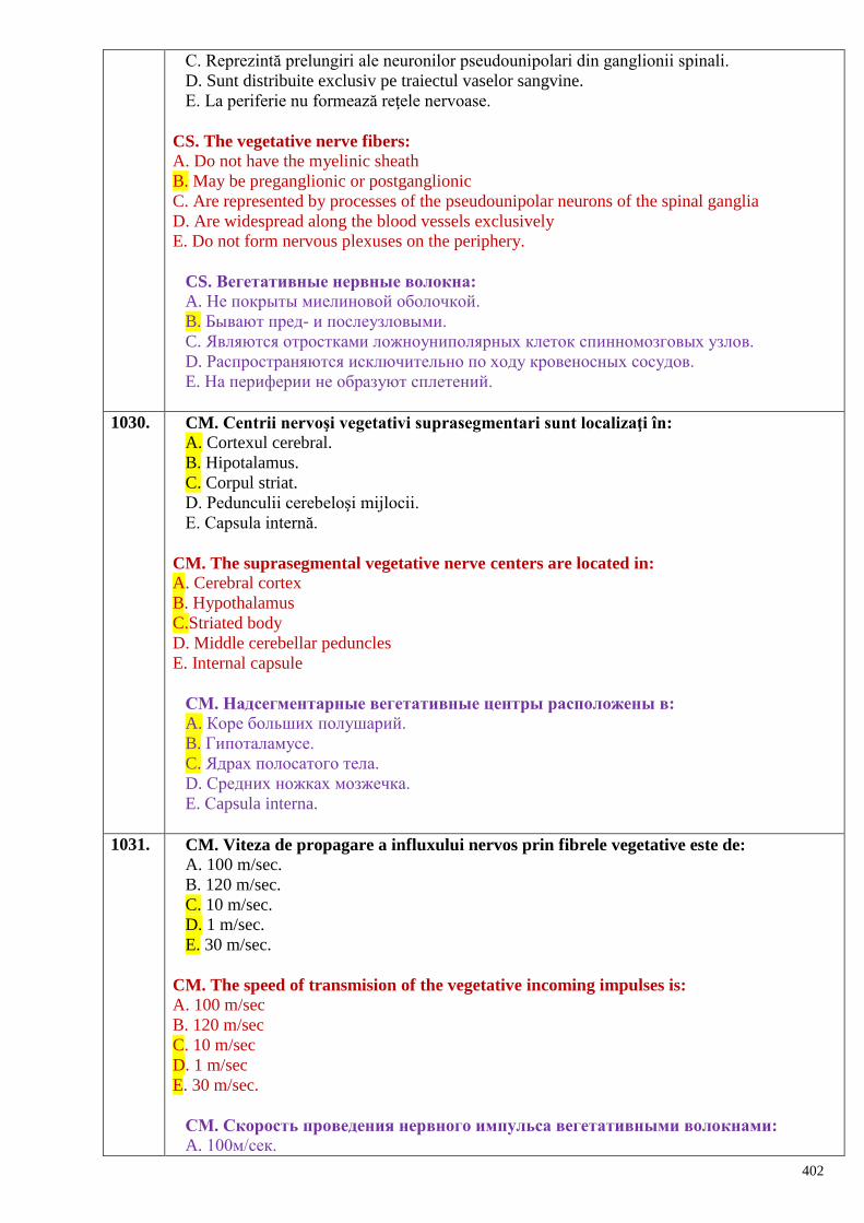
402
C. Reprezintă prelungiri ale neuronilor pseudounipolari din ganglionii spinali.
D. Sunt distribuite exclusiv pe traiectul vaselor sangvine.
E. La periferie nu formează reţele nervoase.
CS. The vegetative nerve fibers:
A. Do not have the myelinic sheath
B. May be preganglionic or postganglionic
C. Are represented by processes of the pseudounipolar neurons of the spinal ganglia
D. Are widespread along the blood vessels exclusively
E. Do not form nervous plexuses on the periphery.
CS. Вегетативные нервные волокна:
A. Не покрыты миелиновой оболочкой.
B. Бывают пред- и послеузловыми.
C. Являются отростками ложноуниполярных клеток спинномозговых узлов.
D. Распространяются исключительно по ходу кровеносных сосудов.
E. На периферии не образуют сплетений.
1030. CM. Centrii nervoşi vegetativi suprasegmentari sunt localizaţi în:
A. Cortexul cerebral.
B. Hipotalamus.
C. Corpul striat.
D. Pedunculii cerebeloşi mijlocii.
E. Capsula internă.
CM. The suprasegmental vegetative nerve centers are located in:
A. Cerebral cortex
B. Hypothalamus
C.Striated body
D. Middle cerebellar peduncles
E. Internal capsule
СМ. Надсегментарные вегетативные центры расположены в:
A. Коре больших полушарий.
B. Гипоталамусе.
C. Ядрах полосатого тела.
D. Средних ножках мозжечка.
E. Сapsula interna.
1031. CM. Viteza de propagare a influxului nervos prin fibrele vegetative este de:
A. 100 m/sec.
B. 120 m/sec.
C. 10 m/sec.
D. 1 m/sec.
E. 30 m/sec.
CM. The speed of transmision of the vegetative incoming impulses is:
A. 100 m/sec
B. 120 m/sec
C. 10 m/sec
D. 1 m/sec
E. 30 m/sec.
СМ. Скорость проведения нервного импульса вегетативными волокнами:
A. 100м/сек.
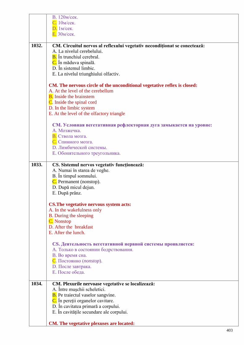
403
B. 120м/сек.
C. 10м/сек.
D. 1м/сек.
E. 30м/сек.
1032. CM. Circuitul nervos al reflexului vegetativ necondiţionat se conectează:
A. La nivelul cerebelului.
B. În trunchiul cerebral.
C. În măduva spinală.
D. În sistemul limbic.
E. La nivelul triunghiului olfactiv.
CM. The nervous circle of the unconditional vegetative reflex is closed:
A. At the level of the cerebellum
B. Inside the brainstem
C. Inside the spinal cord
D. In the limbic system
E. At the level of the olfactory triangle
СМ. Условная вегетативная рефлекторная дуга замыкается на уровне:
A. Мозжечка.
B. Ствола мозга.
C. Спинного мозга.
D. Лимбической системы.
E. Обонятельного треугольника.
1033. CS. Sistemul nervos vegetativ funcţionează:
A. Numai în starea de veghe.
B. În timpul somnului.
C. Permanent (nonstop).
D. După micul dejun.
E. După prânz.
CS.The vegetative nervous system acts:
A. In the wakefulness only
B. During the sleeping
C. Nonstop
D. After the breakfast
E. After the lunch.
CS. Деятельность вегетативной нервной системы проявляется:
A. Только в состоянии бодрствования.
B. Во время сна.
C. Постоянно (nonstop).
D. После завтрака.
E. После обеда.
1034. CM. Plexurile nervoase vegetative se localizează:
A. Între muşchii scheletici.
B. Pe traiectul vaselor sangvine.
C. În pereţii organelor cavitare.
D. În cavitatea primară a corpului.
E. În cavităţile secundare ale corpului.
CM. The vegetative plexuses are located:
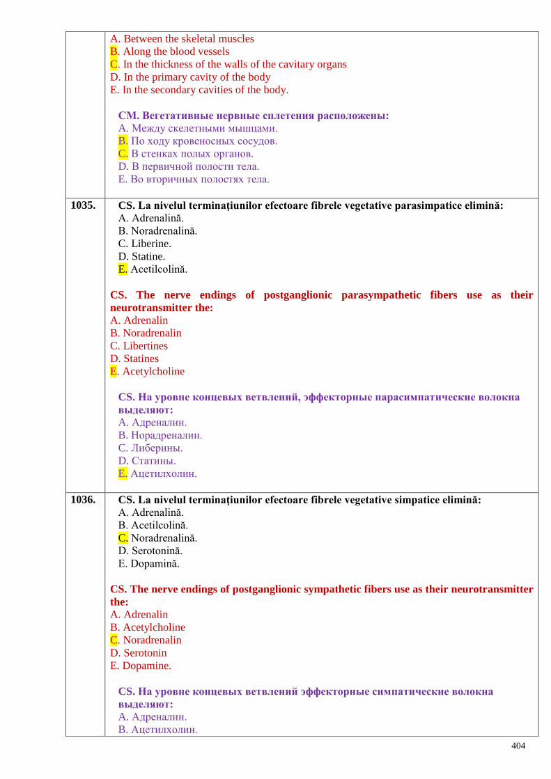
404
A. Between the skeletal muscles
B. Along the blood vessels
C. In the thickness of the walls of the cavitary organs
D. In the primary cavity of the body
E. In the secondary cavities of the body.
СМ. Вегетативные нервные сплетения расположены:
A. Между скелетными мышцами.
B. По ходу кровеносных сосудов.
C. В стенках полых органов.
D. В первичной полости тела.
E. Во вторичных полостях тела.
1035. CS. La nivelul terminaţiunilor efectoare fibrele vegetative parasimpatice elimină:
A. Adrenalină.
B. Noradrenalină.
C. Liberine.
D. Statine.
E. Acetilcolină.
CS. The nerve endings of postganglionic parasympathetic fibers use as their
neurotransmitter the:
A. Adrenalin
B. Noradrenalin
C. Libertines
D. Statines
E. Acetylcholine
CS. На уровне концевых ветвлений, эффекторные парасимпатические волокна
выделяют:
A. Адреналин.
B. Норадреналин.
C. Либерины.
D. Статины.
E. Ацетилхолин.
1036. CS. La nivelul terminaţiunilor efectoare fibrele vegetative simpatice elimină:
A. Adrenalină.
B. Acetilcolină.
C. Noradrenalină.
D. Serotonină.
E. Dopamină.
CS. The nerve endings of postganglionic sympathetic fibers use as their neurotransmitter
the:
A. Adrenalin
B. Acetylcholine
C. Noradrenalin
D. Serotonin
E. Dopamine.
CS. На уровне концевых ветвлений эффекторные симпатические волокна
выделяют:
A. Адреналин.
B. Ацетилхолин.
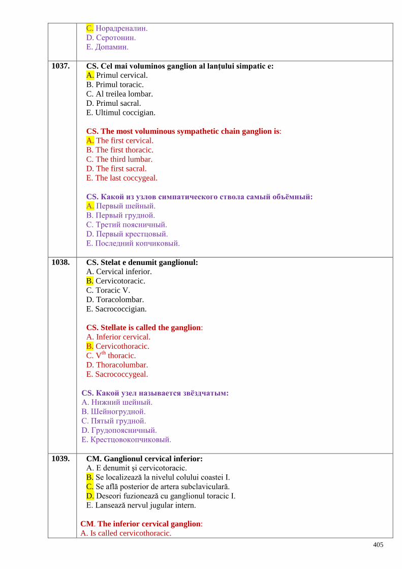
405
C. Норадреналин.
D. Серотонин.
E. Допамин.
1037. CS. Cel mai voluminos ganglion al lanţului simpatic e:
A. Primul cervical.
B. Primul toracic.
C. Al treilea lombar.
D. Primul sacral.
E. Ultimul coccigian.
CS. The most voluminous sympathetic chain ganglion is:
A. The first cervical.
B. The first thoracic.
C. The third lumbar.
D. The first sacral.
E. The last coccygeal.
CS. Какой из узлов симпатического ствола самый объѐмный:
А. Первый шейный.
В. Первый грудной.
С. Третий поясничный.
D. Первый крестцовый.
E. Последний копчиковый.
1038. CS. Stelat e denumit ganglionul:
A. Cervical inferior.
B. Cervicotoracic.
C. Toracic V.
D. Toracolombar.
E. Sacrococcigian.
CS. Stellate is called the ganglion:
A. Inferior cervical.
B. Cervicothoracic.
C. Vth
thoracic.
D. Thoracolumbar.
E. Sacrococcygeal.
CS. Какой узел называется звѐздчатым:
А. Нижний шейный.
В. Шейногрудной.
С. Пятый грудной.
D. Грудопоясничный.
E. Крестцовокопчиковый.
1039. CM. Ganglionul cervical inferior:
A. E denumit şi cervicotoracic.
B. Se localizează la nivelul colului coastei I.
C. Se află posterior de artera subclaviculară.
D. Deseori fuzionează cu ganglionul toracic I.
E. Lansează nervul jugular intern.
CM. The inferior cervical ganglion:
A. Is called cervicothoracic.
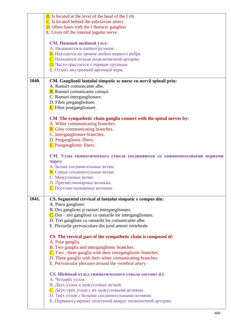
406
B. Is located at the level of the head of the I rib.
C. Is located behind the subclavian artery.
D. Often fuses with the I thoracic ganglion
E. Gives off the internal jugular nerve.
CМ. Нижний шейный узел:
А. Называется и шейногрудным.
В. Находится на уровне шейки первого ребра.
С. Находится позади подключичной артерии.
D. Часто срастается с первым грудным.
E. Отдаѐт внутренний яремный нерв.
1040. CM. Ganglionii lanţului simpatic se unesc cu nervii spinali prin:
A. Ramuri comunicante albe.
B. Ramuri comunicante cenuşii.
C. Ramuri interganglionare.
D. Fibre preganglionare.
E. Fibre postganglionare.
CM. The sympathetic chain ganglia connect with the spinal nerves by:
A. White communicating branches.
B. Gray communicating branches.
C. Interganglionare branches.
D. Preganglionic fibers.
E. Postganglionic fibers.
CМ. Узлы симпатического ствола соединяются со спинномозговыми нервами
через:
А. Белые соединительные ветви.
В. Серые соединительные ветви.
С. Межузловые ветви.
D. Преганглионарные волокна.
E. Постганглионарные волокна.
1041. CS. Segmentul cervical al lanţului simpatic e compus din:
A. Patru ganglioni.
B. Doi ganglioni şi ramuri interganglionare.
C. Doi – trei ganglioni cu ramurile lor interganglionare.
D. Trei ganglioni cu ramurile lor comunicante albe.
E. Plexurile perivasculare din jurul arterei vertebrale.
CS. The cervical part of the sympathetic chain is composed of:
A. Four ganglia.
B. Two ganglia and interganglionic branches.
C. Two - three ganglia with their interganglionic branches.
D. Three ganglia with their white cmmunicating branches.
E. Perivascular plexuses around the vertebral artery.
CS. Шейный отдел симпатического ствола состоит из:
А. Четырѐх узлов.
В. Двух узлов и межузловых ветвей.
С. Двух-трѐх узлов с их межузловыми ветвями.
D. Трѐх узлов с белыми соединительными ветвями.
E. Периваскулярных сплетений вокруг позвоночной артерии.
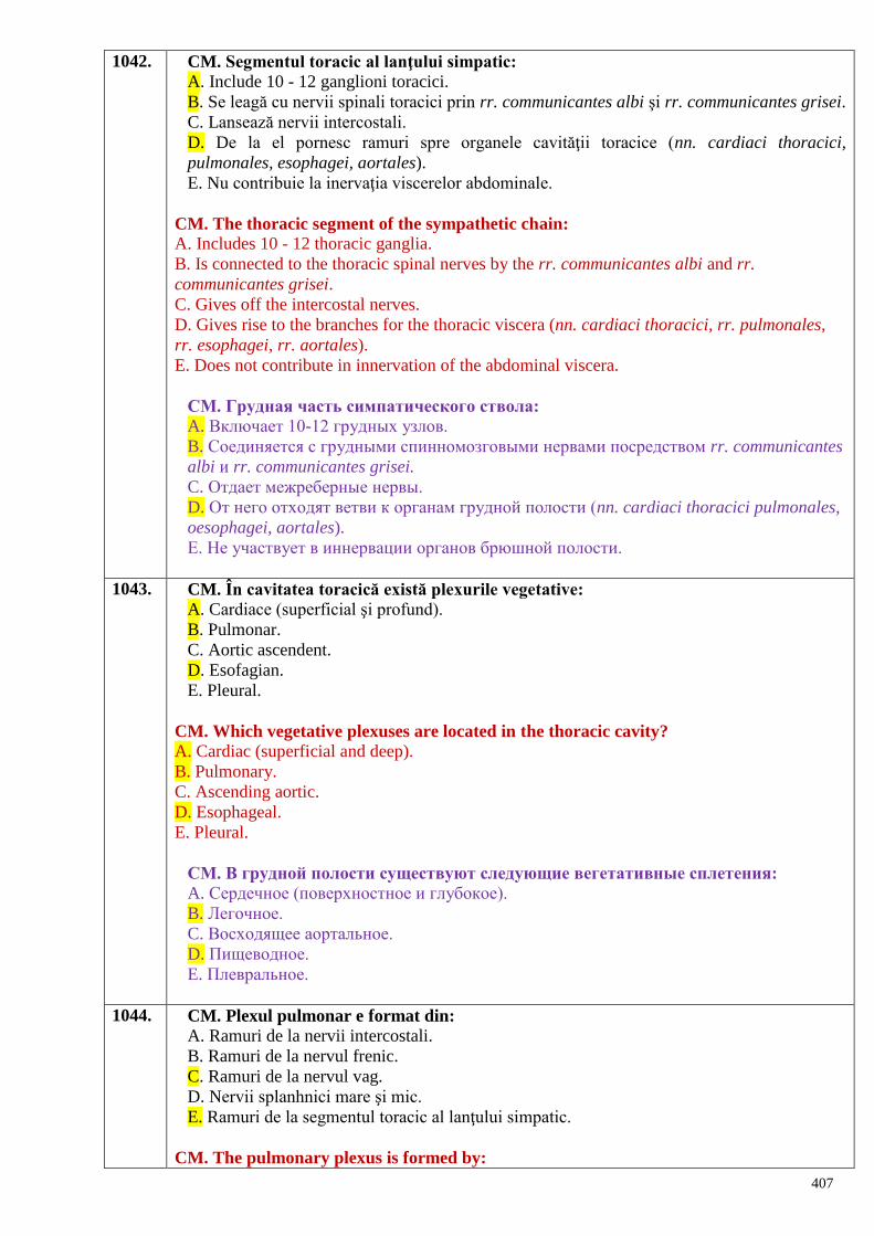
407
1042. CM. Segmentul toracic al lanţului simpatic:
A. Include 10 - 12 ganglioni toracici.
B. Se leagă cu nervii spinali toracici prin rr. communicantes albi şi rr. communicantes grisei.
C. Lansează nervii intercostali.
D. De la el pornesc ramuri spre organele cavităţii toracice (nn. cardiaci thoracici,
pulmonales, esophagei, aortales).
E. Nu contribuie la inervaţia viscerelor abdominale.
CM. The thoracic segment of the sympathetic chain:
A. Includes 10 - 12 thoracic ganglia.
B. Is connected to the thoracic spinal nerves by the rr. communicantes albi and rr.
communicantes grisei.
C. Gives off the intercostal nerves.
D. Gives rise to the branches for the thoracic viscera (nn. cardiaci thoracici, rr. pulmonales,
rr. esophagei, rr. aortales).
E. Does not contribute in innervation of the abdominal viscera.
СМ. Грудная часть симпатического ствола:
A. Включает 10-12 грудных узлов.
B. Соединяется с грудными спинномозговыми нервами посредством rr. communicantes
albi и rr. communicantes grisei.
C. Отдает межреберные нервы.
D. От него отходят ветви к органам грудной полости (nn. cardiaci thoracici pulmonales,
oesophagei, aortales).
E. Не участвует в иннервации органов брюшной полости.
1043. CM. În cavitatea toracică există plexurile vegetative:
A. Cardiace (superficial şi profund).
B. Pulmonar.
C. Aortic ascendent.
D. Esofagian.
E. Pleural.
CM. Which vegetative plexuses are located in the thoracic cavity?
A. Cardiac (superficial and deep).
B. Pulmonary.
C. Ascending aortic.
D. Esophageal.
E. Pleural.
СМ. В грудной полости существуют следующие вегетативные сплетения:
A. Сердечное (поверхностное и глубокое).
B. Легочное.
C. Восходящее аортальное.
D. Пищеводное.
E. Плевральное.
1044. CM. Plexul pulmonar e format din:
A. Ramuri de la nervii intercostali.
B. Ramuri de la nervul frenic.
C. Ramuri de la nervul vag.
D. Nervii splanhnici mare şi mic.
E. Ramuri de la segmentul toracic al lanţului simpatic.
CM. The pulmonary plexus is formed by:

408
A. The branches of the intercostal nerves.
B. The branches of the phrenic nerve.
C. The branches of the vagus nerve.
D. The greater and lesser splanhnic nerves.
E. The branches of the thoracic segment of sympathetic chain.
CM. Легочное сплетение образовано:
A. Ветвями межреберных нервов.
B. Ветвями диафрагмального нерва.
C. Ветвями блуждающего нерва.
D. Большими и малыми внутренностными нервами.
E. Ветвями от грудной части симпатического ствола.
1045. CS. Nervii splanhnici mare şi mic:
A. Pornesc de la nervul vag.
B. Pornesc de la toţi ganglionii lanţului simpatic.
C. Conţin predominant fibre postganglionare.
D. Participă la inervaţia organelor cavităţi toracice.
E. În componenţa lor predomină fibrele preganglionare.
CS. The greater and lesser splanchnic nerves:
A. They are branches of the vagus nerve
B. They start from all ganglia of the sympathetic chain
C. They contain predominantly the postganglionic fibers
D. They participate in the innervation of the thoracic cavity organs.
E. They contain predominantly the preganglionic fibers.
СS. Большой и малый внутренностные нервы:
A. Отходят от блуждающего нерва.
B. Отходят от всех узлов симпатического ствола.
C. Содержат в основном постганглионарные волокна.
D. Участвуют в иннервации органов грудной полости.
E. Содержат в основном преганглионарные волокна.
1046. CM. La formarea plexului esofagian participă ramuri de la nervii:
A. Vag.
B. Segmentul toracic al lanţului simpatic.
C. Intercostali.
D. Frenic.
E. Splanhnici mare şi mic.
CM. The esophageal is formed by the folowing nerves:
A. Vagus nerve.
B. Thoracic segment of the sympathetic chain.
C. Intercostal nerves.
D. Phrenic nerve.
E. Greater and lesser splanchnic nerves.
СМ. Пищеводное сплетение образуется ветвями следующих нервов:
A. Блуждающего.
B. Грудного отдела симпатического ствола.
C. Межрѐберных.
D. Диафрагмального.
E. Большого и малого внутренностных нервов.
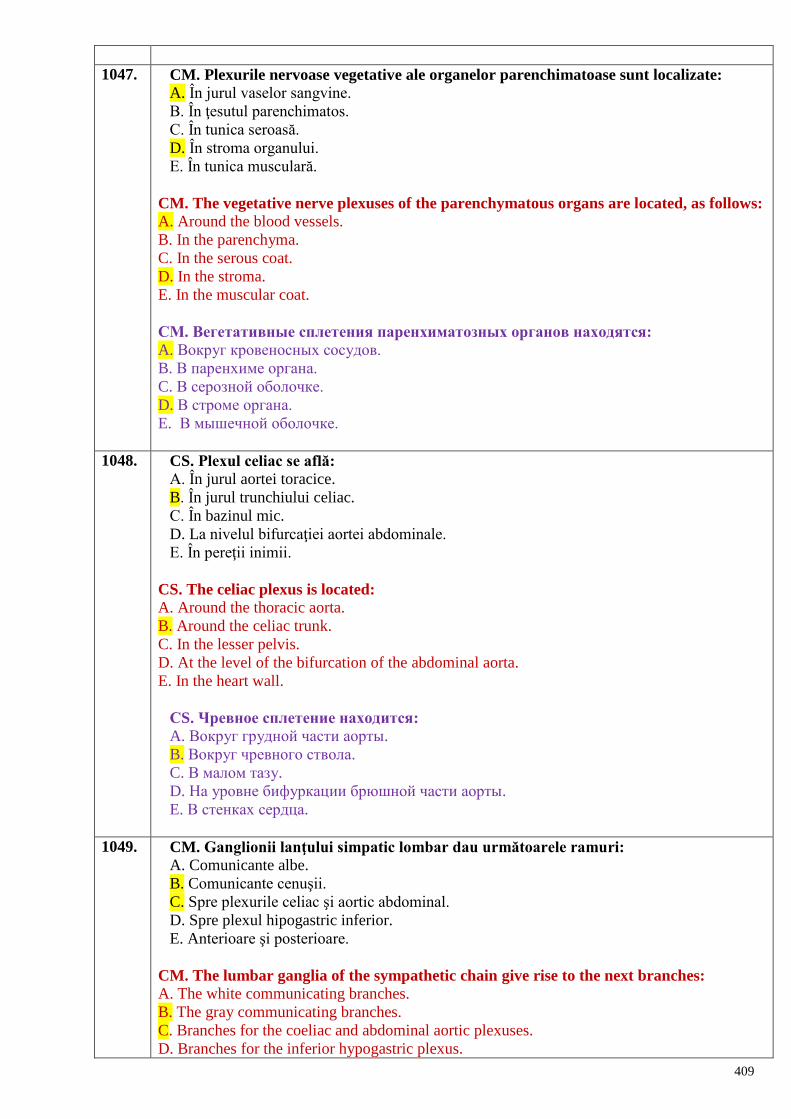
409
1047. CM. Plexurile nervoase vegetative ale organelor parenchimatoase sunt localizate:
A. În jurul vaselor sangvine.
B. În ţesutul parenchimatos.
C. În tunica seroasă.
D. În stroma organului.
E. În tunica musculară.
CM. The vegetative nerve plexuses of the parenchymatous organs are located, as follows:
A. Around the blood vessels.
B. In the parenchyma.
C. In the serous coat.
D. In the stroma.
E. In the muscular coat.
СМ. Вегетативные сплетения паренхиматозных органов находятся:
A. Вокруг кровеносных сосудов.
B. В паренхиме органа.
C. В серозной оболочке.
D. В строме органа.
E. В мышечной оболочке.
1048. CS. Plexul celiac se află:
A. În jurul aortei toracice.
B. În jurul trunchiului celiac.
C. În bazinul mic.
D. La nivelul bifurcaţiei aortei abdominale.
E. În pereţii inimii.
CS. The celiac plexus is located:
A. Around the thoracic aorta.
B. Around the celiac trunk.
C. In the lesser pelvis.
D. At the level of the bifurcation of the abdominal aorta.
E. In the heart wall.
СS. Чревное сплетение находится: A. Вокруг грудной части аорты.
B. Вокруг чревного ствола.
C. В малом тазу.
D. На уровне бифуркации брюшной части аорты.
E. В стенках сердца.
1049. CM. Ganglionii lanţului simpatic lombar dau următoarele ramuri:
A. Comunicante albe.
B. Comunicante cenuşii.
C. Spre plexurile celiac şi aortic abdominal.
D. Spre plexul hipogastric inferior.
E. Anterioare şi posterioare.
CM. The lumbar ganglia of the sympathetic chain give rise to the next branches:
A. The white communicating branches.
B. The gray communicating branches.
C. Branches for the coeliac and abdominal aortic plexuses.
D. Branches for the inferior hypogastric plexus.
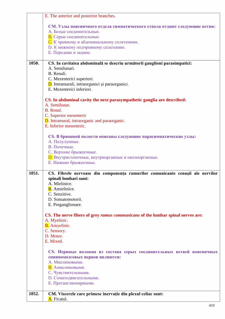
410
E. The anterior and posterior branches.
СМ. Узлы поясничного отдела симпатического ствола отдают следующие ветви: A. Белые соединительные.
B. Серые соединительные.
C. К чревному и абдоминальному сплетениям.
D. К нижнему подчревному сплетению.
E. Передние и задние.
1050. CS. In cavitatea abdominală se descriu următorii ganglioni parasimpatici:
A. Semilunari.
B. Renali.
C. Mezenterici superiori.
D. Intramurali, intraorganici şi paraorganici.
E. Mezenterici inferiori.
CS. In abdominal cavity the next parasympathetic ganglia are described:
A. Semilunar.
B. Renal.
C. Superior mesenteric
D. Intramural, intraorganic and paraorganic.
E. Inferior mesenteric.
СS. В брюшной полости описаны следующие парасимпатические узлы: A. Полулунные.
B. Почечные.
C. Верхние брыжеечные.
D. Внутристеночные, внутриорганные и околоорганные.
E. Нижние брыжеечные.
1051. CS. Fibrele nervoase din componenţa ramurilor comunicante cenuşii ale nervilor
spinali lombari sunt:
A. Mielinice.
B. Amielinice.
C. Senzitive.
D. Somatomotorii.
E. Preganglionare.
CS. The nerve fibers of grey ramus communicans of the lumbar spinal nerves are:
A. Myelinic.
B. Amyelinic.
C. Sensory.
D. Motor.
E. Mixed.
СS. Нервные волокна из состава серых соединительных ветвей поясничных
спинномозговых нервов являются: A. Миелиновыми.
B. Амиелиновыми.
C. Чувствительными.
D. Соматодвигательными.
E. Преганглионарными.
1052. CM. Viscerele care primesc inervaţie din plexul celiac sunt:
A. Ficatul.
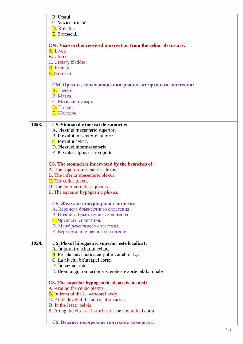
411
B. Uterul.
C. Vezica urinară.
D. Rinichii.
E. Stomacul.
CM. Viscera that received innervation from the celiac plexus are:
A. Liver.
B. Uterus.
C. Urinary bladder.
D. Kidney.
E. Stomach
СМ. Органы, получающие иннервацию от чревного сплетения: A. Печень.
B. Матка.
C. Мочевой пузырь.
D. Почки.
E. Желудок.
1053. CS. Stomacul e inervat de ramurile:
A. Plexului mezenteric superior.
B. Plexului mezenteric inferior.
C. Plexului celiac.
D. Plexului intermezenteric.
E. Plexului hipogastric superior.
CS. The stomach is innervated by the branches of:
A. The superior mesenteric plexus.
B. The inferior mesenteric plexus.
C. The celiac plexus.
D. The intermesenteric plexus.
E. The superior hypogastric plexus.
СS. Желудок иннервирован ветвями: A. Верхнего брыжеечного сплетения.
B. Нижнего брыжеечного сплетения.
C. Чревного сплетения.
D. Межбрыжеечного сплетения.
E. Верхнего подчревного сплетения.
1054. CS. Plexul hipogastric superior este localizat:
A. În jurul trunchiului celiac.
B. Pe faţa anterioară a corpului vertebrei L5.
C. La nivelul bifurcaţiei aortei.
D. În bazinul mic.
E. De-a lungul ramurilor viscerale ale aortei abdominale.
CS. The superior hypogastric plexus is located:
A. Around the celiac plexus.
B. In front of the L5 vertebral body.
C. At the level of the aortic bifurcation.
D. In the lesser pelvis.
E. Along the visceral branches of the abdominal aorta.
СS. Верхнее подчревное сплетение находится:

412
A. Вокруг чревного ствола.
B. На передней поверхности тела позвонка L5.
C. На уровне бифуркации аорты.
D. В малом тазу.
E. Вдоль висцеральных ветвей брюшной аорты.
1055. CM. Plexul hipogastric inferior se găseşte:
A. Pe bifurcaţia aortei.
B. În bazinul mic.
C. În jurul aortei abdominale.
D. Pe m. levator ani.
E. De-a lungul ramurilor viscerale ale aortei abdominale.
CM. The inferior hypogastric plexus is located:
A. At the level of the aortic bifurcation.
B. In the lesser pelvis.
C. Around the abdominal aorta.
D. Upon m. levator ani.
E. Along the visceral branches of the abdominal aorta.
СМ. Нижнее подчревное сплетение находится: A. На бифуркации аорты.
B. В малом тазу.
C. Вокруг брюшной аорты.
D. На m. levator ani.
E. Вдоль висцеральных ветвей брюшной аорты.
1056. CM. Ganglionii lanţului simpatic sacrat dau următoarele ramuri:
A. Comunicante albe.
B. Comunicante cenuşii.
C. Spre plexul hipogastric superior.
D. Spre plexul hipogastric inferior.
E. Spre plexul celiac.
CM. The sacral ganglia of sympathetic chain give rise to the next branches:
A. The white communicating branches.
B. The gray communicating branches.
C. Towards the superior hypogastric plexus.
D. Towards the inferior hypogastric.
E. Towards the superior celiac plexus.
СМ. Узлы крестцового отдела симпатического ствола отдают следующие ветви: A. Белые соединительные.
B. Серые соединительные.
C. К верхнему подчревному сплетению.
D. К нижнему подчревному сплетению.
E. К чревному сплетению.
1057. CM. Există următoarele plexuri nervoase ale organelor cavitare din cavitatea
abdominală:
A. Submuscular.
B. Subseros.
C. Mienteric (Auerbach).
D. Interseros.
E. Submucos (Meissner).
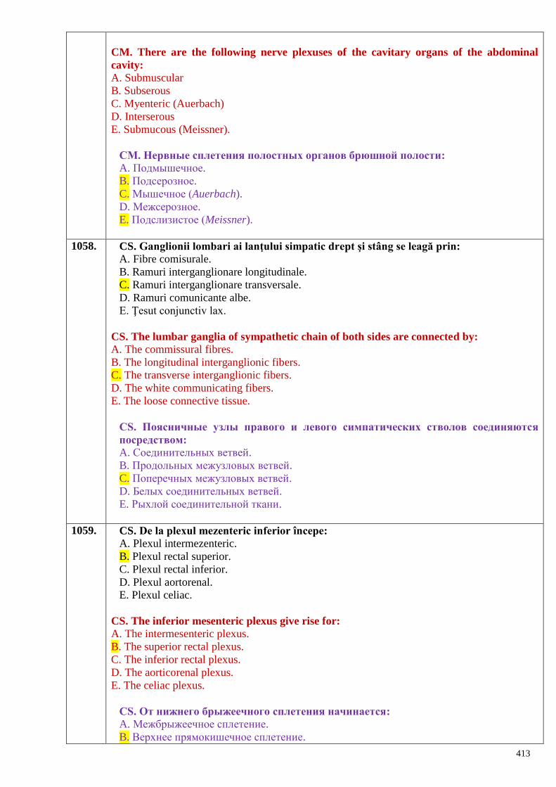
413
CM. There are the following nerve plexuses of the cavitary organs of the abdominal
cavity:
A. Submuscular
B. Subserous
C. Myenteric (Auerbach)
D. Interserous
E. Submucous (Meissner).
СМ. Нервные сплетения полостных органов брюшной полости: A. Подмышечное.
B. Подсерозное.
C. Мышечное (Auerbach).
D. Межсерозное.
E. Подслизистое (Meissner).
1058. CS. Ganglionii lombari ai lanţului simpatic drept şi stâng se leagă prin:
A. Fibre comisurale.
B. Ramuri interganglionare longitudinale.
C. Ramuri interganglionare transversale.
D. Ramuri comunicante albe.
E. Ţesut conjunctiv lax.
CS. The lumbar ganglia of sympathetic chain of both sides are connected by:
A. The commissural fibres.
B. The longitudinal interganglionic fibers.
C. The transverse interganglionic fibers.
D. The white communicating fibers.
E. The loose connective tissue.
СS. Поясничные узлы правого и левого симпатических стволов соединяются
посредством: A. Соединительных ветвей.
B. Продольных межузловых ветвей.
C. Поперечных межузловых ветвей.
D. Белых соединительных ветвей.
E. Рыхлой соединительной ткани.
1059. CS. De la plexul mezenteric inferior începe:
A. Plexul intermezenteric.
B. Plexul rectal superior.
C. Plexul rectal inferior.
D. Plexul aortorenal.
E. Plexul celiac.
CS. The inferior mesenteric plexus give rise for:
A. The intermesenteric plexus.
B. The superior rectal plexus.
C. The inferior rectal plexus.
D. The aorticorenal plexus.
E. The celiac plexus.
СS. От нижнего брыжеечного сплетения начинается: A. Межбрыжеечное сплетение.
B. Верхнее прямокишечное сплетение.
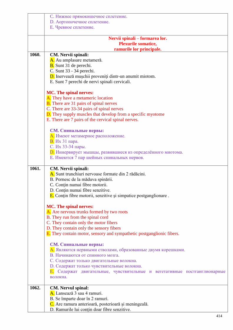
414
C. Нижнее прямокишечное сплетение.
D. Аортопочечное сплетение.
E. Чревное сплетение.
Nervii spinali – formarea lor.
Plexurile somatice,
ramurile lor principale.
1060. CM. Nervii spinali:
A. Au amplasare metameră.
B. Sunt 31 de perechi.
C. Sunt 33 - 34 perechi.
D. Inervează muşchii proveniţi dintr-un anumit miotom.
E. Sunt 7 perechi de nervi spinali cervicali.
MC. The spinal nerves:
A. They have a metameric location
B. There are 31 pairs of spinal nerves
C. There are 33-34 pairs of spinal nerves
D. They supply muscles that develop from a specific myotome
E. There are 7 pairs of the cervical spinal nerves.
CM. Спинальные нервы:
А. Имеют метамерное расположение.
В. Их 31 пара.
С. Их 33-34 пары.
D. Иннервирует мышцы, развившиеся из определѐнного миотома.
E. Имеются 7 пар шейных спинальных нервов.
1061. CM. Nervii spinali:
A. Sunt trunchiuri nervoase formate din 2 rădăcini.
B. Pornesc de la măduva spinării.
C. Conţin numai fibre motorii.
D. Conţin numai fibre senzitive.
E. Conţin fibre motorii, senzitive şi simpatice postganglionare .
MC. The spinal nerves:
A. Are nervous trunks formed by two roots
B. They run from the spinal cord
C. They contain only the motor fibers
D. They contain only the sensory fibers
E. They contain motor, sensory and sympathetic postganglionic fibers.
CM. Спинальные нервы:
А. Являются нервными стволами, образованные двумя корешками.
В. Начинаются от спинного мозга.
С. Содержат только двигательные волокна.
D. Содержат только чувствительные волокна.
E. Содержат двигательные, чувствительные и вегетативные постганглионарные
волокна.
1062. CM. Nervul spinal:
A. Lansează 3 sau 4 ramuri.
B. Se împarte doar în 2 ramuri.
C. Are ramura anterioară, posterioară şi meningeală.
D. Ramurile lui conţin doar fibre senzitive.
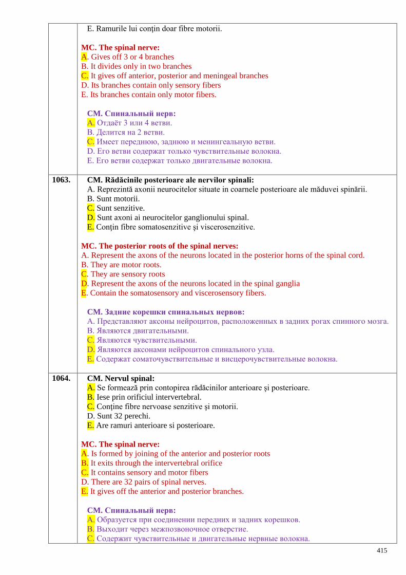
415
E. Ramurile lui conţin doar fibre motorii.
MC. The spinal nerve:
A. Gives off 3 or 4 branches
B. It divides only in two branches
C. It gives off anterior, posterior and meningeal branches
D. Its branches contain only sensory fibers
E. Its branches contain only motor fibers.
CM. Спинальный нерв:
А. Отдаѐт 3 или 4 ветви.
В. Делится на 2 ветви.
С. Имеет переднюю, заднюю и менингеальную ветви.
D. Его ветви содержат только чувствительные волокна.
E. Его ветви содержат только двигательные волокна.
1063. CM. Rădăcinile posterioare ale nervilor spinali:
A. Reprezintă axonii neurocitelor situate in coarnele posterioare ale măduvei spinării.
B. Sunt motorii.
C. Sunt senzitive.
D. Sunt axoni ai neurocitelor ganglionului spinal.
E. Conţin fibre somatosenzitive şi viscerosenzitive.
MC. The posterior roots of the spinal nerves:
A. Represent the axons of the neurons located in the posterior horns of the spinal cord.
B. They are motor roots.
C. They are sensory roots
D. Represent the axons of the neurons located in the spinal ganglia
E. Contain the somatosensory and viscerosensory fibers.
CM. Задние корешки спинальных нервов:
А. Представляют аксоны нейроцитов, расположенных в задних рогах спинного мозга.
В. Являются двигательными.
С. Являются чувствительными.
D. Являются аксонами нейроцитов спинального узла.
E. Содержат соматочувствительные и висцерочувствительные волокна.
1064. CM. Nervul spinal:
A. Se formează prin contopirea rădăcinilor anterioare şi posterioare.
B. Iese prin orificiul intervertebral.
C. Conţine fibre nervoase senzitive şi motorii.
D. Sunt 32 perechi.
E. Are ramuri anterioare si posterioare.
MC. The spinal nerve:
A. Is formed by joining of the anterior and posterior roots
B. It exits through the intervertebral orifice
C. It contains sensory and motor fibers
D. There are 32 pairs of spinal nerves.
E. It gives off the anterior and posterior branches.
CM. Спинальный нерв:
А. Образуется при соединении передних и задних корешков.
В. Выходит через межпозвоночное отверстие.
С. Содержит чувствительные и двигательные нервные волокна.
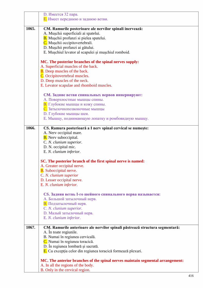
416
D. Имеется 32 пара.
E. Имеет переднюю и заднюю ветви.
1065. CM. Ramurile posterioare ale nervilor spinali inervează:
A. Muşchii superficiali ai spatelui.
B. Muşchii profunzi si pielea spatelui.
C. Muşchii occipitovertebrali.
D. Muşchii profunzi ai gâtului.
E. Muşchiul levator al scapulei şi muşchiul romboid.
MC. The posterior branches of the spinal nerves supply:
A. Superficial muscles of the back.
B. Deep muscles of the back.
C. Occipitovertebral muscles.
D. Deep muscles of the neck.
E. Levator scapulae and rhomboid muscles.
CM. Задние ветви спинальных нервов иннервируют:
А. Поверхностные мышцы спины.
В. Глубокие мышцы и кожу спины.
С. Затылочнопозвоночные мышцы
D. Глубокие мышцы шеи.
E. Мышцу, поднимающую лопатку и ромбовидную мышцу.
1066. CS. Ramura posterioară a I nerv spinal cervical se numeşte:
A. Nerv occipital mare.
B. Nerv suboccipital.
C. N. clunium superior.
D. N. occipital mic.
E. N. clunium inferior.
SC. The posterior branch of the first spinal nerve is named:
A. Greater occipital nerve.
B. Suboccipital nerve.
C. N. clunium superior
D. Lesser occipital nerve.
E. N. clunium inferior.
CS. Задняя ветвь I-го шейного спинального нерва называется:
А. Большой затылочный нерв.
В. Подзатылочный нерв.
С. N. clunium superior.
D. Малый затылочный нерв.
E. N. clunium inferior.
1067. CM. Ramurile anterioare ale nervilor spinali păstrează structura segmentară:
A. În toate regiunile.
B. Numai în regiunea cervicală.
C. Numai în regiunea toracică.
D. În regiunea lombară şi sacrată.
E. Cu excepţia celor din regiunea toracică formează plexuri.
MC. The anterior branches of the spinal nerves maintain segmental arrangement:
A. In all the regions of the body.
B. Only in the cervical region.
417
C. Only in the thoracic region.
D. Only in the lumbar and sacral regions.
E. Excepting those of the thoracic region they form plexuses.
CM. Передние ветви спинальных нервов сохраняют сегментарное строение:
А. Во всех областях.
В. Только в шейной области.
С. Только в грудной области.
D. В поясничной и крестцовой областях.
E. За исключением грудной области образуют сплетения.
1068. CS. Plexul cervical are ramuri:
A. Numai cutanate.
B. Numai motorii.
C. Numai mixte.
D. Cutanate, motorii.
E. Cutanate, motorii şi mixte.
SC. The cervical plexus gives off the following branches:
A. Only cutaneous branches.
B. Only motor branches.
C. Only mixed branches.
D. Cutaneous and motor branches.
E. Cutaneous, motor and mixed branches.
CS. Шейное сплетение отдаѐт ветви:
А. Только кожные.
В. Только двигательные.
С. Только смешанные.
D. Кожные, двигательные.
E. Кожные, двигательные и смешанные.
1069. CM. Nervii motori ai plexului cervical inervează:
A. Pielea de pe faţa anterioară a gâtului.
B. Muşchii platisma şi milohioidian.
C. Muşchii scaleni anterior, mediu şi posterior.
D. Muşchii lungi al capului şi al gâtului.
E. M. levator al scapulei.
MC. The motor branches of the cervical plexus innervate:
A. The skin of the anterior region of the neck.
B. The platysma and mylohyoid muscles.
C. The anterior, middle and posterior scalene muscles.
D. The longus colli and longus capitis muscles.
E. The levator scapulae muscle.
CM. Двигательные нервы шейного сплетения иннервируют:
А. Кожу передней поверхности шеи.
В. Подкожную и челюстно-подъязычную мышцы.
С. Переднюю, среднюю и заднюю лестничные мышцы.
D. Длинную мышцу головы и шеи.
E. Мышцу, поднимающую лопатку.
1070. CM. Plexul cervical are următoarele ramuri cutanate:
A. N. occipital mare.
418
B. N. occipital mic.
C. N. auricular mare.
D. N. transversal al gâtului.
E. Nn. supraclaviculari.
SC. The cutaneous branches of the cervical plexus are:
A. The greater occipital nerve.
B. The lesser occipital nerve.
C. The great auricular nerve.
D. The transverse cervical nerve.
E. The supraclavicular nerves.
CM. Шейное сплетение имеет следующие кожные ветви:
А. Большой затылочный нерв.
В. Малый затылочный нерв.
С. Большой ушной нерв.
D. Поперечный нерв шеи.
E. Надключичные нервы.
1071. CM. Nervul frenic:
A. Este ramură musculară a plexului cervical.
B. Este ramură mixtă a plexului cervical.
C. Trece prin apertura toracică superioară.
D. Inervează pleura, pericardul şi m. phrenicus.
E. Trece anterior de rădăcina plămânului
MC. The phrenic nerve:
A. Is a motor branch of the cervical plexus.
B. Is a mixed branch of the cervical plexus.
C. Passes through the superior thoracic aperture.
D. Innervates the pleura, pericardium and diaphragm (m. phrenicus).
E. Passes in front of the pulmonary root.
CM. Диафрагмальный нерв:
А. Является двигательной ветвью шейного сплетения.
В. Является смешанной ветвью шейного сплетения.
С. Проходит через верхнюю апертуру грудной клетки.
D. Иннервирует плевру, перикард и m. phrenicus.
E. Проходит спереди корня лѐгкого.
1072. CS. Plexul cervical se formează din:
A. Ramurile anterioare ale NS C1 - C4
B. Ramurile posterioare ale NS C1 - C4
C. Ramurile anterioare ale NS C1 - C8
D. Ramurile posterioare ale NS C1 - C8
E. Ramurile meningiene ale NS C1 - C4
SC. The cervical plexus is formed by:
A. Anterior branches of the C1 - C4 spinal nerves.
B. Posterior branches of the C1 - C4 spinal nerves.
C. Anterior branches of the C1 - C8 spinal nerves.
D. Posterior branches of the C1 - C8 spinal nerves.
E. Meningeal branches of the C1 - C4 spinal nerves.
CS. Шейное сплетение образуется из:
419
А. Передних ветвей СМН C1 – C4.
В. Задних ветвей СМН C1 – C4.
С. Передних ветвей СМН C1 – C8.
D. Задних ветвей СМН C1 – C8.
Е. Менингеальных ветвей СМН C1 – C4.
1073. CS. Care din ramurile nervilor spinali participă la formarea plexurilor?
A. Comunicante albe.
B. Comunicante cenuşii.
C. Meningiene.
D. Anterioare.
E. Posterioare.
SC. Which branches of the spinal nerves participate in formation of the plexuses?
A. The white communicating branches.
B. The grey communicating branches.
C. The meningeal branches.
D. The anterior branches.
E. The posterior branches.
CS. Какие ветви спинальных нервов участвуют в образовании сплетений?
А. Белые соединительные.
В. Серые соединительные.
С. Менингеальные.
D. Передние.
Е. Задние.
1074. CM. Formaţiuni anatomice inervate de nervus phrenicus:
A. Diafragma.
B. Pleura.
C. Pericardul.
D. Peritoneul.
E. Pericarionul.
MC. Name the anatomical structures innervated by the phrenic nerve (nervus
phrenicus):
A. The diaphragm.
B. The pleura.
C. The pericardium.
D. The peritoneum.
E. The pericarion.
CМ. Анатомические образования, иннервируемые nervus phrenicus:
А. Диафрагма.
В. Плевра.
С. Перикард.
D. Брюшина.
E. Перикарион.
1075. CS. Ramurile plexului brahial sunt grupate în:
A. Mediale şi laterale.
B. Anterioare şi posterioare.
C. Superioare şi inferioare.
D. Groase şi subţiri.
E. Scurte şi lungi.
420
CS. The branches of the brachial plexus are grouped in:
A. Medial and lateral
B. Anterior and posteriori
C. Superior and inferior
D. Thick and thin
E. Short and long.
СS. Ветви плечевого сплетения делятся на группы:
A. Медиальные и латеральные.
B. Передние и задние.
C. Верхние и нижние.
D. Толстые и тонкие.
E. Короткие и длинные.
1076. CM. Din ramurile lungi ale plexului brahial fac parte:
A. N. axillaris.
B. N. radialis.
C. N. ulnaris.
D. N. thoracicus longus.
E. N. medianus.
CM. The long branches of the brachial plexus are:
A. N. axillaris
B. N. radialis
C. N. ulnaris
D. N. thoracicus longus
E. N. medianus.
СM. Из длинных ветвей плечевого сплетения участвуют:
A. N. axillaris.
B. N. radialis.
C. N. ulnaris.
D. N. thoracicus longus.
E. N. medianus.
1077. CM. Nervii intercostali:
A. Reprezintă ramuri ale lanţului simpatic.
B. Există în număr de 12 perechi.
C. Reprezintă ramurile anterioare ale nervilor toracici I – XII.
D. Ramura anterioară a nervului toracic XII e denumită nerv subcostal.
E. Se localizează pe faţa laterală a coastelor.
CM. The intercostals nerves:
A. Represent the branches of the sympathetic chain
B. There are 12 pairs in number.
C. Represent the anterior branches of the I – XI thoracic spinal nerves.
D. The anterior branch of the XII thoracic nerve is called the subcostal nerve.
E. They are located on the lateral surface of the ribs.
СМ. Межрѐберные нервы:
A. Представляют ветви симпатического ствола.
B. Существуют 12 пар.
C. Представляют передние ветви грудных нервов I-XII.
D. Передняя ветвь грудного XII нерва, названного подрѐберным нервом.
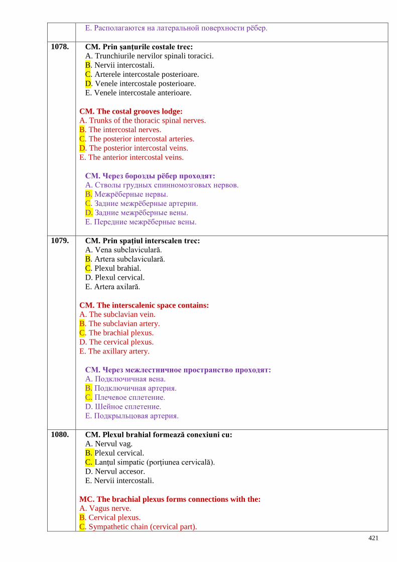
421
E. Располагаются на латеральной поверхности рѐбер.
1078. CM. Prin şanţurile costale trec:
A. Trunchiurile nervilor spinali toracici.
B. Nervii intercostali.
C. Arterele intercostale posterioare.
D. Venele intercostale posterioare.
E. Venele intercostale anterioare.
CM. The costal grooves lodge:
A. Trunks of the thoracic spinal nerves.
B. The intercostal nerves.
C. The posterior intercostal arteries.
D. The posterior intercostal veins.
E. The anterior intercostal veins.
СМ. Через борозды рѐбер проходят:
A. Стволы грудных спинномозговых нервов.
B. Межрѐберные нервы.
C. Задние межрѐберные артерии.
D. Задние межрѐберные вены.
E. Передние межрѐберные вены.
1079. CM. Prin spaţiul interscalen trec:
A. Vena subclaviculară.
B. Artera subclaviculară.
C. Plexul brahial.
D. Plexul cervical.
E. Artera axilară.
CM. The interscalenic space contains:
A. The subclavian vein.
B. The subclavian artery.
C. The brachial plexus.
D. The cervical plexus.
E. The axillary artery.
СМ. Через межлестничное пространство проходят:
A. Подключичная вена.
B. Подключичная артерия.
C. Плечевое сплетение.
D. Шейное сплетение.
E. Подкрыльцовая артерия.
1080. CM. Plexul brahial formează conexiuni cu:
A. Nervul vag.
B. Plexul cervical.
C. Lanţul simpatic (porţiunea cervicală).
D. Nervul accesor.
E. Nervii intercostali.
MC. The brachial plexus forms connections with the:
A. Vagus nerve.
B. Cervical plexus.
C. Sympathetic chain (cervical part).
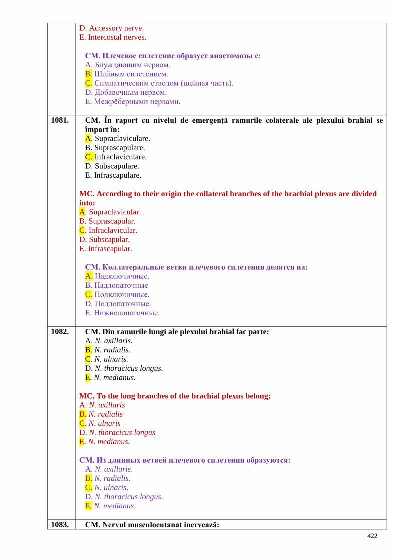
422
D. Accessory nerve.
E. Intercostal nerves.
СМ. Плечевое сплетение образует анастомозы с:
A. Блуждающим нервом.
B. Шейным сплетением.
C. Симпатическим стволом (шейная часть).
D. Добавочным нервом.
E. Межрѐберными нервами.
1081. CM. În raport cu nivelul de emergenţă ramurile colaterale ale plexului brahial se
împart în:
A. Supraclaviculare.
B. Suprascapulare.
C. Infraclaviculare.
D. Subscapulare.
E. Infrascapulare.
MC. According to their origin the collateral branches of the brachial plexus are divided
into:
A. Supraclavicular.
B. Suprascapular.
C. Infraclavicular.
D. Subscapular.
E. Infrascapular.
СМ. Коллатеральные ветви плечевого сплетения делятся на:
A. Надключичные.
B. Надлопаточные
C. Подключичные.
D. Подлопаточные.
E. Нижнелопаточные.
1082. CM. Din ramurile lungi ale plexului brahial fac parte:
A. N. axillaris.
B. N. radialis.
C. N. ulnaris.
D. N. thoracicus longus.
E. N. medianus.
MC. To the long branches of the brachial plexus belong:
A. N. axillaris
B. N. radialis
C. N. ulnaris
D. N. thoracicus longus
E. N. medianus.
СМ. Из длинных ветвей плечевого сплетения образуются:
A. N. axillaris.
B. N. radialis.
C. N. ulnaris.
D. N. thoracicus longus.
E. N. medianus.
1083. CM. Nervul musculocutanat inervează:
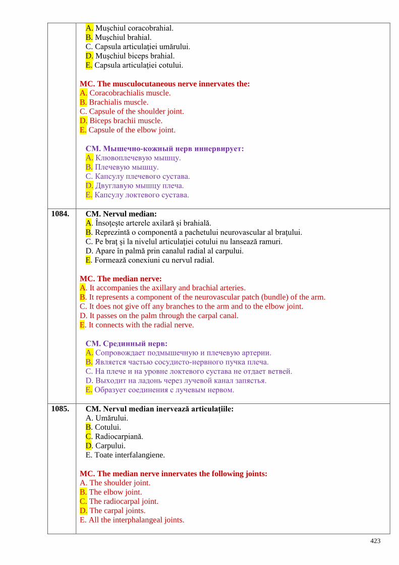
423
A. Muşchiul coracobrahial.
B. Muşchiul brahial.
C. Capsula articulaţiei umărului.
D. Muşchiul biceps brahial.
E. Capsula articulaţiei cotului.
MC. The musculocutaneous nerve innervates the:
A. Coracobrachialis muscle.
B. Brachialis muscle.
C. Capsule of the shoulder joint.
D. Biceps brachii muscle.
E. Capsule of the elbow joint.
СМ. Мышечно-кожный нерв иннервирует:
A. Клювоплечевую мышцу.
B. Плечевую мышцу.
C. Капсулу плечевого сустава.
D. Двуглавую мышцу плеча.
E. Капсулу локтевого сустава.
1084. CM. Nervul median:
A. Însoţeşte arterele axilară şi brahială.
B. Reprezintă o componentă a pachetului neurovascular al braţului.
C. Pe braţ şi la nivelul articulaţiei cotului nu lansează ramuri.
D. Apare în palmă prin canalul radial al carpului.
E. Formează conexiuni cu nervul radial.
MC. The median nerve:
A. It accompanies the axillary and brachial arteries.
B. It represents a component of the neurovascular patch (bundle) of the arm.
C. It does not give off any branches to the arm and to the elbow joint.
D. It passes on the palm through the carpal canal.
E. It connects with the radial nerve.
СМ. Срединный нерв:
A. Сопровождает подмышечную и плечевую артерии.
B. Является частью сосудисто-нервного пучка плеча.
C. На плече и на уровне локтевого сустава не отдает ветвей.
D. Выходит на ладонь через лучевой канал запястья.
E. Образует соединения с лучевым нервом.
1085. CM. Nervul median inervează articulaţiile:
A. Umărului.
B. Cotului.
C. Radiocarpiană.
D. Carpului.
E. Toate interfalangiene.
MC. The median nerve innervates the following joints:
A. The shoulder joint.
B. The elbow joint.
C. The radiocarpal joint.
D. The carpal joints.
E. All the interphalangeal joints.
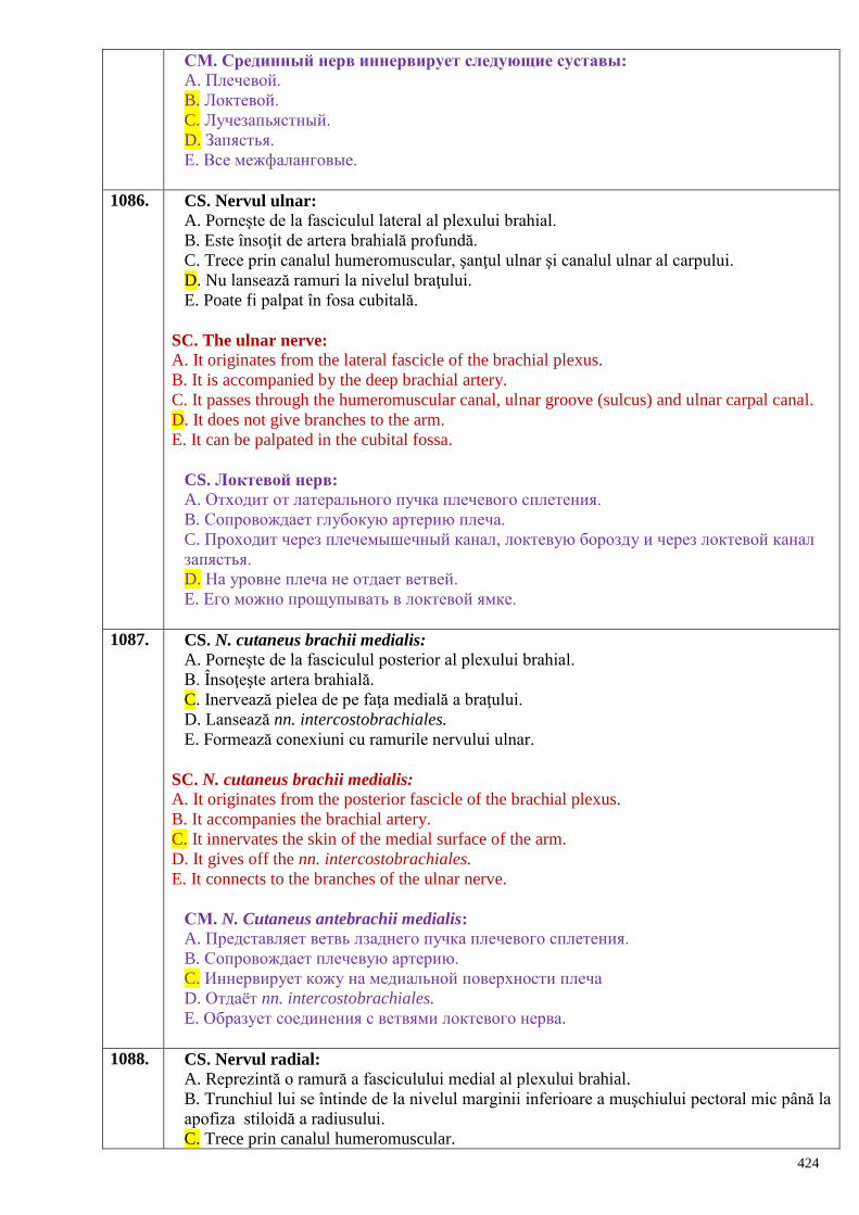
424
СМ. Срединный нерв иннервирует следующие суставы:
A. Плечевой.
B. Локтевой.
C. Лучезапьястный.
D. Запястья.
E. Все межфаланговые.
1086. CS. Nervul ulnar:
A. Porneşte de la fasciculul lateral al plexului brahial.
B. Este însoţit de artera brahială profundă.
C. Trece prin canalul humeromuscular, şanţul ulnar şi canalul ulnar al carpului.
D. Nu lansează ramuri la nivelul braţului.
E. Poate fi palpat în fosa cubitală.
SC. The ulnar nerve:
A. It originates from the lateral fascicle of the brachial plexus.
B. It is accompanied by the deep brachial artery.
C. It passes through the humeromuscular canal, ulnar groove (sulcus) and ulnar carpal canal.
D. It does not give branches to the arm.
E. It can be palpated in the cubital fossa.
CS. Локтевой нерв: A. Отходит от латерального пучка плечевого сплетения.
B. Сопровождает глубокую артерию плеча.
C. Проходит через плечемышечный канал, локтевую борозду и через локтевой канал
запястья.
D. На уровне плеча не отдает ветвей.
E. Его можно прощупывать в локтевой ямке.
1087. CS. N. cutaneus brachii medialis:
A. Porneşte de la fasciculul posterior al plexului brahial.
B. Însoţeşte artera brahială.
C. Inervează pielea de pe faţa medială a braţului.
D. Lansează nn. intercostobrachiales.
E. Formează conexiuni cu ramurile nervului ulnar.
SC. N. cutaneus brachii medialis:
A. It originates from the posterior fascicle of the brachial plexus.
B. It accompanies the brachial artery.
C. It innervates the skin of the medial surface of the arm.
D. It gives off the nn. intercostobrachiales.
E. It connects to the branches of the ulnar nerve.
СМ. N. Cutaneus antebrachii medialis:
A. Представляет ветвь лзаднего пучка плечевого сплетения.
B. Сопровождает плечевую артерию.
C. Иннервирует кожу на медиальной поверхности плеча
D. Отдаѐт nn. intercostobrachiales.
E. Образует соединения с ветвями локтевого нерва.
1088. CS. Nervul radial:
A. Reprezintă o ramură a fasciculului medial al plexului brahial.
B. Trunchiul lui se întinde de la nivelul marginii inferioare a muşchiului pectoral mic până la
apofiza stiloidă a radiusului.
C. Trece prin canalul humeromuscular.
425
D. Este însoţit de artera brahială.
E. În leziunile lui mâna îmbracă aspect de gheară.
SC. The radial nerve:
A. It is a branch of the medial fascicle of the brachial plexus.
B. It trunk extends from the inferior margin of the pectoralis minor muscle towards the styloid
process of the radius.
C. It passes through the humeromuscular canal.
D. It is accompanied by the brachial artery.
E. In its lesions the hand takes a shape of a claw.
CS. Лучевой нерв: A. Представляет собой ветвь медиального пучка плечевого сплетения.
B. Его ствол начинается от уровня нижнего края малого грудного нерва и до
шиловидного отростка лучевой кости.
C. Проходит через плечемышечный канал.
D. Сопровождает плечевую артерию.
E. При его повреждении рука принимает форму когтя.
1089. CM. Muşchii anteriori ai antebraţului sunt inervaţi din:
A. N. medianus.
B. N. radialis.
C. N. ulnaris.
D. N. musculocutaneus.
E. N. cutaneus antebrachii medialis.
MC. The anterior muscles of the forearm are innervated by the:
A. N. medianus
B. N. radialis
C. N. ulnaris
D. N. musculocutaneus
E. N. cutaneus antebrachii medialis.
СМ. Передние мышцы предплечья иннервируются:
A. N. medianus.
B. N. radialis.
C. N. ulnaris.
D. N. musculocutaneus.
E. N. cutaneus antebrachii medialis.
1090. CM. Muşchii posteriori ai antebraţului sunt irigaţi şi inervaţi de:
A. A. interossea posterior.
B. N. medianus.
C. A. radialis.
D. N. ulnaris.
E. N. radialis.
MC. The posterior muscles of the forearm are vascularized and innervated by the:
A. A. interossea posterior
B. N. medianus
C. A. radialis
D. N. ulnaris
E. N. radialis.
СМ. Задние мышцы предплечья кровоснабжаются и иннервируются:
426
A. A. interossea posterior.
B. N. medianus.
C. A. radialis.
D. N. ulnaris.
E. N. radialis.
1091. CM. Pielea antebraţului este inervată de:
A. N. cutaneus antebrachii anterior.
B. N. cutaneus antebrachii medialis.
C. N. cutaneus antebrachii superior.
D. N. cutaneus antebrachii inferior.
E. N. cutaneus antebrachii posterior.
MC. The skin of the forearm is innervated by the:
A. N. cutaneus antebrachii anterior.
B. N. cutaneus antebrachii medialis.
C. N. cutaneus antebrachii superior.
D. N. cutaneus antebrachii inferior.
E. N. cutaneus antebrachii posterior.
CM. Кожу предплечья иннервируют:
A. N. cutaneus antebrachii anterior.
B. N. cutaneus antebrachii medialis.
C. N. cutaneus antebrachii superior.
D. N. cutaneus antebrachii inferior.
E. N. cutaneus antebrachii posterior
1092. CM. Pielea de pe faţa palmară a mâinii e inervată de:
A. Nervul radial.
B. Nervul interosos anterior.
C. Nervul median.
D. Nervul cutanat anterior al mâinii.
E. Nervul ulnar.
MC. The skin of the palmar surface of the hand is innervated by the:
A. Radial nerve.
B. Anterior interosseous nerve.
C. Median nerve.
D. Anterior cutaneous nerve of the hand.
E. Ulnar nerve.
СМ. Кожу ладонной поверхности кисти иннервируют:
A. Лучевой нерв.
B. Передний межкостный нерв.
C. Срединный нерв.
D. Передний кожный нерв кисти.
E. Локтевой нерв.
1093. CS. Muşchiii anteriori ai braţului sunt inervaţi de:
A. Nervul median.
B. Nervul radial.
C. Nervul musculocutanat.
D. Nervul ulnar.
E. Nervul cutanat brahial medial.
427
SC. The anterior muscles of the arm are innervated by the:
A. Median nerve.
B. Radial nerve.
C. Musculocutaneous nerve.
D. Ulnar nerve.
E. Medial cutaneous brachial nerve.
CS. Передние мышцы плеча иннервируются: A. Срединным нервом.
B. Лучевым нервом.
C. Мышечнокожным нервом.
D. Локтевым нервом.
E. Медиальным кожным нервом плеча.
1094. CS. Muşchiii posteriori ai braţului sunt inervaţi de nervul:
A. Cutanat posterior al braţului.
B. Axilar.
C. Radial.
D. Median.
E. Ulnar.
SC. The posterior muscles of the arm are innervated by the:
A. Posterior cutaneous brachial nerve.
B. Axillary nerve.
C. Radial nerve.
D. Median nerve.
E. Ulnar nerve.
CS. Задняя группа мышц плеча иннервируется нервом: A. Задним кожным нервом плеча.
B. Подмышечным.
C. Лучевым.
D. Срединным.
E. Локтевым.
1095. CS. Prin canalul humeromuscular trec:
A. Artera brahială.
B. Nervul median.
C. Artera brahială profundă cu nervul radial.
D. Nervul ulnar.
E. Artera radială.
SC. Through the humeromuscular canal pass:
A. The brachial artery.
B. The median nerve.
C. The deep brachial artery and the radial nerve.
D. The ulnar nerve.
E. The radial artery.
CS. Состав плечемышечного канала: A. Плечевая артерия.
B. Срединный нерв.
C. Глубокая артерия плеча с лучевым нервом.
D. Локтевой нерв.
E. Лучевая артерия.
428
1096. CM. Componenţa pachetului vasculonevros al braţului:
A. Artera ulnară.
B. Artera brahială.
C. Nervul median.
D. Venele brahiale.
E. Artera radială.
MC. The neurovascular patch of the arm includes:
A. The ulnar artery.
B. The brachial artery.
C. The median nerve.
D. The brachial veins.
E. The radial artery.
СМ. Состав сосудистонервного пучка плеча:
A. Локтевая артерия.
B. Плечевая артерия.
C. Срединный нерв.
D. Плечевые вены.
E. Лучевая артерия.
1097. CS. Prin canalul carpian trec:
A. Nervul ulnar.
B. Nervul musculocutanat.
C. Nervul median.
D. Nervul radial.
E. Nervul cutanat antebrahial medial.
SC. Through the carpal canal passes:
A. The ulnar nerve.
B. The musculocutaneous nerve.
C. The median nerve.
D. The radial nerve.
E. The n. cutaneus antebrachii medialis.
CS. Через канал запястья проходят:
A. Локтевой нерв.
B. Мышечнокожный нерв.
C. Срединный нерв.
D. Лучевой нерв.
E. Медиальный кожный нерв предплечья.
1098. CM. Referitor la plexul lombar:
A. Se formează din ramurile anterioare ale nervilor spinali L1 - L3, parţial T12 şi L4.
B. Dă doar ramuri motorii.
C. Se formează din ramurile anterioare ale nervilor spinali L2 - L4 cu participarea celor din L1
si L5.
D. Este principala sursă de inervaţie a muşchilor bazinului.
E. Este una din principalele surse de inervaţie a muşchilor coapsei.
CM. Regarding to the lumbar plexus:
A. It is formed by the anterior branches of the L1 - L3, and partially the T12 and L4 spinal
nerves.
B. It gives off only motor branches
429
C. It is formed by the anterior branches of the L2 - L4 with the participation of those of the L1
and L5.
D. It is the main source of the innervation of the muscles of the pelvis
E. It is one of the main sources of the innervation of the muscles of the thigh.
СM. Поясничное сплетение:
A. Образуется передними ветвями спинномозговых нервов L1-L3, частично T12 и L4.
B. Отдаѐт только двигательные ветви.
C. Образуется передними ветвями спинномозговых нервов L2-L4, с участием тех из L1
и L5.
D. Является главным источником иннервации мышц таза.
E. Является одним из главных источников иннервации мышц бедра.
1099. CS. Care dintre nervii enumeraţi inervează muşchii anteriori ai coapsei?
A. Femural.
B. Sciatic.
C. Obturator.
D. Ilioinghinal.
E. Fesier inferior.
CS. Which of the following nerves supply anterior muscles of the thigh?
A. Femoral nerve.
B. Sciatic nerve.
C. Obturator nerve.
D. Ilioinguinal nerve.
E. Inferior gluteal nerve.
СS. Какие из перечисленных нервов иннервируют переднюю группу мышц
бедра?
A. Бедренный.
B. Седалищный.
C. Запирательный.
D. Подвздошно-паховый.
E. Нижний ягодичный.
1100. CS. Nervul obturator iese pe coapsă prin:
A. Orificiul suprapiriform.
B. Lacuna vasculară.
C. Canalul obturator.
D. Canalul femural.
E. Canalul inghinal.
CS. The obturator nerve reaches the thigh through the:
A. Suprapiriform foramen.
B. Vascular lacuna.
C. Obturator canal.
D. Femoral canal.
E. Inguinal canal.
СS. Запирательный нерв выходит на бедро через:
A. Надгрушевидное отверстие.
B. Сосудистую лакуну.
C. Запирательный канал.
D. Бедренный канал.
E. Паховый канал.
430
1101. CS. Care dintre nervii plexului lombar inervează muşchii anteriori ai abdomenului?
A. Genitofemural.
B. Ilioinghinal.
C. Iliohipogastric.
D. Obturator.
E. Femural.
CS. Which branches of the lumbar plexus innervate the anterior muscles of the
abdomen?
A. Genitofemoral nerve.
B. Ilioinguinal nerve.
C. Iliohypogastric nerve.
D. Obturator nerve.
E. Femoral nerve.
СS. Какие из нервов поясничного сплетения иннервируют мышцы передней
стенки живота?
A. Бедренно-половой.
B. Подвздошно-паховый.
C. Подвздошно-подчревный.
D. Запирательный.
E. Бедренный.
1102. CM. La inervaţia muşchilor abdomenului participă:
A. Nervul frenic.
B. Nervii intercostali inferiori.
C. Nervul genitofemural.
D. Nervul ilioinghinal.
E. Nervul iliohipogastric.
CM. The innervation of the abdominal muscles is assured by the following nerves:
A. Phrenic nerve
B. Iinferior intercostal nerves
C. Genitofemoral nerve
D. Ilioinguinal nerve
E. Iliohypogastric nerve.
СМ. В иннервации мышц живота участвуют:
A. Диафрагмальный нерв.
B. Нижние межрѐберные нервы.
C. Бедренно-половой нерв.
D. Подвздошно-паховый нерв.
E. Подвздошно-подчревный нерв.
1103. CM. Nervul obturator dă ramuri pentru:
A. Muşchii anteriori ai coapsei.
B. Muşchii mediali ai coapsei.
C. Articulaţia coxofemurală.
D. Pielea feţei anterioare a coapsei.
E. Pielea feţei mediale a coapsei.
CM. The obturator nerve gives branches to the:
A. Anterior muscles of the thigh.
B. Medial muscles of the thigh
431
C. Hip joint
D. Skin of the anterior surface of the thigh
E. Skin of the medial surface of the thigh.
СМ. Запирательный нерв отдаѐт ветви для:
A. Передней группы мышц бедра.
B. Медиальной группы мышц бедра.
C. Тазобедренного сустава.
D. Кожи передней поверхности бедра.
E. Кожи медиальной поверхности бедра.
1104. CM. Nervul femural:
A. Se localizează în triunghiul femural între artera şi vena femurale.
B. Trimite ramuri musculare grupului anterior de muşchi ai coapsei.
C. Pătrunde în canalul adductor împreună cu vasele omonime.
D. Nervul safen este cea mai lungă ramură a nervului femural.
E. Trimite ramuri cutanate femurale anterioare ce se distribuie în pielea feţei anteromediale a
coapsei.
CM. The femoral nerve:
A. It is located within the femoral triangle between the femoral artery and femoral vein.
B. It gives off muscular branches to the anterior group of muscles of the thigh.
C. It enters within the adductor canal together with the homonymous vessels.
D. The saphenous nerve is the longest branch of the femoral nerve.
E. It gives off anterior cutaneous branches that supply the skin of the anteromedial surface of
the thigh.
СМ. Бедренный нерв:
A. В бедренном треугольнике располагается между бедренными артерией и веной.
B. Отдаѐт мышечные ветви для передней группы мышц бедра.
C. Заходит в приводящий канал вместе с одноимѐнными сосудами.
D. Подкожный нерв является наиболее длинной ветвью бедренного нерва.
E. Отдаѐт передние кожные ветви, которые иннервируют кожу переднемедиальной
поверхности бедра.
1105. CM. Muşchii coapsei sunt inervaţi de:
A. N. ilioinguinalis.
B. N. obturatorius.
C. N. isciadicus.
D. N. genitofemuralis.
E. N. femuralis.
CM. The muscles of the thigh are innervated by the:
A. N. ilioinguinalis
B. N. obturatorius
C. N. isciadicus
D. N. genitofemuralis
E. N. femuralis.
СМ. Мышцы бедра получают иннервацию от:
A. N. ilioinguinalis.
B. N. obturatorius.
C. N. ischiadicus.
D. N. genitofemoralis.
E. N. femoralis.
432
1106. CM. Plexul lombar:
A. Face parte din plexurile somatice.
B. Cea mai lungă ramură a lui este nervul femural.
C. Nervul obturator este una din ramurile lui scurte.
D. Poate exista şi nervul obturator accesor.
E. O altă ramură scurtă a lui este nervul pudend.
CM. The lumbar plexus:
A. It is a somatic plexus.
B. Its longest branch is the femoral nerve.
C. The obturator nerve is one of its short branches.
D. In some cases the accessory obturator nerve can be distinguished.
E. Another short branch of the lumbar plexus is the pudendal nerve
СМ. Поясничное сплетение:
A. Относится к соматическим сплетениям.
B. Бедренный нерв является самой длинной ветвью этого сплетения.
C. Запирательный нерв является одной из коротких ветвей этого сплетения.
D. Может иметь и добавочный запирательный нерв.
E. Другой его короткой ветвью является половой нерв.
1107. CM. Articulaţia coxofemurală primeşte ramuri de la:
A. N. obturatorius.
B. N. genitofemoralis.
C. N. femoralis.
D. N. cutaneus femoris lateralis.
E. N. ilioinguinalis.
CM. The hip joint receives branches from the:
A. N. obturatorius
B. N. genitofemoralis
C. N. femoralis
D. N. cutaneus femoris lateralis
E. N. ilioinguinalis.
СM. Тазобедренный сустав получает иннервацию от:
A. N. obturatorius.
B. N. genitofemoralis.
C. N. femoralis.
D. N. cutaneus femoris lateralis.
E. N. ilioinguinalis.
1108. CM. Formaţiunile capsuloligamentare ale articulației genunchiului sunt inervate de:
A. N. suralis.
B. N. cutaneus femoris lateralis.
C. N. obturatorius.
D. N. iliohypogastricus.
E. N. femoralis.
CM. The capsule and ligaments of the knee joint are innervated by the:
A. N. suralis
B. N. cutaneus femoris lateralis
C. N. obturatorius
D. N. iliohypogastricus

433
E. N. femoralis.
СM. Капсульно-связочный аппарат коленного сустава получает иннервацию от:
A. N. suralis.
B. N. cutaneus femoris lateralis.
C. N. obturatorius.
D. N. ilioihypogastricus.
E. N. femoralis.
1109. CS. M. qudriceps femoris este inervat de:
A. N. obturatorius.
B. N. gluteus inferior.
C. N. pudendus.
D. N. ischiadicus.
E. N. femoralis.
CS. The quadriceps femoris muscle is innervated by the:
A. N. obturatorius.
B. N. gluteus inferior.
C. N. pudendus.
D. N. ischiadicus.
E. N. femoralis.
СS. M. quadriceps femoris получает иннервацию от:
A. N. obturatorius.
B. N. gluteus inferior.
C. N. pudendus.
D. N. ischiadicus.
E. N. femoralis.
1110. CS. Ramurile plexului lombar inervează în totalitate:
A. Organele genitale interne.
B. Organele genitale externe.
C. Muşchii flexori ai coapsei.
D. Muşchii abdomenului.
E. Pielea regiunii lombare.
CS. The branches of the lumbar plexus assure total innervation of the:
A. Internal genital organs.
B. External genital organs.
C. Flexor muscles of the thigh.
D. Abdominal muscles.
E. Skin of the lumbar region.
СS. Ветви поясничного сплетения иннервируют полностью:
A. Внутренние половые органы.
B. Наружные половые органы.
C. Группу мышц сгибателей бедра.
D. Мышцы живота.
E. Кожу поясничной области.
1111. CM. Ramuri lungi ale plexului sacrat sunt:
A. N. tibialis.
B. N. cutaneus femoris posterior.
C. N. peroneus communis.
434
D. N. ischiadicus.
E. N. pudendus.
CM. The long branches of the sacral plexus are:
A. N. tibialis
B. N. cutaneus femoris posterior
C. N. peroneus communis
D. N. ischiadicus
E. N. pudendus.
СМ. Длинными ветвями крестцового сплетения являются:
A. N. tibialis.
B. N. cutaneus femoris posterior.
C. N. peroneus communis.
D. N. ischiadicus.
E. N. pudendus.
1112. CM. N. ischiadicus:
A. Este cel mai voluminos nerv al corpului uman.
B. Iese din cavitatea bazinului prin orificiul suprapiriform.
C. Se ramifică în n. tibialis şi n. peroneus superficialis.
D. Părăseşte bazinul prin orificiul infrapiriform.
E. Părăseşte bazinul prin orificiul sciatic mic.
CM. The n. ischiadicus:
A. It is the largest and thickest nerve of the human body
B. It leaves the pelvis through the suprapiriform foramen
C. It divides into the n. tibialis and n. peroneus superficialis
D. It leaves the pelvis through the infrapiriform foramen
E. It exits from the pelvis through the lesser sciatic foramen.
СМ. N. ischiadicus:
A. Является самым крупным нервом тела человека.
B. Выходит из полости таза через надгрушевидное отверстие.
C. Делится на n. tibialis и n. peroneus superficialis.
D. Выходит из полости таза через подгрушевидное отверстие.
E. Выходит из полости таза через foramen ischiadicum minus.
1113. CM. Prin orificiul infrapiriform trec:
A. Nervul obturator.
B. Nervul fesier inferior.
C. Nervul pudend.
D. Nervul sciatic.
E. Nervul fesier superior.
CM. Through the infrapiriform foramen pass the:
A. Obturator nerve.
B. Inferior gluteal nerve.
C. Pudendal nerve.
D. Sciatic nerve.
E. Superior gluteal nerve.
СM. Через подгрушевидное отверстие проходят:
A. Запирательный нерв.
B. Нижний ягодичный нерв.
435
C. Половой нерв.
D. Седалищный нерв.
E. Верхний ягодичный нерв.
1114. CS. La inervaţia muşchilor posteriori ai coapsei participă nervii:
A. Obturator.
B. Fesier inferior.
C. Sciatic.
D. Pudend.
E. Tibial.
CS. The posterior group of muscles of the thigh are innervated by the:
A. Obturator nerve.
B. Inferior gluteal nerve.
C. Sciatic nerve.
D. Pudendal nerve.
E. Tibial nerve.
СS. В иннервации задней группы мышц бедра участвуют нервы:
A. Запирательный.
B. Нижний ягодичный.
C. Седалищный.
D. Половой.
E. Большеберцовый.
1115. CS. Numiţi nervul care trece prin fosa ischioanală şi inervează muşchii perineului:
A. Genitofemural.
B. Sciatic.
C. Pudend.
D. Obturator.
E. Fesier inferior.
CS. Name the nerve that passes through the ischiorectal fossa and innervates the muscles
of the perineum:
A. Genitofemoral nerve.
B. Sciatic nerve
C. Pudendal nerve
D. Obturator nerve
E. Inferior gluteal nerve.
СS. Назовите нерв, который проходит через седалищно-прямокишечную ямку и
иннервирует мышцы промежности:
A. Бедренно-половой.
B. Седалищный.
C. Половой.
D. Запирательный.
E. Нижний ягодичный.
1116. CS. Plexul sacrat este format din:
A. Ramurile anterioare L1 - L4.
B. Ramurile anterioare S1 - S4, L5 şi o parte din ramura L4.
C. Parţial din ramura anterioară L5.
D. Ramurile anterioare L4 - L5 şi S1 - S3.
E. Ramurile posterioare S1 - S4.
436
CS. The sacral plexus is formed by the:
A. Anterior branches of the L1 - L4 spinal nerves
B. Anterior branches of the S1 - S4, L5 and partially by a branch of the L4 spinal nerves
C. Partially by the anterior branch of the L5 spinal nerve
D. Anterior branches of the L4 - L5 and S1 - S3 spinal nerves
E. Posterior branches of the S1 - S4 spinal nerves.
СS. Крестцовое сплетение образовано:
A. Передними ветвями L1-L4.
B. Передними ветвями S1-S4, L5 и частью L4.
C. Частично передней ветвью L5.
D. Передними ветвями L4-L5 и S1-S3.
E. Задними ветвями S1-S4.
1117. CM. Plexul sacrat:
A. Este localizat în cavitatea bazinului, lateral de muşchiul piriform
B. Are forma unui triunghi, cu baza orientată spre orificiile sacrate pelviene.
C. Este constituit doar din fibre motorii.
D. Este localizat pe faţa anterioară a muşchiului piriform.
E. Are forma unui triunghi, cu vârful orientat spre orificiul sciatic mare.
CM. The sacral plexus:
A. It is located in the pelvis cavity, laterally the piriform muscle.
B. It has the shape of a triangle, with the base facing sacral pelvic openings.
C. It consists of only motor fibers.
D. It is located on the anterior surface of the piriform muscle.
E. It has the shape of a triangle, pointing to the greater sciatic opening
СМ. Крестцовое сплетение:
A. Расположено в полости таза, латеральнее грушевидной мышцы.
B. По форме напоминает треугольник, основание которого находится у тазовых
крестцовых отверстий.
C. Состоит только из двигательных нервных волокон.
D. Находится на передней поверхности грушевидной мышцы.
E. По форме напоминает треугольник, верхушка которого обращена к большому
седалищному отверствию
1118. CM. Cu privire la ramurile plexului sacrat:
A. Inervează muşchii, participanţi la rotirea externă a coapsei.
B. Administrarea intramusculară a soluţiilor medicamentoase se realizează ţinându-se cont
de distribuirea ramurilor nervului fesier superior.
C. Lezarea nervului pudend provoacă dereglări de micţiune şi defecaţie.
D. Nervul peronier comun poate fi palpat la nivelul fosei poplitee.
E. Inervează muşchii adductori ai coapsei.
CM. The sacral plexus branches:
A. Innervate the muscles that perform the external rotation of the thigh.
B. Intramuscular administration of the medicinal solutions is performed taking into account the
superior gluteal nerve distribution branches.
C. Pudendal nerve damage causes urination and defecation disorders.
D. Common peroneal nerve can be palpated in the popliteal fossa.
E. Innervate the adductor muscles of the thigh.
СМ Относительно ветвей крестцового сплетения: A. Иннервируют мышцы, вращающие бедро кнаружи.
437
B. При внутримышечном введении лекарственных растворов учитывается
распределение ветвей верхнего ягодичного нерва.
C. Повреждение полового нерва приводит к нарушениям мочеиспускания и
дефекации.
D. Общий малоберцовый нерв прощупывается на уровне подколенной ямки.
E. Иннервирует приводящие мышцы бедра.
1119. CM. Sunt ramuri ale plexului sacrat:
A. N. genitofemoralis.
B. N. pudendus.
C. N. femoralis.
D. N. gluteus superior.
E. N. cutaneus femoris posterior.
CM. The sacral plexus branches are:
A. N. genitofemoralis.
B. N. pudendus.
C. N. femoralis.
D. N. gluteus superior.
E. N. cutaneus femoris posterior.
СМ. Ветвями крестцового сплетения являются:
A. N. genitofemoralis.
B. N. pudendus.
C. N. femoralis.
D. N. gluteus superior.
E. N. cutaneus femoris posterior.
1120. CM. Ramuri scurte ale plexului sacrat sunt:
A. N. femoralis.
B. N. genitofemoralis.
C. N. gluteus superior.
D. N. pudendus.
E. N. cutaneus femoris posterior.
CM. The short branches of the sacral plexus are:
A. N. femoralis
B. N. genitofemoralis
C. N. gluteus superior
D. N. pudendus
E. N. cutaneus femoris posterior
СМ. К коротким ветвям крестцового сплетения относятся: A. N. femoralis.
B. N. genitofemoralis.
C. N. gluteus superior.
D. N. pudendus.
E. N. cutaneus femoris posterior.
1121. CM. Nervul tibial:
A. Reprezintă ramura medială a nervului sciatic.
B. Continuă direcţia nervului sciatic.
C. Trece prin canalul Pirogov.
D. Trece pe sub retinaculul flexorilor.
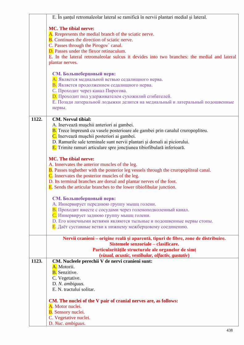
438
E. În şanţul retromaleolar lateral se ramifică în nervii plantari medial şi lateral.
MC. The tibial nerve:
A. Rrepresents the medial branch of the sciatic nerve.
B. Continues the direction of sciatic nerve.
C. Passes through the Pirogov` canal.
D. Passes under the flexor retinaculum.
E. In the lateral retromaleolar sulcus it devides into two branches: the medial and lateral
plantar nerves.
СМ. Большеберцовый нерв: A. Является медиальной ветвью седалищного нерва.
B. Является продолжением седалищного нерва.
C. Проходит через канал Пирогова.
D. Проходит под удерживателем сухожилий сгибателей.
E. Позади латеральной лодыжки делится на медиальный и латеральный подошвенные
нервы.
1122. CM. Nervul tibial:
A. Inervează muşchii anteriori ai gambei.
B. Trece împreună cu vasele posterioare ale gambei prin canalul cruropopliteu.
C. Inervează muşchii posteriori ai gambei.
D. Ramurile sale terminale sunt nervii plantari şi dorsali ai piciorului.
E. Trimite ramuri articulare spre joncţiunea tibiofibulară inferioară.
MC. The tibial nerve:
A. Innervates the anterior muscles of the leg.
B. Passes toghether with the posterior leg vessels through the cruropopliteal canal.
C. Innervates the posterior muscles of the leg.
D. Its terminal branches are dorsal and plantar nerves of the foot.
E. Sends the articular branches to the lower tibiofibular junction.
СМ. Большеберцовый нерв: A. Иннервирует переднюю группу мышц голени.
B. Проходит вместе с сосудами через голеноподколенный канал.
C. Иннервирует заднюю группу мышц голени.
D. Его конечными ветвями являются тыльные и подошвенные нервы стопы.
E. Даѐт суставные ветви к нижнему межберцовому соединению.
Nervii cranieni – origine reală şi aparentă, tipuri de fibre, zone de distribuire.
Sistemele senzoriale – clasificare.
Particularităţile structurale ale organelor de simţ
(vizual, acustic, vestibular, olfactiv, gustativ)
1123. CM. Nucleele perechii V de nervi cranieni sunt:
A. Motorii.
B. Senzitive.
C. Vegetative.
D. N. ambiguus.
E. N. tractului solitar.
CM. The nuclei of the V pair of cranial nerves are, as follows:
A. Motor nuclei.
B. Sensory nuclei.
C. Vegetative nuclei.
D. Nuc. ambiguus.
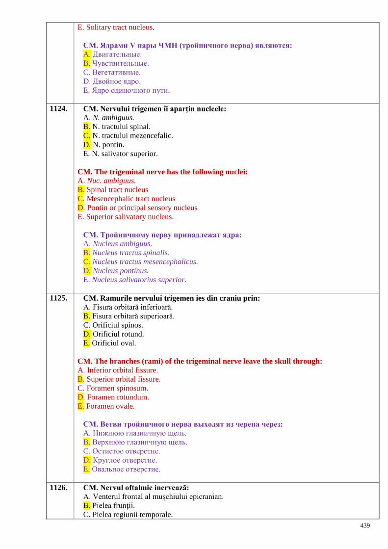
439
E. Solitary tract nucleus.
CM. Ядрами V пары ЧМН (тройничного нерва) являются: A. Двигательные.
B. Чувствительные.
C. Вегетативные.
D. Двойное ядро.
E. Ядро одиночного пути.
1124. CM. Nervului trigemen îi aparţin nucleele:
A. N. ambiguus.
B. N. tractului spinal.
C. N. tractului mezencefalic.
D. N. pontin.
E. N. salivator superior.
CM. The trigeminal nerve has the following nuclei:
A. Nuc. ambiguus.
B. Spinal tract nucleus
C. Mesencephalic tract nucleus
D. Pontin or principal sensory nucleus
E. Superior salivatory nucleus.
СМ. Тройничному нерву принадлежат ядра:
A. Nucleus ambiguus.
B. Nucleus tractus spinalis.
C. Nucleus tractus mesencephalicus.
D. Nucleus pontinus.
E. Nucleus salivatorius superior.
1125. CM. Ramurile nervului trigemen ies din craniu prin:
A. Fisura orbitară inferioară.
B. Fisura orbitară superioară.
C. Orificiul spinos.
D. Orificiul rotund.
E. Orificiul oval.
CM. The branches (rami) of the trigeminal nerve leave the skull through:
A. Inferior orbital fissure.
B. Superior orbital fissure.
C. Foramen spinosum.
D. Foramen rotundum.
E. Foramen ovale.
СМ. Ветви тройничного нерва выходят из черепа через:
A. Нижнюю глазничную щель.
B. Верхнюю глазничную щель.
C. Остистое отверстие.
D. Круглое отверстие.
E. Овальное отверстие.
1126. CM. Nervul oftalmic inervează:
A. Venterul frontal al mușchiului epicranian.
B. Pielea frunţii.
C. Pielea regiunii temporale.
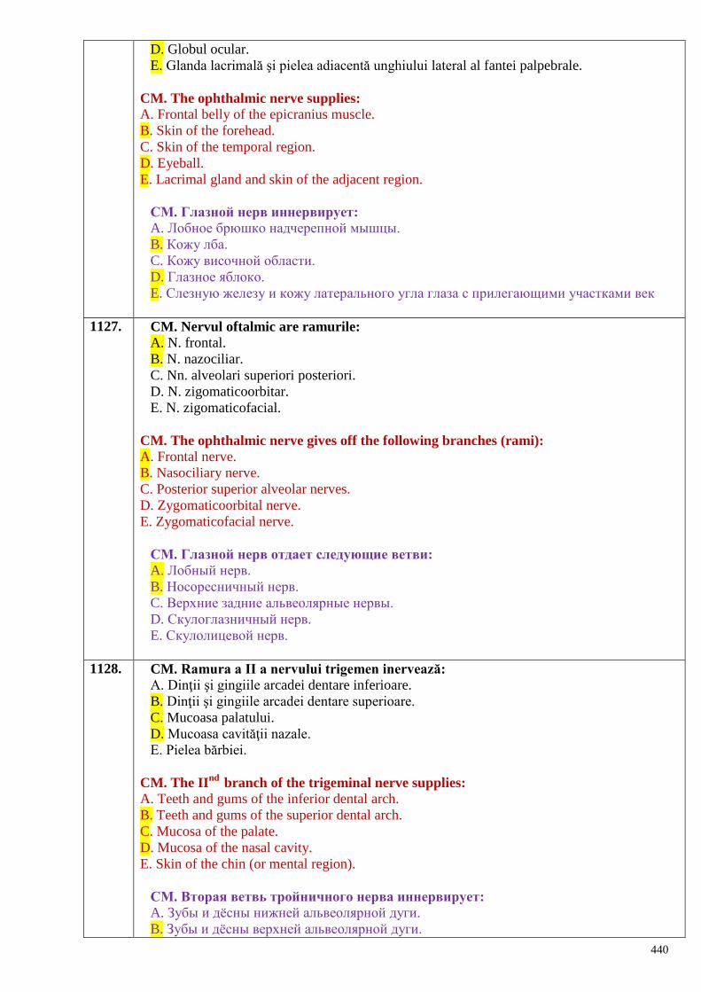
440
D. Globul ocular.
E. Glanda lacrimală şi pielea adiacentă unghiului lateral al fantei palpebrale.
CM. The ophthalmic nerve supplies:
A. Frontal belly of the epicranius muscle.
B. Skin of the forehead.
C. Skin of the temporal region.
D. Eyeball.
E. Lacrimal gland and skin of the adjacent region.
СМ. Глазной нерв иннервирует:
A. Лобное брюшко надчерепной мышцы.
B. Кожу лба.
C. Кожу височной области.
D. Глазное яблоко.
E. Слезную железу и кожу латерального угла глаза с прилегающими участками век
1127. CM. Nervul oftalmic are ramurile:
A. N. frontal.
B. N. nazociliar.
C. Nn. alveolari superiori posteriori.
D. N. zigomaticoorbitar.
E. N. zigomaticofacial.
CM. The ophthalmic nerve gives off the following branches (rami):
A. Frontal nerve.
B. Nasociliary nerve.
C. Posterior superior alveolar nerves.
D. Zygomaticoorbital nerve.
E. Zygomaticofacial nerve.
СМ. Глазной нерв отдает следующие ветви:
A. Лобный нерв.
B. Носоресничный нерв.
C. Верхние задние альвеолярные нервы.
D. Скулоглазничный нерв.
E. Скулолицевой нерв.
1128. CM. Ramura a II a nervului trigemen inervează:
A. Dinţii şi gingiile arcadei dentare inferioare.
B. Dinţii şi gingiile arcadei dentare superioare.
C. Mucoasa palatului.
D. Mucoasa cavităţii nazale.
E. Pielea bărbiei.
CM. The IInd
branch of the trigeminal nerve supplies:
A. Teeth and gums of the inferior dental arch.
B. Teeth and gums of the superior dental arch.
C. Mucosa of the palate.
D. Mucosa of the nasal cavity.
E. Skin of the chin (or mental region).
СМ. Вторая ветвь тройничного нерва иннервирует:
A. Зубы и дѐсны нижней альвеолярной дуги.
B. Зубы и дѐсны верхней альвеолярной дуги.
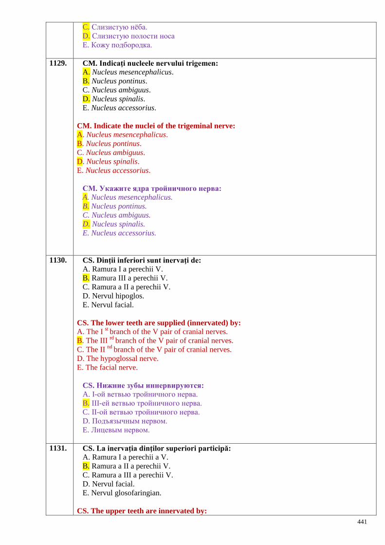
441
C. Слизистую нѐба.
D. Слизистую полости носа
E. Кожу подбородка.
1129. CM. Indicaţi nucleele nervului trigemen:
A. Nucleus mesencephalicus.
B. Nucleus pontinus.
C. Nucleus ambiguus.
D. Nucleus spinalis.
E. Nucleus accessorius.
CM. Indicate the nuclei of the trigeminal nerve:
A. Nucleus mesencephalicus.
B. Nucleus pontinus.
C. Nucleus ambiguus.
D. Nucleus spinalis.
E. Nucleus accessorius.
СМ. Укажите ядра тройничного нерва:
A. Nucleus mesencephalicus.
B. Nucleus pontinus.
C. Nucleus ambiguus.
D. Nucleus spinalis.
E. Nucleus accessorius.
1130. CS. Dinţii inferiori sunt inervaţi de:
A. Ramura I a perechii V.
B. Ramura III a perechii V.
C. Ramura a II a perechii V.
D. Nervul hipoglos.
E. Nervul facial.
CS. The lower teeth are supplied (innervated) by:
A. The I st
branch of the V pair of cranial nerves.
B. The III rd
branch of the V pair of cranial nerves.
C. The II nd
branch of the V pair of cranial nerves.
D. The hypoglossal nerve.
E. The facial nerve.
CS. Нижние зубы иннервируются:
A. I-ой ветвью тройничного нерва.
B. III-ей ветвью тройничного нерва.
C. II-ой ветвью тройничного нерва.
D. Подъязычным нервом.
E. Лицевым нервом.
1131. CS. La inervaţia dinţilor superiori participă:
A. Ramura I a perechii a V.
B. Ramura a II a perechii V.
C. Ramura a III a perechii V.
D. Nervul facial.
E. Nervul glosofaringian.
CS. The upper teeth are innervated by:
442
A. The I st
branch of the V pair of cranial nerves.
B. The II nd
branch of the V pair of cranial nerves.
C. The III rd
branch of the V pair of cranial nerves.
D. The facial nerve.
E. The glossopharyngeal nerve.
CS. В иннервации верхних зубов участвуют:
A. I-ая ветвь тройничного нерва.
B. II-ая ветвь тройничного нерва.
C. III-яя ветвь тройничного нерва.
D. Лицевым нервом.
E. Языкоглоточным нервом.
1132. CS. Muşchii masticatori sunt inervaţi de:
A. N. facial.
B. N. accesor.
C. N. trigemen.
D. N. abducens.
E. N. hipoglos.
CS. Muscles of mastication are innervated by:
A. Facial nerve.
B. Accessory nerve.
C. Trigeminal nerve.
D. Abducens nerve.
E. Hypoglossal nerve.
CS. Жевательные мышцы иннервируются:
A. Лицевым нервом.
B. Добавочным нервом.
C. Тройничным нервом.
D. Отводящим нервом.
E. Подъязычным нервом.
1133. CS. Inervaţia parasimpatică a glandei sublingvale este asigurată de:
A. Perechea VII de nervi cranieni
B. Perechea IX de nervi cranieni
C. Perechea X de nervi cranieni
D. Fibre emergente de la ganglionul cervical superior al lanțului simpatic
E. Ramuri emergente de la ganglionul cervical inferior al lanțului simpatic
CS. The parasympathetic innervation of the sublingual gland is assured by:
A. The VII pair of cranial nerves.
B. The IX pair of cranial nerves.
C. The X pair of cranial nerves.
D. The fibers from the superior cervical ganglion of the sympathetic trunk.
E. The rami from the inferior cervical ganglion of the sympathetic trunk.
СS. Парасимпатическая иннервация подъязычной железы обеспечена:
A. VII-ой парой ЧМН.
B. IX-ой парой ЧМН.
C. X-ой парой ЧМН.
D. Волокнами идущими от верхнего шейного узла симпатического ствола.
E. Волокнами идущими от нижнего шейного узла симпатического ствола.
443
1134. CM. Indicaţi nucleele perechii a VII de n. cranieni:
A. N. tractului solitar.
B. N. ambiguus.
C. N. salivator superior.
D. N. salivator inferior.
E. N. motor al n. facial.
CM. Indicate the nuclei of the VII pair of cranial nerves:
A. Solitary tract nucleus.
B. Nuc. ambiguus.
C. Superior salivatory nucleus.
D. Inferior salivatory nucleus.
E. Motor nucleus of the facial nerve.
СМ. Укажите ядра VII-ой пары чмн:
A. Nucleus tractus solitarii.
B. Nucleus ambiguus.
C. Nucleus salivatorius superior.
D. Nucleus salivatorius inferior.
E. Nucleus motorius nervi facialis.
1135. CS. Muşchii mimici sunt inervaţi motor de:
A. N. abducens.
B. N. trigemen.
C. N. facial.
D. N. oculomotor.
E. N. accesor.
CS. Muscles of facial exprtession are supplied by:
A. Abducens nerve.
B. Trigeminal nerve.
C. Facial nerve.
D. Oculomotor nerve.
E. Accessory nerve.
CS. Мимические мышцы получают двигательную иннервацию от:
A. Отводящего нерва.
B. Тройничного нерва.
C. Лицевого нерва.
D. Глазодвигательного нерва.
E. Добавочного нерва.
1136. CM. Nervul facial are următoarele ramuri intracanaliculare:
A. Nervul pietros mic.
B. Nervul pietros mare.
C. Nervul m. digastric.
D. Coarda timpanică.
E. Ramuri temporale.
CM. The intracanalicular part of the facial nerve gives off the following branches:
A. Lesser petrosal nerve.
B. Greater petrosal nerve.
C. Digastric branch.
D. Chorda tympani nerve.
E. Temporal branches.
444
СМ. В одноимѐнном канале, лицевой нерв отдает ветви:
A. Малый каменистый нерв.
B. Большой каменистый нерв.
C. Нерв двубрюшной мышцы.
D. Барабанная струна.
E. Височные ветви.
1137. CM. Care dintre glandele enumerate sunt inervate de nervul facial?
A. Gl. parotidă.
B. Gl. lacrimală.
C. Gl. tiroidă.
D. Gl. sublingvală.
E. Gl. submandibulară.
CM. Which of the following glands are supplied by the facial nerve?
A. Parotid gland.
B. Lacrimal gland.
C. Thyroid gland.
D. Sublingual gland.
E. Submandibular gland.
СМ. Какие железы получают иннервацию от лицевого нерва?
A. Околоушная железа.
B. Слезная железа.
C. Щитовидная железа.
D. Подъязычная железа.
E. Поднижнечелюстная железа.
1138. CM. Sensibilitatea gustativă a limbii este asigurată de:
A. Perechea V.
B. Perechea VII.
C. Perechea IX.
D. Perechea X.
E. Perechea XII.
CM. The gustatory sensitivity of the tongue is assured by:
A. The V pair of cranial nerves.
B. The VII pair of cranial nerves.
C. The IX pair of cranial nerves.
D. The X pair of cranial nerves.
E. The XII pair of cranial nerves.
СМ. Вкусовая чувствительность языка обеспечена:
A. V-ой парой ЧМН.
B. VII-ой парой ЧМН.
C. IX-ой парой ЧМН.
D. X-ой парой ЧМН.
E. XII-ой парой ЧМН.
1139. CM. Care dintre nervii indicaţi mai jos asigură sensibilitatea generală (nespecifică) a
mucoasei lingvale:
A. Perechea V.
B. Perechea VII.
C. Perechea IX.
445
D. Perechea X.
E. Perechea XII.
CM. Which of the following nerves assure the general sensitivity of the lingual mucosa:
A. The V pair of cranial nerves.
B. The VII pair of cranial nerves.
C. The IX pair of cranial nerves.
D. The X pair of cranial nerves.
E. The XII pair of cranial nerves.
СМ. Какие из перечисленных нервов обеспечивают общую чувствительность
(неспецифическую) для слизистой языка:
A. V-ая пара ЧМН.
B. VII-ая пара ЧМН.
C. IX-ая пара ЧМН.
D. X-ая пара ЧМН.
E. XII-ая пара ЧМН.
1140. CM. De la plexul parotidian (al nervului facial) pornesc următoarele ramuri:
A. Digastrică.
B. Stilohioidiană.
C. Temporale.
D. Bucale.
E. Zigomatice.
CM. The parotid plexus gives off the following branches:
A. Digastric branch.
B. Stylohyoid branch.
C. Temporal branches.
D. Buccal branches.
E. Zygomatic branches.
СМ. От околоушного сплетения отходят следующие ветви:
A. Двубрюшная.
B. Шилоподъязычная.
C. Височные.
D. Щечные.
E. Скуловые.
1141. CM. Perechea a VII de n. cranieni inervează parasimpatic următoarele glande:
A. Lacrimală.
B. Parotidă.
C. Submandibulară.
D. Sublingvală.
E. Tiroidă.
CM. The VII pair of cranial nerves supplies (parasympathetic innervation) the following
glands:
A. Lacrimal gland.
B. Parotid gland.
C. Submandibular gland.
D. Sublingual gland.
E. Thyroid gland.
СМ. VII-ая пара чмн обеспечивает парасимпатической иннервацией следующие
446
железы:
A. Слезную.
B. Околоушную.
C. Поднижнечелюстную.
D. Подъязычную.
E. Щитовидную.
1142. CS. Sursa inervaţiei gustative la nivelul celor 2/3 anterioare ale limbii:
A. Nervul facial.
B. Nervul oftalmic.
C. Nervul maxilar.
D. Nervul mandibular.
E. Nervul glosofaringian.
CS. The source of gustatory innervation of the anterior 2/3 of the tongue is:
A. Facial nerve.
B. Ophthalmic nerve.
C. Maxillary nerve.
D. Mandibular nerve.
E. Glossopharyngeal nerve
CS. Источник вкусовой иннервации для передних 2/3 языка:
A. Лицевой нерв.
B. Глазной нерв.
C. Верхнечелюстной нерв.
D. Нижнечелюстной нерв.
E. Языкоглоточный нерв.
1143. CM. Nucleii nervului facial sunt:
A. Nucleus salivatorius superior.
B. Nucleus salivaorius inferior.
C. Nucleus motorius.
D. Nucleus tractus solitarii.
E. Nucleus ambiguus.
CM. The nuclei of facial nerve are, as follows:
A. Nucleus salivatorius superior.
B. Nucleus salivaorius inferior.
C. Nucleus motorius.
D. Nucleus tractus solitarii.
E. Nucleus ambiguus.
СМ. Являются ядрами лицевого нерва:
A. Nucleus salivatorius superior.
B. Nucleus salivatorius inferior.
C. Nucleus motorius.
D. Nucleus tractus solitarii.
E. Nucleus ambiguus.
1144. CS. Nervus facialis iese din craniu prin:
A. Foramen ovale.
B. Foramen stylomastoideum.
C. Fissura petrotympanica.
D. Foramen spinosum.
E. Foramen lacerum.
447
CS. Nervus facialis leaves the skull through:
A. Foramen ovale.
B. Foramen stylomastoideum.
C. Fissura petrotympanica.
D. Foramen spinosum.
E. Foramen lacerum.
CS. Nervus facialis выходит из черепа через:
A. Foramen ovale.
B. Foramen stylomastoideum.
C. Fissura petrotympanica.
D. Foramen spinosum.
E. Foramen lacerum.
1145. CS. Indicaţi sursa de inervaţie parasimpatică a glandei lacrimale:
A. Nervus facialis.
B. Nervus glossopharyngeus.
C. Nervus vagus.
D. Nervus ophthalmicus.
E. Nervus lacrimalis.
CS. Indicate the source of parasympathetic innervation of the lacrimal gland:
A. Nervus facialis.
B. Nervus glossopharyngeus.
C. Nervus vagus.
D. Nervus ophthalmicus.
E. Nervus lacrimalis.
CS. Укажите источник парасимпатической иннервации слезной железы:
A. Nervus facialis.
B. Nervus glossopharyngeus.
C. Nervus vagus.
D. Nervus ophthalmicus.
E. Nervus lacrimalis.
1146. CS. Indicaţi sursa de inervaţie motorie a muşchilor mimici:
A. Nervus facialis.
B. Nervus ophthalmicus.
C. Nervus maxillaris.
D. Nervus mandibularis.
E. Nervus accessorius.
CS. Indicate the source of motor innervation of the muscles of facial expression:
A. Nervus facialis.
B. Nervus ophthalmicus.
C. Nervus maxillaris.
D. Nervus mandibularis.
E. Nervus accessorius.
CS. Укажите источник двигательной иннервации мимических мышц:
A. Nervus facialis.
B. Nervus ophthalmicus.
C. Nervus maxillaris.
D. Nervus mandibularis.
448
E. Nervus accessorius.
1147. CS. Indicaţi sursa de inervaţie motorie a platismei:
A. Nervus facialis.
B. Nervus vagus.
C. Rami musculares plexus cervicalis.
D. Ansa cervicalis.
E. Nervus transversus colli.
CS. Indicate the source of motor innervation of the platysma muscle:
A. Nervus facialis.
B. Nervus vagus.
C. Rami musculares plexus cervicalis.
D. Ansa cervicalis.
E. Nervus transversus colli.
CS. Укажите источник двигательной иннервации подкожной мышцы:
A. Nervus facialis.
B. Nervus vagus.
C. Rami musculares plexus cervicalis.
D. Ansa cervicalis.
E. Nervus transversus colli.
1148. CS. Indicaţi sursa de inervaţie parasimpatică a glandei submandibulare:
A. Nervus facialis.
B. Nervus vagus.
C. Nervus glossopharyngeus.
D. Ansa cervicalis.
E. Nervus mandibularis.
CS. Indicate the source of parasympathetic innervation of the submandibular gland:
A. Nervus facialis.
B. Nervus vagus.
C. Nervus glossopharyngeus.
D. Ansa cervicalis.
E. Nervus mandibularis.
CS. Укажите источник парасимпатической иннервации поднижнечелюстной
железы:
A. Nervus facialis.
B. Nervus vagus.
C. Nervus glossopharyngeus.
D. Ansa cervicalis.
E. Nervus mandibularis.
1149. CS. Nucleul salivator superior aparţine nervului:
A. V.
B. VIII.
C. IX.
D. VII.
E. X.
CS. The superior salivatory nucleus belongs to:
A. The V pair of cranial nerves.
B. The VIII pair of cranial nerves.
449
C. The IX pair of cranial nerves.
D. The VII pair of cranial nerves.
E. The X pair of cranial nerves.
CS. Верхнее слюноотделительное ядро принадлежит:
A. V–ой паре ЧМН.
B. VIII–ой паре ЧМН.
C. IX–ой паре ЧМН.
D. VII–ой паре ЧМН.
E. X–ой паре ЧМН.
1150. CM. Perechea a X de nervi cranieni are următoarele nuclee:
A. N. salivator superior.
B. N. salivator inferior.
C. N. ambiguus.
D. N. tractului solitar.
E. N. dorsal.
CM. The X cranial nerve posseses the following nuclei:
A. Nucleus salivatorius superior.
B. Nucleus salivatorius inferior.
C. Nucleus ambiguus.
D. Nucleus tractus solitarii.
E. Nucleus dorsalis.
СМ. Х-ая пара ЧМН имеет следующие ядра:
A. Nucleus salivatorius superior.
B. Nucleus salivatorius inferior.
C. Nucleus ambiguus.
D. Nucleus tractus solitarii.
E. Nucleus dorsalis.
1151. CM. Topografic nervului vag i se disting porţiunile:
A. Sacrată.
B. Craniană.
C. Cervicală.
D. Toracică.
E. Abdominală.
CM. According to the topography the vagus nerve has the following portions:
A. Sacral part.
B. Cranial part.
C. Cervical part.
D. Thoracic part.
E. Abdominal part.
СМ. У блуждающего нерва топографически различаются отделы:
A. Крестцовый.
B. Головной.
C. Шейный.
D. Грудной.
E. Брюшной.
1152. CM. Indicaţi 2 nervi cranieni ce inervează faringele:
A. N. glosofaringian.
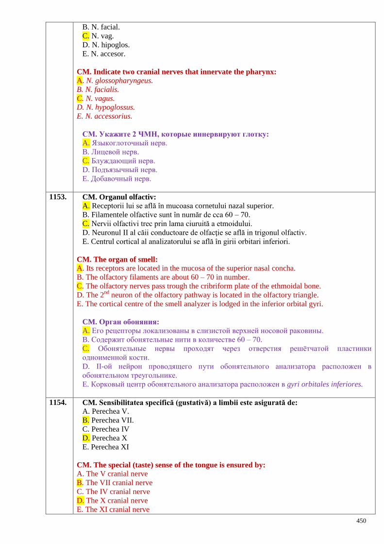
450
B. N. facial.
C. N. vag.
D. N. hipoglos.
E. N. accesor.
CM. Indicate two cranial nerves that innervate the pharynx:
A. N. glossopharyngeus.
B. N. facialis.
C. N. vagus.
D. N. hypoglossus.
E. N. accessorius.
СМ. Укажите 2 ЧМН, которые иннервируют глотку:
A. Языкоглоточный нерв.
B. Лицевой нерв.
C. Блуждающий нерв.
D. Подъязычный нерв.
E. Добавочный нерв.
1153. CM. Organul olfactiv:
A. Receptorii lui se află în mucoasa cornetului nazal superior.
B. Filamentele olfactive sunt în număr de cca 60 – 70.
C. Nervii olfactivi trec prin lama ciuruită a etmoidului.
D. Neuronul II al căii conductoare de olfacţie se află in trigonul olfactiv.
E. Centrul cortical al analizatorului se află în girii orbitari inferiori.
CM. The organ of smell:
A. Its receptors are located in the mucosa of the superior nasal concha.
B. The olfactory filaments are about 60 – 70 in number.
C. The olfactory nerves pass trough the cribriform plate of the ethmoidal bone.
D. The 2nd
neuron of the olfactory pathway is located in the olfactory triangle.
E. The cortical centre of the smell analyzer is lodged in the inferior orbital gyri.
CM. Орган обоняния:
A. Его рецепторы локализованы в слизистой верхней носовой раковины.
B. Содержит обонятельные нити в количестве 60 – 70.
C. Обонятельные нервы проходят через отверстия решѐтчатой пластинки
одноименной кости.
D. II-ой нейрон проводящего пути обонятельного анализатора расположен в
обонятельном треугольнике.
E. Корковый центр обонятельного анализатора расположен в gyri orbitales inferiores.
1154. CM. Sensibilitatea specifică (gustativă) a limbii este asigurată de:
A. Perechea V.
B. Perechea VII.
C. Perechea IV
D. Perechea X
E. Perechea XI
CM. The special (taste) sense of the tongue is ensured by:
A. The V cranial nerve
B. The VII cranial nerve
C. The IV cranial nerve
D. The X cranial nerve
E. The XI cranial nerve
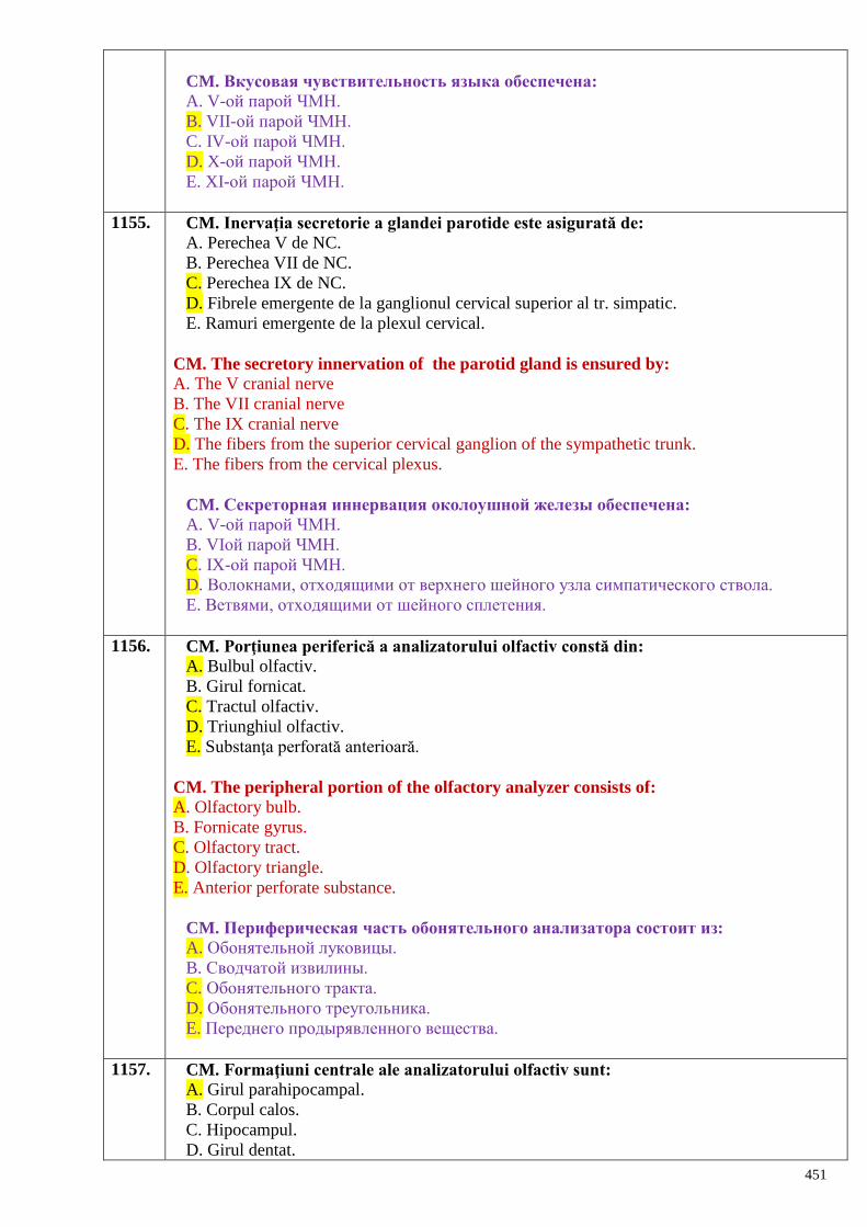
451
CM. Вкусовая чувствительность языка обеспечена:
A. V-ой парой ЧМН.
B. VII-ой парой ЧМН.
C. IV-ой парой ЧМН.
D. X-ой парой ЧМН.
E. XI-ой парой ЧМН.
1155. CM. Inervaţia secretorie a glandei parotide este asigurată de:
A. Perechea V de NC.
B. Perechea VII de NC.
C. Perechea IX de NC.
D. Fibrele emergente de la ganglionul cervical superior al tr. simpatic.
E. Ramuri emergente de la plexul cervical.
CM. The secretory innervation of the parotid gland is ensured by:
A. The V cranial nerve
B. The VII cranial nerve
C. The IX cranial nerve
D. The fibers from the superior cervical ganglion of the sympathetic trunk.
E. The fibers from the cervical plexus.
CM. Секреторная иннервация околоушной железы обеспечена:
A. V-ой парой ЧМН.
B. VIой парой ЧМН.
C. IX-ой парой ЧМН.
D. Волокнами, отходящими от верхнего шейного узла симпатического ствола.
E. Ветвями, отходящими от шейного сплетения.
1156. CM. Porţiunea periferică a analizatorului olfactiv constă din:
A. Bulbul olfactiv.
B. Girul fornicat.
C. Tractul olfactiv.
D. Triunghiul olfactiv.
E. Substanţa perforată anterioară.
CM. The peripheral portion of the olfactory analyzer consists of:
A. Olfactory bulb.
B. Fornicate gyrus.
C. Olfactory tract.
D. Olfactory triangle.
E. Anterior perforate substance.
CM. Периферическая часть обонятельного анализатора состоит из:
A. Обонятельной луковицы.
B. Сводчатой извилины.
C. Обонятельного тракта.
D. Обонятельного треугольника.
E. Переднего продырявленного вещества.
1157. CM. Formaţiuni centrale ale analizatorului olfactiv sunt:
A. Girul parahipocampal.
B. Corpul calos.
C. Hipocampul.
D. Girul dentat.
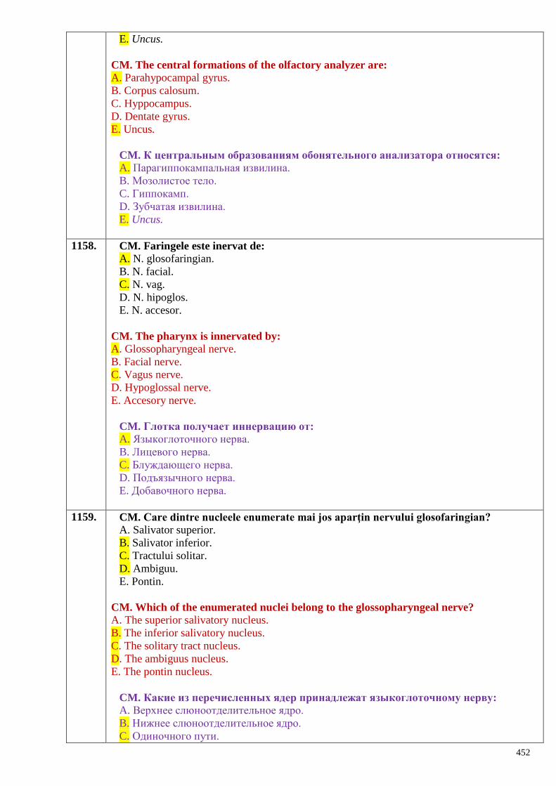
452
E. Uncus.
CM. The central formations of the olfactory analyzer are:
A. Parahypocampal gyrus.
B. Corpus calosum.
C. Hyppocampus.
D. Dentate gyrus.
E. Uncus.
CM. К центральным образованиям обонятельного анализатора относятся:
A. Парагиппокампальная извилина.
B. Мозолистое тело.
C. Гиппокамп.
D. Зубчатая извилина.
E. Uncus.
1158. CM. Faringele este inervat de:
A. N. glosofaringian.
B. N. facial.
C. N. vag.
D. N. hipoglos.
E. N. accesor.
CM. The pharynx is innervated by:
A. Glossopharyngeal nerve.
B. Facial nerve.
C. Vagus nerve.
D. Hypoglossal nerve.
E. Accesory nerve.
CM. Глотка получает иннервацию от:
A. Языкоглоточного нерва.
B. Лицевого нерва.
C. Блуждающего нерва.
D. Подъязычного нерва.
E. Добавочного нерва.
1159. CM. Care dintre nucleele enumerate mai jos aparţin nervului glosofaringian?
A. Salivator superior.
B. Salivator inferior.
C. Tractului solitar.
D. Ambiguu.
E. Pontin.
CM. Which of the enumerated nuclei belong to the glossopharyngeal nerve?
A. The superior salivatory nucleus.
B. The inferior salivatory nucleus.
C. The solitary tract nucleus.
D. The ambiguus nucleus.
E. The pontin nucleus.
CM. Какие из перечисленных ядер принадлежат языкоглоточному нерву:
A. Верхнее слюноотделительное ядро.
B. Нижнее слюноотделительное ядро.
C. Одиночного пути.
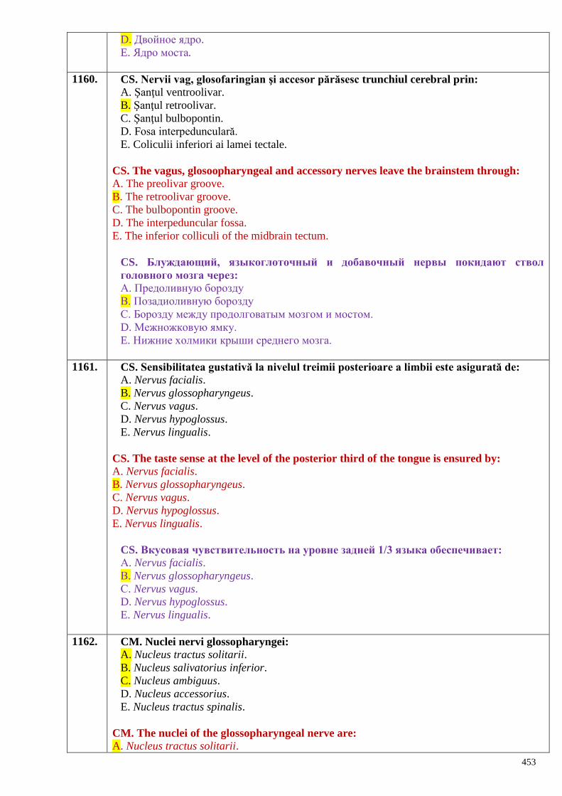
453
D. Двойное ядро.
E. Ядро моста.
1160. CS. Nervii vag, glosofaringian şi accesor părăsesc trunchiul cerebral prin:
A. Şanţul ventroolivar.
B. Şanţul retroolivar.
C. Şanţul bulbopontin.
D. Fosa interpedunculară.
E. Coliculii inferiori ai lamei tectale.
CS. The vagus, glosoopharyngeal and accessory nerves leave the brainstem through:
A. The preolivar groove.
B. The retroolivar groove.
C. The bulbopontin groove.
D. The interpeduncular fossa.
E. The inferior colliculi of the midbrain tectum.
CS. Блуждающий, языкоглоточный и добавочный нервы покидают ствол
головного мозга через:
А. Предоливную борозду
В. Позадиоливную борозду
С. Борозду между продолговатым мозгом и мостом.
D. Межножковую ямку.
E. Нижние холмики крыши среднего мозга.
1161. CS. Sensibilitatea gustativă la nivelul treimii posterioare a limbii este asigurată de:
A. Nervus facialis.
B. Nervus glossopharyngeus.
C. Nervus vagus.
D. Nervus hypoglossus.
E. Nervus lingualis.
CS. The taste sense at the level of the posterior third of the tongue is ensured by:
A. Nervus facialis.
B. Nervus glossopharyngeus.
C. Nervus vagus.
D. Nervus hypoglossus.
E. Nervus lingualis.
CS. Вкусовая чувствительность на уровне задней 1/3 языка обеспечивает:
А. Nervus facialis.
В. Nervus glossopharyngeus.
С. Nervus vagus.
D. Nervus hypoglossus.
E. Nervus lingualis.
1162. CM. Nuclei nervi glossopharyngei:
A. Nucleus tractus solitarii.
B. Nucleus salivatorius inferior.
C. Nucleus ambiguus.
D. Nucleus accessorius.
E. Nucleus tractus spinalis.
CM. The nuclei of the glossopharyngeal nerve are:
A. Nucleus tractus solitarii.
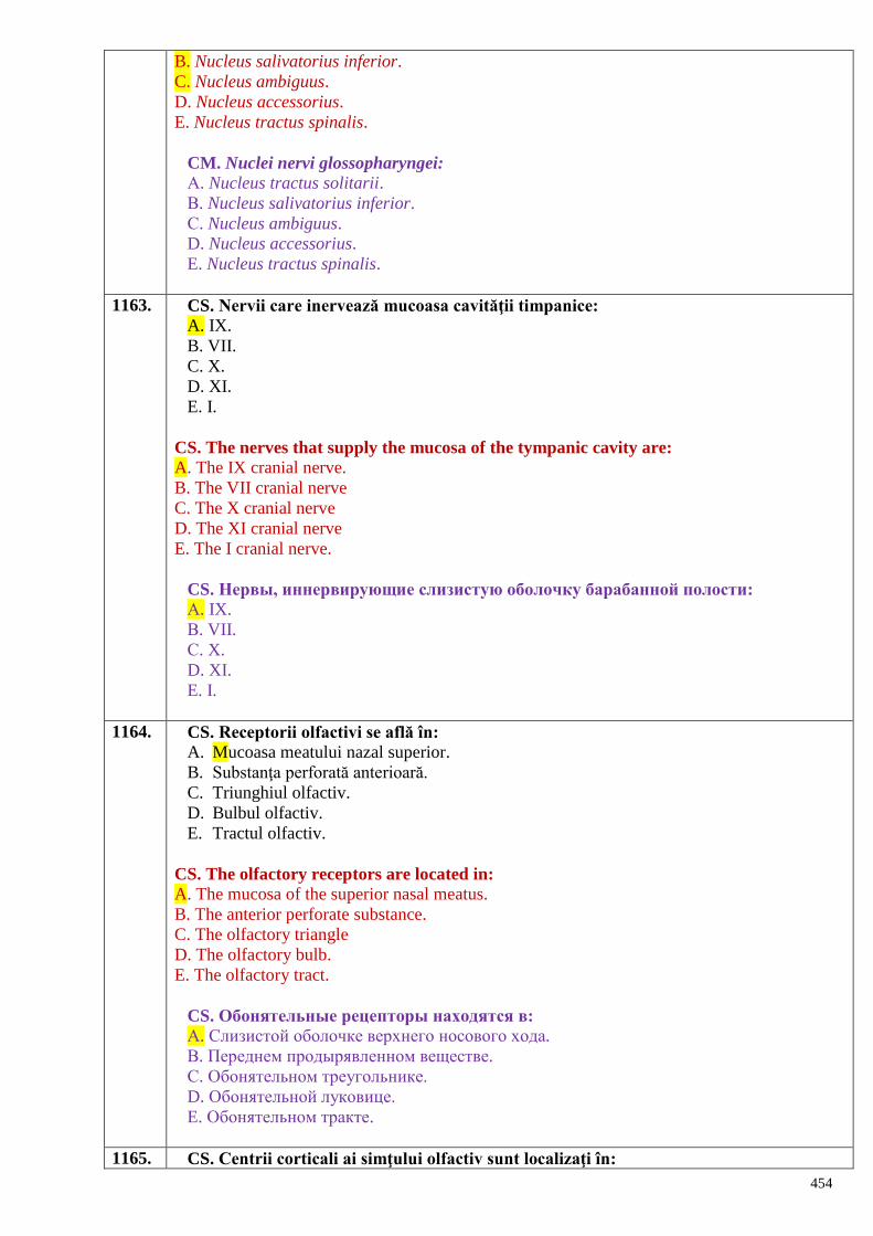
454
B. Nucleus salivatorius inferior.
C. Nucleus ambiguus.
D. Nucleus accessorius.
E. Nucleus tractus spinalis.
CM. Nuclei nervi glossopharyngei:
А. Nucleus tractus solitarii.
В. Nucleus salivatorius inferior.
С. Nucleus ambiguus.
D. Nucleus accessorius.
E. Nucleus tractus spinalis.
1163. CS. Nervii care inervează mucoasa cavităţii timpanice:
A. IX.
B. VII.
C. X.
D. XI.
E. I.
CS. The nerves that supply the mucosa of the tympanic cavity are:
A. The IX cranial nerve.
B. The VII cranial nerve
C. The X cranial nerve
D. The XI cranial nerve
E. The I cranial nerve.
CS. Нервы, иннервирующие слизистую оболочку барабанной полости:
А. IX.
В. VII.
С. X.
D. XI.
E. I.
1164. CS. Receptorii olfactivi se află în:
A. Mucoasa meatului nazal superior.
B. Substanţa perforată anterioară.
C. Triunghiul olfactiv.
D. Bulbul olfactiv.
E. Tractul olfactiv.
CS. The olfactory receptors are located in:
A. The mucosa of the superior nasal meatus.
B. The anterior perforate substance.
C. The olfactory triangle
D. The olfactory bulb.
E. The olfactory tract.
CS. Обонятельные рецепторы находятся в:
А. Слизистой оболочке верхнего носового хода.
В. Переднем продырявленном веществе.
С. Обонятельном треугольнике.
D. Обонятельной луковице.
E. Обонятельном тракте.
1165. CS. Centrii corticali ai simţului olfactiv sunt localizaţi în:
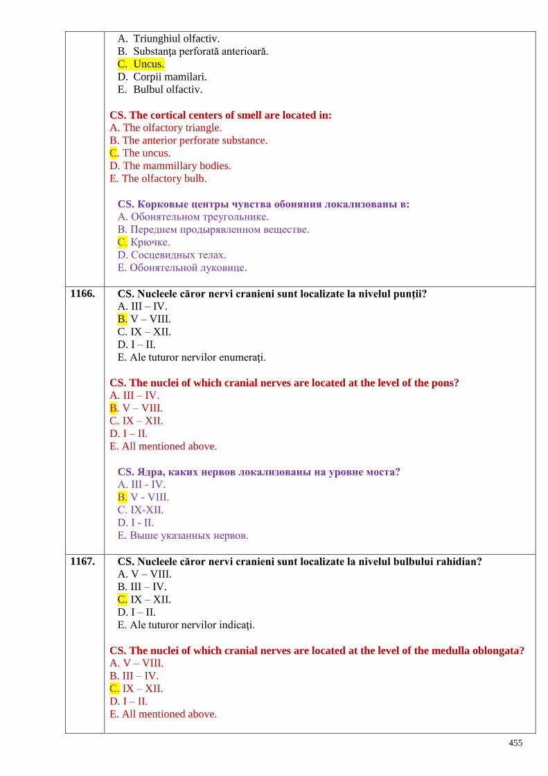
455
A. Triunghiul olfactiv.
B. Substanţa perforată anterioară.
C. Uncus.
D. Corpii mamilari.
E. Bulbul olfactiv.
CS. The cortical centers of smell are located in:
A. The olfactory triangle.
B. The anterior perforate substance.
C. The uncus.
D. The mammillary bodies.
E. The olfactory bulb.
CS. Корковые центры чувства обоняния локализованы в:
А. Обонятельном треугольнике.
В. Переднем продырявленном веществе.
С. Крючке.
D. Сосцевидных телах.
E. Обонятельной луковице.
1166. CS. Nucleele căror nervi cranieni sunt localizate la nivelul punţii?
A. III – IV.
B. V – VIII.
C. IX – XII.
D. I – II.
E. Ale tuturor nervilor enumeraţi.
CS. The nuclei of which cranial nerves are located at the level of the pons?
A. III – IV.
B. V – VIII.
C. IX – XII.
D. I – II.
E. All mentioned above.
CS. Ядра, каких нервов локализованы на уровне моста?
А. III - IV.
В. V - VIII.
С. IX-XII.
D. I - II.
E. Выше указанных нервов.
1167. CS. Nucleele căror nervi cranieni sunt localizate la nivelul bulbului rahidian?
A. V – VIII.
B. III – IV.
C. IX – XII.
D. I – II.
E. Ale tuturor nervilor indicaţi.
CS. The nuclei of which cranial nerves are located at the level of the medulla oblongata?
A. V – VIII.
B. III – IV.
C. IX – XII.
D. I – II.
E. All mentioned above.
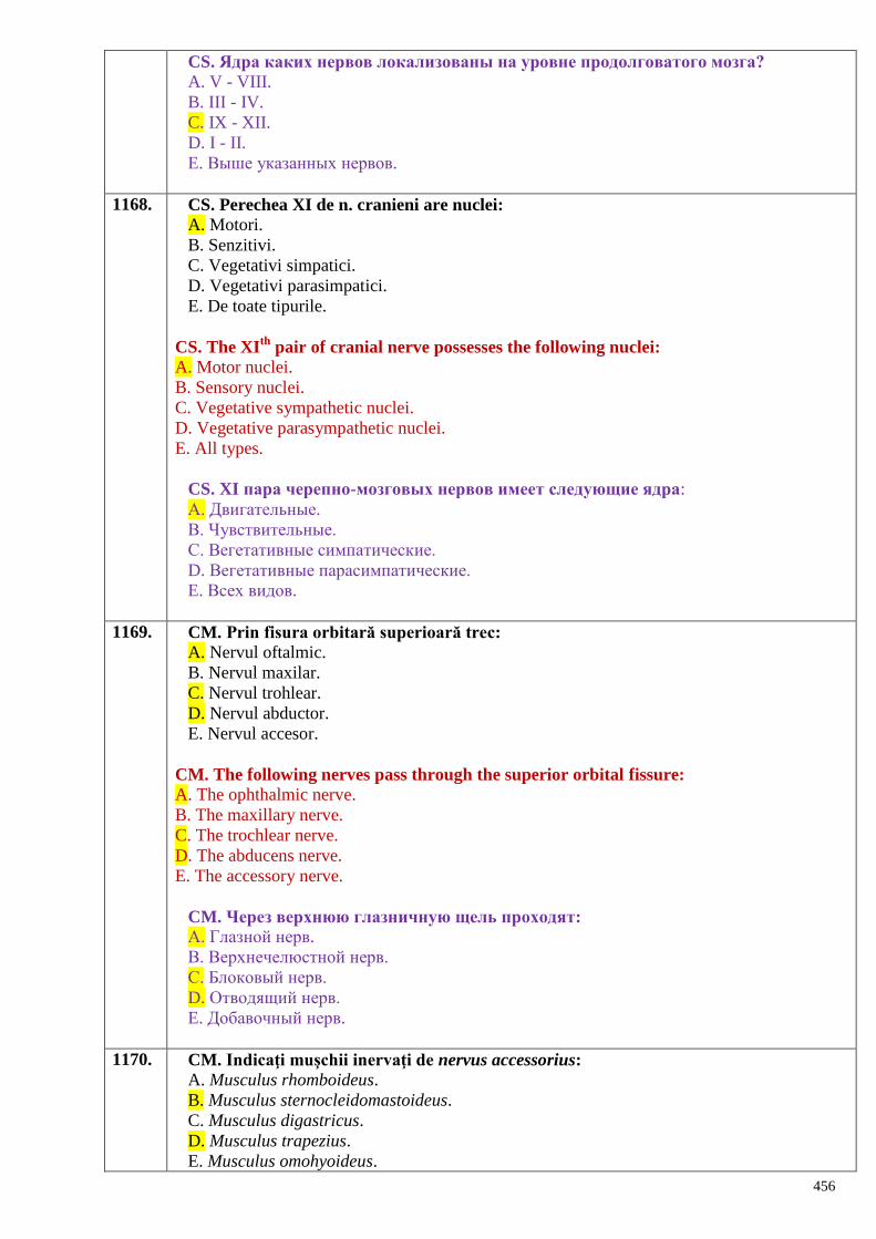
456
CS. Ядра каких нервов локализованы на уровне продолговатого мозга?
A. V - VIII.
B. III - IV.
C. IX - XII.
D. I - II.
E. Выше указанных нервов.
1168. CS. Perechea XI de n. cranieni are nuclei:
A. Motori.
B. Senzitivi.
C. Vegetativi simpatici.
D. Vegetativi parasimpatici.
E. De toate tipurile.
CS. The XIth
pair of cranial nerve possesses the following nuclei:
A. Motor nuclei.
B. Sensory nuclei.
C. Vegetative sympathetic nuclei.
D. Vegetative parasympathetic nuclei.
E. All types.
CS. XI пара черепно-мозговых нервов имеет следующие ядра:
А. Двигательные.
В. Чувствительные.
С. Вегетативные симпатические.
D. Вегетативные парасимпатические.
E. Всех видов.
1169. CM. Prin fisura orbitară superioară trec:
A. Nervul oftalmic.
B. Nervul maxilar.
C. Nervul trohlear.
D. Nervul abductor.
E. Nervul accesor.
CM. The following nerves pass through the superior orbital fissure:
A. The ophthalmic nerve.
B. The maxillary nerve.
C. The trochlear nerve.
D. The abducens nerve.
E. The accessory nerve.
CM. Через верхнюю глазничную щель проходят:
А. Глазной нерв.
В. Верхнечелюстной нерв.
С. Блоковый нерв.
D. Отводящий нерв.
E. Добавочный нерв.
1170. CM. Indicaţi muşchii inervaţi de nervus accessorius:
A. Musculus rhomboideus.
B. Musculus sternocleidomastoideus.
C. Musculus digastricus.
D. Musculus trapezius.
E. Musculus omohyoideus.
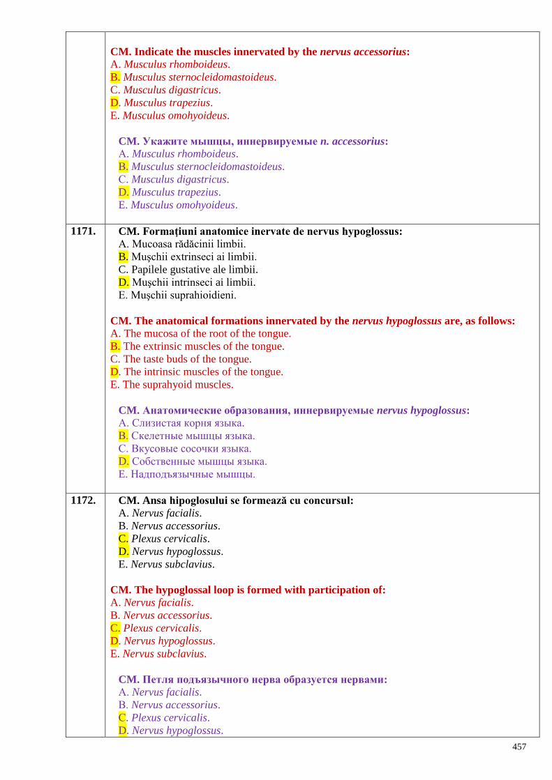
457
CM. Indicate the muscles innervated by the nervus accessorius:
A. Musculus rhomboideus.
B. Musculus sternocleidomastoideus.
C. Musculus digastricus.
D. Musculus trapezius.
E. Musculus omohyoideus.
CM. Укажите мышцы, иннервируемые n. accessorius:
А. Musculus rhomboideus.
В. Musculus sternocleidomastoideus.
С. Musculus digastricus.
D. Musculus trapezius.
E. Musculus omohyoideus.
1171. CM. Formaţiuni anatomice inervate de nervus hypoglossus:
A. Mucoasa rădăcinii limbii.
B. Muşchii extrinseci ai limbii.
C. Papilele gustative ale limbii.
D. Muşchii intrinseci ai limbii.
E. Muşchii suprahioidieni.
CM. The anatomical formations innervated by the nervus hypoglossus are, as follows:
A. The mucosa of the root of the tongue.
B. The extrinsic muscles of the tongue.
C. The taste buds of the tongue.
D. The intrinsic muscles of the tongue.
E. The suprahyoid muscles.
CM. Анатомические образования, иннервируемые nervus hypoglossus:
А. Слизистая корня языка.
В. Скелетные мышцы языка.
С. Вкусовые сосочки языка.
D. Собственные мышцы языка.
E. Надподъязычные мышцы.
1172. CM. Ansa hipoglosului se formează cu concursul:
A. Nervus facialis.
B. Nervus accessorius.
C. Plexus cervicalis.
D. Nervus hypoglossus.
E. Nervus subclavius.
CM. The hypoglossal loop is formed with participation of:
A. Nervus facialis.
B. Nervus accessorius.
C. Plexus cervicalis.
D. Nervus hypoglossus.
E. Nervus subclavius.
CM. Петля подъязычного нерва образуется нервами:
А. Nervus facialis.
В. Nervus accessorius.
С. Plexus cervicalis.
D. Nervus hypoglossus.
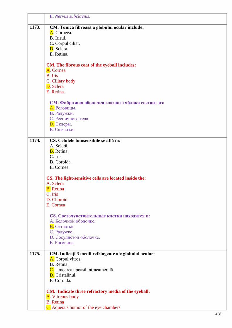
458
E. Nervus subclavius.
1173. CM. Tunica fibroasă a globului ocular include:
A. Corneea.
B. Irisul.
C. Corpul ciliar.
D. Sclera.
E. Retina.
CM. The fibrous coat of the eyeball includes:
A. Cornea
B. Iris
C. Ciliary body
D. Sclera
E. Retina.
СМ. Фиброзная оболочка глазного яблока состоит из:
A. Роговицы.
B. Радужки.
C. Ресничного тела.
D. Склеры.
E. Сетчатки.
1174. CS. Celulele fotosensibile se află în:
A. Scleră.
B. Retină.
C. Iris.
D. Coroidă.
E. Cornee.
CS. The light-sensitive cells are located inside the:
A. Sclera
B. Retina
C. Iris
D. Choroid
E. Cornea
CS. Светочувствительные клетки находятся в:
A. Белочной оболочке.
B. Сетчатке.
C. Радужке.
D. Сосудистой оболочке.
E. Роговице.
1175. CM. Indicaţi 3 medii refringente ale globului ocular:
A. Corpul vitros.
B. Retina.
C. Umoarea apoasă intracamerală.
D. Cristalinul.
E. Coroida.
CM. Indicate three refractory media of the eyeball:
A. Vitreous body
B. Retina
C. Aqueous humor of the eye chambers
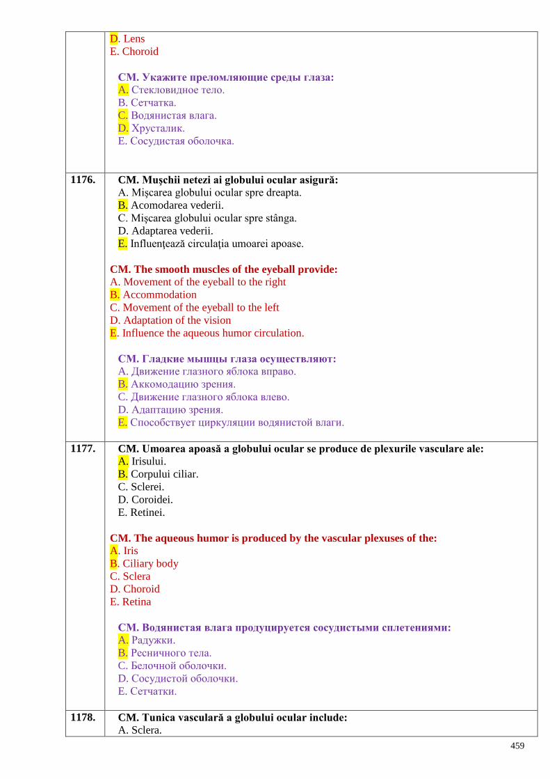
459
D. Lens
E. Choroid
СМ. Укажите преломляющие среды глаза:
A. Стекловидное тело.
B. Сетчатка.
C. Водянистая влага.
D. Хрусталик.
E. Сосудистая оболочка.
1176. CM. Muşchii netezi ai globului ocular asigură:
A. Mişcarea globului ocular spre dreapta.
B. Acomodarea vederii.
C. Mişcarea globului ocular spre stânga.
D. Adaptarea vederii.
E. Influenţează circulaţia umoarei apoase.
CM. The smooth muscles of the eyeball provide:
A. Movement of the eyeball to the right
B. Accommodation
C. Movement of the eyeball to the left
D. Adaptation of the vision
E. Influence the aqueous humor circulation.
СМ. Гладкие мышцы глаза осуществляют:
A. Движение глазного яблока вправо.
B. Аккомодацию зрения.
C. Движение глазного яблока влево.
D. Адаптацию зрения.
E. Способствует циркуляции водянистой влаги.
1177. CM. Umoarea apoasă a globului ocular se produce de plexurile vasculare ale:
A. Irisului.
B. Corpului ciliar.
C. Sclerei.
D. Coroidei.
E. Retinei.
CM. The aqueous humor is produced by the vascular plexuses of the:
A. Iris
B. Ciliary body
C. Sclera
D. Choroid
E. Retina
СМ. Водянистая влага продуцируется сосудистыми сплетениями:
A. Радужки.
B. Ресничного тела.
C. Белочной оболочки.
D. Сосудистой оболочки.
E. Сетчатки.
1178. CM. Tunica vasculară a globului ocular include:
A. Sclera.
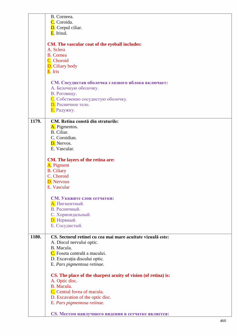
460
B. Corneea.
C. Coroida.
D. Corpul ciliar.
E. Irisul.
CM. The vascular coat of the eyeball includes:
A. Sclera
B. Cornea
C. Choroid
D. Ciliary body
E. Iris
СМ. Сосудистая оболочка глазного яблока включает:
A. Белочную оболочку.
B. Роговицу.
C. Собственно сосудистую оболочку.
D. Ресничное тело.
E. Радужку.
1179. CM. Retina constă din straturile:
A. Pigmentos.
B. Ciliar.
C. Coroidian.
D. Nervos.
E. Vascular.
CM. The layers of the retina are:
A. Pigment
B. Ciliary
C. Choroid
D. Nervous
E. Vascular
СМ. Укажите слои сетчатки:
A. Пигментный.
B. Ресничный.
C. Хориоидальный.
D. Нервный.
E. Сосудистый.
1180. CS. Sectorul retinei cu cea mai mare acuitate vizuală este:
A. Discul nervului optic.
B. Macula.
C. Foseta centrală a maculei.
D. Excavaţia discului optic.
E. Pars pigmentosa retinae.
CS. The place of the sharpest acuity of vision (of retina) is:
A. Optic disc.
B. Macula.
C. Central fovea of macula.
D. Excavation of the optic disc.
E. Pars pigmentosa retinae.
CS. Местом наилучшего видения в сетчатке является:
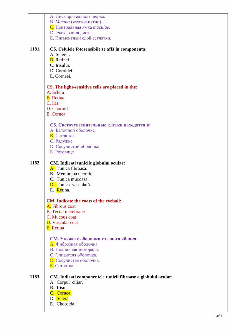
461
A. Диск зрительного нерва.
B. Macula (желтое пятно).
C. Центральная ямка maculaе.
D. Экскавация диска.
E. Пигментный слой сетчатки.
1181. CS. Celulele fotosensibile se află în componența:
A. Sclerei.
B. Retinei.
C. Irisului.
D. Coroidei.
E. Corneei.
CS. The light-sensitive cells are placed in the:
A. Sclera
B. Retina
C. Iris
D. Choroid
E. Cornea.
CS. Светочувствительные клетки находятся в:
A. Белочной оболочке.
B. Сетчатке.
C. Радужке.
D. Сосудистой оболочке.
E. Роговице.
1182. CM. Indicați tunicile globului ocular:
A. Tunica fibroasă.
B. Membrana tectorie.
C. Tunica mucoasă.
D. Tunica vasculară.
E. Retina.
CM. Indicate the coats of the eyeball:
A. Fibrous coat
B. Tectal membrane
C. Mucous coat
D. Vascular coat
E. Retina
CM. Укажите оболочки глазного яблока:
A. Фиброзная оболочка.
B. Покровная мембрана.
C. Слизистая оболочка.
D. Сосудистая оболочка.
E. Сетчатка.
1183. CM. Indicați componentele tunicii fibroase a globului ocular:
A. Corpul ciliar.
B. Irisul.
C. Cornea.
D. Sclera.
E. Choroida.
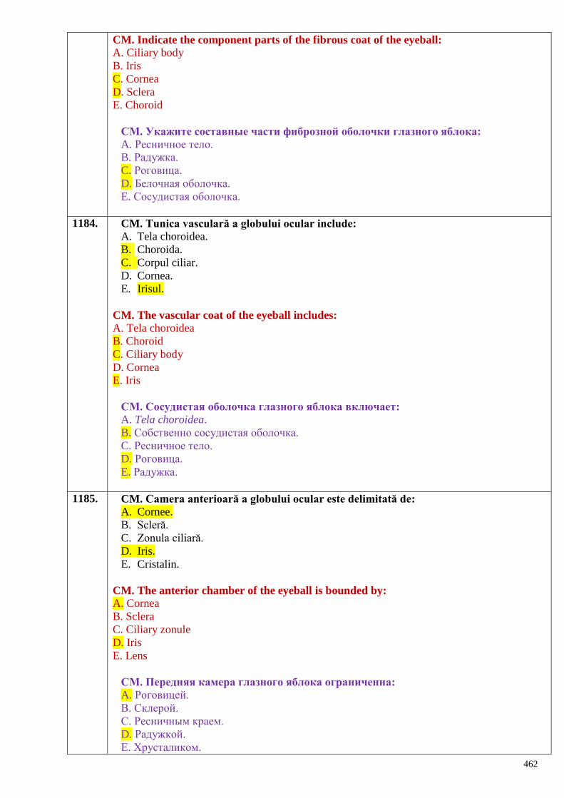
462
CM. Indicate the component parts of the fibrous coat of the eyeball:
A. Ciliary body
B. Iris
C. Cornea
D. Sclera
E. Choroid
СМ. Укажите составные части фиброзной оболочки глазного яблока:
A. Ресничное тело.
B. Радужка.
C. Роговица.
D. Белочная оболочка.
E. Сосудистая оболочка.
1184. CM. Tunica vasculară a globului ocular include:
A. Tela choroidea.
B. Choroida.
C. Corpul ciliar.
D. Cornea.
E. Irisul.
CM. The vascular coat of the eyeball includes:
A. Tela choroidea
B. Choroid
C. Ciliary body
D. Cornea
E. Iris
СМ. Сосудистая оболочка глазного яблока включает:
A. Tela choroidea.
B. Собственно сосудистая оболочка.
C. Ресничное тело.
D. Роговица.
E. Радужка.
1185. CM. Camera anterioară a globului ocular este delimitată de: A. Cornee.
B. Scleră.
C. Zonula ciliară.
D. Iris.
E. Cristalin.
CM. The anterior chamber of the eyeball is bounded by:
A. Cornea
B. Sclera
C. Ciliary zonule
D. Iris
E. Lens
СМ. Передняя камера глазного яблока ограниченна:
A. Роговицей.
B. Склерой.
C. Ресничным краем.
D. Радужкой.
E. Хрусталиком.
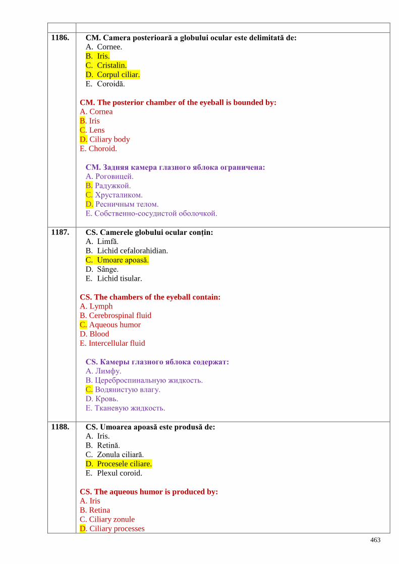
463
1186. CM. Camera posterioară a globului ocular este delimitată de: A. Cornee.
B. Iris.
C. Cristalin.
D. Corpul ciliar.
E. Coroidă.
CM. The posterior chamber of the eyeball is bounded by:
A. Cornea
B. Iris
C. Lens
D. Ciliary body
E. Choroid.
СМ. Задняя камера глазного яблока ограничена:
A. Роговицей.
B. Радужкой.
C. Хрусталиком.
D. Ресничным телом.
E. Собственно-сосудистой оболочкой.
1187. CS. Camerele globului ocular conțin: A. Limfă.
B. Lichid cefalorahidian.
C. Umoare apoasă.
D. Sânge.
E. Lichid tisular.
CS. The chambers of the eyeball contain:
A. Lymph
B. Cerebrospinal fluid
C. Aqueous humor
D. Blood
E. Intercellular fluid
СS. Камеры глазного яблока содержат:
A. Лимфу.
B. Цереброспинальную жидкость.
C. Водянистую влагу.
D. Кровь.
E. Тканевую жидкость.
1188. CS. Umoarea apoasă este produsă de: A. Iris.
B. Retină.
C. Zonula ciliară.
D. Procesele ciliare.
E. Plexul coroid.
CS. The aqueous humor is produced by:
A. Iris
B. Retina
C. Ciliary zonule
D. Ciliary processes
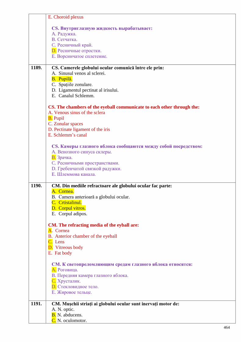
464
E. Choroid plexus
СS. Внутриглазную жидкость вырабатывает:
A. Радужка.
B. Сетчатка.
C. Ресничный край.
D. Ресничные отростки.
E. Ворсинчатое сплетение.
1189. CS. Camerele globului ocular comunică între ele prin: A. Sinusul venos al sclerei.
B. Pupilă.
C. Spațiile zonulare.
D. Ligamentul pectinat al irisului.
E. Canalul Schlemm.
CS. The chambers of the eyeball communicate to each other through the:
A. Venous sinus of the sclera
B. Pupil
C. Zonular spaces
D. Pectinate ligament of the iris
E. Schlemm’s canal
СS. Камеры глазного яблока сообщаются между собой посредством:
A. Венозного синуса склеры.
B. Зрачка.
C. Ресничными пространствами.
D. Гребенчатой связкой радужки.
E. Шлеммова канала.
1190. CM. Din mediile refractoare ale globului ocular fac parte:
A. Cornea.
B. Camera anterioară a globului ocular.
C. Crtistalinul.
D. Corpul vitros.
E. Corpul adipos.
CM. The refracting media of the eyball are:
A. Cornea
B. Anterior chamber of the eyeball
C. Lens
D. Vitreous body
E. Fat body
СМ. К светопреломляющим средам глазного яблока относятся:
A. Роговица.
B. Передняя камера глазного яблока.
C. Хрусталик.
D. Стекловидное тело.
E. Жировое тельце.
1191. CM. Muşchii striaţi ai globului ocular sunt inervaţi motor de:
A. N. optic.
B. N. abducens.
C. N. oculomotor.
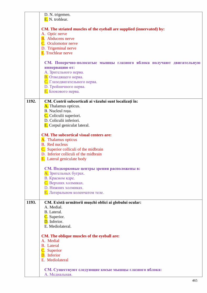
465
D. N. trigemen.
E. N. trohlear.
CM. The striated muscles of the eyeball are supplied (innervated) by:
A. Optic nerve
B. Abducens nerve
C. Oculomotor nerve
D. Trigeminal nerve
E. Trochlear nerve
СМ. Поперечно-полосатые мышцы глазного яблока получают двигательную
иннервацию от:
A. Зрительного нерва.
B. Отводящего нерва.
C. Глазодвигательного нерва.
D. Тройничного нерва.
E. Блокового нерва.
1192. CM. Centrii subcorticali ai văzului sunt localizaţi în:
A. Thalamus opticus.
B. Nucleul roşu.
C. Coliculii superiori.
D. Coliculii inferiori.
E. Corpul geniculat lateral.
CM. The subcortical visual centers are:
A. Thalamus opticus
B. Red nucleus
C. Superior colliculi of the midbrain
D. Inferior colliculi of the midbrain
E. Lateral geniculate body
СМ. Подкорковые центры зрения расположены в:
A. Зрительных буграх.
B. Красном ядре.
C. Верхних холмиках.
D. Нижних холмиках.
E. Латеральном коленчатом теле.
1193. CM. Există următorii muşchi oblici ai globului ocular:
A. Medial.
B. Lateral.
C. Superior.
D. Inferior.
E. Mediolateral.
CM. The oblique muscles of the eyeball are:
A. Medial
B. Lateral
C. Superior
D. Inferior
E. Mediolateral
СМ. Существуют следующие косые мышцы глазного яблока:
A. Медиальная.
466
B. Латеральная.
C. Верхняя.
D. Нижняя.
E. Медиолатеральная.
1194. CS. Nervul optic este format din axonii celulelor:
A. Bipolare.
B. Fotosensibile.
C. Ganglionare.
D. Conilor şi bastonaşelor.
E. Melanocitelor.
CS. The axons of which neurons form the optic nerve?
A. Bipolar
B. Photosensitive
C. Ganglionic
D. Rods and cones
E. Melanocytes
СS. Зрительный нерв образован аксонами клеток:
A. Биполярных.
B. Светочувствительных.
C. Узловых.
D. Палочек и колбочек.
E. Меланоцитов.
1195. CM. Pe imaginea oftalmoscopică a fundului de ochi se observă:
A. A. centrală a retinei.
B. V. centrală a retinei.
C. Zonele ciliare.
D. Pata oarbă.
E. Pata galbenă.
CM. The structures seen on the ophthalmoscopic image are:
A. Central artery of the retina
B. Central vein of the retina
C. Ciliary zonules
D. Blind spot (optic disc)
E. Yellow spot (macula).
СМ. При офтальмоскопии глазного дна видны:
A. Центральная артерия сетчатки.
B. Центральная вена сетчатки.
C. Ресничные зоны.
D. Слепое пятно.
E. Желтое пятно.
1196. CM. Aparatul lacrimal include:
A. Pleoapele.
B. Glandele tarsale.
C. Glanda lacrimală.
D. Râul lacrimal.
E. Lacul lacrimal.
CM. The lacrimal apparatus includes:
467
A. Eyelids
B. Tarsal glands
C. Lacrimal gland
D. Lacrimal river
E. Lacrimal lake
СМ. Слѐзный аппарат содержит:
A. Веки.
B. Тарсальные железы.
C. Слѐзную железу.
D. Слѐзный ручей.
E. Слѐзное озеро.
1197. CS. Ganglionul ciliar se află:
A. În fosa pterigopalatină.
B. La baza externa a craniului.
C. În orbită.
D. În fosa submandibulară.
E. Pe fața laterală a nervului mandibular.
CS. The ciliary ganglion is located:
A. In the pterygopalatine fossa
B. On the exobase of the skull
C. In the orbit
D. In the submandibular fossa
E. On the lateral side of the mandibular nerve.
СS. Ресничный узел находится: A. В крылонебной ямке.
B. У наружного основания черепа.
C. В глазнице.
D. В поднижнечелюстной ямке.
E. На латеральной поверхности нижнечелюстного нерва.
1198. CS. Nervul trohlear inervează:
A. Muşchiul drept superior.
B. Muşchiul levator al palpebrei superioare.
C. Muşchiul oblic superior.
D. Muşchiul oblic inferior.
E. Muşchiul drept lateral.
CS. The trochlear nerve supplies:
A. Superior rectus muscle
B. Levator palpebrae superior muscle
C. Superior oblique muscle
D. Inferior oblique muscle
E. Lateral rectus muscle
СS. Блоковой нерв иннервирует:
A. Верхнюю прямую мышцу.
B. Мышцу поднимающую верхнее веко.
C. Верхнюю косую мышцу.
D. Нижнюю косую мышцу.
E. Латеральную прямую мышцу.
468
1199. CS. Segmentul analizatorului care generează impulsul nervos:
A. Conductor.
B. Central.
C. Celulele neurogliale.
D. Receptor.
E. Cortical.
CS. The segment of the analyzer generating the nerve impulse is:
A. Transmitter
B. Central part
C. Neuroglial cells
D. Receptor
E. Cortical end.
CS. Сегмент анализатора где генерируется нервный импульс:
A. Проводниковый.
B. Центральный.
C. Нейроглиальных клеток.
D. Рецепторный.
E. Корковый.
1200. CS. Centrul cortical al analizatorului vizual este situat în:
A. Pulvinar thalami.
B. Corpul geniculat lateral.
C. Pe marginile şanţului calcarin.
D. Hipocamp.
E. Girii orbitari.
CS. The cortical center of the visual analyzer is located in:
A. Pulvinar of the thalamus
B. Lateral geniculate body
C. Borders of the calcarine groove
D. Hippocampus
E. Orbital gyri.
CS. Корковый центр зрительного анализатора расположен в:
A. Подушке зрительного бугра.
B. Латеральном коленчатом теле.
C. По краям шпорной борозды.
D. Гиппокампе.
E. Глазничных извилинах.
1201. CM. Centrii subcorticali ai văzului sunt localizaţi în:
A. Diencefal.
B. Nucleul roşu.
C. Coliculii cvadrigemeni superiori.
D. Coliculii cvadrigemeni inferiori.
E. Corpul geniculat lateral.
CM. The subcortical visual centers are:
A. Diencephalon
B. Red nucleus
C. Superior collicli of the tectal lamina
D. Inferior collicli of the tectal lamina
E. Lateral geniculate body
469
СМ. Подкорковые центры зрения расположены в:
A. Промежуточном мозге.
B. Красном ядре.
C. Верхних холмиках четверохолмия.
D. Нижних холмиках четверохолмия.
E. Латеральном коленчатом теле.
1202. CS. Nervul abducens inervează următorii muşchi ai globului ocular:
A. Drept superior.
B. Oblic superior.
C. Drept lateral.
D. Drept medial.
E. Oblic inferior.
CS. The muscles supplied by the abducent nerve are:
A. Superior rectus muscle
B. Oblique superior muscle
C. Lateral rectus muscle
D. Medial rectus muscle
E. Inferior oblique muscle
CS. Отводящий нерв иннервирует следующие мышцы глазного яблока:
A. Верхнюю прямую.
B. Верхнюю косую.
C. Латеральную прямую.
D. Медиальную прямую.
E. Нижнюю косую.
1203. CM. Nervi care inervează motor muşchii globului ocular:
A. N. optic.
B. N. abducens.
C. N. oculomotor.
D. N. trigemen.
E. N. trohlear.
CM. The motor innervation of the muscles of the eyeball is provided by:
A. Optic nerve
B. Abducens nerve
C. Oculomotor nerve
D. Trigeminal nerve
E. Trochlear nerve.
СМ. Нервы, иннервирующие мышцы глазного яблока:
A. Зрительный нерв.
B. Отводящий нерв.
C. Глазодвигательный нерв.
D. Тройничный нерв.
E. Блоковой нерв.
1204. CS. Nervul oculomotor iese din craniu prin:
A. Foramen ovale.
B. Fissura orbitalis superior.
C. Fissura orbitalis inferior.
D. Canalis opticus.
470
E. Foramen supraorbitalis.
CS. The oculomotor nerve leaves the skull through:
A. Foramen ovale.
B. Fissura orbitalis superior.
C. Fissura orbitalis inferior.
D. Canalis opticus.
E. Foramen supraorbitalis.
CS. Глазодвигательный нерв выходит из черепа через:
A. Овальное отверстие.
B. Верхнюю глазничную щель.
C. Нижнюю глазничную щель.
D. Зрительный канал.
E. Надглазничное отверстие.
1205. CM. Anexele organului văzului sunt reprezentate prin:
A. Mușchii globului ocular.
B. Pleoape.
C. Tunica conjunctivă.
D. Aparatul lacrimal.
E. Tunica vasculară.
CM. The auxiliary apparatus of the eyeball comprises:
A. Muscles of the eyeball
B. Eyelids
C. Conjunctiva
D. Lacrimal apparatus
E. Vascular coat.
СМ. К вспомогательным органам глаза относятся:
A. Мышцы глазного яблока.
B. Веки.
C. Конъюнктива.
D. Слезный аппарат.
E. Сосудистая оболочка.
1206. CM. Elementele anatomice ale urechii externe sunt:
A. Conductul auditiv extern.
B. Tuba auditivă.
C. Oscioarele auditive.
D. Pavilionul urechii.
E. Cavitatea timpanică.
CM. The anatomcal elements of external ear are, as follows:
A. External auditory meatus.
B. Auditory tube.
C. Auditory ossicles.
D. Auricle.
E. Tympanic cavity.
СМ. Анатомическими образованиями наружного уха являются:
A. Наружный слуховой проход.
B. Слуховая труба.
C. Слуховые косточки.
471
D. Ушная раковина.
E. Барабанная полость.
1207. CM. Urechea medie include:
A. Membrana timpanică.
B. Cavitatea timpanică.
C. Labirintul osos şi membranos.
D. Oscioarele auditive.
E. Tuba auditivă.
CM. The middle ear includes:
A. Tympanic membrane.
B. Tympanic cavity.
C. Bony and membranous labyrinths.
D. Auditory ossicles.
E. Auditory tube.
СМ. Среднее ухо включает:
A. Барабанную перепонку.
B. Барабанную полость.
C. Костный и перепончатый лабиринт.
D. Слуховые косточки.
E. Слуховую трубу.
1208. CM. Urechea externă include:
A. Auricula.
B. Meatul auditiv intern.
C. Membrana timpanică.
D. Meatul auditiv extern.
E. Cavitatea timpanică.
CM. The external ear includes:
A. Auricule.
B. Internal auditory meatus.
C. Tympanic membrane.
D. External auditory meatus.
E. Tympanic cavity.
СМ. Наружное ухо включает:
A. Ушную раковину.
B. Внутренний слуховой проход.
C. Барабанную перепонку.
D. Наружный слуховой проход.
E. Барабанную полость.
1209. CM. Membranei timpanice i se disting:
A. Partea mucoasă.
B. Partea tensionată.
C. Partea flască.
D. Partea membranoasă.
E. Partea seroasă.
CM. The following parts can be distinguished in the tympanic membrane:
A. Mucous part.
B. Tense part.
472
C. Flaccid part.
D. Membranous part.
E. Serous part.
СМ. Барабанная перепонка имеет:
A. Слизистую часть.
B. Натянутую часть.
C. Ненатянутую часть.
D. Перепончатую часть.
E. Серозную часть.
1210. CM. În cavitatea timpanică sunt localizaţi muşchii:
A. Tensor al vălului palatin.
B. Tensor al timpanului.
C. Auricular posterior.
D. Scăriței.
E. Nicovalei.
CM. The following muscles are located in the tympanic cavity:
A. Tensor veli palatini muscle.
B. Tensor tympani muscle.
C. Posterior auricular muscle.
D. Stapedius muscle.
E. Incudal muscle.
СМ. В барабанной полости расположены мышцы:
A. Напрягающую небную занавеску.
B. Напрягающую барабанную перепонку.
C. Задняя ушная.
D. Стременная мышца.
E. Мышца наковальни.
1211. CM. Indicați părțile labirintului osos:
A. Vestibulul labirintului.
B. Peștera mastoidiană.
C. Canalele semicirculare.
D. Melcul.
E. Căluțul de mare.
CM. Indicate the parts of bony labyrinth:
A. Vestibule.
B. Mastoid antrum.
C. Semicircular canals.
D. Cochlea.
E. Seahorse.
СМ. Укажите части костного лабиринта:
A. Преддверие лабиринта.
B. Сосцевидная пещера.
C. Полукружные каналы.
D. Улитка.
E. Гиппокамп
1212. CS. Urechea internă include:
A. Tuba auditivă.
473
B. Oscioarele auditive.
C. Labirintul osos şi cel membranos.
D. Cavitatea timpanică.
E. Muşchiul scăriţei.
CS. The internal ear includes:
A. Auditory tube.
B. Auditory ossicles.
C. Bony and membranous labyrinths.
D. Tympanic cavity.
E. Stapedius muscle.
CS. Внутреннее ухо включает:
A. Слуховую трубу.
B. Слуховые косточки.
C. Костный и перепончатый лабиринты.
D. Барабанную полость.
E. Стременную мышцу.
1213. CM. Centrii subcorticali ai analizatorului auditiv sunt:
A. Talamusul.
B. Nucleele coliculilor superiori.
C. Nucleele coliculilor inferiori.
D. Corpul geniculat lateral.
E. Corpul geniculat medial.
CM. The subcortical centers of auditory analyser are, as follows:
A. Thalamus.
B. Nuclei of the superior colliculi.
C. Nuclei of the inferior colliculi.
D. Lateral geniculate body.
E. Medial geniculate body.
СМ. Подкорковыми центрами слухового анализатора являются:
A. Зрительный бугор.
B. Ядра верхних холмиков.
C. Ядра нижних холмиков.
D. Латеральное коленчатое тело.
E. Медиальное коленчатое тело.
1214. CM. Se disting canalele semicirculare ale labirintului osos:
A. Anterior.
B. Medial.
C. Posterior.
D. Superior.
E. Lateral.
CM. The following semicircular canals are distinguished in the bony labyrinth:
A. Anterior.
B. Medial.
C. Posterior.
D. Superior.
E. Lateral.
СМ. Различают костные полукружные каналы:
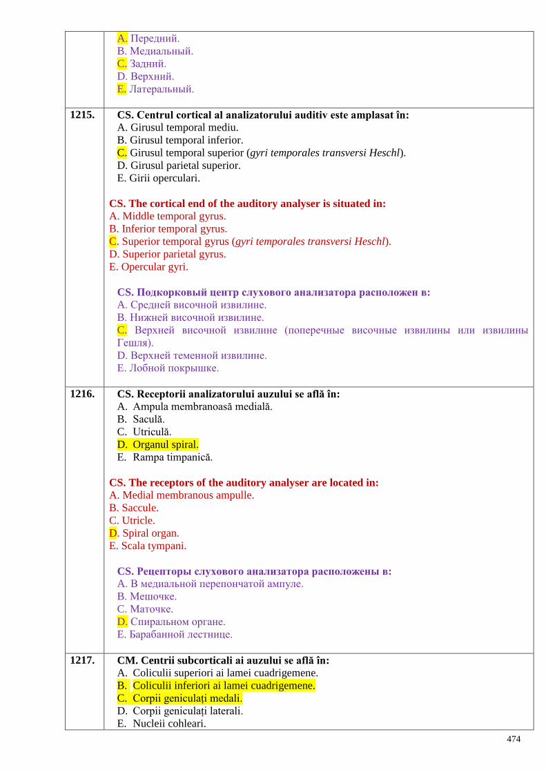
474
A. Передний.
B. Медиальный.
C. Задний.
D. Верхний.
E. Латеральный.
1215. CS. Centrul cortical al analizatorului auditiv este amplasat în:
A. Girusul temporal mediu.
B. Girusul temporal inferior.
C. Girusul temporal superior (gyri temporales transversi Heschl).
D. Girusul parietal superior.
E. Girii operculari.
CS. The cortical end of the auditory analyser is situated in:
A. Middle temporal gyrus.
B. Inferior temporal gyrus.
C. Superior temporal gyrus (gyri temporales transversi Heschl).
D. Superior parietal gyrus.
E. Opercular gyri.
CS. Подкорковый центр слухового анализатора расположен в:
A. Средней височной извилине.
B. Нижней височной извилине.
C. Верхней височной извилине (поперечные височные извилины или извилины
Гешля).
D. Верхней теменной извилине.
E. Лобной покрышке.
1216. CS. Receptorii analizatorului auzului se află în:
A. Ampula membranoasă medială.
B. Saculă.
C. Utriculă.
D. Organul spiral.
E. Rampa timpanică.
CS. The receptors of the auditory analyser are located in:
A. Medial membranous ampulle.
B. Saccule.
C. Utricle.
D. Spiral organ.
E. Scala tympani.
CS. Рецепторы слухового анализатора расположены в:
A. В медиальной перепончатой ампуле.
B. Мешочке.
C. Маточке.
D. Спиральном органе.
E. Барабанной лестнице.
1217. CM. Centrii subcorticali ai auzului se află în:
A. Coliculii superiori ai lamei cuadrigemene.
B. Coliculii inferiori ai lamei cuadrigemene.
C. Corpii geniculați medali.
D. Corpii geniculați laterali.
E. Nucleii cohleari.
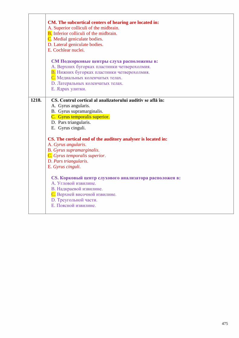
475
CM. The subcortical centers of hearing are located in:
A. Superior colliculi of the midbrain.
B. Inferior colliculi of the midbrain.
C. Medial geniculate bodies.
D. Lateral geniculate bodies.
E. Cochlear nuclei.
CM Подкорковые центры слуха расположены в:
A. Верхних бугорках пластинки четверохолмия.
B. Нижних бугорках пластинки четверохолмия.
C. Медиальных коленчатых телах.
D. Латеральных коленчатых телах.
E. Ядрах улитки.
1218. CS. Centrul cortical al analizatorului auditiv se află în:
A. Gyrus angularis.
B. Gyrus supramarginalis.
C. Gyrus temporalis superior.
D. Pars triangularis.
E. Gyrus cinguli.
CS. The cortical end of the auditory analyser is located in:
A. Gyrus angularis.
B. Gyrus supramarginalis.
C. Gyrus temporalis superior.
D. Pars triangularis.
E. Gyrus cinguli.
CS. Корковый центр слухового анализатора расположен в:
A. Угловой извилине.
B. Надкраевой извилине.
C. Верхней височной извилине.
D. Треугольной части.
E. Поясной извилине.