Embed Size (px)
Citation preview
5
RiassuntoNel 2011, ceppi di lignaggio 1 del virus della West Nile (WNV) sono stati identificati per la prima volta in Sardegna. A differenza di quanto osservato in altre regioni italiane, il virus è stato rinvenuto in esemplari di Culex modestus ed è stato in grado di evocare sintomatologia clinica negli uccelli infettati. L’analisi filogenetica basata su un frammento del gene codificante la proteina NS3 ha svelato un elevato grado di similitudine tra i ceppi sardi ed i ceppi di WNV che hanno circolato recentemente nei paesi del Bacino Mediterraneo. Tuttavia, le sequenze ottenute dagli isolati sardi del 2011 sono raggruppate in un subcluster distinto. L’analisi delle sequenze ha confermato la presenza di differenti genotipi virali del WNV, in particolare NS3-249P e NS3-249T, a conferma della contemporanea circolazione di diverse popolazioni virali nel corso del focolaio. Tali differenze genotipiche, tuttavia, non sono risultate associate a variazioni della patogenicità.
Parole chiaveGene codificante NS3,Italia,Lignaggio 1,Sardegna,Uccelli selvatici,Virus della West Nile.
Descrizione dei focolai di West Nile disease nel 2011nella regione Sardegna, Italia
SummaryIn 2011, strains of West Nile Virus (WNV) belonging to lineage 1 spread for the first time in Sardinia region (Italy). In contrast to previous WNV Italian incursion, the strains were found in Culex modestus and, more surprisingly, they were able to cause severe clinical signs in the affected birds. Based on the partial sequence of the NS3 encoding gene, the Sardinian WNV strains demonstrated a high similarity with the other WNV strains recently detected in the Mediterranean Basin. Nonetheless, the 2011 Sardinian sequences were grouped in a distinct sub-cluster. Both the NS3-249P and NS3-249T genotypes were detected in the Sardinian outbreaks confirming that the co-circulation of different genotypes in the affected population might be common for WNV as for many RNA viruses. No association, however, was observed between virulence and viral genotype.
KeywordsItaly,Lineage 1,NS3 encoding gene,Sardinia,West Nile virus,Wild birds.
Veterinaria Italiana 2015, 51 (1), 5-16. doi: 10.12834/VetIt.260.2386.2Accepted: 21.08.2014 | Available on line: 23.01.2015
1 Istituto Zooprofilattico Sperimentale dell’Abruzzo e del Molise ‘G. Caporale’, Campo Boario, 64100 Teramo, Italy.2 Clinica Veterinaria ‘Duemari’, Via Cagliari 313, 09170 Oristano, Italy.
3 Istituto Zooprofilattico Sperimentale della Sardegna, Dipartimento di Oristano, Via Atene ZI, 09170 Oristano, Italy.
* Corresponding author at: Istituto Zooprofilattico Sperimentale dell’Abruzzo e del Molise ‘G. Caporale’,Campo Boario, 64100 Teramo, Italy.
Tel: + 39 0861 332446, Fax: +39 0861 332251, e-mail: [email protected].
Federica Monaco1*, Maria Goffredo1, Paolo Briguglio2, Chiara Pinoni1, Andrea Polci1,Simona Iannetti1, Stefano Pinto2, Giuseppe Marruchella1, Gabriella Di Francesco1,
Annapia Di Gennaro1, Monica Pais2, Liana Teodori1, Rossana Bruno1,Monica Catalani1, Angelo Ruiu3, Rossella Lelli1 & Giovanni Savini1
The 2011 West Nile disease outbreakin Sardinia region, Italy
reading frame that encodes 4 structural proteins (the nucleocapsid, the pre-membrane, the membrane, the envelope) and 7 non-structural proteins (NS1, NS2A, NS2B, NS3, NS4A, NS4B and NS5).
Based on phylogenetic analysis the worldwide circulating WN strains are clustered in 8 lineages (Mackenzie and Williams 2009, Vazquez et al. 2010),
IntroductionWest Nile virus (WNV) is a mosquito-borne zoonotic flavivirus member of the Japanese encephalitis serogroup, which includes other important neuro-invasive viruses such as the Japanese encephalitis virus, the Murray Valley virus and St Louis virus. The WNV genome is composed of a single open
6 Veterinaria Italiana 2015, 51 (1), 5-16. doi: 10.12834/VetIt.260.2386.2
West Nile virus in Sardinia region Monaco et al.
Materials and methods
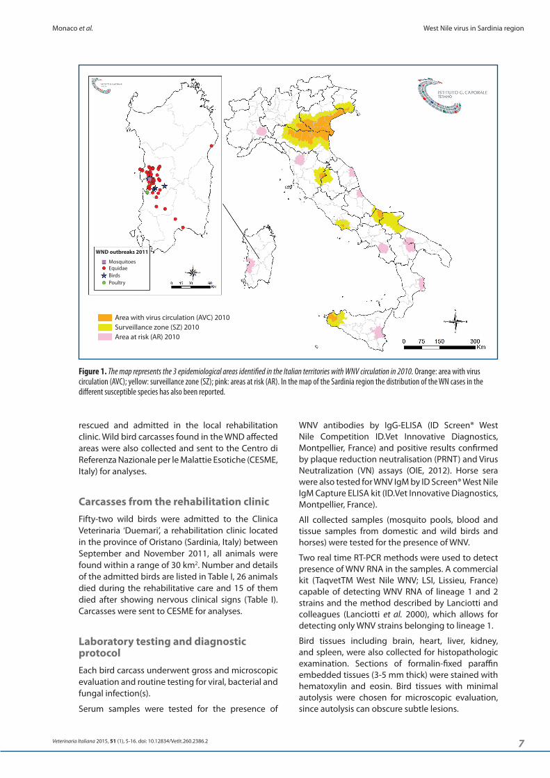
National surveillance programIn Italy, a WNV National surveillance plan is in place since 2002 (Calistri et al. 2010). Although continuously revised according to the new epidemiological scenarios, the plan selected 10 risky areas within the entire Italian territory. Such areas are characterized by the presence of a significant number of wild birds including species of migratory birds. In these areas, WNV circulation is monitored each year, between March and November, the monitoring encompasses sentinel animals (chicken and horses), wild bird carcasses, and mosquitoes caught by specific traps. In Sardinia, the sentinel chickens were housed in Arborea (39°81’ N - 08°56’ E) and Santa Giusta, (39°83’ N - 08°60’ E), 2 municipalities of the Oristano province, which are close to humid areas with great abundance of wild birds. The mosquito traps were located in the wetland of S’Ena Arrubia (39°49’ N - 08°34’ E), in the Arborea municipality, where Culex pipiens resulted to be the most abundant species collected since 2002. Sentinel horses, 28 animals, were located in Arborea (39°79’ N - 08°55’ E). Serum samples were collected from sentinel animals 3 times per year (April - August - October) (Figure 1).
Clinical and neurological investigations were conducted on all horses of the affected farms. Blood and serum samples were also collected from all animals and sent to the OIE and National reference laboratory for WND, CESME, Istituto Zooprofilattico Sperimentale dell’Abruzzo e del Molise, Italy (IZSAM).
After the first WNV confirmed cases, the routine monitoring was extended on horses with WND-like symptoms living within 4 km of radius from the infected premises.
Mosquito surveyBetween September the 14th and 18th 2011, 10 mosquito collections were performed, using Centers for Disease Control (CDC) light traps, BG Sentinel traps (CDC, Atlanta, USA) and manual aspiration. The collected insects were identified at species level (Severini et al. 2009) and divided in pools to be tested for WNV by real time RT-PCR (TaqvetTM West Nile WNV; LSI, Lissieu, France). Within each collection and species, males, engorged and non-engorged females were tested separately.
Birds monitoringDuring the Sardinian 2011 WND epidemic, an unusual number of wild birds showing neurologic illness was noted in the field. Some of them were
with the strains belonging to lineage 1 and 2 being the most widely disseminated.
Strains of lineage 2 are endemic in Southern African Countries and were first detected in Europe in 2004 in a goshawk (Accipiter gentilis) of a national park in South-east Hungary (Bakonyi et al. 2006, Erdélyi et al. 2007) and then in Austria (Wodak et al. 2012). Other WND outbreaks caused by lineage 2 strains were reported in Russia (Platonov et al. 2012) and in Romania (Sirbu et al. 2011). A strain of lineage 2 has recently circulated in Greece (Danis et al. 2011) and Balcan countries (WAHID 2014, Petrovic T. et al. 2013).
In Italy, WNV first appeared in 1998 in the Padule di Fucecchio mash area, in Tuscany, (Autorino et al. 2002); and then it was detected in the North-Eastern part of Italy in 2008 (Calistri et al. 2010b, Monaco et al. 2010) where it became endemic (Monaco et al. 2009). Unrelated new foci were also reported in Central and Southern Italy (Calistri et al. 2010a) and, more recently, lineage 2 strains were detected in Central (Bagnarelli et al. 2011) and Northern-eastern parts of Italy, as well as in Sardinia (Savini et al. 2012, Capelli et al. 2013).
As for the pathogenicity, strains of lineages 1 and 2 have been associated to severe disease in birds, horses, and/or humans (Kutasi et al. 2011). In humans and horses, disease is a spill over event emerging from the enzootic cycle, which involves vertebrates, mainly birds and mosquitoes, Culex mosquitoes and passerine birds being, respectively, the main vectors and vertebrate hosts for virus spread.
In Italy, clinical cases have been reported both in human and in horses but never in birds, at least before 2011, even if birds have been found in the past to be infected by WNV (Lelli et al. 2012). Magpies (Pica pica), carrion crows (Corvus corone) and rock pigeons (Columba livia) are the species most commonly found infected by WNV. Ochlerotatus caspius and Culex pipiens are, instead, the most abundant mosquitos found in Italy (Thompson et al. 1994) and those in which WNV has been detected (Monaco et al. 2010, Capelli et al. 2013).
In September 2011, a severe WND epidemic was first reported in Sardinia. During this epidemic, numerous horses became infected, some of them died or were euthanized because of the severity of the clinical signs, others recovered after showing classical nervous symptoms. Interestingly, in the same area and in the same period, an unusual number of wild birds dying after showing neurologic illness was also noted. This paper describes the Sardinian outbreak with special emphasis on the clinical signs and virological findings due to WNV infection observed in several birds admitted to a rehabilitation clinic or collected in the field.
7
Monaco et al. West Nile virus in Sardinia region
Veterinaria Italiana 2015, 51 (1), 5-16. doi: 10.12834/VetIt.260.2386.2
WNV antibodies by IgG-ELISA (ID Screen® West Nile Competition ID.Vet Innovative Diagnostics, Montpellier, France) and positive results confirmed by plaque reduction neutralisation (PRNT) and Virus Neutralization (VN) assays (OIE, 2012). Horse sera were also tested for WNV IgM by ID Screen® West Nile IgM Capture ELISA kit (ID.Vet Innovative Diagnostics, Montpellier, France).
All collected samples (mosquito pools, blood and tissue samples from domestic and wild birds and horses) were tested for the presence of WNV.
Two real time RT-PCR methods were used to detect presence of WNV RNA in the samples. A commercial kit (TaqvetTM West Nile WNV; LSI, Lissieu, France) capable of detecting WNV RNA of lineage 1 and 2 strains and the method described by Lanciotti and colleagues (Lanciotti et al. 2000), which allows for detecting only WNV strains belonging to lineage 1.
Bird tissues including brain, heart, liver, kidney, and spleen, were also collected for histopathologic examination. Sections of formalin-fixed paraffin embedded tissues (3-5 mm thick) were stained with hematoxylin and eosin. Bird tissues with minimal autolysis were chosen for microscopic evaluation, since autolysis can obscure subtle lesions.
rescued and admitted in the local rehabilitation clinic. Wild bird carcasses found in the WND affected areas were also collected and sent to the Centro di Referenza Nazionale per le Malattie Esotiche (CESME, Italy) for analyses.
Carcasses from the rehabilitation clinicFifty-two wild birds were admitted to the Clinica Veterinaria ‘Duemari’, a rehabilitation clinic located in the province of Oristano (Sardinia, Italy) between September and November 2011, all animals were found within a range of 30 km2. Number and details of the admitted birds are listed in Table I, 26 animals died during the rehabilitative care and 15 of them died after showing nervous clinical signs (Table I). Carcasses were sent to CESME for analyses.
Laboratory testing and diagnostic protocolEach bird carcass underwent gross and microscopic evaluation and routine testing for viral, bacterial and fungal infection(s).
Serum samples were tested for the presence of
Figure 1. The map represents the 3 epidemiological areas identified in the Italian territories with WNV circulation in 2010. Orange: area with virus circulation (AVC); yellow: surveillance zone (SZ); pink: areas at risk (AR). In the map of the Sardinia region the distribution of the WN cases in the different susceptible species has also been reported.
WND outbreaks 2011
MosquitoesEquidaeBirdsPoultry
Area with virus circulation (AVC) 2010Surveillance zone (SZ) 2010Area at risk (AR) 2010
8 Veterinaria Italiana 2015, 51 (1), 5-16. doi: 10.12834/VetIt.260.2386.2
West Nile virus in Sardinia region Monaco et al.
Table I. Birds admitted to the Clinica Veterinaria ‘Duemari’ (Oristano, Sardinia, Italy), between September and November 2011, and included in the study.
Common name Species Family No. deaths/admitted birds
WND+/nervous symptoms
Grey heron Ardea cinerea Ardeidae 1/2 0/1
Mallard Anas platyrhynchos Anatidae 4/4 1/4
European herring gull Larus argentatus Laridae 2/2 0/2
Barbary partridge Alectoris barbara Fasianidae 1/3 0/0
Eurasian stone curlew Burhinus oedicnemus Burhinidae 0/1 n.d.
Greater flamingo Phoenicopterus roseus Fenicotteridae 1/1 0/0
House sparrow Passer domesticus Passeridae 1/1 0/0
Purple swamphen Poprhyrio porphyrio Rallidae 0/1 n.d.
Little grebe Tachybaptus ruficollis Podicipedidae 1/1 0/0
Common buzzard Buteo buteo Accipitridae 5/13 2/3
Eurasian jay Garrulus glandarius
Corvidae 5/6
2/2
Hooded crow Corvus corone cornix 0/0
Carrion crow Corvus corone 0/0
Little owl Athena noctua Strigidae 2/4 1/2
Western barn owl Tyto alba Tytonidae 0/4 n.d.
Turtle dove Streptopelia turtur Columbidae 0/3 n.d.
Common starling Sturnus vugaris Sturnidae 1/1 0/0
Peregrine falcon Falco peregrinusFalconidae 2/5
0/0
Common kestrel Falco tinnunculus 0/1
Total 26/52 6/15n.d. = data not available.
using the amplifying primers and 4 additional internal primers (data not shown). Sequencing was performed using the BigDye Version 3.1 Dye Terminator Cycle Sequencing Kit (Applied Biosystems, Foster City, CA, USA) on ABI PRISM 3130xl automated capillary sequencer after a cleaning step with Cleanseq (Beckman Coulter, Brea, California, USA).
Raw sequence data were assembled using Contig Express (Vector NTI suite 9.1; Invitrogen, Carlsbad, California, USA) and consensus sequences aligned with the homologous sequences deposited in the Genbank database with Clustal-W (Thompson et al. 1994). Both, the nucleic and the deduced amino acid sequences were compared using Vector NTI suite 9.1 (Invitrogen, Carlsbad, California, USA).
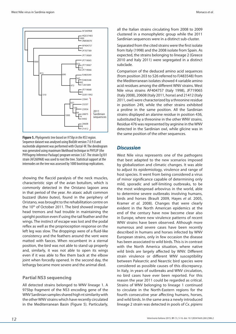
The phylogenetic analysis was conducted on 975bp in the NS3 region from the viral strains listed in Table II. Sequence dataset was analysed using BioEdit version 7.0.9.0 and nucleotide alignment was performed with Clustal-W. Aligned sequences were compared and dendrogram generated using maximum likelihood technique in PHYLIP (the PHYlogeny Inference Package) program version 3.67. The tree obtained was rooted with Eg101 strain (AF260968). Statistical support at the internodes on the tree was assessed by 1000 bootstrap replications.
Molecular characterizationIn order to characterize the viral strain, WNV positive samples were partially sequenced. Briefly: total RNA was extracted from collected samples by the automated BioSprint 96 One-For-All Vet kit (Qiagen, Leipzig, Germany ) according to manufacturer's instructions and collected in 100μl of elution buffer. A 1099bp fragment of the NS3 gene (genomic position 5216-6190 on AF404757 ITA98) was amplified by using the primer pair WN_7_5199F: 5’-CGGTGCCGGTAAAACAAG-3’ and WN_7_6297R: 5’-CCTCCGATCGTGGTATGACA-3’. The gel based RT-PCR was performed using Transcriptor One-Step RT-PCR kit (Roche Applied Science, Madison, Wisconsin, USA). The kit contains a blend consisting of Taq DNA polymerase and a proofreading polymerase, which minimizes the possibility of mutations offering high yield and fidelity in PCR.
RT-PCR cycling conditions for the amplification of WNV partial NS3 gene were 50°C 15 minutes, 94°C 7 minutes followed by 35 cycles of denaturation at 94°C for 10 seconds, annealing at 58°C for 30 seconds and extension at 68°C for 2 minutes.
Gel based RT-PCR amplicons were purified with the Qiaquick PCR Purification kit (Qiagen, Leipzig, Germany) and directly sequenced in both directions
9
Monaco et al. West Nile virus in Sardinia region
Veterinaria Italiana 2015, 51 (1), 5-16. doi: 10.12834/VetIt.260.2386.2
C.I. = 15.00-37.6%) died. West Nile virus infection was confirmed in all dead animals by serology and/or molecular tests. The temporal progression of the infection is detailed in the on-line bulletin edited by CESME1. Virus circulation involved a restricted area extended for about 2720 square kilometres of the Sardinia territories, 94 equine cases (Monaco et al. 2010) were reported from 4 provinces (Oristano, Cagliari, Nuoro and Medio Campidano). Thirty-eight different stables were involved, 33 of which located in the Oristano province, 2 in the Medio Campidano and Cagliari provinces and 1 in the Nuoro province (Table III). Four WNV cases of encephalitis have been reported in humans in the provinces of Oristano and Olbia (Rizzo et al. 2012).
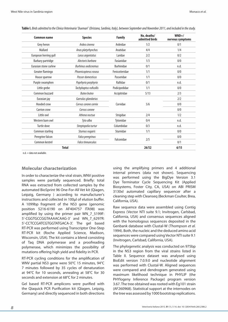
Mosquito surveyA total of 310 mosquitoes were collected on the affected area. They belonged to 5 genera and 8 species: Anopheles maculipennis, Culex pipiens, Culex modestus, Culex theileri, Ochlerotatus caspius, Culiseta annulata, Culiseta longiareolata, Coquillettidia
Results
National Surveillance ProgramSeroconversion was first detected by ELISA in a sentinel chickens in Arborea on the 6th of July 2011. The VN assay however was not able to confirm it. A second ELISA seroconversion occurred on the 18th of July in a sentinel chicken located in Santa Giusta, but again the VN test was not able to confirm the positive reaction. Neutralising antibodies were first detected on the 7th of September in 2 chicken sera from Santa Giusta, whereas on the 8th of October 3 sentinel chickens were found viraemic. Four sentinel horses first seroconverted (IgG-ELISA, IgM-ELISA, PRNT and VN assays) were detected on the 4th of October.
Case reportClinical cases were first observed on the 14th
of September in 5 horses located in 2 different stables within the Oristano province. By the end of the season, clinical signs ranging from fever to muscle fasciculation, paralysis/paresis of the limbs, proprioceptive deficits or inability to maintain the standing station were reported in 53 horses (56.38% of the confirmed cases), 13 of them (24.53%, 95%
Table II. Details of the WNV strains included in the phylogenetic analysis. For each viral strain, the GenBank identification code of the sequences included in the analysis is provided. The strains were obtained from samples collected in the Oristano province between September and November 2011.
Strain Location Year Lineage Host GenBank20608 Sardinia 2011 1 Equine KJ562347
21412 Sardinia 2011 1 Little owl KJ562350
20875 Sardinia 2011 1 Eurasian jay KJ562349
23237 Sardinia 2011 1 Sentinel chickens KJ562351
9492 Sardinia 2011 1 Mosquito pool KJ562353
23954 Sardinia 2011 1 Carrion crow KJ562352
23941 Sardinia 2011 1 Carrion crow KJ562348
WN Italy 1998 equine Italy 1998 1 Equine AF404757
15217 Italy 2008 1 Magpie FJ483548
15803 Italy 2008 1 Magpie FJ483549
Italy/2008/J-242853 Italy 2008 1 Eurasian jay JF719065
Italy/2008/M-203204 Italy 2008 1 Magpie JF719066
12010 09 Italy 2009 1 Magpie KJ562354
Ita09 Italy 2009 1 Human (bood donor) GU011992
Italy/2009/G-223184 Italy 2009 1 Gull JF719067
Italy/2009/J-225677 Italy 2009 1 Jay JF719068
Spain/2010/H-1b Spain 2010 1 Equine JF719069
HU6365/08 Spain 2008 1 Culex perexiguus JF707789
Nea Santa-Greece-2010 Italy 2010 2 Culex pipiens mosquito pool HQ537483
Italy/2011/AN-2 Italy 2011 2 Human JN858070
Eg101 Egypt 1951 1 Human AF260968
1 Istituto Zooprofilattico Sperimentale dell’Abruzzo e del Molise 2011. West Nile virus bulletin http://sorveglianza.izs.it/emergenze/west_nile/emergenze.htm.
10 Veterinaria Italiana 2015, 51 (1), 5-16. doi: 10.12834/VetIt.260.2386.2
West Nile virus in Sardinia region Monaco et al.
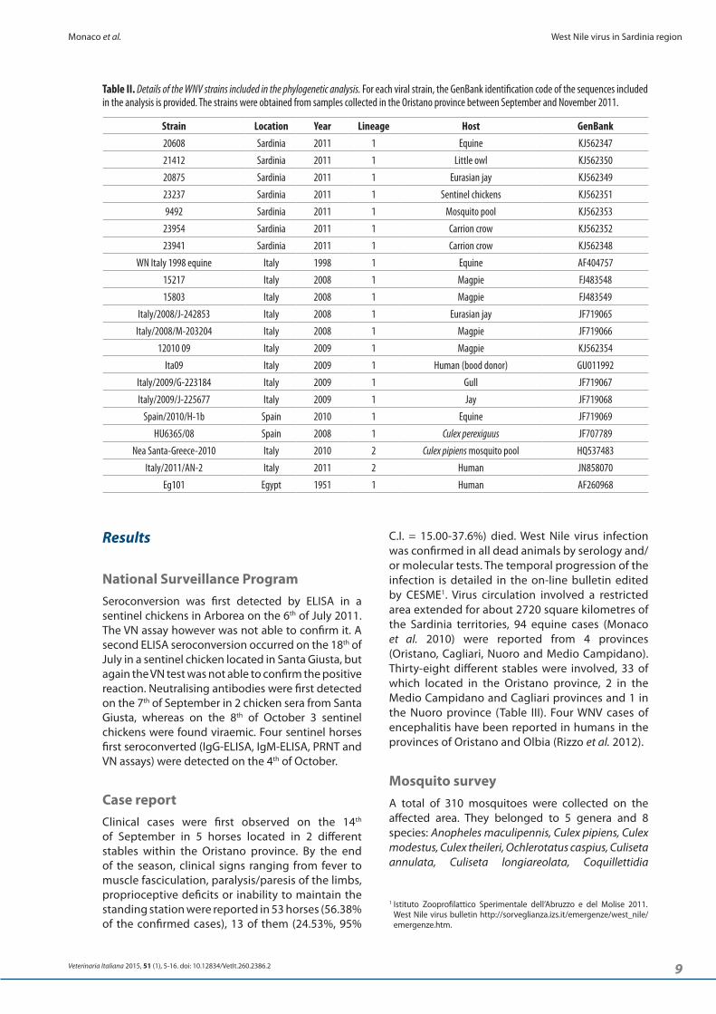
Birds monitoringA total of 132 wild birds were examined for the presence of West Nile virus, 106 were found dead in the field and 26 were from the rehabilitation clinic. Out of the 132 wild birds examined, 10 resulted positive to WNV RT-PCR (Table V). Of these, 4 (3 hooded crows and 1 Eurasian jay) were from the field and 6 from the clinic collections. All 6 birds from the rehabilitation facility suffered from nervous clinical signs before dying (Table I). In 4 occasions it was also possible to isolate the virus in cell culture. Neither traumatic lesions nor routine viral, fungal and bacterial infections were detected in the birds showing clinical signs. Because of autolysis, only few tissues could be processed, the most consistent histopathologic finding was the myocarditis (Figure 3).
Clinical casesTwo adult Eurasian jays (Garrulus glandarius) were admitted to the clinic the 5th of September 2011. They showed aspecific clinical signs characterised by drowsy, incapability of flying or walking properly, ruffle feathers, pectoral atrophy, and absence of the flight instinct. Both birds died within 24 hours from
richiardii (Figure 2). The mosquitoes were divided in 41 pools (Table IV) and only 1, consisting of 3 non-engorged females of Culex modestus collected by a BG-Sentinel trap, was positive when tested for WNV. Within the overall collected mosquitoes, the minimum infection rate (number of positive pools/total number of tested insects) was 0.32%.
Table III. Equine cases. The case fatality rate was calculated as the number of fatalities due to the WNV infection on the number of confirmed clinical cases. For each proportion obtained in this study 95% confidence intervals were calculated using the Bayesian approach through the beta distribution.
Province N. outbreaks
N. outbreaks with clinical symptoms
Equids in the outbreaksPrevalence total cases
Prevalence clinical cases
Case fatality
rateFarmed equids Total cases Clinical
symptomsDead/
euthanized273 89 48 9
Cagliari 2 2 36 2 2 2 5.56% 5.56% 100.00%
Medio Campidano 2 2 10 2 2 2 20.00% 20.00% 100.00%
Nuoro 1 1 2 1 1 0 50.00% 50.00% 0.00%
Total 38 36 321 94 53 13 29.28% 16.51% 24.53%Source: West Nile virus bulletin, available at: http://sorveglianza.izs.it/emergenze/west_nile/emergenze.htm.
Culex theileriCulex
modestus
Coquillettidia richiardii
Anopheles maculipennis
Culex pipiens
Culiseta annulata
Culiseta longiareolata
Ochlerotatus caspius
Figure 2. Relative abundance of mosquito species collected in Oristano province (Sardinia, Italy), between September the 14th-18th 2011 (total mosquitoes 310).
Table IV. West Nile virus in mosquitoes collected in Oristano province (Sardinia, Italy) between the 14th and 18th of September 2011.
SpeciesEngorged females (pools)
Non-engorged females (pools)
Males (pools)
Total (pools)
Implicated in WNV
transmission elsewhere
Possible bridge vector (biting both humans
and birds
Human biting
Bird biting
Anopheles maculipennis 105 (5) 3 (3) 9 (3) 117 (11) x x
Coquillettidia richiardii 1 (1) 1 (1) 0 2 (2) x x x x
Culex modestus 4 (2) 0 4 (2) x x x x
Culex pipiens 5 (2) 79 (7) 8 (2) 92 (11) x x x x
Culex theileri 14 (4) 0 14 (4) x x
Culiseta annulata 5 (2) 0 5 (2) x x x
Culiseta longiareolata 7 (2) 7 (2) x
Ochlerotatus caspius 68 (6) 1 (1) 69 (7) x x
Total 118 (10) 174 (25) 18 (6) 310 (41)
11
Monaco et al. West Nile virus in Sardinia region
Veterinaria Italiana 2015, 51 (1), 5-16. doi: 10.12834/VetIt.260.2386.2
up by using the tail feather and the wings. Both, the pupillary and corneal reflexes, were present. In the second day clinical signs became more severe. The corneal reflex was lost and the animal was not anymore capable of standing up although it still tried to fly when encouraged (Figure 4). It died at the end of the second day. On the 29th of September 2011, an adult male mallard (Anas plathyrynchos) was rescued at the Marrubiu (OR) periphery. The bird showed a complete flaccid paralysis of the legs and even if still present, the instinct to escape was precluded by the leg paralysis. Neck and wing movements were still under control and the sensorium was still awake. In the following day, the bird progressively lost the wing muscle contractile capability and the instinct of escape. In the third day, the animal died without
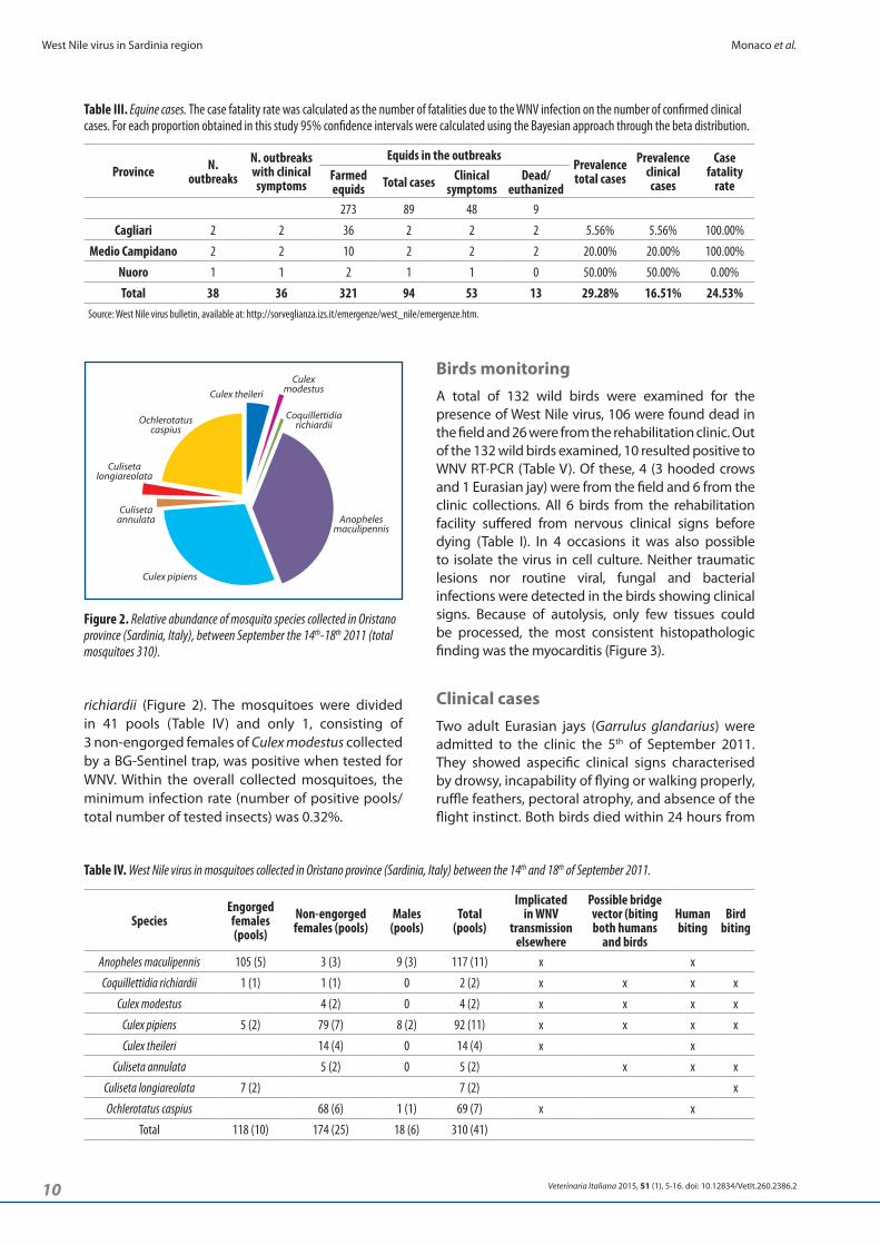
the admission. On the 21st of September 2011, an adult common buzzard originating from the town of Oristano was admitted to the clinic. It showed lethargy, head tremors, drooping wings and inability to fly due to the flaccid paralysis of the wing muscles. The legs were kept flexed and the bird was not able to stand up. The podal reflex was lost whereas both, the pupillary and corneal reflexes were still present. The animal died few hours after the admission. A little owl (Athene noctua) found close by Santa Giusta (OR), was bought to the rehabilitation centre on the 24th of September 2011. The first day it showed ataxia, incoordination, reluctance or inability to fly properly, head tilt and anisocoria. It was able to stand
Table V. West Nile virus in wild birds collected in Oristano province (Sardinia, Italy) between September and November 2011.
Family Species Common name WNV+/Total % of positiveAccipitridae Buteo buteo Common buzzard 2/5 40%
Anatidae Anas platyrhynchos Mallard 1/5 20%
Ardeide Ardea cinerea Grey heron 0/1 0%
Corvidae
Corvus corone Carrion crow 0/2 0%
Corvus corone cornix Hooded crow 3/93 3.2%
Garrulus glandarius Eurasian jay 3/11 27.3%
FalconidaeFalco peregrinus Peregrine falcon 0/1 0%
Falcus tinnunculus Common kestrel 0/2 0%
Fasanidae Alectoris barbara Barbary partridge 0/1 0%
Laride Larus argentatus European herring gull 0/3 0%
Passeridae Passer domesticus House sparrow 0/1 0%
Phoenicocpteridae Phoenicopterus roseus Greater flamingo 0/1 0%
Podicepidae Tachybaptus ruficollis Little grebe 0/1 0%
Strigidae Athena noctua Little owl 1/2 50%
Sturnidae Sturnus vugaris Common starling 0/1 0%
Columbidae Streptopelia turtur Turtle dove 0/1 0%
Columbidae Columba livia Rock pigeon 0/1 0%
Total 10/132 7.6%
Figure 3. Little owl (Athena noctua), heart. Foci of interstitial myocarditis. Hematoxylin & eosin. Final magnification = x 400.
Figure 4. Little owl (Athena noctua) with nervous clinical symptoms. The animal was admitted to the Clinica Veterinaria ‘Duemari’ (Oristano, Sardinia, Italy) on the 24th of September 2011.
12 Veterinaria Italiana 2015, 51 (1), 5-16. doi: 10.12834/VetIt.260.2386.2
West Nile virus in Sardinia region Monaco et al.
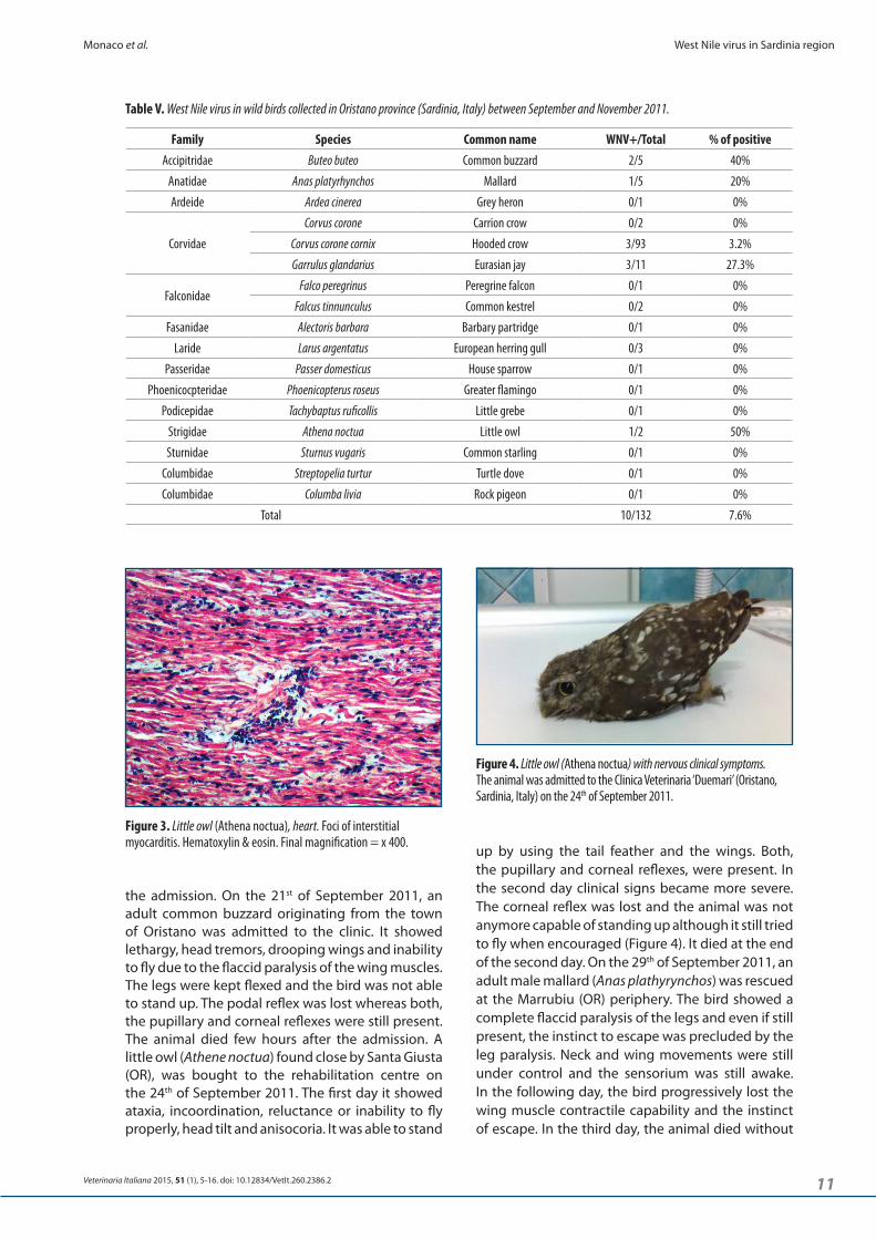
all the Italian strains circulating from 2008 to 2009 clustered in a monophyletic group while the 2011 Sardinian sequences were in a distinct sub-cluster.
Separated from the cited strains were the first isolate from Italy (1998) and the 2008 isolate from Spain. As expected, the strains belonging to lineage 2 (Greece 2010 and Italy 2011) were segregated in a distinct subclade.
Comparison of the deduced amino acid sequences (from position 203 to 526 referred to FJ483548) from the Mediterranean isolates showed 4 variable amino acid residues among the different WNV strains. West Nile virus strains AF404757 (Italy 1998), JF719065 (Italy 2008), 20608 (Italy 2011, horse) and 21412 (Italy 2011, owl) were characterized by a threonine residue in position 249, while the other strains exhibited a proline in the same position. All the Sardinian strains displayed an alanine residue in position 436, substituted by a threonine in the other WNV strains. Residue 476 was represented by arginine in the WNV detected in the Sardinian owl, while glicine was in the same position of the other sequences.
DiscussionWest Nile virus represents one of the pathogens that best adapted to the new scenarios imposed by globalization and climatic changes. It was able to adjust its epidemiology, virulence and range of host species. It went from being considered a virus of minor significance capable of determining only mild, sporadic and self-limiting outbreaks, to be the most widespread arbovirus in the world, able to determine severe outbreaks involving humans, birds and horses (Brault 2009, Hayes et al. 2005, Kramer et al. 2008). Changes that were clearly evident in the North American epidemics at the end of the century have now become clear also in Europe, where new virulence patterns of recent WNV strains have been observed. Although more numerous and severe cases have been recently described in humans and horses infected by WNV European strains, only in few occasion the disease has been associated to wild birds. This is in contrast with the North America situation, where native wild birds are largely affected by WNV. Different strain virulence or different WNV susceptibility between Palearctic and Nearctic bird species were considered as possible causes of this discrepancy. In Italy, in years of outbreaks and WNV circulation, no bird cases have ever been reported. For this reason the year 2011 could be regarded as critical. Strains of WNV belonging to lineage 1 continued to circulate in the North-Eastern regions for the fourth consecutive year affecting humans, horses, and wild birds. In the same area a newly introduced lineage 2 strain was detected in pools of Cx. pipiens
showing the flaccid paralysis of the neck muscles, characteristic sign of the avian botulism, which is commonly detected in the Oristano lagoon area in that period of the year. An ataxic adult common buzzard (Buteo buteo), found in the periphery of Oristano, was brought to the rehabilitation centre on the 10th of October 2011. The bird showed irregular head tremors and had trouble in maintaining the upright position even if using the tail feather and the wings. The instinct of escape was lost and the podal reflex as well as the proprioception response on the left leg was slow. The droppings were of a fluid-like consistency and the feathers around the vent were matted with faeces. When recumbent in a sternal position, the bird was not able to stand up properly and, similarly, it was not able to open its wings even if it was able to flex them back at the elbow joint when forcedly opened. In the second day, the lethargy became more severe and the animal died.
Partial NS3 sequencingAll detected strains belonged to WNV lineage 1. A 975bp fragment of the NS3 encoding gene of the WNV Sardinian samples shared high nt similarity with the other WNV strains which have recently circulated in the Mediterranean Basin (Figure 5). Particularly,
1000
562
653
512
547
492
883
631
472
139
69
74
80421412
20608
9492
20875
23954
23237
23941
GU011992
2011Sardiniansequences
JF719068
1201009
FJ483548
FJ483549
JF719066
JF719067
JF719065
JF719069
JF707789
AF404757
JN858070
HQ537483
AF260968
510
815 690
1000
1000
Figure 5. Phylogenetic tree based on 975bp in the NS3 region. Sequence dataset was analysed using BioEdit version 7.0.9.0 and nucleotide alignment was performed with Clustal-W. The dendrogram was generated using maximum likelihood technique in PHYLIP (the PHYlogeny Inference Package) program version 3.67. The strain Eg101 strain (AF260968) was used to root the tree. Statistical support at the internodes on the tree was assessed by 1000 bootstrap replications.
13
Monaco et al. West Nile virus in Sardinia region
Veterinaria Italiana 2015, 51 (1), 5-16. doi: 10.12834/VetIt.260.2386.2
The Italian strains belong to the ‘Western Mediterranean group’, regardless their year and place of isolation. Nevertheless the Italian strains seemed to evolve independently, as demonstrated by their distance from the most recent Spanish strains isolated in 2008 and 2010.
As a consequence, it is likely that the strains which have circulated in the Sardinia region during 2011, do not represent a new introduction from other Mediterranean countries with viral circulation but from the endemic areas of Northern Italy, likely through some short distance migratory Passeriformes birds (Spina and Volponi 2009). These species became infected in the Italian mainland and then spread the infection in Sardinia region where favourable environmental conditions permitted the establishment of the infection through local mosquitoes and resident bird species. In the latter, the Sardinian WNV strains were capable of causing clinical signs and death. It was the first time that an Italian strain of WNV showed virulence for birds. The WNV strain isolated in New York in 1999 (NY99) is regarded as the prototype of the pathogenic strain due to the fast spread, the high neurovirulence and the high fatality rate in birds, humans and animals, (Ceccaldi et al., 2004, Ciota and Kramer 2010). The NS3 gene sequence (Genbank accession number AF196835) is characterised by the substitution of a threonin-to-proline in the position 249 (NS3-T249P) when compared with other lineage 1 isolates (Lanciotti et al. 1999, Lanciotti et al. 2002). Brault and colleagues (Brault et al. 2007) showed, with a site-directed mutagenesis experiment, a strict correlation between the NS3-T249P and the increased avian virulence: this point mutation is claimed to be responsible of the increased efficiency of viral replication in avian hosts, due to the generation of temperature resistant phenotype and improving the ability in delaying innate antiviral response (Fredericksen et al. 2004, Kinney et al. 2006). All these factors could facilitate mosquito transmission, which in turn might affect the incidence of human and horse infections. Both, the NS3-249P and NS3-249T genotypes were detected in the Sardinian outbreaks confirming that co-circulation of different genotypes in the affected population might be common for WNV as for many RNA viruses. No association, however, was observed between virulence and the NS3 proline strains. Neurological clinical signs were seen in birds affected, by the putative ‘mild’ NS3-249T genotype. Furthermore the case-fatality rate evidenced in horses in the 2011 Sardinian outbreaks (24.53%, I.C. 95%: 15-37.6%) does not significantly differ from that found in northern Italian outbreaks (15.6% I.C. 95%: 7.0-31.9%). These data confirmed what observed in studies on mice (Sotelo et al. 2009) and redleg partridge (Sotelo et al. 2011) and clearly indicates that the role of NS3-249
and in the organs of a dead wild bird (Savini et al. 2012). Another lineage 2 strain was also detected in a human patient in the eastern coast of central Italy (Bagnarelli et al. 2011). In Sardinia, as reported in this study, an unusual increase of the wild bird mortality was observed in the area where WNV was circulating and the presence of WNV was confirmed in nearly 8% of the carcasses. In some birds characteristic clinical nervous signs were also observed. The virus was found in the brain and other organs of the affected birds. It was the first time that the presence of severe and lethal acute nervous clinical signs could be associated to WNV infection in Italy indicating that at least some Italian WNV strains are pathogenic in native wild bird species. In line with other WND associated cases described in European or US wild birds (Bakonyi et al. 2006, Höfle et al. 2008, Jiménez-Clavero et al. 2008, Jourdain et al. 2008, Ludwig et al. 2010, Saito et al. 2007), also in this epidemic Accipitridae and Corvidae were the bird families more often involved. Even if some authors sustained that, as predators, these species could then become infected by feeding on WNV infected prey (Garmendia et al. 2000), it is still difficult to understand whether these higher susceptibility might be determined by host related factors or ecological factors. Oral transmission of WNV through the ingestion of experimentally infected prey and mosquitoes has already been documented in great horned owls, crows, and other passerines (Komar et al. 2003). Equally, it has been shown that, if exposed at high WNV infection rates, small mammals could serve as reservoirs (Root et al. 2006). Although numerous studies have been tackled this topic, the real impact of WNV on the predatory bird population is still unknown.
In a similar complex scenario, beside the classical epidemiological approach, the analysis of the viral genome provided a powerful tool to infer the geographical and temporal correlations between the circulating WNV strains. Among the WNV genes, the NS3 encoding gene revealed a high capability to retain a strong phylogenetic signal (Gray et al. 2010) and the NS3 helicase domain is also 1 of the putative sites of the virulence determinants. For this reason it was the selected target for the molecular characterisation of the Sardinian samples.
Previous phylogenetic studies clustered the WNV lineage 1 strains circulating in the Mediterranean countries in the so called ‘Western Mediterranean group’ which has been traced back to a single introduction in the Mediterranean area before 1996 (Sotelo et al. 2009). More recently, the virus has been probably able to establish an endemic cycle of transmission between resident birds and local mosquitoes, showing its ability to overwinter in temperate areas (Monaco et al. 2009).
14
West Nile virus in Sardinia region Monaco et al.
Veterinaria Italiana 2015, 51 (1), 5-16. doi: 10.12834/VetIt.260.2386.2
IV). In this survey, however, Culex pipiens and Ochlerotatus caspius, which are the most abundant WNV vectors in Italy (Toma et al. 2008), tested negative for WNV. Whether this was because of the small size of the sample tested or because of the minor capability of Sardinian strain adaptation for these species is hard to say. Surprisingly, the Sardinian WNV strains were detected in Cx. modestus, which is known to be a competent vector of WNV, both in the field and under laboratory conditions (Hannoun et al. 1964, Balenghien et al. 2006, Balenghien et al. 2007, Balenghien et al. 2008). This is a Mediterranean species, one of the most aggressive against humans, able to bite also during daytime and to overwinter as adult by diapause. Culex modestus feeds also on birds and horses, thus it can also act as bridge vector for WNV (Medlock et al. 2005, Severini et al. 2009).
In conclusion, this investigation confirms that Italian WNV lineage 1 strains might have a severe effect on native wild birds, especially on free-ranging raptors. Whether it depends on a particular virulence of the strains involved or on other factors related to the host susceptibility and vectors, is not clear and requires further investigations.
residue as virulence determinant is far from being elucidated in the Mediterranean ecosystem at least. It is likely that the WNV pathogenicity is the result of a complex series of events, which involve the virus, the vectors and the hosts.
Thus, although the term ‘vector’ implies a lack of significant biological interaction between arthropods and the pathogens they carry, it has become clear in recent years that such interactions are complex and are likely dominant forces shaping the evolution of arboviruses including their virulence (Ciota and Kramer 2010). Interestingly, NS3 helicase has also been shown to determine the WNV natural host fitness (mosquitoes and birds) (Ebel et al. 2011). In the WNV transmission cycle, different host types differentially influence the virus population. Whereas infection of mosquitoes leads to high levels of population variation and consequent adaptive plasticity, vertebrate infection maintains high fitness through strong purifying selection. All mosquito species collected in this survey can be considered capable to sustain the virus circulation, since they have been implicated in WNV transmission elsewhere, and/or possible bridge vectors between birds and mammals (Table
Autorino G.L., Battisti A., Deubel V., Ferrari G., Forletta R., Giovannini A., Lelli R., Murri S. & Scicluna M.T. 2002. West Nile virus epidemic in horses, Tuscany region, Italy. Emerging Infec Dis, 8, 1372-1378.
Bagnarelli P., Marinelli K., Trotta D., Monachetti A., Tavio M., Del Gobbo R., Capobianchi M., Menzo S., Nicoletti L., Magurano F. & Varaldo P. 2011. Human case of autochthonous West Nile virus lineage 2 infection in Italy, September 2011. Euro Surveill, 16 (43), pii=20002. http://www.eurosur vei l lance.org/ViewAr ticle.aspx?ArticleId=20002.
Balenghien T., Fouque F., Sabatier P. & Bicout D.J. 2006. Horse-, bird-, and human-seeking behaviour and seasonal abundance of mosquitoes in a West Virus Focus of Southern France. J Med Entomol, 43 (5), 937-946.
Balenghien T., Vazeille M., Reiter P., Schaffner F., Zeller H. & Bicout D.J. 2007. Evidence of laboratory vector competence of Culex modestus for West Nile virus. J Am Mosq Control Assoc, 23 (2), 233-236.
Balenghien T., Vazeille M., Grandadam M., Schaffner F., Zeller H., Reiter P., Sabatier P., Fouque F. & Bicout D.J. 2008. Vector competence of some French Culex and Ades mosquitoes for West Nile Virus. Vector Borne Zoonotic Dis, 8 (5), 589-595.
Bakonyi T., Ivanics E., Erdelyi K., Ursu K., Ferenczi E., Weissenbock H. & Nowotny N. 2006. Lineage 1 and 2 strains of encephalitic West Nile Virus, Central Europe. Emerging Infec Dis, 12, 618-623.
References
Brault A.C., Huang C.Y., Langevin S.A., Kinney R.M., Bowen R.A., Ramey W.N., Panella N.A., Holmes E.C., Powers A.M. & Miller B.R. 2007. A single positively selected West Nile viral mutation confers increased virogenesis in American crows. Nat Genet, 39 (9), 1162-1166.
Brault A.C. 2009. Changing patterns of West Nile virus transmission: altered vector competence and host susceptibility. Vet Res, 40, 43-62.
Calistri P., Monaco F., Savini G., Guercio A., Purpari G., Vicari D., Cascio S. & Lelli R. 2010a. Further spread of West Nile virus in Italy. Vet Ital, 46 (4), 467-470.
Calistri, P., Giovannini A., Savini G. Monaco F., Bonfanti L., Ceolin C., Terregino C., Tamba M., Cordioli P. & Lelli R. 2010b. West Nile virus transmission in 2008 in North-Eastern Italy. Zoonoses Public Health, 57, 211-219.
Capelli G., Ravagnan S., Montarsi F., Ciocchetta S., Cazzin S., Bonfanti L., Di Gennaro A., Portanti O., Mulatti P., Monne I., Cattoli G., Cester G., Russo F., Savini G. & Marangon S. 2013. Further evidence of lineage 2 West Nile Virus in Culex pipiens of North-Eastern Italy. Vet Ital, 49 (3), 263-268.
Ceccaldi P.E., M. Lucas & Depres P. 2004. New insights on neuropathogenicity of West Nile virus. FEMS Microbiology Letters, 233, 1-6.
Ciota A.T. & Kramer L.D. 2010. Insights into arbovirus evolution and adaptation from experimental studies. Viruses, 2, 2594-2617.
15
Monaco et al. West Nile virus in Sardinia region
Veterinaria Italiana 2015, 51 (1), 5-16. doi: 10.12834/VetIt.260.2386.2
Kutasi O., Bakonyi T., Lecollinet S., Biksi I., Ferenczi E., Bahuon C., Sardi S., Zientara S. & Szenci O. 2011. Equine encephalomyelitis caused by a genetic lineage 2 West Nile virus in Hungary. J Vet Intern Med, 25, 586-591.
Lanciotti R.S., Roehring J.T, Deubel V., Smith J., Parker M., Steele K., Crise B., Volpe K.E., Crabtree M.B., Scherret J.H., Hall R.A., MacKenzie J.S., Cropp C.B., Panigrahy B., Ostlund E., Schmitt B., Malkinson M., Banet C., Weissman J., Komar N., Savage H.M., Stone W., McNamara T. & Gubler D.J. 1999. Origin of the West Nile virus responsible for an outbreak of encephalitis in the northeastern United States. Science, 286, 2333-2337.
Lanciotti R.S., Kerst A.J., Nasci R.S., Godsey M.S., Mitchell C.J., Savage H.M., Komar N., Panella N.A., Allen B.C., Volpe K.E., Davis B.S. & Roehrig J.T. 2000. Rapid detection of West Nile virus from human clinical specimens, field-collected mosquitoes, and avian samples by a TaqMan reverse transcriptase-PCR assay. J Clin Microbiol, 38, 4066-4071.
Lanciotti R.S., Ebel G.D., Deubel V., Kerst A.J., Murri S., Meyer R., Bowen M., McKinney N., Morrill W.E., Crabtree M.B., Kramer L.D. & Roehring J.T. 2002. Complete genome sequencing and phylogenetic analysis of West Nile virus strains isolated from the United States, Europe, and the Middle East. Virology, 298, 96-105.
Lelli R., Calistri P., Bruno R., Monaco F., Savini G., Di Sabatino D., Corsi I. & Pascucci I. 2012. West Nile transmission in resident birds in Italy. Transbound Emerg Dis, 59 (5), 421-428.
Ludwig A., Bigras-Poulin M., Michel P. & Bélanger D. 2010. Risk factors associated with West Nile virus mortality in American Crow populations in Southern Quebec. J Wild Dis, 46( 1), 195-208.
Mackenzie J.S. & Williams D.T. 2009. The zoonotic flaviviruses of southern, south-eastern and eastern Asia, and Australasia: the potential for emergent viruses. Zoonoses Public Health, 56, 338-356.
Medlock J.M., Snow K.R. & Leach S. 2005. Potential transmission of West Nile virus in the British Isles: an ecological review of candidate mosquito bridge vectors. Med Vet Entomol, 19 (1), 2-21.
Monaco F., Lelli R., Teodori L., Pinoni C., Di Gennaro A., Polci A., Calistri P. & Savini G. 2010. Re-emergence of West Nile virus in Italy. Zoonoses Public Health, 57 (7-8), 476-486.
Monaco F., Savini G., Calistri P., Polci A., Pinoni C., Bruno R. & Lelli R. 2011. 2009 West Nile disease epidemic in Italy: first evidence of overwintering in Western Europe? Res Vet Sci, 91 (2), 321-326.
Petrovic T., Blazquez A.B., Lupulovic D., Lazić G., Escribano-Romero E., Fabijan D., Kapetanov M., Lazić S. & Saiz J. 2013. Monitoring West Nile virus (WNV) infection in wild birds in Serbia during 2012: first isolation and characterisation of WNV strains from Serbia. Euro Surveill, 18 (44), pii: 20622. http://www.eurosurveillance.org/ViewArticle.aspx?ArticleId=20622.
Platonov A.E., Karan' L.S., Shopenskaia T.A., Fedorova M.V., Koliasnikova N.M., Rusakova N.M., Shishkina L.V., Arshba T.E., Zhuravlev V.I., Govorukhina M.V., Valentseva A.A. & Shipulin G.A. 2011. Genotyping of West Nile fever virus strains circulating in southern Russia as an
Danis K., Papa A., Papanikolaou E., Dougas G. , Terzaki I., Baka A., Vrioni G., Kapsimali V., Tsakris A., Kansouzidou A., Tsiodras S., Vakalis N., Bonovas S. & Kremastinou J. 2011. Ongoing outbreak of West Nile virus infection in humans, Greece, July to August 2011. Euro Surveill,16 34), pii=19951.http://www.eurosurveillance.org/ViewArticle.aspx?ArticleId=19762
Ebel G.D., Fitzpatrick K.A., Lim P.Y., Bennett C.J., Deardorff E.R., Jerzak G.V., Kramer L.D., Zhou Y., Shi P.Y. & Bernard K.A. 2011. Nonconsensus West Nile virus genomes arising during mosquito infection suppress pathogenesis and modulate virus fitness in vivo. J Virol, 85, 12605-12613.
Erdélyi K., Ursu K., Ferenczi E., Szeredi L., Rátz F., Skáre J. & Bakonyi T. 2007. Clinical and pathologic features of lineage 2 West Nile virus infections in birds of prey in Hungary. Vector Borne Zoonotic Dis, 7 (2), 181-188.
Fredericksen B.L., Smith M., Katze M.G, Shi P.Y. & Gale M.Jr. 2004. The host response to West Nile virus infection limits viral spread through the activation of the interferon regulatory factor 3 pathway. J Virol, 78, 7737-7747.
Garmendia A.E., Van Kruiningen H.J., French R.A., Anderson J.F., Andreadis T.G., Kumar A. & West A.B. 2000. Recovery and identification of West Nile virus from a hawk in winter. J Clin Microbiol, 38 (8), 3110-3111.
Gray R.R., Veras N.M., Santos L.A. & Salemi M. 2010. Evolutionary characterization of the West Nile Virus complete genome. Mol Phylogenet Evol, 56 (1), 195-200.
Hannoun C., Panthier R., Mouchet J. & Eouzan J.P. 1964. Isolement en France du virus West Nile à partir de malades et du vecteur Culex modestus Ficalbi. CR Acad Sci Paris, 259, 4170-4172.
Hayes E.B., Komar N., Nasci R.S., Montgomery S.P., O'Leary D.R. & Campbell G.L., 2005. Epidemiology and transmission dynamics of West Nile virus disease. Emerg Infect Dis, 11, 1167-1173.
Höfle U., Blanco J.M., Crespo E., Naranjo V., Jiménez-Clavero M.A., Sanchez A., de la Fuente J. & Gortazar C. 2008. West Nile virus in the endangered Spanish imperial eagle. Vet Microbiol, 129 (1-2), 171-178.
Jiménez-Clavero M.A., Sotelo E., Fernandez-Pinero J., Llorente F., Blanco J.M., Rodriguez-Ramos J., Perez-Ramirez E. & Höfle U. 2008. West Nile virus in golden eagles, Spain, 2007. Emerg Infect Dis, 14 (9), 1489-1491.
Jourdain E., Gauthier-Clerc M., Sabatier P., Grège O., Greenland T., Leblond A., Lafaye M. & Zeller H.G. 2008. Magpies as hosts for West Nile virus, southern France. Emerg Infect Dis, 14 (1), 158-160.
Kinney R.B., Huang. C.Y.H., Whiteman M.C., Bowen R.A., Langevin S.A. & Miller B.R. 2006. Avian virulence and thermostable replication of the North American strain of West Nile virus. J Gen Virol, 87, 3611-3622.
Kramer L.D., Styer L.M. & Ebel G.D. 2008. A global perspective on the epidemiology of West Nile virus. Annu Rev Entomol, 53, 61-81.
Komar N., Langevin S., Hinten S., Nemeth N., Edwards E., Hettler D., Davis B., Bowen R. & Bunning M. 2003. Experimental Infection of North American Birds with the New York 1999 Strain of West Nile Virus. Emerg Infect Dis, 9 (3), 311-322.
16
West Nile virus in Sardinia region Monaco et al.
Sotelo E., Gutierrez-Guzmán A.V., Del Amo J., Llorente F., El-Harrak M., Pérez-Ramírez E., Blanco J.M., Höfle U. & Jiménez-Clavero M.A. 2011. Pathogenicity of two recent Western Mediterranean West Nile virus isolates in a wild bird species indigenous to Southern Europe: the red-legged partridge. Vet Res, 42 (1), 11.
Spina F. & Volponi S. 2009. Atlante della Migrazione degli Uccelli in Italia. Vol. 2 - Passeriformi. ISPRA, Roma, 631 pp.
Thompson J.D., Higgins D.G. & Gibson T.J. 1994. CLUSTAL W: improving the sensitivity of progressive multiple sequence alignment through sequence weighting, position-specific gap penalties and weight matrix choice. Nucleic Acids Res, 22, 4673-4680.
Toma L., Cipriani M., Goffredo M., Romi R. & Lelli R. 2008. First report on entomological field activities for the surveillance of West Nile disease in Italy. Vet Ital, 44 (3), 499-512.
Vazquez A., Sanchez-Seco M.P., Ruiz S., Molero F., Hernandez L., Moreno J., Magallanes A., Tejedor C.G. & A. Tenorio. 2010. Putative new lineage of West Nile virus, Spain. Emerg Infect Dis, 16, 549-552.
Wodak E., Richter S., Bagó Z., Revilla-Fernández S., Weissenböck H., Nowotny N. & Winter P. 2011. Detection and molecular analysis of West Nile virus infections in birds of prey in the eastern part of Austria in 2008 and 2009. Vet Microbiol, 149 (3-4), 358-366.
World Animal Health Information Disease Database (WAHID). 2014. West Nile fever, Turkey. World Organisation for Animal Health, Paris. www.oie.int/wahis_2/public/wahid.php/Reviewreport/Review/viewsummary?reportid=16002 accessed on 20th
January 2015.
World Organization for Animal Health (OIE) Terrestrial Manual 2014, Chapter 2.1.20. West Nile fever. (http://www.oie.int/fileadmin/Home/eng/Health_standards/tahm/2.01.20_WEST_NILE.pdf, accessed on 12/01/2015).
epidemiological investigation method: principles and results. Zh Mikrobiol Epidemiol Immunobiol, 2, 29-37.
Rizzo C., Salcuni P., Nicoletti L., Ciufolini M.G., Russo F., Masala R., Frongia O., Finarelli A.C., Gramegna M., Gallo L., Pompa M.G., Rezza G., Salmaso S. & Declich S. 2012. Epidemiological surveillance of West Nile neuroinvasive diseases in Italy, 2008 to 2011. Euro Surveill, 17 (20), pii=20172. http://www.eurosurveillance.org/View Article.aspx?ArticleId=20172.
Root J.J., Oesterle P.T., Nemeth N.M., Klenk K., Gould D.H., McLean R.G., Clark L. & Hall J.S. 2006. Experimental infection of fox squirrels (Sciurus niger) with West Nile virus. Am J Trop Med Hyg, 75(4), 697-701.
Saito E.K., Sileo L., Green D.E., Meteyer C.U., McLaughlin G.S., Converse K.A. & Docherty D.E. 2007. Raptor mortality due to West Nile virus in the United States, 2002. J Wild Dis, 43(2), 206-213.
Savini G., Capelli G., Monaco F., Polci A., Russo F., Di Gennaro A., Marini V., Teodori L., Montarsi F., Pinoni C., Pisciella M., Terregino C., Marangon S., Capua I. & Lelli R. 2012. Evidence of West Nile virus lineage 2 circulation in Northern Italy. Vet Microbiol, 158 (3-4), 267-273.
Severini F., Toma L., Di Luca M. & Romi R. 2009. Le zanzare italiane: generalità e identificazione degli adulti (Diptera, Culicidae). Fragmenta Entomologica, 41 (2), 213-372.
Sirbu A., Ceianu C.S., Panculescu-Gatej R.I., Vazquez A., Tenorio A., Rebreanu R., Niedrig M., Nicolescu G. & Pistol A. 2011. Outbreak of West Nile virus infection in humans, Romania, July to October 2010. Euro Surveill, 16 (2), pii=19762. http://www.eurosurveillance.org/ViewArticle.aspx?ArticleId=19762.
Sotelo E., Fernandez-Pinero J., Llorente F., Agüero M., Hoefle U., Blanco J.M. & Jiménez-Clavero M.A. 2009. Characterization of West Nile virus isolates from Spain: new insights into the distinct West Nile virus eco-epidemiology in the Western Mediterranean. Virology, 395 (2), 289-297.
Veterinaria Italiana 2015, 51 (1), 5-16. doi: 10.12834/VetIt.260.2386.2