Embed Size (px)
Citation preview
The Anatomy and Physiology of the
Respiratory System
Functions of the Respiratory System
Air Distributor Gas exchanger Filters, warms, and humidifies
air Influences speech Allows for sense of smell
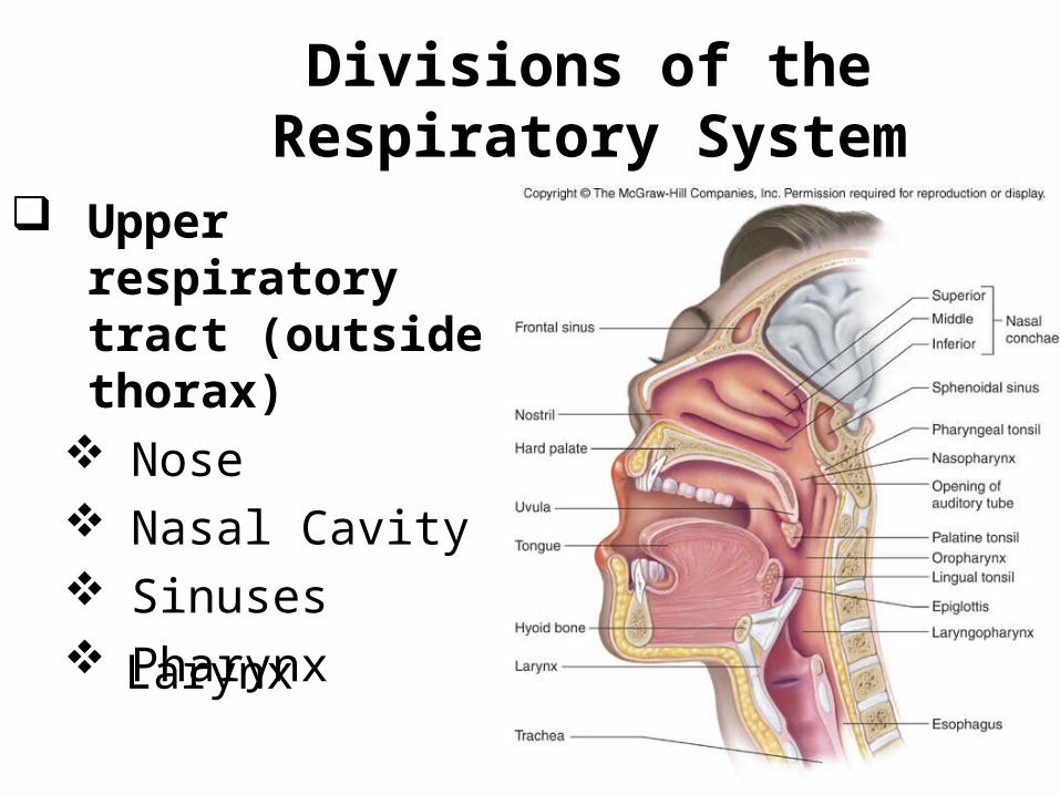
Divisions of the Respiratory System
Upper respiratory tract (outside thorax)
Nose Nasal Cavity Sinuses PharynxLarynx
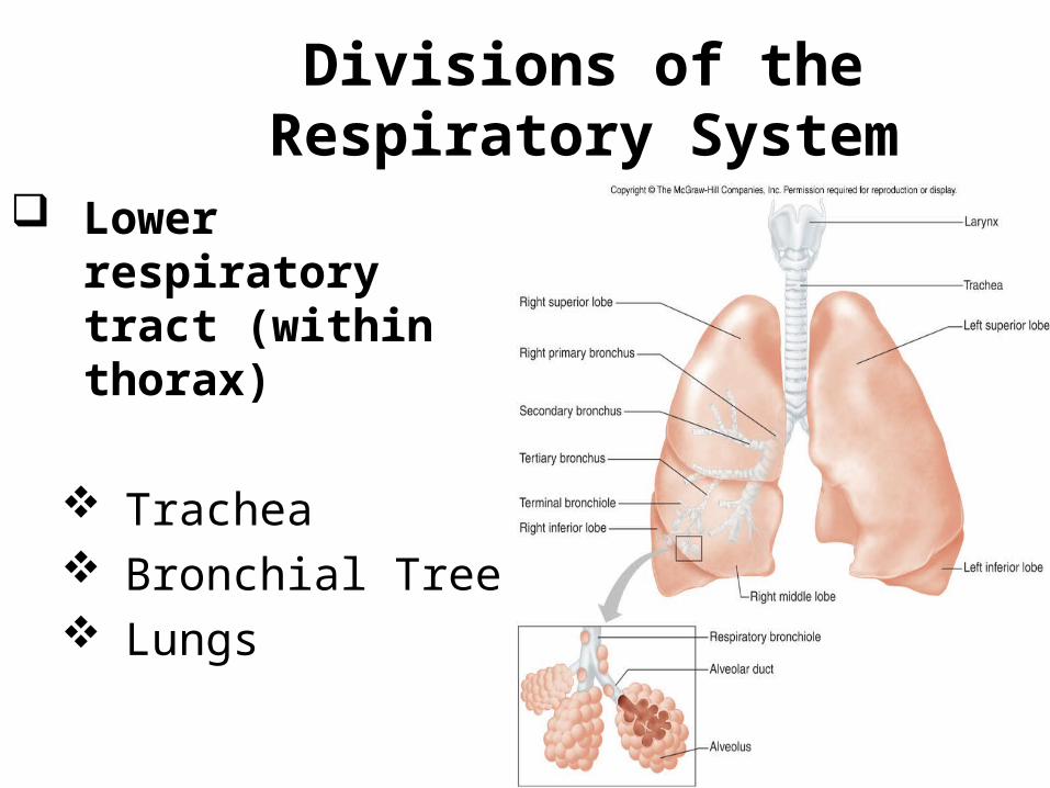
Divisions of the Respiratory System
Lower respiratory tract (within thorax)
Trachea Bronchial Tree Lungs
Structures of the Upper Respiratory Tract
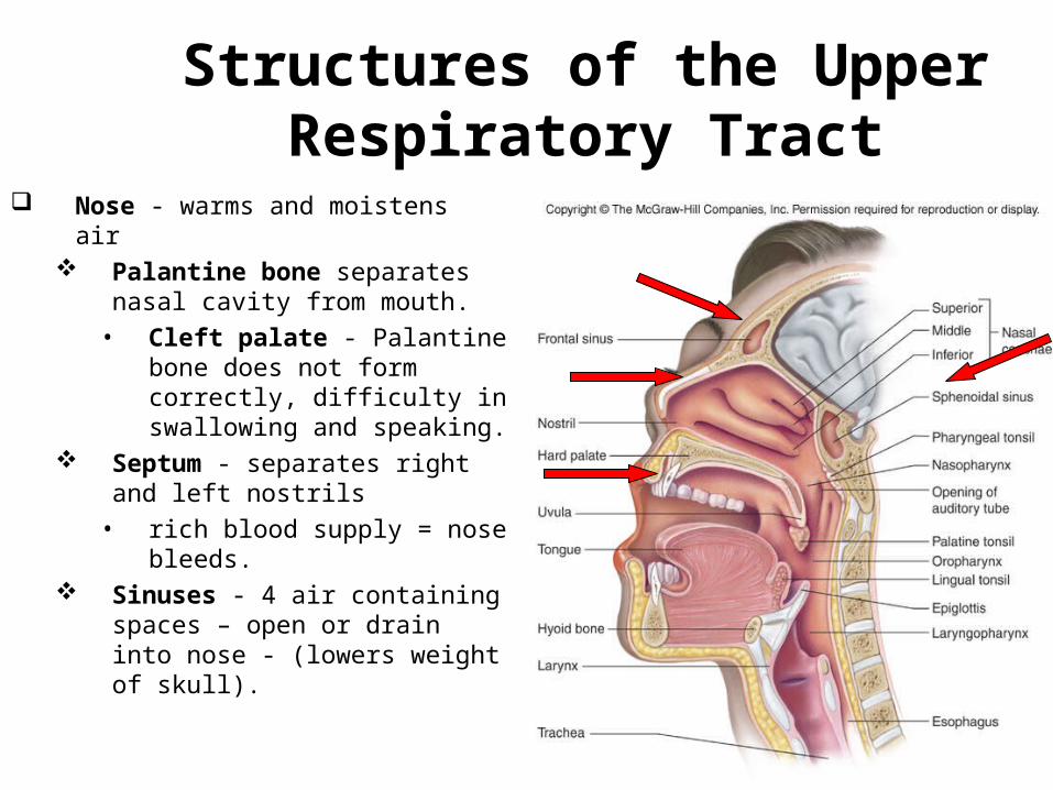
Nose - warms and moistens air
Palantine bone separates nasal cavity from mouth.
• Cleft palate - Palantine bone does not form correctly, difficulty in swallowing and speaking.
Septum - separates right and left nostrils
• rich blood supply = nose bleeds.
Sinuses - 4 air containing spaces – open or drain into nose - (lowers weight of skull).
Structures of the Upper Respiratory Tract
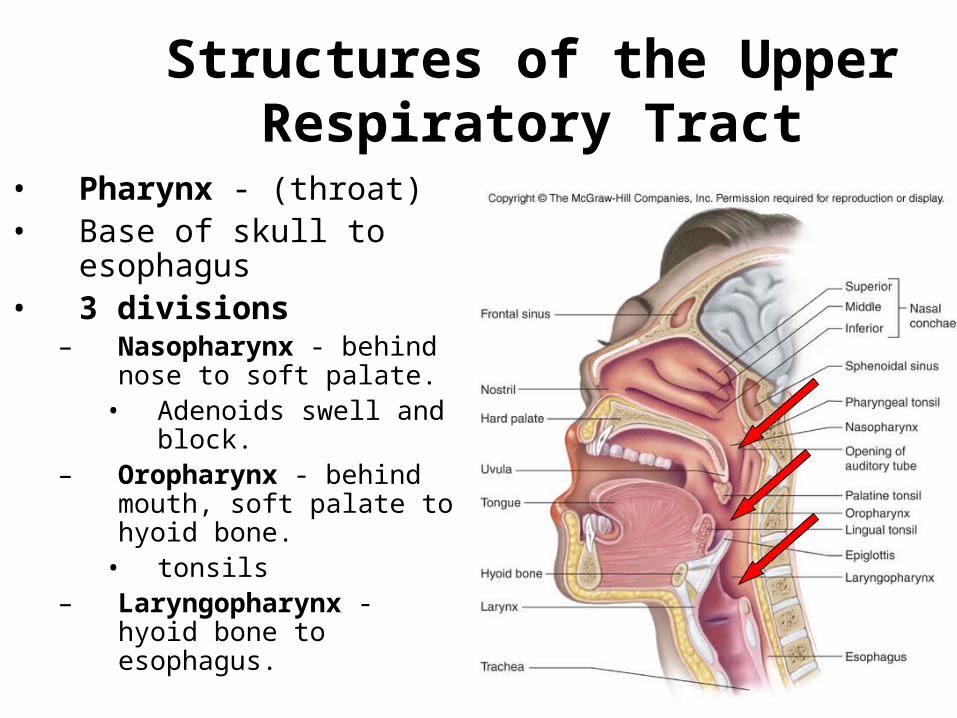
• Pharynx - (throat)• Base of skull to
esophagus• 3 divisions
– Nasopharynx - behind nose to soft palate.
• Adenoids swell and block.
– Oropharynx - behind mouth, soft palate to hyoid bone.
• tonsils– Laryngopharynx -
hyoid bone to esophagus.
Structures of the Upper Respiratory Tract Pharynx
Continued
• Changes shape to allow for vowel sounds = phonation.
Structures of the Lower Respiratory Tract
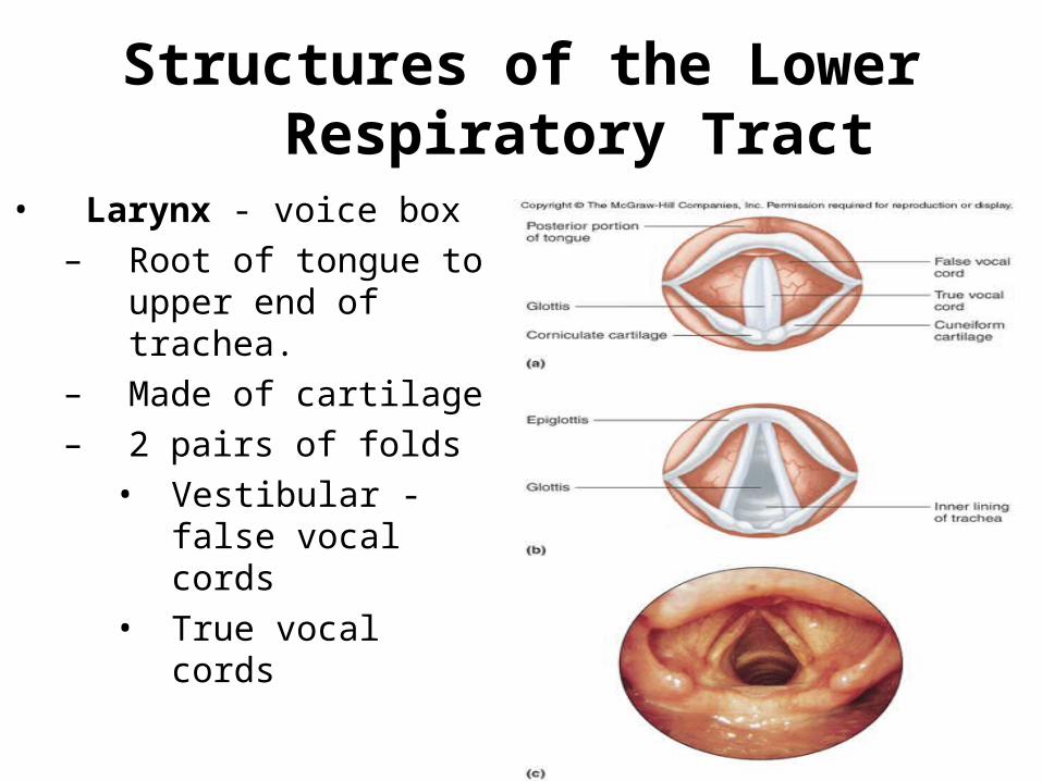
• Larynx - voice box– Root of tongue to
upper end of trachea.
– Made of cartilage– 2 pairs of folds
• Vestibular - false vocal cords
• True vocal cords
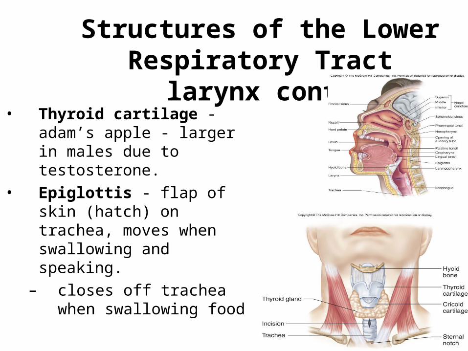
Structures of the Lower Respiratory Tract larynx
cont…• Thyroid cartilage -
adam’s apple - larger in males due to testosterone.
• Epiglottis - flap of skin (hatch) on trachea, moves when swallowing and speaking.
– closes off trachea when swallowing food
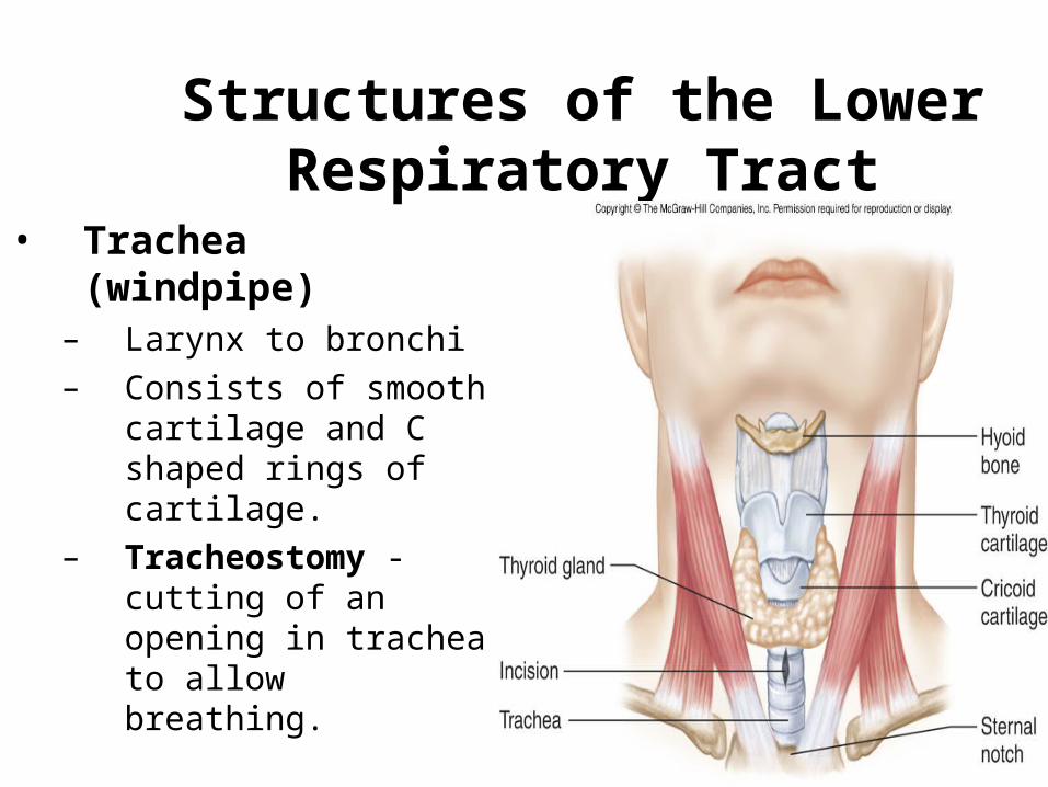
Structures of the Lower Respiratory Tract
• Trachea (windpipe)
– Larynx to bronchi– Consists of smooth
cartilage and C shaped rings of cartilage.
– Tracheostomy - cutting of an opening in trachea to allow breathing.
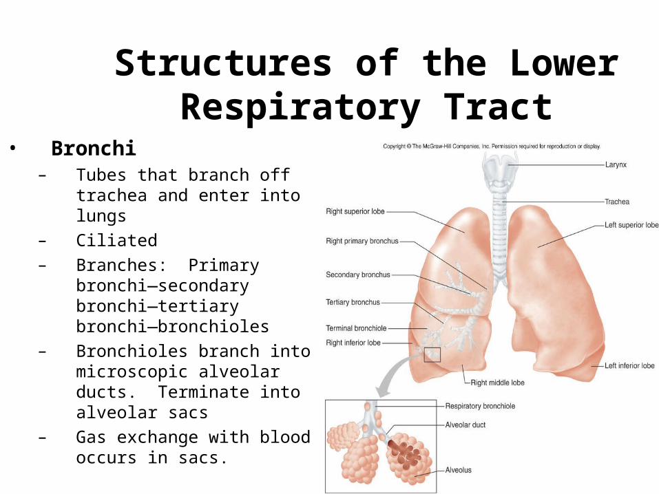
Structures of the Lower Respiratory Tract
• Bronchi– Tubes that branch off
trachea and enter into lungs
– Ciliated– Branches: Primary
bronchi—secondary bronchi—tertiary bronchi—bronchioles
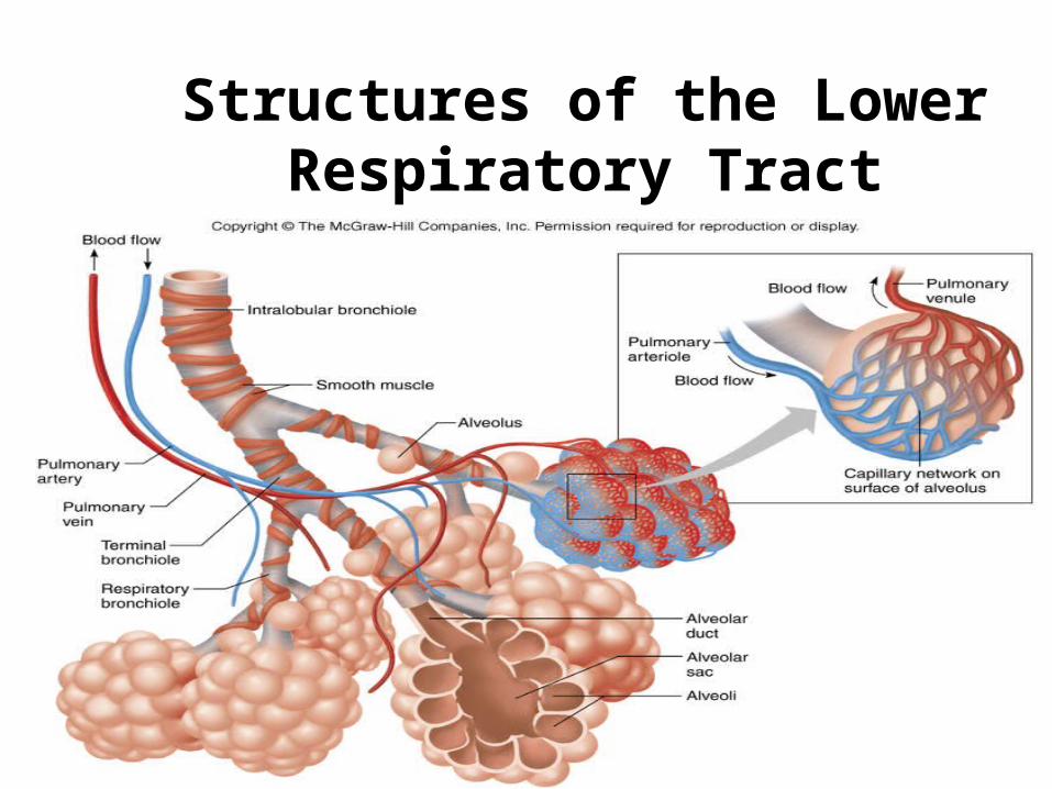
– Bronchioles branch into microscopic alveolar ducts. Terminate into alveolar sacs
– Gas exchange with blood occurs in sacs.
Structures of the Lower Respiratory Tract
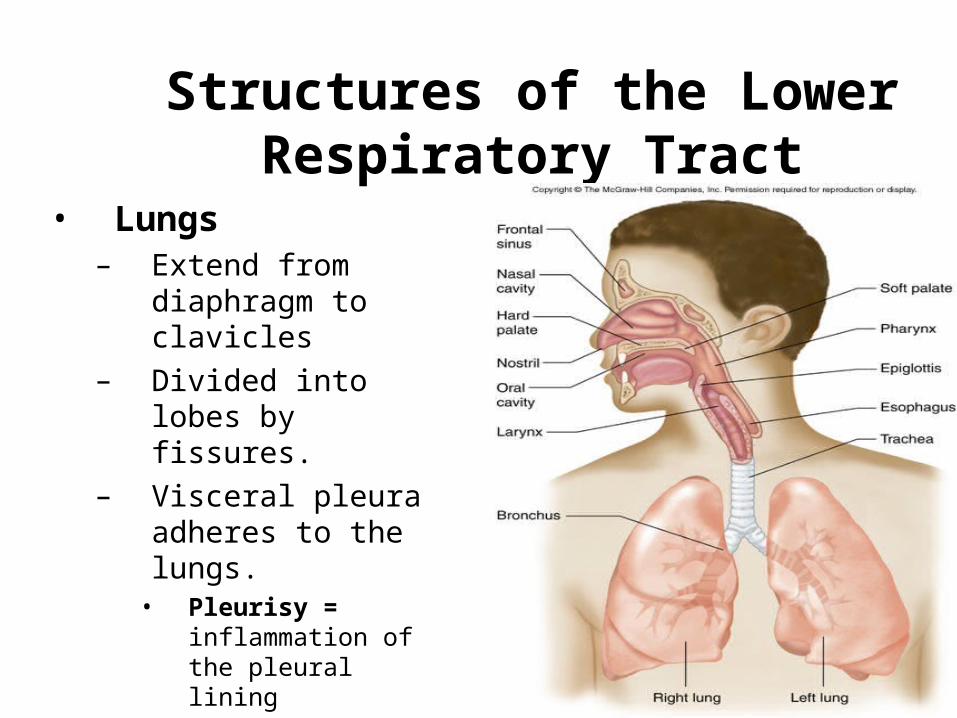
Structures of the Lower Respiratory Tract
• Lungs– Extend from
diaphragm to clavicles
– Divided into lobes by fissures.
– Visceral pleura adheres to the lungs.
• Pleurisy = inflammation of the pleural lining
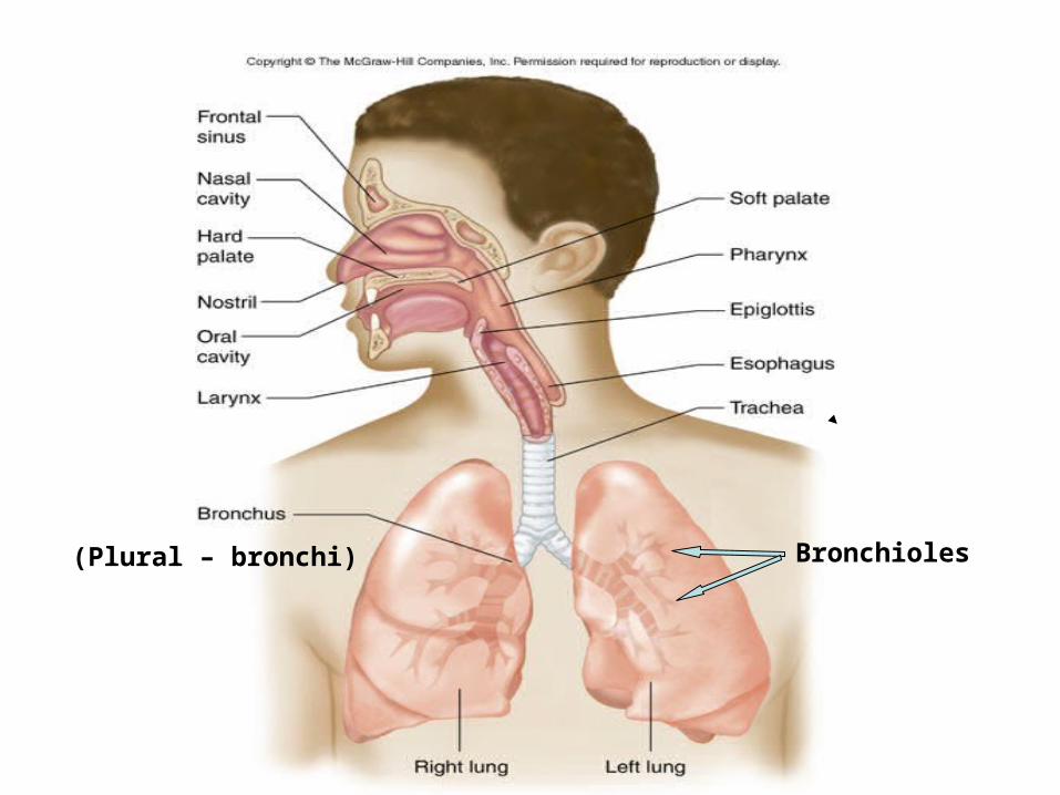
Bronchioles(Plural – bronchi)
Respiratory Physiology
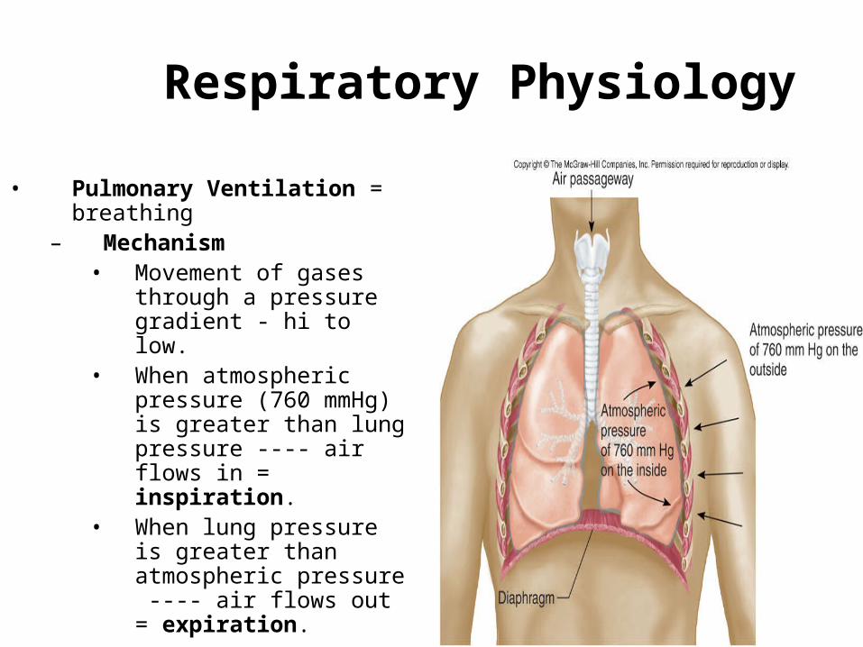
• Pulmonary Ventilation = breathing
– Mechanism• Movement of gases
through a pressure gradient - hi to low.
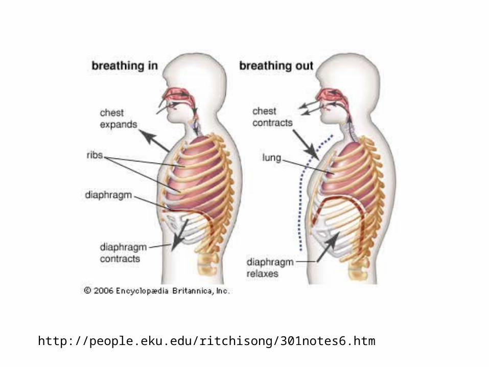
• When atmospheric pressure (760 mmHg) is greater than lung pressure ---- air flows in = inspiration.
• When lung pressure is greater than atmospheric pressure ---- air flows out = expiration.
Respiratory Physiology
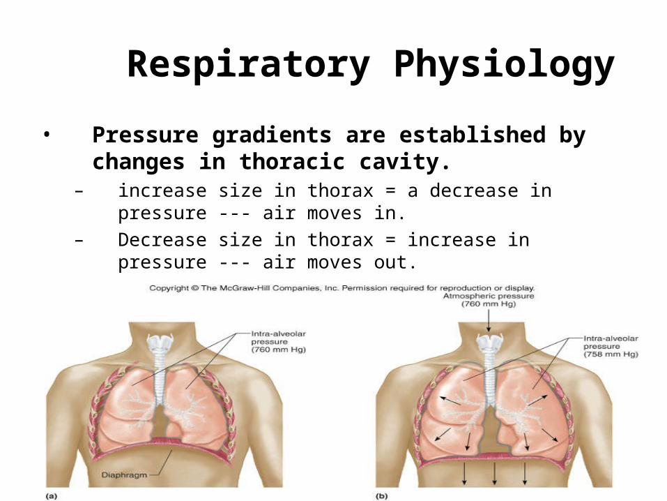
• Pressure gradients are established by changes in thoracic cavity.
– increase size in thorax = a decrease in pressure --- air moves in.
– Decrease size in thorax = increase in pressure --- air moves out.
http://people.eku.edu/ritchisong/301notes6.htm
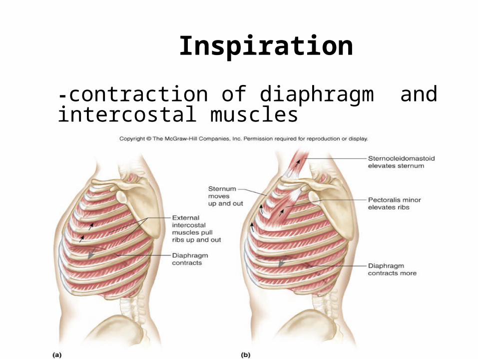
Inspiration
-contraction of diaphragm and intercostal muscles

Expiration • relaxation of diaphragm and
intercostal muscles
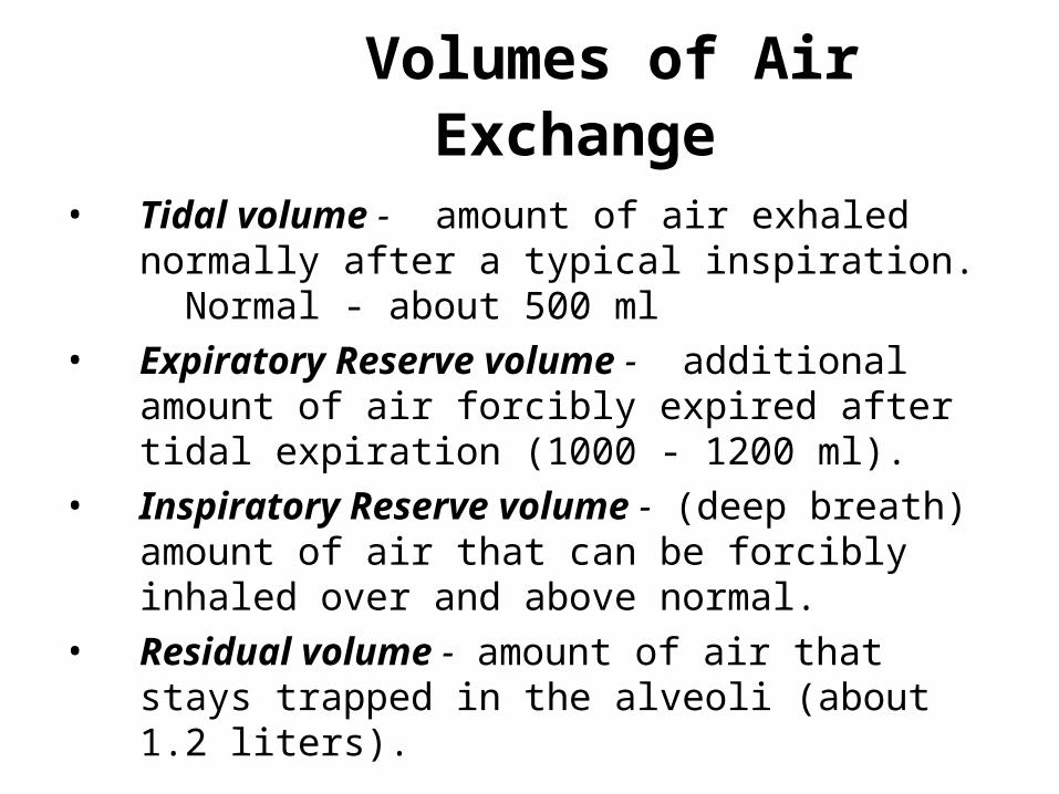
Volumes of Air Exchange
• Tidal volume - amount of air exhaled normally after a typical inspiration. Normal - about 500 ml
• Expiratory Reserve volume - additional amount of air forcibly expired after tidal expiration (1000 - 1200 ml).
• Inspiratory Reserve volume - (deep breath) amount of air that can be forcibly inhaled over and above normal.
• Residual volume - amount of air that stays trapped in the alveoli (about 1.2 liters).
Volumes of Air Exchange
• Vital capacity - the largest volume of air an individual can move in and out of the lungs.
• Vital capacity = sum of IRV+TV+ERV
• Depends of many factors• size of thoracic cavity• posture• volume of blood in lungs congestive
heart failure, emphysema, disease, etc…
Volumes of Air Exchange
• Eupnea - normal quiet breathing, 12-17 breaths per minute.
• Hyperpnea - increase in breathing to meet an increased demand by body for oxygen.
• Hyperventilation - increase in pulmonary ventilation in excess of the need for oxygen.
– Someone hysterical Breathe into – exertion paper bag.
• Hypoventilation - decrease in pulmonary ventilation.
• Apnea - temporary cessation of breathing at the end of normal expiration.
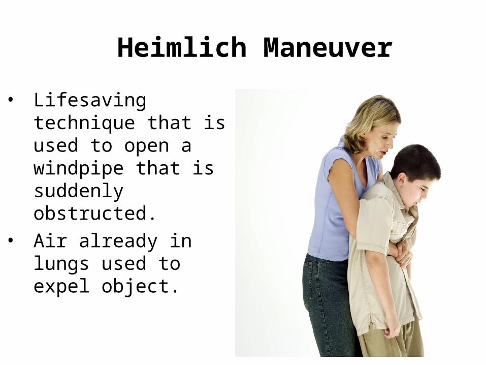
Heimlich Maneuver
• Lifesaving technique that is used to open a windpipe that is suddenly obstructed.
• Air already in lungs used to expel object.
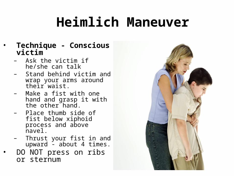
Heimlich Maneuver
• Technique - Conscious victim
– Ask the victim if he/she can talk
– Stand behind victim and wrap your arms around their waist.
– Make a fist with one hand and grasp it with the other hand.
– Place thumb side of fist below xiphoid process and above navel.
– Thrust your fist in and upward - about 4 times.
• DO NOT press on ribs or sternum
Heimlich Maneuver
– Technique - Unconscious victim• Catch victim if they begin to fall - place on
floor face up.• Straddle hips• Place one hand on top of other on the
victims abdomen - above navel and below xiphoid process.
• Forceful upward thrusts with heel of hand - several times if necessary.
Review Questions
1 Which of the following is not a function of the respiratory system?
A. influence speech
B. Distribution of oxygen to cells
C. Filtration of air
D. Warming of air B
2
The common name for the trachea is _______
Windpipe
3
• The structure known as the Adam’s Apple located in neck is the _____
Thyroid Cartilage
4
• The smallest branches of the bronchial tree are the
a. Primary bronchi
b. Secondary bronchi
c. Tertiary bronchi
d. BronchiolesD
5
• The flap or opening to the trachea is the
a. Larynx
b. Pharynx
c. Epiglottis
d. Vocal cords
6
• The structure that separates the right and left nasal cavities is the ____________
Septum
7
• The incorrect formation of the palantine bone during gestation is known as a __________
Cleft Palate
8
During inspiration which of thefollowing does not occur?
A.Diaphragm contractsB.Intercostals relaxC.Diaphragm flattensD.Size of thorax increases
B
9
Which of the following activities is thebest analogy of respiration?
A.Exchanging giftsB.Giving a giftC.Receiving a giftD.Sitting in a chair
A
10
Air is forced into the lungs by thecontraction of the…
A.AlveoliB.BronchiolesC.DiaphragmD.Heart
C