Embed Size (px)
Citation preview
The Cell Cycle and
Cell Division
Chapter 7

Chapter 7 The Cell Cycle
and Cell Division Key Concepts
7.1 Different Life Cycles Use Different Modes of Cell
Reproduction
7.2 Both Binary Fission and Mitosis Produce Genetically
Identical Cells
7.3 Cell Reproduction Is Under Precise Control
7.4 Meiosis Halves the Nuclear Chromosome Content and
Generates Diversity
7.5 Programmed Cell Death Is a Necessary Process in Living
Organisms

Chapter 7 Opening
QuestionHow does infection with HPV result in uncontrolled cell
reproduction?
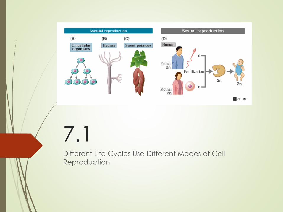
7.1Different Life Cycles Use Different Modes of Cell
Reproduction
Different Life Cycles Use
Different Modes of Cell
Reproduction The lifespan of an organism is linked to cell
reproduction, or cell division: a parent cell duplicates
its genetic material and then divides into two similar
cells.
Cell division is important in growth and repair of
multicellular organisms and the reproduction of all
organisms.
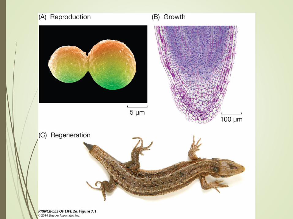
Figure 7.1 The Importance
of Cell Division
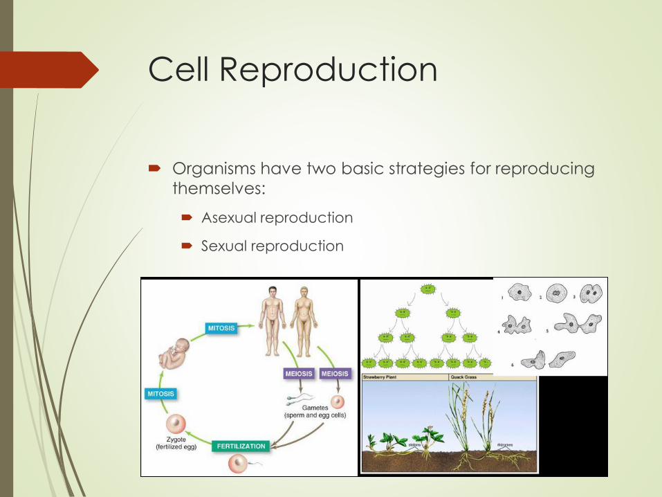
Cell Reproduction
Organisms have two basic strategies for reproducing
themselves:
Asexual reproduction
Sexual reproduction
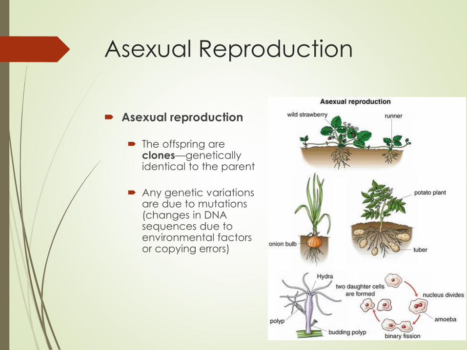
Asexual Reproduction
Asexual reproduction
The offspring are clones—genetically identical to the parent
Any genetic variations are due to mutations (changes in DNA sequences due to environmental factors or copying errors)
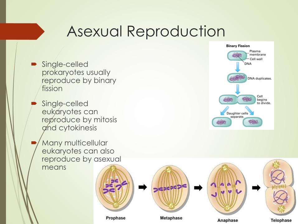
Asexual Reproduction
Single-celled prokaryotes usually reproduce by binary fission
Single-celled eukaryotes can reproduce by mitosis and cytokinesis
Many multicellular eukaryotes can also reproduce by asexual means

Figure 7.2 Asexual
Reproduction on a Large Scale
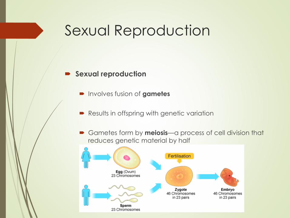
Sexual Reproduction
Sexual reproduction
Involves fusion of gametes
Results in offspring with genetic variation
Gametes form by meiosis—a process of cell division that reduces genetic material by half
Sexual Reproduction
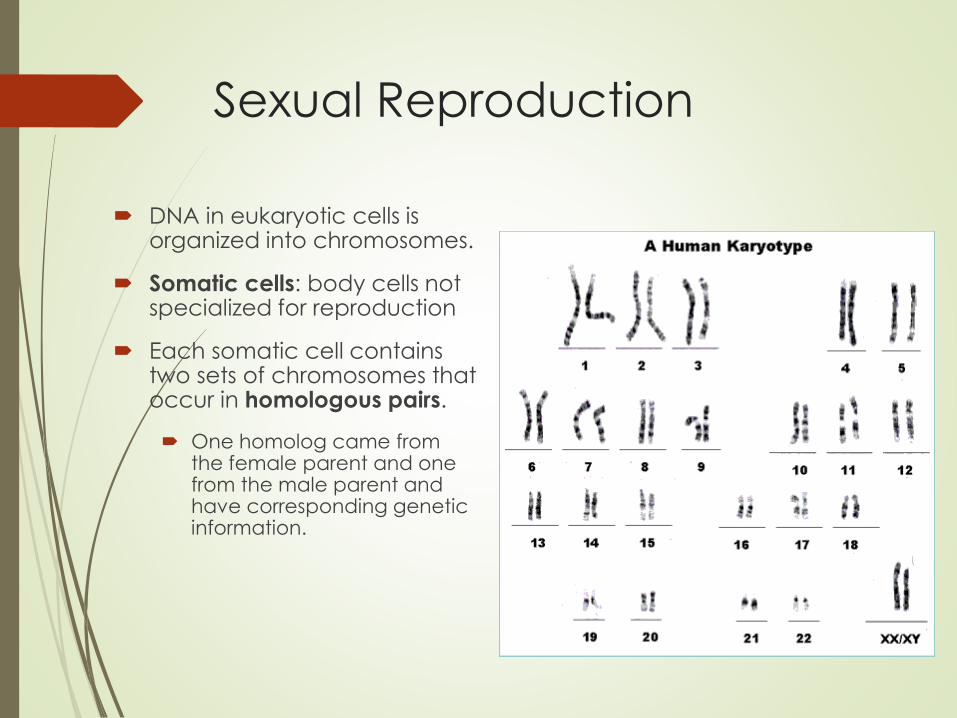
DNA in eukaryotic cells is organized into chromosomes.
Somatic cells: body cells not specialized for reproduction
Each somatic cell contains two sets of chromosomes that occur in homologous pairs.
One homolog came from the female parent and one from the male parent and have corresponding genetic information.
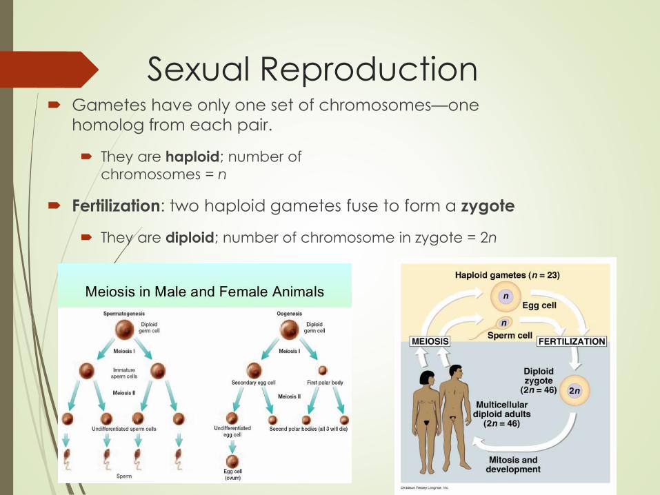
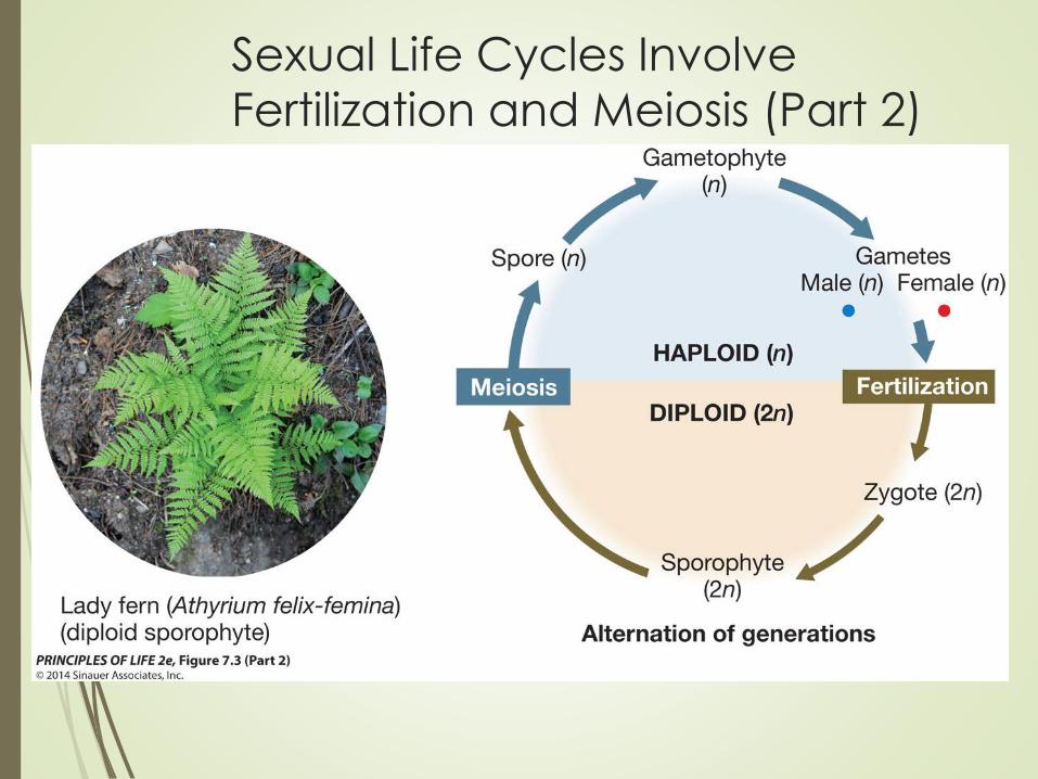
Sexual Reproduction Gametes have only one set of chromosomes—one
homolog from each pair.
They are haploid; number of
chromosomes = n
Fertilization: two haploid gametes fuse to form a zygote
They are diploid; number of chromosome in zygote = 2n

Sexual Reproduction
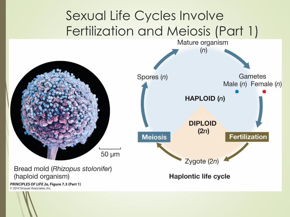
All sexual life cycles involve meiosis:
Gametes may develop immediately after meiosis
Or each haploid cell may develop into a haploid organism (haploid stage of the life cycle) that eventually produces gametes by mitosis
Fertilization results in a zygote and begins the diploid stage of the life cycle.
Sexual Life Cycles Involve
Fertilization and Meiosis (Part 1)
Sexual Life Cycles Involve
Fertilization and Meiosis (Part 2)

Sexual Life Cycles Involve
Fertilization and Meiosis (Part 3)
Sexual Reproduction
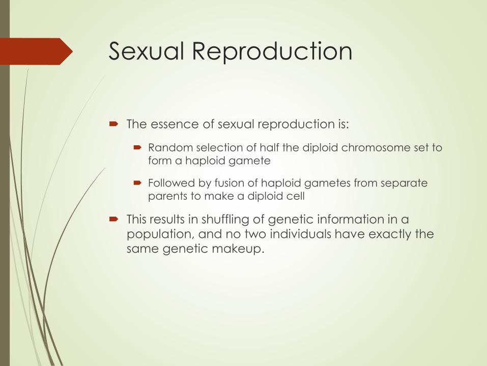
The essence of sexual reproduction is:
Random selection of half the diploid chromosome set to
form a haploid gamete
Followed by fusion of haploid gametes from separate
parents to make a diploid cell
This results in shuffling of genetic information in a
population, and no two individuals have exactly the
same genetic makeup.

Sexual Reproduction
Four events in cell division:
• Reproductive signals initiate cell division
• DNA replication
• DNA segregation—distribution of the DNA into the two
new cells
• Cytokinesis—division of the cytoplasm and separation
of the two new cells
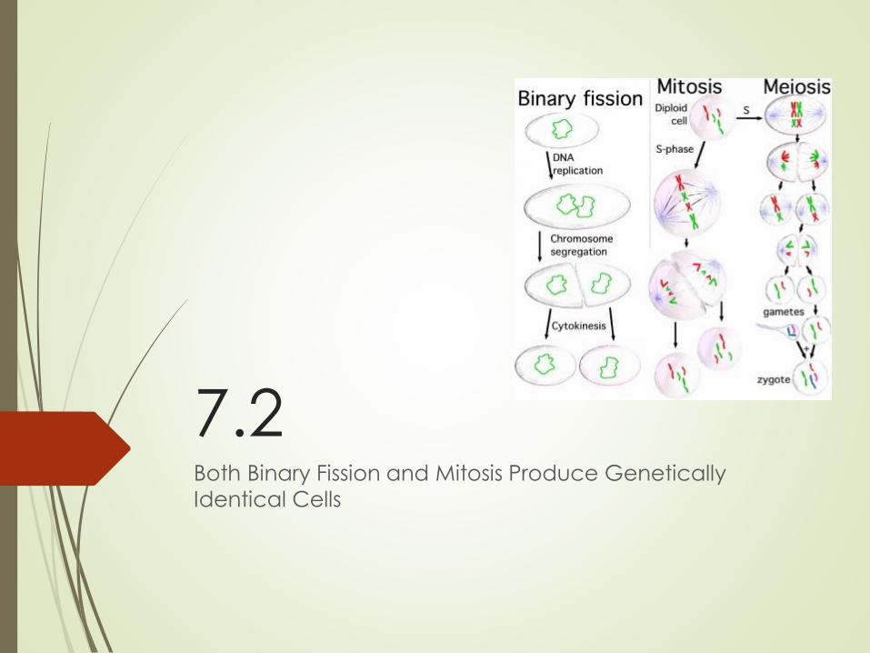
7.2 Both Binary Fission and Mitosis Produce Genetically
Identical Cells
Binary Fission
Prokaryotes divide by binary fission: results in
reproduction of the entire organism.
Reproductive signals may be environmental factors
such as nutrient availability.
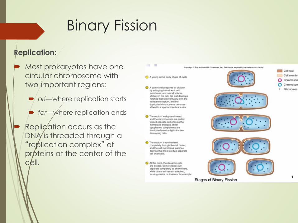
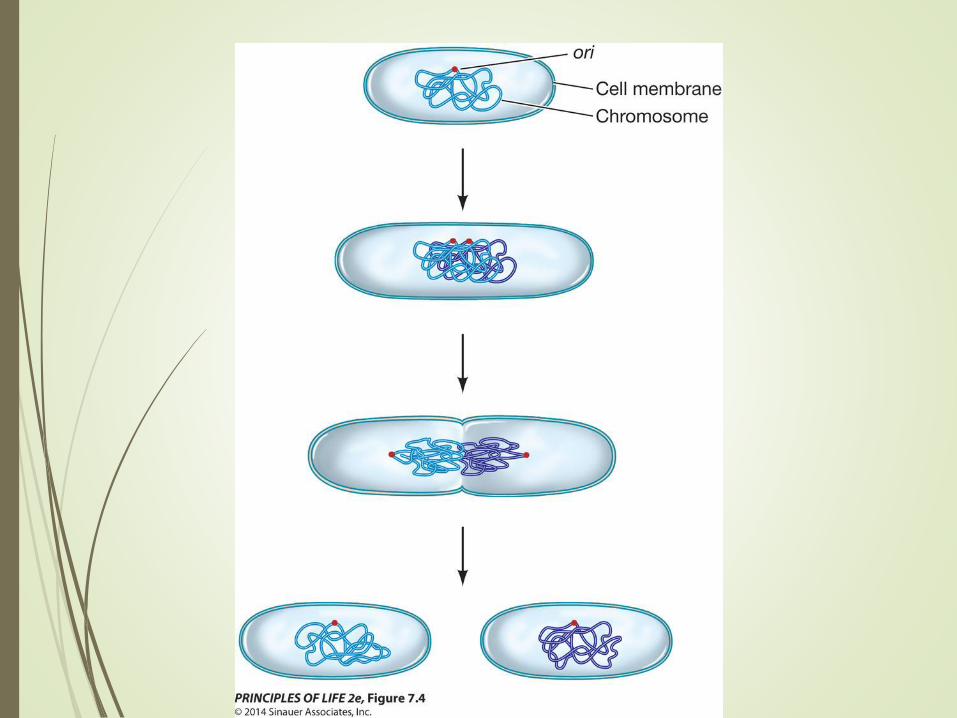
Binary Fission
Replication:
Most prokaryotes have one
circular chromosome with
two important regions:
ori—where replication starts
ter—where replication ends
Replication occurs as the
DNA is threaded through a
“replication complex” of proteins at the center of the
cell.
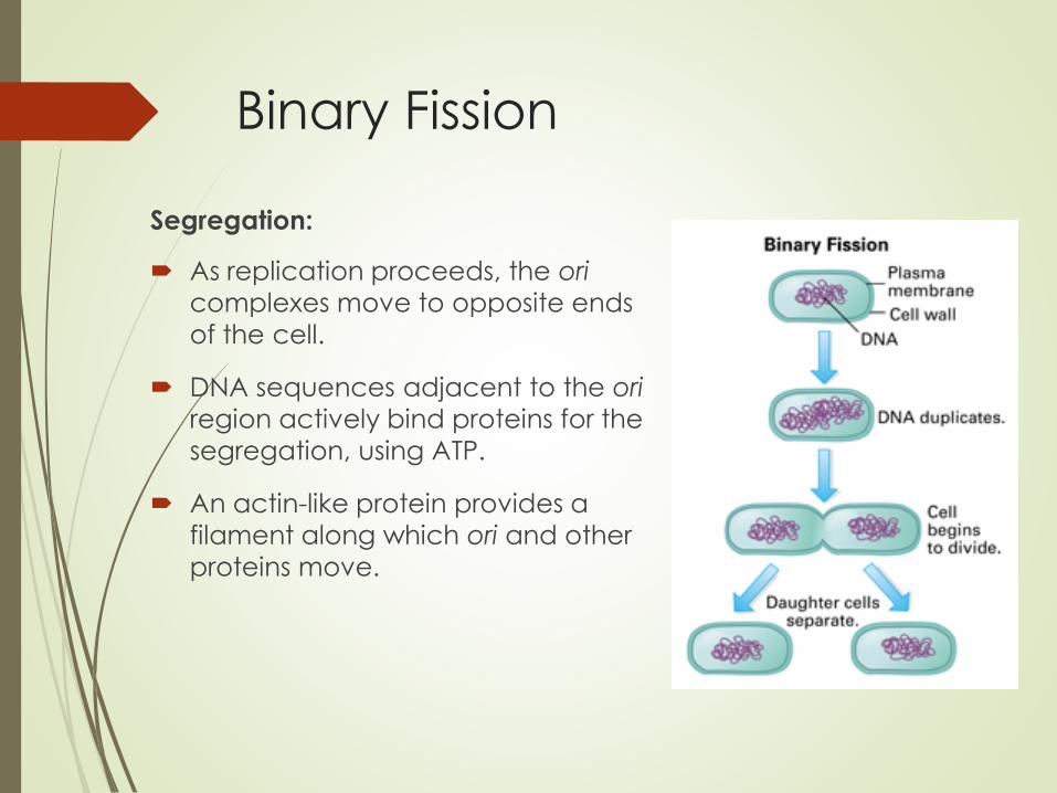
Binary Fission
Segregation:
As replication proceeds, the ori
complexes move to opposite ends
of the cell.
DNA sequences adjacent to the ori
region actively bind proteins for the
segregation, using ATP.
An actin-like protein provides a
filament along which ori and other
proteins move.
Binary Fission
Cytokinesis:
After chromosome segregation, the cell membrane
pinches in by contraction of a ring of protein fibers
under the surface.
As the membrane pinches in, new cell wall materials
are deposited, resulting in separation of the two cells.

Eukaryotes - Mitosis
Eukaryotic cells divide by mitosis followed by
cytokinesis.
Reproductive signals are usually related to functions of
the entire organism, not the environment of a single cell.
Most cells in a multicellular organism are specialized and
do not divide.
Eukaryotes
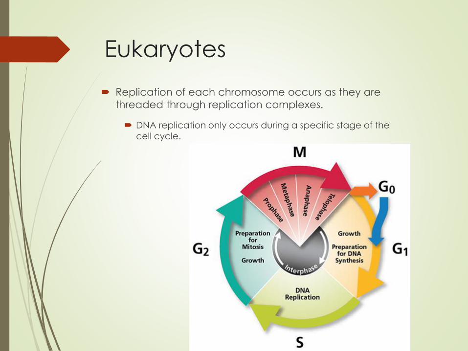
Replication of each chromosome occurs as they are
threaded through replication complexes.
DNA replication only occurs during a specific stage of the cell cycle.

Eukaryotes
In segregation, one copy of each chromosome ends up
in each of the two new cells.
More complex than in prokaryotes: eukaryotes have a nuclear envelope, and there are multiple chromosomes.
Cytokinesis in plant cells (which have cell walls) is
different than in animal cells (no cell walls).
Eukaryotes
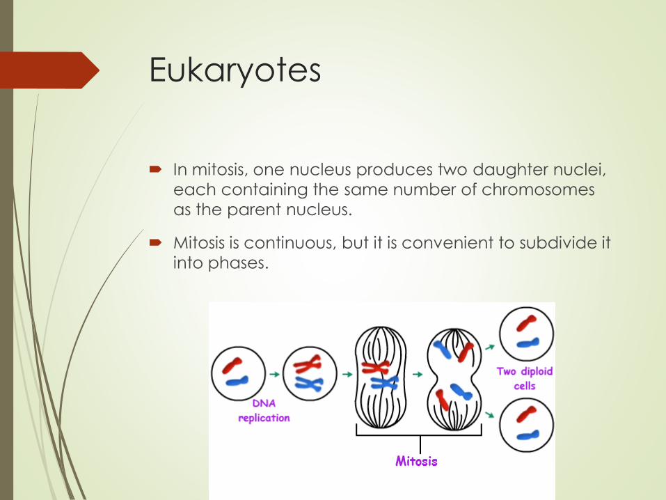
In mitosis, one nucleus produces two daughter nuclei,
each containing the same number of chromosomes
as the parent nucleus.
Mitosis is continuous, but it is convenient to subdivide it
into phases.
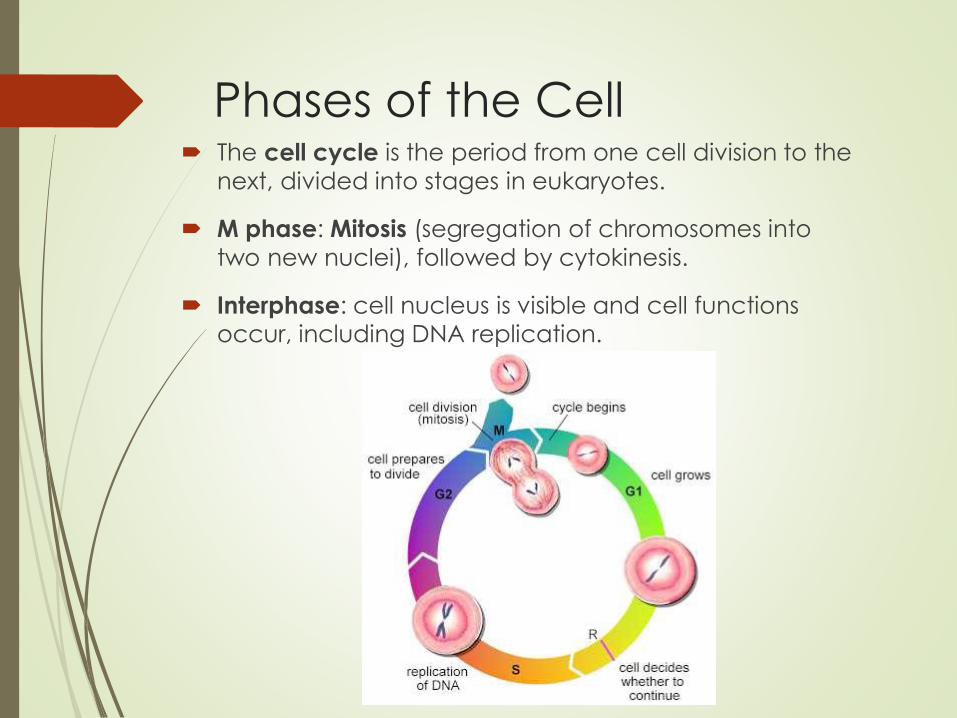
Phases of the Cell The cell cycle is the period from one cell division to the
next, divided into stages in eukaryotes.
M phase: Mitosis (segregation of chromosomes into
two new nuclei), followed by cytokinesis.
Interphase: cell nucleus is visible and cell functions occur, including DNA replication.
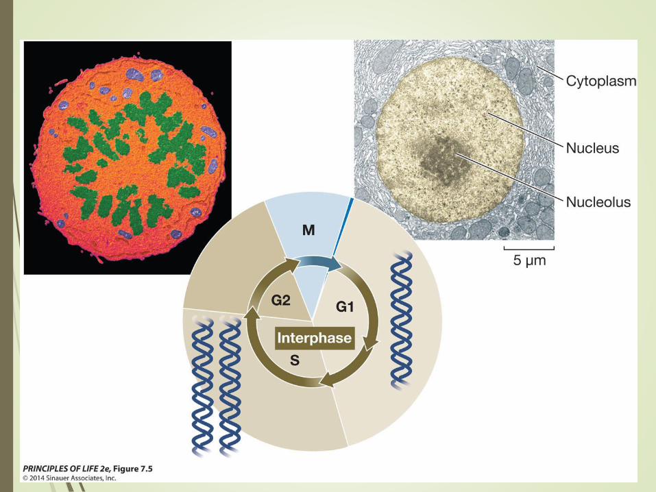
Figure 7.5 The Phases of the
Eukaryotic Cell Cycle
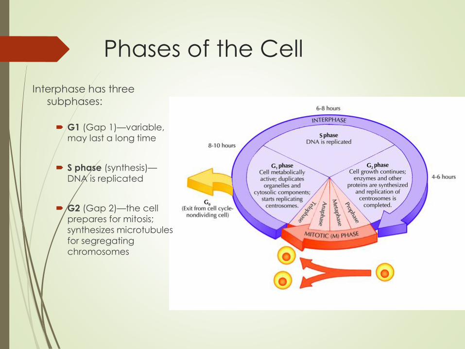
Phases of the Cell
Interphase has three
subphases:
G1 (Gap 1)—variable, may last a long time
S phase (synthesis)—DNA is replicated
G2 (Gap 2)—the cell prepares for mitosis; synthesizes microtubules for segregating chromosomes

Mitosis
Prophase: three structures appear
Condensed chromosomes
Reoriented centrosomes
Spindle
Mitosis
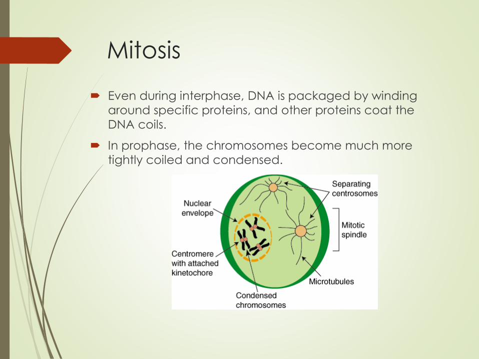
Even during interphase, DNA is packaged by winding
around specific proteins, and other proteins coat the
DNA coils.
In prophase, the chromosomes become much more
tightly coiled and condensed.
Mitosis
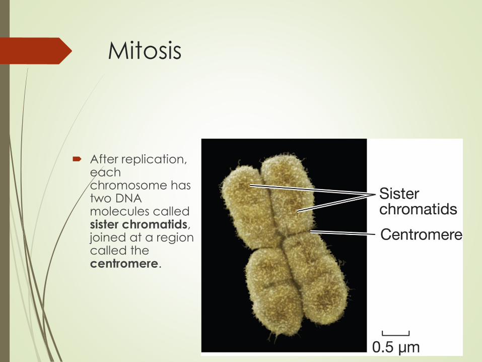
After replication, each chromosome has two DNA molecules called sister chromatids, joined at a region called the centromere.

Both Binary Fission and Mitosis
Produce Genetically Identical
Cells Karyotype: the condensed chromosomes for a given
organism can be distinguished by their sizes and
centromere positions
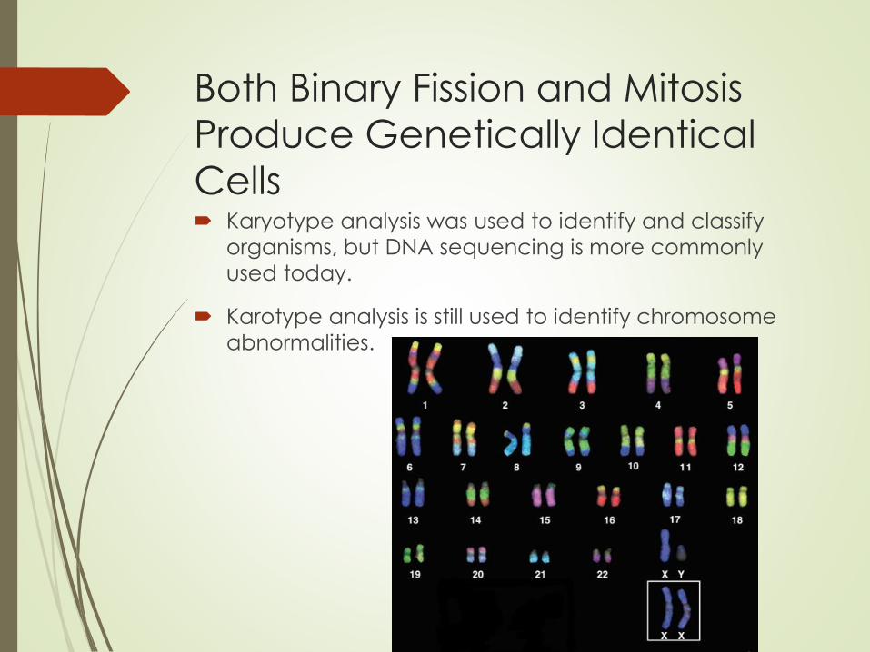
Both Binary Fission and Mitosis
Produce Genetically Identical
Cells Karyotype analysis was used to identify and classify
organisms, but DNA sequencing is more commonly
used today.
Karotype analysis is still used to identify chromosome
abnormalities.
Mitosis
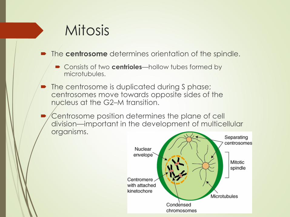
The centrosome determines orientation of the spindle.
Consists of two centrioles—hollow tubes formed by microtubules.
The centrosome is duplicated during S phase; centrosomes move towards opposite sides of the nucleus at the G2–M transition.
Centrosome position determines the plane of cell division—important in the development of multicellular organisms.
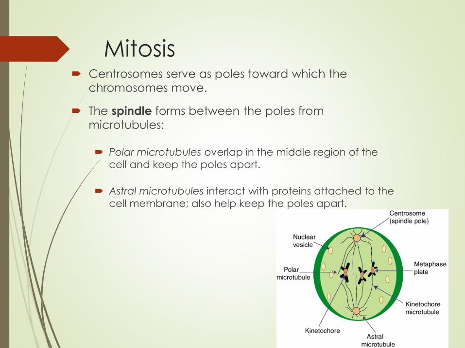
Mitosis Centrosomes serve as poles toward which the
chromosomes move.
The spindle forms between the poles from
microtubules:
Polar microtubules overlap in the middle region of the
cell and keep the poles apart.
Astral microtubules interact with proteins attached to the
cell membrane; also help keep the poles apart.

Mitosis
Kinetochore microtubules attach to kinetochores on the chromatid centromeres.
Sister chromatids attach to kinetochore microtubules from opposite sides so that the two chromatids will move to opposite poles.
Sister chromatids become daughter chromatids after separation.
Mitosis
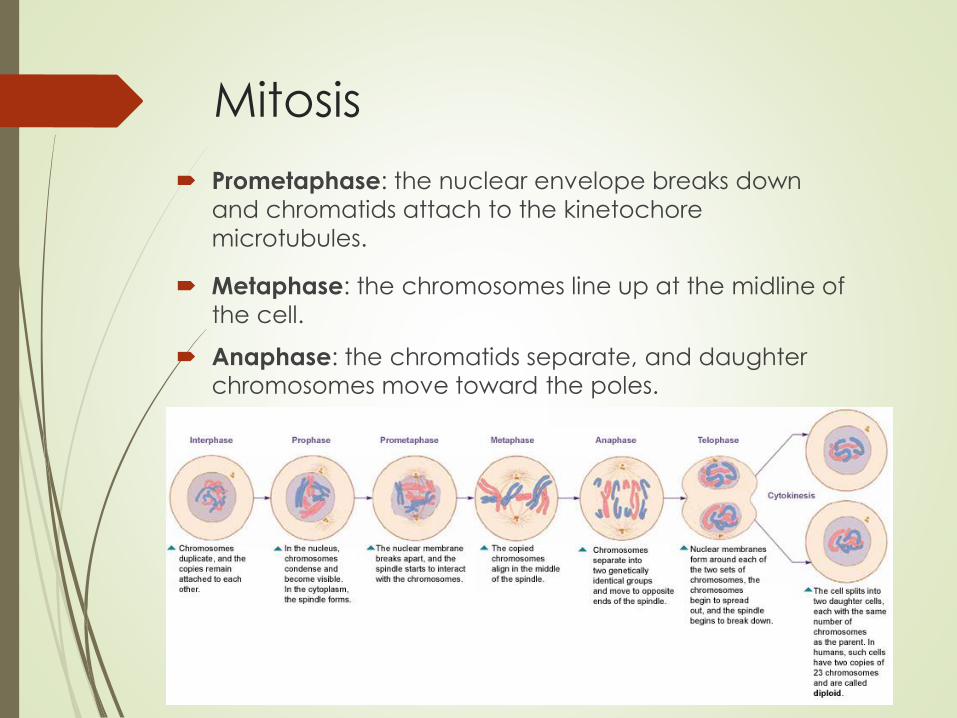
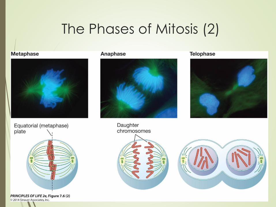
Prometaphase: the nuclear envelope breaks down
and chromatids attach to the kinetochore
microtubules.
Metaphase: the chromosomes line up at the midline of
the cell.
Anaphase: the chromatids separate, and daughter chromosomes move toward the poles.
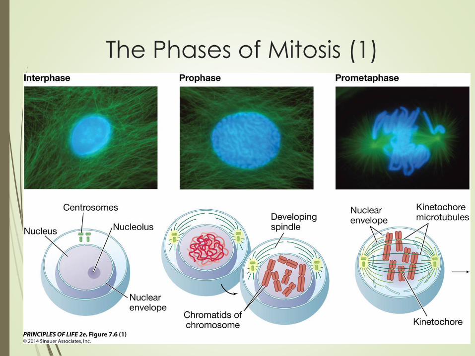
The Phases of Mitosis (1)
The Phases of Mitosis (2)

Mitosis
Two mechanisms move the chromosomes to opposite poles:
Kinetochores have molecular motor proteins (kinesin and
dynein), which move the chromosomes along the
microtubules.
The kinetochore microtubules shorten from the poles,
drawing the chromosomes toward the poles.

Mitosis Telophase: nuclear envelopes form around each set
of chromosomes and nucleoli appear, and the spindle
breaks down and chromosomes become less
compact.

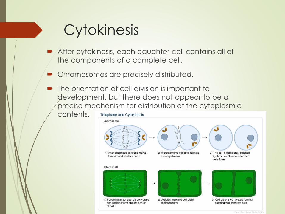
Cytokinesis Cytokinesis:
In animal cells, the cell membrane pinches in between
the nuclei.
A contractile ring of actin and myosin microfilaments
forms on the inner surface of the cell membrane; the
two proteins produce a contraction to pinch the cell
in two.
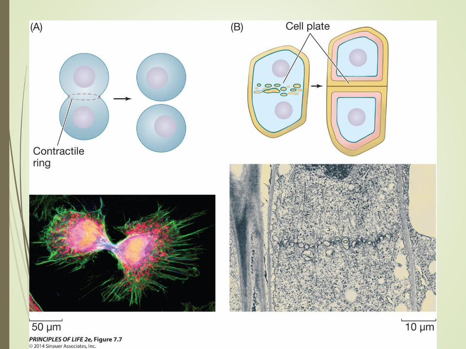
Figure 7.7 Cytokinesis Differs
in Animal and Plant Cells

Cytokinesis
In plant cells, vesicles
from the Golgi
apparatus appear
along the plane of cell
division.
The vesicles fuse to form
a new cell membrane.
Contents of vesicles
also contribute to
forming the cell plate—
the beginning of the
new cell wall.
Cytokinesis
After cytokinesis, each daughter cell contains all of
the components of a complete cell.
Chromosomes are precisely distributed.
The orientation of cell division is important to
development, but there does not appear to be a
precise mechanism for distribution of the cytoplasmic
contents.
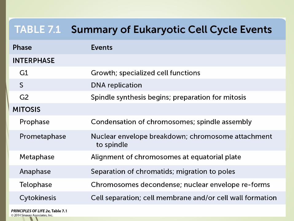
Table 7.1
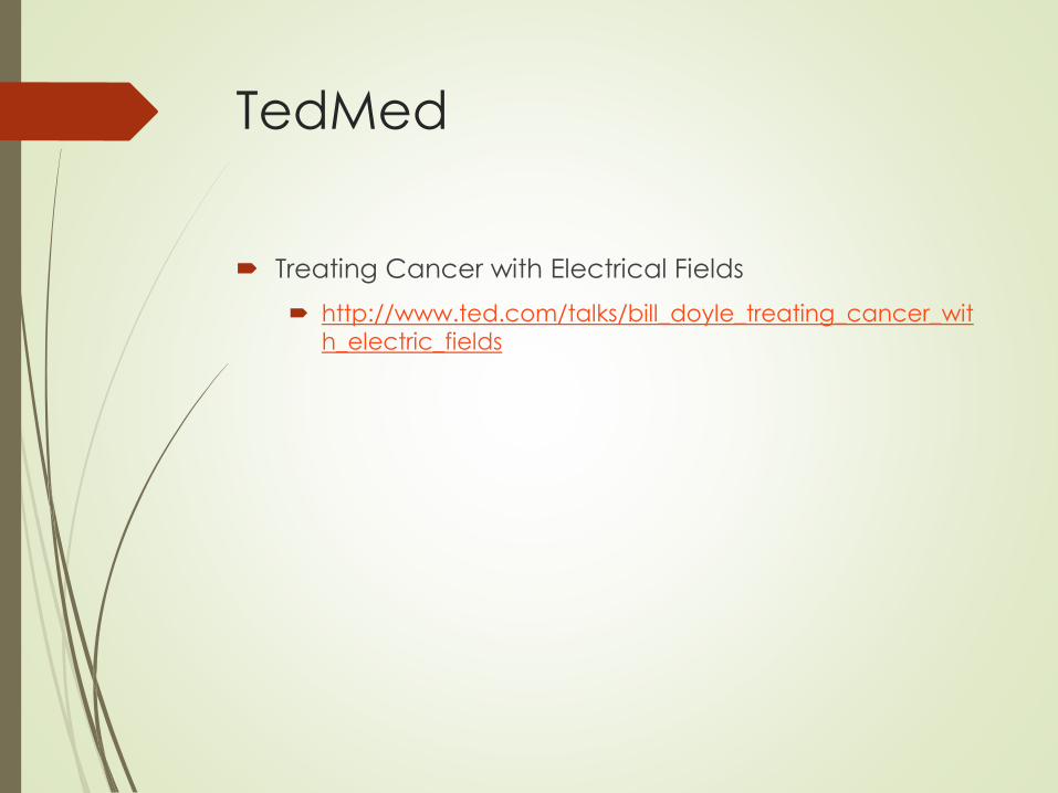
TedMed
Treating Cancer with Electrical Fields
http://www.ted.com/talks/bill_doyle_treating_cancer_wit
h_electric_fields

7.3Cell Reproduction is Under Precise Control
Cell Control
Cell reproduction must be under precise control.
If single-celled organisms had no control over
reproduction, they would soon overrun the
environment and starve to death.
In multicellular organisms, cell reproduction must be
controlled to maintain body form and function.
Cell Control
Prokaryotic cells divide in response to environmental
conditions.
In eukaryotes, cell division is related to the needs of
the entire organism.
Mammals produce growth factors that stimulate cell division and differentiation.
Example: platelets in the blood secrete growth factors
that stimulate cells to divide to heal wounds.

Cell Control
Progression through the
eukaryotic cell cycle is
tightly regulated.
The G1–S transition is
called R, the restriction
point.
Passing this point
usually means the cell
will proceed with the
cell cycle and divide.


Cell Fusion Investigation
Specific substances trigger the transition from one
phase to another.
The first evidence for these substances came from cell
fusion experiments.
Fusion of mammalian cells at G1 and S phases showed
that a cell in S phase produces a substance that
activates DNA replication.
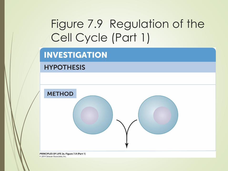
Figure 7.9 Regulation of the
Cell Cycle (Part 1)

Figure 7.9 Regulation of the
Cell Cycle (Part 2)
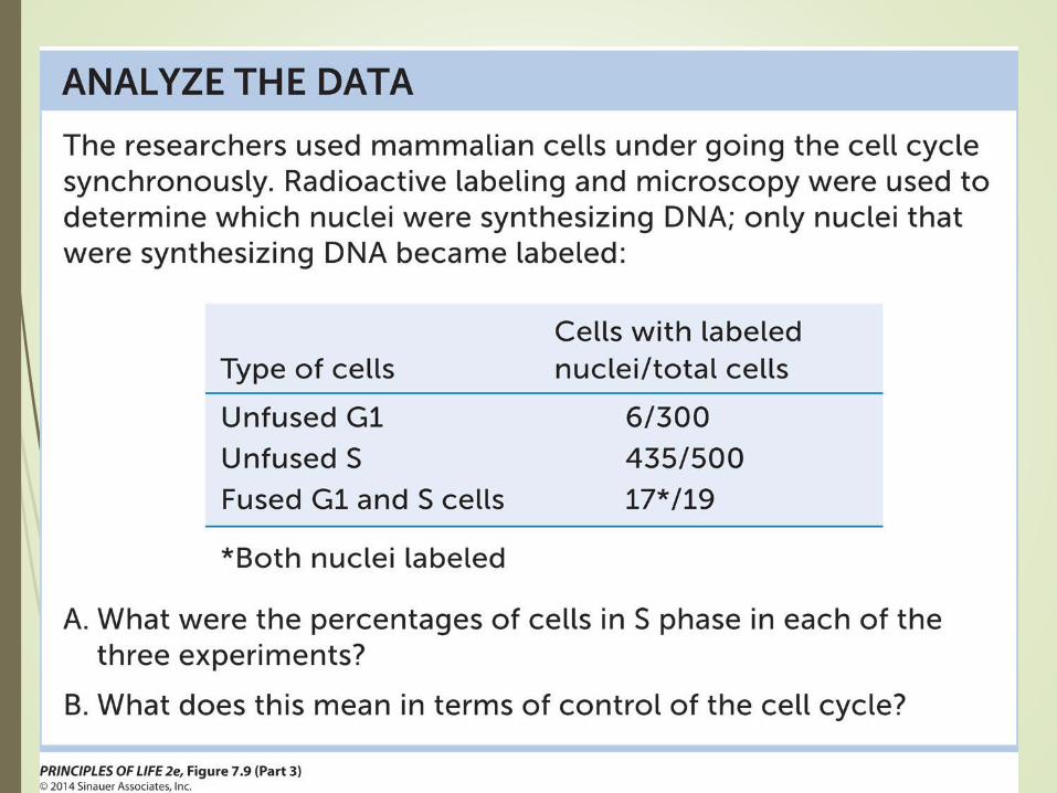
Figure 7.9 Regulation of the
Cell Cycle (Part 3)
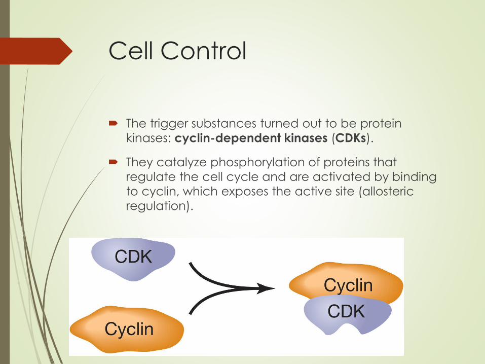
Cell Control
The trigger substances turned out to be protein
kinases: cyclin-dependent kinases (CDKs).
They catalyze phosphorylation of proteins that
regulate the cell cycle and are activated by binding
to cyclin, which exposes the active site (allosteric
regulation).
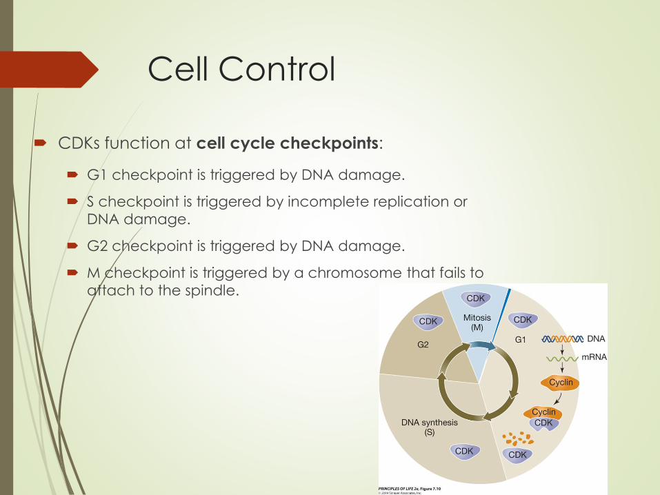
Cell Control
CDKs function at cell cycle checkpoints:
G1 checkpoint is triggered by DNA damage.
S checkpoint is triggered by incomplete replication or
DNA damage.
G2 checkpoint is triggered by DNA damage.
M checkpoint is triggered by a chromosome that fails to
attach to the spindle.

Cell Control
Each CDK has a cyclin to activate
it, which is made only at the right
time.
After the CDK acts, the cyclin is
broken down by a protease.
Synthesis and breakdown of cyclins
is important in controlling the cell
cycle.
Cyclins are synthesized in response
to various signals, such as growth
factors.
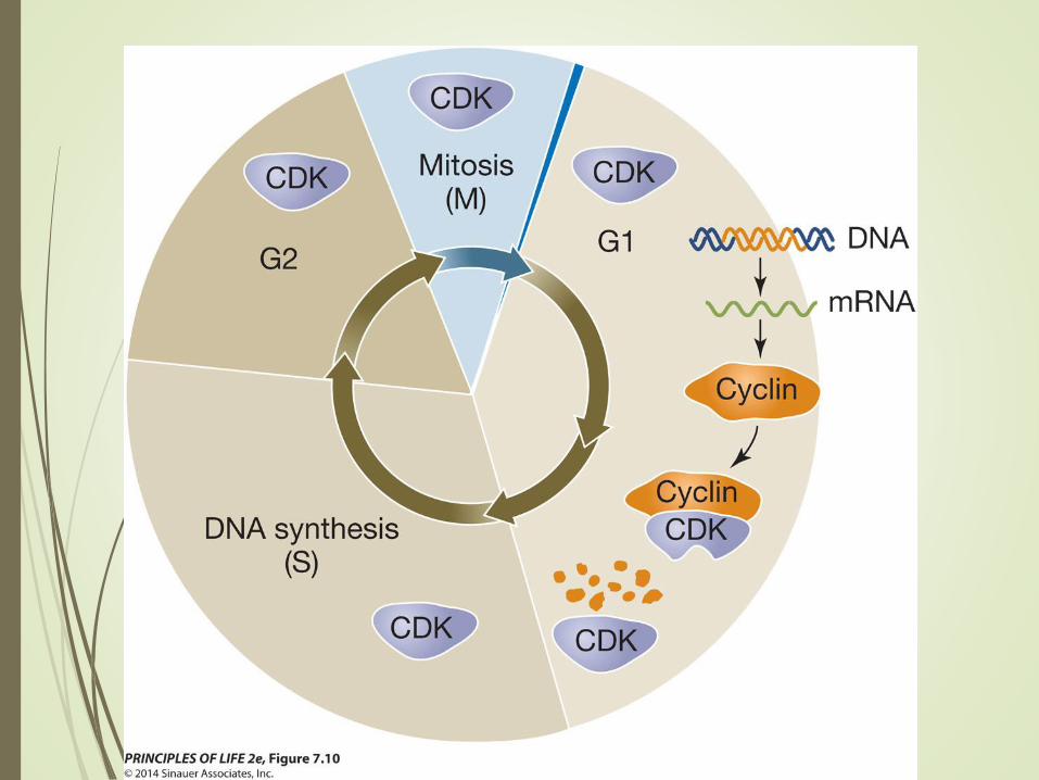
Figure 7.10 Cyclins Are
Transient in the Cell Cycle
Cell Control
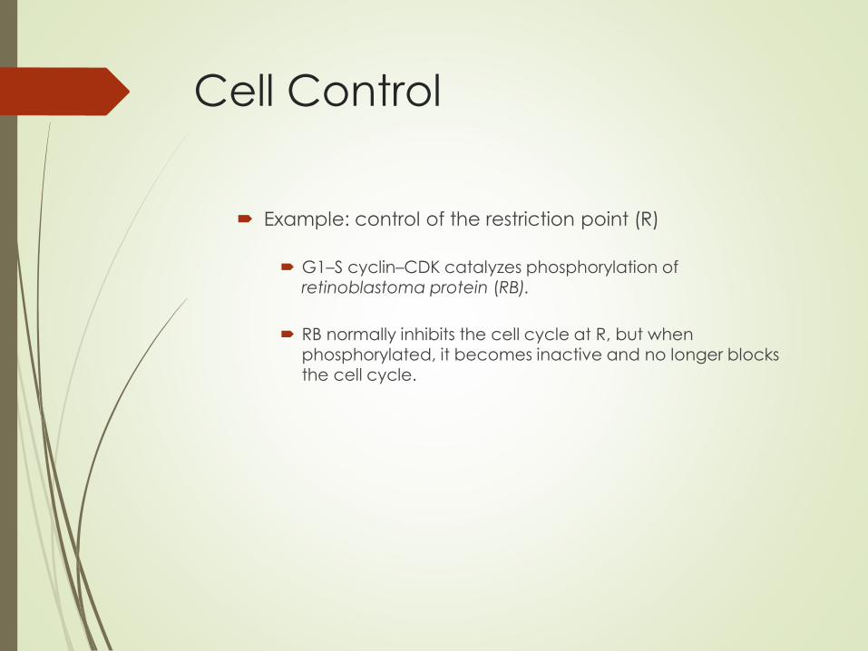
Example: control of the restriction point (R)
G1–S cyclin–CDK catalyzes phosphorylation of
retinoblastoma protein (RB).
RB normally inhibits the cell cycle at R, but when phosphorylated, it becomes inactive and no longer blocks the cell cycle.

7.4Meiosis Halves the Nuclear Chromosome Content and
Generates Diversity

Meiosis
Meiosis consists of two nuclear divisions but DNA is replicated only once.
The haploid cells produced by meiosis are genetically different from one another and from the parent cell.
Meiosis
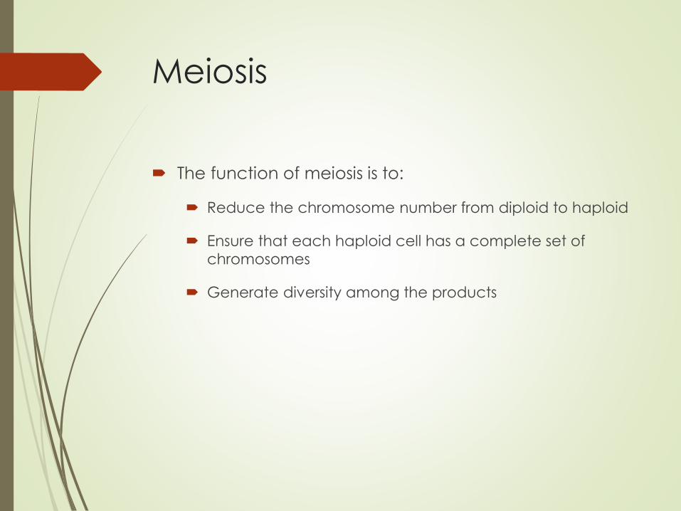
The function of meiosis is to:
Reduce the chromosome number from diploid to haploid
Ensure that each haploid cell has a complete set of
chromosomes
Generate diversity among the products
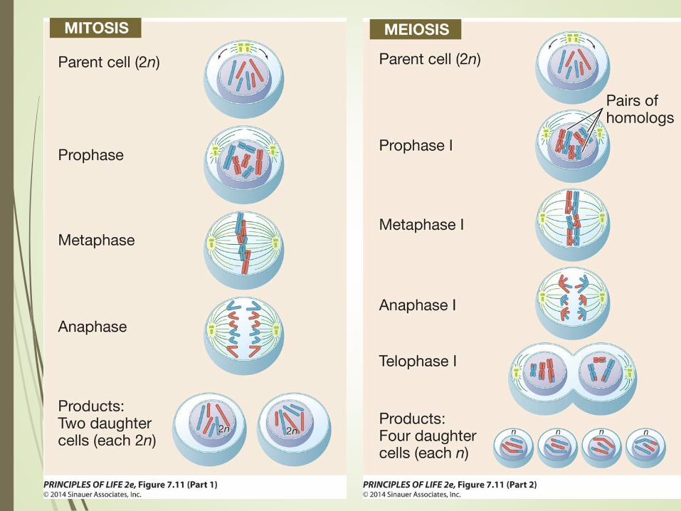
Meiosis
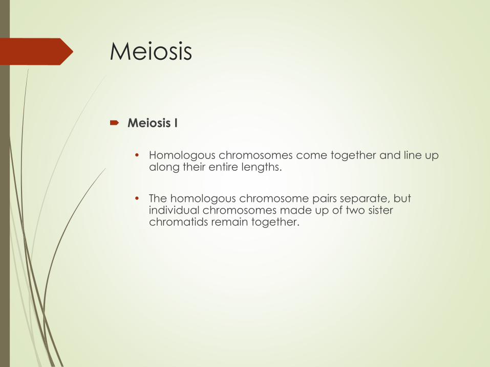
Meiosis I
• Homologous chromosomes come together and line up along their entire lengths.
• The homologous chromosome pairs separate, but individual chromosomes made up of two sister chromatids remain together.
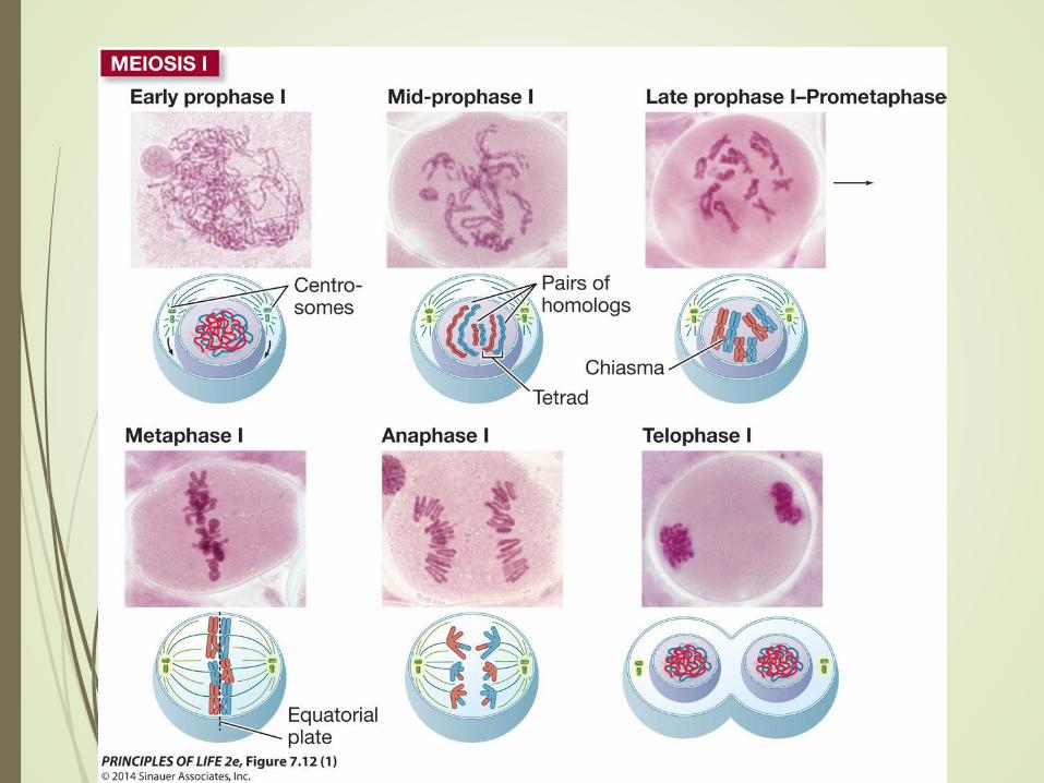
Figure 7.12 Meiosis:
Generating Haploid Cells (1)
Meiosis
Meiosis I is preceded by an S phase during which DNA
is replicated.
Each chromosome then consists of two sister
chromatids.
At the end of meiosis I, two nuclei form, each with half
the original chromosomes (one member of each
homologous pair).
The centromeres did not separate, so each
chromosome is still two sister chromatids.
Meiosis
Meiosis II
• Not preceded by DNA replication
• Sister chromatids separate
• End result: four haploid cells that are not genetically
identical
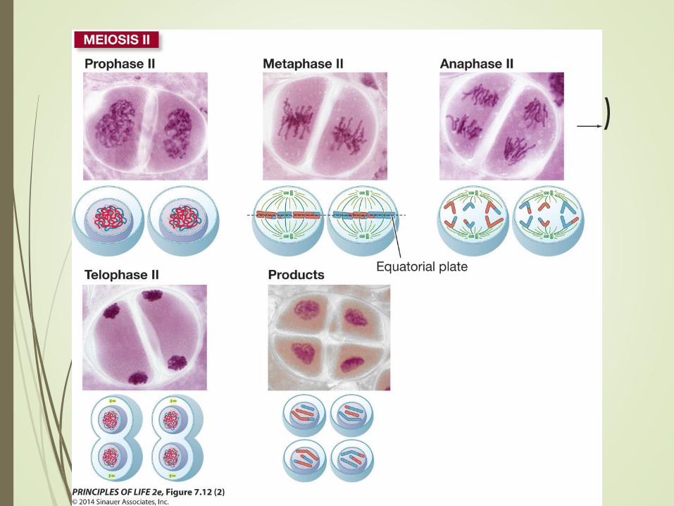
Figure 7.12 Meiosis:
Generating Haploid Cells (2)
Meiosis
Shuffling of genetic material during meiosis occurs by
two processes:
Crossing over
In prophase I homologous chromosomes (synapsis) and the four chromatids form a tetrad, or bivalent.

Meiosis
The homologs seem to repel each other at the centromeres but remain attached at chiasmata.
Meiosis
Genetic material is exchanged between nonsister chromatids at the chiasmata.
Any of the four chromatids in the tetrad can participate, and a single chromatid can exchange material at more than one point.
Crossing over results in recombinant chromatids and increases genetic variability of the products.

Meiosis
Prophase I may last a long time.
Human males: prophase I lasts about 1 week, and 1 month for
entire meiotic cycle
Human females: prophase I begins before birth, meiosis
continues up to decades later during the monthly ovarian cycle
and is completed only after fertilization.

Meiosis
Independent assortment
At anaphase I, it is a matter of chance which member of a homologous pair goes to which daughter cell.
The greater the number of chromosomes, the greater the potential for genetic diversity.
In humans, 223 (8,388,608) different combinations of maternal and paternal chromosomes can be produced.

Meiosis
Meiosis is complex, and errors can occur.
Nondisjunction
• Homologous pair fails to separate at anaphase I
• Sister chromatids fail to separate at anaphase II
Both result in aneuploidy—an abnormal number of
chromosomes.


Meiosis
Most human embryos from aneuploid zygotes do not
survive. Many miscarriages are due to this.
The most common human aneuploidy is trisomy 16.
Trisomy 21 (Down syndrome) is one of the few
aneuploidies that allow survival.

Meiosis
Polyploidy
Sometimes, organisms with triploid (3n), tetraploid (4n),
and even higher numbers can form.
This can occur through an extra round of DNA
replication before meiosis, or lack of spindle formation
in meiosis II.
Polyploidy occurs naturally in some species and can
be desirable in plants.
Meiosis
Translocation
Crossing over between non-homologous
chromosomes in meiosis I
Location of genes relative to other DNA sequences is
important, and translocations can have profound
effects on gene expression.
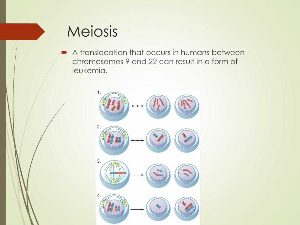
Meiosis
A translocation that occurs in humans between
chromosomes 9 and 22 can result in a form of
leukemia.
7.5Programmed Cell Death Is a Necessary Process in Living
Organisms
Programmed Cell Death
Cells can die in one of two ways:
• In necrosis, the cell is damaged or starved for oxygen or
nutrients. The cell swells and bursts.
Cell contents are released to the extracellular environment and can cause inflammation.
Programmed Cell Death
• Apoptosis is genetically programmed cell death. Two possible reasons:
The cell is no longer needed (e.g., the connective tissue between the fingers of a fetus)
Old cells are prone to genetic damage that can lead to cancer—especially true of epithelial cells that die after days or weeks
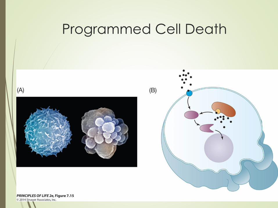
Programmed Cell Death
Events of apoptosis:
• Cell detaches from its neighbors
• DNA is cut into small fragments
• Membranous lobes (“blebs”) form and break into fragments
• Surrounding living cells usually ingest remains of the dead
cell by phagocytosis
Programmed Cell Death
Programmed Cell Death
Plants use apoptosis in the hypersensitive response.
They protect themselves from disease by undergoing
apoptosis at the site of infection by a fungus or
bacterium, preventing spread to other parts of the
plant.
Programmed Cell Death
Programmed cell death is controlled by signals:
Internal signals may be linked to cell age or damaged
DNA.
Both internal and external signals lead to activation of
caspases, which hydrolyze target proteins in a cascade
of events.
The cell dies as caspases hydrolyze proteins of the
nuclear envelope, nucleosomes, and cell membrane.
Answer to Opening Question
Human papilloma virus (HPV) stimulates the cell cycle
when it infects the cervix.
Two proteins regulate the cell cycle:
Oncogene proteins are mutated positive regulators of
the cell cycle—in cancer cells they are overactive or
present in excess.
Answer to Opening Question
Tumor suppressors are negative regulators of the cell cycle, but are inactive in cancer cells.
Example: RB blocks the cell cycle at R. HPV causes synthesis of E7 protein, which fits into the protein-binding site of RB, thereby inactivating it.
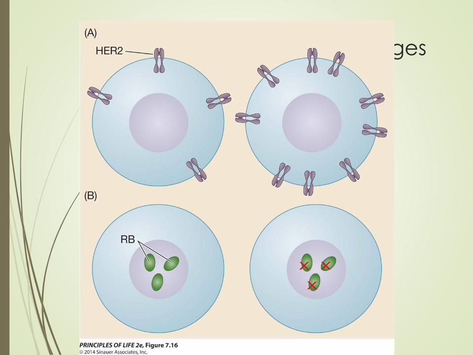
Figure 7.16 Molecular Changes
Regulate the Cell Cycle in
Cancer Cells

Answer to Opening Question
Chemotherapy drugs stop cell division by targeting
cell cycle events.
Some drugs block DNA replication; others damage
DNA, stopping cells at G2; and still others prevent
normal functioning of the mitotic spindle.
Unfortunately, these drugs also act on normal cells
and are toxic to rapidly dividing cells in the intestines,
skin, and bone marrow.

Answer to Opening Question
Research into more specific chemotherapy drugs is
ongoing.
Example: a drug has been identified that affects the
protein produced as a result of the translocation
between chromosomes 9 and 22.
It has been successful at treating leukemia caused by this
translocation.