Embed Size (px)
Citation preview
Biochimica et Biophysica Acta, 379 (1975) 360-369 © Elsevier Scientific Publishing Company, Amsterdam - - Printed in The Netherlands
BBA 36975
THE ISOLATION AND CHARACTERISATION OF A PLATELET-SPECIFIC r -GLOBULIN (f l -THROMBOGLOBULIN) AND THE DETECTION OF ANTI- UROKINASE AND ANTIPLASMIN RELEASED FROM THROMBIN- A G G R E G A T E D WASHED H U M A N PLATELETS
STEPHEN MOORE, DUNCAN S. PEPPER and JOHN D. CASH Edinburgh and South-East Scotland Regional Blood Transfusion Service, Royal Infirmary, Edinburgh* and Department of Medicine, University of Edinburgh (U.K.)
(Received July 2nd, 1974)
SUMMARY
A protein fraction was isolated from the supernatant of thrombin-aggregated washed human platelets and was shown, by immunodiffusion techniques, to contain a platelet-specific fl-globulin (fl-thromboglobulin) as the major component.
A molecular weight of 35 800 was determined for fl-thromboglobulin from the measured sedimentation coefficient of 3.0 S and Stokes radius of 2.85 nm.
fl-Thromboglobulin was detected in the serum from whole blood and the supernatant of 48-h-old platelet-rich plasma and 28-day-old citrated whole blood, but not in platelet-poor plasma.
The fraction containing fl-thromboglobulin was shown to possess an anti- urokinase activity but was devoid of antiplasmin activity.
A further fraction of approximate molecular weight 70 000 was also isolated which contained an antiplasmin but was devoid of antiurokinase activity.
INTRODUCTION
The presence of proteins in the soluble material released during thrombin- induced aggregation of washed platelets has been previously described [1, 2]. These studies have indicated the existence of platelet-specific a- and r-globulins in such material [2].
Other studies have shown that platelets contain separate substances with antiplasmin and antiplasminogen activator activity. These investigations have been confined to supernatant preparations from damaged platelets either following lyophilisation or freezing and thawing [3-14].
The following communication describes a series of studies designed to separate the proteins released from thrombin-aggregated washed platelets and to measure their antiplasmin and antiurokinase activity. Further studies are described which investigate the presence of platelet-specific proteins.
* Address for reprint requests.
361
MATERIALS AND METHODS
Preparation of soluble release products from thrombin aggregated human plate- lets. Human platelets, less than 24 h old, were obtained from S-E. Scotland Regional Blood Transfusion Service as platelet concentrates prepared at room temperature from acid/citrate/dextrose anticoagulated whole blood. Units of the same ABO blood group were pooled and processed at room temperature, using plastic containers, by the following method: residual red cells were removed by differential centrifugation and the platelets harvested by centrifugation at 4000 x g for 10 min followed by washing in a solution composed of Ringers lactate, 5 ~o glucose, 10~ trisodium citrate (7:2:1, by vol.) to remove plasma proteins. Washed platelets from 12 platelet concentrates were resuspended in 100 ml of Ringers lactate/glucose/citrate and equi- librated to 37 °C. Platelet aggregation was induced by the addition of bovine thrombin and CaC12 solution to a final concentration of 1 I.U./ml and 10 mM respectively; complete aggregation occurred within 15 s. After 2 min the aggregated suspension was centrifuged at 12 000 x g for 10 rain at 4 °C. The supernatant containing the soluble platelet release products was carefully separated.
Determination of sedimentation coefficient(s). Linear sucrose density gradients (6-30 ~o w/w, total volume = 6.0 ml), buffered by 25 mM Tris, 1 mM EDTA, 3.8 mM borate, 0.15 M NaC1, pH 8.8, were prepared in 6.5-ml thin-walled polycarbonate tubes (MSE 59203). Samples (0.15 ml) were overlayered on the gradients and cen- trifuged at 60 000 rev./min (420 000 x g at max. radius) in a 3 x 6.5 ml titanium swing out rotor (MSE 59587) using an MSE superspeed 65 preparative ultracen- trifuge. All three gradients were fractionated simultaneously into 30 equal fractions (0.2 ml) by aspiration from the bottom of the gradient using a three-channel peristaltic pump. Fractions were analysed for protein. The sedimentation distance was expressed in terms of the number of fractions moved by the protein peak from the origin. No corrections were made for protein concentration. Proteins of known S20, w were used to calibrate the system.
Molecular weights. Molecular weights were determined using the Svedberg equation, diffusion coefficients (D) being calculated from rs using the Stokes-Einstein equation, and assuming the partial specific volume to be 0.733.
Ultrafiltration. Ultrafiltration to concentrate protein solutions was performed at room temperature using an Amicon PM10 Diaflo membrane (Amicon Corp. Lexington, Mass., U.S.A.) at 25 lb/inch 2.
Desalting. Desalting was performed on a Sephadex G-25 column, of bed dimensions 430 m m x 25 ram, equilibrated and eluted with 0.05 M ammonia, formic acid, pH 8.8. The flow rate was 115 ml/h and 11.5-ml fractions were collected.
Protein. Protein was determined by a modification of the Lowry method using human serum albumin as standard [17].
Antiurokinase activity. Antiurokinase activity was detected by a fibrin plate method: 6 ml of 1 ~o human fibrinogen (Kabi Pharmaceuticals: grade L) plus 24 ml of 0.05 M Tris, 0.15 M NaC1, pH 7.4, was mixed with 0.6 ml of human thrombin (Parke Davis, Detroit, Mich., U.S.A.), 10 I.U./ml, and immediately poured into a 100 m m x 100 mm square plastic dish on a level surface and left for 30 min at 37 °C. A mixture (30/A) of 10 parts sample and 1 part urokinase solution (Leo Pharma- ceuticals), 100 Ploug units/ml, was spotted on the surface of the fibrin plate and
362
incubated for 18 h at 37 °C. Lysis was visualised using 0.1 ~ bromothymol blue in 50 ~ ethanol plus 0.25 M NaOH. The extent of lysis was expressed as the reciprocal of the lysis area calculated from the product of two measurements of the lysis diam- eter. Specific activities were calculated by comparison with the values obtained from 30 ffl each of 10, 5, 2.5, 1.25, 0.63 and 0.31 Ploug units/ml respectively of urokinase and expressed as Ploug units inhibited per mg protein.
Antiplasmin activity. Antiplasmin activity was determined using a fibrin plate prepared as for the antiactivator assay. A mixture (30 #1) of 10 parts sample and 1 part plasmin (2.5 caseinolytic units (C.U.) ml) was spotted on the surface of the fibrin plate and incubated for 18 h. The lysis areas were visualised and measured as before and compared with the lysis areas for 30 ffl each of 10, 5, 2.5, 1.25, 0.625 and 0.31 C.U./ml plasmin.
Antithrombin activity. Antithrombin activity was determined at room temper- ature. Platelet-poor plasma (0.3 ml) plus 0.1 ml sample or control plus 0.1 ml thrombin in 7.14 mM sodium barbitone, 7.14 mM sodium acetate, 0.15 M NaCI, 10 mM CaCI2, pH 7.4, were mixed in a siliconised tube and the clotting time determined visually. The thrombin concentration was such as to give a control time of 22 s.
Electrophoresis. Electrophoresis was performed on cellulose acetate strips, 25 mm × 170 mm (Cellogel, Reeve Angel Ltd), buffered by 0.04 M sodium barbitone, pH 9.9, at 18 V/cm for 25 min. The strips were stained in 0.1 ~ amido black 10B (E. Gurr Ltd, London) in 7 ~ acetic acid for 5 min. Excess stain was removed by washing in 7 ~ acetic acid.
Rabbit antibody serum production. A sterile solution of peak III (10 mg/ml) was prepared by filtration through a 0.22-ffm filter membrane (Millipore Corp., Bedford, Mass., U.S.A.).
The injection schedule was as follows. Day 1 : 3 mg intravenously and 1.5 mg with Freunds complete adjuvant in the left buttock and 1.5 mg without adjuvant in the right buttock. Day 8 : 3 mg into the left buttock. Day 15:3 mg into the right buttock. Day 22: 3 mg into the left buttock. Day 29 : 3 mg into the right buttock.
Four days after the last injection the rabbit was exsanguinated and the blood collected into 10 ~ trisodium citrate. Platelet-free plasma was obtained by immediate centrifugation at 4000 × g for 10 min and fine debris removed by centrifugation at 48 000 x g for 10 rain to prevent release of rabbit platelet products.
Ouchterlony immunodiffusion. A 1~o molten solution of agarose (Miles- Seravac) in 25 mM Tris, 1 mM EDTA, 3.8 mM borate, 0.15 M NaCI, pH 8.8, was prepared by heating to 100 ° C for 20 min and then 20 ml was poured into a 100 mm × 100 mm square plastic dish and allowed to solidify. Samples of 100 #1 were placed in 6-mm-diameter wells punched on a 17-mm-diameter circle from the central well con- taining the antibody serum. Diffusion took place for 48 h followed by washing in 0.15 M NaCI to remove soluble proteins and washing three times in distilled water, 15 min each, to remove salts. The gel was dried at 60 °C for 2 h and stained with amido black 10B in 7 ~o acetic acid for 2 min and destained in 7 ~ acetic acid.
Gel chromatography. The void volume (1"Io) and the total bed volume (Vt) were determined from the elution volumes (Ve) of dextran blue 2000 (Vo) and glucose (Vt), respectively. Proteins of known Stokes radius (rs) were eluted and the distribution coefficient (K~) calculated from the relationship Ka-~ (V e-- Vo)/(Vt- Vo). Inverse error functions of the distribution coefficient, erfc -1 (1--Ka), were calculated using

363
tables of the error function [15]. The relatiOnship rs = ao + bo erfc -1 (1--Kd), was used to give a linear plot of rs against erfc -1 (1 --Kd) [16].
Biogel A15m (4% agarose), 200-400 mesh, was obtained as an aqueous sus- pension from Bio-Rad Laboratories, Sephadex G-200 and G-25, 20-80 ktm particle
1400
1200
IOO0
~E 800
v .C
6o0 o
40C
200
30
V o V t = Fract ion 84,5
/ J I i I 40 50 6O
Frac t ion No
1 (a)
% i I i I
70 80
600~-
E "~. 500 V t = Frac t ion ~ 83 c ~ 400
o. Vo 300
200 I
10C
(b)
i
3O 4O 5O 60 70
Fraction NO.
Fig. 1. Chromatography of soluble material remaining after tbrombin-induced platelet aggregation. (a) From platelets less than 24 h old. (b) From 48-h-old platelets. The soluble material was concen- trated by ultrafiltration to a protein concentration of 17.5 mg/ml and 6 ml applied to a column con- taining Biogel A15m, of bed dimensions 1070 mm x 22 mm, equilibrated and eluted with 25 mM Tris, 1 mM EDTA, 3.8 mM borate, 0.15 M NaC1, pH 8.8, at 22 ml/h. Fractions of 4.75 ml were col- lected and analysed for protein (D--Vq).
364
size, from Pharmacia Ltd. Both were washed to remove fines according to the manufacturer's instructions.
Ovalbumin (salt-free crystallised) was obtained as a lyophilised preparation from Sigma Chemical Co., cytochrome c and chymotrypsinogen A from Miles- Seravac, Myoglobin from Schwartz-Mann. Transferrin was a gift from Dr G. A. Jamieson, American National Red Cross, Washington D.C., U.S.A. Human serum albumin was obtained from Koch-Light as a lyophilised preparation. Haemoglobin was a crude solution prepared from lysed washed human red cells. IgG was obtained from the Protein Fractionation Centre, Edinburgh.
All other material was of reagent grade or better.
RESULTS
Fraetionation of platelet release products A preliminary fractionation of soluble platelet release products to separate
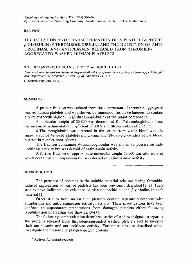
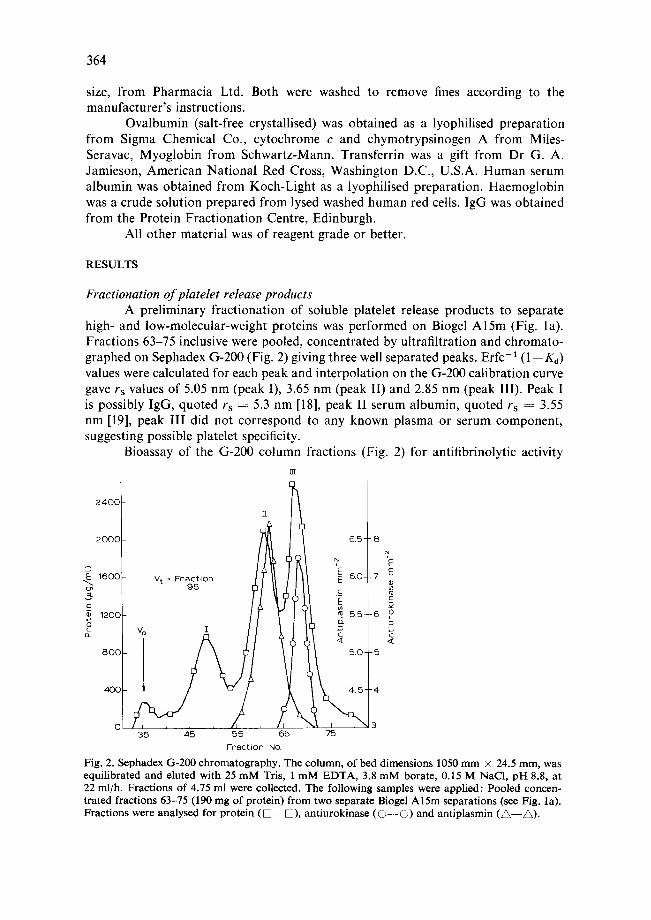
high- and low-molecular-weight proteins was performed on Biogel Al5m (Fig. la). Fractions 63-75 inclusive were pooled, concentrated by ultrafiltration and chromato- graphed on Sephadex G-200 (Fig. 2) giving three well separated peaks. Erfc-1 (1--K d) values were calculated for each peak and interpolation on the G-200 calibration curve gave rs values of 5.05 nm (peak I), 3.65 nm (peak II) and 2.85 nm (peak III). Peak I is possibly IgG, quoted rs = 5.3 nm [18], peak II serum albumin, quoted rs = 3.55 nm [19], peak III did not correspond to any known plasma or serum component, suggesting possible platelet specificity.
Bioassay of the G-200 column fractions (Fig. 2) for antifibrinolytic activity rn
2400 z
2000 / ~ ~ 6.5-- 8
't' .EE 1600 V t = Fraction 6.0--7 E 96 ! c "~ 120C 5.5--6 XO
80C [ / t 5.0- -5
J l \
,',.l i % 3 35 46 55 66 75
Fraction NQ
Fig. 2. Sephadex 6-200 chromatography. The column, of bed dimensions 1050 mm × 24.5 mm, was equilibrated and eluted with 25 mM Tris, 1 mM EDTA, 3.8 mM borate, 0.15 M NaC1, pH 8.8, at 22 ml/h. Fractions of 4.75 ml were collected. The following samples were applied: Pooled concen- trated fractions 63-75 (190 mg of protein) from two separate Biogel A15m separations (see Fig. la). Fractions were analysed for protein ( D - - D ) , antiurokinase ( O - - O ) and antiplasmin ( A - - ~ ) .

365
showed the complete separation of a minor peak of antiplasmin activity (spec. act. = 0.5 C.U./mg protein) in peak II and a large peak of antiurokinase activity (spec. act. = 19.6 Ploug units/mg protein) in peak III.
Peak III fractions 66-74 were pooled and concentrated by ultrafiltration giving a final protein concentration of 8.3 mg/ml, and 7 ml of this material was desalted on Sephadex G-25 and lyophilised.
Thrombin treatment of 48-h-old washed platelets A washed platelet suspension of 48-h-old platelets was treated with thrombin/
CaCI2, the supernatant isolated by centrifugation, concentrated and chromatographed on Biogel A15m exactly as for 24-h-old platelets. The fractions analysed for protein showed only one major protein peak, rs = 3.6 nm, corresponding exactly to peak II albumin (rs = 3.65 nm) found for 24-h-old platelets (Fig. la). There was no visible aggregation of platelets after the addition of thrombin and no peak of ultraviolet- absorbing material (259 nm) corresponding to the presence of nucleotides [1]. A large peak of ultraviolet-absorbing material eluted at Vt on Biogel A 15m when the soluble release products from fresh platelets were chromatographed.
ii~ii~i~iii~ iiiii~ili~ii !̧iiiiiii~iiiii!!iiiii!iii!~ii!i~iiiiiiiiili~!ii!i!iiiiiii~iii~ii!iii~ ~iiiiiiiiiiiii~!i!iiiiii~ii!!Tiiiiiiii<~!~i!ii:iiii~i?i!!ii >~i!Y :
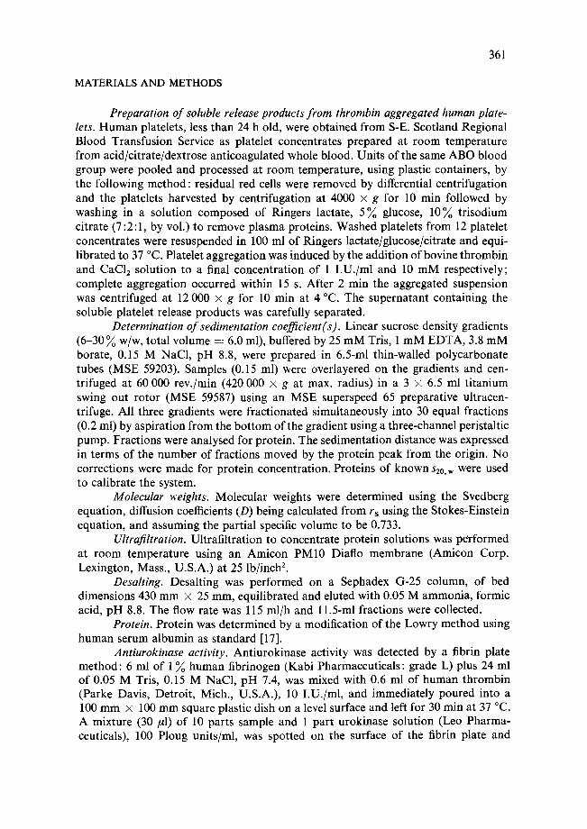
Fig. 3. Cellulose acetate electrophoresis. Sodium barb i tone 0.04 M buffer, p H 9.9, 18 V/cm for 25 min. Samples were applied as follows: (1) Lyophil ised peak I I I (3/~l, 3 mg/ml) (see text). (2) Undi lu ted h u m a n serum (3 yl). The ar row indicates the posi t ion of the a lbumin present in trace amount s in peak III which does not show in the pho tograph .
366
Electrophoresis Cellulose acetate electrophoresis of lyophilised peak III was performed (Fig. 3).
A broad protein band with a mobility between the serum t3- and v-globulins was noted and a trace amount of albumin resulting from contamination from peak II.
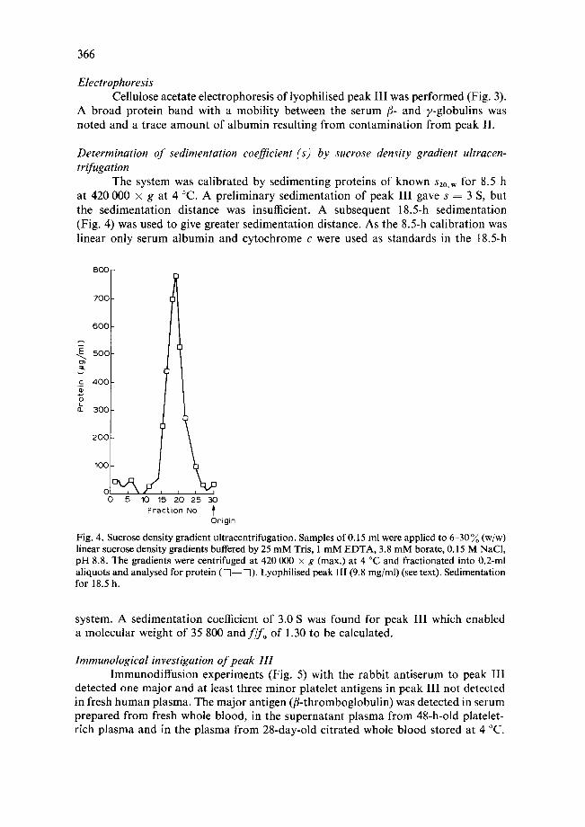
Determination of sedimentation coefficient (s) by sucrose density gradient ultracen- trifugation
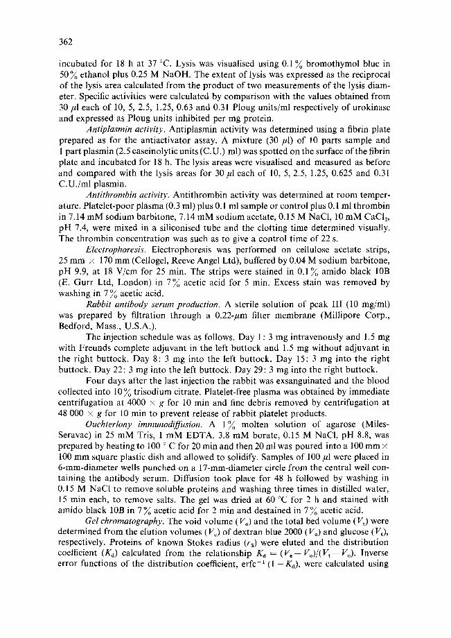
The system was calibrated by sedimenting proteins of known s20.w for 8.5 h at 420 000 × g at 4 °C. A preliminary sedimentation of peak III gave s = 3 S, but the sedimentation distance was insufficient. A subsequent 18.5-h sedimentation (Fig. 4) was used to give greater sedimentation distance. As the 8.5-h calibration was linear only serum albumin and cytochrome e were used as standards in the 18.5-h
8 0 0
7 0 0
6 0 0
5 0 0
c 4 0 0
a. 3 0 0
200 I
, ' d , , 5 10 15 20 25 30
F rac t ion NO. Origin
Fig. 4. Sucrose density gradient ultracentrifugation. Samples of 0.15 ml were applied to 6-30 % (w/w) linear sucrose density gradients buffered by 25 mM Tris, 1 mM EDTA, 3.8 mM borate, 0.15 M NaCI, pH 8.8. The gradients were centrifuged at 420 000 x g (max.) at 4 °C and fractionated into 0.2-ml aliquots and analysed for protein ([~--D). Lyophilised peak II! (9.8 mg/ml) (see text). Sedimentation for 18.5 h.
system. A sedimentation coefficient of 3.0 S was found for peak III which enabled a molecular weight of 35 800 and fifo of 1.30 to be calculated.
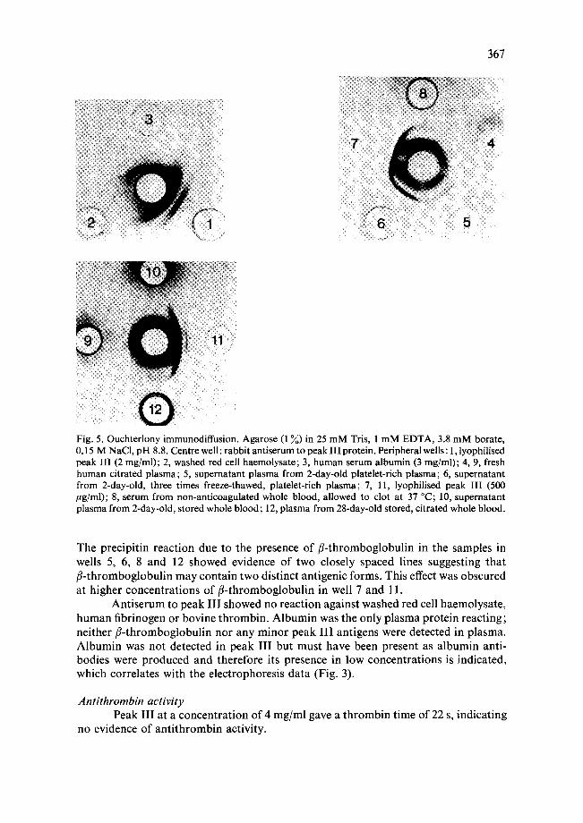
Immunological investigation of peak III Immunodiffusion experiments (Fig. 5) with the rabbit antiserum to peak II1
detected one major and at least three minor platelet antigens in peak III not detected in fresh human plasma. The major antigen (fl-thromboglobulin) was detected in serum prepared from fresh whole blood, in the supernatant plasma from 48-h-old platelet- rich plasma and in the plasma from 28-day-old citrated whole blood stored at 4 °C.
367
Fig. 5. Ouchterlony immunodiffusion. Agarose (1 ~) in 25 mM Tris, 1 mM EDTA, 3.8 mM borate, 0.15 M NaCI, pH 8.8. Centre well: rabbit antiserum to peak III protein. Peripheral wells: 1, lyophilised peak III (2 mg/ml); 2, washed red cell haemolysate; 3, human serum albumin (3 mg/ml); 4, 9, fresh human citrated plasma; 5, supernatant plasma from 2-day-old platelet-rich plasma; 6, supernatant from 2-day-old, three times freeze-thawed, platelet-rich plasma; 7, 11, lyophilised peak III (500 /~g/ml); 8, serum from non-anticoagulated whole blood, allowed to clot at 37 °C; 10, supernatant plasma from 2-day-old, stored whole blood; 12, plasma from 28-day-old stored, citrated whole blood.
The precipitin reaction due to the presence of fl-thromboglobulin in the samples in wells 5, 6, 8 and 12 showed evidence of two closely spaced lines suggesting that fl-thromboglobulin may contain two distinct antigenic forms. This effect was obscured at higher concentrations of fl-thromboglobulin in well 7 and 11.
Antiserum to peak I | I showed no reaction against washed red cell haemolysate, human fibrinogen or bovine thrombin. Albumin was the only plasma protein reacting; neither fl-thromboglobulin nor any minor peak I I I antigens were detected in plasma. Albumin was not detected in peak I I I but must have been present as albumin anti- bodies were produced and therefore its presence in low concentrations is indicated, which correlates with the electrophoresis data (Fig. 3).
Antithrombin activity Peak I I I at a concentration of 4 mg/ml gave a thrombin time of 22 s, indicating
no evidence of anti thrombin activity.

368
DISCUSSION
There are two previous reports of platelet-specific fl-globulins. A thrombin- sensitive protein with coagulation factor XIII activity (fibrin-stabilising activity) and with a molecular weight of l l0 000-150 000 has been isolated from homogenised whole platelets [20]. A platelet-specific fl-globulin has also been described in the release products of thrombin-aggregated washed platelets [2].
In the studies reported here a protein fraction (peak III) was isolated by Sephadex G-200 chromatography. The major platelet specific component migrated as a fl-globulin and had a Stokes radius of 2.85 nm and sedimentation coefficient of 3.0 S from which a molecular weight of 35 800 and frictional ratio of 1.30 were calculated. This suggested a compact shape for the molecule. This major component was named fl-thromboglobulin. Antiurokinase activity with a specific activity of 19.6 Ploug units/mg protein was detected in this fraction. It was not known whether this activity was a property of fl-thromboglobulin or of one of the minor antigen components of peak III.
We were unable to detect thrombin-induced aggregation, release of nucleotides or release of fl-thromboglobulin in a preparation of 48-h-old washed platelets. fl-Thromboglobulin was however detected in the supernatant plasma of 48-h-old platelet concentrates which suggested that in vitro ageing accompanied by leakage of platelet constituents had occurred. It was concluded that fl-thromboglobulin was specifically released during thrombin-induced platelet aggregation as aggregation and the release of nucleotides and fl-thromboglobulin was detected in preparations of fresh washed platelets, fl-Thromboglobulin was also detected in serum prepared from clotted whole blood, the immunodiffusion data suggesting that it may be com- posed of two similar components (Fig. 5).
The antiplasmin activity present in minor quantities in the Sephadex G-200 fractions had a low specific activity and an approximate molecular weight of 70 000. This latter observation does not correspond to previously reported values of 120 000 [10], 40 000 [ll], 35 000 [12] and 20 000 [14], suggesting the existence of multiple forms of antiplasmin activity or degradation during isolation procedures. The low specific activity, however, is in agreement with other work [21]. The anti- plasmin was completely separated from antiurokinase activity.
The relevance of the discovery of antiurokinase activity in platelet release products to the in vivo balance of the fibrinolytic system is uncertain. Urokinase is not the naturally occurring activator in plasma. It has been reported that platelet antiactivator has the highest activity against urokinase but less against heart tissue and plasma activators. A molecular weight of approx. 45 000 has been reported for an antiplasminogen activator isolated from lyophilised platelets [23] and, therefore it is possible that our activity is the same molecule. The serum plasminogen anti- activator investigated by Aoki and von Kaulla [24] has a molecular weight of 80 000 and is clearly distinct from platelet antiactivator described here. fl-Thromboglobulin, the major platelet specific component, of peak III, may be a constituent of the fraction containing platelet-specific fl-globutins isolated by Davey [2].
The isolation of a platelet-specific protein of high purity from the soluble material released during platelet aggregation has potential application in the study of platelet function and in the development of an immunological assay for in vivo

369
p la te le t dest ruct ion, f l -Thromboglobul in , if p repa red in a high pur i ty form, would be sui table for the deve lopment of such an assay. Wi th the possible except ion o f a lbumin, f l - th romboglobu l in is the mos t a b u n d a n t p ro te in present in t h r o m b i n release p ro- ducts, a fac tor o f impor t ance , since the c i rcula t ing levels of soluble pla te le t mater ia l resul t ing f rom in vivo pla te le t des t ruct ion are l ikely to be low. Fu r the r studies on the r emova l o f the con taminan t s present in the p r e p a r a t i o n are requi red to isolate fl- t h r o m b o g l o b u l i n in a high pur i ty fo rm and to de te rmine whether i t is composed of two s imilar molecu la r species present in s imilar concentra t ions . Such studies would also a id the ident i f icat ion o f the ant igen posessing an t iu rok inase activity.
ACKNOWLEDGEMENTS
Our thanks to D r R. A. C u m m i n g for p rov id ing the facilities for this work, Dr G. McVie for an t i se rum produc t ion , D r P. C, Das for helpful advice, Mrs R. J. M c L e a n for pe r fo rming the f ibr inolyt ic assays and Mrs A. Ma lone for technical assistance.
REFERENCES
1 Grette, K. (1962) Acta Physiol. Scand. 56, Suppl. 195 2 Davey, M. G. and Luscher, E. F. (1968) Biochim. Biophys. Acta 165, 490-506 3 Johnson, S. A. and Schneider, C. L. (1953) Science 117, 229-230 4 Stefanini, R. and Murphy, 1. S. (1956) J. Clin. Invest. 35, 355-361 5 Alkjaersig, N. (1961) Blood Platelet (Johnson, S. A., Monto, R. W., Rebuck, J. W. and Horn, Jr,
R. C., eds), pp. 329-336, Little Brown, Boston 6 Kwaan, H. C. and Suwanwela, N. (1971) Br. J. Haematol. 21, 313-322 7 Den Ottolander, G. J. H., Leijnse, B. and Cremer-Elfrink, H. M. J. (1967) Thromb. Diath.
Haemorrh. 18, 404-415 8 Den Ottolander, G. J. H., Leijnse, B. and Cremer-Elfrink, H. M. J. (1969) Thromb. Diath.
Haemorrh. 21, 26-34 9 Den Ottolander, G. J. H., Leijnse, B. and Cremer-Elfrink, H. M. J. (1969) Thromb. Diath.
Haemorrh. 21, 35-41 10 McDonagh, J., Kiesselbach, T. H. and Wagner, R. H. (1969) Am. J. Physiol. 216, 508-513 11 Wakabayashi, K., Fujikawa, K. and Abe, T. (1970) Thromb. Diath. Haemorrh. 24, 76-84 12 Siegel-Ralston, A. and Astrup, T. (1971) Nat. New Biol. 234, 180-181 13 Ganguly, P. (1972) Clin. Chim. Acta 39, 466-468 14 Hume, R. (1958) Scott. Med. J. 3, 479 15 Tables of the error function and its derivatives (1954) N.B.S. Applied Mathematics Series 41,
United States Government Printing Office, Washington D.C. 16 Ackers, G. K. (1967) J. Biol. Chem. 242, 3237-3238 17 Eggstein, M. and Kreutz, F. H. (1955) Klin. Wochenschr. 33, 879-884 18 Smith, M. H. (1968) Handbook of Biochemistry, Selected Data for Molecular Biology (Sober,
H. A., ed.), The Chemical Rubber Co., Cleveland 19 Andrews, P. (1970) Methods Biochem. Anal. 18, 1-53 20 Ganguly, P. (1971) J. Biol. Chem. 246, 4286-4290 21 Joist, J. H., Niewiarowski, S. and Mustard, J. F. (1971) Thrombolytic Therapy (Mammen, E. F.,
Anderson, G. F. and Barnhart, M. I., eds), Thromb. Diath. Haemorrh. Suppl. 47, 113-119 22 Brackman, P., Snyder, J., Henderson, E. S. and Astrup, T. (1970) Br. J. Haematol. 18, 135-145 23 Ogston, D., Murray, J. and Douglas, A. S. (1973) Br. J. Haematol. 24, 665-666 24 Aoki, N. and von Kaulla, K. N. (1971) Am. J. Physiol. 220, 1137-1145