Embed Size (px)
Citation preview

The management and complications of head and
neck malignancy
Waseem Jerjes
The nature of cancer
• H&N Cancer (6th)• Incidence in developing countries• Variety of histological types (SCC)• Mouth, larynx and hypopharynx• Male:female

• 500 000 cases worldwide
• 270 000 deaths per annum
• 6% incidence of all cancers (excl. skin)
• 5% mortality of all cancers
• 75% oral cavity/pharynx
• 25% larynx
Head and Neck Cancer: Epidemiology
• Geographical distribution - wide variations
– France (supraglottic, oral cancers)
– Hong Kong (nasopharyngeal cancers)
– India (oral cancers)
• Race - African Americans vs Caucasians
• Gender - Men > Women
– trend towards increased incidence in women?
Head and Neck Cancer: Epidemiology
The nature of cancer
• Carcinogens (Smoking-p53 mutations and IL 4 receptor)
• RR of <7cig/d = 2.4• RR of >25 cig/day = 16.4• Cessation reduction 70%

The nature of cancer
• Carcinogens (Alcohol)• Resveratrol (Chemopreventive)• RR of 7-21u/w (no wine) = 3• RR of 7-21u/w (30% wine) = 0.5• RR of >21u/w (no wine) = 5.2• RR of >21u/w (30% wine) = 1.7
The nature of cancer
• Site specific carcinogens
• Nickel and chromate dust• Hardwood dust• Nitrosamines in a salted fish diet <10y age

The nature of cancer
• Iron deficiency (Paterson-Kelly)
• Postcricoid carcinoma • Young age and common in women• Mild symptoms and slow progression• Iron + Vit B

The nature of cancer
• 2 Viruses (HPV and EBV)
• HPV types (6, 18 and 33) – p53 and pRb• EBV – NPC• Serum anti-EBV IgA = endoscopic
examination + blind biopsies
• HIV
The nature of cancer
• Family History
• RR of 3.5-3.79 HNSCC family history• RR of 7.89 1st degree relative of patients
with multiple HNSCC

keratosis dysplasia carcinoma
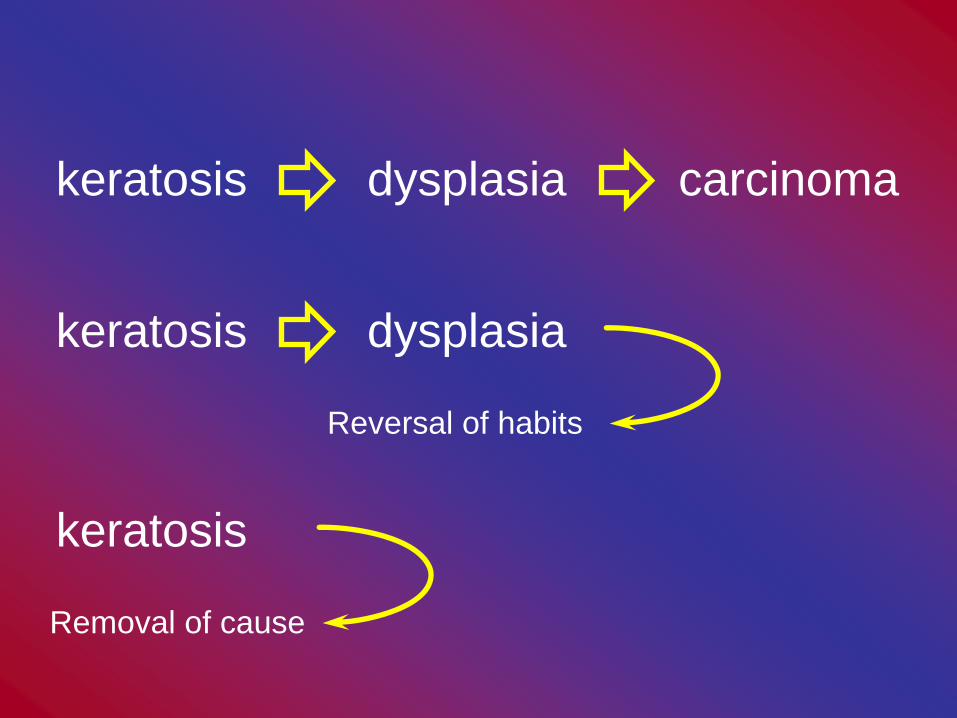
keratosis dysplasia carcinoma
keratosis dysplasia
Reversal of habits
keratosis
Removal of cause
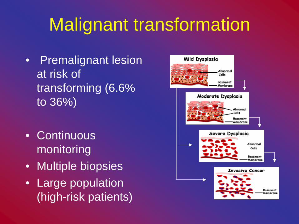
Malignant transformation
• Premalignant lesion at risk of transforming (6.6% to 36%)
• Continuous monitoring
• Multiple biopsies• Large population
(high-risk patients)

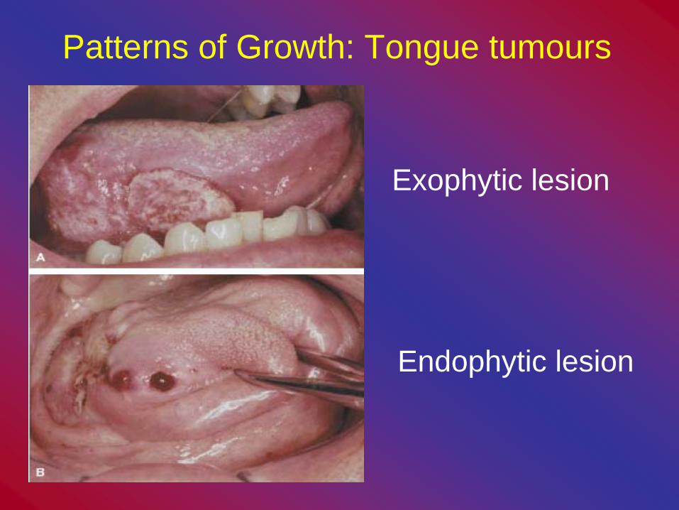
Patterns of Growth: Tongue tumours
Exophytic lesion
Endophytic lesion

PatternPattern 22 Pattern 3Pattern 3 Pattern 4Pattern 4Pattern 1Pattern 1
GOODGOOD
CohesiveCohesive
BADBAD
NonNon--cohesivecohesiveInvasion
The team
• Surgeons• Oncologists• General practitioners
(Medical and Dental)• Nurses • SALT• Dieticians
• Occupational health team
• McMillan nurses• Oral Hygienists • Physiotherapists • Others

Assessment of cancer patients
• Potentially treatable and not all curable
• Decision (fit for surgery?)• Palliative (Chemoradio?)
• History (no difference)• Age + social circumstance + tumour
biology

Assessment of cancer patients
• Examination• Primary site (think T staging) by inspection
and palpation
• Neck (triangles)• Tips: tension off SCM, JD-LN, JO-LN,
space of Burns• Normal structures: C1, C2, thyroid, carotid
bulb
Assessment of cancer patients
• Triangles of the neck
• Anterior (submental, submandibular, carotid, muscular)
• Posterior (occipital, subclavian)

Assessment of cancer patients
• TNM• T classification differs between tumours
• NM classification are universal for most head and neck tumours
• cTNM vs. pTNM
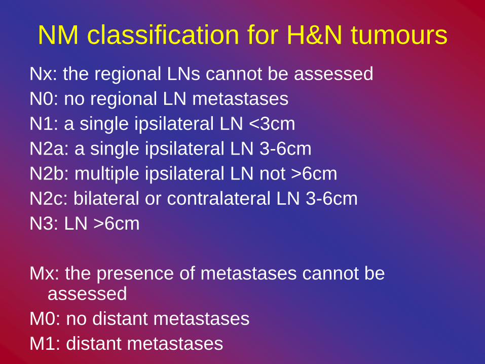
NM classification for H&N tumoursNx: the regional LNs cannot be assessedN0: no regional LN metastasesN1: a single ipsilateral LN <3cmN2a: a single ipsilateral LN 3-6cmN2b: multiple ipsilateral LN not >6cmN2c: bilateral or contralateral LN 3-6cmN3: LN >6cm
Mx: the presence of metastases cannot be assessed
M0: no distant metastasesM1: distant metastases
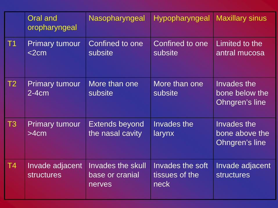
Oral and oropharyngeal
Nasopharyngeal Hypopharyngeal Maxillary sinus
T1 Primary tumour <2cm
Confined to one subsite
Confined to one subsite
Limited to the antral mucosa
T2 Primary tumour 2-4cm
More than one subsite
More than one subsite
Invades the bone below the Ohngren’s line
T3 Primary tumour >4cm
Extends beyond the nasal cavity
Invades the larynx
Invades the bone above the Ohngren’s line
T4 Invade adjacent structures
Invades the skull base or cranial nerves
Invades the soft tissues of the neck
Invade adjacent structures
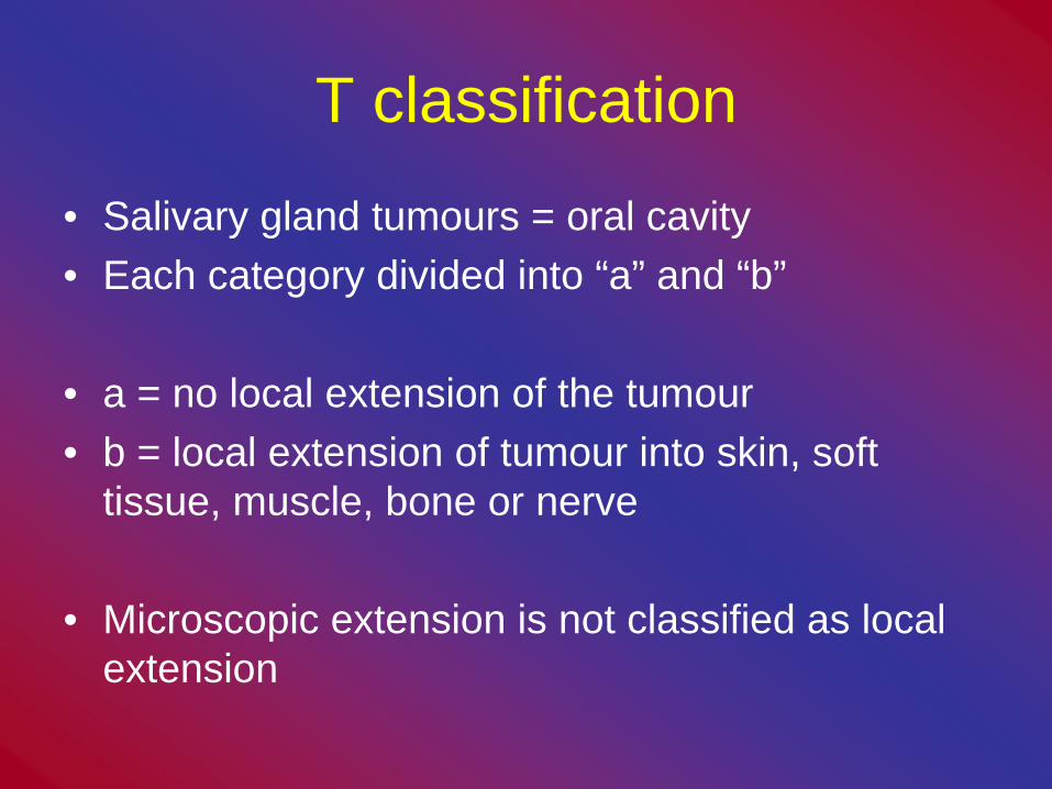
T classification
• Salivary gland tumours = oral cavity• Each category divided into “a” and “b”
• a = no local extension of the tumour• b = local extension of tumour into skin, soft
tissue, muscle, bone or nerve
• Microscopic extension is not classified as local extension
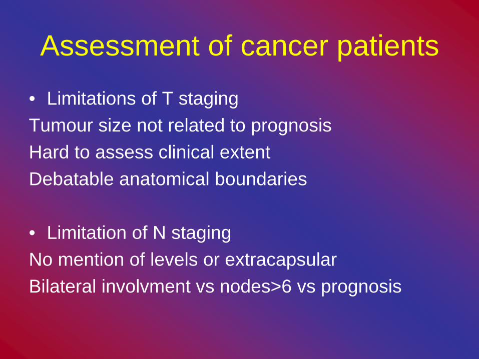
Assessment of cancer patients
• Limitations of T stagingTumour size not related to prognosisHard to assess clinical extentDebatable anatomical boundaries
• Limitation of N stagingNo mention of levels or extracapsularBilateral involvment vs nodes>6 vs prognosis
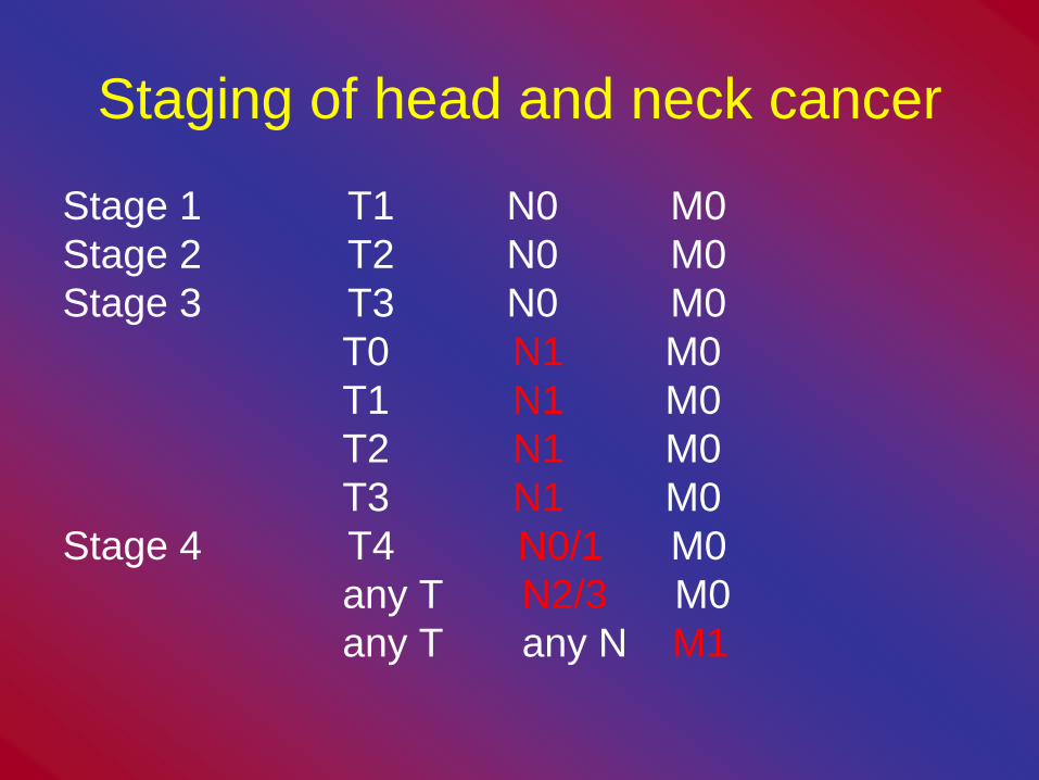
Staging of head and neck cancer
Stage 1 T1 N0 M0Stage 2 T2 N0 M0Stage 3 T3 N0 M0
T0 N1 M0T1 N1 M0T2 N1 M0T3 N1 M0
Stage 4 T4 N0/1 M0any T N2/3 M0any T any N M1
Assessment of cancer patients
• Staging cancer• Planning therapy, aid to prognosis,
comparison of results and epidemiology
• I, II (low risk)• III, IV (high risk)

Therapeutic options
• Early stage disease (Stage I and II)– Surgery vs radical radiotherapy
• Late stage disease (Stage III and IV)– Surgery + PORT– Radical RT– Radical chemo-RT
+/- Surgical salvage

Assessment of cancer patients
• General examination• Anaesthetic opinion
• Bloods, CXR, ECG…DVT• Special imaging
• Nutritional status

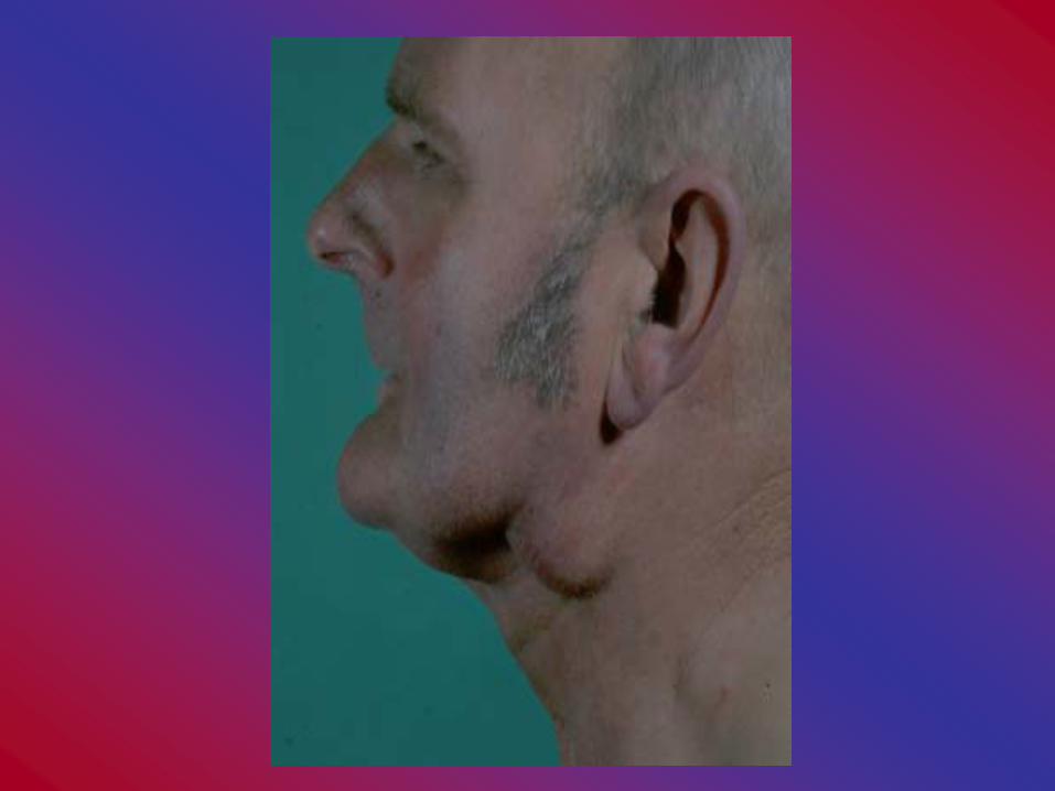
• LocalDepends on subsiteOral cavity – ulcer, mass, bleeding, pain, dysarthria,
loose denturesOropharynx – pain, dysphagia, odynophagia, otalgiaLarynx – dysphonia, pain, dysphagia, odynophagiaHypopharynx - pain, dysphagia, odynophagia, otalgia
• RegionalNodal metastases
• Systemic (<10%): Lung, Liver, Bone
Head and Neck Cancer: symptoms
Assessment of cancer patients
• Radiology• Plain films-CXR• Contrast studies-barium swallow• What do you assess?• Tongue movement• Soft palate elevation• Epiglottic tilt• Laryngeal closure• Pharyngo-oesophageal segment and pharyngeal
perstalsis
Assessment of cancer patients
• Biopsy of the nodeFNAC, if –ve, consider open biopsy• Panendoscopy: synchronous tumoursNasopharynx, oropharynx, hypopharynx,
oesophagus, stomach, larynx, trachea and upper bronchi
Biopsies from pyriform fossa, nasopharynx, tonsillar fossa and base of tongue can be acquired

Assessment of cancer patients
• Ultrasound• Not use ionizing radiation• Solid vs cystic mass
• Bone, cartilage and gas• Deep structures
• Doppler US
Assessment of cancer patients
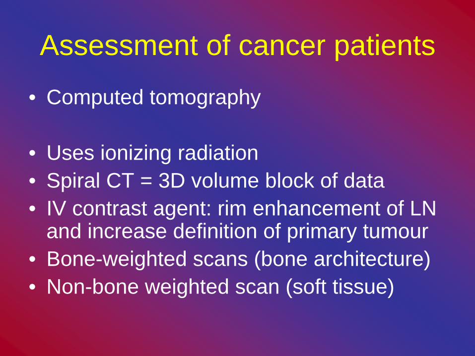
• Computed tomography
• Uses ionizing radiation• Spiral CT = 3D volume block of data• IV contrast agent: rim enhancement of LN
and increase definition of primary tumour • Bone-weighted scans (bone architecture)• Non-bone weighted scan (soft tissue)

Assessment of cancer patients• Magnetic resonance imaging
• Not use ionizing radiation• Claustrophobic patients, ferromagnatic surgical
clips, embolization coils, pace makers• Superior soft tissue contrast• T1-weighted images (fat as white)• T2-weighted (water as white)• STIR sequence (suppression of fat signal)• Inflammation (high water content)
Assessment of cancer patients
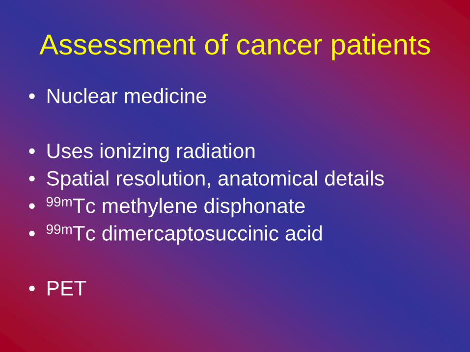
• Nuclear medicine
• Uses ionizing radiation• Spatial resolution, anatomical details• 99mTc methylene disphonate• 99mTc dimercaptosuccinic acid
• PET
Treatment options
• Surgery, Radiotherapy, Chemotherapy• Removal of primary tumour ± neck
dissection ± reconstruction• SCC (locoregional disease)• Uncontrolled disease primary site


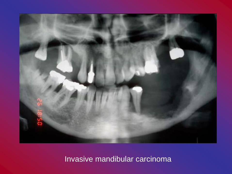
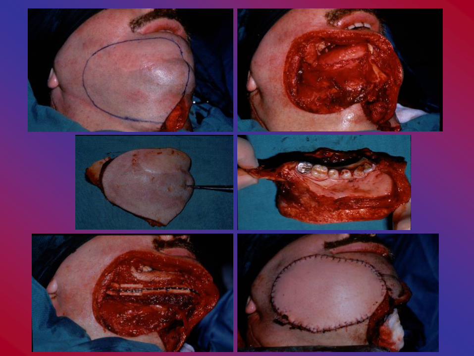
Invasive mandibular carcinoma
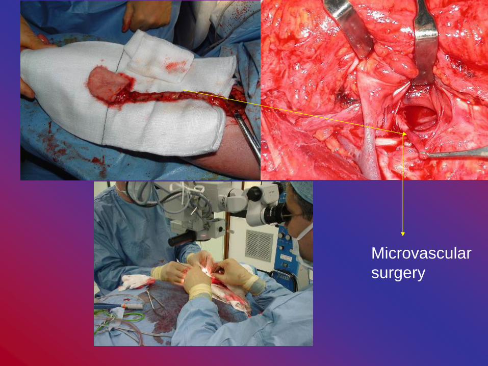
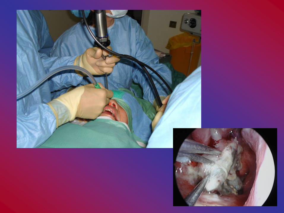
Microvascular surgery

Treatment options
• Levels (regions) within the neck which contains group of lymphnodes
• Level I: submental + submandibular• Level II: upper jugular• Level III: middle jugular• Level IV: lower jugular • Level V: posterior triangle • Level VI: anterior compartment• Level VII: upper anterior mediastinum
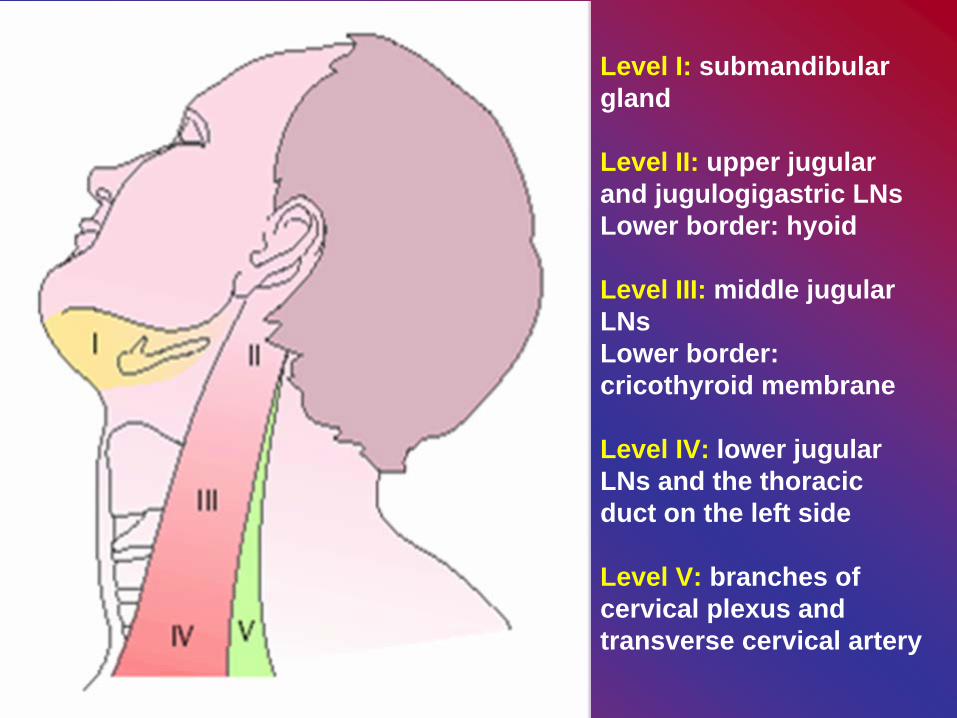
Level I: submandibular gland
Level II: upper jugular and jugulogigastric LNsLower border: hyoid
Level III: middle jugular LNsLower border: cricothyroid membrane
Level IV: lower jugular LNs and the thoracic duct on the left side
Level V: branches of cervical plexus and transverse cervical artery
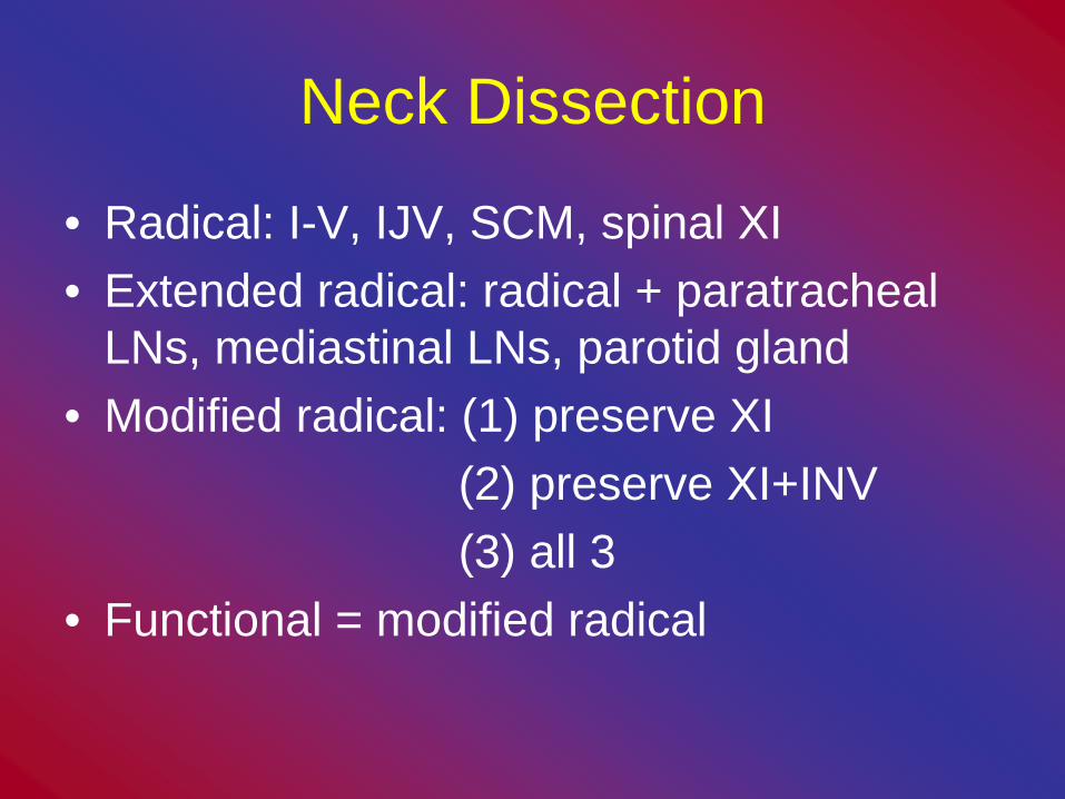
Neck Dissection
• Radical: I-V, IJV, SCM, spinal XI• Extended radical: radical + paratracheal
LNs, mediastinal LNs, parotid gland• Modified radical: (1) preserve XI
(2) preserve XI+INV(3) all 3
• Functional = modified radical
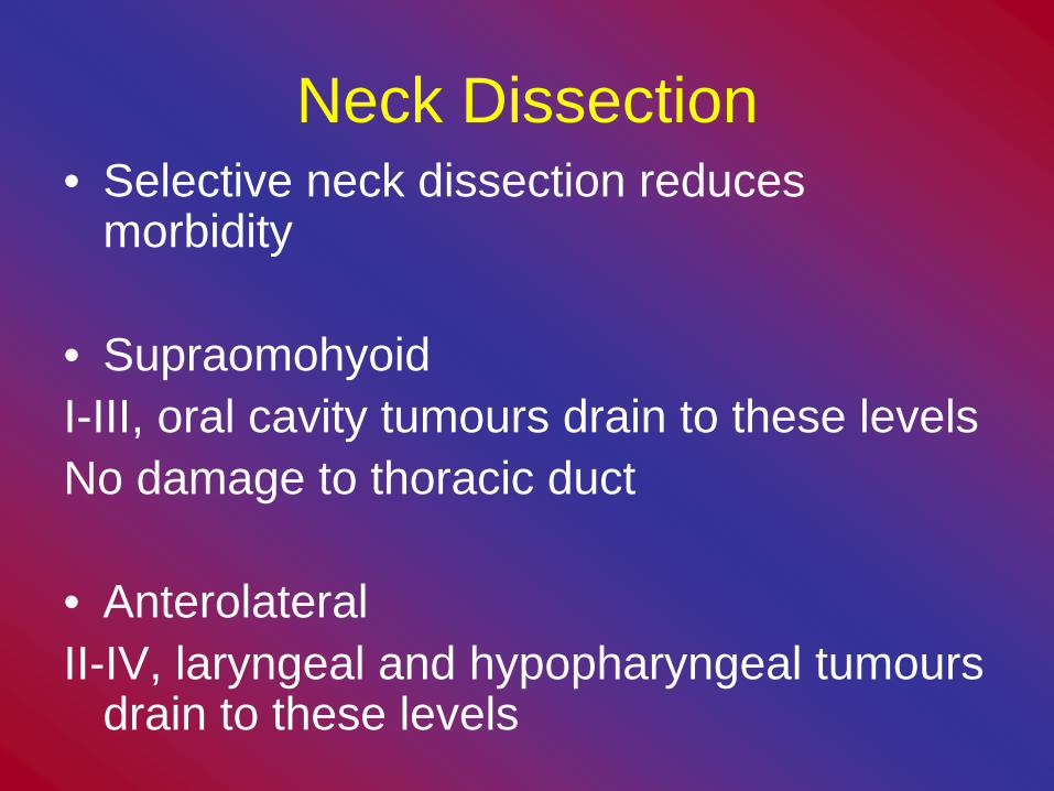
Neck Dissection• Selective neck dissection reduces
morbidity
• SupraomohyoidI-III, oral cavity tumours drain to these levelsNo damage to thoracic duct
• AnterolateralII-IV, laryngeal and hypopharyngeal tumours
drain to these levels
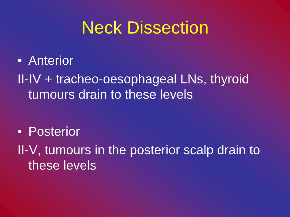
Neck Dissection
• AnteriorII-IV + tracheo-oesophageal LNs, thyroid
tumours drain to these levels
• PosteriorII-V, tumours in the posterior scalp drain to
these levels

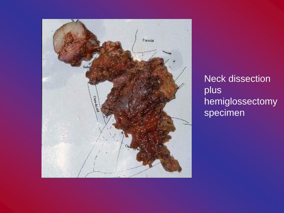
Neck dissection plus hemiglossectomy specimen
When do we dissect?
• Radical ND (tumours with N2 neck, recurrent disease, invasive nodal disease)
• Modified radical/selective ND (T3, T4, N1 tumours)
• T1, T2…lots of debate
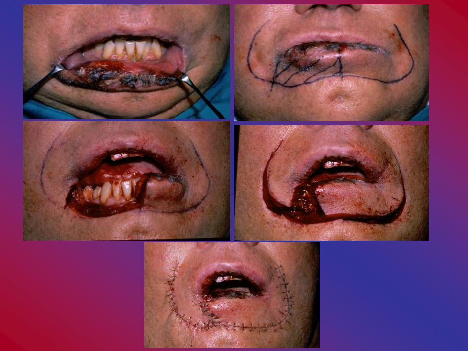
Treatment options• Reconstruction
• Free grafts • ST skin graft• FT skin graft• Composite FT skin and cartilage grafts• Pinch grafts (free skin grafts)• Dermal and fat grafts• Fascial grafts• Chondromucosal grafts


Treatment options
• Flaps• Local flaps (random, axial pattern)• Distant axial flaps• Myocutaneous flaps • Free flaps
• Radial forearm, fibula, latissimus dorsi, DCIA, scapula, jejunum, rectus abdominus

Postoperative care
• Tracheostomy nursing care
• Removal of secretions• Humidification• Changing of tracheostomy tube• Care of inflatable cuff• Breathing exercises• Dressings
Postoperative care
• Leaking drains• Type of drainage• Removal of drains• Feeding• DVT prophylaxis

Postoperative care• Flap monitoring
Clinical parameter
Normal circulation
Venous occlusion
Arterial Occlusion
Capillary refill time
Pink Blue/purple Pale, mottled blue
Capillary refill time
1-2secs <1sec >2secs
Temperature Warm Warm-cold Cooler
Tissue turgor Full Distended Hollow
Dermal bleeding Bright red blood Dark red to pink. Brisky.
Minimal to absence (serum)
Postoperative care
• Medications • Dressing and sutures• Postoperative examination
• Discharge and follow up
Complications
• Specific early intraoperativeBleedingAir embolusPneumothoraxCarotid artery injuryNerve injury (phrenic, vagus, brachial
plexus, lingual, hypoglossal, glossopharyngeal)
Complications• Specific intermediateSkin flap necrosisCarotid blowout (radiotherapy)Chyle leak (thoracic duct)• Specific lateScar contractureNeuroma formation (cervical plexus)Shoulder pain syndrome (accessory nerve)Cellulitis and facial oedema
Recurrent disease• Chance of salvage depends on
– TNM stage of recurrence
– Prior therapy
– Performance status
– Co-morbidities
• Frequent episodes of recurrence BEFORE systemic spread
• Large volume recurrence (primary or nodal) carries a poor prognosis
Palliative therapy
• Pain control• Shrink tumour (airway)• Control bleeding
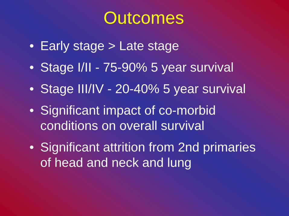
Outcomes• Early stage > Late stage
• Stage I/II - 75-90% 5 year survival
• Stage III/IV - 20-40% 5 year survival
• Significant impact of co-morbid conditions on overall survival
• Significant attrition from 2nd primaries of head and neck and lung
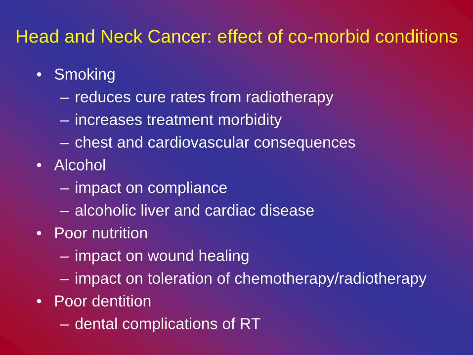
• Smoking– reduces cure rates from radiotherapy– increases treatment morbidity– chest and cardiovascular consequences
• Alcohol– impact on compliance– alcoholic liver and cardiac disease
• Poor nutrition– impact on wound healing– impact on toleration of chemotherapy/radiotherapy
• Poor dentition– dental complications of RT
Head and Neck Cancer: effect of co-morbid conditions
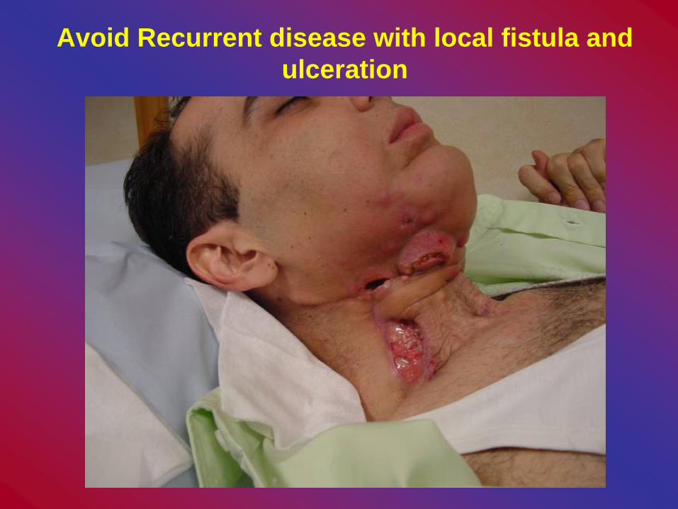
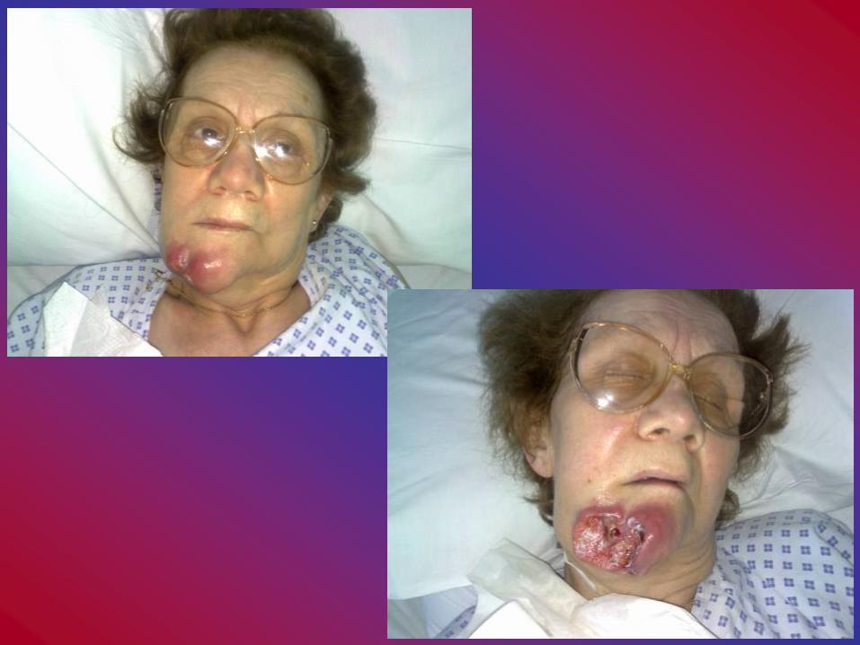
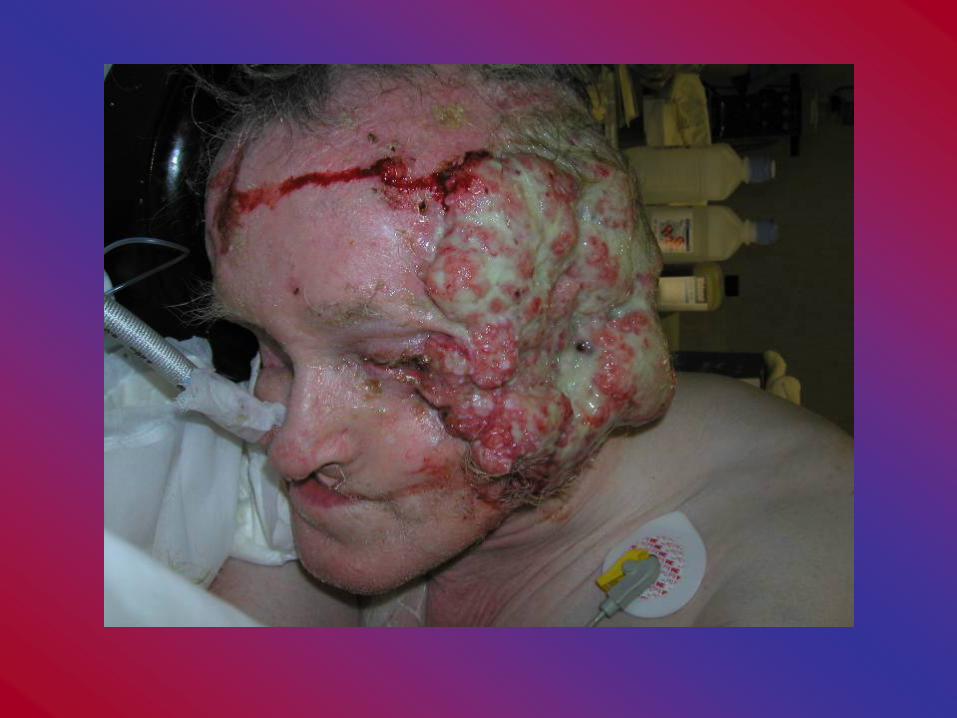
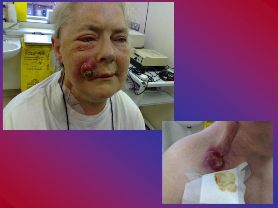
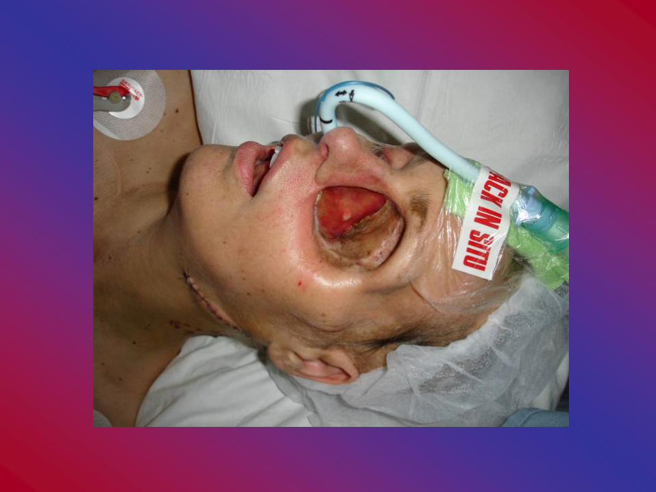
Avoid Recurrent disease with local fistula and ulceration

Acknowledgment Mr Colin Hopper
Mr Laurence Newman
Thank you


![Maternal characteristics and obstetrical complications ... · complications during birth and could be prevented [4, 5]. The vast majority of perinatal deaths occur in develop-ing](https://img.pdfslide.tips/doc/110x75/5f8916eb226ec04e4f13d2fa/maternal-characteristics-and-obstetrical-complications-complications-during.jpg)