Embed Size (px)
Citation preview
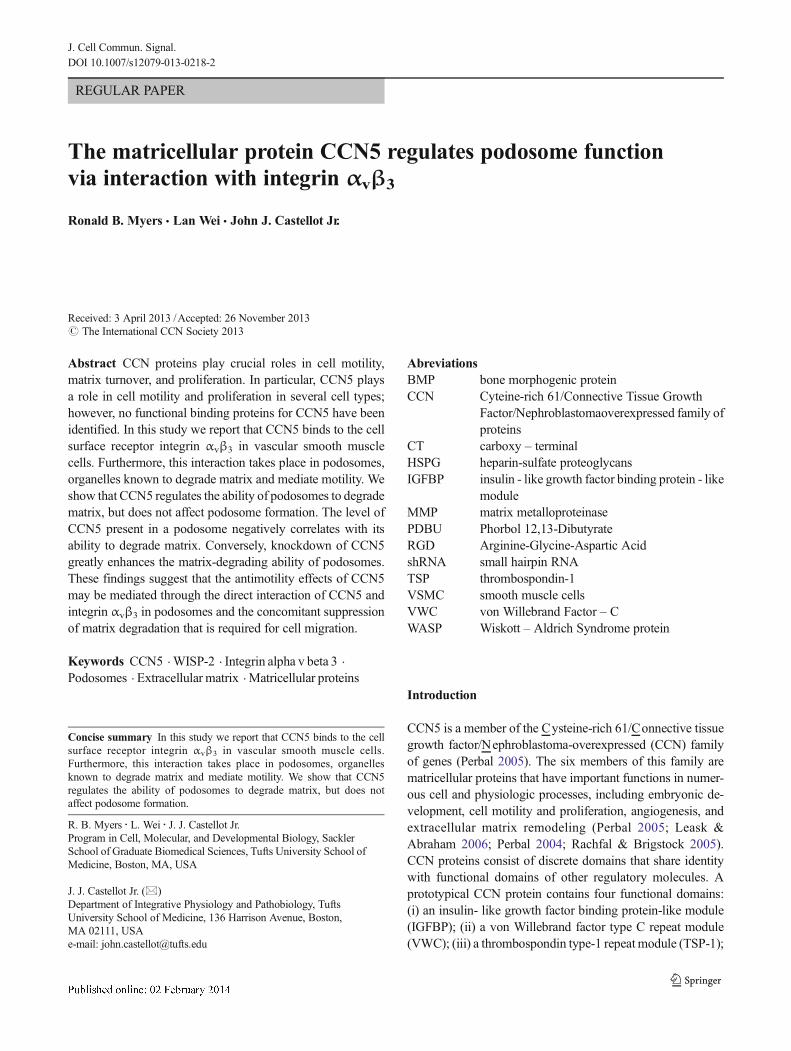
REGULAR PAPER
The matricellular protein CCN5 regulates podosome functionvia interaction with integrin αvβ3
Ronald B. Myers & Lan Wei & John J. Castellot Jr.
Received: 3 April 2013 /Accepted: 26 November 2013# The International CCN Society 2013
Abstract CCN proteins play crucial roles in cell motility,matrix turnover, and proliferation. In particular, CCN5 playsa role in cell motility and proliferation in several cell types;however, no functional binding proteins for CCN5 have beenidentified. In this study we report that CCN5 binds to the cellsurface receptor integrin αvβ3 in vascular smooth musclecells. Furthermore, this interaction takes place in podosomes,organelles known to degrade matrix and mediate motility. Weshow that CCN5 regulates the ability of podosomes to degradematrix, but does not affect podosome formation. The level ofCCN5 present in a podosome negatively correlates with itsability to degrade matrix. Conversely, knockdown of CCN5greatly enhances the matrix-degrading ability of podosomes.These findings suggest that the antimotility effects of CCN5may be mediated through the direct interaction of CCN5 andintegrin αvβ3 in podosomes and the concomitant suppressionof matrix degradation that is required for cell migration.
Keywords CCN5 .WISP-2 . Integrin alpha v beta 3 .
Podosomes . Extracellular matrix . Matricellular proteins
AbreviationsBMP bone morphogenic proteinCCN Cyteine-rich 61/Connective Tissue Growth
Factor/Nephroblastomaoverexpressed family ofproteins
CT carboxy – terminalHSPG heparin-sulfate proteoglycansIGFBP insulin - like growth factor binding protein - like
moduleMMP matrix metalloproteinasePDBU Phorbol 12,13-DibutyrateRGD Arginine-Glycine-Aspartic AcidshRNA small hairpin RNATSP thrombospondin-1VSMC smooth muscle cellsVWC von Willebrand Factor – CWASP Wiskott – Aldrich Syndrome protein
Introduction
CCN5 is a member of the Cysteine-rich 61/Connective tissuegrowth factor/Nephroblastoma-overexpressed (CCN) familyof genes (Perbal 2005). The six members of this family arematricellular proteins that have important functions in numer-ous cell and physiologic processes, including embryonic de-velopment, cell motility and proliferation, angiogenesis, andextracellular matrix remodeling (Perbal 2005; Leask &Abraham 2006; Perbal 2004; Rachfal & Brigstock 2005).CCN proteins consist of discrete domains that share identitywith functional domains of other regulatory molecules. Aprototypical CCN protein contains four functional domains:(i) an insulin- like growth factor binding protein-like module(IGFBP); (ii) a von Willebrand factor type C repeat module(VWC); (iii) a thrombospondin type-1 repeat module (TSP-1);
Concise summary In this study we report that CCN5 binds to the cellsurface receptor integrin αvβ3 in vascular smooth muscle cells.Furthermore, this interaction takes place in podosomes, organellesknown to degrade matrix and mediate motility. We show that CCN5regulates the ability of podosomes to degrade matrix, but does notaffect podosome formation.
R. B. Myers : L. Wei : J. J. Castellot Jr.Program in Cell, Molecular, and Developmental Biology, SacklerSchool of Graduate Biomedical Sciences, Tufts University School ofMedicine, Boston, MA, USA
J. J. Castellot Jr. (*)Department of Integrative Physiology and Pathobiology, TuftsUniversity School of Medicine, 136 Harrison Avenue, Boston,MA 02111, USAe-mail: [email protected]
J. Cell Commun. Signal.DOI 10.1007/s12079-013-0218-2

and (iv) a carboxy terminal domain (CT). CCN5, also knownas WISP-2 (Pennica et al. 1998), rCop-1(Zhang et al. 1998),COP-1(Delmolino et al. 2001), and CTGF-L (Kumar et al.1999), is highly conserved among vertebrates and is the onlyCCN protein lacking the C-terminal domain (GrayMR 2005).
Originally discovered as a heparin-induced gene in vascu-lar smooth muscle cells (VSMC), CCN5 behaves as a growth-arrest-specific gene in this cell type (Delmolino et al. 2001;Lake et al. 2003). CCN5 expression decreases in uterinefibroids and in the arterial wall after balloon angioplasty, andwhen over-expressed in in vivo models of fibroids and vascu-lar injury, it significantly reduced SMC proliferation(Delmolino et al. 2001; Lake et al. 2003; Mason et al.2004a). Cell culture studies demonstrate that CCN5 over-expression inhibits SMC proliferation and motility in vitro ,whereas CCN5 knock-down via siRNA inhibits proliferationand increases basal locomotion (Lake et al. 2003; Mason et al.2004a; Lake & Castellot 2003).
CCN proteins have been shown to mediate many of theiractivities through interactions with the integrin class of celladhesion receptors (Lau & Lam 2005). The first documentedinteraction of integrins with CCN proteins is the direct bindingof CCN1 to integrinαvβ3 to mediate endothelial cell adhesion(Kireeva et al. 1998). Since then, many studies have demon-strated the potential interaction of at least seven other integrinsas signaling receptors for a diverse array of CCN-mediatedfunctions including adhesion, migration, cell survival, andDNA synthesis (Chen & Lau 2009). Functional binding pro-teins and receptors for CCN5, however, have remained elu-sive. CCN5 has been shown to compete with fibrinogen forbinding to integrin αvβ3 (Kumar et al. 1999), but no directevidence of binding to integrin αvβ3 has been demonstrated.Like all CCN proteins, CCN5 lacks the prototypical integrinrecognition sequence Arg-Gly-Asp (RGD), but it does containa stretch of amino acids that is very similar to the integrinαvβ3 binding site in CCN1 (Chen et al. 2004) (Fig. 1).
Based on these considerations, we hypothesized that CCN5interacts with integrinαvβ3, and that this interaction mediates,
at least in part, the ability of CCN5 to inhibit cell proliferationand migration. In the present study, we show that CCN5 andintegrin αvβ3 co-immunoprecipitate and co-localize inpodosomes, cellular structures that degrade matrix and regu-late cell motility and adhesion. Furthermore, we show thatCCN5 strongly down-regulates the matrix-degrading activityof podosomes.
Materials and methods
Cell culture Vascular smooth muscle cells (VSMC) wereobtained from aortae of Sprague–Dawley rats (CharlesRiver Breeding Laboratories, Inc. Wilmington MA). Theywere isolated, cultured, and characterized as previouslydescribed (Castellot et al. 1982). Briefly, the abdominalsegment of the aorta was removed and the fascia cleanedaway under a dissecting microscope. The aorta was cutlongitudinally, and small pieces of the media were careful-ly stripped from the vessel wall. Two or three such stripswere placed in 60 mm dishes. Within 1–2 weeks, VSMCmigrated from the explants; they were capable of beingpassaged approximately 1 week after the first appearanceof cells. They were identified as VSMC by their character-istic hill-and-valley growth pattern and indirect immuno-fluorescence staining for VSMC specific α-actin. All cul-tures were maintained in RPMI-1640 medium containing10 % bovine growth serum (BGS) at 37 °C in a humidified,5 % CO2/95 % air atmosphere.
Adenovirus production and infection An adenovirus express-ing both Green Fluorescent Protein (GFP) or CCN5, eachtagged by a nine amino acid HA epitope on the C-terminus,was produced using the Ad-Easy system (He et al. 1998), aspreviously described (Lake et al. 2003). Briefly, cells werewashed and exposed to various amounts of virus for 2 h inserum free RPMI-1640 with occasional gentle agitation.RPMI-1640 medium containing 10 % BGS was then addedback to the cells. After 1–2 days, infected cells can betrypsinized and used for experiments. Infection can be mon-itored quickly by observing the fraction of cells expressingGFP in the population. Using this method, we achieve >90 %infection efficiency (Lake et al. 2003).
Design, construction, and Use of lentiviral shRNA against RatCCN5 Rat CCN5 (RefSeq NM_031590) genomic sequenceand splicing sites were obtained from University of CaliforniaSanta Cruz (UCSC) Genome Bioinformatics database.shRNAs against CCN5 were designed by siDESIGN inDharmacon website:
( h t t p : / / www. d h a rm a c o n . c om / d e s i g n c e n t e r /designcenterpage.aspx)
One shRNA used the following oligos:
Fig. 1 CCN5 contains region analogous to CCN1 Integrin αvβ3 bindingdomain. The 20 amino acid sequence (corresponding to amino acids 116–135 of the mouse CCN1) in the Von Willebrand factor type C domain ofCCN1, as shown by (Chen et al. 2004) is critical for CCN1 binding tointegrin αvβ3. CCN5 has an analogous region in amino acids 116–135that contains Asp-125 as well flanking Cys residues (shown in red),which are necessary for binding of CCN1 to integrin αvβ3 as well asother conserved Cys residues (shown in blue) enhance binding affinity
R.B. Myers et al.
Forward :CCGGTCCCATCTCTTAAGCACTCGCAAATTCAAGA- GATTTGCGAGTGCTTAAGAGATTTTTG;Reverse : AATTCAAAAATCTCTTA-AGCACTCGCAAATCTCTTGAATTTGCGAGTGCTTAAGAGATGGGA.
The second shRNA used the following oligos:Forward : CCGGTCCCAGATGATGAA- TGAACTCG
TATTCAAGAGATACGAGTTCATTCATCATCTTTTTG;Reverse :AATTCAAAAAGATGATGAATGAACTCGT
ATCTCTTGAATACGAGTT-CATTCATCATCTGGGA. Alloligos were synthesized by Invitrogen, then cloned into vectorpLK0.1puro (Addgene, Cambridge MA) at AgeI/EcoRI sites.Identified clones were confirmed by NdeI/EcorI digestion andsequencing.
Constructs of pLK0.1puro-sh-CCN5 variants were mixedwith two packing vectors, pCMV-dR8.2-dvpr and pCMV-VSV-G, at a ratio of 3:2:1. Mixed plasmids were co-transfected with Lipofectamine 2000 (Invitrogen, CarlsbadCA) into 293 T cells, following the protocol previously de-scribed (Wei, 2009). 48 h after transfection, culture mediacontaining lentivirus was filtered with 0.45 μmMillex syringedriven filter unit (Millipore, Billerica MA). Target cells weregrown in 6-well plates for lentiviral infection and seeded ontomulti-well slides (Erie Scientific, Portsmouth NH) or grownand infected directly on slides for immunofluorescence mi-croscopy. When cells reached approximately 50 % conflu-ence, we replaced the growth media with the filtered viruscontaining media, and then added polybrene to a final con-centration of 10 μg/ml. After overnight infection, cells wereswitched back to normal media. Media containing 2.5ug/mlpuromycin was used for selection of infected cells.
Antibodies
CCN5 protein was detected using a highly specific, peptideaffinity-purified rabbit polyclonal antibody to a polypeptidefragment from amino acids 103–117 of the von WillebrandFactor-C (VWC) domain of CCN5 (Gray MR 2005; Lakeet al. 2003; Lake & Castellot 2003; Mason et al. 2004b). Thisrecognizes full-length 27 kDa CCN5 protein onWestern blots(Gray MR 2005; Lake et al. 2003; Lake & Castellot 2003;Mason et al. 2004b). Alternatively, CCN5 protein was detect-ed using a highly specific mouse monoclonal antibody to thevon Willebrand Factor-C domain of CCN5 (Wei et al. 2009).Antibodies against the following proteins were also used:cortactin (Abcam, Cambridge MA); integrin αvβ3, integrinαv, integrin β3, and integrin αIIB (Millipore, Billerica MA);and HA (Roche, Indianapolis IN).
Western blot analysis
Proteins were denatured at 95 °C for 5 min with 2X loadingbuffer (100 mM Tris-Cl, pH6.8, 4 % SDS, 20 % glycerol,200 mM dithiothreitol, 0.2 % Bromophenol Blue), and then
separated by 12 % SDS-PAGE and transferred onto 0.2-μmpore size Immuno-Blot polyvinylidene fluoride membranes(PVDF) (Bio-Rad, Hercules CA). Dried membranes were re-wetted and blocked with 5 % non-fat milk in TBST (137 mMNaCl, 20 mM Tris, pH7.6, 0.2 % Tween 20). Primary anti-bodies were diluted in milk-TBST; horseradish peroxidase(HRP)-conjugated secondary antibody anti-mouse or anti-rabbit IgG (Santa Cruz Biotechnology, Santa Cruz CA) wasdiluted 1:5000. All washing steps were done three times withPBST for 10 min each. Membranes were developed with ECLreagent (GE-Amersham, Amersham UK), and exposed withX-Omat Blue XB-1 Film (Kodak, Rochester NY).
Immunocytochemistry
In experiments in which podosomes were induced, cells weretreated with 2 μM phorbol 12,13-dibutyrate (PDBU) (Fisher,Hampton NH) for 1 h at 37°C prior to processing for immu-nostaining. Cells were fixed with 3 % formaldehyde in PBSfor 7–10 min. In some cases cells were permeabilized bywashing three times with 1x PBST (PBS containing 0.1 %Triton X−100) for 3 min each at room temperature. Wellswere washed with PBS three times before adding primaryantibody. Unless otherwise noted, primary antibodies werenormally diluted 1:100 in PBS, and the secondary antibodywas diluted 1:300 in PBS. Cells were incubated with anti-bodies for 1 h at room temperature. All washing stepsbetween incubations were done 3 times with PBS for5 min each. Coverslips were mounted using VECTASHIELD (Vector Laboratories, Burlingame CA), which con-tains DAPI to visualize nuclei. Pictures were taken at roomtemperature through a Carl Zeiss Axiomat fluorescencemicroscope with a digital camera system (SPOT; Diagnos-tic Instruments) and analyzed by NIS-Elements softwareby Nikon (Tokyo, Japan).
Co-immunoprecipitation
Cultured cells were washed twice with cold PBS for 5 mineach, then lysed with buffer (1 % NP-40, 0.15 M NaCl,0.05 M Tris–HCl, pH8.0, 0.001 M MgCl2, 0.001 M CaCl2)plus 1:50 protease inhibitor (Sigma, P8340). Immunoprecip-itations were carried out with Protein-G-conjugatedDynabeads (Invitrogen, Carlsbad CA), according to the man-ufacturer’s protocol. Briefly, protein-G coated magnetic beadswere bound to antibody in buffer (0.1 M sodium phosphate,0.01%Tween 20, pH 7.4) for 10min at RT. Dynabeads boundto antibody were then incubated with lysates for 30 min at RT.Beads were then washed in buffer (PBS, 0.01 % Tween 20)three times at RT. The bound protein complexes were theneluted from the beads at 70°C for 10 min in loading buffer(100 mM Tris-Cl, pH6.8, 4 % SDS, 20 % glycerol, 200 mMDTT, 0.2 % Bromophenol Blue). Bound proteins in the eluate
The matricellular protein CCN5 regulates podosome function

were resolved by SDS-PAGE and analyzed by Western Blotusing an antibody against the desired protein.
In situ zymography
In situ zymographywas performed essentially as described byBowden et al (Bowden et al. 2001). Glass coverslips werecoated with 50 μg/mL fluorescein-conjugated gelatin(Invitrogen, Carlsbad CA), cross-linked for 15 min with0.25 % glutaraldehyde in PBS at 37°C, and incubated for3 min with 5 mg/ml NaBH4 in PBS at 37°C. After quenchingwith RPMI at 37°C, cells were plated on coated coverslips inRPMI containing 10%BGS and incubated overnight at 37°C.Cells were then treated with 2 μM Phorbol 12,13-Dibutyrate(PDBU) (Fisher, Hampton NH) for 1 h at 37°C to inducepodosome formation before processing for immunostaining.Pictures were taken at room temperature through a Carl ZeissAxiomat fluorescence microscope with a digital camera sys-tem (SPOT; Diagnostic Instruments) and analyzed by NIS-Elements software by Nikon (Tokyo, Japan). Uniform mag-nification and exposure times for each channel were used forevery photograph. Pixel intensities were quantified with Ado-be Photoshop CS by recording the mean pixel intensity/areafor the CCN5 signal and gelatin remaining under eachpodosome.
Results
CCN5 Interacts with integrin αvβ3
To determine if CCN5 and integrin αvβ3 interact, VSMCwere infected with either adenovirus expressing HA-taggedCCN5 or adenovirus expressing GFP prior to making wholecell lysates. We then carried out immunoprecipitation withanti-HA antibody on these lysates, followed by immunoblot-ting with both anti-integrin αv and anti-integrin β3 antibodies.AWestern blot using integrin αIIB was performed as a controlfor nonspecific binding as it is the only other integrin subunitknown to form a heterodimer with integrin β3. Binding be-tween HA-CCN5 and integrin αvβ3 was demonstrated basedon the ability to detect integrin αv and integrin β3 on theWestern blot (Fig. 2a).
Additional immunoprecipitation studies were performedusing growth-arrested VSMCs to determine if this interactionwas observed with endogenous CCN5.Whole cell lysates weremade and immunoprecipitation was carried out using eitheranti-integrin β3 antibody or mixed IgG, followed by immuno-blotting with mouse monoclonal anti-CCN5 antibody. Bindingof integrin β3 and endogenous CCN5 was demonstrated basedon the ability to detect CCN5 on the Western blot of the anti-integrinβ3 immunoprecipitation, but not on theWestern blot ofthe mixed IgG immunoprecipitation (Fig. 2b). These
observations strongly suggest that CCN5 has a specific inter-action with integrin αvβ3.
CCN5 Interacts with integrin αvβ3 in podosomes
The two most prominent subcellular localizations of integrinαvβ3 are found at focal adhesions and podosomes. To ascer-tain if CCN5 was interacting with integrin αvβ3 at either ofthese cell structures, we performed immunofluorescence stud-ies to look for colocalization of CCN5 and integrin αvβ3.CCN5 could be seen in relatively large and densely stainingstructures (Fig. 3). These structures appeared morphologicallydistinct from focal adhesion-like structures and strongly re-semble podosomes.
To confirm that the observed CCN5-containing structureswere podosomes, we co-labeled VSMCs with CCN5 and theclassic podosomal marker, cortactin. The CCN5-positivestructures also displayed cortactin labeling >95 % of the time(Fig. 4a). Individual podosomes assemble and form higherorder conglomerates of podosomes known as rosettes, due totheir flower-like appearance (Tarone et al. 1985). We alsoobserved a colocalization of CCN5 and cortactin in rosettestructures in close proximity to the nucleus (Fig. 4b), as wellas on the periphery of the cell in what appear to be filopodia(Fig. 4c). Because it has been well established that the phorbolester PDBU induces podosome formation in VSMC (Hai et al.2002), we treated VSMC with PDBU and observed that thepercentage of cells with podosomes containing CCN5 in-creased in a strongly dose- dependent fashion (Fig. 4d).CCN2 and CCN5 have opposite expression patterns inVSMC. We therefore examined CCN2 expression inpodosomes (Fig. 4e). Unlike CCN5, CCN2 was completelyexcluded from podosomes. These observations indicate thatCCN5 is present in virtually all podosomes and is likely tointeract with integrin αvβ3 in these structures.
CCN5 Over-expression does Not induce podosome formation
After establishing that CCN5 was present in podosomes, weinvestigated the possibility that CCN5 was involved inpodosome formation. The formation of podosomes is an ex-tremely complex process that is still not completely under-stood, though the basic players involved are well character-ized (Osiak et al. 2005; Linder et al. 2000a; Linder et al. 1999;Burns et al. 2001). CCN5 knockdown results in structuralchanges in the actin cytoskeleton (Lake & Castellot 2003)and therefore may be playing a role in regulating assembly ofactin-based structures such as podosomes. To address thispossibility, we overexpressed CCN5 in VSMC using an ade-novirus (Fig. 5a-b), after which cells were treated with PDBUto induce podosome formation. The fraction of cells withpodosomes was then calculated for each experimental condi-tion (Fig. 5c). Cells overexpressing CCN5 did not show a
R.B. Myers et al.

significant change in podosome formation with respect toGFP or uninfected controls. As a complementary experiment,we knocked down CCN5 in VSMCs using a lentivirallydelivered shRNA (Fig. 6a-b), after which cells were treatedwith PDBU to induce podosome formation. The fraction ofcells with podosomes was then calculated for each condition(Fig. 6c). Cells with CCN5 knockdown did not show a sig-nificant change in podosome formation with respect to GFP oruninfected controls. These data indicate that altered expres-sion of CCN5 does not result in a change in the number ofpodosomes.
CCN5 Regulates the matrix degrading capacityof podosomes
While examining CCN5 containing podosomes with immu-nofluorescence, we noticed that the intensity of CCN5 was
widely variable between podosomes. To assess whether theCCN5 level in a podosome correlated with its ability todegrade the matrix we carried out an in situ zymographyassay as described by Tatin et al . (Tatin et al. 2006). VSMCwere seeded onto glass slides pre-coated with cross-linked,fluorsceinated gelatin and treated for 1 h with PDBU. Follow-ing PDBU treatment, gelatin degradation was visualized asdark areas in the matrix, and podosomes were visualized byCCN5 labeling. As essentially all CCN5-labeled structuresthat resemble podosomes also label with cortactin, CCN5 isa suitable marker for podosomes. Most of the non-fluorescentpatches (i.e., degraded matrix) co-localized with podosomes.Podosomes with robust areas of matrix degradation belowthem always had a much lower intensity of CCN5 labeling(Fig. 7a,c), whereas podosomes with little or no matrix deg-radation below themwere invariably higher in CCN5 intensity(Fig. 7b,d). This correlation was quantified by measuring
Fig. 2 CCN5 Binds Integrin αvβ3. a Exponentially growing VSMCwere infected with either adenovirus expressing an HA tagged CCN5(lane 1) or adenovirus expressing GFP (lane 2), after which cell lysateswere prepared in an NP-40 based lysis buffer. Equal amounts of lysateprotein were immunoprecipitated using anti-HA bound magnetic beads.Bound proteins were resolved by SDS-PAGE and analyzed by Westernblotting using an antibody against Integrin β3, Integrin αv, Integrin αIIB
(as a control for nonspecific integrin binding), or HA (to demonstrate HA-
CCN5 binding to beads). b VSMC were growth arrested by serumstarvation, after which, cell lysates were prepared in an NP-40 based lysisbuffer. Equal amounts of lysate protein were immunoprecipitated usingmagnetic beads bound to either antibody against Integrin β3 (lane 1) ormixed IgG (lane 2). Bound proteins were resolved by SDS-PAGE andanalyzed by Western blotting using an antibody against CCN5, orIntegrin β3 (to demonstrate binding of Integrin β3 to beads). Bothexperiments replicated three times
Fig. 3 Integrin αvβ3 and CCN5 colocalize at structures which resemble podosomes. Exponentially growing VSMCs were fixed and stained withantibodies for Integrin αvβ3 (green), CCN5 (red), and DAPI (blue). Bars=5 μm
The matricellular protein CCN5 regulates podosome function

pixel intensity (Fig. 7e). Due to the linear appearance of thedata as well as its apparent non-parametric distribution wecalculated the Spearman correlation coefficient (rs =0.828) onthe rank order of all the data points from three separateexperiments. This analysis indicates a very high likelihoodthat CCN5 levels and matrix degradation are negativelycorrelated.
The results above correlate CCN5 levels with matrix deg-radation by podosomes, but do not indicate if a causal rela-tionship exists. To demonstrate the direction of causality, weknocked down CCN5 using shRNA. VSMC were infectedwith lentivirus vectors containing one of two shRNAs againstCCN5, or a scrambled shRNA. Infected the cells were drug-selected to ensure a pure population of infected cells. Western
Fig. 4 CCN5 colocalizes withcortactin in podosomes of severalclassic morphologies, and CCN2does not a-c Podosomes inducedby stimulation of VSMCs with2 μM PDBU phorbol esterphorbol 12,13-dibutyrate (PDBu)for 1 h. Cells were then fixed andstained with antibodies forcortactin (green), CCN5 (red),and DAPI (blue). Bars=10 μm dPodosomes induced bystimulation of VSMCs with 0-2 μM PDBU phorbol esterphorbol 12,13-dibutyrate (PDBU)for 1 h. Percent of cells containingpodosomes positive for CCN5and/or cortactin were counted andgraphed against increasing PDBUconcentration. Error Bars=standard deviation, n =3. ePodosomes induced bystimulation of VSMCs with 2 μMPDBU phorbol ester phorbol12,13-dibutyrate (PDBu) for 1 h.Cells were then fixed and stainedwith antibodies for cortactin (red),CCN5 (green), and DAPI (blue).Bars=10 μm
R.B. Myers et al.

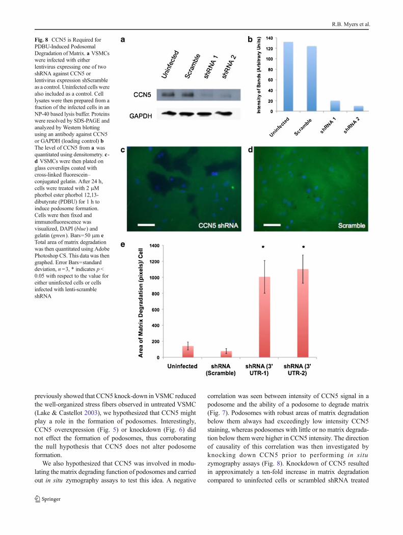
Blot analysis confirmed that both the shRNAs directed againstCCN5 reduced CCN5 protein levels >85 %, whereas thescrambled control did not affect CCN5 levels (Fig. 8a,b).
To assess the effect of CCN5 knock-down on podosomematrix degradation, VSMC were seeded onto glass slides pre-coated with cross-linked, fluoresceinated gelatin and treatedfor 1 h with PDBU. A group of uninfected cells were alsoseeded on gelatin-coated slides as a control. Following PDBUtreatment, gelatin degradation was visualized as dark areas inthe fluorescent matrix. To quantify this effect, three represen-tative photographs were taken of each slide and matrix deg-radation assessed by measuring the total amount of black area(degraded gelatin) in the three pictures; that number wasdivided by the total number of cells in the three pictures. Thisexperiment was repeated three times using different VSMCisolates and averaged. Knockdown of CCN5with either of ourtwo shRNAs resulted in approximately a ten-fold increaseabove uninfected cells; cells infected with the scrambledshRNA showed no change in matrix degradation (Fig. 8c,d,e).
The ability of podosomes to degrade matrix is generallyattributed to the presence of matrix metalloproteinases(MMPs). Furthermore, prior studies from our laboratory
demonstrated that the expression of MMP-2 in VSMC isinversely correlated with expression of CCN5 (12). To exam-ine the possibility that decreased MMP2 in the presence ofCCN5 occurred at the level of the podosome, we carried outimmunofluorescence studies using two different MMP2 anti-bodies, but were unable to detect MMP2 labeling inpodosomes. No MMP2 was detected with either antibody(data not shown). Immunofluorescence labeling with an anti-body to the membrane-bound form ofMMP2—MT-MMP2—also failed to reveal MT-MMP2 in podosomes. The samenegative labeling result was seen with an anti-MMP9 anti-body. To ensure that we were capable of detecting MMPs inpodosomes, we labeled SMC with an anti–MT-MMP1 anti-body. MT-MMP1 labeling was seen in all podosomes whetheror not CCN5 was also present in the podosome (data notshown).
Discussion
In this study we report several novel findings that shed light onthe mechanism of action of CCN5 and the regulation of
Fig. 5 CCN5 overexpression does not alter podosome formation. aVSMCs were infected with either adenovirus expressing CCN5 (lane 1)or adenovirus expressing GFP (lane 2). Cell lysates were then preparedfrom a fraction of the infected cells in an NP-40 based lysis buffer.Proteins were resolved by SDS-PAGE and analyzed by Western blottingusing an antibody against CCN5 or GAPDH (loading control) b Thelevel of CCN5 from a was quantitated using densitometry. c Anotherfraction of the cells infected with adenovirus (both CCN5 and GFP), as
well as a group of uninfected cells were trypsinized and plated on glassslides. Half of each of the aforementioned three groups of cells were thentreated with 2 μMphorbol ester phorbol 12,13-dibutyrate (PDBU) for 1 hto induce podosome formation, while the other half was untreated. Cellswere then fixed and stained for cortactin (podosomal marker). Percentageof cells containing podosomes were then calculated and graphed. ErrorBars=standard deviation, n =3
The matricellular protein CCN5 regulates podosome function

podosome function: 1) integrin αvβ3 is a CCN5 interactingprotein; 2) CCN5 and integrin αvβ3 co-localize inpodosomes; 3) CCN5 protein levels do not regulate the num-ber of podosomes in cells; and 4) CCN5 negatively regulatesmatrix degradation by podosomes.
To date, a functional cell surface binding protein for CCN5has not been identified. We have identified integrin αvβ3 as aCCN5 binding protein, based on co-immunoprecipitation ofintegrin αvβ3 with both endogenous and adenovirallyexpressed CCN5 (Fig. 2). Furthermore, CCN5 and integrinαvβ3 co-localize in podosomes using immunofluorescence(Fig. 3). Integrin αvβ3 is known to bind to a broad range ofboth RGD-containing and non-RGD-containing ligands(Brooks et al. 1996; Piali et al. 1995; Ruoslahti &Pierschbacher 1987). The primary sequence of CCN5 doesnot contain the RGD recognition motif present integrin αvβ3
ligands such as vitronectin, fibronectin, fibrinogen,thrombospondin , and osteopont in (Ruoslaht i &Pierschbacher 1987). In addition to CCN5, at least three otherintegrin αvβ3 ligands do not contain the consensus RGDsequence: a cell surface molecule from the immunoglobulinsuperfamily CD31/PECAM-1 (Piali et al. 1995), the matrix
metalloproteinase MMP-2 (Brooks et al. 1996), and the CCNfamily member CCN1 (Chen et al. 2004). Interestingly, CCN5contains a stretch of amino acids in its vonWillebrand Factor-C domain that is very similar to a known integrin αvβ3
binding site in CCN1 (Fig. 1) (Chen et al. 2004).The finding that CCN5 interacts with integrin αvβ3 also
provides a possible explanation for the ability of CCN5 toinhibit cell migration. Following the binding of extracellularligands, integrins are known to initiate “outside-in” signalingcascades, namely the phosphorylation and activation of focaladhesion kinase (FAK), and subsequent activation of Ras/Raf/Erk, known to regulate cell proliferation and Rho/Rac/cdc42,known to regulate cell adhesion and motility (Giancotti &Ruoslahti 1999). CCN proteins have been shown to actthrough integrin αvβ3 in fibroblasts and endothelial cells topromote cell proliferation and migration (Grzeszkiewicz et al.2001; Leu et al. 2002; Lin et al. 2005). Therefore, the inter-action of CCN5 with integrin αvβ3 could be regulating theknown anti-proliferation and anti-motility effects of CCN5 ina similar fashion.
Podosomes are highly dynamic adhesion structures with ahalf-life of approximately 2–12 min (Destaing et al. 2003).
Fig. 6 CCN5 shRNA knockdown does not alter podosome formation. aVSMCs were infected with either lentivirus expressing shRNA againstCCN5 (lane 1) or lentivirus expressing GFP (lane 2). Cell lysates werethen prepared from a fraction of the infected cells in an NP-40 based lysisbuffer. Proteins were resolved by SDS-PAGE and analyzed by Westernblotting using an antibody against CCN5 or GAPDH (loading control) bThe level of CCN5 from a was quantitated using densitometry. c .Another fraction of the cells infected with lentivirus (both CCN5 shRNA
and GFP), as well as a group of uninfected cells were trypsinized andplated on glass slides. Half of each of the aforementioned three groups ofcells were then treated with 2 μM phorbol ester phorbol 12,13-dibutyrate(PDBU) for 1 h to induce podosome formation, while the other half wasuntreated Cells were then fixed and stained for cortactin (podosomalmarker). Percentage of cells containing podosomes were then calculatedand graphed. Error Bars=standard deviation, n =3
R.B. Myers et al.

They have been documented in several cells including mac-rophages, osteoclasts, transformed fibroblasts, dendritic cells,and VSMCs (Marchisio et al. 1987; Hiura et al. 1995; Linder& Aepfelbacher 2003; Burgstaller & Gimona 2005), and theirpresence has been correlated with the invasiveness and abilityof these cells to move across anatomical boundaries(Marchisio et al. 1987). Subcellular position of podosomescorresponds to sites of localized proteolytic activity (Chenet al. 1984). We demonstrated that the podosome-like struc-tures containing CCN5 were indeed podosomes, as they were
labeled by the podosomemarker cortactin, and their formationwas inducible by the phorbol ester PDBU in a dose-dependentmanner. Interestingly, we observed that unlike CCN5, CCN2did not localize to podosomes. This lack of CCN2 inpodosomes is consistent with the opposite expression patternsof CCN2 and CCN5 observed in VSMC. CCN2 over-expression induces VSMC proliferation and increases MMP-2 expression, and CCN5 over-expression reduces proliferationand MMP-2 expression. CCN5 expression decreases whereasCCN2 expression increases in VSMC during the proliferativephase of balloon angioplasty injury (Lake et al. 2003; Lake &Castellot 2003; Ando et al. 2004; Fan et al. 2000; Fan &Karnovsky 2002).
The humanMMP family currently consists of 24members,including both secreted isoforms as well as MMPs that arelocalized to the cell surface through membrane anchors ortransmembrane stretches (MT-MMPs). MT1-MMP andMMP-9 are present at podosomes in osteoclasts whereas,MT1-MMP and MMP-2 are present in thepodosomes ofendothelial cells. We were unsuccessful in showing a correla-tion between CCN5 and MMPs in podosomes using immu-nofluorescence, with the exception of MT-MMP1, which weused as a positive control. The failure of MMP2 andMMP9 tolocalize to podosomes is not surprising, since they are secretedMMPs and may not be present at detectable levels bound to apodosome. While our negative data suggests that MT-MMP1,MMP2, MT-MMP2, and MMP-9 are not involved in theability of CCN5 to regulate matrix degradation in podosomes,this intriguing question deserves further exploration.
The defining characteristic of podosome structure is anactin-rich core (Marchisio et al. 1984; Pfaff & Jurdic 2001;Baldassarre et al. 2006). Proteins involved in actin nucleationincludingWiskott–Aldrich syndrome protein (WASP) (Linderet al. 1999), cortactin (Bowden et al. 1999), and Arp2/3complex (Linder et al. 2000a) are present throughout theactin-rich core. This intracellular core also contains integrins,including mostly β2 and β3 integrins (Gaidano et al. 1990)and integrin associated proteins such as talin and paxillin(Pfaff & Jurdic 2001; Bowden et al. 1999), thereby allowingthe podosome to interact with matricellular proteins such asCCN5.
We investigated the role CCN5 was playing in podosomesby examining whether it regulated podosome formation. Theformation of an F-actin core is necessary for podosome for-mation as F-actin disassembly through cytochalasins orlatrunculin results in disappearance of podosomes (Destainget al. 2003; Lehto et al. 1982; Linder et al. 2000b). Thisassembly of the F-actin core is catalyzed by CDC42-dependent activation of Wasp/N-WASP, which in turn acti-vates the actin-nucleating Arp2/3 complex (Linder et al.2000b). Given the dynamic nature of podosomes, which lastonly 2-12 min (Destaing et al. 2003), disassembly of the F-actin core is just as crucial as its formation. Because we
Fig. 7 CCN5 level in a podosome correlates with decreased the ability ofpodosomes to degrade matrix. VSMCs were plated on glass coverslipscoated with cross-linked fluorescein–conjugated gelatin. After 24 h, cellswere treated with 2 μM phorbol ester phorbol 12,13-dibutyrate (PDBU)for 1 h to induce podosome formation. Cells were then fixed and stainedfor CCN5 and DAPI. a–b Images of gelatin (green) and DAPI (blue). c-d Superimposed images of the same fields in a-b corresponding toCCN5 (red) and DAPI (blue). Bars=10 μm e CCN5 signal intesity/areaof podosome and gelatin signal intesity/area of below correspondingpodosome was quantitated using Adobe Photoshop CS. This data wasthen graphed in a scatterplot. n =57. Spearman rank-order correlationcoefficient (rs)=0.828
The matricellular protein CCN5 regulates podosome function
previously showed that CCN5 knock-down inVSMC reducedthe well-organized stress fibers observed in untreated VSMC(Lake & Castellot 2003), we hypothesized that CCN5 mightplay a role in the formation of podosomes. Interestingly,CCN5 overexpression (Fig. 5) or knockdown (Fig. 6) didnot effect the formation of podosomes, thus corroboratingthe null hypothesis that CCN5 does not alter podosomeformation.
We also hypothesized that CCN5 was involved in modu-lating the matrix degrading function of podosomes and carriedout in situ zymography assays to test this idea. A negative
correlation was seen between intensity of CCN5 signal in apodosome and the ability of a podosome to degrade matrix(Fig. 7). Podosomes with robust areas of matrix degradationbelow them always had exceedingly low intensity CCN5staining, whereas podosomes with little or no matrix degrada-tion below them were higher in CCN5 intensity. The directionof causality of this correlation was then investigated byknocking down CCN5 prior to performing in situzymography assays (Fig. 8). Knockdown of CCN5 resultedin approximately a ten-fold increase in matrix degradationcompared to uninfected cells or scrambled shRNA treated
Fig. 8 CCN5 is Required forPDBU-Induced PodosomalDegradation of Matrix. a VSMCswere infected with eitherlentivirus expressing one of twoshRNA against CCN5 orlentivirus expression shScrambleas a control. Uninfected cells werealso included as a control. Celllysates were then prepared from afraction of the infected cells in anNP-40 based lysis buffer. Proteinswere resolved by SDS-PAGE andanalyzed by Western blottingusing an antibody against CCN5or GAPDH (loading control) bThe level of CCN5 from a wasquantitated using densitometry. c-d VSMCs were then plated onglass coverslips coated withcross-linked fluorescein–conjugated gelatin. After 24 h,cells were treated with 2 μMphorbol ester phorbol 12,13-dibutyrate (PDBU) for 1 h toinduce podosome formation.Cells were then fixed andimmunofluorescence wasvisualized, DAPI (blue) andgelatin (green). Bars=50 μm eTotal area of matrix degradationwas then quantitated using AdobePhotoshop CS. This data was thengraphed. Error Bars=standarddeviation, n =3, * indicates p<0.05 with respect to the value foreither uninfected cells or cellsinfected with lenti-scrambleshRNA
R.B. Myers et al.

cells, thus strongly supporting the hypothesis that CCN5regulates podosome function.
These results suggest the importance of CCN5 inpodosome-mediated extracellular matrix degradation, a pro-cess thought to underlie the motogenic phenotype of VSMC.The involvement of CCN5 in podosomes could potentially beimportant in restenosis following vascular surgery. Followingprocedures such as angioplasty or coronary artery bypassgrafts, VSMC migrate from their original location in themedia of a vessel wall, through the internal elastic laminaand into the lumen, where they proliferate and block the arteryin a process called restenosis. Cortactin, a classic marker ofpodosomes, shows high expression in neointima, implicatingpodosomes in the VSMC migration seen in restenosis (Mintaet al. 2006). We have shown that ectopic expression of CCN5in a mouse model for restenosis strongly suppresses VSMCmigration and proliferation (unpublished work). The results ofthe present paper suggest the possibility that CCN5-mediatedinhibition of restenosis may occur via blocking the ability ofmedial VSMC podosomes to degrade matrix, thus preventingmigration into the intima.
Another example may be found in melanoma. CCN5 isthe most highly expressed mRNA of the CCN family innormal human skin cells. UV irradiation of human skinreduced the levels of CCN5 mRNA by 50 % in a time-dependent manner and did not return to basal levels until48 h post irradiation (Quan et al. 2009). In addition, theinvolvement of podosomes in the invasive capacity ofhuman melanoma has been well documented (Tague et al.2004; Nakahara et al. 2003).
The observations reported here raise many intriguing ques-tions about the mechanism of action of CCN5 and its role inpodosome function. Among these are: Does CCN5 bind di-rectly to integrins, and/or are other proteins are involved?What downstream signaling pathways are controlled byCCN5? Does CCN5 block the action of specific proteolyticenzymes like MMP-2, which we have previously shown to bestrongly inhibited by CCN5? Experiments designed to answerthese important questions are currently underway in ourlaboratory.
References
AndoH, Fukuda N, Kotani M, Yokoyama S, Kunimoto S, Matsumoto K,Saito S, Kanmatsuse K, Mugishima H (2004) Chimeric DNA-RNAhammerhead ribozyme targeting transforming growth factor-beta 1mRNA inhibits neointima formation in rat carotid artery after bal-loon injury. Eur J Pharmacol 483:207–214
Baldassarre M, Ayala I, Beznoussenko G, Giacchetti G, Machesky LM,Luini A, Buccione R (2006) Actin dynamics at sites of extracellularmatrix degradation. Eur J Cell Biol 85:1217–1231
Bowden ET, Barth M, Thomas D, Glazer RI, Mueller SC (1999) Aninvasion-related complex of cortactin, paxillin and PKCmu
associates with invadopodia at sites of extracellular matrix degrada-tion. Oncogene 18:4440–4449
Bowden ET, Coopman PJ, Mueller SC (2001) Invadopodia: uniquemethods for measurement of extracellular matrix degradationin vitro . Methods Cell Biol 63:613–627
Brooks PC, Stromblad S, Sanders LC, von Schalscha TL, Aimes RT,Stetler-StevensonWG, Quigley JP, ChereshDA (1996) Localizationof matrix metalloproteinase MMP-2 to the surface of invasive cellsby interaction with integrin alpha v beta 3. Cell 85:683–693
Burgstaller G, Gimona M (2005) Podosome-mediated matrix resorptionand cell motility in vascular smooth muscle cells. Am J PhysiolHeart Circ Physiol 288:H3001–3005
Burns S, Thrasher AJ, Blundell MP, Machesky L, Jones GE (2001)Configuration of human dendritic cell cytoskeleton by RhoGTPases, the WAS protein, and differentiation. Blood 98:1142–1149
Castellot JJ Jr, Favreau LV, Karnovsky MJ, Rosenberg RD (1982)Inhibition of vascular smooth muscle cell growth by endothelialcell-derived heparin, Possible role of a platelet endoglycosidase. JBiol Chem 257:11256–11260
Chen CC, Lau LF (2009) Functions and mechanisms of action of CCNmatricellular proteins. Int J Biochem Cell Biol 41:771–783
Chen WT, Olden K, Bernard BA, Chu FF (1984) Expression oftransformation-associated protease(s) that degrade fibronectin at cellcontact sites. J Cell Biol 98:1546–1555
Chen N, Leu SJ, Todorovic V, Lam SC, Lau LF (2004) Identification of anovel integrin alphavbeta3 binding site in CCN1 (CYR61) criticalfor pro-angiogenic activities in vascular endothelial cells. J BiolChem 279:44166–44176
Delmolino LM, Stearns NA, Castellot JJ Jr (2001) COP-1, a member ofthe CCN family, is a heparin-induced growth arrest specific gene invascular smooth muscle cells. J Cell Physiol 188:45–55
Destaing O, Saltel F, Geminard JC, Jurdic P, Bard F (2003) Podosomesdisplay actin turnover and dynamic self-organization in osteoclastsexpressing actin-green fluorescent protein. Mol Biol Cell 14:407–416
Fan WH, KarnovskyMJ (2002) IncreasedMMP-2 expression in connec-tive tissue growth factor over-expression vascular smooth musclecells. J Biol Chem 277:9800–9805
FanWH, PechM, KarnovskyMJ (2000) Connective tissue growth factor(CTGF) stimulates vascular smooth muscle cell growth and migra-tion in vitro . Eur J Cell Biol 79:915–923
Gaidano G, Bergui L, Schena M, Gaboli M, Cremona O, Marchisio PC,Caligaris-Cappio F (1990) Integrin distribution and cytoskeletonorganization in normal and malignant monocytes. Leukemia 4:682–687
Giancotti FG, Ruoslahti E (1999) Integrin signaling. Science 285:1028–1032
Gray MR, Castellot JJ (2005) Function and regulation of CCN5. In:T.M.e. Perbal BV (Ed) CCN proteins: a new family of cell growthand differentiation regulators. Imperial College Press, London
Grzeszkiewicz TM, Kirschling DJ, Chen N, Lau LF (2001) CYR61stimulates human skin fibroblast migration through Integrin alphavbeta 5 and enhances mitogenesis through integrin alpha vbeta 3,independent of its carboxyl-terminal domain. J Biol Chem 276:21943–21950
Hai CM, Hahne P, Harrington EO, Gimona M (2002) Conventionalprotein kinase C mediates phorbol-dibutyrate-induced cyto-skeletal remodeling in a7r5 smooth muscle cells. Exp CellRes 280:64–74
He TC, Zhou S, da Costa LT, Yu J, Kinzler KW, Vogelstein B (1998) Asimplified system for generating recombinant adenoviruses. ProcNatl Acad Sci U S A 95:2509–2514
Hiura K, Lim SS, Little SP, Lin S, Sato M (1995) Differentiation depen-dent expression of tensin and cortactin in chicken osteoclasts. CellMotil Cytoskeleton 30:272–284
The matricellular protein CCN5 regulates podosome function

Kireeva ML, Lam SC, Lau LF (1998) Adhesion of human umbilical veinendothelial cells to the immediate-early gene product Cyr61 ismediated through integrin alphavbeta3. J Biol Chem 273:3090–3096
Kumar S, Hand AT, Connor JR, Dodds RA, Ryan PJ, Trill JJ, Fisher SM,Nuttall ME, Lipshutz DB, Zou C, Hwang SM, Votta BJ, James IE,RiemanDJ, GowenM, Lee JC (1999) Identification and cloning of aconnective tissue growth factor-like cDNA from human osteoblastsencoding a novel regulator of osteoblast functions. J Biol Chem 274:17123–17131
Lake AC, Castellot JJ Jr (2003) CCN5 modulates the antiproliferativeeffect of heparin and regulates cell motility in vascular smoothmuscle cells. Cell Commun Signal 1:5
Lake AC, Bialik A, Walsh K, Castellot JJ Jr (2003) CCN5 is a growtharrest-specific gene that regulates smooth muscle cell proliferationand motility. Am J Pathol 162:219–231
Lau LF, Lam SC (2005) Stephen Integrin-Mediated CCN Functions. In:T.M.e. Perbal BV (Ed) CCN proteins: a new family of cell growthand differentiation regulators. Imperial College Press, London
Leask A, Abraham DJ (2006) All in the CCN family: essentialmatricellular signaling modulators emerge from the bunker. J CellSci 119:4803–4810
Lehto VP, Hovi T, Vartio T, Badley RA, Virtanen I (1982) Reorganizationof cytoskeletal and contractile elements during transition of humanmonocytes into adherent macrophages. Lab Invest 47:391–399
Leu SJ, Lam SC, Lau LF (2002) Pro-angiogenic activities of CYR61(CCN1) mediated through integrins alphavbeta3 and alpha6beta1 inhuman umbilical vein endothelial cells. J Biol Chem 277:46248–46255
Lin CG, Chen CC, Leu SJ, Grzeszkiewicz TM, Lau LF (2005) Integrin-dependent functions of the angiogenic inducer NOV (CCN3): im-plication in wound healing. J Biol Chem 280:8229–8237
Linder S, Aepfelbacher M (2003) Podosomes: adhesion hot-spots ofinvasive cells. Trends Cell Biol 13:376–385
Linder S, Nelson D, Weiss M, Aepfelbacher M (1999) Wiskott-Aldrichsyndrome protein regulates podosomes in primary human macro-phages. Proc Natl Acad Sci U S A 96:9648–9653
Linder S, Higgs H, Hufner K, Schwarz K, Pannicke U, Aepfelbacher M(2000a) The polarization defect of Wiskott-Aldrich syndrome mac-rophages is linked to dislocalization of the Arp2/3 complex. JImmunol 165:221–225
Linder S, Hufner K, Wintergerst U, Aepfelbacher M (2000b)Microtubule-dependent formation of podosomal adhesion structuresin primary human macrophages. J Cell Sci 113(Pt 23):4165–4176
Marchisio PC, Cirillo D, Naldini L, Primavera MV, Teti A, Zambonin-Zallone A (1984) Cell-substratum interaction of cultured avianosteoclasts is mediated by specific adhesion structures. J Cell Biol99:1696–1705
Marchisio PC, Cirillo D, Teti A, Zambonin-Zallone A, Tarone G (1987)Rous sarcoma virus-transformed fibroblasts and cells of monocyticorigin display a peculiar dot-like organization of cytoskeletal pro-teins involved in microfilament-membrane interactions. Exp CellRes 169:202–214
Mason HR, Lake AC, Wubben JE, Nowak RA, Castellot JJ Jr (2004a)The growth arrest-specific gene CCN5 is deficient in humanleiomyomas and inhibits the proliferation and motility of culturedhuman uterine smooth muscle cells. Mol Hum Reprod 10:181–187
Mason HR, Grove-Strawser D, Rubin BS, Nowak RA, Castellot JJ Jr(2004b) Estrogen induces CCN5 expression in the rat uterus in vivo .Endocrinology 145:976–982
Minta JO, Yun JJ, Kabiawu O, Jones J (2006) mRNA differential displayidentification of vascular smooth muscle early response genes reg-ulated by PDGF. Mol Cell Biochem 281:63–75
Nakahara H, Otani T, Sasaki T, Miura Y, Takai Y, Kogo M (2003)Involvement of Cdc42 and Rac small G proteins in invadopodiaformation of RPMI7951 cells. Genes Cells 8:1019–1027
Osiak AE, Zenner G, Linder S (2005) Subconfluent endothelial cells formpodosomes downstream of cytokine and RhoGTPase signaling. ExpCell Res 307:342–353
Pennica D, Swanson TA, Welsh JW, Roy MA, Lawrence DA, Lee J,Brush J, Taneyhill LA, Deuel B, Lew M, Watanabe C, Cohen RL,MelhemMF, Finley GG, Quirke P, Goddard AD, Hillan KJ, GurneyAL, Botstein D, Levine AJ (1998) WISP genes are members of theconnective tissue growth factor family that are up-regulated in wnt-1-transformed cells and aberrantly expressed in human colon tu-mors. Proc Natl Acad Sci U S A 95:14717–14722
Perbal B (2004) CCN proteins: multifunctional signalling regulators.Lancet 363:62–64
Perbal BV (2005) CCN Proteins: a new family of cell growth anddifferentiation regulators. Imperial College Press, London
Pfaff M, Jurdic P (2001) Podosomes in osteoclast-like cells: structuralanalysis and cooperative roles of paxillin, proline-rich tyrosinekinase 2 (Pyk2) and integrin alphaVbeta3. J Cell Sci 114:2775–2786
Piali L, Hammel P, Uherek C, Bachmann F, Gisler RH, Dunon D, ImhofBA (1995) CD31/PECAM-1 is a ligand for alpha v beta 3 integrininvolved in adhesion of leukocytes to endothelium. J Cell Biol 130:451–460
Quan T, Shin S, Qin Z, Fisher GJ (2009) Expression of CCN family ofgenes in human skin in vivo and alterations by solar-simulatedultraviolet irradiation. J Cell Commun Signal 3:19–23
Rachfal AW, Brigstock DR (2005) Structural and functional properties ofCCN proteins. Vitam Horm 70:69–103
Ruoslahti E, Pierschbacher MD (1987) New perspectives in cell adhe-sion: RGD and integrins. Science 238:491–497
Tague SE,MuralidharanV, D’Souza-Schorey C (2004) ADP-ribosylationfactor 6 regulates tumor cell invasion through the activation of theMEK/ERK signaling pathway. Proc Natl Acad Sci U SA 101:9671–9676
Tarone G, Cirillo D, Giancotti FG, Comoglio PM, Marchisio PC (1985)Rous sarcoma virus-transformed fibroblasts adhere primarily atdiscrete protrusions of the ventral membrane called podosomes.Exp Cell Res 159:141–157
Tatin F, Varon C, Genot E, Moreau V (2006) A signalling cascadeinvolving PKC, Src and Cdc42 regulates podosome assembly incultured endothelial cells in response to phorbol ester. J Cell Sci 119:769–781
Wei L, McKeon F, Russo JW, Lemire J, Castellot J (2009) Domain-andspecies-specific monoclonal antibodies recognize the VonWillebrand Factor-C domain of CCN5. J Cell Commun Signal 3:65–77
Zhang R, Averboukh L, ZhuW, Zhang H, Jo H, Dempsey PJ, Coffey RJ,Pardee AB, Liang P (1998) Identification of rCop-1, a new memberof the CCN protein family, as a negative regulator for cell transfor-mation. Mol Cell Biol 18:6131–6141
R.B. Myers et al.