Embed Size (px)
Citation preview
Brief Communications
The Matrix Protein Hikaru genki Localizes to CholinergicSynaptic Clefts and Regulates Postsynaptic Organization inthe Drosophila Brain
Minoru Nakayama, Fumiya Matsushita, and X Chihiro HamaDepartment of Molecular Biosciences, Faculty of Life Sciences, Kyoto Sangyo University, Kita-ku, Kyoto 603-8555, Japan
The synaptic cleft, a crucial space involved in neurotransmission, is filled with extracellular matrix that serves as a scaffold for synapticdifferentiation. However, little is known about the proteins present in the matrix and their functions in synaptogenesis, especially in theCNS. Here, we report that Hikaru genki (Hig), a secreted protein with an Ig motif and complement control protein domains, localizesspecifically to the synaptic clefts of cholinergic synapses in the Drosophila CNS. The data indicate that this specific localization is achievedby capture of secreted Hig in synaptic clefts, even when it is ectopically expressed in glia. In the absence of Hig, the cytoskeletal scaffoldprotein DLG accumulated abnormally in cholinergic postsynapses, and the synaptic distribution of acetylcholine receptor (AchR) sub-units D�6 and D�7 significantly decreased. hig mutant flies consistently exhibited resistance to the AchR agonist spinosad, which causeslethality by specifically activating the D�6 subunit, suggesting that loss of Hig compromises the cholinergic synaptic activity mediated byD�6. These results indicate that Hig is a specific component of the synaptic cleft matrix of cholinergic synapses and regulates theirpostsynaptic organization in the CNS.
Key words: acetylcholine receptor; DLG; Drosophila; matrix; synaptic cleft; synaptogenesis
IntroductionIn both vertebrates and invertebrates, proteins secreted to theextracellular synaptic spaces regulate clustering of neurotrans-mitter receptors (Broadie et al., 2011; Nakayama and Hama,2011). For example, Agrin in vertebrates (Wu et al., 2010) andLEV-9 and OIG-4 in Caenorhabditis elegans (Gendrel et al., 2009;Rapti et al., 2011) induce clustering of AchR at cholinergic neu-romuscular junctions (NMJs). In the mouse cerebellum, Cbln1,which links Neurexin to the glutamate receptor GluD2 at syn-apses (Matsuda et al., 2010; Uemura et al., 2010), is capable ofprompting GluD2 clustering in heterologous cells. In Drosophilaglutamatergic NMJs, clustering of glutamate receptors dependson the secreted protein Mind-the-Gap (Rohrbough et al., 2007).Although these secreted proteins have been identified in a fewspecific types of synapses, the identities of the matrix proteinsthat regulate the organization of cholinergic synapses in the CNSremain unknown.
The hikaru genki (hig) gene was identified by analysis of Dro-sophila mutants exhibiting reduced locomotor behavior. Thisgene is expressed in a subset of neurons in the CNS of embryosand continues to be expressed in the nervous system until theadult stage. Hig is a secretory protein with an immunoglobulin(Ig) domain and a maximum of five complement control protein(CCP) domains (Hoshino et al., 1993). Immunoelectron micros-copy reveals that Hig localizes to the synaptic clefts of subsets ofsynapses in the adult brain and in nascent synapses during pupalstages (Hoshino et al., 1996). This protein is present in the secre-tory apparatus (Hoshino et al., 1996) and is secreted from cul-tured cells to the medium (Hoshino et al., 1999). Mutantphenotypes of hig flies can be rescued by transient expression ofHig during middle pupal stages, suggesting that Hig plays a rolein differentiation of synapses (Hoshino et al., 1996).
Here, we show that Hig is a matrix protein localized at thesynaptic clefts of cholinergic synapses in the CNS and regulatesthe distribution of AchR subunits and DLG in postsynaptic ter-minals. This study indicates a possible avenue for revealing thecomposition of synaptic cleft matrix and determining how thematrix is involved in differentiation of cholinergic synapses inthe CNS.
Materials and MethodsDrosophila strains. higdd37 flies harbor a null mutation caused by adeficiency in the hig gene (Hoshino et al., 1993). The mutant strainnAcR�-30DDAS1 (hereafter D�6DAS1) (Watson et al., 2010) was obtainedfrom the Bloomington Stock Center. D�7P�EY6, D�7-GAL4, and UAS-D�7-EGFP were obtained from H.J. Bellen (Fayyazuddin et al., 2006).To generate UAS-hig-GFP, hig-GFP was amplified by PCR from
Received April 18, 2014; revised Aug. 14, 2014; accepted Aug. 31, 2014.Author contributions: M.N. and C.H. designed research; M.N. and F.M. performed research; M.N. analyzed data;
M.N. and C.H. wrote the paper.This work was supported by Japan Society for the Promotion of Science Grant-in-Aid for Challenging Exploratory
Research Grant 23650170. We thank Hugo J. Bellen, Ulrich Thomas, Hermann Aberle, Dick R. Nassel, Tadashi Ue-mura, and the Developmental Studies Hybridoma Bank for antibodies BDGP for cDNAs; Jing Wang, Hugo J. Bellen,and the Bloomington Stock Center for fly stocks; Shin-ichi Tsunoda for the use of structured illumination microscopysystem; and Nobuaki Tanaka for technical instruction of GCaMP imaging.
The authors declare no competing financial interests.Correspondence should be addressed to Dr. Chihiro Hama, Faculty of Life Sciences, Kyoto Sangyo University,
Kita-ku, Kyoto 603-8555, Japan. E-mail: [email protected]:10.1523/JNEUROSCI.1585-14.2014
Copyright © 2014 the authors 0270-6474/14/3413872-06$15.00/0
13872 • The Journal of Neuroscience, October 15, 2014 • 34(42):13872–13877
pCasperHS-higD30GFP, which contains the longest form of hig cDNAfused with GFP. The resulting fragment was cloned into the EcoRI site ofpUASTattB (Bischof et al., 2007). Transgenic fly lines expressing UAS-hig-GFP were generated by Phi-C31-mediated site-specific integration atattP2, P18, and P40.
Antibody production. To generate anti-Hig antibody, a fragment of higcDNA encoding the N-terminal fragment (amino acids 40 –266) wasamplified by PCR from GH23986 (BDGP) and cloned into pET15b (No-vagen). Hexahistidine fusion proteins were expressed in Escherichia coliBL21 (DE3), purified, and injected into guinea pigs. To generate anti-D�6 antibody, DNA encoding the cytoplasmic loop (amino acids 392–460) was amplified by PCR from GH15518 (BDGP) and cloned intopGEX4T-1 (GE Healthcare). GST fusion proteins were expressed in E.
coli BL21 (DE3), purified, and injected intorabbits. Anti-Hig and anti-D�6 antibodieswere purified by affinity binding to the respec-tive antigens.
Immunohistochemistry. Brains of adult maleflies were dissected in PBS, fixed with 4% para-formaldehyde (PFA) in PBS on ice for 1 h, andstained with the following antibodies: anti-Hig(1:1000), anti-Bruchpilot (nc82, 1:20, DSHB),anti-ChAT (ChAT4B1, 1:20, DSHB), anti-DVGLUT (1:500, Mahr and Aberle, 2006), anti-Rdl (1:1000, Enell et al., 2007), anti-GABARbII(1:1000, Enell et al., 2007), anti-DLG (4F3, 1:100,DSHB), anti-DN-cadherin (Ex#7, 1:20, DSHB),or anti-GFP (1:500, Invitrogen). AlexaFluor(-488, -568, -594, and -647)-conjugated anti-bodies (1:200, Invitrogen) were used as sec-ondaries. For staining with anti-D�7 (1:1000)and anti-D�6 (1:1000), dissected brains werefixed with 4% PFA in PBS for 10 min on ice.Samples were observed by sequential scans onan SP2 confocal microscope (Leica) or ELYRAS.1 microscope (Zeiss). Quantitation of confo-cal images was performed using the Leica con-focal software, with fluorescence intensitycompared with that of control samples stainedsimultaneously in the same tube. At least sevensamples were examined for the purposes of sta-tistical analysis.
Insecticide assay. Insecticide resistance assayswere performed as previously reported (Wat-son et al., 2010) with slight modifications.Spinosad was dissolved at appropriate concen-trations in 2:1 acetone:10% sucrose solution,and then 0.1 ml of each solution was applied tovials (inner diameter 2.1 cm) containing 5 mlof blue agar (1% agar, 1% Brilliant Blue FCF).Vials were dried overnight. Ten male flies (3- to5-d-old) were introduced to each vial, andmortality was examined over 24 h. To assess theamount of food ingested by surviving flies, thebodies were separated from heads, homoge-nized in PBS, and centrifuged at 13,000 rpm for25 min. After the supernatants were recentri-fuged, absorbance was measured at 625 nm.
ResultsHig localizes predominantly atcholinergic synapsesHig was detected most abundantly in syn-aptic regions that labeled positively for thepresynaptic active-zone protein Bruchpi-lot (Brp; Wagh et al., 2006) in the adultbrain (Fig. 1A). This Hig signal disap-peared in the hig mutant higdd37, confirm-ing the specificity of the anti-Hig antibody
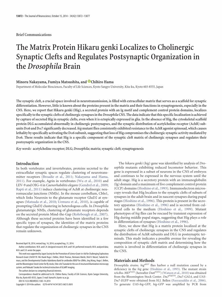
(Fig. 1A). To identify the neurotransmitter associated with theHig-positive synapses, we performed double immunolabeling forseveral synaptic markers and Hig (Fig. 1B–E). The antennal lobesare well-analyzed regions containing synapses that use acetylcho-line, glutamate, and GABA as neurotransmitters (Liu and Wil-son, 2013). Synaptic Hig was closely associated with ChAT, apresynaptic marker of cholinergic neurons, whereas vesicularglutamate transporter (VGLUT) and GABA receptors (Rdl andGABAbRII) were largely excluded from Hig-positive zones (Fig.1B–E). To confirm the distribution of Hig at cholinergic syn-apses, we used structured illumination microscopy to obtain
Figure 1. Hig localizes in the synaptic clefts of cholinergic synapses in the brain. A, Distribution of Hig in the brain. Frontalsections of the anterior and posterior brain regions are shown. Hig was observed in the entire synaptic region labeled with Brp inthe WT brain, whereas Hig signals disappeared in higdd37. al, Antennal lobe; cx, calyx of the MB. B–E, Double labeling of synapsesin the antennal lobe with antibodies against synaptic proteins and Hig. Hig was closely associated with ChAT (B) but not VGLUT (C),Rdl (D), or GABAbRII (E). Single glomeruli surrounded by dotted squares are magnified in the three right panels. F, G, High-resolution images of synapses in the antennal-lobe glomerulus obtained by structured illumination microscopy. Each Hig-positivesynapse in focus is colabeled with D�7-GFP, driven by D�7-GAL4 (F ). In most synapses, Hig (red at the top, gray at the bottom)localizes between Brp (blue) and D�7-GFP (green) (G). Serial optical images were obtained at Z intervals of 0.3 �m. Scale bar, 100nm. H, Schematic model for the distribution of Brp, Hig, and D�7 in a microglomerulus formed by a single presynaptic bouton (pb)of olfactory projection neurons and the dendrites (d) of MB Kenyon cells (for the structure of the microglomerulus and distributionof synaptic markers, see Kremer et al., 2010). I–K, Distribution pattern of Hig in microglomeruli of the calyx. The single microglom-erulus in a dotted square is enlarged in the three lower panels. Most Hig-positive microglomeruli were associated with ChAT (I ).Discrete signals of Hig in microglomeruli were juxtaposed with D�7 (J ) and the active-zone marker Brp (K ). K, Arrowheadsindicate synapses without Hig, which are not cholinergic.
Nakayama et al. • Hig Protein Regulates Postsynaptic Differentiation J. Neurosci., October 15, 2014 • 34(42):13872–13877 • 13873
high-resolution images of synapses, tak-ing advantage of the surface localizationof the antennal lobes in the brain. We ex-pressed D�7-GFP, a GFP-tagged AchRsubunit D�7, using a D�7-GAL4 driverline; when the center of each signal wasfocused, the D�7-GFP signals were closelyassociated with Hig (Fig. 1F). Structuredillumination microscopy also revealedfine images of a single synapse in whichHig was present between the presynapticmarker Brp and the postsynaptic markerD�7-GFP, indicating that Hig was local-ized to the synaptic cleft (Fig. 1G). Themicroglomerulus of the mushroom body(MB) calyx is a specialized synaptic com-plex that uses acetylcholine as a neu-rotransmitter (Fig. 1 I, J) (Yasuyama et al.,2002) and is surrounded by noncholin-ergic synapses (Fig. 1K). This complexcomprises an axon terminal of the projec-tion neuron at its center and the dendritesof MB Kenyon cells (Fig. 1H) (Kremer etal., 2010). In most microglomeruli, a dis-crete pattern of Hig staining was observedin the surrounding regions of ChAT-positive projection neuron axon terminals(Fig. 1I). These immunohistochemical datafrom antennal lobe, MB, and other brain re-gions, including the optic lobes (see Fig.3A,B), indicated that Hig predominantlylocalized to cholinergic synapses in thebrain. To further analyze the discrete pat-tern of Hig in the microglomeruli, brainswere simultaneously stained for D�7 andHig or Brp and Hig. Hig did not simplydiffuse over the entire space of the synap-tic clefts but was instead juxtaposed withD�7 on the postsynaptic membrane (Fig.1H, J); it also colocalized with Brp, anactive-zone marker in the presynaptic ter-minal (Fig. 1H,K). We conclude that Higis predominantly distributed in the cho-linergic synapses and localizes at the syn-aptic cleft region adjacent to the activezone.
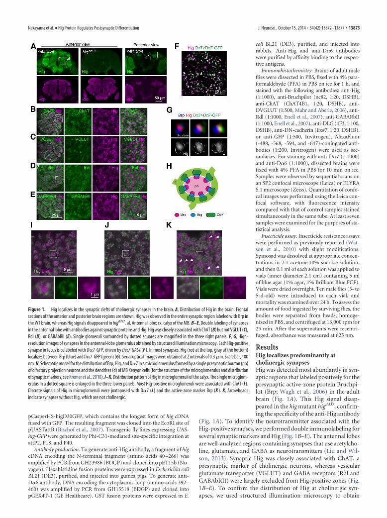
Hig is extracellularly diffused andtrapped by cholinergic synapsesBecause Hig is predominantly distributed in the cholinergic syn-apses, we predicted that the hig mutant phenotype could be res-cued by expression of transgenic hig in the cholinergic neurons.To test this idea, we expressed GFP-tagged Hig (Hig-GFP) in thehigdd37 background under the control of several GAL4 drivers.The higdd37 homozygotes exhibited reduced longevity, and thisphenotype was rescued by expression of Hig-GFP in all neurons(elav-Gal4) and cholinergic neurons (Cha-Gal4), as expected(Fig. 2A). However, the longevity of higdd37 was also rescued byhig expression in glutamatergic neurons (OK371-Gal4 andVGlut-Gal4) as well as in glial cells (Eaat-Gal4 and Repo-Gal4)(Fig. 2A). By contrast, Gal4 lines driving expression in a smallnumber of neurons (5HTR1B-Gal4, Yuan et al., 2005; Ddc-Gal4,Liu et al., 2012) were unable to rescue the mutant phenotype.
Nevertheless, when expression of Hig-GFP was enhanced by ad-dition of one more copy of the hig transgene, Ddc-Gal4 couldprolong longevity (Fig. 2A). These results indicate that Hig doesnot need to be expressed in cholinergic neurons. Indeed, endog-enous Hig was detected in the cell bodies of a large number ofglutamatergic and cholinergic neurons (Fig. 2B).
The results of these rescue experiments and the synaptic dis-tribution of Hig suggested that secreted and diffused Hig is even-tually integrated into cholinergic synaptic clefts. To test thispossibility, we examined the distribution of Hig-GFP expressedby a glia-specific driver (Fig. 2C). Secreted Hig-GFP was detectedin most synaptic regions throughout the brain, notably in thecholinergic microglomeruli of the MB calyx (Fig. 2C), whereasHig-GFP was not detected in noncholinergic synapses outside of
Figure 2. Secreted Hig-GFP is captured by the cholinergic synapses. A, Rescue of the hig phenotype by ectopic expression ofHig-GFP. The indicated GAL4 lines were used to drive Hig-GFP expression in cholinergic, glutamatergic, or dopaminergic neurons,or glia, in the hig mutant background. The genotype of the control is higdd37; UAS-hig-GFP/ �. UAS-hig-GFP on the third chromo-some was used for most of the experiments, unless otherwise stated. Ddc�1st: UAS-hig-GFP/ Y; Ddc-Gal4, higdd37/ higdd37;�/�.Ddc � 3rd: �/ Y; Ddc-Gal4, higdd37/ higdd37; UAS-hig-GFP/�. Ddc � 1st 3rd: UAS-hig-GFP/ Y; Ddc-Gal4, higdd37/ higdd37; UAS-hig-GFP/�. Error bars indicate�SEM. B, Expression of endogenous Hig in cholinergic and glutamatergic neurons. Cholinergic andglutamatergic neurons were labeled with nuclear GFP driven by Cha-Gal4 and VGlut-Gal4, respectively. Filled and open arrowheadsindicate Hig-expressing neurons with and without GFP signals, respectively. C, Synaptic localization of Hig-GFP ectopically ex-pressed in the glia. Eaat1-GAL4 was used for glial expression of membrane-bound mCD8-GFP and Hig-GFP. Hig-GFP is detected inmost synaptic regions labeled with Brp in both the anterior (ant) and posterior (post) brain sections, whereas mCD8-GFP labels glialmembranes (yellow arrowheads). In MB calyx of the posterior section, Hig-GFP was detected in cholinergic microglomeruli (ar-rows) but excluded from noncholinergic synapses outside microglomeruli (inset, arrowheads). Scale bar: C, inset, 5 �m. D,Microglomerulus-specific localization of endogenous Hig and pan-neuronally expressed Hig-GFP. As in C, inset, endogenous Hig(top) and Hig-GFP driven by elav-Gal4 (bottom) were only detected in cholinergic microglomeruli, but not in other synapses(arrowheads). mCD8-GFP driven by elav-Gal4 labeled the whole synaptic region of the calyx (middle).
13874 • J. Neurosci., October 15, 2014 • 34(42):13872–13877 Nakayama et al. • Hig Protein Regulates Postsynaptic Differentiation
the microglomeruli (Fig. 2C, inset). This microglomerulus-specific localization was also observed for endogenous Hig andHig-GFP driven by elav-Gal4 in all neurons (Fig. 2D, insets).Thus, Hig is produced in cholinergic and noncholinergic neu-rons, secreted to extracellular spaces, and finally trapped in cho-linergic synaptic clefts by a specific mechanism.
Hig regulates the synaptic distribution of D�7, D�6,and DLGBecause Hig is required for normal locomotor activity and lon-gevity, we predicted that the neural function of cholinergic syn-apses would be partly impaired in the hig mutant. We thereforeexamined the distribution of several proteins localized to cholin-
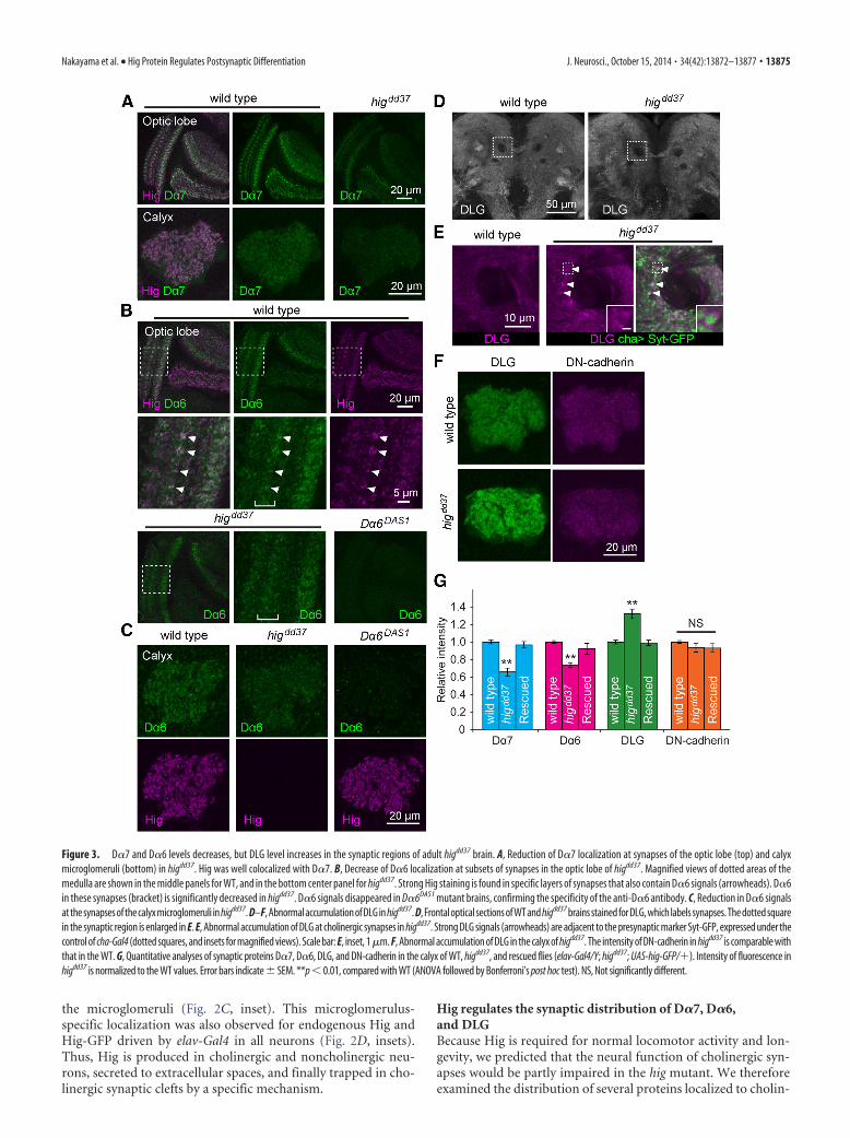
Figure 3. D�7 and D�6 levels decreases, but DLG level increases in the synaptic regions of adult higdd37 brain. A, Reduction of D�7 localization at synapses of the optic lobe (top) and calyxmicroglomeruli (bottom) in higdd37. Hig was well colocalized with D�7. B, Decrease of D�6 localization at subsets of synapses in the optic lobe of higdd37. Magnified views of dotted areas of themedulla are shown in the middle panels for WT, and in the bottom center panel for higdd37. Strong Hig staining is found in specific layers of synapses that also contain D�6 signals (arrowheads). D�6in these synapses (bracket) is significantly decreased in higdd37. D�6 signals disappeared in D�6DAS1 mutant brains, confirming the specificity of the anti-D�6 antibody. C, Reduction in D�6 signalsat the synapses of the calyx microglomeruli in higdd37. D–F, Abnormal accumulation of DLG in higdd37. D, Frontal optical sections of WT and higdd37 brains stained for DLG, which labels synapses. The dotted squarein the synaptic region is enlarged in E. E, Abnormal accumulation of DLG at cholinergic synapses in higdd37. Strong DLG signals (arrowheads) are adjacent to the presynaptic marker Syt-GFP, expressed under thecontrol of cha-Gal4 (dotted squares, and insets for magnified views). Scale bar: E, inset, 1�m. F, Abnormal accumulation of DLG in the calyx of higdd37. The intensity of DN-cadherin in higdd37 is comparable withthat in the WT. G, Quantitative analyses of synaptic proteins D�7, D�6, DLG, and DN-cadherin in the calyx of WT, higdd37, and rescued flies (elav-Gal4/Y; higdd37; UAS-hig-GFP/�). Intensity of fluorescence inhigdd37 is normalized to the WT values. Error bars indicate � SEM. **p � 0.01, compared with WT (ANOVA followed by Bonferroni’s post hoc test). NS, Not significantly different.
Nakayama et al. • Hig Protein Regulates Postsynaptic Differentiation J. Neurosci., October 15, 2014 • 34(42):13872–13877 • 13875
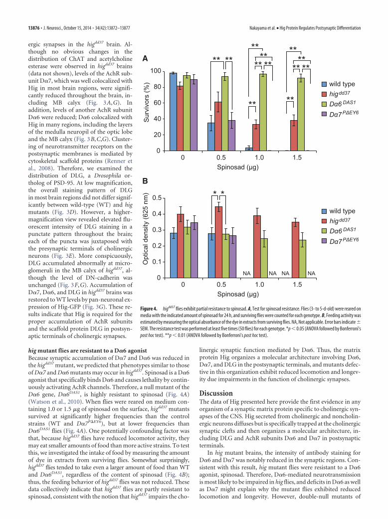
ergic synapses in the higdd37 brain. Al-though no obvious changes in thedistribution of ChAT and acetylcholineesterase were observed in higdd37 brains(data not shown), levels of the AchR sub-unit D�7, which was well colocalized withHig in most brain regions, were signifi-cantly reduced throughout the brain, in-cluding MB calyx (Fig. 3A,G). Inaddition, levels of another AchR subunitD�6 were reduced; D�6 colocalized withHig in many regions, including the layersof the medulla neuropil of the optic lobeand the MB calyx (Fig. 3B,C,G). Cluster-ing of neurotransmitter receptors on thepostsynaptic membranes is mediated bycytoskeletal scaffold proteins (Renner etal., 2008). Therefore, we examined thedistribution of DLG, a Drosophila or-tholog of PSD-95. At low magnification,the overall staining pattern of DLGin most brain regions did not differ signif-icantly between wild-type (WT) and higmutants (Fig. 3D). However, a higher-magnification view revealed elevated flu-orescent intensity of DLG staining in apunctate pattern throughout the brain;each of the puncta was juxtaposed withthe presynaptic terminals of cholinergicneurons (Fig. 3E). More conspicuously,DLG accumulated abnormally at micro-glomeruli in the MB calyx of higdd37, al-though the level of DN-cadherin wasunchanged (Fig. 3F,G). Accumulation ofD�7, D�6, and DLG in higdd37 brains wasrestored to WT levels by pan-neuronal ex-pression of Hig-GFP (Fig. 3G). These re-sults indicate that Hig is required for theproper accumulation of AchR subunitsand the scaffold protein DLG in postsyn-aptic terminals of cholinergic synapses.
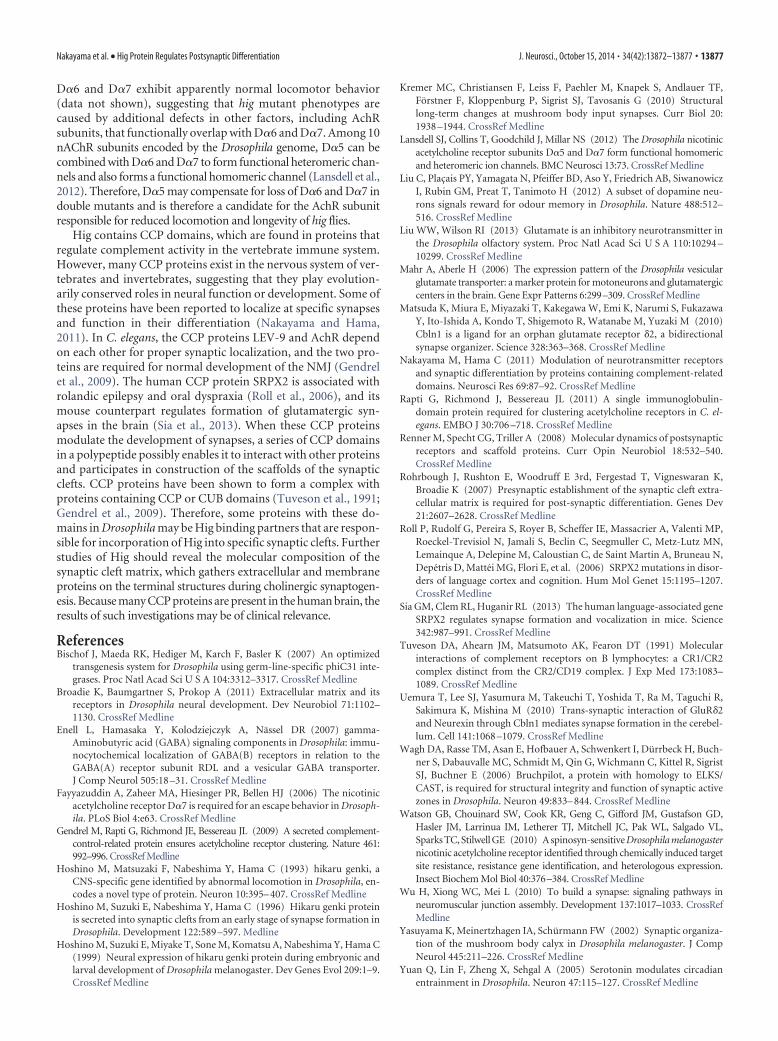
hig mutant flies are resistant to a D�6 agonistBecause synaptic accumulation of D�7 and D�6 was reduced inthe higdd37 mutant, we predicted that phenotypes similar to thoseof D�7 and D�6 mutants may occur in higdd37. Spinosad is a D�6agonist that specifically binds D�6 and causes lethality by contin-uously activating AchR channels. Therefore, a null mutant of theD�6 gene, D�6DAS1, is highly resistant to spinosad (Fig. 4A)(Watson et al., 2010). When flies were reared on medium con-taining 1.0 or 1.5 �g of spinosad on the surface, higdd37 mutantssurvived at significantly higher frequencies than the controlstrains (WT and D�7P�EY6), but at lower frequencies thanD�6DAS1 flies (Fig. 4A). One potentially confounding factor wasthat, because higdd37 flies have reduced locomotor activity, theymay eat smaller amounts of food than more active strains. To testthis, we investigated the intake of food by measuring the amountof dye in extracts from surviving flies. Somewhat surprisingly,higdd37 flies tended to take even a larger amount of food than WTand D�6DAS1, regardless of the content of spinosad (Fig. 4B);thus, the feeding behavior of higdd37 flies was not reduced. Thesedata collectively indicate that higdd37 flies are partly resistant tospinosad, consistent with the notion that higdd37 impairs the cho-
linergic synaptic function mediated by D�6. Thus, the matrixprotein Hig organizes a molecular architecture involving D�6,D�7, and DLG in the postsynaptic terminals, and mutants defec-tive in this organization exhibit reduced locomotion and longev-ity due impairments in the function of cholinergic synapses.
DiscussionThe data of Hig presented here provide the first evidence in anyorganism of a synaptic matrix protein specific to cholinergic syn-apses of the CNS. Hig secreted from cholinergic and noncholin-ergic neurons diffuses but is specifically trapped at the cholinergicsynaptic clefts and then organizes a molecular architecture, in-cluding DLG and AchR subunits D�6 and D�7 in postsynapticterminals.
In hig mutant brains, the intensity of antibody staining forD�6 and D�7 was notably reduced in the synaptic regions. Con-sistent with this result, hig mutant flies were resistant to a D�6agonist, spinosad. Therefore, D�6-mediated neurotransmissionis most likely to be impaired in hig flies, and deficits in D�6 as wellas D�7 might explain why the mutant flies exhibited reducedlocomotion and longevity. However, double-null mutants of
wild typehigdd37
Dα6 DAS1
Dα7PΔEY6
0 0.5 1.0 1.5Spinosad (μg)
Sur
vivo
rs (%
)0
20
40
60
80
100
wild typehigdd37
Dα6 DAS1
Dα7PΔEY6
0 0.5 1.0 1.5Spinosad (μg)
Opt
ical
den
sity
(625
nm
)
0
0.1
0.2
0.3
0.4
0.5
A
B
****
*
NA
*
NA NA NA
****
**
**
****
**
**
**
**
Figure 4. higdd37 flies exhibit partial resistance to spinosad. A, Test for spinosad resistance. Flies (3- to 5-d-old) were reared onmedia with the indicated amount of spinosad for 24 h, and surviving flies were counted for each genotype. B, Feeding activity wasestimated by measuring the optical absorbance of the dye in extracts from surviving flies. NA, Not applicable. Error bars indicate �SEM. The resistance test was performed at least five times (50 flies) for each genotype. *p � 0.05 (ANOVA followed by Bonferroni’spost hoc test). **p � 0.01 (ANOVA followed by Bonferroni’s post hoc test).
13876 • J. Neurosci., October 15, 2014 • 34(42):13872–13877 Nakayama et al. • Hig Protein Regulates Postsynaptic Differentiation
D�6 and D�7 exhibit apparently normal locomotor behavior(data not shown), suggesting that hig mutant phenotypes arecaused by additional defects in other factors, including AchRsubunits, that functionally overlap with D�6 and D�7. Among 10nAChR subunits encoded by the Drosophila genome, D�5 can becombined with D�6 and D�7 to form functional heteromeric chan-nels and also forms a functional homomeric channel (Lansdell et al.,2012). Therefore, D�5 may compensate for loss of D�6 and D�7 indouble mutants and is therefore a candidate for the AchR subunitresponsible for reduced locomotion and longevity of hig flies.
Hig contains CCP domains, which are found in proteins thatregulate complement activity in the vertebrate immune system.However, many CCP proteins exist in the nervous system of ver-tebrates and invertebrates, suggesting that they play evolution-arily conserved roles in neural function or development. Some ofthese proteins have been reported to localize at specific synapsesand function in their differentiation (Nakayama and Hama,2011). In C. elegans, the CCP proteins LEV-9 and AchR dependon each other for proper synaptic localization, and the two pro-teins are required for normal development of the NMJ (Gendrelet al., 2009). The human CCP protein SRPX2 is associated withrolandic epilepsy and oral dyspraxia (Roll et al., 2006), and itsmouse counterpart regulates formation of glutamatergic syn-apses in the brain (Sia et al., 2013). When these CCP proteinsmodulate the development of synapses, a series of CCP domainsin a polypeptide possibly enables it to interact with other proteinsand participates in construction of the scaffolds of the synapticclefts. CCP proteins have been shown to form a complex withproteins containing CCP or CUB domains (Tuveson et al., 1991;Gendrel et al., 2009). Therefore, some proteins with these do-mains in Drosophila may be Hig binding partners that are respon-sible for incorporation of Hig into specific synaptic clefts. Furtherstudies of Hig should reveal the molecular composition of thesynaptic cleft matrix, which gathers extracellular and membraneproteins on the terminal structures during cholinergic synaptogen-esis. Because many CCP proteins are present in the human brain, theresults of such investigations may be of clinical relevance.
ReferencesBischof J, Maeda RK, Hediger M, Karch F, Basler K (2007) An optimized
transgenesis system for Drosophila using germ-line-specific phiC31 inte-grases. Proc Natl Acad Sci U S A 104:3312–3317. CrossRef Medline
Broadie K, Baumgartner S, Prokop A (2011) Extracellular matrix and itsreceptors in Drosophila neural development. Dev Neurobiol 71:1102–1130. CrossRef Medline
Enell L, Hamasaka Y, Kolodziejczyk A, Nassel DR (2007) gamma-Aminobutyric acid (GABA) signaling components in Drosophila: immu-nocytochemical localization of GABA(B) receptors in relation to theGABA(A) receptor subunit RDL and a vesicular GABA transporter.J Comp Neurol 505:18 –31. CrossRef Medline
Fayyazuddin A, Zaheer MA, Hiesinger PR, Bellen HJ (2006) The nicotinicacetylcholine receptor D�7 is required for an escape behavior in Drosoph-ila. PLoS Biol 4:e63. CrossRef Medline
Gendrel M, Rapti G, Richmond JE, Bessereau JL (2009) A secreted complement-control-related protein ensures acetylcholine receptor clustering. Nature 461:992–996. CrossRef Medline
Hoshino M, Matsuzaki F, Nabeshima Y, Hama C (1993) hikaru genki, aCNS-specific gene identified by abnormal locomotion in Drosophila, en-codes a novel type of protein. Neuron 10:395– 407. CrossRef Medline
Hoshino M, Suzuki E, Nabeshima Y, Hama C (1996) Hikaru genki proteinis secreted into synaptic clefts from an early stage of synapse formation inDrosophila. Development 122:589 –597. Medline
Hoshino M, Suzuki E, Miyake T, Sone M, Komatsu A, Nabeshima Y, Hama C(1999) Neural expression of hikaru genki protein during embryonic andlarval development of Drosophila melanogaster. Dev Genes Evol 209:1–9.CrossRef Medline
Kremer MC, Christiansen F, Leiss F, Paehler M, Knapek S, Andlauer TF,Forstner F, Kloppenburg P, Sigrist SJ, Tavosanis G (2010) Structurallong-term changes at mushroom body input synapses. Curr Biol 20:1938 –1944. CrossRef Medline
Lansdell SJ, Collins T, Goodchild J, Millar NS (2012) The Drosophila nicotinicacetylcholine receptor subunits D�5 and D�7 form functional homomericand heteromeric ion channels. BMC Neurosci 13:73. CrossRef Medline
Liu C, Placais PY, Yamagata N, Pfeiffer BD, Aso Y, Friedrich AB, SiwanowiczI, Rubin GM, Preat T, Tanimoto H (2012) A subset of dopamine neu-rons signals reward for odour memory in Drosophila. Nature 488:512–516. CrossRef Medline
Liu WW, Wilson RI (2013) Glutamate is an inhibitory neurotransmitter inthe Drosophila olfactory system. Proc Natl Acad Sci U S A 110:10294 –10299. CrossRef Medline
Mahr A, Aberle H (2006) The expression pattern of the Drosophila vesicularglutamate transporter: a marker protein for motoneurons and glutamatergiccenters in the brain. Gene Expr Patterns 6:299–309. CrossRef Medline
Matsuda K, Miura E, Miyazaki T, Kakegawa W, Emi K, Narumi S, FukazawaY, Ito-Ishida A, Kondo T, Shigemoto R, Watanabe M, Yuzaki M (2010)Cbln1 is a ligand for an orphan glutamate receptor �2, a bidirectionalsynapse organizer. Science 328:363–368. CrossRef Medline
Nakayama M, Hama C (2011) Modulation of neurotransmitter receptorsand synaptic differentiation by proteins containing complement-relateddomains. Neurosci Res 69:87–92. CrossRef Medline
Rapti G, Richmond J, Bessereau JL (2011) A single immunoglobulin-domain protein required for clustering acetylcholine receptors in C. el-egans. EMBO J 30:706 –718. CrossRef Medline
Renner M, Specht CG, Triller A (2008) Molecular dynamics of postsynapticreceptors and scaffold proteins. Curr Opin Neurobiol 18:532–540.CrossRef Medline
Rohrbough J, Rushton E, Woodruff E 3rd, Fergestad T, Vigneswaran K,Broadie K (2007) Presynaptic establishment of the synaptic cleft extra-cellular matrix is required for post-synaptic differentiation. Genes Dev21:2607–2628. CrossRef Medline
Roll P, Rudolf G, Pereira S, Royer B, Scheffer IE, Massacrier A, Valenti MP,Roeckel-Trevisiol N, Jamali S, Beclin C, Seegmuller C, Metz-Lutz MN,Lemainque A, Delepine M, Caloustian C, de Saint Martin A, Bruneau N,Depetris D, Mattei MG, Flori E, et al. (2006) SRPX2 mutations in disor-ders of language cortex and cognition. Hum Mol Genet 15:1195–1207.CrossRef Medline
Sia GM, Clem RL, Huganir RL (2013) The human language-associated geneSRPX2 regulates synapse formation and vocalization in mice. Science342:987–991. CrossRef Medline
Tuveson DA, Ahearn JM, Matsumoto AK, Fearon DT (1991) Molecularinteractions of complement receptors on B lymphocytes: a CR1/CR2complex distinct from the CR2/CD19 complex. J Exp Med 173:1083–1089. CrossRef Medline
Uemura T, Lee SJ, Yasumura M, Takeuchi T, Yoshida T, Ra M, Taguchi R,Sakimura K, Mishina M (2010) Trans-synaptic interaction of GluR�2and Neurexin through Cbln1 mediates synapse formation in the cerebel-lum. Cell 141:1068 –1079. CrossRef Medline
Wagh DA, Rasse TM, Asan E, Hofbauer A, Schwenkert I, Durrbeck H, Buch-ner S, Dabauvalle MC, Schmidt M, Qin G, Wichmann C, Kittel R, SigristSJ, Buchner E (2006) Bruchpilot, a protein with homology to ELKS/CAST, is required for structural integrity and function of synaptic activezones in Drosophila. Neuron 49:833– 844. CrossRef Medline
Watson GB, Chouinard SW, Cook KR, Geng C, Gifford JM, Gustafson GD,Hasler JM, Larrinua IM, Letherer TJ, Mitchell JC, Pak WL, Salgado VL,Sparks TC, Stilwell GE (2010) A spinosyn-sensitive Drosophila melanogasternicotinic acetylcholine receptor identified through chemically induced targetsite resistance, resistance gene identification, and heterologous expression.Insect Biochem Mol Biol 40:376–384. CrossRef Medline
Wu H, Xiong WC, Mei L (2010) To build a synapse: signaling pathways inneuromuscular junction assembly. Development 137:1017–1033. CrossRefMedline
Yasuyama K, Meinertzhagen IA, Schurmann FW (2002) Synaptic organiza-tion of the mushroom body calyx in Drosophila melanogaster. J CompNeurol 445:211–226. CrossRef Medline
Yuan Q, Lin F, Zheng X, Sehgal A (2005) Serotonin modulates circadianentrainment in Drosophila. Neuron 47:115–127. CrossRef Medline
Nakayama et al. • Hig Protein Regulates Postsynaptic Differentiation J. Neurosci., October 15, 2014 • 34(42):13872–13877 • 13877
![Disclaimer - Seoul National Universitys-space.snu.ac.kr/bitstream/10371/133663/1/000000141364.pdf · 2019. 11. 14. · localizes at membrane protrusions [6-12]. CD133 was identified](https://img.pdfslide.tips/doc/110x75/5fe31516fcaa3d03414f6a89/disclaimer-seoul-national-universitys-spacesnuackrbitstream103711336631.jpg)


![Journal of Molecular and Cellular Cardiology€¦ · Cardif, and VISA) adapter protein and promote MAVS oligomerization [42,44,78,101,102]. MAVS localizes primarily to the outer mitochon-drial](https://img.pdfslide.tips/doc/110x75/5ff8125ed446ec04280eefb4/journal-of-molecular-and-cellular-cardiology-cardif-and-visa-adapter-protein-and.jpg)