Embed Size (px)
Citation preview
Biochem. J. (2004) 379, 107–118 (Printed in Great Britain) 107
The periplasmic serine protease inhibitor ecotin protects bacteria againstneutrophil elastaseChristopher T. EGGERS*, Iain A. MURRAY†1, Valerie A. DELMAR†, Anthony G. DAY‡ and Charles S. CRAIK*†2
*Department of Biochemistry and Biophysics, University of California at San Francisco, San Francisco, CA 94143-92280, U.S.A., †Department of Pharmaceutical Chemistry,University of California at San Francisco, San Francisco, CA 94143-92280, U.S.A., and ‡Genencor International Inc., 925 Page Mill Road, Palo Alto, CA 94304, U.S.A.
Ecotin is a dimeric periplasmic protein from Escherichia colithat has been shown to inhibit potently many trypsin-fold serineproteases of widely varying substrate specificity. To help elucidatethe physiological function of ecotin, we examined the family ofecotin orthologues, which are present in a subset of Gram-negativebacteria. Phylogenetic analysis suggested that ecotin has an ex-ogenous target, possibly neutrophil elastase. Recombinant proteinwas expressed and purified from E. coli, Yersinia pestis andPseudomonas aeruginosa, all species that encounter the mam-malian immune system, and also from the plant pathogen Pantoeacitrea. Notably, the Pa. citrea variant inhibits neutrophil elastase1000-fold less potently than the other orthologues. All four ortho-logues are dimeric proteins that potently inhibit (< 10 pM) thepancreatic digestive proteases trypsin and chymotrypsin, whileshowing more variable inhibition (5 pM to 24 µM) of the blood
proteases Factor Xa, thrombin and urokinase-type plasminogenactivator. To test whether ecotin does, in fact, protect bacteriafrom neutrophil elastase, an ecotin-deficient strain was generatedin E. coli. This strain is significantly more sensitive in cell-killingassays to human neutrophil elastase, which causes increasedpermeability of the outer membrane that persists even duringrenewed bacterial growth. Ecotin affects primarily the ability ofE. coli to recover and grow following treatment with neutrophilelastase, rather than the actual rate of killing. This suggests thatan important part of the antimicrobial mechanism of neutrophilelastase may be a periplasmic bacteriostatic effect of protease thathas translocated across the damaged outer membrane.
Key words: ecotin, Gram-negative bacteria, neutrophil elastase,outer membrane permeability, serine protease inhibitor.
INTRODUCTION
Neutrophils are leucocytes that represent the first line of defenceagainst invading micro-organisms [1]. In a process called phago-cytosis, bacteria are engulfed into an intracellular compartment,the phagosome, where an array of antimicrobial agents are re-leased. Two pathways of killing have generally been considered:the oxidative and non-oxidative pathways. Oxidative killing in-volves the generation, through the action of NADPH oxidase andmyeloperoxidase, of oxidants that can be directly toxic to bacteria[2]. The non-oxidative pathway includes a number of proteins withmembrane permeabilizing [defensins and bacterial/permeability-increasing protein (BPI)], sugar hydrolysing (lysozyme), andproteolytic [neutrophil elastase (NE), cathepsin G (CatG) andproteinase 3] activities [3].
While the bactericidal nature of neutrophil proteases has beenknown for some time, recent genetic studies in mice have high-lighted their importance in microbial killing. It has recently beenproposed that the major role for the oxidative pathway may, infact, be to cause the release and activation of granule proteases,which are principally responsible for bacterial killing [4]. Micedeficient in NE were found to be more susceptible to infectionby the Gram-negative bacteria Escherichia coli and Klebsiellapneumoniae, but not the Gram-positive bacterium Staphylococcusaureus [5]. This same species selectivity was seen during killingof bacteria in vitro by purified NE, which was found to cleaveOmpA (outer membrane protein A). E. coli deficient in OmpA
Abbreviations used: BPI, bactericidal/permeability-increasing protein; CatG, cathepsin G; CFU, colony-forming unit; DOE JGI, Department of EnergyJoint Genome Institute; NE, neutrophil elastase; Omp, outer membrane protein; TSA, tryptic soya agar; TSB, tryptic soya broth; uPA, urokinase-typeplasminogen activator; WT, wild-type.
1 Present address: Krebs Institute of Biomolecular Sciences, Department of Molecular Biology and Biotechnology, University of Sheffield, WesternBank, Sheffield S10 2TN, U.K.
2 To whom correspondence should be addressed (e-mail [email protected]).
were no longer susceptible to killing by purified NE and weretherefore no longer affected by whether or not mice were deficientin NE [6]. Furthermore, low concentrations of NE were foundto cleave preferentially enterobacterial virulence factors fromShigella, Salmonella and Yersinia, and abrogation of NE activityin neutrophils allowed Shigella to escape the phagosome [7].
Mice deficient in CatG were not found to be impaired in theirability to fight off Gram-negative infections [8], but both thesemice and their neutrophils appear to be impaired in killing St.aureus [4]. The granule proteases therefore appear to have acertain specificity of action, with NE and CatG playing relativelydistinct roles in the killing of Gram-negative and Gram-positivebacteria respectively. Although the bactericidal properties of anumber of neutrophil components have been described, it is stillnot clear exactly how bacteria are killed within the phagosome orhow bacteria may attempt to defend themselves from this lethalenvironment. Given the prominent role of proteases in the immuneresponse to bacterial infections, we investigated whether bacteriautilize protease inhibitors to counter host defences.
Protease inhibitors from many plant and animal species havebeen characterized on the level of protein sequence, structure, in-hibitory specificity and kinetics [9,10]. The most well-studiedclass comprises the ‘small’ serine protease inhibitors, which in-hibit in a ‘substrate-like’ fashion. The residues flanking the scissilebond are denoted P1 and P1′, with the P1 residue binding inthe primary specificity pocket [11]. Although the function of thisclass of proteins is clearly to prevent proteolysis by binding stably
c© 2004 Biochemical Society
108 C. T. Eggers and others
to the active site of a protease, their exact physiological functionshave not often been completely elucidated. Even less is knownabout the role of protease inhibitors from bacteria. Bacillus brevis[12,13] and Prevotella intermedia [14] both secrete proteaseinhibitors that are thought to protect the cell against externalproteolytic attack. Many of the known periplasmic inhibitorstarget endogenous proteases. Species such as Pseudomonasaeruginosa, Serratia marcescens and Erwinia chrysanthemiproduce a periplasmic inhibitor that is thought to protect peri-plasmic proteins from serralysin-family metalloproteases prior totheir secretion [15,16].
Only one protease inhibitor has been isolated from E. coli [17],the periplasmic protein ecotin. Ecotin is a homodimer of 16 kDasubunits that is able to inhibit serine proteases of widely varyingsubstrate specificities, such as trypsin, chymotrypsin and elastase[18]. A crystal structure of ecotin bound to trypsin revealed aheterotetrameric complex in which each trypsin molecule makescontacts with both subunits of ecotin [19]. Ecotin is folded intoa β-barrel, with the protease contact surface consisting mainly offour loops connecting the β-strands. The primary binding siteof ecotin includes the 80s loop, an extended loop fitting into theprotease active site in a substrate-like fashion, and the 50s loop,which stabilizes the 80s loop through disulphide and hydrogenbonds. The same molecule of trypsin makes contacts with theother ecotin subunit through a smaller secondary binding site,consisting of the 60s and 100s loops.
Dimerization of ecotin is mediated primarily through an ex-changed C-terminal arm. Dimerization creates an exceptionallylarge surface for interaction with proteases, and co-operativitybetween the two binding sites helps ecotin inhibit with extremelybroad specificity [20]. In addition to the pancreatic serine pro-teases, ecotin potently inhibits Factor Xa, Factor XIIa, plasmakallikrein and NE, among others [21,22]. Although its physio-logical function is unknown, its inhibition of pancreatic digestiveproteases has led researchers to postulate a role in protecting thebacterium from external proteolytic attack in the mammalian gut[18].
To help elucidate the function of this inhibitor, we investigatedthe distribution of orthologues in other bacterial species, whichsuggested a potential role in protection against NE. To determinewhether ecotin variants from different species could share thiscommon target, we cloned three orthologous genes, recombin-antly expressed the proteins, and characterized them for oligo-merization and for inhibition against a panel of proteases. Finally,we generated an ecotin-deficient strain of E. coli, which showedthat ecotin protects the bacterium against NE and allowed a closerexamination of the mechanism of NE-mediated killing.
EXPERIMENTAL
Compiling sequences of ecotin orthologues
A list of ecotin-containing species was assembled by TBLASTNsearches of microbial genomes on the NCBI site (http://www.ncbi.nlm.nih.gov/sutils/genom table.cgi), as well as searches of in-complete genomes at the Sanger Center, the Department of EnergyJoint Genome Institute (DOE JGI) and The Institute for GenomicResearch microbial database (http://www.tigr.org/tdb/mdb/mdbinprogress.html). DNA sequences for ecotin coding regionswere retrieved from published genome sequences and from thefollowing unpublished data. From the Sanger Center (http://www.sanger.ac.uk/Projects/Microbes/), we received the unpublishedsequence data for Yersinia enterocolitica, Se. marcescens (assem-bled from multiple shotgun reads), Burkholderia pseudomalleiand Bu. cepacia, and searched the unpublished sequence of
Erwinia carotovora. Preliminary sequence data for Bu. malleiwere obtained from The Institute for Genomic Research website athttp://www.tigr.org. Preliminary data from the unfinished genomesequence of Salmonella dublin was obtained from the Universityof Illinois, Urbana-Champaign, IL, U.S.A. The sequence ofK. pneumoniae ecotin was obtained from the unpublished genomesequence at the Washington University Sequencing Center. Whilethe native protein sequence was unambiguous, a single base pairsequencing error seemed likely in the signal peptide region, sincethere was no apparent initiating methionine. Several differentsingle base pair changes would give signal peptides predictedto end at the same point, so the native form of the protein wascalculated from this. The Ps. fluorescens ecotin sequencewas obtained from the genome sequence at the DOE JGI. Thepreviously unpublished ecotin protein sequence from Pantoeacitrea was obtained from the genome sequence obtained byGenencor International Inc. (Palo Alto, CA, U.S.A.). TBLASTNsearches were also conducted on the unpublished sequences at theDOE JGI of Bu. fungorum and Ps. syringae pv. Tomato DC3000.
Full-length protein sequences were calculated from assembledDNA sequence data. The start of the native sequence was calcu-lated using the signal prediction program SignalP v1.1 (http://www.cbs.dtu.dk/services/SignalP/) [23]. Multiple sequencealignment was performed using ClustalW (version 1.4) in theMacVector 7.0 suite of programs, using the default parameters. Aphylogram was constructed from the ClustalW Guide Tree.
Cloning of ecotin orthologues
The DNA encoding the Ps. aeruginosa PAO1 strain ecotin genewas obtained in an M13 vector from the University of WashingtonGenome Center as part of the Pseudomonas Genome Project[24]. A PCR product containing the gene was generated usingthe primers 5′ TAG TGG GAT CCA TCG ATG CTT AGG AGGTCA TAT GAA AGC ACT ACT GAT CGC CGC C 3′ and 5′ TAGTGA AGC TTT TAT TCG CTG ACC GCT TTC TCG AC 3′. Afterdigestion with BamHI and HindIII, the insert was subcloned intothe expression vector pTacTac [25]. The Ps. aeruginosa signalpeptide was later replaced with that of E. coli by PCR (using theforward primer 5′ GCT ACG ACG TCC GCC TGG GCG GCCAAA CTG GAT GAA AAG G 3′) and subcloning into AatII andHindIII sites of the ecotin expression vector. The free cysteineat position 108 was replaced with arginine (by overlap-extensionPCR using the primers 5′ CGC TAC AAC AGC AAG CTG CCGATC G 3′ and 5′ GGC AGC TTG CTG TTG TAG CGC AGCAGG AAG CCT TCG C 3′) to match the sequence found in thePA103 strain of Ps. aeruginosa and all other ecotin orthologues.The Pa. citrea gene was amplified from genomic DNA from thesequenced strain, using the primers 5′ GCT ACG ACG TCC GCCTGG GCG GCA ACG GAG ATA ACT GAT GCA CAG 3′ and5′ ATA AGC TTA ACG AAC CTG TGA CGG GAC AAT CTG3′. The product was digested with AatII and HindIII, and insertedinto the pTacTac vector after the E. coli ecotin signal peptide.The Y. pestis ecotin gene was generated by oligonucleotidereconstruction from the published sequence and inserted intopTacTac after the E. coli ecotin signal peptide. The inactive ecotinvariant, 80A6 mEcotin, was constructed in pTacTac by subcloningthe BamHI/BsmI fragment from the 81–86A ecotin variant [26]into the monomeric ecotin variant, mEcotin [20]. The sequencesof the cloned ecotin orthologues were determined, confirming theabsence of mutations arising from DNA manipulations.
Construction of ecotin knockout strains of E. coli
A SalI/EcoRI restriction fragment from the pBR322 clone usedin the original cloning [27], which contained the ecotin gene and
c© 2004 Biochemical Society
Ecotin protects bacteria against neutrophil elastase 109
flanking DNA, was subcloned into pUC18. A double-strandedBamHI linker oligonucleotide (CGGATCCG) was inserted intothe unique BstZ17I site in the middle of Tyr118 of ecotin, as wellas the HpaI site immediately downstream of the catIII chloram-phenicol resistance gene of plasmid pUC18:IM3 [28]. The BamHIfragment containing catIII and its promoter was inserted into thenew BamHI site in ecotin. The entire construct was excised withHindIII and moved into pBSII, where flanking ApaI and NotIsites were used to subclone into the pBIP3 suicide phagemidvector [29].
A pBIP3 lysate was prepared by transforming the pBIP3-ecotinvector into JM109 cells and infecting with f1R189 helper phage[29]. X90 cells were infected with the lysate and selected for kana-mycin and chloramphenicol resistance. Individual colonies weregrown up and plated on 5 % (w/v) sucrose plus 25 µg/ml chloram-phenicol to select for cells that had lost the WT (wild-type) genecopy and retained the disrupted copy. Kanamycin sensitivity andPCR analysis confirmed that the strains contained only the inser-tionally inactivated ecotin gene. P1 bacteriophage transduction,followed by selection for chloramphenicol resistance, was usedto move the ecotin disruption into JM101 to give the strainIM�ecoJ, which was used for protein expression. The knockoutwas moved from IM�ecoJ into the K12 sequence strain MG1655by three sequential rounds of P1 transduction to generate the strainMG1655�eco. MG1655 and MG1655�eco were used in the cell-killing experiments, where they are referred to as WT and eco−
E. coli respectively. Both strains were verified by immunoblotting(see Figure 2B). MG1655 E. coli and P1 bacteriophage werekindly provided by Dr Carol Gross (Department of Microbiologyand Immunology, University of California at San Francisco).Mid-exponential cell pellets were boiled in loading buffer and sub-jected to SDS/PAGE on 10 % tricine gels. Protein was transferredto nitrocellulose and probed with rabbit anti-ecotin polyclonalantibodies.
Protein expression and purification
Expression vectors were transformed into the ecotin knockoutstrain IM�ecoJ. One-litre cultures were grown at 37 ◦C in Luria–Bertani broth to D600 = 0.7 and induced with 0.5 mM isopropylβ-D-thiogalactoside. After overnight culture, the bacteria werepelleted and resuspended in 20 ml of 25 % sucrose/10 mM Tris/HCl, pH 8.0. Chicken egg white lysozyme (2 ml of 15 mg/ml) in40 mM EDTA were added to the suspension and incubated withrocking for about 1 h. After pelleting the cells, the supernatant wastaken as the periplasmic fraction. At this point the four orthologueproteins were treated in separate ways. E. coli ecotin was dialysedinto 1 mM HCl, centrifuged and the supernatant was brought toa final concentration of 20 mM Tris/HCl/5 mM MgCl2, pH 7.5,by the addition of a × 25 stock. The protein was passed overa DEAE-Sepharose column equilibrated with the same buffer,and the ecotin was collected from the flow-through fractions,pooled and concentrated. The protein was then purified by reverse-phase HPLC as described previously [26]. Y. pestis ecotin wasprecipitated from the periplasmic fraction with 65 % ammoniumsulphate and dialysed into 10 mM Tris/HCl, pH 8.0. Proteinwas purified on a Mono Q HR 10/10 strong anion exchangecolumn (Pharmacia) equilibrated in the same buffer, eluting ina gradient of NaCl at around 10 mM. The Ps. aeruginosa ecotinperiplasmic fraction was loaded on to a DEAE-Sepharose columnequilibrated with 10 mM Tris/HCl, pH 8.0, and eluted by theaddition of 120 mM NaCl. Ps. aeruginosa ecotin was purified bystrong anion exchange in the same manner as Y. pestis ecotin.Pa. citrea ecotin was purified on a DEAE column in the sameway as E. coli ecotin. The protein was dialysed into 10 mM
Mes, pH 6.0, and loaded on to an SP Sepharose Fast Flow strongcation-exchange column (Pharmacia) equilibrated with the samebuffer and eluted with a gradient up to 100 mM NaCl. All ecotinorthologues were concentrated, further purified on a HiLoad26/60 Superdex 75 preparative gel filtration column (Pharmacia)equilibrated with 20 mM Tris/HCl/150 mM NaCl, pH 8.0, andconcentrated again. Protein concentrations were determinedat A280 based on predicted molar absorption coefficients [30]and by titration with 4-methylumbelliferyl p-guanidinobenzoate-quantified trypsin. Protein masses, as measured by matrix-assisted laser-desorption ionization–time-of-flight MS, were con-sistent with masses predicted from amino acid sequences.
Gel filtration analysis of ecotin orthologues
Oligomerization of ecotin orthologues was analysed on aPharmacia Superdex 200 10/30 column run at 0.5 ml/min with20 mM Tris/HCl/150 mM NaCl, pH 8.0. Samples (100 µl) of10 µM inhibitor, which was either uncomplexed or pre-incubatedwith 20 µM rat trypsin with a Ser195 → Ala substitution, wereloaded on to the column, and peaks were assigned at A280.Inactive rat trypsin, with a Ser195 → Ala substitution, was preparedas described previously [20]. Apparent molecular masses weredetermined from a standard curve of ferritin (440 kDa), aldolase(158 kDa), BSA (67 kDa), ovalbumin (43 kDa), myoglobin(17.6 kDa) and cytochrome c (12.3 kDa).
Measurement of protease inhibition
Equilibrium inhibitory constants (K i values) were determined byincubating varying inhibitor concentrations with a given proteaseconcentration, which ranged from 0.1 to 3 nM, depending uponactivity. The length of incubation was determined based on theconcentrations used so as to ensure that protease and inhibitorreached equilibrium. All assays were performed in 200 µlof 50 mM Tris/HCl/100 mM NaCl/20 mM CaCl2/0.05 % (v/v)Tween 20, pH 8.0, in a Molecular Devices microplate reader. K i
values greater than 500 nM were fitted to the Michaelis–Mentenequation after separate determination of the Km for the substrate:
v = Vmax[S]/{[S] + Km(1 + [I]/Ki)}For K i values less than 500 nM, the equation for tight-bindinginhibitors was used to fit the apparent equilibrium inhibitionconstant, K i
∗:
vi/vo = 1 − {Eo + Io + Ki∗ − [(Eo + Io +Ki
∗)2 − 4EoIo]0.5}/(2Eo)
For those assays in which inhibitor, enzyme and substrate wereall in equilibrium during the course of the assay (K i > 2 nM), theK i value was calculated from the K i
∗ by the equation
Ki = (Ki∗[S])/([S] + Km)
Otherwise, the two values were considered equal. Although the K i
values were corrected for substrate-induced dissociation, the in-hibitory constant must still be considered an apparent value, sincethe monomer/dimer equilibrium leads to a more complex situationthan a simple binary interaction [20]. Uncertainty values representthe sample S.D. of at least two independent assays.
Enzymes and substrates for kinetic assays were as follows:human uPA (urokinase-type plasminogen activator; AmericanDiagnostica) with Spectrozyme UK (American Diagnostica);human α-thrombin (Haematologic Technologies Inc.) withSpectrozyme TH (American Diagnostica); human Factor Xa(Haematologic Technologies Inc.) with S-2765 (DiaPharma);bovine chymotrypsin (Sigma) with suc-AAPF-pNA (Bachem);
c© 2004 Biochemical Society
110 C. T. Eggers and others
bovine trypsin (Sigma) with Z-GPR-pNA (Sigma); humanCatG (Calbiochem) with suc-AAPF-SBzl (Bachem); human NE(Calbiochem) with suc-AAPV-pNA (Bachem) (where suc issuccinyl, pNA is p-nitroanaline, Z is benzyloxycarbonyl and Bzlis benzyl).
Bacterial cell-killing assays
Because the balance between protease-mediated killing and cellgrowth was so important, we found that the specifics of thecell preparation and incubation conditions played a large role inthe outcome of experiments. MG1655 (WT) and MG1655�eco(eco−) E. coli were plated on tryptic soya agar (TSA) from − 80 ◦Cstocks, grown overnight at 37 ◦C and placed at 4 ◦C for not morethan 5 days. Single colonies were picked into tryptic soya broth(TSB) and grown overnight at 37 ◦C with shaking. Samples werediluted 1:1000 into 10 ml of 10 mM sodium phosphate plus 5 %(v/v) TSB, pH 7.4, and grown in a 50 ml Corning tube at 37 ◦Cuntil a D540 of 0.3 was reached (approx. 220 min). A 1 ml sampleof each strain was spun at 1500 g for 2 min, washed once andresuspended in 1 ml of 10 mM sodium phosphate plus 1 %TSB, pH 7.4. The absorbance of a 2-fold dilution was taken andthe samples were diluted in the same buffer to a D540 of 0.01[approx. 4000 CFU (colony-forming unit)/µl]. Either an equalmix of both strains or individual samples were then added toNE (final concentration 3.4 µM) or its storage buffer (50 mMsodium acetate/150 mM NaCl, pH 5.0). Uncertainty values werecalculated as the S.E.M. of the log10 of CFUs for quintuplicateassays. Statistical significance was determined by the unpairedStudent’s t test, with P < 0.001 indicated on the graphs by anasterisk. Single time-point experiments were performed with50 µl volumes in 0.6 ml tubes in a 37 ◦C shaker. After 6 h ofincubation, samples were serially diluted into TSB and plated onto TSA. In experiments using equal mixtures of both strains,samples were plated on both TSA and TSA plus 20 µg/mlchloramphenicol to select for the knockout strain. Time-courseexperiments were performed in larger volumes (250–520 µl) in1.5 ml tubes in an end-over-end rotator at 37 ◦C. For experimentsinvolving bacteriostatic tetracycline, 5 µg/ml of the antibiotic wasadded to samples at time zero. For experiments monitoring outermembrane permeability, samples were also plated on to TSA plus5 µg/ml rifampicin, with or without 20 µg/ml chloramphenicol.NE was obtained from Elastin Products Company (Owensville,MO, U.S.A.).
RESULTS
Distribution and sequences of ecotin homologues
The amino acid sequence of E. coli ecotin was used as the querysequence to perform a TBLASTN search on the NCBI BLASTserver of microbial genomes, as well as the non-redundant data-base, genome survey sequence and unfinished high-throughputgenomic sequences. Individual genomes that were not part ofthese databases were searched at the Sanger Centre, TIGR, UWGenome Project and DOE JGI, as well as the complete genomesequence of the plant pathogen Pa. citrea performed at Genencor.
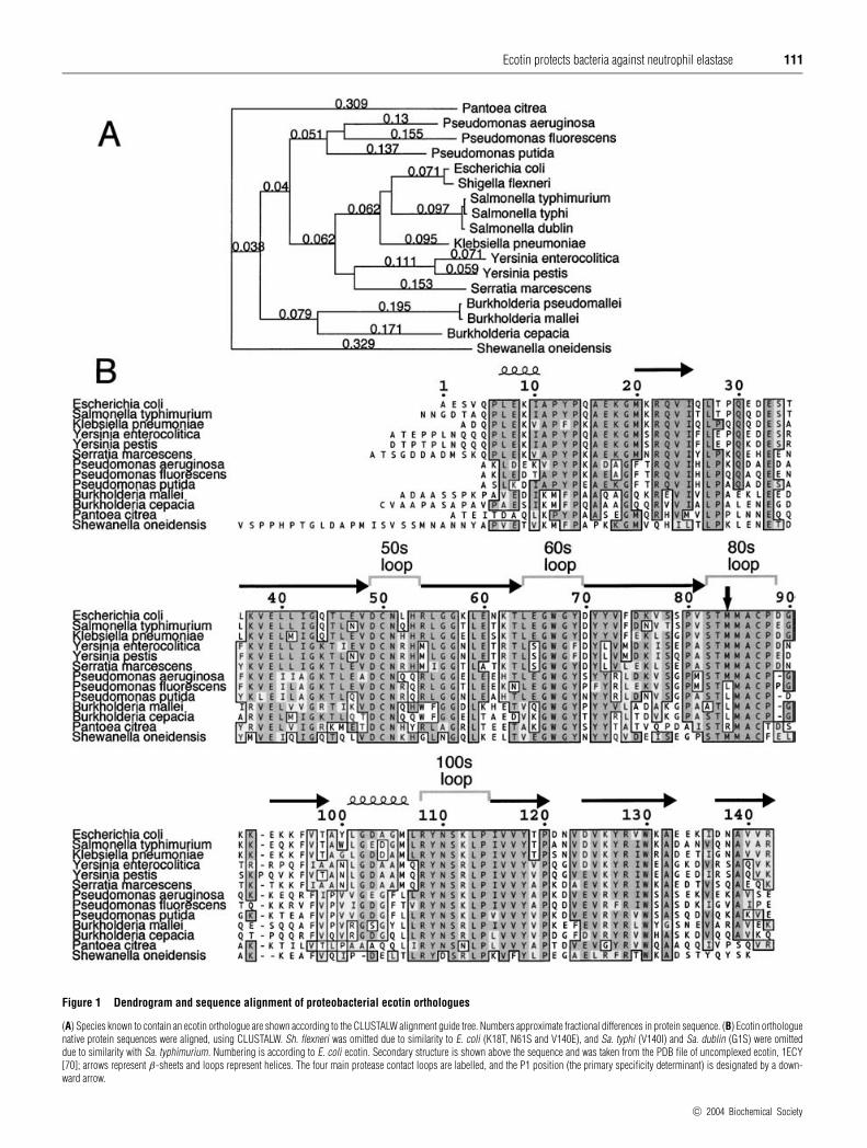
While ecotin did not appear to have significant homology to anyother E. coli proteins, orthologous proteins were found in severalproteobacteria (Figure 1A). Within the gamma subdivision,ecotin orthologues were found in the genuses Pseudomonas andShewanella, in addition to many members of the Enterobacter-iaceae family: Escherichia, Shigella, Salmonella, Yersinia,Serratia, Klebsiella and Pantoea. The beta subdivision genusBurkholderia contained an orthologue, but many other com-pletely sequenced proteobacteria did not, including Vibrio,
Haemophilus, Pasteurella, Helicobacter, Campylobacter, Neis-seria and Mesorhizobium.
In general, those species containing ecotin encountered mam-malian hosts, but were not obligate intracellular parasites. In thealpha subdivision, the genus Rickettsia appeared to be in the pro-cess of removing the gene [31], since Rickettsia conorii andR. sibirica, but not R. prowazekii, contained an ecotin pseudogene.Within the Enterobacteriaceae family, neither the plant pathogenErwinia, nor the insect endosymbionts, Buchnera aphidicola andWigglesworthia glossinidia, have the ecotin gene. Within thegenus Pseudomonas, ecotin is present in the species Ps. aerugi-nosa, Ps. putida, and Ps. fluorescens, but not Ps. syringae pv.tomato str. DC3000, which infects tomato and Arabidopsis. Thegenus Burkholderia, similar to Pseudomonas, represents ubiquit-ous environmental bacteria that can, in the case of Bu. cepacia, Bu.mallei, and Bu. pseudomallei, become opportunistic mammalianpathogens. Ecotin is present in those species, but not in B. fungo-rum, which belongs to a clade not implicated as pathogenic [32].A seeming exception to this rule is Pa. citrea, the phytopathogenresponsible for pink disease in pineapple [33]. The ecotin proteinsequences for Pa. citrea and Shewanella oneidensis are the mostdifferent from E. coli, even though both species are closer inevolutionary distance than Pseudomonas or Burkholderia, asmeasured by the 16 S rRNA sequence. This could signify adivergence in the protease target.
Outside of the proteobacteria, homology to ecotin was foundonly in the marine unicellular cyanobacterium Prochlorococcusmarinus and the protozoal parasites, Trypanasoma brucei andLeishmania major. Within Pr. marinus, ecotin is present only instrain MIT9313 and not in the smaller genomes of strains MED4and SS120, or in the closely related Synechococcus sp. strainWWH8102. Comparison of these genomes shows a dynamicprocess of genetic change involving gene loss, rearrangement andacquisition [34–36]. In MIT9313, the ecotin orthologue flanks onesuch region in flux that includes a group of nitrogen usage genes,some of which appear to have been gained through lateralgene transfer from proteobacteria [34]. This strain may have onlyrecently acquired ecotin, which may not be expressed or playany functional role in the cyanobacterium. Outside of bacteria,the homologous genes in T. brucei and L. major lack many of theconserved protease contact residues, as well as the disulphidebond between positions 50 and 87 that stabilizes the substrate-likeloop. The sequence conservation is mainly in the adjacent β-sheetresidues making up the protein core. Recombinantly expressedand purified T. brucei homologue protein had no detectableinhibitory activity (results not shown).
While the presence of ecotin in enterobacteria is consistentwith a putative protective function against digestive proteases inthe gut, such an explanation seems inadequate for such bacteriaas Pseudomonas, Burkholderia or Shewanella. However, a widernumber of these species would be expected to encounter themammalian immune system, suggesting the possibility of a targetprotease involved in immunity. Consistent with this notion arethe reports that NE plays a direct role in the killing of E. coli,K. pneumoniae, Shigella flexneri and Ps. aeruginosa, all specieswith ecotin orthologues [5–7,37].
Alignment of protein sequences
Complete DNA sequences were assembled for each of theorthologues, the signal sequence cleavage sites were predictedusing SignalP V1.1 [23], and the native protein sequences werealigned using CLUSTALW (Figure 1B). The Pa. citrea ecotinsequence is published here for the first time. The published se-quence of the PAO1 strain of Ps. aeruginosa contained an unusual
c© 2004 Biochemical Society
Ecotin protects bacteria against neutrophil elastase 111
Figure 1 Dendrogram and sequence alignment of proteobacterial ecotin orthologues
(A) Species known to contain an ecotin orthologue are shown according to the CLUSTALW alignment guide tree. Numbers approximate fractional differences in protein sequence. (B) Ecotin orthologuenative protein sequences were aligned, using CLUSTALW. Sh. flexneri was omitted due to similarity to E. coli (K18T, N61S and V140E), and Sa. typhi (V140I) and Sa. dublin (G1S) were omitteddue to similarity with Sa. typhimurium. Numbering is according to E. coli ecotin. Secondary structure is shown above the sequence and was taken from the PDB file of uncomplexed ecotin, 1ECY[70]; arrows represent β-sheets and loops represent helices. The four main protease contact loops are labelled, and the P1 position (the primary specificity determinant) is designated by a down-ward arrow.
c© 2004 Biochemical Society
112 C. T. Eggers and others
free cysteine at position 108, which was arginine in all otherspecies. Cloning the gene directly from PAO1 confirmed thepublished sequencing results, but sequencing of the cytotoxicPA103 strain of Ps. aeruginosa revealed the conserved arginineat that position; this latter sequence is used in the alignment.
Of the 142 residues of ecotin, 28 of them are completelyconserved among all species. Many of these are cysteine, glycineand proline residues that play important roles in defining thesecondary structure, whereas others have side chains involvedin core packing. However, about half of the conserved residuesare in the four surface loops that contact target proteases. Asexpected, the residues showing the most variation were generallysolvent exposed in edge strands of the β-sheet or in loops notcontacting the protease. Insertions or deletions were present atthe N-terminus and in the residues immediately following thesubstrate-like loop, two regions that were disordered in the ecotin–trypsin structure [19]. While the substrate-like 80s loop was wellconserved from the P3 to the P4′ positions (residues 82–88),suggesting a similar protease target, the important P1 residue(residue 84) showed some variation. The P1 methionine of E. coliecotin has been hypothesized to play a role in the pan-specificityof the inhibitor, since it is able to fit into many different primaryspecificity pockets [27]. Therefore, the P1 leucine of orthologuesfrom some Pseudomonas and Burkholderia species could indicatea different or more specific target. Likewise, the arginine at theP1 of Pa. citrea ecotin could indicate a trypsin-like target.
Searches for functionally linked proteins
Several lines of evidence suggest that ecotin orthologues donot share an endogenous target. First of all, ecotin has onlybeen shown to inhibit trypsin-fold serine proteases in the S1Asubfamily of clan PA, of which E. coli has no members [38,39].While E. coli does have three periplasmic proteases in theS1C family (DegP, DegQ and DegS), ecotin is known not toinhibit DegP [18], which forms a hexameric complex presumablyinaccessible to ecotin [40]. DegQ also is thought to form a largeoligomeric complex [41]. Ecotin has been shown not to inhibitthe following E. coli proteases: Do, Re, Mi, Fa, So, La, Ci, Pi, andproteases I, II, IV, V and VI [18,42]. Furthermore, no interactingE. coli proteins were detected when a periplasmic fraction waspassed over an ecotin–agarose affinity column (results not shown).
Finally, we utilized a comparative genomics approach to searchfor proteins with orthologues in the same set of fully sequencedgenomes as ecotin, since proteins involved in the same functionwould be expected to be maintained or eliminated from a genomein a correlated fashion [43]. Because ecotin is not in the COGdatabase, we utilized both the HOBACGEN database of proteinfamilies (release #10, Feb, 2002) [44] and the ComprehensiveMicrobial Resource (www.tigr.org) to conduct searches basedon taxonomic relationships. Genes were selected that containedhomologues in E. coli, Sa. typhimurium, Y. pestis and Ps.aeruginosa, but not in closely related proteobacteria lacking anecotin orthologue. The amino acid sequences of genes from thissubset of candidates were used for individual TBLASTN searchesof the microbial database. No genes were found with the samephylogenetic profile as ecotin, suggesting that ecotin orthologuesare not functionally linked to a common endogenous protein andstrengthening the case for an exogenous protease target.
Cloning, expression and purification of ecotin orthologues
To determine whether mammalian proteases, such as NE, maybe targets of ecotin in multiple bacterial species, we cloned theecotin genes from Y. pestis, Ps. aeruginosa and Pa. citrea. Unlike
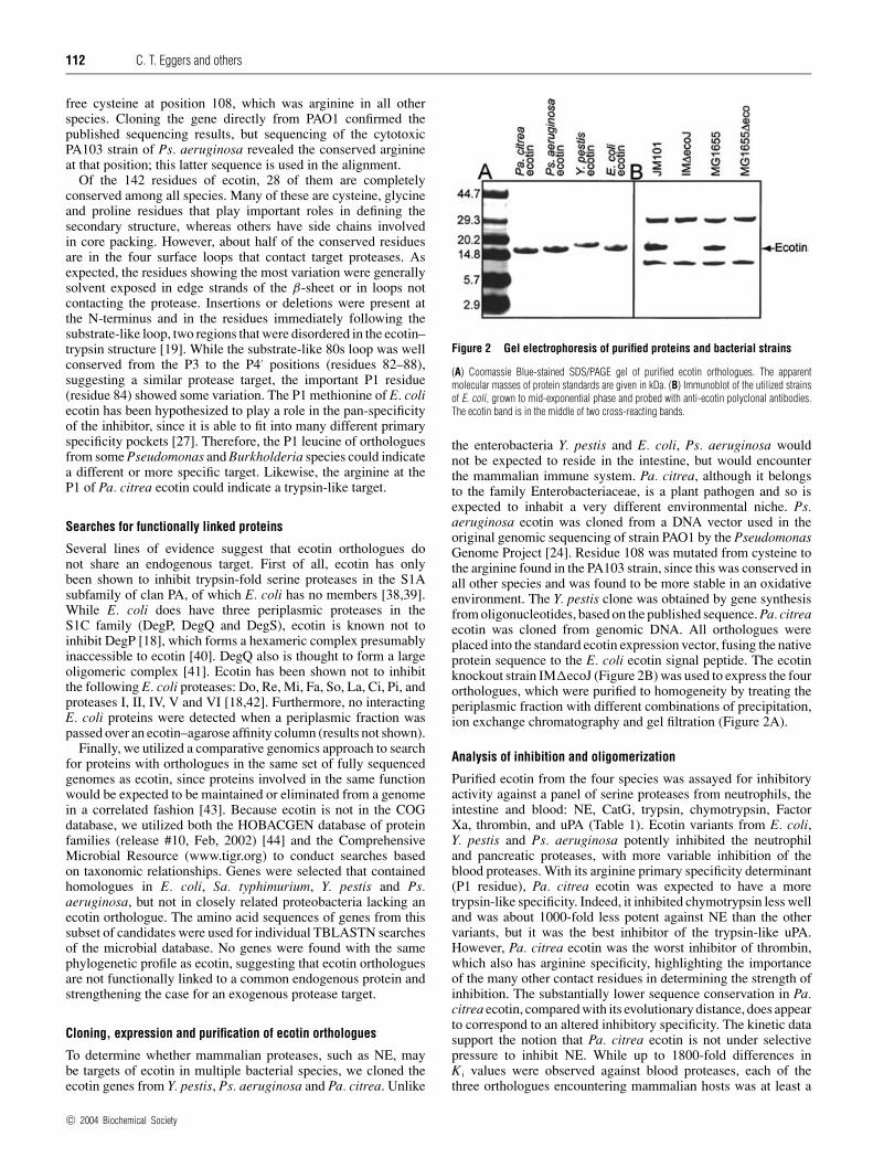
Figure 2 Gel electrophoresis of purified proteins and bacterial strains
(A) Coomassie Blue-stained SDS/PAGE gel of purified ecotin orthologues. The apparentmolecular masses of protein standards are given in kDa. (B) Immunoblot of the utilized strainsof E. coli, grown to mid-exponential phase and probed with anti-ecotin polyclonal antibodies.The ecotin band is in the middle of two cross-reacting bands.
the enterobacteria Y. pestis and E. coli, Ps. aeruginosa wouldnot be expected to reside in the intestine, but would encounterthe mammalian immune system. Pa. citrea, although it belongsto the family Enterobacteriaceae, is a plant pathogen and so isexpected to inhabit a very different environmental niche. Ps.aeruginosa ecotin was cloned from a DNA vector used in theoriginal genomic sequencing of strain PAO1 by the PseudomonasGenome Project [24]. Residue 108 was mutated from cysteine tothe arginine found in the PA103 strain, since this was conserved inall other species and was found to be more stable in an oxidativeenvironment. The Y. pestis clone was obtained by gene synthesisfrom oligonucleotides, based on the published sequence. Pa. citreaecotin was cloned from genomic DNA. All orthologues wereplaced into the standard ecotin expression vector, fusing the nativeprotein sequence to the E. coli ecotin signal peptide. The ecotinknockout strain IM�ecoJ (Figure 2B) was used to express the fourorthologues, which were purified to homogeneity by treating theperiplasmic fraction with different combinations of precipitation,ion exchange chromatography and gel filtration (Figure 2A).
Analysis of inhibition and oligomerization
Purified ecotin from the four species was assayed for inhibitoryactivity against a panel of serine proteases from neutrophils, theintestine and blood: NE, CatG, trypsin, chymotrypsin, FactorXa, thrombin, and uPA (Table 1). Ecotin variants from E. coli,Y. pestis and Ps. aeruginosa potently inhibited the neutrophiland pancreatic proteases, with more variable inhibition of theblood proteases. With its arginine primary specificity determinant(P1 residue), Pa. citrea ecotin was expected to have a moretrypsin-like specificity. Indeed, it inhibited chymotrypsin less welland was about 1000-fold less potent against NE than the othervariants, but it was the best inhibitor of the trypsin-like uPA.However, Pa. citrea ecotin was the worst inhibitor of thrombin,which also has arginine specificity, highlighting the importanceof the many other contact residues in determining the strength ofinhibition. The substantially lower sequence conservation in Pa.citrea ecotin, compared with its evolutionary distance, does appearto correspond to an altered inhibitory specificity. The kinetic datasupport the notion that Pa. citrea ecotin is not under selectivepressure to inhibit NE. While up to 1800-fold differences inK i values were observed against blood proteases, each of thethree orthologues encountering mammalian hosts was at least a
c© 2004 Biochemical Society

Ecotin protects bacteria against neutrophil elastase 113
Table 1 Protease inhibition by ecotin orthologues
The K i inhibitory constant was measured for ecotin orthologues from four species against a panel of serine proteases.
K i (nM) of ecotin
Protease Organism . . . E. coli Y. pestis Ps. aeruginosa Pa. citrea
NE 0.012 +− 0.004 0.016 +− 0.003 < 0.005 8.8 +− 0.4CatG < 0.005 < 0.005 < 0.005 < 0.005Trypsin < 0.001 < 0.001 < 0.001 < 0.001Chymotrypsin < 0.002 < 0.002 < 0.002 0.005 +− 0.002Factor Xa 0.0046 +− 0.001 0.23 +− 0.01 0.007 +− 0.001 0.22 +− 0.005Thrombin 770 +− 80 1400 +− 100 1500 +− 100 24 000 +− 3000uPA 670 +− 20 13 900 +− 1500 7.8 +− 0.4 1.1 +− 0.2
Table 2 Size-exclusion chromatography of ecotin orthologues
The four ecotin orthologues were run on an analytical gel filtration column either alone or complexed with an excess of trypsin. Predicted molecular mass was deduced from the amino acid sequence.Apparent molecular mass was calculated from comparison of elution volumes with known protein standards.
Ecotin orthologue Predicted mass (kDa) Uncomplexed elution volume (ml) Apparent mass (kDa) Complexed elution volume (ml) Apparent mass of complex (kDa)
E. coli 16.098 14.89 37.4 13.28 85.5Y. pestis 16.727 14.78 39.6 13.26 86.4Ps. aeruginosa 15.507 15.06 34.3 13.36 82.1Pa. citrea 15.800 15.06 34.3 13.37 81.7
low picomolar inhibitor of neutrophil and pancreatic proteases,indicating their potential as physiological targets.
Analytical gel filtration analysis showed that all four ortho-logues form the same oligomeric complexes (Table 2). On theirown, ecotin variants ran as homodimers, with apparent molecularmasses slightly higher than double the predicted monomeric massdue to the elongated shape of ecotin. When ecotin was pre-incubated with an excess of inactive rat trypsin, the complex ranwith an apparent molecular mass equal to that of a heterotetramer.
Ecotin protects E. coli from NE
Given the species distribution and inhibitory activities of ecotinorthologues, NE appeared to be a promising candidate for aphysiological target of ecotin. To test whether ecotin would protectbacteria from NE, we disrupted, by homologous recombination,the ecotin gene in E. coli with a marker for chloramphenicolresistance. The disruption was moved into the K12 sequence strainMG1655 by three rounds of P1 bacteriophage transduction, andthis eco− strain was verified by PCR and by immunoblotting(Figure 2B).
NE-mediated killing experiments were performed essentially asdescribed previously [5], with some modification (see the Experi-mental section). At concentrations of 3.4 µM NE (100 µg/ml),substantial killing of E. coli was observed compared with thebuffer control, and the eco− strain was significantly more sensitiveto NE. After 6 h of incubation with NE, 2.76 +− 0.04 orders ofmagnitude (570-fold) fewer viable ecotin-deficient E. coli cellsremained than WT cells (Figure 3A).
Transforming into eco− E. coli a plasmid overexpressingeither WT ecotin or an inactive ecotin variant showed that thisdifference in sensitivity was due to inhibition by ecotin, ratherthan any extraneous strain differences, such as expression of theantibiotic marker. The K i for NE of this inactive variant, whichhas a disrupted primary binding site and dimerization interface,was greater than 10 µM, or 106-fold worse than WT ecotin.Expression of WT ecotin made the cells more resistant to NEthan expression of inactive ecotin, yielding 2.1 +− 0.4 orders of
magnitude (140-fold) higher cell counts (Figure 3B). Since theecotin-deficient strain contained an antibiotic-resistance marker,a direct competition could be performed between the two strains inone test tube, quantifying viable bacteria by plating both with andwithout chloramphenicol. This procedure assured that each strainwas exposed to the exact same environment. Again, the eco− strainyielded 2.6 +− 0.2 orders of magnitude (390-fold) fewer coloniesthan WT E. coli after 6 h incubation with NE (Figure 3C).
The observation that the presence of ecotin has an effect duringdirect competition of the two strains requires that ecotin playsits protective role while localized to the bacterium producing it,rather than after release into solution, where it would inhibit NEactivity against both strains. Since ecotin is found in the periplasm,this strongly suggests that NE is crossing the outer membrane andthat a bactericidal or bacteriostatic effect is due to periplasmicNE activity. If this were the case, one would expect an ecotin-independent outer membrane permeability to precede killing.Outer membrane permeability is generally accompanied bysensitivity to detergents, such as SDS, and to certain hydrophobicantibiotics, such as rifampicin and actinomycin D [45]. In orderto observe how the presence of ecotin affected the rate of bothouter membrane damage and cell killing, we followed a timecourse of NE treatment of an equal mixture of eco− and WT E.coli, monitoring cell viability on agar plates with and withoutrifampicin as a gauge of outer membrane permeability.
It was immediately apparent that the majority of the effect ofecotin was due to different lengths of time taken to recover andstart growing again following NE treatment (Figure 4A). After30 min, net killing of WT E. coli essentially stopped, whereasthe ecotin-deficient strain continued to be killed for the first fewhours. At this point, both strains began growing at the same rate,even though virtually all of the cells had damaged, permeableouter membranes, as judged by sensitivity to rifampicin. The outermembrane was permeabilized at a faster rate than killing, andsensitivity to 3% SDS showed the same effect (results notshown). Although the greater amount of new growth by WT E. coliled to somewhat more rifampicin-resistant bacteria, the fractionof cells resistant to rifampicin decreased at identical rates for
c© 2004 Biochemical Society
114 C. T. Eggers and others
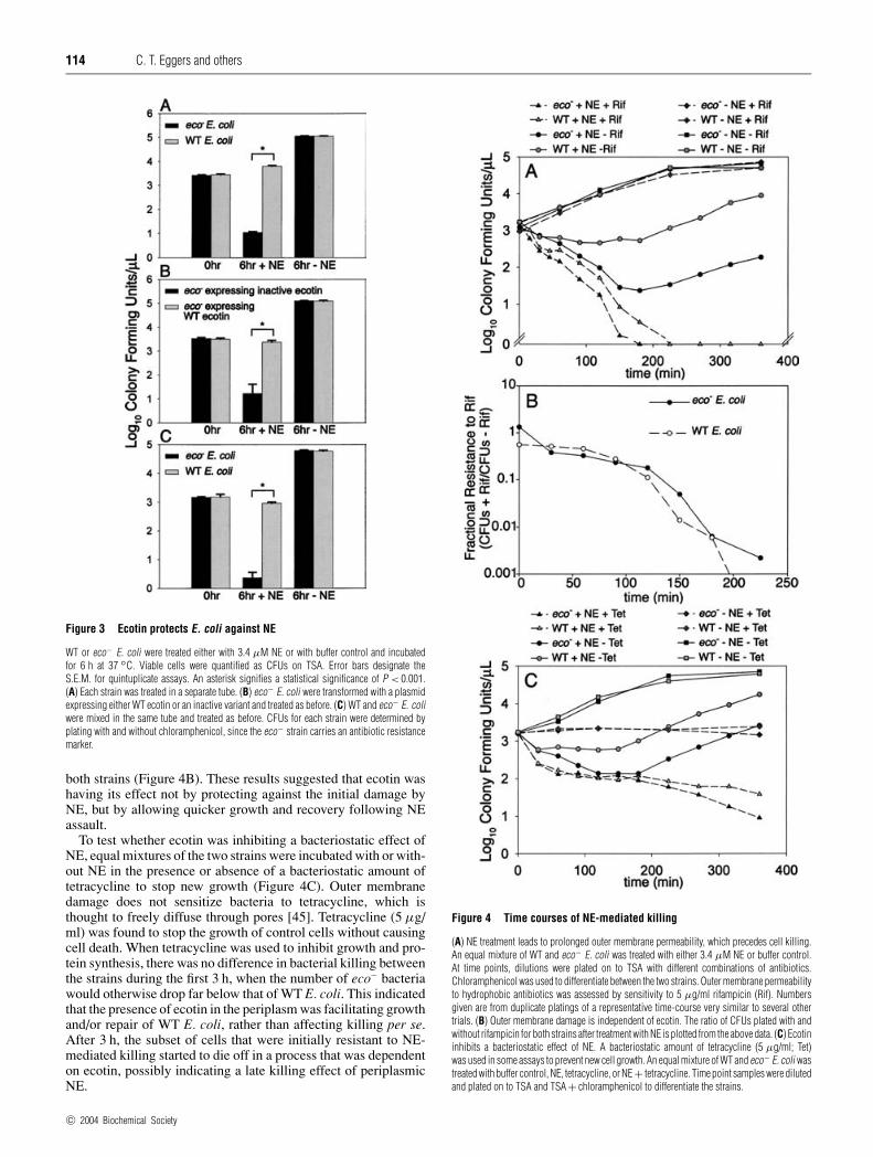
Figure 3 Ecotin protects E. coli against NE
WT or eco− E. coli were treated either with 3.4 µM NE or with buffer control and incubatedfor 6 h at 37 ◦C. Viable cells were quantified as CFUs on TSA. Error bars designate theS.E.M. for quintuplicate assays. An asterisk signifies a statistical significance of P < 0.001.(A) Each strain was treated in a separate tube. (B) eco− E. coli were transformed with a plasmidexpressing either WT ecotin or an inactive variant and treated as before. (C) WT and eco− E. coliwere mixed in the same tube and treated as before. CFUs for each strain were determined byplating with and without chloramphenicol, since the eco− strain carries an antibiotic resistancemarker.
both strains (Figure 4B). These results suggested that ecotin washaving its effect not by protecting against the initial damage byNE, but by allowing quicker growth and recovery following NEassault.
To test whether ecotin was inhibiting a bacteriostatic effect ofNE, equal mixtures of the two strains were incubated with or with-out NE in the presence or absence of a bacteriostatic amount oftetracycline to stop new growth (Figure 4C). Outer membranedamage does not sensitize bacteria to tetracycline, which isthought to freely diffuse through pores [45]. Tetracycline (5 µg/ml) was found to stop the growth of control cells without causingcell death. When tetracycline was used to inhibit growth and pro-tein synthesis, there was no difference in bacterial killing betweenthe strains during the first 3 h, when the number of eco− bacteriawould otherwise drop far below that of WT E. coli. This indicatedthat the presence of ecotin in the periplasm was facilitating growthand/or repair of WT E. coli, rather than affecting killing per se.After 3 h, the subset of cells that were initially resistant to NE-mediated killing started to die off in a process that was dependenton ecotin, possibly indicating a late killing effect of periplasmicNE.
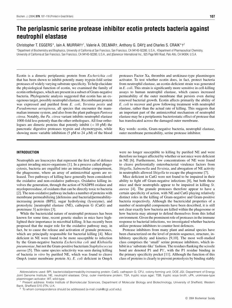
Figure 4 Time courses of NE-mediated killing
(A) NE treatment leads to prolonged outer membrane permeability, which precedes cell killing.An equal mixture of WT and eco− E. coli was treated with either 3.4 µM NE or buffer control.At time points, dilutions were plated on to TSA with different combinations of antibiotics.Chloramphenicol was used to differentiate between the two strains. Outer membrane permeabilityto hydrophobic antibiotics was assessed by sensitivity to 5 µg/ml rifampicin (Rif). Numbersgiven are from duplicate platings of a representative time-course very similar to several othertrials. (B) Outer membrane damage is independent of ecotin. The ratio of CFUs plated with andwithout rifampicin for both strains after treatment with NE is plotted from the above data. (C) Ecotininhibits a bacteriostatic effect of NE. A bacteriostatic amount of tetracycline (5 µg/ml; Tet)was used in some assays to prevent new cell growth. An equal mixture of WT and eco− E. coli wastreated with buffer control, NE, tetracycline, or NE + tetracycline. Time point samples were dilutedand plated on to TSA and TSA + chloramphenicol to differentiate the strains.
c© 2004 Biochemical Society
Ecotin protects bacteria against neutrophil elastase 115
DISCUSSION
As part of the immune response to bacterial infection, neutrophilsdirect a large arsenal of antimicrobial agents against invading bac-teria. Many of these agents demonstrate specificity in their anti-microbial profiles, showing bactericidal or bacteriostatic effectsmainly against certain subsets of bacteria or fungi. The importantrole of proteases in neutrophil-mediated killing has been demons-trated by recent genetic studies in mice. The serine protease NEhas been shown to be important for neutrophil killing of suchGram-negative bacteria as Escherichia, Shigella and Klebsiella.While the exact mechanism of protease-mediated killing remainsunknown, NE cleavage of OmpA in E. coli appears to be important[6]. OmpA is thought to be important in maintaining outermembrane stability, and OmpA-deficient E. coli are more sensitiveto environmental stresses [46].
Given the killing specificity of NE, it is interesting to notethat those bacteria that have shown sensitivity to NE belong tothe small subset of bacteria containing the periplasmic proteaseinhibitor ecotin. This raises the question of what the physiologicaltarget of ecotin is and whether it plays a role in protecting bacteriaagainst NE. Do only bacteria susceptible to NE require ecotin, andwhat would make certain species naturally resistant to NE? Ecotinorthologues appear to be present mainly in straight-rod Gram-negative bacteria, as opposed to curved, helical, or coccobacillaryrods. Certain cell morphologies may be more susceptible to NEcleavage of OmpA and the concomitant breakdown of the cell en-velope. It is known that OmpA is important for cell morphology[47], and it is possible that sequence differences in the extracellu-lar loops of OmpA could lead to differential susceptibility to NE.
Bacteria may also be less sensitive to the immune system orexogenous proteases during certain growth phases. For example,in stressful environments, bacteria tend to form biofilms, whichare more protected against host defences, such as neutrophils[48–50]. In regulating the formation of biofilms, bacteria appearto monitor the concentration of acetyl phosphate as an indicator ofthe nutritional status of the cell [51]. Interestingly, ecotin and theperiplasmic inhibitor of vertebrate lysozyme, ivy, were two of 25genes significantly upregulated by higher concentrations of acetylphosphate, which tends to promote the free-living planktonic statethat is more susceptible to neutrophils [51].
In order to gain insight into what exogenous targets ecotin andits orthologues may inhibit, we cloned, expressed and purifiedecotin from three additional species: Y. pestis, Ps. aeruginosa andPa. citrea. All four ecotin variants were found to form homodimersand to inhibit trypsin in a heterotetrameric complex (Table 2).By allowing the inhibitor to make contacts with the proteaseat two distinct protein interfaces, dimerization has the effect ofmaking inhibition more potent and less specific [20]. For thatreason, we expected the orthologues to share relatively broadinhibitory profiles. However, Pa. citrea, as a plant pathogen,was expected to inhabit a different environment from the otherthree and so potentially encounter different proteases. Its genesequence shows a fair amount of divergence in the protease contactloops, especially in having arginine as the primary specificitydeterminant (P1), rather than the methionine observed in the otherthree. While all variants were tight inhibitors of the pancreaticproteases trypsin and chymotrypsin and the neutrophil proteaseCatG, Pa. citrea ecotin was about 1000-fold less potent againstNE, which the others inhibited tightly (Table 1).
Interestingly, a P1 methionine is also present in the naturalphysiological inhibitor of NE, α1-proteinase inhibitor (α1-antitrypsin). Imbalance between NE and its inhibitor is seennot only in α1-proteinase inhibitor deficiency, but also in cysticfibrosis, which is typified by chronic respiratory infection in
the presence of a vast influx of neutrophils [52]. Degranulatingneutrophils lead to very high concentrations of elastase, averaging100 µg/ml in cystic fibrosis sputum, the same concentrationused in our cell-killing assays [53,54]. The vast majority ofthe NE is free and active, since the physiological inhibitorstend to be degraded or otherwise inactivated [54,55]. The mostimportant pathogens causing chronic infections in adult cysticfibrosis patients are Ps. aeruginosa and Bu. cepacia, two ecotin-containing species [56,57]. More broadly, such species as K.pneumoniae, E. coli, Se. marcescens and Ps. aeruginosa areoften responsible for nosocomial respiratory infections, especiallyin neonatal and elderly populations [58–61]. Many of thesepulmonary infections are marked not only by increased numbersof neutrophils, but also by high levels of free uninhibited elastase[62]. Even when excess α1-proteinase inhibitor is present in thelung, release of granules from neutrophils can lead to bursts ofvery high concentrations of free NE immediately surroundingthe neutrophil [63,64]. Therefore, the respiratory system couldrepresent a location where ecotin-containing bacteria encounternot only neutrophils, but also high concentrations of free NE.
Some of the ecotin-containing bacteria are known to be suscep-tible to NE-mediated killing, and ecotin inhibits NE, but doesecotin localized to the periplasm actually protect the bacteriumagainst external NE? To answer this question, we generated anecotin-deficient strain of E. coli and treated it and an isogenicWT strain with purified NE. After 6 h of incubation with NE,cells producing functional ecotin were present at 140- to 570-foldhigher numbers than those without ecotin (Figure 3). This dif-ference was due to the inhibitory activity of ecotin (Figure 3B) andinvolved protein that was localized, rather than released into bulksolution (Figure 3C). The concentrations of any released ecotinwould be so low in our assays that it could not inhibit the activityof the vast excess of NE. This situation probably holds in vivo, aswell. The number of NE molecules in a single neutrophil is about3 × 107 [65,66], which is 10000-fold higher than the 3000 ecotinmolecules per E. coli estimated from immunoblotting (results notshown). Therefore, it seems likely that ecotin in the periplasm issituated to inhibit the small fraction of NE molecules that manageto permeate the outer membrane.
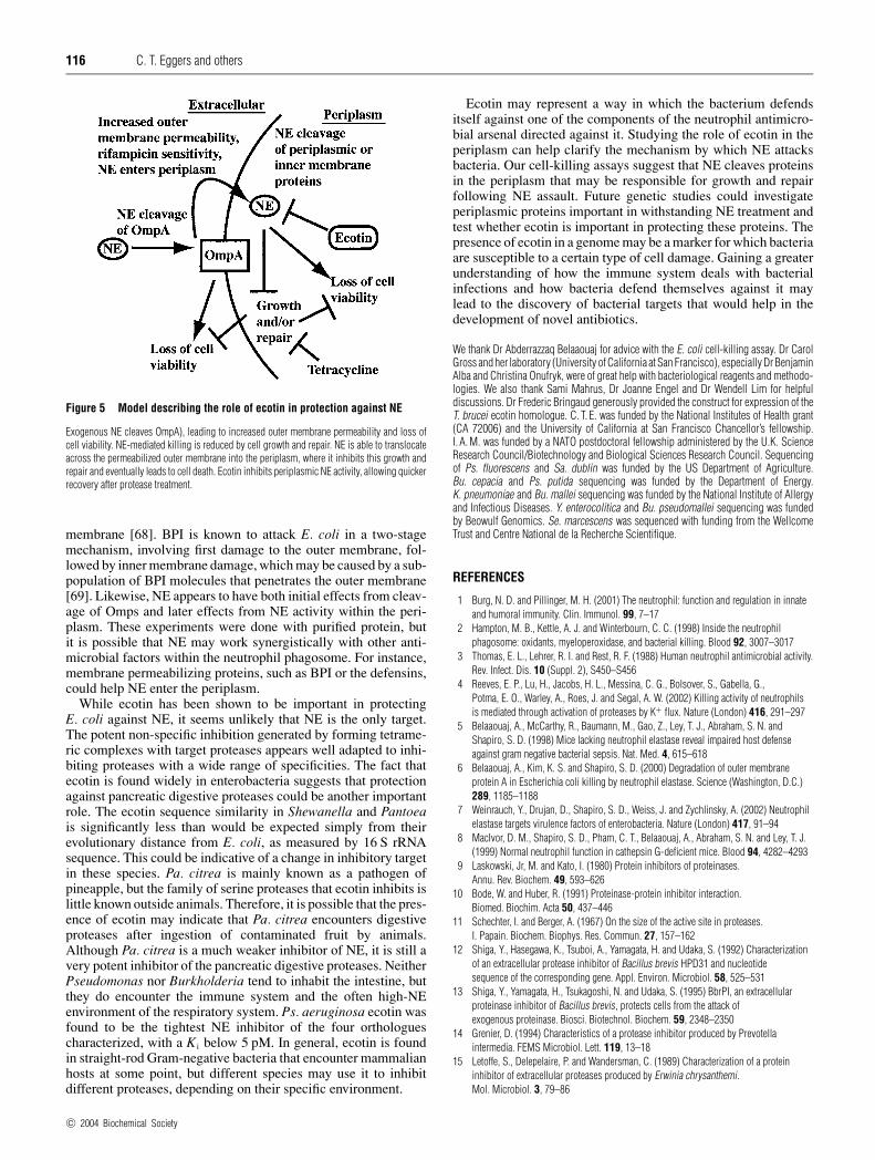
Time courses of bacterial killing demonstrated that the effect ofecotin was in promoting growth after NE assault. The presenceof ecotin did not affect the rate of outer membrane damage by NE,as assayed by sensitivity to the hydrophobic antibiotic rifampicin(Figures 4A and 4B). When a bacteriostatic amount of tetracyclinewas added in order to prevent new growth, the strain differencein sensitivity disappeared, although late ecotin-dependent deathdid occur (Figure 4C). Taken together, these results suggest thefollowing model for NE-mediated killing of E. coli (Figure 5). NEinitially cleaves OmpA, leading to outer membrane damage andincreased permeability. Such damage can lead directly to loss ofcell viability, although in any population of bacteria, not all cellsappear to be equally sensitive. Killing by NE is balanced by growthand repair of cells. However, permeability of the outer membraneallows NE to translocate into the periplasm, where it can inhibitthese processes, leading to a bacteriostatic effect. Ecotin inhibitsthis periplasmic activity, allowing quicker recovery following NEtreatment.
While the periplasmic targets of NE remain unknown, theycould include proteins involved in envelope stress responses.Several pathways exist in E. coli, including Sigma(E) and Cpx,for transmitting periplasmic stress signals to promote cell wallbiosynthesis and protein folding [67]. The OmpR pathway ofosmotic regulation of outer membrane proteins has been shownto be essential for allowing E. coli to withstand low doses of BPIand for preventing the progression to lethal damage of the inner
c© 2004 Biochemical Society
116 C. T. Eggers and others
Figure 5 Model describing the role of ecotin in protection against NE
Exogenous NE cleaves OmpA), leading to increased outer membrane permeability and loss ofcell viability. NE-mediated killing is reduced by cell growth and repair. NE is able to translocateacross the permeabilized outer membrane into the periplasm, where it inhibits this growth andrepair and eventually leads to cell death. Ecotin inhibits periplasmic NE activity, allowing quickerrecovery after protease treatment.
membrane [68]. BPI is known to attack E. coli in a two-stagemechanism, involving first damage to the outer membrane, fol-lowed by inner membrane damage, which may be caused by a sub-population of BPI molecules that penetrates the outer membrane[69]. Likewise, NE appears to have both initial effects from cleav-age of Omps and later effects from NE activity within the peri-plasm. These experiments were done with purified protein, butit is possible that NE may work synergistically with other anti-microbial factors within the neutrophil phagosome. For instance,membrane permeabilizing proteins, such as BPI or the defensins,could help NE enter the periplasm.
While ecotin has been shown to be important in protectingE. coli against NE, it seems unlikely that NE is the only target.The potent non-specific inhibition generated by forming tetrame-ric complexes with target proteases appears well adapted to inhi-biting proteases with a wide range of specificities. The fact thatecotin is found widely in enterobacteria suggests that protectionagainst pancreatic digestive proteases could be another importantrole. The ecotin sequence similarity in Shewanella and Pantoeais significantly less than would be expected simply from theirevolutionary distance from E. coli, as measured by 16 S rRNAsequence. This could be indicative of a change in inhibitory targetin these species. Pa. citrea is mainly known as a pathogen ofpineapple, but the family of serine proteases that ecotin inhibits islittle known outside animals. Therefore, it is possible that the pres-ence of ecotin may indicate that Pa. citrea encounters digestiveproteases after ingestion of contaminated fruit by animals.Although Pa. citrea is a much weaker inhibitor of NE, it is still avery potent inhibitor of the pancreatic digestive proteases. NeitherPseudomonas nor Burkholderia tend to inhabit the intestine, butthey do encounter the immune system and the often high-NEenvironment of the respiratory system. Ps. aeruginosa ecotin wasfound to be the tightest NE inhibitor of the four orthologuescharacterized, with a K i below 5 pM. In general, ecotin is foundin straight-rod Gram-negative bacteria that encounter mammalianhosts at some point, but different species may use it to inhibitdifferent proteases, depending on their specific environment.
Ecotin may represent a way in which the bacterium defendsitself against one of the components of the neutrophil antimicro-bial arsenal directed against it. Studying the role of ecotin in theperiplasm can help clarify the mechanism by which NE attacksbacteria. Our cell-killing assays suggest that NE cleaves proteinsin the periplasm that may be responsible for growth and repairfollowing NE assault. Future genetic studies could investigateperiplasmic proteins important in withstanding NE treatment andtest whether ecotin is important in protecting these proteins. Thepresence of ecotin in a genome may be a marker for which bacteriaare susceptible to a certain type of cell damage. Gaining a greaterunderstanding of how the immune system deals with bacterialinfections and how bacteria defend themselves against it maylead to the discovery of bacterial targets that would help in thedevelopment of novel antibiotics.
We thank Dr Abderrazzaq Belaaouaj for advice with the E. coli cell-killing assay. Dr CarolGross and her laboratory (University of California at San Francisco), especially Dr BenjaminAlba and Christina Onufryk, were of great help with bacteriological reagents and methodo-logies. We also thank Sami Mahrus, Dr Joanne Engel and Dr Wendell Lim for helpfuldiscussions. Dr Frederic Bringaud generously provided the construct for expression of theT. brucei ecotin homologue. C. T. E. was funded by the National Institutes of Health grant(CA 72006) and the University of California at San Francisco Chancellor’s fellowship.I. A. M. was funded by a NATO postdoctoral fellowship administered by the U.K. ScienceResearch Council/Biotechnology and Biological Sciences Research Council. Sequencingof Ps. fluorescens and Sa. dublin was funded by the US Department of Agriculture.Bu. cepacia and Ps. putida sequencing was funded by the Department of Energy.K. pneumoniae and Bu. mallei sequencing was funded by the National Institute of Allergyand Infectious Diseases. Y. enterocolitica and Bu. pseudomallei sequencing was fundedby Beowulf Genomics. Se. marcescens was sequenced with funding from the WellcomeTrust and Centre National de la Recherche Scientifique.
REFERENCES
1 Burg, N. D. and Pillinger, M. H. (2001) The neutrophil: function and regulation in innateand humoral immunity. Clin. Immunol. 99, 7–17
2 Hampton, M. B., Kettle, A. J. and Winterbourn, C. C. (1998) Inside the neutrophilphagosome: oxidants, myeloperoxidase, and bacterial killing. Blood 92, 3007–3017
3 Thomas, E. L., Lehrer, R. I. and Rest, R. F. (1988) Human neutrophil antimicrobial activity.Rev. Infect. Dis. 10 (Suppl. 2), S450–S456
4 Reeves, E. P., Lu, H., Jacobs, H. L., Messina, C. G., Bolsover, S., Gabella, G.,Potma, E. O., Warley, A., Roes, J. and Segal, A. W. (2002) Killing activity of neutrophilsis mediated through activation of proteases by K+ flux. Nature (London) 416, 291–297
5 Belaaouaj, A., McCarthy, R., Baumann, M., Gao, Z., Ley, T. J., Abraham, S. N. andShapiro, S. D. (1998) Mice lacking neutrophil elastase reveal impaired host defenseagainst gram negative bacterial sepsis. Nat. Med. 4, 615–618
6 Belaaouaj, A., Kim, K. S. and Shapiro, S. D. (2000) Degradation of outer membraneprotein A in Escherichia coli killing by neutrophil elastase. Science (Washington, D.C.)289, 1185–1188
7 Weinrauch, Y., Drujan, D., Shapiro, S. D., Weiss, J. and Zychlinsky, A. (2002) Neutrophilelastase targets virulence factors of enterobacteria. Nature (London) 417, 91–94
8 MacIvor, D. M., Shapiro, S. D., Pham, C. T., Belaaouaj, A., Abraham, S. N. and Ley, T. J.(1999) Normal neutrophil function in cathepsin G-deficient mice. Blood 94, 4282–4293
9 Laskowski, Jr, M. and Kato, I. (1980) Protein inhibitors of proteinases.Annu. Rev. Biochem. 49, 593–626
10 Bode, W. and Huber, R. (1991) Proteinase-protein inhibitor interaction.Biomed. Biochim. Acta 50, 437–446
11 Schechter, I. and Berger, A. (1967) On the size of the active site in proteases.I. Papain. Biochem. Biophys. Res. Commun. 27, 157–162
12 Shiga, Y., Hasegawa, K., Tsuboi, A., Yamagata, H. and Udaka, S. (1992) Characterizationof an extracellular protease inhibitor of Bacillus brevis HPD31 and nucleotidesequence of the corresponding gene. Appl. Environ. Microbiol. 58, 525–531
13 Shiga, Y., Yamagata, H., Tsukagoshi, N. and Udaka, S. (1995) BbrPI, an extracellularproteinase inhibitor of Bacillus brevis, protects cells from the attack ofexogenous proteinase. Biosci. Biotechnol. Biochem. 59, 2348–2350
14 Grenier, D. (1994) Characteristics of a protease inhibitor produced by Prevotellaintermedia. FEMS Microbiol. Lett. 119, 13–18
15 Letoffe, S., Delepelaire, P. and Wandersman, C. (1989) Characterization of a proteininhibitor of extracellular proteases produced by Erwinia chrysanthemi.Mol. Microbiol. 3, 79–86
c© 2004 Biochemical Society
Ecotin protects bacteria against neutrophil elastase 117
16 Duong, F., Lazdunski, A., Cami, B. and Murgier, M. (1992) Sequence of a cluster of genescontrolling synthesis and secretion of alkaline protease in Pseudomonas aeruginosa:relationships to other secretory pathways. Gene 121, 47–54
17 Maurizi, M. R. (1992) Proteases and protein degradation in Escherichia coli. Experientia48, 178–201
18 Chung, C. H., Ives, H. E., Almeda, S. and Goldberg, A. L. (1983) Purification fromEscherichia coli of a periplasmic protein that is a potent inhibitor of pancreatic proteases.J. Biol. Chem. 258, 11032–11038
19 McGrath, M. E., Erpel, T., Bystroff, C. and Fletterick, R. J. (1994) Macromolecularchelation as an improved mechanism of protease inhibition: structure of the ecotin–trypsin complex. EMBO J. 13, 1502–1507
20 Eggers, C. T., Wang, S. X., Fletterick, R. J. and Craik, C. S. (2001) The role of ecotindimerization in protease inhibition. J. Mol. Biol. 308, 975–991
21 Seymour, J. L., Lindquist, R. N., Dennis, M. S., Moffat, B., Yansura, D., Reilly, D.,Wessinger, M. E. and Lazarus, R. A. (1994) Ecotin is a potent anticoagulant and reversibletight-binding inhibitor of factor Xa. Biochemistry 33, 3949–3958
22 Ulmer, J. S., Lindquist, R. N., Dennis, M. S. and Lazarus, R. A. (1995) Ecotin is a potentinhibitor of the contact system proteases factor XIIa and plasma kallikrein. FEBS Lett.365, 159–163
23 Nielsen, H., Engelbrecht, J., Brunak, S. and von Heijne, G. (1997) Identification ofprokaryotic and eukaryotic signal peptides and prediction of their cleavage sites.Protein Eng. 10, 1–6
24 Stover, C. K., Pham, X. Q., Erwin, A. L., Mizoguchi, S. D., Warrener, P., Hickey, M. J.,Brinkman, F. S., Hufnagle, W. O., Kowalik, D. J., Lagrou, M. et al. (2000) Completegenome sequence of Pseudomonas aeruginosa PA01, an opportunistic pathogen.Nature (London) 406, 959–964
25 McGrath, M. E., Erpel, T., Browner, M. F. and Fletterick, R. J. (1991) Expression of theprotease inhibitor ecotin and its co-crystallization with trypsin. J. Mol. Biol. 222,139–142
26 Yang, S. Q., Wang, C. I., Gillmor, S. A., Fletterick, R. J. and Craik, C. S. (1998) Ecotin:a serine protease inhibitor with two distinct and interacting binding sites. J. Mol. Biol.279, 945–957
27 McGrath, M. E., Hines, W. M., Sakanari, J. A., Fletterick, R. J. and Craik, C. S. (1991) Thesequence and reactive site of ecotin. A general inhibitor of pancreatic serine proteasesfrom Escherichia coli. J. Biol. Chem. 266, 6620–6625
28 Murray, I. A., Hawkins, A. R., Keyte, J. W. and Shaw, W. V. (1988) Nucleotide sequenceanalysis and overexpression of the gene encoding a type III chloramphenicolacetyltransferase. Biochem. J. 252, 173–179
29 Slater, S. and Maurer, R. (1993) Simple phagemid-based system for generating allelereplacements in Escherichia coli. J. Bacteriol. 175, 4260–4262
30 Gill, S. C. and von Hippel, P. H. (1989) Calculation of protein extinction coefficients fromamino acid sequence data. Anal. Biochem. 182, 319–326
31 Ogata, H., Audic, S., Renesto-Audiffren, P., Fournier, P. E., Barbe, V., Samson, D.,Roux, V., Cossart, P., Weissenbach, J., Claverie, J. M. and Raoult, D. (2001) Mechanismsof evolution in Rickettsia conorii and R. prowazekii. Science (Washington, D.C.) 293,2093–2098
32 Fain, M. G. and Haddock, J. D. (2001) Phenotypic and phylogenetic characterization ofBurkholderia (Pseudomonas) sp. strain LB400. Curr. Microbiol. 42, 269–275
33 Cha, J. S., Pujol, C. and Kado, C. I. (1997) Identification and characterization of a Pantoeacitrea gene encoding glucose dehydrogenase that is essential for causing pink disease ofpineapple. Appl. Environ. Microbiol. 63, 71–76
34 Rocap, G., Larimer, F. W., Lamerdin, J., Malfatti, S., Chain, P., Ahlgren, N. A., Arellano, A.,Coleman, M., Hauser, L., Hess, W. R. et al. (2003) Genome divergence in twoProchlorococcus ecotypes reflects oceanic niche differentiation. Nature (London) 424,1042–1047
35 Palenik, B., Brahamsha, B., Larimer, F. W., Land, M., Hauser, L., Chain, P., Lamerdin, J.,Regala, W., Allen, E. E., McCarren, J. et al. (2003) The genome of a motile marineSynechococcus. Nature (London) 424, 1037–1042
36 Dufresne, A., Salanoubat, M., Partensky, F., Artiguenave, F., Axmann, I. M., Barbe, V.,Duprat, S., Galperin, M. Y., Koonin, E. V., Le Gall, F. et al. (2003) Genome sequence of thecyanobacterium Prochlorococcus marinus SS120, a nearly minimal oxyphototrophicgenome. Proc. Natl. Acad. Sci. U.S.A. 100, 10020–10025
37 Belaaouaj, A. (2002) Neutrophil elastase-mediated killing of bacteria: lessons fromtargeted mutagenesis. Microbes Infect. 4, 1259–1264
38 Rawlings, N. D., O’Brien, E. and Barrett, A. J. (2002) MEROPS: the protease database.Nucleic Acids Res. 30, 343–346
39 Barrett, A., Rawlings, N. D. and Woessner, J. F. (eds) (1998) Handbook of ProteolyticEnzymes, Academic Press, London
40 Krojer, T., Garrido-Franco, M., Huber, R., Ehrmann, M. and Clausen, T. (2002) Crystalstructure of DegP (HtrA) reveals a new protease-chaperone machine. Nature (London)416, 455–459
41 Kolmar, H., Waller, P. R. and Sauer, R. T. (1996) The DegP and DegQ periplasmicendoproteases of Escherichia coli: specificity for cleavage sites and substrateconformation. J. Bacteriol. 178, 5925–5929
42 Palmer, S. M. and St John, A. C. (1987) Characterization of a membrane-associatedserine protease in Escherichia coli. J. Bacteriol. 169, 1474–1479
43 Pellegrini, M., Marcotte, E. M., Thompson, M. J., Eisenberg, D. and Yeates, T. O. (1999)Assigning protein functions by comparative genome analysis: protein phylogeneticprofiles. Proc. Natl. Acad. Sci. U.S.A. 96, 4285–4288
44 Perriere, G., Duret, L. and Gouy, M. (2000) HOBACGEN: database system for comparativegenomics in bacteria. Genome Res. 10, 379–385
45 Vaara, M. (1992) Agents that increase the permeability of the outer membrane.Microbiol. Rev. 56, 395–411
46 Wang, Y. (2002) The function of OmpA in Escherichia coli. Biochem. Biophys.Res. Commun. 292, 396–401
47 Sonntag, I., Schwarz, H., Hirota, Y. and Henning, U. (1978) Cell envelope and shape ofEscherichia coli: multiple mutants missing the outer membrane lipoprotein and othermajor outer membrane proteins. J. Bacteriol. 136, 280–285
48 Mah, T. F. and O’Toole, G. A. (2001) Mechanisms of biofilm resistance to antimicrobialagents. Trends Microbiol. 9, 34–39
49 Kharazmi, A. (1991) Mechanisms involved in the evasion of the host defence byPseudomonas aeruginosa. Immunol. Lett. 30, 201–205
50 Hume, E. B., Stapleton, F. and Willcox, M. D. (2003) Evasion of cellular ocular defensesby contact lens isolates of Serratia marcescens. Eye Contact Lens 29, 108–112
51 Wolfe, A. J., Chang, D. E., Walker, J. D., Seitz-Partridge, J. E., Vidaurri, M. D.,Lange, C. F., Pruss, B. M., Henk, M. C., Larkin, J. C. and Conway, T. (2003) Evidencethat acetyl phosphate functions as a global signal during biofilm development.Mol. Microbiol. 48, 977–988
52 Doring, G. (1999) Serine proteinase inhibitor therapy in α1-antitrypsin inhibitordeficiency and cystic fibrosis. Pediatr. Pulmonol. 28, 363–375
53 Witko-Sarsat, V., Halbwachs-Mecarelli, L., Schuster, A., Nusbaum, P., Ueki, I.,Canteloup, S., Lenoir, G., Descamps-Latscha, B. and Nadel, J. A. (1999) Proteinase 3,a potent secretagogue in airways, is present in cystic fibrosis sputum. Am. J. Respir.Cell Mol. Biol. 20, 729–736
54 Goldstein, W. and Doring, G. (1986) Lysosomal enzymes from polymorphonuclearleukocytes and proteinase inhibitors in patients with cystic fibrosis. Am. Rev. Respir. Dis.134, 49–56
55 Birrer, P., McElvaney, N. G., Rudeberg, A., Sommer, C. W., Liechti-Gallati, S., Kraemer, R.,Hubbard, R. and Crystal, R. G. (1994) Protease-antiprotease imbalance in the lungs ofchildren with cystic fibrosis. Am. J. Respir. Crit. Care Med. 150, 207–213
56 Rajan, S. and Saiman, L. (2002) Pulmonary infections in patients with cystic fibrosis.Semin. Respir. Infect. 17, 47–56
57 Govan, J. R. and Deretic, V. (1996) Microbial pathogenesis in cystic fibrosis: mucoidPseudomonas aeruginosa and Burkholderia cepacia. Microbiol. Rev. 60, 539–574
58 Watanabe, A., Kikuchi, T., Lutfor, A. B., Tokue, Y., Takahashi, H., Fujimura, S., Shoji, S.,Honda, Y., Nakai, Y. and Nukiwa, T. (1999) In vitro antimicrobial activity and penetrationrate into sputum of gatifloxacin, a novel 6-fluoro-8-methoxy quinolone, and itstherapeutic efficacy in respiratory infections. J. Infect. Chemother. 5, 149–155
59 Bjornson, H. S., Ramirez-Ronda, C., Saavedra, S., Rivera-Vazquez, C. R., Liu, C. andHinthorn, D. R. (1993) Comparison of empiric aztreonam and aminoglycoside regimensin the treatment of serious Gram-negative lower respiratory infections. Clin. Ther. 15,65–78
60 Ramirez, J. A. (1994) The choice of empirical antibiotic therapy for nosocomialpneumonia. J. Chemother. 6 (suppl. 2), 47–50
61 Cordero, L., Ayers, L. W. and Davis, K. (1997) Neonatal airway colonization with Gram-negative bacilli: association with severity of bronchopulmonary dysplasia. Pediatr. Infect.Dis. J. 16, 18–23
62 Schaaf, B., Wieghorst, A., Aries, S. P., Dalhoff, K. and Braun, J. (2000) Neutrophilinflammation and activation in bronchiectasis: comparison with pneumonia andidiopathic pulmonary fibrosis. Respiration 67, 52–59
63 Liou, T. G. and Campbell, E. J. (1995) Nonisotropic enzyme–inhibitor interactions:a novel nonoxidative mechanism for quantum proteolysis by human neutrophils.Biochemistry 34, 16171–16177
64 Liou, T. G. and Campbell, E. J. (1996) Quantum proteolysis resulting from release ofsingle granules by human neutrophils: a novel, nonoxidative mechanism of extracellularproteolytic activity. J. Immunol. 157, 2624–2631
65 Campbell, E. J., Silverman, E. K. and Campbell, M. A. (1989) Elastase and cathepsin G ofhuman monocytes. Quantification of cellular content, release in response to stimuli, andheterogeneity in elastase-mediated proteolytic activity. J. Immunol. 143, 2961–2968
66 Damiano, V. V., Kucich, U., Murer, E., Laudenslager, N. and Weinbaum, G. (1988)Ultrastructural quantitation of peroxidase- and elastase-containing granules in humanneutrophils. Am. J. Pathol. 131, 235–245
c© 2004 Biochemical Society
118 C. T. Eggers and others
67 Raivio, T. L. and Silhavy, T. J. (2001) Periplasmic stress and ECF sigma factors.Annu. Rev. Microbiol. 55, 591–624
68 Prohinar, P., Forst, S. A., Reed, D., Mandic-Mulec, I. and Weiss, J. (2002)OmpR-dependent and OmpR-independent responses of Escherichia coli to sublethalattack by the neutrophil bactericidal/permeability increasing protein. Mol. Microbiol.43, 1493–1504
69 Mannion, B. A., Weiss, J. and Elsbach, P. (1990) Separation of sublethal and lethal effectsof the bactericidal/permeability increasing protein on Escherichia coli. J. Clin. Invest. 85,853–860
70 Shin, D. H., Song, H. K., Seong, I. S., Lee, C. S., Chung, C. H. and Suh, S. W. (1996)Crystal structure analyses of uncomplexed ecotin in two crystal forms: implications for itsfunction and stability. Protein Sci. 5, 2236–2247
Received 21 November 2003; accepted 6 January 2004Published as BJ Immediate Publication 6 January 2004, DOI 10.1042/BJ20031790
c© 2004 Biochemical Society