Embed Size (px)
Citation preview
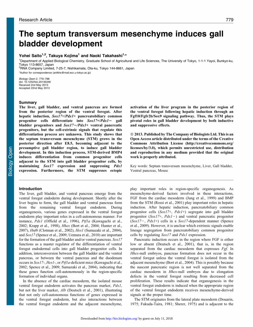
The septum transversum mesenchyme induces gallbladder development
Yohei Saito1,2, Takuya Kojima1 and Naoki Takahashi1,*1Department of Applied Biological Chemistry, Graduate School of Agricultural and Life Sciences, The University of Tokyo, 1-1-1 Yayoi, Bunkyo-ku,Tokyo 113-8657, Japan2RNA Company Limited, 7-25-7, Nishikamata, Ota-ku, Tokyo 144-8661, Japan
*Author for correspondence ([email protected])
Biology Open 2, 779–788doi: 10.1242/bio.20135348Received 2nd May 2013Accepted 22nd May 2013
SummaryThe liver, gall bladder, and ventral pancreas are formed
from the posterior region of the ventral foregut. After
hepatic induction, Sox17+/Pdx1+ pancreatobiliary common
progenitor cells differentiate into Sox17+/Pdx12 gall
bladder progenitors and Sox172/Pdx1+ ventral pancreatic
progenitors, but the cell-extrinsic signals that regulate this
differentiation process are unknown. This study shows that
the septum transversum mesenchyme (STM) grows in the
posterior direction after E8.5, becoming adjacent to the
presumptive gall bladder region, to induce gall bladder
development. In this induction process, STM-derived BMP4
induces differentiation from common progenitor cells
adjacent to the STM into gall bladder progenitor cells, by
maintaining Sox17 expression and suppressing Pdx1
expression. Furthermore, the STM suppresses ectopic
activation of the liver program in the posterior region of
the ventral foregut following hepatic induction through an
Fgf10/Fgfr2b/Sox9 signaling pathway. Thus, the STM plays
pivotal roles in gall bladder development by both inductive
and suppressive effects.
� 2013. Published by The Company of Biologists Ltd. This is an
Open Access article distributed under the terms of the Creative
Commons Attribution License (http://creativecommons.org/
licenses/by/3.0), which permits unrestricted use, distribution
and reproduction in any medium provided that the original
work is properly attributed.
Key words: Septum transversum mesenchyme, Liver, Gall bladder,
Ventral pancreas, Mouse
IntroductionThe liver, gall bladder, and ventral pancreas emerge from the
ventral foregut endoderm during development. Shortly after the
liver begins to form, the gall bladder and ventral pancreas form
from the remaining ventral foregut endoderm. During
organogenesis, various genes expressed in the ventral foregut
endoderm play important roles in a cell-autonomous manner. For
instance, Pdx1 (Offield et al., 1996), Ptf1a (Kawaguchi et al.,
2002; Krapp et al., 1998), Hhex (Bort et al., 2004; Hunter et al.,
2007), Hnf6 (Clotman et al., 2002), Hes1 (Sumazaki et al., 2004),
and Sox17 (Spence et al., 2009; Uemura et al., 2010) are important
for the formation of the gall bladder and/or ventral pancreas. Sox17
functions as a master regulator of the differentiation of ventral
foregut endodermal cells into gall bladder progenitor cells. In
addition, interconversion between the gall bladder and the ventral
pancreas, or between the ventral pancreas and the duodenum
occurs in Sox17-, Hes1-, or Ptf1a-deficient mice (Kawaguchi et al.,
2002; Spence et al., 2009; Sumazaki et al., 2004), indicating that
these genes function cell-autonomously in the region-specific
formation of individual organs.
In the absence of the cardiac mesoderm, the isolated mouse
ventral foregut endoderm activates the pancreas marker, Pdx1,
but not the liver marker, Alb (Deutsch et al., 2001), illustrating
that not only cell-autonomous functions of genes expressed in
the ventral foregut endoderm, but also interactions between
the ventral foregut endoderm and the adjacent mesenchyme,
play important roles in region-specific organogenesis. As
mesenchyme-derived factors involved in these interactions,
FGF from the cardiac mesoderm (Jung et al., 1999) and BMP
from the STM (Rossi et al., 2001) play important roles in hepatic
induction. After hepatic induction, pancreatobiliary common
progenitor cells (Sox17+, Pdx1+) segregate into gall bladder
progenitor (Sox17+, Pdx12) and ventral pancreatic progenitor
(Sox172, Pdx1+) cells in a Sox17-dependent manner (Spence
et al., 2009). However, it is unclear which extrinsic signals enable
lineage segregation from pancreatobiliary common progenitor
cells by regulating Sox17 and Pdx1 expression.
Pancreatic induction occurs in the region where FGF is either
low or absent (Deutsch et al., 2001), that is, in the region
separated from the cardiac mesoderm that expresses Fgf. In
Hhex-null embryos, pancreas formation does not occur in the
ventral foregut unless the ventral foregut is isolated from the
adjacent mesenchyme (Bort et al., 2004). This is possibly because
the nascent pancreatic region is not well separated from the
cardiac mesoderm in Hhex-null embryos due to elongation
defects in the ventral foregut resulting from decreased cell
proliferation. These results indicate that organogenesis in the
ventral foregut endoderm is induced when the appropriate region
of the ventral foregut endoderm receives mesenchyme-derived
signals at the proper time.
The STM originates from the lateral plate mesoderm (Douarin,
1975; Fukuda-Taira, 1981; Sherer, 1975) and is adjacent to the
Research Article 779
Bio
logy
Open
by guest on July 11, 2018http://bio.biologists.org/Downloaded from
ventral foregut endoderm during ventral foregut-derivedorganogenesis. Gata4 (Watt et al., 2004) and Mab21l2 (Saitoet al., 2012) are essential for STM formation. Gata4 and Mab21l2
are expressed in the STM, and in Gata4- and Mab21l2-deficientembryos, defective morphogenesis of the STM occurs. The STMis involved not only in hepatic induction, but also in the growth
and survival of hepatoblasts, which are bipotential progenitors forhepatocytes and cholangiocytes (Zaret, 2002), demonstrating thatthe STM is important for liver development. However, it is notclear from previous studies using Gata4- or Mab21l2-deficient
mice what roles the STM plays during gall bladder and ventralpancreas development. Therefore, the aim of this study was todetermine at which point during development the STM is
adjacent to the nascent gall bladder and/or ventral pancreas in theventral foregut, and how the STM is involved in the developmentof these organs using Mab21l2-null embryos as a model for STM
loss.
In this study, we show that the STM becomes adjacent to the
nascent gall bladder region after embryonic day 9.0 (E9.0). Thiscellular event triggers the differentiation of pancreatobiliarycommon progenitor cells into gall bladder progenitor cells by
maintaining Sox17 expression and suppressing Pdx1 expression.Moreover, after hepatic induction, STM-derived signals act onthe nascent gall bladder and ventral pancreas regions to suppress
ectopic induction of the liver program. Taken together, theseobservations indicate that following hepatic induction, the STMinduces gall bladder development, while suppressing ectopic
activation of the liver program in the posterior region of theventral foregut.
Materials and MethodsMiceThe generation of the mutant mice used in this study has been reported previously(Yamada et al., 2004). Mice were backcrossed to the ICR strain. Knockoutembryos are more readily obtained via the interbreeding of heterozygous miceusing the ICR strain, because the litter size of the ICR strain is larger than that ofthe B6 strain. There are no differences in phenotype or lethality rates at eachdevelopmental stage between the ICR and B6 strains. Mice were maintained inaccordance with protocols approved by the Animal Care and Use Committee of theUniversity of Tokyo.
In situ hybridizationWhole-mount in situ hybridization was performed as described previously (Nietoet al., 1996) at 65 C in 50% formamide containing 56SSC. Paraffin sections werehybridized in situ at 65 C in 50% formamide, 20 mM Tris-HCl (pH 8.0), 300 mMNaCl, 0.2% Sarkosyl, 16Denhart’s solution, 10% dextran sulfate, and 0.5 mg/mLyeast tRNA. All probes were labeled with digoxigenin using standard procedures.Details for probes will be provided upon request.
HistologyEmbryos were dissected in phosphate-buffered saline (PBS) and fixed in 4%paraformaldehyde in PBS overnight at 4 C. Fixed embryos were dehydratedthrough a graded alcohol series and embedded in paraffin, sectioned (8 mm thick),and stained with hematoxylin and eosin.
Detection of proliferating or apoptotic cellsParaffin sections were deparaffinized with xylene and dehydrated through a gradedethanol series. Sections were boiled in 10 mM citrate buffer (pH 6.0) for 10 minand washed with PBS. Endogenous peroxidase activity was blocked with 3%hydrogen peroxide in TBS for 10 min. After washing with PBS, sections wereincubated with 1/400 anti-PHH3 antibody (CST) overnight. After washing withPBS, sections were incubated with HRP-conjugated goat anti-rabbit IgG and 1%BSA in PBS for 1 hour. Immunoreactive sites were visualized with DAB andH2O2. Sections were counterstained with hematoxylin.
TdT-mediated dUTP nick-end labeling (TUNEL) analysis was performed asfollows. Paraffin sections (8 mm thick) of embryos were incubated in 3% H2O2 for15 min, Proteinase K solution for 10 min, and then TdT reaction solution (0.2 mMfluorescein-12-dUTP (Roche), 0.2 mM dATP, 1 mM CoCl2, 30 mM Tris-HCl
(pH 7.5), 140 mM sodium cacodylate, and 40 U terminal deoxynucleotidyltransferase (Roche)). Reactive sites were detected with an alkaline phosphatase-conjugated anti-fluorescein antibody (Roche) in a solution containing aphosphatase substrate (Fast Red Tablets, Roche). Sections were counterstainedwith hematoxylin.
Whole-embryo cultureE9.0 embryos were dissected from the uteri for in vitro culture. All littermateembryos were then cultured for 6 hours in medium (DMEM, Gibco) containing4 nM Noggin (Peprotech), 20 nM BMP4 (R&D Systems), and BSA at 37 C in thepresence of 5% CO2. Thereafter, embryos were fixed in 4% paraformaldehyde.
ResultsAfter E9.0, the STM is adjacent to the Sox17, but not the Pdx1,expressing region of the foregut
During gall bladder and ventral pancreas development,pancreatobiliary common progenitor cells expressing both
Sox17 and Pdx1 differentiate into Sox17+/Pdx12 gall bladderprogenitor cells, and Sox172/Pdx1+ ventral pancreaticprogenitor cells (Spence et al., 2009). However, how this
differentiation process is regulated is unknown. Interactionsbetween the endodermal epithelium and adjacent mesenchymeare important during ventral foregut-derived organ development,
and mesenchyme-derived signals regulate organ induction in theventral foregut (Deutsch et al., 2001; Jung et al., 1999; Rossiet al., 2001). Therefore, we examined the relationship betweenthe expression of Sox17 and Pdx1 and the position of the adjacent
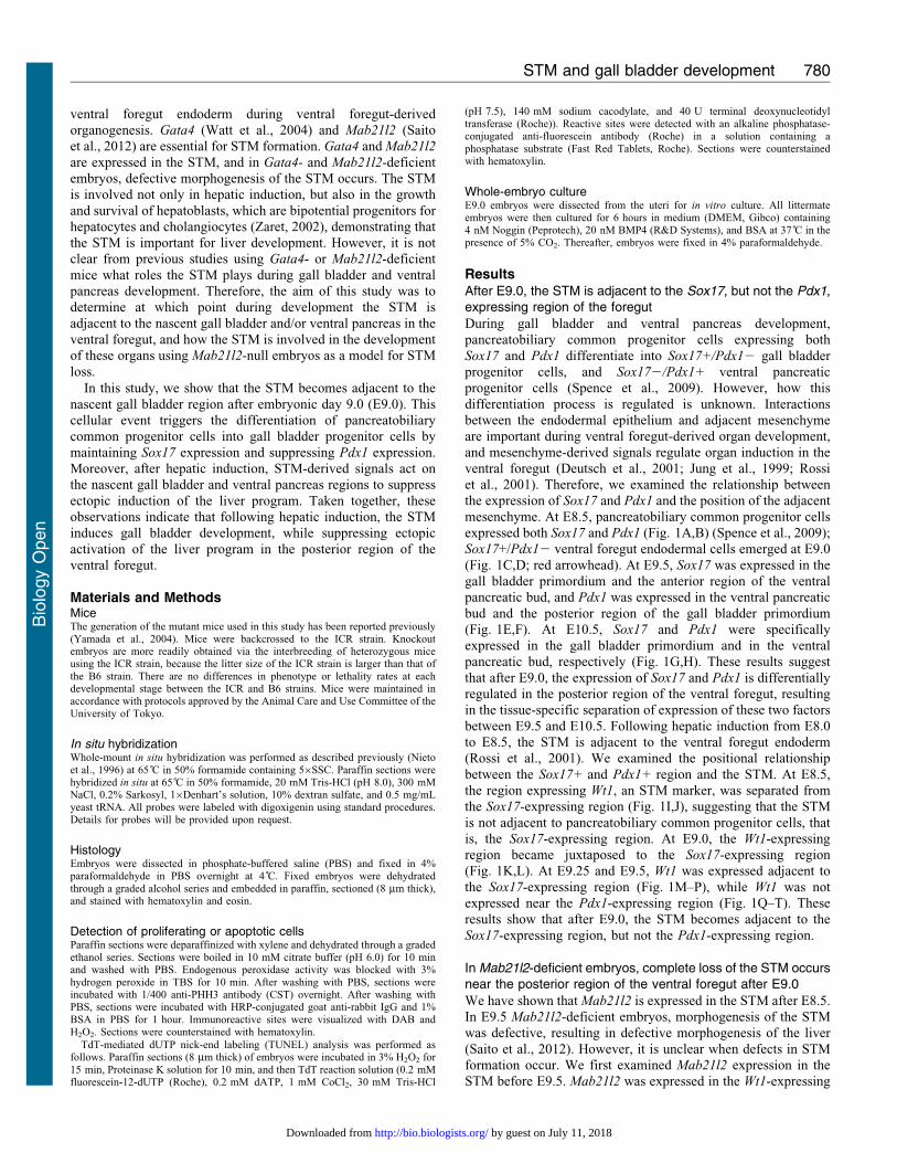
mesenchyme. At E8.5, pancreatobiliary common progenitor cellsexpressed both Sox17 and Pdx1 (Fig. 1A,B) (Spence et al., 2009);Sox17+/Pdx12 ventral foregut endodermal cells emerged at E9.0
(Fig. 1C,D; red arrowhead). At E9.5, Sox17 was expressed in thegall bladder primordium and the anterior region of the ventralpancreatic bud, and Pdx1 was expressed in the ventral pancreatic
bud and the posterior region of the gall bladder primordium(Fig. 1E,F). At E10.5, Sox17 and Pdx1 were specificallyexpressed in the gall bladder primordium and in the ventral
pancreatic bud, respectively (Fig. 1G,H). These results suggestthat after E9.0, the expression of Sox17 and Pdx1 is differentiallyregulated in the posterior region of the ventral foregut, resultingin the tissue-specific separation of expression of these two factors
between E9.5 and E10.5. Following hepatic induction from E8.0to E8.5, the STM is adjacent to the ventral foregut endoderm(Rossi et al., 2001). We examined the positional relationship
between the Sox17+ and Pdx1+ region and the STM. At E8.5,the region expressing Wt1, an STM marker, was separated fromthe Sox17-expressing region (Fig. 1I,J), suggesting that the STM
is not adjacent to pancreatobiliary common progenitor cells, thatis, the Sox17-expressing region. At E9.0, the Wt1-expressingregion became juxtaposed to the Sox17-expressing region(Fig. 1K,L). At E9.25 and E9.5, Wt1 was expressed adjacent to
the Sox17-expressing region (Fig. 1M–P), while Wt1 was notexpressed near the Pdx1-expressing region (Fig. 1Q–T). Theseresults show that after E9.0, the STM becomes adjacent to the
Sox17-expressing region, but not the Pdx1-expressing region.
In Mab21l2-deficient embryos, complete loss of the STM occursnear the posterior region of the ventral foregut after E9.0
We have shown that Mab21l2 is expressed in the STM after E8.5.In E9.5 Mab21l2-deficient embryos, morphogenesis of the STMwas defective, resulting in defective morphogenesis of the liver
(Saito et al., 2012). However, it is unclear when defects in STMformation occur. We first examined Mab21l2 expression in theSTM before E9.5. Mab21l2 was expressed in the Wt1-expressing
STM and gall bladder development 780
Bio
logy
Open
by guest on July 11, 2018http://bio.biologists.org/Downloaded from
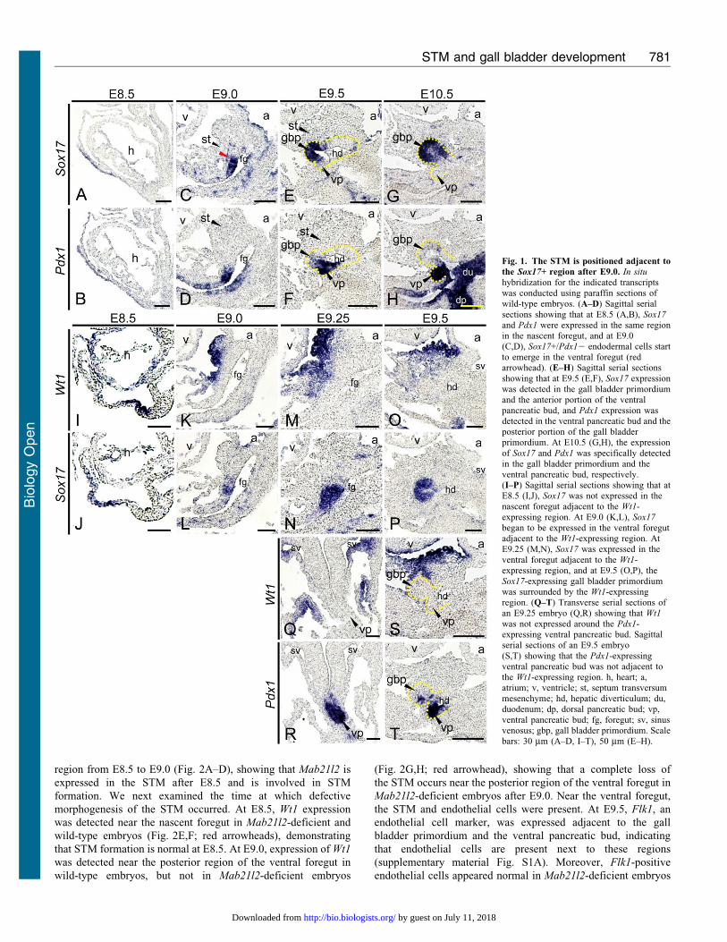
region from E8.5 to E9.0 (Fig. 2A–D), showing that Mab21l2 is
expressed in the STM after E8.5 and is involved in STM
formation. We next examined the time at which defective
morphogenesis of the STM occurred. At E8.5, Wt1 expression
was detected near the nascent foregut in Mab21l2-deficient and
wild-type embryos (Fig. 2E,F; red arrowheads), demonstrating
that STM formation is normal at E8.5. At E9.0, expression of Wt1
was detected near the posterior region of the ventral foregut in
wild-type embryos, but not in Mab21l2-deficient embryos
(Fig. 2G,H; red arrowhead), showing that a complete loss of
the STM occurs near the posterior region of the ventral foregut in
Mab21l2-deficient embryos after E9.0. Near the ventral foregut,
the STM and endothelial cells were present. At E9.5, Flk1, an
endothelial cell marker, was expressed adjacent to the gall
bladder primordium and the ventral pancreatic bud, indicating
that endothelial cells are present next to these regions
(supplementary material Fig. S1A). Moreover, Flk1-positive
endothelial cells appeared normal in Mab21l2-deficient embryos
Fig. 1. The STM is positioned adjacent to
the Sox17+ region after E9.0. In situ
hybridization for the indicated transcriptswas conducted using paraffin sections ofwild-type embryos. (A–D) Sagittal serial
sections showing that at E8.5 (A,B), Sox17
and Pdx1 were expressed in the same regionin the nascent foregut, and at E9.0(C,D), Sox17+/Pdx12 endodermal cells startto emerge in the ventral foregut (redarrowhead). (E–H) Sagittal serial sections
showing that at E9.5 (E,F), Sox17 expressionwas detected in the gall bladder primordiumand the anterior portion of the ventralpancreatic bud, and Pdx1 expression wasdetected in the ventral pancreatic bud and theposterior portion of the gall bladderprimordium. At E10.5 (G,H), the expression
of Sox17 and Pdx1 was specifically detectedin the gall bladder primordium and theventral pancreatic bud, respectively.(I–P) Sagittal serial sections showing that atE8.5 (I,J), Sox17 was not expressed in thenascent foregut adjacent to the Wt1-
expressing region. At E9.0 (K,L), Sox17
began to be expressed in the ventral foregutadjacent to the Wt1-expressing region. AtE9.25 (M,N), Sox17 was expressed in theventral foregut adjacent to the Wt1-expressing region, and at E9.5 (O,P), the
Sox17-expressing gall bladder primordiumwas surrounded by the Wt1-expressingregion. (Q–T) Transverse serial sections ofan E9.25 embryo (Q,R) showing that Wt1
was not expressed around the Pdx1-expressing ventral pancreatic bud. Sagittalserial sections of an E9.5 embryo
(S,T) showing that the Pdx1-expressingventral pancreatic bud was not adjacent tothe Wt1-expressing region. h, heart; a,atrium; v, ventricle; st, septum transversummesenchyme; hd, hepatic diverticulum; du,duodenum; dp, dorsal pancreatic bud; vp,
ventral pancreatic bud; fg, foregut; sv, sinusvenosus; gbp, gall bladder primordium. Scalebars: 30 mm (A–D, I–T), 50 mm (E–H).
STM and gall bladder development 781
Bio
logy
Open
by guest on July 11, 2018http://bio.biologists.org/Downloaded from
compared to wild-type embryos at E9.0 (supplementary material
Fig. S1B,C), indicating that the STM is only lost near the
posterior region of the ventral foregut in Mab21l2-deficient
embryos. Therefore, we examined the function of the STM in
organogenesis in the posterior region of the ventral foregut after
E9.0 using Mab21l2-deficient embryos as a model of STM loss.
Defective morphogenesis of the extrahepatic biliary system andgall bladder occurs in Mab21l2-deficient embryos
If the STM influences the differentiation of the pancreatobiliary
common progenitor cells, then STM loss should affect the
formation of the gall bladder and/or ventral pancreas. We
examined the formation of the gall bladder and ventral pancreas
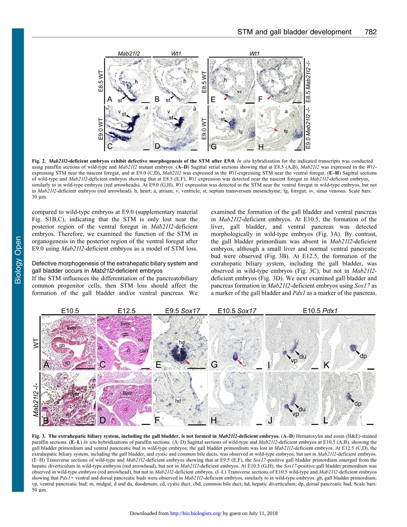
in Mab21l2-deficient embryos. At E10.5, the formation of the
liver, gall bladder, and ventral pancreas was detected
morphologically in wild-type embryos (Fig. 3A). By contrast,
the gall bladder primordium was absent in Mab21l2-deficient
embryos, although a small liver and normal ventral pancreatic
bud were observed (Fig. 3B). At E12.5, the formation of the
extrahepatic biliary system, including the gall bladder, was
observed in wild-type embryos (Fig. 3C), but not in Mab21l2-
deficient embryos (Fig. 3D). We next examined gall bladder and
pancreas formation in Mab21l2-deficient embryos using Sox17 as
a marker of the gall bladder and Pdx1 as a marker of the pancreas.
Fig. 2. Mab21l2-deficient embryos exhibit defective morphogenesis of the STM after E9.0. In situ hybridization for the indicated transcripts was conductedusing paraffin sections of wild-type and Mab21l2 mutant embryos. (A–D) Sagittal serial sections showing that at E8.5 (A,B), Mab21l2 was expressed in the Wt1-expressing STM near the nascent foregut, and at E9.0 (C,D), Mab21l2 was expressed in the Wt1-expressing STM near the ventral foregut. (E–H) Sagittal sectionsof wild-type and Mab21l2-deficient embryos showing that at E8.5 (E,F), Wt1 expression was detected near the nascent foregut in Mab21l2-deficient embryos,
similarly to in wild-type embryos (red arrowheads). At E9.0 (G,H), Wt1 expression was detected in the STM near the ventral foregut in wild-type embryos, but notin Mab21l2-deficient embryos (red arrowhead). h, heart; a, atrium; v, ventricle; st, septum transversum mesenchyme; fg, foregut; sv, sinus venosus. Scale bars:30 mm.
Fig. 3. The extrahepatic biliary system, including the gall bladder, is not formed in Mab21l2-deficient embryos. (A–D) Hematoxylin and eosin (H&E)-stained
paraffin sections. (E–L) In situ hybridizations of paraffin sections. (A–D) Sagittal sections of wild-type and Mab21l2-deficient embryos at E10.5 (A,B), showing thegall bladder primordium and ventral pancreatic bud in wild-type embryos; the gall bladder primordium was lost in Mab21l2-deficient embryos. At E12.5 (C,D), theextrahepatic biliary system, including the gall bladder, and cystic and common bile ducts, was observed in wild-type embryos, but not in Mab21l2-deficient embryos.(E–H) Transverse sections of wild-type and Mab21l2-deficient embryos showing that at E9.5 (E,F), the Sox17-positive gall bladder primordium emerged from thehepatic diverticulum in wild-type embryos (red arrowhead), but not in Mab21l2-deficient embryos. At E10.5 (G,H), the Sox17-positive gall bladder primordium wasobserved in wild-type embryos (red arrowhead), but not in Mab21l2-deficient embryos. (I–L) Transverse sections of E10.5 wild-type and Mab21l2-deficient embryos
showing that Pdx1+ ventral and dorsal pancreatic buds were observed in Mab21l2-deficient embryos, similarly to in wild-type embryos. gb, gall bladder primordium;vp, ventral pancreatic bud; m, midgut; d and du, duodenum; cd, cystic duct; cbd, common bile duct; hd, hepatic diverticulum; dp, dorsal pancreatic bud. Scale bars:50 mm.
STM and gall bladder development 782
Bio
logy
Open
by guest on July 11, 2018http://bio.biologists.org/Downloaded from
At E9.5, a Sox17+ gall bladder primordium arose from the hepatic
diverticulum in wild-type embryos (Fig. 3E; red arrowhead), but
not in Mab21l2-deficient embryos (Fig. 3F). Similarly, at E10.5,
the Sox17+ gall bladder primordium was observed in wild-type
embryos (Fig. 3G; red arrowhead), but not in Mab21l2-deficient
embryos (Fig. 3H). Moreover, the formation of ventral and dorsal
pancreas was normal in Mab21l2-deficient embryos at E10.5
(Fig. 3J,L), similar to wild-type embryos (Fig. 3I,K). These results
reveal that the STM is required for formation of the gall bladder,
but not the pancreas.
Reduced expression of Sox17 and ectopic expression of Pdx1
occur in Mab21l2-deficient embryos
Loss of the gall bladder in Mab21l2-deficient embryos suggested
that defects occurred during the differentiation of pancreatobiliary
common progenitor cells into gall bladder progenitor cells. As
Sox17 plays an essential role in this process (Spence et al., 2009),
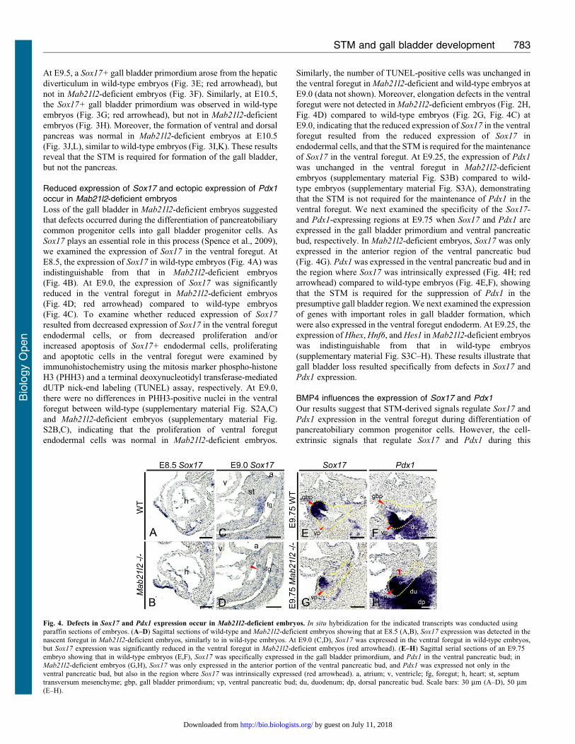
we examined the expression of Sox17 in the ventral foregut. At
E8.5, the expression of Sox17 in wild-type embryos (Fig. 4A) was
indistinguishable from that in Mab21l2-deficient embryos
(Fig. 4B). At E9.0, the expression of Sox17 was significantly
reduced in the ventral foregut in Mab21l2-deficient embryos
(Fig. 4D; red arrowhead) compared to wild-type embryos
(Fig. 4C). To examine whether reduced expression of Sox17
resulted from decreased expression of Sox17 in the ventral foregut
endodermal cells, or from decreased proliferation and/or
increased apoptosis of Sox17+ endodermal cells, proliferating
and apoptotic cells in the ventral foregut were examined by
immunohistochemistry using the mitosis marker phospho-histone
H3 (PHH3) and a terminal deoxynucleotidyl transferase-mediated
dUTP nick-end labeling (TUNEL) assay, respectively. At E9.0,
there were no differences in PHH3-positive nuclei in the ventral
foregut between wild-type (supplementary material Fig. S2A,C)
and Mab21l2-deficient embryos (supplementary material Fig.
S2B,C), indicating that the proliferation of ventral foregut
endodermal cells was normal in Mab21l2-deficient embryos.
Similarly, the number of TUNEL-positive cells was unchanged in
the ventral foregut in Mab21l2-deficient and wild-type embryos at
E9.0 (data not shown). Moreover, elongation defects in the ventral
foregut were not detected in Mab21l2-deficient embryos (Fig. 2H,
Fig. 4D) compared to wild-type embryos (Fig. 2G, Fig. 4C) at
E9.0, indicating that the reduced expression of Sox17 in the ventral
foregut resulted from the reduced expression of Sox17 in
endodermal cells, and that the STM is required for the maintenance
of Sox17 in the ventral foregut. At E9.25, the expression of Pdx1
was unchanged in the ventral foregut in Mab21l2-deficient
embryos (supplementary material Fig. S3B) compared to wild-
type embryos (supplementary material Fig. S3A), demonstrating
that the STM is not required for the maintenance of Pdx1 in the
ventral foregut. We next examined the specificity of the Sox17-
and Pdx1-expressing regions at E9.75 when Sox17 and Pdx1 are
expressed in the gall bladder primordium and ventral pancreatic
bud, respectively. In Mab21l2-deficient embryos, Sox17 was only
expressed in the anterior region of the ventral pancreatic bud
(Fig. 4G). Pdx1 was expressed in the ventral pancreatic bud and in
the region where Sox17 was intrinsically expressed (Fig. 4H; red
arrowhead) compared to wild-type embryos (Fig. 4E,F), showing
that the STM is required for the suppression of Pdx1 in the
presumptive gall bladder region. We next examined the expression
of genes with important roles in gall bladder formation, which
were also expressed in the ventral foregut endoderm. At E9.25, the
expression of Hhex, Hnf6, and Hes1 in Mab21l2-deficient embryos
was indistinguishable from that in wild-type embryos
(supplementary material Fig. S3C–H). These results illustrate that
gall bladder loss resulted specifically from defects in Sox17 and
Pdx1 expression.
BMP4 influences the expression of Sox17 and Pdx1
Our results suggest that STM-derived signals regulate Sox17 and
Pdx1 expression in the ventral foregut during differentiation of
pancreatobiliary common progenitor cells. However, the cell-
extrinsic signals that regulate Sox17 and Pdx1 during this
Fig. 4. Defects in Sox17 and Pdx1 expression occur in Mab21l2-deficient embryos. In situ hybridization for the indicated transcripts was conducted usingparaffin sections of embryos. (A–D) Sagittal sections of wild-type and Mab21l2-deficient embryos showing that at E8.5 (A,B), Sox17 expression was detected in thenascent foregut in Mab21l2-deficient embryos, similarly to in wild-type embryos. At E9.0 (C,D), Sox17 was expressed in the ventral foregut in wild-type embryos,but Sox17 expression was significantly reduced in the ventral foregut in Mab21l2-deficient embryos (red arrowhead). (E–H) Sagittal serial sections of an E9.75embryo showing that in wild-type embryos (E,F), Sox17 was specifically expressed in the gall bladder primordium, and Pdx1 in the ventral pancreatic bud; inMab21l2-deficient embryos (G,H), Sox17 was only expressed in the anterior portion of the ventral pancreatic bud, and Pdx1 was expressed not only in the
ventral pancreatic bud, but also in the region where Sox17 was intrinsically expressed (red arrowhead). a, atrium; v, ventricle; fg, foregut; h, heart; st, septumtransversum mesenchyme; gbp, gall bladder primordium; vp, ventral pancreatic bud; du, duodenum; dp, dorsal pancreatic bud. Scale bars: 30 mm (A–D), 50 mm(E–H).
STM and gall bladder development 783
Bio
logy
Open
by guest on July 11, 2018http://bio.biologists.org/Downloaded from
differentiation process are unknown. Various genes that encode
secreted ligands such as BMP, FGF, and HGF are expressed inthe STM (Berg et al., 2007; Rossi et al., 2001; Schmidt et al.,
1995). However, which of these genes are involved in the
regulation of Sox17 and Pdx1 expression during gall bladder and/
or ventral pancreas development is not clear. FoxF1, whichencodes a forkhead transcription factor, is expressed in the STM
and plays an important role in gall bladder formation because
haploinsufficiency of FoxF1 resulted in defects in gall bladder
development (Kalinichenko et al., 2002). Furthermore, previousstudies have shown that expression of Bmp4 is regulated by
FoxF1 (Mahlapuu et al., 2001), and that the BMP receptors
BMPRIA, BMPRII, and ActRIIA are expressed in the ventral
foregut endoderm (Mishina et al., 1995; Roelen et al., 1997). At
E9.0, Bmp4 was expressed in the STM in wild-type embryos(Fig. 5A,B), but the expression of Bmp4 was not detected near
the posterior region of the ventral foregut in Mab21l2-deficient
embryos (Fig. 5C,D; red arrowhead), suggesting the possibility
that STM-derived BMP4 may regulate the expression of Sox17
and Pdx1 in the ventral foregut endoderm. To test this hypothesis,
we performed several experiments using whole-embryo cultures.
We cultured E9.0 embryos for 6 hours in medium containing
4 nM Noggin, a BMP antagonist, and evaluated its effects onSox17 and Pdx1 expression. Noggin had a tendency to reduce the
expression of Sox17 in the Hhex-positive foregut region (n53;
Fig. 5G,H) compared to embryos cultured in control medium
containing BSA (Fig. 5E,F). The expression of Sox17 inendothelial cells around the ventral foregut (red arrowheads)
and the size of the Sox17-expressing region were the same in
control and Noggin-treated embryos. These results indicate that
BMPs are involved in the regulation of Sox17 in the ventralforegut endodermal cells. Noggin did not influence Pdx1
expression (data not shown). We next cultured E9.0 Mab21l2-deficient embryos in medium containing 20 nM BMP4 for
6 hours, to determine whether the expression of Sox17 could berescued by BMP4. In Mab21l2-deficient embryos cultured incontrol medium containing BSA, the expression of Sox17 wassignificantly reduced in the Hhex-positive foregut region
(Fig. 5I,J). By contrast, in Mab21l2-deficient embryos culturedin medium containing BMP4, Sox17 expression had a tendency tobe rescued in the Hhex-positive foregut region (n53; Fig. 5K,L)
and was similar to wild-type embryos cultured in control medium(Fig. 5F). These results suggest that BMP4 is involved in themaintenance of Sox17 in the ventral foregut. In E9.0 wild-type
embryos cultured in medium containing 20 nM BMP4 for6 hours, BMP4 had a tendency to increase Sox17 expressionmoderately (Fig. 5O) and to reduce Pdx1 expression (Fig. 5P)compared with embryos cultured in control medium (n53;
Fig. 5M,N). These results demonstrate that BMP4 maintainsSox17 expression and suppresses Pdx1 expression in the ventralforegut.
STM loss results in ectopic Alb expression and defects in theFgf10/Fgfr2b/Sox9 signaling pathway in the posterior region ofthe ventral foregut
Previous studies have shown that FGF from the cardiacmesoderm and BMP from the STM induce liver development
(Jung et al., 1999; Rossi et al., 2001), while hepatic induction issuppressed in the region where FGF is either low or absent(Deutsch et al., 2001). However, the role of the STM insuppressing liver development is not known, and we therefore
examined the function of the STM in the posterior region of theventral foregut. At E9.25, the expression of Alb was only detectedin the anterior region of the Hhex-positive ventral foregut (i.e.,
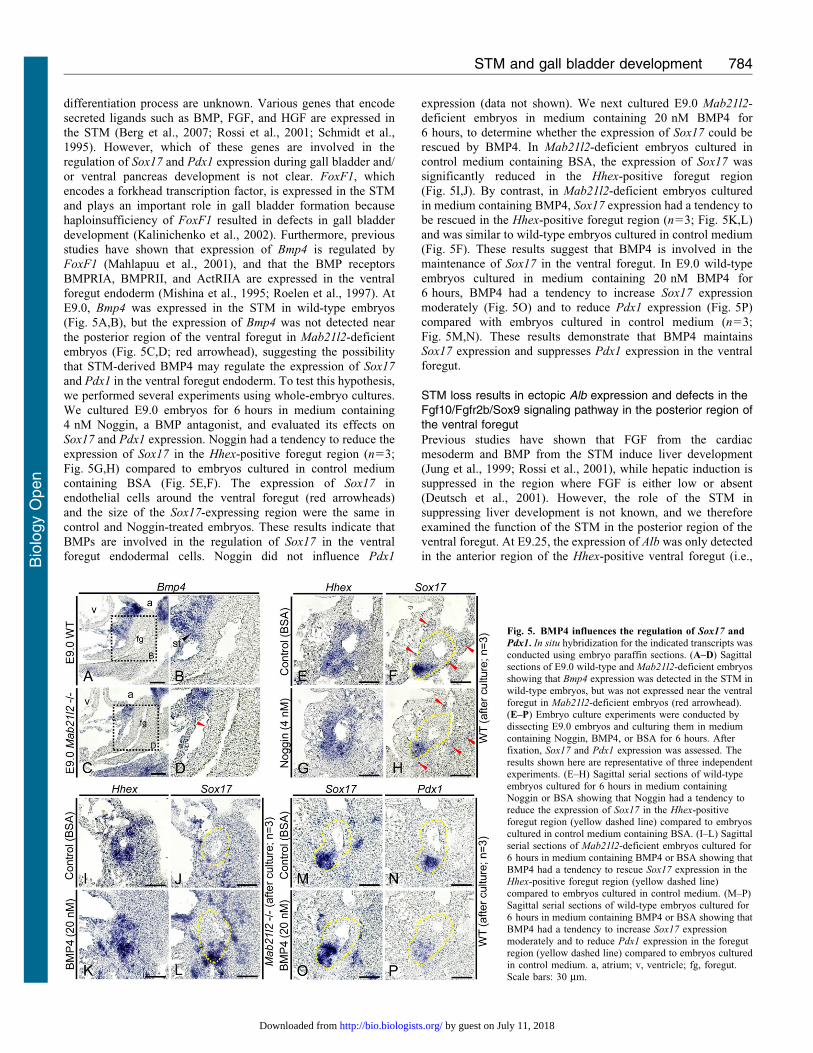
Fig. 5. BMP4 influences the regulation of Sox17 and
Pdx1. In situ hybridization for the indicated transcripts wasconducted using embryo paraffin sections. (A–D) Sagittalsections of E9.0 wild-type and Mab21l2-deficient embryosshowing that Bmp4 expression was detected in the STM in
wild-type embryos, but was not expressed near the ventralforegut in Mab21l2-deficient embryos (red arrowhead).(E–P) Embryo culture experiments were conducted bydissecting E9.0 embryos and culturing them in mediumcontaining Noggin, BMP4, or BSA for 6 hours. Afterfixation, Sox17 and Pdx1 expression was assessed. The
results shown here are representative of three independentexperiments. (E–H) Sagittal serial sections of wild-typeembryos cultured for 6 hours in medium containingNoggin or BSA showing that Noggin had a tendency toreduce the expression of Sox17 in the Hhex-positiveforegut region (yellow dashed line) compared to embryoscultured in control medium containing BSA. (I–L) Sagittal
serial sections of Mab21l2-deficient embryos cultured for6 hours in medium containing BMP4 or BSA showing thatBMP4 had a tendency to rescue Sox17 expression in theHhex-positive foregut region (yellow dashed line)compared to embryos cultured in control medium. (M–P)Sagittal serial sections of wild-type embryos cultured for
6 hours in medium containing BMP4 or BSA showing thatBMP4 had a tendency to increase Sox17 expressionmoderately and to reduce Pdx1 expression in the foregutregion (yellow dashed line) compared to embryos culturedin control medium. a, atrium; v, ventricle; fg, foregut.Scale bars: 30 mm.
STM and gall bladder development 784
Bio
logy
Open
by guest on July 11, 2018http://bio.biologists.org/Downloaded from
the presumptive liver region) in wild-type embryos (Fig. 6A,B).By contrast, the expression of Alb in the Hhex-positive ventral
foregut was detected not only in the anterior region but also in theposterior region (i.e., the presumptive gall bladder and ventralpancreas region) in Mab21l2-deficient embryos (Fig. 6C,D; redarrowhead). Moreover, in E9.75 Mab21l2-deficient embryos,
Sox17 was only expressed in part of the anterior region of theventral pancreatic bud (Fig. 6G). Alb was expressed not only inthe nascent liver, as in wild-type embryos (Fig. 6E,F), but also in
the posterior region of the ventral foregut including the ventralpancreatic bud and the region where Sox17 was intrinsicallyexpressed (Fig. 6H; red arrowhead). These results suggest that
STM loss results in ectopic activation of the liver program in thepresumptive gall bladder and ventral pancreas regions. Previousstudies have shown hepatic induction to occur in the ventral anddorsal foregut (Bossard and Zaret, 1998; Bossard and Zaret,
2000; Gualdi et al., 1996). During pancreas development, theFGF signaling pathway suppresses the liver program in theventral and dorsal pancreas through an Fgf10/Fgfr2b/Sox9
pathway (Seymour et al., 2012). As Fgf10 is expressed in theSTM at E9.0 (Berg et al., 2007), this signaling pathway maysuppress the liver program during the initial stages of gall bladder
and ventral pancreas development. To examine when Fgf10 wasexpressed in the STM, we examined the expression of the STMmarker Wt1 and of Fgf10 before E9.0. At E8.5, Fgf10 expression
was not detected in the Wt1-expressing region (Fig. 6I,J; redarrowheads). At E9.0, Fgf10 expression was observed in the Wt1-expressing STM near the ventral foregut (Fig. 6K,L), aspreviously reported. These results indicate that Fgf10
expression in the STM begins at E9.0. At E9.25, Fgf10 wasexpressed in the STM, and Fgfr2b, the main receptor for Fgf10,was expressed in the ventral foregut in the Sox17-expressing
region (Fig. 6M–P). Sox17 was not expressed in the Alb+
presumptive liver region (Fig. 6Q,R), indicating that Fgfr2b isspecifically expressed in the posterior region of the ventral
foregut endoderm, that is, in the nascent gall bladder and ventralpancreas, but not in the nascent liver. These results indicate thatafter E9.0, the FGF signaling pathway functions specifically inthe presumptive gall bladder and ventral pancreas regions
through Fgf10/Fgfr2b. Sox9, a downstream target of Fgf10/Fgfr2b signaling (Seymour et al., 2012), was mainly expressed inthe presumptive gall bladder and ventral pancreas regions,
including the Sox17-expressing region (Fig. 6S,T). Thus, theFGF signaling pathway may function through Fgf10/Fgfr2b/Sox9to suppress the liver program in the nascent gall bladder and
ventral pancreas shortly after liver formation begins (Fig. 6U). AtE9.25, the expression of Fgf10 was lost near the ventral foregutin Mab21l2-deficient embryos (Fig. 6W; red arrowhead)
compared to wild-type embryos (Fig. 6V), and Sox9 expressionwas also significantly reduced in the ventral foregut endoderm(Fig. 6Y; red arrowhead) compared to wild-type embryos(Fig. 6X). These results suggest that after E9.0 (i.e., after
hepatic induction), the STM suppresses ectopic activation ofthe liver program in the posterior region of the ventral foregutthrough the Fgf10/Fgfr2b/Sox9 signaling pathway.
DiscussionThe STM is required for induction of gall bladder development
Mab21l2 is expressed in the STM, but not in endothelial andventral foregut endodermal cells. In Mab21l2-deficient embryos,the extrahepatic biliary system including the gall bladder was
completely lost. At E9.0, the STM was completely absent nearthe posterior region of the ventral foregut, but normal endothelial
cells were present. Previous studies have shown that the bloodvessel endothelium plays an important role in the induction ofpancreatic differentiation (Lammert et al., 2001) and that Flk1,which is expressed in endothelial cells adjacent to the liver bud,
is required to promote liver bud growth (Matsumoto et al., 2001).These studies demonstrated that endothelial cells are essential fororganogenesis of ventral foregut-derived tissues. However, the
expression of Flk1 was unchanged in Mab21l2-deficient embryoscompared to wild-type embryos at E9.0, suggesting that theendothelial cells are normal. Thus, our results suggest that gall
bladder loss resulted from loss of the STM, not from defects inthe endothelial cells. Therefore, the STM is essential for theinduction of gall bladder development.
STM-derived BMP4 regulates differentiation of pancreatobiliarycommon progenitor cells into gall bladder progenitor cells byregulating Sox17 and Pdx1 expression
This study showed that Sox17 and Pdx1 were expressed in thesame region, in the pancreatobiliary common progenitor cells, atE8.5, and that Sox17+/Pdx12 ventral foregut endodermal cells
began to emerge at E9.0. These results indicate that after E9.0,the expression of Sox17 and Pdx1 in pancreatobiliary commonprogenitor cells is differentially regulated. Moreover, betweenE9.5 and E10.5, Sox17 expression is specifically detected in the
gall bladder primordium and Pdx1 expression in the ventralpancreatic bud, showing that between E9.5 and E10.5 thedifferential regulation is complete. Our study also showed that
after E9.0, the STM is adjacent only to the Sox17+, Pdx12
region, and is involved in the differentiation of pancreatobiliarycommon progenitor cells, most likely by maintaining Sox17
expression and suppressing Pdx1 expression. A previous studyshowed that the regulation of Sox17 and Pdx1 plays an importantrole in the differentiation of pancreatobiliary common progenitor
cells into gall bladder progenitor cells and ventral pancreaticprogenitor cells. Sox17 is a master regulator of gall bladderformation (Spence et al., 2009). Sox17 expression in Mab21l2-deficient embryos was reduced in the ventral foregut and Pdx1
was ectopically expressed in the region where Sox17 wasintrinsically expressed, indicating that gall bladder loss inMab21l2-deficient embryos resulted from defects in the
regulation of Sox17 and Pdx1. Thus, these results show that theSTM is required to maintain Sox17 expression and suppress Pdx1
expression in the ventral foregut; the STM induces gall bladder
development by differentially regulating Sox17 and Pdx1.
Using whole-embryo cultures, we showed that Noggin reducedthe expression of Sox17 in the Hhex-positive foregut region ofwild-type embryos, while Sox17 expression in endothelial cells
did not differ from expression in embryos cultured in controlmedium. This result suggests that Noggin specifically reducesSox17 expression in the foregut endodermal cells. Furthermore,
Hhex expression in the foregut region was unchanged, suggestingthat reduced expression of Sox17 is unlikely to result fromsecondary effects of Noggin addition. Therefore, our results
suggest that BMPs function to maintain Sox17 expression in theventral foregut. Several BMPs are expressed in the STMincluding BMP2, BMP4, BMP5, and BMP7 (Hogan, 1996;
Zhao, 2003). Previous studies have shown that Bmp4 is regulatedby FoxF1, which is expressed in the STM and is important forgall bladder development (Kalinichenko et al., 2002; Mahlapuu
STM and gall bladder development 785
Bio
logy
Open
by guest on July 11, 2018http://bio.biologists.org/Downloaded from
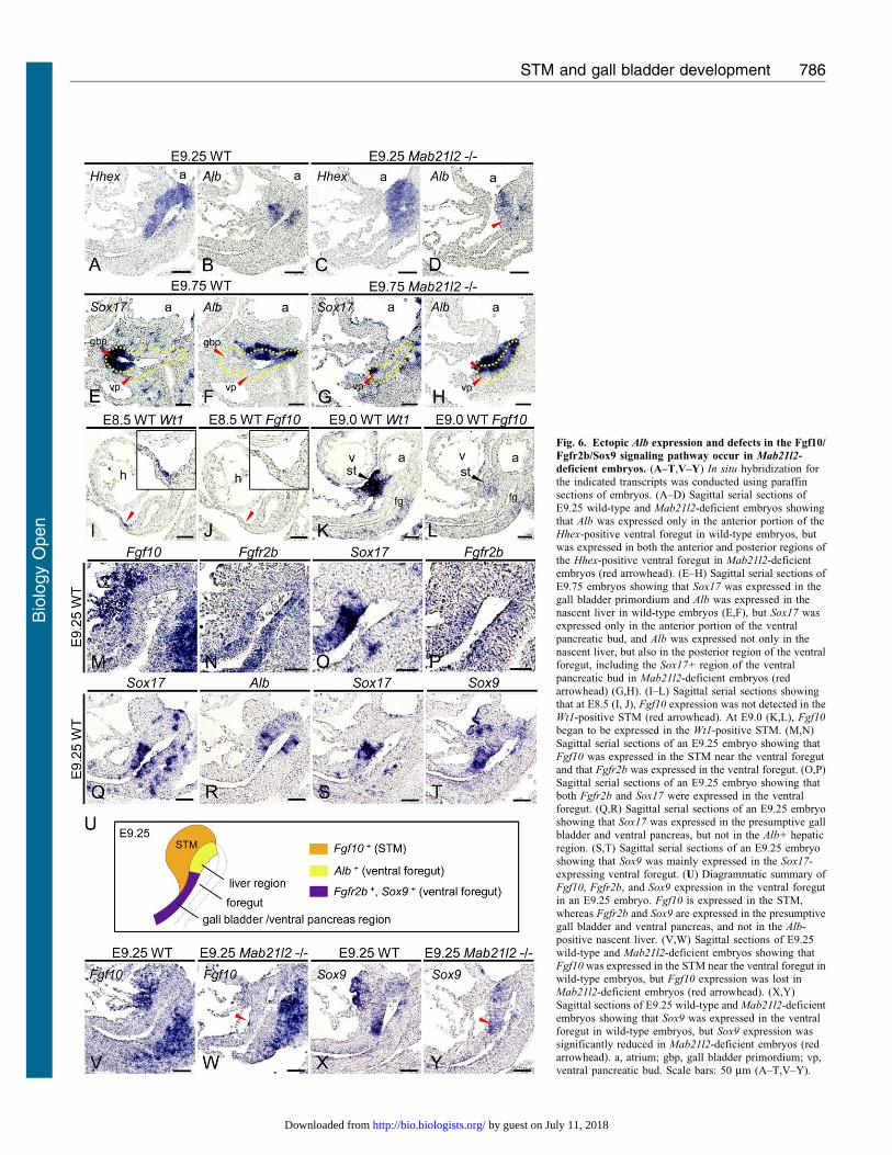
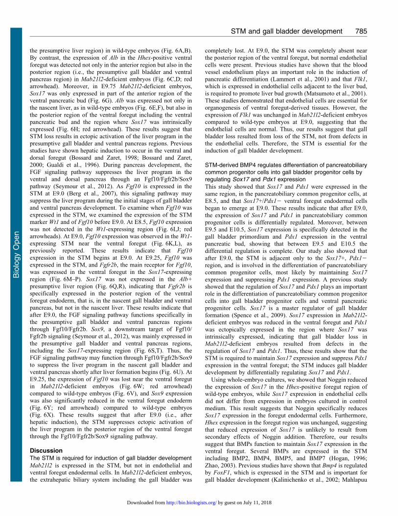
Fig. 6. Ectopic Alb expression and defects in the Fgf10/
Fgfr2b/Sox9 signaling pathway occur in Mab21l2-
deficient embryos. (A–T,V–Y) In situ hybridization forthe indicated transcripts was conducted using paraffinsections of embryos. (A–D) Sagittal serial sections of
E9.25 wild-type and Mab21l2-deficient embryos showingthat Alb was expressed only in the anterior portion of theHhex-positive ventral foregut in wild-type embryos, butwas expressed in both the anterior and posterior regions ofthe Hhex-positive ventral foregut in Mab21l2-deficientembryos (red arrowhead). (E–H) Sagittal serial sections of
E9.75 embryos showing that Sox17 was expressed in thegall bladder primordium and Alb was expressed in thenascent liver in wild-type embryos (E,F), but Sox17 wasexpressed only in the anterior portion of the ventralpancreatic bud, and Alb was expressed not only in thenascent liver, but also in the posterior region of the ventralforegut, including the Sox17+ region of the ventral
pancreatic bud in Mab21l2-deficient embryos (redarrowhead) (G,H). (I–L) Sagittal serial sections showingthat at E8.5 (I, J), Fgf10 expression was not detected in theWt1-positive STM (red arrowhead). At E9.0 (K,L), Fgf10
began to be expressed in the Wt1-positive STM. (M,N)Sagittal serial sections of an E9.25 embryo showing that
Fgf10 was expressed in the STM near the ventral foregutand that Fgfr2b was expressed in the ventral foregut. (O,P)Sagittal serial sections of an E9.25 embryo showing thatboth Fgfr2b and Sox17 were expressed in the ventralforegut. (Q,R) Sagittal serial sections of an E9.25 embryoshowing that Sox17 was expressed in the presumptive gallbladder and ventral pancreas, but not in the Alb+ hepatic
region. (S,T) Sagittal serial sections of an E9.25 embryoshowing that Sox9 was mainly expressed in the Sox17-expressing ventral foregut. (U) Diagrammatic summary ofFgf10, Fgfr2b, and Sox9 expression in the ventral foregutin an E9.25 embryo. Fgf10 is expressed in the STM,whereas Fgfr2b and Sox9 are expressed in the presumptive
gall bladder and ventral pancreas, and not in the Alb-positive nascent liver. (V,W) Sagittal sections of E9.25wild-type and Mab21l2-deficient embryos showing thatFgf10 was expressed in the STM near the ventral foregut inwild-type embryos, but Fgf10 expression was lost inMab21l2-deficient embryos (red arrowhead). (X,Y)
Sagittal sections of E9.25 wild-type and Mab21l2-deficientembryos showing that Sox9 was expressed in the ventralforegut in wild-type embryos, but Sox9 expression wassignificantly reduced in Mab21l2-deficient embryos (redarrowhead). a, atrium; gbp, gall bladder primordium; vp,ventral pancreatic bud. Scale bars: 50 mm (A–T,V–Y).
STM and gall bladder development 786
Bio
logy
Open
by guest on July 11, 2018http://bio.biologists.org/Downloaded from
et al., 2001). These observations suggest that Bmp4 is involved ingall bladder development through regulation of Sox17 and Pdx1
expression. Bmp4 was expressed in the STM at E8.5 when hepaticinduction occurred (Rossi et al., 2001). However, in Mab21l2-deficient embryos at E9.0, STM loss resulted in the loss of Bmp4
expression near the posterior region of the ventral foregut,
including the presumptive gall bladder region, suggesting thatreduced expression of Sox17 may be a consequence of the loss ofBMP4 signaling. BMP4 rescued Sox17 expression in the foregut
region of cultured Mab21l2-deficient embryos. A previous studyshowed that BMP4 promotes cell proliferation of Sox17-expressing cells (Sneddon et al., 2012). However, our study
showed that Noggin did not influence the size of the Sox17-expressing region, suggesting that BMP4 is the primary regulatormaintaining Sox17 expression at this developmental stage.Moreover, BMP4 reduced Pdx1 expression in the foregut of
cultured wild-type embryos, suggesting that BMP4 also suppressesPdx1 expression. Therefore, our results demonstrate that, duringgall bladder development, STM-derived BMP4 induces
differentiation of Sox17+/Pdx1+ pancreatobiliary commonprogenitor cells adjacent to the STM into Sox17+/Pdx12 gallbladder progenitor cells by maintaining Sox17 expression and
suppressing that of Pdx1. The positional relationship between theSTM and the ventral foregut endoderm thus determines theboundary between the gall bladder and the ventral pancreas
(supplementary material Fig. S5).
Previous studies have shown that Hgf is expressed in the STMsurrounding the hepatoblasts (Schmidt et al., 1995), and that theHGF receptor c-met is expressed in hepatoblasts (Bladt et al.,
1995). During liver development, the Hgf-c-met signalingpathway promotes cell proliferation and survival ofhepatoblasts through pro-mitogenic and anti-apoptotic actions
(Birchmeier et al., 2003; Schmidt et al., 1995), indicating that theSTM is involved in liver differentiation and growth. At E9.5, Hgf
was expressed in the STM surrounding the gall bladder
primordium (supplementary material Fig. S4A,B), and c-met
was expressed in the gall bladder primordium (supplementarymaterial Fig. S4C,D), indicating that the STM promotes theproliferation and survival of gall bladder primordial cells.
Therefore, the STM is involved in both differentiation andgrowth of the gall bladder.
The STM suppresses ectopic activation of the liver program inthe presumptive gall bladder and ventral pancreas
Previous studies in chick and Xenopus suggest that during
endoderm patterning, a concentration gradient of Wnt and FGFsecreted from the adjacent mesoderm regulates the regionalidentity of the endoderm (foregut, midgut, or hindgut) (Dessimozet al., 2006; McLin et al., 2007; Wells and Melton, 2000).
Moreover, inhibition of the Wnt signaling pathway in theXenopus posterior endoderm induces ectopic activation of theliver program in the intestine (McLin et al., 2007). These studies
indicate that for each organ to develop in the appropriateposition, not only inductive signals but also suppressive signalsare required. After endoderm patterning, FGF from the cardiac
mesoderm induces or suppresses liver development in the foregutendoderm, depending on the region (Deutsch et al., 2001; Junget al., 1999). In Mab21l2-deficient embryos, in which the STM
was completely lost near the posterior region of the ventralforegut, the expression of Alb was activated not only in thenascent liver but also in the remaining posterior region of the
ventral foregut, suggesting that after hepatic induction, the STM
normally suppresses ectopic activation of the liver program in the
presumptive gall bladder and ventral pancreas regions.
Various genes that encode secreted ligands, including Fgf10,
are expressed in the STM. During pancreas development, Fgf10
is expressed in the mesenchyme surrounding the pancreatic
epithelium, and Fgfr2b and Sox9 are expressed in the pancreatic
epithelium. In addition, the Fgf10-Fgfr2b signaling pathway
suppresses the liver program in the pancreatic epithelium by
regulating Sox9 (Seymour et al., 2012), suggesting that FGF10
from the STM has similar functions. Here, we showed that Fgf10
is expressed in the STM at E9.0, and Fgfr2b and Sox9 were
expressed in presumptive gall bladder and ventral pancreas
regions, and not in the nascent liver. This expression pattern
enables the Fgf10/Fgfr2b/Sox9 signaling pathway to function in a
region-specific manner in the presumptive gall bladder and
ventral pancreas. In Mab21l2-deficient embryos, STM loss
resulted in the loss of Fgf10 expression near the ventral
foregut. In addition, Sox9 expression in the ventral foregut was
significantly reduced. These results suggest that FGF10 from the
STM is involved in suppression of the liver program through the
coordinated expression of Fgf10/Fgfr2b/Sox9 in the presumptive
gall bladder and ventral pancreas and that the resulting
expression pattern determines the boundary between hepatic
and pancreatobiliary common progenitors. Therefore, the STM
functions not only in hepatic induction and liver growth, but also
in suppression of the liver program in the nascent gall bladder
and ventral pancreas (supplementary material Fig. S5).
Taken together, this study revealed that the establishment of an
appropriate positional relationship between the STM and the
ventral foregut endoderm is required for proper organogenesis in
the ventral foregut, and that the STM induces the formation first
of the liver and then of the gall bladder through the changes in the
positional relationship of the STM to the ventral foregut at each
developmental stage (supplementary material Fig. S5).
AcknowledgementsWe appreciate the critiques received from members of the Takahashilaboratory during this study and thank Yoshiakira Kanai for valuablecomments on the manuscript. This work was supported by a Grant-in-Aid from the Ministry of Education, Culture, Sports, Science andTechnology to N.T. and a Predoctoral Fellowship from JSPS to Y.S.
Competing InterestsThe authors have no competing interests to declare.
ReferencesBerg, T., Rountree, C. B., Lee, L., Estrada, J., Sala, F. G., Choe, A., Veltmaat, J. M.,
De Langhe, S., Lee, R., Tsukamoto, H. et al. (2007). Fibroblast growth factor 10 iscritical for liver growth during embryogenesis and controls hepatoblast survival via
beta-catenin activation. Hepatology 46, 1187-1197.
Birchmeier, C., Birchmeier, W., Gherardi, E. and Vande Woude, G. F. (2003). Met,metastasis, motility and more. Nat. Rev. Mol. Cell Biol. 4, 915-925.
Bladt, F., Riethmacher, D., Isenmann, S., Aguzzi, A. and Birchmeier, C. (1995).
Essential role for the c-met receptor in the migration of myogenic precursor cells intothe limb bud. Nature 376, 768-771.
Bort, R., Martinez-Barbera, J. P., Beddington, R. S. and Zaret, K. S. (2004). Hex
homeobox gene-dependent tissue positioning is required for organogenesis of theventral pancreas. Development 131, 797-806.
Bossard, P. and Zaret, K. S. (1998). GATA transcription factors as potentiators of gut
endoderm differentiation. Development 125, 4909-4917.
Bossard, P. and Zaret, K. S. (2000). Repressive and restrictive mesodermal interactions
with gut endoderm: possible relation to Meckel’s Diverticulum. Development 127,
4915-4923.
Clotman, F., Lannoy, V. J., Reber, M., Cereghini, S., Cassiman, D., Jacquemin, P.,
Roskams, T., Rousseau, G. G. and Lemaigre, F. P. (2002). The onecut transcription
STM and gall bladder development 787
Bio
logy
Open
by guest on July 11, 2018http://bio.biologists.org/Downloaded from
factor HNF6 is required for normal development of the biliary tract. Development
129, 1819-1828.Dessimoz, J., Opoka, R., Kordich, J. J., Grapin-Botton, A. and Wells, J. M. (2006).
FGF signaling is necessary for establishing gut tube domains along the anterior-posterior axis in vivo. Mech. Dev. 123, 42-55.
Deutsch, G., Jung, J., Zheng, M., Lora, J. and Zaret, K. S. (2001). A bipotentialprecursor population for pancreas and liver within the embryonic endoderm.Development 128, 871-881.
Douarin, N. M. (1975). An experimental analysis of liver development. Med. Biol. 53,427-455.
Fukuda-Taira, S. (1981). Hepatic induction in the avian embryo: specificity of reactiveendoderm and inductive mesoderm. J. Embryol. Exp. Morphol. 63, 111-125.
Gualdi, R., Bossard, P., Zheng, M., Hamada, Y., Coleman, J. R. and Zaret,K. S. (1996). Hepatic specification of the gut endoderm in vitro: cell signaling andtranscriptional control. Genes Dev. 10, 1670-1682.
Hogan, B. L. (1996). Bone morphogenetic proteins in development. Curr. Opin. Genet.
Dev. 6, 432-438.Hunter, M. P., Wilson, C. M., Jiang, X., Cong, R., Vasavada, H., Kaestner,
K. H. and Bogue, C. W. (2007). The homeobox gene Hhex is essential for properhepatoblast differentiation and bile duct morphogenesis. Dev. Biol. 308, 355-367.
Jung, J., Zheng, M., Goldfarb, M. and Zaret, K. S. (1999). Initiation of mammalianliver development from endoderm by fibroblast growth factors. Science 284, 1998-2003.
Kalinichenko, V. V., Zhou, Y., Bhattacharyya, D., Kim, W., Shin, B., Bambal,
K. and Costa, R. H. (2002). Haploinsufficiency of the mouse Forkhead Box f1 genecauses defects in gall bladder development. J. Biol. Chem. 277, 12369-12374.
Kawaguchi, Y., Cooper, B., Gannon, M., Ray, M., MacDonald, R. J. and Wright,C. V. (2002). The role of the transcriptional regulator Ptf1a in converting intestinal topancreatic progenitors. Nat. Genet. 32, 128-134.
Krapp, A., Knofler, M., Ledermann, B., Burki, K., Berney, C., Zoerkler, N.,Hagenbuchle, O. and Wellauer, P. K. (1998). The bHLH protein PTF1-p48 isessential for the formation of the exocrine and the correct spatial organization of theendocrine pancreas. Genes Dev. 12, 3752-3763.
Lammert, E., Cleaver, O. and Melton, D. (2001). Induction of pancreaticdifferentiation by signals from blood vessels. Science 294, 564-567.
Mahlapuu, M., Ormestad, M., Enerback, S. and Carlsson, P. (2001). The forkheadtranscription factor Foxf1 is required for differentiation of extra-embryonic and lateralplate mesoderm. Development 128, 155-166.
Matsumoto, K., Yoshitomi, H., Rossant, J. and Zaret, K. S. (2001). Liverorganogenesis promoted by endothelial cells prior to vascular function. Science
294, 559-563.McLin, V. A., Rankin, S. A. and Zorn, A. M. (2007). Repression of Wnt/beta-catenin
signaling in the anterior endoderm is essential for liver and pancreas development.Development 134, 2207-2217.
Mishina, Y., Suzuki, A., Ueno, N. and Behringer, R. R. (1995). Bmpr encodes a type Ibone morphogenetic protein receptor that is essential for gastrulation during mouseembryogenesis. Genes Dev. 9, 3027-3037.
Nieto, M. A., Patel, K. and Wilkinson, D. G. (1996). In situ hybridization analysis ofchick embryos in whole mount and tissue sections. Methods Cell Biol. 51, 219-235.
Offield, M. F., Jetton, T. L., Labosky, P. A., Ray, M., Stein, R. W., Magnuson,
M. A., Hogan, B. L. and Wright, C. V. (1996). PDX-1 is required for pancreaticoutgrowth and differentiation of the rostral duodenum. Development 122, 983-995.
Roelen, B. A., Goumans, M. J., van Rooijen, M. A. and Mummery, C. L. (1997).Differential expression of BMP receptors in early mouse development. Int. J. Dev. Biol.
41, 541-549.
Rossi, J. M., Dunn, N. R., Hogan, B. L. and Zaret, K. S. (2001). Distinct mesodermalsignals, including BMPs from the septum transversum mesenchyme, are required incombination for hepatogenesis from the endoderm. Genes Dev. 15, 1998-2009.
Saito, Y., Kojima, T. and Takahashi, N. (2012). Mab21l2 is essential for embryonicheart and liver development. PLoS ONE 7, e32991.
Schmidt, C., Bladt, F., Goedecke, S., Brinkmann, V., Zschiesche, W., Sharpe, M.,
Gherardi, E. and Birchmeier, C. (1995). Scatter factor/hepatocyte growth factor isessential for liver development. Nature 373, 699-702.
Seymour, P. A., Shih, H. P., Patel, N. A., Freude, K. K., Xie, R., Lim, C. J. and
Sander, M. (2012). A Sox9/Fgf feed-forward loop maintains pancreatic organidentity. Development 139, 3363-3372.
Sherer, G. K. (1975). Tissue interaction in chick liver development: a reevaluation. II.Parenchymal differentiation: mesenchymal influence and morphogenetic independence.Dev. Biol. 46, 296-308.
Sneddon, J. B., Borowiak, M. and Melton, D. A. (2012). Self-renewal of embryonic-stem-cell-derived progenitors by organ-matched mesenchyme. Nature 491, 765-768.
Spence, J. R., Lange, A. W., Lin, S. C., Kaestner, K. H., Lowy, A. M., Kim, I.,
Whitsett, J. A. and Wells, J. M. (2009). Sox17 regulates organ lineage segregationof ventral foregut progenitor cells. Dev. Cell 17, 62-74.
Sumazaki, R., Shiojiri, N., Isoyama, S., Masu, M., Keino-Masu, K., Osawa, M.,
Nakauchi, H., Kageyama, R. and Matsui, A. (2004). Conversion of biliary systemto pancreatic tissue in Hes1-deficient mice. Nat. Genet. 36, 83-87.
Uemura, M., Hara, K., Shitara, H., Ishii, R., Tsunekawa, N., Miura, Y., Kurohmaru,
M., Taya, C., Yonekawa, H., Kanai-Azuma, M. et al. (2010). Expression andfunction of mouse Sox17 gene in the specification of gallbladder/bile-duct progenitorsduring early foregut morphogenesis. Biochem. Biophys. Res. Commun. 391, 357-363.
Watt, A. J., Battle, M. A., Li, J. and Duncan, S. A. (2004). GATA4 is essential forformation of the proepicardium and regulates cardiogenesis. Proc. Natl. Acad. Sci. USA
101, 12573-12578.
Wells, J. M. and Melton, D. A. (2000). Early mouse endoderm is patterned by solublefactors from adjacent germ layers. Development 127, 1563-1572.
Yamada, R., Mizutani-Koseki, Y., Koseki, H. and Takahashi, N. (2004).Requirement for Mab21l2 during development of murine retina and ventral bodywall. Dev. Biol. 274, 295-307.
Zaret, K. S. (2002). Regulatory phases of early liver development: paradigms oforganogenesis. Nat. Rev. Genet. 3, 499-512.
Zhao, G. Q. (2003). Consequences of knocking out BMP signaling in the mouse.Genesis 35, 43-56.
STM and gall bladder development 788
Bio
logy
Open
by guest on July 11, 2018http://bio.biologists.org/Downloaded from