Embed Size (px)
Citation preview

1072 THE JOURNAL OF ANTIBIOTICS AUG. 1986
THE STRUCTURES OF THE NOVEL PROTEIN KINASE C
INHIBITORS K-252a, b, c AND d
TOHRU YASUZAWA, TAKAO IIDA, MAYUMI YOSHIDA, NORIAKI HIRAYAMA,
MITSURU TAKAHASHI, KUNIKATSU SHIRAHATA and HIROSHI SANO*
Tokyo Research Laboratories, Kyowa Hakko Kogyo, Co., Ltd., Machida-shi, Tokyo, Japan
(Received for publication February 6, 1986)
The structures of four new protein kinase C inhibitors of microbial origin, K-252a, b, c and d were determined by spectral studies and chemical conversion.
K-252a (1)1), b (2), c (3) and d (4)2) are inhibitors of protein kinase C produced by Nocardiopsis
sp. K-252 and sp. K-290, respectively. The production and isolation of these compounds has been
reported by KASE et a1.1.2). In this paper we wish to report their complete structures.
K-252a (1) was obtained as pale yellow crystals by recrystallization from acetone - methanol. It
gave positive tests to anisaldehyde, Ehrlich and Dragendorf reactions and was negative to Ninhydrin and ferrous chloride reactions. Physico-chemical properties of 1 are listed in Table 1.
The molecular formula of 1 was determined as C,,H,,N,O, by high resolution electron impact
mass spectrum and elemental analysis data. The IR spectrum indicated the existence of NH and
OH (3420 cm-1), ester (1730 cm-1) and amide (1663 cm-1) groups. The UV spectrum of 1 was
quite similar to that of staurosporine3), which suggested the same chromophore in both compounds. 1H and 13C NMR data are summarized in Tables 2 and 3. The data show the presence of one
secondary amide group, one methoxy carbonyl group, one tertiary hydroxyl group, eighteen Sp2 carbons,
one quaternary carbon, one methine, two methylenes and one methyl group. Proton homo decoupl-
ing, 1H-13C selective decoupling and 1H-13C long range selective decoupling experiments revealed four
structural moieties; two 1,2-disubstituted benzenes (I, II), a i-lactam (III) and a sugar moiety (IV).
The y-lactam (III) contained two quaternary carbons (6 119.5 and 132.9). Another two quaternary
carbons (5 114.6 and 115.8) are found in the partial structures I and II, respectively, and a quaternary
Fig. 1. 1H-13C Long range coupling and NOE experiment of K-252a (1) .
IH-73C Long range coupling

Table 1. Physico-chemical properties of K-252a (1), b (2), c (3) and d (4).
Appearance MP
[a] U
Molecular formula Anal Calcd
Found Mass (m/z)
HR-MS Calcd Found UV d„ uY nm (s)
IR (KBr) cm-'
1
Pale yellow crystals 262 - 273°C (dec) -23° (c 0 .5, CHCl3), +52° (c 0.1, McOH) C27H2,N3Os C 69.37, H 4.52, N 8.98 C 69.08, H 4.47, N 8.79 467 (M+), 424, 406, 364, 337, 321 467.1482 467.1462 367 (21,000), 350 (19,000), 333 (26,000), 320 (sh, 19,000), 290 (102,000), 280 (sh, 68,000), 264 (sh, 44,000), 248 (41,000), 226 (42,000)-
3420, 1730, 1663, 1630, 1590, 1450, 1250, 1130, 950, 740
2
Pale yellow powder 262 266°C (dec) +97° (c 0.6, DMF)
C2OH10N3O5 C 68.87, H 4.22, N 9.27 C 68.71, H 4.35, N 9.01 453 (M+)
371 (13,000), 353 (13,000), 337 (16,000), 323 (12,000), 291 (62,000), 280 (sh, 42,000), 268 (sh, 29,000), 246 (28,000). 232(29 ,000)6
3430, 1720, 1665, 1632, 1591, 1460, 1313, 1279, 1235, 1133, 740
3
Pale yellow needles > 300°C
C26H23N3O5
311 (M+), 282, 255, 155, 141, 127 311.1058 311.1079 358 (11,000), 341 (16,000), 331 (20,000), 320 (sh, 16,000), 287 (86,000), 257 (sh, 29,000), 246 (sh, 28,000), 238 (sh, 34,000), 230 (37,000)-
3590,3440,3320,3200,1648, 1585, 1453, 1410, 1330, 1239, 1108, 1030, 1011, 778, 737
4
Pale yellow needles 240245°C (dec) d-35° (c 0.4, MeOH)
C26H23N3O5
458 (M+1)+, 440, 354, 340, 311, 282, 268, 255 457.1636 457.1650 364 (13,000), 347 (16,000), 335 (23,000), 322 (17,000), 290 (95,000), 280 (sh, 61,000), 268 (sh, 38,000), 260 (sh, 34,000), 248 (sh, 31,000), 242 (sh, 36,000), 223 (40,000)a3340, 1650, 1605, 1584, 1454, 1402, 1330, 1246, 1112, 1045, 744
' Measured in MeOH . b Measured in H2O.
1074 THE JOURNAL OF ANTIBIOTICS AUG. 1986
carbon (8 123.9) is connected to the sugar moiety (IV) through a hetero atom. It is apparent that these
five quaternary carbons and a remaining quaternary carbon (8 128.3) constitute another aromatic ring
system. Observation of the nuclear Overhauser effect (NOE) (7.0 %) of the methylene protons (8 5.04,
5.00) to H-8 indicates the position of methylene to be at 7, thus confirming the chromophore of 1,
which is identical with that of staurosporine.
Correlation spectroscopy via long range couplings (COLOC)4) is an useful tool to determine a
structure of an aliphatic system containing a quaternary carbon or a hetero atom. The long range
coupling pattern of the sugar moiety found by the COLOC spectrum (Fig. 2) is shown in Fig. 1. From
the observation of long range coupling: The 2'-methylene protons to the 3', 4' and carbonyl carbons;
the methyl proton to the 3' and 4' carbons; and the 1' proton to the 4' carbon, it is obvious that the
methoxy carbonyl group is bonded to the 3'-position and the methyl group is at 4'. The 1' and 2'
positions were determined as methine and methylene, respectively, by proton homo decoupling ex-
periment. These facts indicate the 2-deoxyfuranoside structure (IV).
Further observations of long range coupling between H-I' and C-12b, and the NOE (9.1 %) be-
tween the 6'-methyl and 11-protons indicate 1' is bonded to N-13 and 4' to N-12. From these findings,
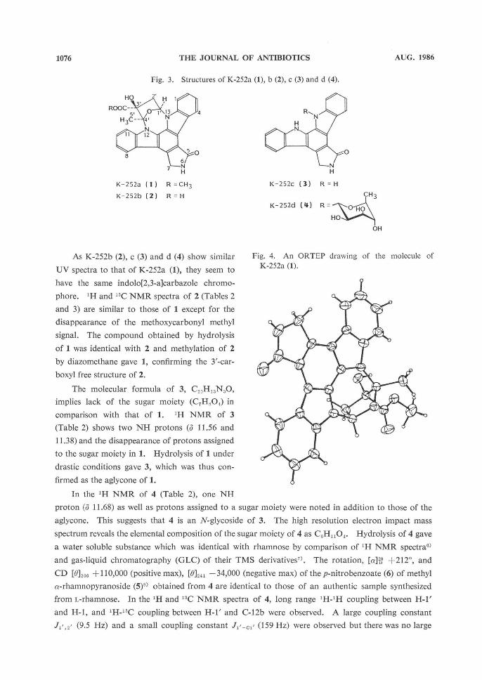
the structure of 1 was determined as shown in Fig. 3. The complete structure with the 3'-configula-
tion was confirmed by single crystal X-ray analysis (Fig. 4)5).
Table 2. 1H NMR data of K-252a (1), b (2), c (3) and d (4) (400 MHz, DMSO-d,).
Position
1
2
3
4
6
7
8
9
10
11
12
13
1'
2'
3'
4'
5'
6'
2'-OH
3'-OH
4'-OH
COOCH3
Chemical shift a (J, Hz)
1
7.90 (d, 8.3) 7.49 (br t)b 7.29 (br t) 9.24 (d, 7.9) 8.64 (br s) 5.04, 5.00
(AB, 17.3) 8.05 (d, 7.8) 7.36 (br t) 7.49 (br t)b 7.95 (d, 8.5)
7.15 (dd, 7.4, 4.9) 3.41 (dd, 14.0, 7.4) 2.04 (dd, 14.0, 4.9)
2.16 (s)
3.94 (s)
2
7.91 (d, 8.4) 7.48 (br t)b 7.28 (br t) 9.22 (d, 7.8) 8.62 (br s) 5.03,5.04 (AB, 17.3) 8.05 (d, 7.8) 7.36 (br t) 7.48 (br t)' 7.98 (d, 8.5)
7.12 (dd, 7.3, 4.9) 3.36 (dd, 13.8, 7.3) 1.98 (dd, 13.8, 4.9)
2.22 (s)
3
7.73 (d, 8.1) 7.44 (br t) 7.24 (br t) 9.24 (d, 7.9) 8.49 (br s) 4.98 (s)
8.05 (d, 7.8) 7.31 (br t) 7.48 (br t) 7.79 (d, 8.1)
11. 56 (br s) 11.38 (br s)
4
7.69 (d, 8.3) 7.49 (br t) 7.27 (br t) 9.47 (d, 7.7) 8.54 (br s) 5.01 (br s)
8.07 (d, 7.8) 7.31 (br t) 7.50 (br t) 7.60 (d, 8.1)
11.68 (br s)
6.40 (br d, 9.5) ca. 4.5 (m)
4.18 (br q)c 4.05 (br t)c4.48 (br q, 7.6) 1.70 (d, 7.2) ca. 5.0d5.40 (d, 3.6) 6.69 (br s)
Chemical shift in ppm from TMS as an internal reference. b These signals are overlapped
.
Coupling constants are ca. 3 Hz. a This signal is overlapped with 7-H_ (d 5.01).

1075VOL. XXXIX NO. 8 THE JOURNAL OF ANTIBIOTICS
Table 3. 13C NMR data of K-252a (1), b (2), c (3) and d (4) (100 MHz, DMSO-d6).
Position
1
2 3 4
4a
4b 4c
5
7 7a 7b
7c 8 9
10 11 1la 12a
12b 13a 1'
2' 3'
4' 5' 6'
COOCH3
1
109.0
125.4 119.4 125.6 122.6
115.8 119.5 171.7
45.4
132.9 114.6 124.1 121.2 120.4
125.0 114.7 139.8 128.3
123.9 136.8 84.95
42.5
84.92 99.3 22.8 172.8 52.6
2
109.0
125.3 119.4 125.6 122.6
115.7 119.5 171.8
45.4
132.9 114.5 124.1 121.2 120.3
124.9 114.8 139.9 128.3
123.9 136.8 85.0 42.5
84.4 99.2 22.8
174.0
3
112.0
125.1 119.0 125.4 123.0
115.7 120.0 172.6 45.4
133.0 114.2 122.7 121.2 120.0
125.1 111.4 139.2 128.0
125.2 139.3
4
109.7 125.1
119.2 125.5 122.2
117.4 118.5 172.2
45.1
133.8 114.8 121.8 121.1 119.7
125.0 111.1 138.9 127.5 124.4
140.1 77.1 66.8 71.6
71.4
76.4 15.3
Chemical shift in ppm from TMS as an internal reference.
Fig. 2. COLOC spectrum of K-252a (1).
180.0 160. 0 140.0 120.0 100.0 80.0 60.0 40.0 20.0 ppm
1076 THE JOURNAL OF ANTIBIOTICS AUG. 1986
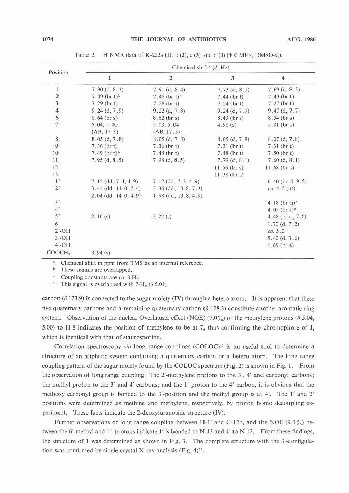
As K-252b (2), c (3) and d (4) show similar
UV spectra to that of K-252a (1), they seem to
have the same indolo[2,3-a]carbazole chromo-
phore. 1H and 13C NMR spectra of 2 (Tables 2
and 3) are similar to those of 1 except for the
disappearance of the methoxycarbonyl methyl
signal. The compound obtained by hydrolysis
of 1 was identical with 2 and methylation of 2
by diazomethane gave 1, confirming the 3'-car-
boxyl free structure of 2.
The molecular formula of 3, C20H13N3O,
implies lack of the sugar moiety (C7H8O4,) in
comparison with that of 1. 1H NMR of 3
(Table 2) shows two NH protons (o 11.56 and
11.38) and the disappearance of protons assigned
to the sugar moiety in 1. Hydrolysis of 1 under
drastic conditions gave 3, which was thus con-
firmed as the aglycone of 1.
In the 1H NMR of 4 (Table 2), one NH
proton (0 11.68) as well as protons assigned to a sugar moiety were noted in addition to those of the
aglycone. This suggests that 4 is an N-glycoside of 3. The high resolution electron impact mass
spectrum reveals the elemental composition of the sugar moiety of 4 as C,H1,0,. Hydrolysis of 4 gave
a water soluble substance which was identical with rhamnose by comparison of 1H NMR spectral)
and gas liquid chromatography (GLC) of their TMS derivatives'. The rotation, [a]26D +212°, and
CD [012,,6 +110,000 (positive max), [0]241 -34,000 (negative max) of the p-nitrobenzoate (6) of methyl
a-rhamnopyranoside (5)6) obtained from 4 are identical to those of an authentic sample synthesized
from L-rhamnose. In the 1H and 13C NMR spectra of 4, long range 1H-1H coupling between H-1'
and H-1, and 1H-13C coupling between H-1' and C-12b were observed. A large coupling constant
J, (9.5 Hz) and a small coupling constant J,'-c,' (159 Hz) were observed but there was no large
Fig. 3. Structures of K-252a (1), b (2), c (3) and d (4).
K-252a (1) R =CH3
K-252b (2) R=H
K-252c (3) R = H
K-252d (4) R =
Fig. 4. An ORTEP drawing of the molecule of
K-252a (1).

1077VOL. XXXIX NO. 8 THE JOURNAL OF ANTIBIOTICS
coupling constant found for J2' ,3',J3',4' and J,4,5'. The structure of 4 is determined, therefore, as 13-
N-(a-L-rhamnopyranosyl)-K-252c, where the rhamnopyranosyl moiety takes the 1C4 conformation.
Experimental
NMR spectra were measured on a Jeol FX100 and Bruker AM400 spectrometer. Mass spectra were obtained on a Hitachi M-80B spectrometer at 70 eV. IR spectra were measured with a Shimadzu IR-27G spectrometer. UV spectra were taken with a Hitachi 200-20 spectrometer. Optical rotations were measured with a Perkin-Elmer 141 polarimeter. CD spectra were taken on a Jasco J-500A spec-tropolarimeter and a Jasco DP-501 data processor. Melting points were taken with a Yanagimoto micro melting point apparatus and were not corrected. GLC was carried out on a Hitachi 263-70 gas chromatograph. Thin-layer chromatography (TLC) was performed on pre-coated plates, Merck Kieselgel 60 F254 and detected with UV light (254 nm) and I % Ce(SO4)2 - 10 % H2SO, reagent.
Hydrolysis of 1 A suspension of K-252a (1, 20 mg) in MeOH (10 ml) and 1 m NaOH solution (1 ml) was stirred
for 5 hours at room temp. The reaction mixture was concentrated in vacuo to remove MeOH. After acidification with 2 m HCl, the solution was extracted with EtOAc. EtOAc solution was washed with brine, dried over MgSO4 and evaporated to give 2 (18 mg, 93 %).
Methylation of 2 To a suspension of K-252b (2, 2.0 mg) in MeOH (3 ml), diazomethane in ether (1 ml) was added and stirred for 4 hours at room temp. The reaction mixture was evaporated to give 1 (2.0 mg, 97%).
Hydrolysis of 4 K-252d (4, 10 mg) was suspended in 2 m hydrochloric acid and heated to reflux for 2 hours. The reaction mixture was filtered to remove the solid (3, 5 mg) and washed with H2O. The filtrate and washings were combined and evaporated to give a colorless solid, 2.5 mg (77 %), which was identical with rhamnose by 'H NMR spectroscopy. Rhamnose was further identified by GLC of its TMS de-rivative as follows.
GLC Analysis of TMS Derivative of Rhamnose
Rhamnose (1 mg), given by hydrolysis of 4, was dissolved in TMS-Pz (Tokyo Kasei, T0623, 0.1 ml) and held for 15 minutes. The reaction mixture was diluted with 10 volumes of acetone and subjected to GLC using a silicone OV-101 25 m (0.25 mm i.d.) FS-WCOT capillary column, detection: FID, flow rate: N2 15 ml/minute, temp: 210°C (injection), 180°C (column), 210°C (detection), t0: a-TMS-rhamnose, 8'12"; j3-TMS-rhamnose, 10'24", which were identical with an authentic sample
prepared from L-rhamnose in a similar manner.
Preparation of 7
Rhamnose (1.5 mg), given by the hydrolysis of 4, was dissolved in 17% HCl in MeOH (1 ml) and heated to reflux for 1 hour. The reaction mixture was evaporated to give methyl a-rhamnopyranoside
(5, 1.6 mg)'), which was dissolved in dry pyridine (0.5 ml), treated with p-nitrobenzoyl chloride (20 mg) and stirred for 2 hours under N2 atmosphere at room temp. The reaction mixture was evaporated to
give a residue, which was chromatographed on a silicagel column, giving a colorless powder (6, 4 mg). Recrystallization from hexane - EtOAc gave colorless needles: MP 103 - 104°C; [a]20D +212° (c 0.2, CHCl,); CD (dioxane) [8]246 +110,000 (positive max), [01211 0110121, -34,000 (negative max), [01223 0,
[8]220 +5,000; 'H NMR (100 MHz, CDCl3) 6 1.41 (3H, d, J=6.1 Hz), 3.54 (3H, s), 4.25 (1H, dq, J= 9.3, 6.1 Hz), 4.93 (1 H, d, J 1.5 Hz), 5.5 5.9 (3H, m), 7.9 8.4 (12H).
References
1) KASE, H.; K. IWAHASHI & Y. MATSUDA: K-252a, a potent inhibitor of protein kinase C from microbial origin. J. Antibiotics 39: 1059- 1065, 1986

1078 THE JOURNAL OF ANTIBIOTICS AUG. 1986
2) NAKANISHI, S.; Y. MATSUDA, K. IWAHASHI & H. KASE: K-252b, c and d, potent inhibitors of protein kinase C from microbial origin. J. Antibiotics 39: 1066 - 1071, 1986
3) OMURA, S.; Y. IWAI, A. HIRANO, A. NAKAGAWA, J. AWAYA, H. TSUCHIYA, Y. TAKAHASHI & R. MASUMA: A new alkaloid AM-2282 of Streptomyces origin. Taxonomy, fermentation, isolation and preliminary
characterization. J. Antibiotics 30: 275-282, 1977 4) KESSLER, H.; C. GRIESINGER, J. ZARBOCK & H. R. LooSLI: Assignment of carbonyl carbons and sequence
analysis in peptides by heteronuclear shift correlation via small coupling constants with broadband de- coupling in t, (COLOC). J. Magn. Reson. 57: 331- 336, 1984
5) HIRAYAMA, N.; T. IIDA & K. SHIRAHATA: Novel protein kinase C inhibitor K-252a. Acta Crystallogra-
phica (C), in preparation 6) DE BRUYN, A. & M. ANTEUNIS: IH-N.m.r. study of L-rhamnose, methyl a-L-rhamnopyranoside, and 4-O-~-D-galactopyranosyl-L-rhamnose in deuterium oxide. Carbohydr. Res. 47: 158163, 1976 7) SWEELEY, C. C.; R. BENTLEY, M. MAKITA & W. W. WELLS: Gas-liquid chromatography of trimethylsilyl
derivatives of sugars and related substances. J. Am. Chem. Soc. 85: 2497 - 2507, 1963 8) BREITMAIER, E.; W. VOELTER, G. JUNG & C. TANZER: Konfigulations-, konformations- and substituen-
teneinflusse auf die 13C-chemischen verschiebungen von glykosiden. Chem. Ber. 104: 1147 - 1154, 1971