Embed Size (px)
Citation preview
JMB—MS 636
J. Mol. Biol. (1995) 250, 659–671
Three-dimensional Solution Structure of the CalciumChannel Antagonist v-Agatoxin IVA: ConsensusMolecular Folding of Calcium Channel Blockers
Jae Il Kim 1*, Shiro Konishi 1, Hideo Iwai 2, Toshiyuki Kohno 1
Hiroaki Gouda 2, Ichio Shimada 2, Kazuki Sato 1 and Yoji Arata 2,3
The three-dimensional solution structure of v-agatoxin IVA, which is a1Mitsubishi Kasei Institute ofspecific blocker of the P-type calcium channel isolated from funnel webLife Sciences, 11 Minamiooyaspider venom and has a molecular mass of 5.2 kDa, was determined by twoMachida-shi, Tokyo 194, Japandimensional 1H NMR spectroscopy, combined with simulated annealing2Faculty of Pharmaceutical calculations. On the basis of 563 experimental constraints, including 516
Sciences, University of Tokyo distance constraints obtained from the nuclear Overhauser effect, 21 torsionBunkyo-ku, Tokyo 113, Japan angle (f,x1) constraints, and 26 constraints associated with hydrogen bonds
and disulfide bonds, a total of 14 converged structures were obtained. The3Water Research Instituteatomic root mean square difference for the 14 converged structures withSengen 2-1-6, Tsukubarespect to the mean coordinates is 0.42 (20.07) Å for the backbone atomsIbaraki 305, Japan(N, Ca, C) and 0.95 (20.15) Å for all heavy atoms of the central part (residues4 to 38) constrained by four disulfide bonds. The N- and C-terminal segments(residues 1 to 3 and 39 to 48, respectively) have a disordered structure inaqueous solution. The molecular structure of v-agatoxin IVA is composed ofa short triple-stranded antiparallel b-sheet, three loops, and the disorderedN- and C-terminal segments. The overall b-sheet topology is +2x, −1, whichis the same as that reported for v-conotoxin GVIA, an N-type calciumchannel blocker. Irrespective of differences in the number of disulfide bondsand low primary sequence homology, these two peptide toxins show asignificant structural similarity in three dimensions. The whole-cell voltage-clamp recording using rat cerebellar slices suggests that the hydrophobicC-terminal segment of v-agatoxin IVA, which does not exist in v-conotoxinGVIA, plays a crucial role in the blocking action of v-agatoxin IVA on theP-type calcium channel in rat cerebellar Purkinje cells. The present studyprovides a molecular basis for the toxin–channel interaction, and therebyprovides insight into the discrimination of different subtypes of calciumchannels.
7 1995 Academic Press Limited
Keywords: calcium channel blocker; v-agatoxin IVA; proton nuclearmagnetic resonance spectroscopy; structure–activity relationships; three*Corresponding authordimensional structure similarity
Introduction
Many peptide toxins have been used as potentpharmacological tools for studying the roles of ionchannels and neurotransmitter receptors (Gray et al.,
1988). As the number of cloned receptors and ionchannels grows, the peptide toxins have becomemore important as specific probes for their charac-terization (Adams & Olivera, 1994). The venoms ofscorpion, spider, and cone snail are major sources of
Abbreviations used: v-Aga-IVA, v-agatoxin IVA; v-Aga-IVB, v-agatoxin IVB; v-CTX-GVIA, v-conotoxin GVIA;ESI-MS, electrospray ionization mass spectrometry; Fmoc, 9-fluorenylmethoxycarbonyl; HPLC, high performanceliquid chromatography; NMR, nuclear magnetic resonance; NOE, nuclear Overhauser effect; p.p.m., parts permillion; RMS, root mean square; RMSD, root-mean-square deviation; TFA, trifluoroacetic acid; DQF-COSY,double-quantum-filtered correlated spectroscopy; HOHAHA, homonuclear Hartmann-Hahn, v-Aga-IIIA, v-agatoxinIIIA; ACSF, artificial cerebrospinal fluid; EGTA, ethylene glycol bis (b-aminoethyl ether)-N,N,N',N'-tetraacetic acid;PE-COSY, primitive exclusive COSY; TPPI, time-proportional phase incrementation.
0022–2836/95/300659–13 $08.00/0 7 1995 Academic Press Limited
JMB—MS 636
NMR Structure of v-Agatoxin IVA660
neurotoxins, and the characterization of toxin actionsis currently a major focus in the field of neuroscience.
The voltage-sensitive calcium channels are classi-fied into several subtypes, according to their electro-physiological and pharmacological properties (Tsienet al., 1988). L-type calcium channels are inhibitedby dihydropyridines (Rampe & Triggle, 1989),and N-type calcium channels are selectivelyblocked by v-conotoxin GVIA (v-CTX-GVIA), a27mer peptide toxin isolated from the predatorymarine snail Conus geographus (Olivera et al., 1984;McCleskey et al., 1987). The P-type calcium channels(Uchitel et al., 1992) in rat Purkinje neurons areselectively blocked by v-agatoxin IVA (v-Aga-IVA),a 48mer peptide toxin isolated from the venom of thefunnel web spider Agelenopsis aperta (Mintz et al.,1992a,b).
The two peptide toxins v-Aga-IVA and v-CTX-GVIA are best characterized, in their biologicalactions, among many calcium channel blockers, andare the toxins most widely used for the identificationof calcium channels associated with particular syn-aptic transmissions. Recently, the three-dimensionalstructure of v-CTX-GVIA (Davis et al., 1993; Pallaghyet al., 1993; Sevilla et al., 1993; Skalicky et al., 1993)was determined using NMR spectroscopy, and itsactive site was revealed by the synthesis of a seriesof analogs with alanine substitution (Sato et al., 1993;Kim et al., 1994). These studies on the structure–activity relationship of v-CTX-GVIA have provideda molecular basis for the mode of interaction, as wellas toxin specificity to calcium channel subtypes.
In the present study, we chemically synthesizedv-Aga-IVA by solid-phase methodology to obtain the
toxin in an amount sufficient for NMR studies. Thethree-dimensional structure of synthetic v-Aga-IVAwas determined in aqueous solution by two-dimensional 1H NMR and simulated annealingcalculations. On the basis of analysis of the deter-mined structure, it appeared that the overall foldingof v-Aga-IVA shows a significant similarity to that ofv-CTX-GVIA. The solution structure of v-Aga-IVAis also discussed in terms of the structure–activityrelationship of the v-agatoxins, determined byelectrophysiological experiments using the C-terminal truncated v-Aga-IVA.
Results
Sequence-specific resonance assignments
Sequence-specific resonance assignments weremade according to the standard method for smallproteins (Wuthrich, 1986). The identification ofamino acid spin systems was based on scalarcoupling patterns observed in double-quantum-filtered-correlated spectroscopy (DQF-COSY) andhomonuclear Hartmann-Hahn (HOHAHA) exper-iments, and complemented with the result of nuclearOverhauser effect spectroscopy (NOESY) measure-ments. The identified spin systems were orderedalong the primary structure of v-Aga-IVA throughinterresidue sequential NOEs observed on theNOESY spectrum. The sequential connectivitieswere carried out by the analysis of the CaH(i )–NH(i + 1) (daN), CbH(i )–NH(i + 1) (dbN), and NH(i )–NH(i + 1) (dNN) NOEs. Since Asp, Tyr, Trp, Ser andAsn are each present only once in the primary
Figure 1. Sequential daN(i, i + 1)NOE connectivities for residues 4 to38 in the NOESY spectrum observedwith a mixing time of 200 ms. The daN
connectivities of the proline residuesare shown for Pro18/Cys19 andPro38/Arg39. Intraresidue NH–CaHcross-peaks not shown in this plot areindicated with asterisks (*). Intra-residue NH–CaH cross-peaks arelabeled with the residue number bystandard single-letter amino acidabbreviations.
JMB—MS 636
NMR Structure of v-Agatoxin IVA 661
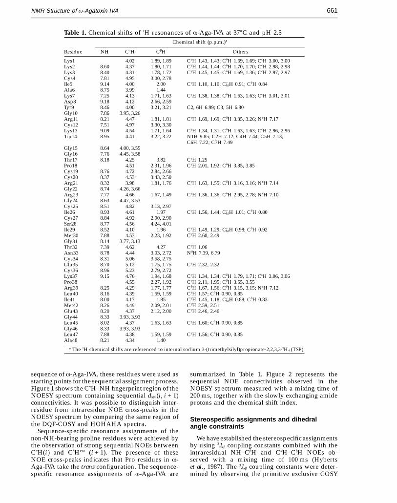
Table 1. Chemical shifts of 1H resonances of v-Aga-IVA at 37°C and pH 2.5Chemical shift (p.p.m.)a
Residue NH CaH CbH Others
Lys1 4.02 1.89, 1.89 CgH 1.43, 1.43; CdH 1.69, 1.69; CeH 3.00, 3.00Lys2 8.60 4.37 1.80, 1.71 CgH 1.44, 1.44; CdH 1.70, 1.70; CeH 2.98, 2.98Lys3 8.40 4.31 1.78, 1.72 CgH 1.45, 1.45; CdH 1.69, 1.36; CeH 2.97, 2.97Cys4 7.81 4.95 3.00, 2.78Ile5 9.14 4.00 2.00 CgH 1.10, 1.10; Cg
MH 0.91; CdH 0.84Ala6 8.75 3.99 1.44Lys7 7.25 4.13 1.71, 1.63 CgH 1.38, 1.38; CdH 1.63, 1.63; CeH 3.01, 3.01Asp8 9.18 4.12 2.66, 2.59Tyr9 8.46 4.00 3.21, 3.21 C2, 6H 6.99; C3, 5H 6.80Gly10 7.86 3.95, 3.26Arg11 8.21 4.47 1.81, 1.81 CgH 1.69, 1.69; CdH 3.35, 3.26; NeH 7.17Cys12 7.51 4.97 3.30, 3.30Lys13 9.09 4.54 1.71, 1.64 CgH 1.34, 1.31; CdH 1.63, 1.63; CeH 2.96, 2.96Trp14 8.95 4.41 3.22, 3.22 N1H 9.85; C2H 7.12; C4H 7.44; C5H 7.13;
C6H 7.22; C7H 7.49Gly15 8.64 4.00, 3.55Gly16 7.76 4.45, 3.58Thr17 8.18 4.25 3.82 CgH 1.25Pro18 4.51 2.31, 1.96 CgH 2.01, 1.92; CdH 3.85, 3.85Cys19 8.76 4.72 2.84, 2.66Cys20 8.37 4.53 3.43, 2.50Arg21 8.32 3.98 1.81, 1.76 CgH 1.63, 1.55; CdH 3.16, 3.16; NeH 7.14Gly22 8.74 4.26, 3.66Arg23 7.77 4.66 1.67, 1.49 CgH 1.36, 1.36; CdH 2.95, 2.78; NeH 7.10Gly24 8.63 4.47, 3.53Cys25 8.51 4.82 3.13, 2.97Ile26 8.93 4.61 1.97 CgH 1.56, 1.44; Cg
MH 1.01; CdH 0.80Cys27 8.84 4.92 2.90, 2.90Ser28 8.77 4.56 4.24, 4.01Ile29 8.52 4.10 1.96 CgH 1.49, 1.29; Cg
MH 0.98; CdH 0.92Met30 7.88 4.53 2.23, 1.92 CgH 2.60, 2.49Gly31 8.14 3.77, 3.13Thr32 7.39 4.62 4.27 CgH 1.06Asn33 8.78 4.44 3.03, 2.72 NdH 7.39, 6.79Cys34 8.31 5.06 3.58, 2.75Glu35 8.70 5.12 1.75, 1.75 CgH 2.32, 2.32Cys36 8.96 5.23 2.79, 2.72Lys37 9.15 4.76 1.94, 1.68 CgH 1.34, 1.34; CdH 1.79, 1.71; CeH 3.06, 3.06Pro38 4.55 2.27, 1.92 CgH 2.11, 1.95; CdH 3.55, 3.55Arg39 8.25 4.29 1.77, 1.77 CdH 1.67, 1.56; CdH 3.15, 3.15; NeH 7.12Leu40 8.16 4.39 1.59, 1.59 CgH 1.57; CdH 0.90, 0.85Ile41 8.00 4.17 1.85 CgH 1.45, 1.18; Cg
MH 0.88; CdH 0.83Met42 8.26 4.49 2.09, 2.01 CgH 2.59, 2.51Glu43 8.20 4.37 2.12, 2.00 CgH 2.46, 2.46Gly44 8.33 3.93, 3.93Leu45 8.02 4.37 1.63, 1.63 CgH 1.60; CdH 0.90, 0.85Gly46 8.33 3.93, 3.93Leu47 7.88 4.38 1.59, 1.59 CgH 1.56; CdH 0.90, 0.85Ala48 8.21 4.34 1.40
a The 1H chemical shifts are referenced to internal sodium 3-(trimethylsilyl)propionate-2,2,3,3-2H4 (TSP).
sequence of v-Aga-IVA, these residues were used asstarting points for the sequential assignment process.Figure 1 shows the CaH–NH fingerprint region of theNOESY spectrum containing sequential daN(i, i + 1)connectivities. It was possible to distinguish inter-residue from intraresidue NOE cross-peaks in theNOESY spectrum by comparing the same region ofthe DQF-COSY and HOHAHA spectra.
Sequence-specific resonance assignments of thenon-NH-bearing proline residues were achieved bythe observation of strong sequential NOEs betweenCaH(i ) and CdHPro (i + 1). The presence of theseNOE cross-peaks indicates that Pro residues in v-Aga-IVA take the trans configuration. The sequence-specific resonance assignments of v-Aga-IVA are
summarized in Table 1. Figure 2 represents thesequential NOE connectivities observed in theNOESY spectrum measured with a mixing time of200 ms, together with the slowly exchanging amideprotons and the chemical shift index.
Stereospecific assignments and dihedralangle constraints
We have established the stereospecific assignmentsby using 3Jab coupling constants combined with theintraresidual NH–CbH and CaH–CbH NOEs ob-served with a mixing time of 100 ms (Hybertset al., 1987). The 3Jab coupling constants were deter-mined by observing the primitive exclusive COSY
JMB—MS 636
NMR Structure of v-Agatoxin IVA662
(PE-COSY) spectrum in 2H2O solution, in which thecross-peaks gave the passive coupling between Ca
and Cb protons. On the basis of the 3Jab couplingconstants and the intensities of intraresidual NOEs,we have established the stereospecific assignments ofthe prochiral b-methylene protons and the range ofx1 side-chain torsion angles for seven residues, i.e.Cys4, Cys19, Cys20, Arg23, Cys25, Cys34, and Lys37.For t2g3, g2g3, and g2t3 conformations around theCa–Cb bond, the x1 angle was constrained in the rangeof −60(240)°, 60(240)°, and 180(240)°, respectively(Wagner et al., 1987).
The 3JHNa coupling constants were estimated onthe DQF-COSY spectrum and converted to f angleconstraints according to the following rules: for3JHNa < 5.5 Hz, and >8 Hz, the f angle was con-strained to be −65(225)° and −120(240)°, respect-ively (Pardi et al., 1984). Five residues (Lys7, Asp8,Trp14, Thr17, and Cys20) with 3JHNa less than 5.5 Hz,and nine residues (Cys12, Lys13, Arg23, Ile26, Cys27,Met30, Thr32, Glu35, and Lys37) with 3JHNa greaterthan 8 Hz, were constrained by these rules.
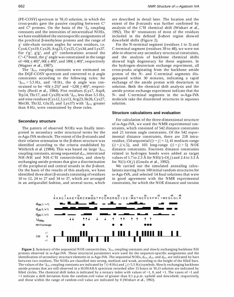
Secondary structure
The pattern of observed NOEs was finally inter-preted in secondary order structural terms for thev-Aga-IVA molecule. The extent of the b-strands andtheir relative orientation in the b-sheet structure wasidentified according to the criteria established byWuthrich et al. (1984). This was based on large 3JHNa
coupling constants, strong sequential daN, interstrandNH–NH and NH–CaH connectivities, and slowlyexchanging amide protons that give a discriminationof the peripheral and central strands in the b-sheet.On the basis of the results of this analysis, we haveidentified three short b-strands consisting of residues10 to 12, 24 to 27 and 34 to 37, which are arrangedin an antiparallel fashion, and several turns, which
are described in detail later. The location and theextent of the b-strands was further confirmed byanalysis of the CaH chemical shift (Wishart et al.,1992). The Ha resonances of most of the residuesincluded in the defined b-sheet region showeddownfield shifts (Figure 2).
For the N-terminal segment (residues 1 to 3) andC-terminal segment (residues 39 to 48), we were notable to observe any secondary structural constraints,and the analysis of backbone chemical shiftsshowed high degeneracy for these segments. Inthe hydrogen–deuterium exchange experiment, allcross-peaks originating from the backbone amideproton of the N- and C-terminal segments dis-appeared within 30 minutes, indicating a rapidexchange of the amide proton with deuterium insolution. Both the chemical shift analysis and theamide proton exchange experiment indicate that theN- and C-terminal segments in the v-Aga-IVAmolecule take the disordered structures in aqueoussolution.
Structure calculations and evaluation
For calculation of the three-dimensional structureof v-Aga-IVA, we used the NMR experimental con-straints, which consisted of 542 distance constraintsand 21 torsion angle constraints. Of the 542 exper-imental distance constraints, there are 218 intra-residue, 154 sequential (=i − j = = 1), 41 medium-range(=i − j =E5), and 103 long-range (=i − j = > 5) NOEdistance constraints. Fourteen distance constraintsrelated to hydrogen bonds were added as targetvalues of 1.7 to 2.3 A for NH(i )–O( j ) and 2.4 to 3.3 Afor N(i )–O( j ) (Gouda et al., 1992).
We carried out the simulated annealing calcu-lations starting from 100 initial random structures forv-Aga-IVA, and selected 14 final solutions that werein good agreement with the NMR experimentalconstraints, for which the NOE distance and torsion
Figure 2. Summary of the sequential NOE connectivities, 3JHNa coupling constants and slowly exchanging backbone NHprotons observed in v-Aga-IVA. These structural parameters were used for the sequence-specific assignments and theidentification of secondary structure elements in v-Aga-IVA. The sequential NOEs, dNN, daN, and dbN, are indicated by barsbetween two residues. The NOEs are classified into strong, medium and weak, according to the height of the filled bars.The values of the 3JHNa coupling constants are indicated by 3 (>8 Hz) and 4 (<5.5 Hz) symbols. Slowly exchanging backboneamide protons that are still observed in a HOHAHA spectrum recorded after 15 hours in 2H2O solution are indicated byfilled circles. The chemical shift index is indicated by a ternary index with values of −1, 0, and +1. The vaues of −1 and+1 indicate a shift deviation from the random-coil value of greater than 0.1 p.p.m. upfield and downfield, respectively,and those within the range of random-coil value are indicated by 0 (Wishart et al., 1992).
JMB—MS 636
NMR Structure of v-Agatoxin IVA 663
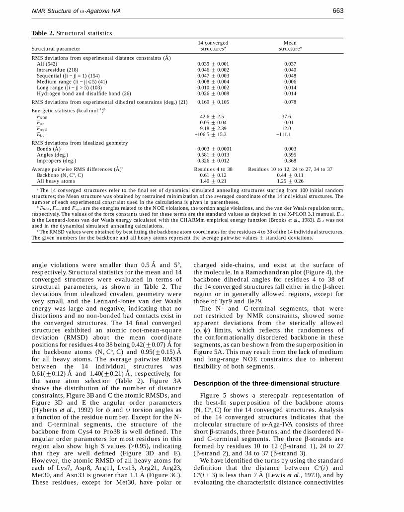
Table 2. Structural statistics14 converged Mean
Structural parameter structuresa structurea
RMS deviations from experimental distance constraints (A)All (542) 0.039 2 0.001 0.037Intraresidue (218) 0.046 2 0.002 0.040Sequential (=i − j= = 1) (154) 0.047 2 0.003 0.048Medium range (=i − j=E5) (41) 0.008 2 0.004 0.006Long range (=i − j= > 5) (103) 0.010 2 0.002 0.014Hydrogen bond and disulfide bond (26) 0.026 2 0.008 0.014
RMS deviations from experimental dihedral constraints (deg.) (21) 0.169 2 0.105 0.078
Energetic statistics (kcal mol−1)b
FNOE 42.6 2 2.5 37.6Ftor 0.05 2 0.04 0.01Frepel 9.18 2 2.39 12.0EL-J −106.5 2 15.3 −111.1
RMS deviations from idealized geometryBonds (A) 0.003 2 0.0001 0.003Angles (deg.) 0.581 2 0.013 0.595Impropers (deg.) 0.326 2 0.012 0.368
Average pairwise RMS differences (A)c Residues 4 to 38 Residues 10 to 12, 24 to 27, 34 to 37Backbone (N, Ca, C) 0.61 2 0.12 0.44 2 0.11All heavy atoms 1.40 2 0.21 1.25 2 0.26a The 14 converged structures refer to the final set of dynamical simulated annealing structures starting from 100 initial random
structures; the Mean structure was obtained by restrained minimization of the averaged coordinate of the 14 individual structures. Thenumber of each experimental constraint used in the calculations is given in parentheses.
b FNOE, Ftor, and Frepel are the energies related to the NOE violations, the torsion angle violations, and the van der Waals repulsion term,respectively. The values of the force constants used for these terms are the standard values as depicted in the X-PLOR 3.1 manual. EL-J
is the Lennard-Jones van der Waals energy calculated with the CHARMm empirical energy function (Brooks et al., 1983). EL-J was notused in the dynamical simulated annealing calculations.
c The RMSD values were obtained by best fitting the backbone atom coordinates for the residues 4 to 38 of the 14 individual structures.The given numbers for the backbone and all heavy atoms represent the average pairwise values 2 standard deviations.
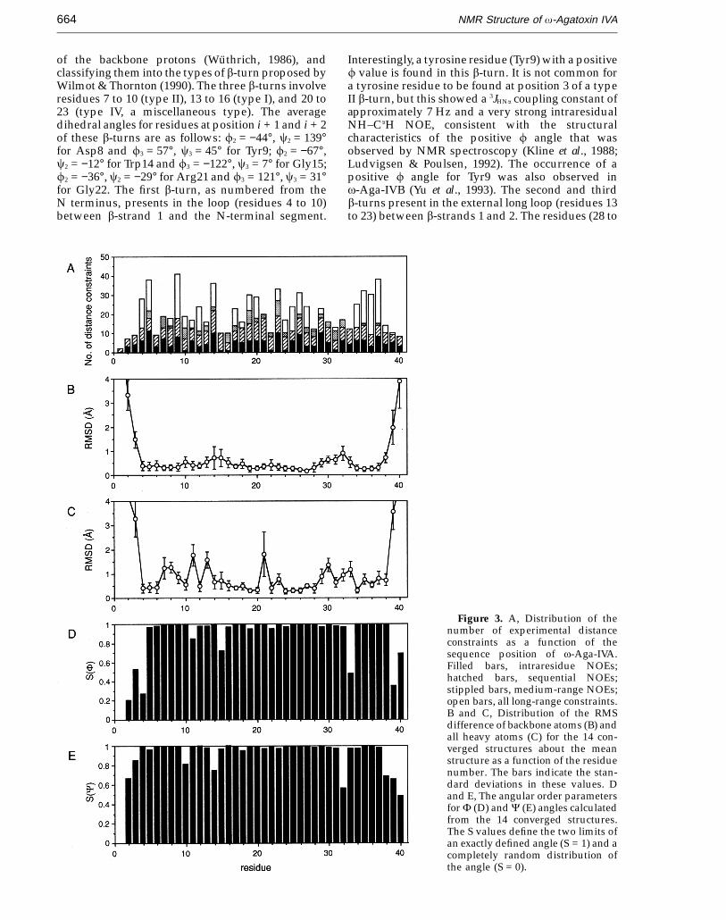
angle violations were smaller than 0.5 A and 5°,respectively. Structural statistics for the mean and 14converged structures were evaluated in terms ofstructural parameters, as shown in Table 2. Thedeviations from idealized covalent geometry werevery small, and the Lennard-Jones van der Waalsenergy was large and negative, indicating that nodistortions and no non-bonded bad contacts exist inthe converged structures. The 14 final convergedstructures exhibited an atomic root-mean-squaredeviation (RMSD) about the mean coordinatepositions for residues 4 to 38 being 0.42(20.07) A forthe backbone atoms (N, Ca, C) and 0.95(20.15) Afor all heavy atoms. The average pairwise RMSDbetween the 14 individual structures was0.61(20.12) A and 1.40(20.21) A, respectively, forthe same atom selection (Table 2). Figure 3Ashows the distribution of the number of distanceconstraints, Figure 3B and C the atomic RMSDs, andFigure 3D and E the angular order parameters(Hyberts et al., 1992) for f and c torsion angles asa function of the residue number. Except for the N-and C-terminal segments, the structure of thebackbone from Cys4 to Pro38 is well defined. Theangular order parameters for most residues in thisregion also show high S values (>0.95), indicatingthat they are well defined (Figure 3D and E).However, the atomic RMSD of all heavy atoms foreach of Lys7, Asp8, Arg11, Lys13, Arg21, Arg23,Met30, and Asn33 is greater than 1.1 A (Figure 3C).These residues, except for Met30, have polar or
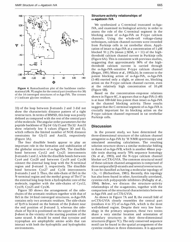
charged side-chains, and exist at the surface ofthe molecule. In a Ramachandran plot (Figure 4), thebackbone dihedral angles for residues 4 to 38 ofthe 14 converged structures fall either in the b-sheetregion or in generally allowed regions, except forthose of Tyr9 and Ile29.
The N- and C-terminal segments, that werenot restricted by NMR constraints, showed someapparent deviations from the sterically allowed(f, c) limits, which reflects the randomness ofthe conformationally disordered backbone in thesesegments, as can be shown from the superposition inFigure 5A. This may result from the lack of mediumand long-range NOE constraints due to inherentflexibility of both segments.
Description of the three-dimensional structure
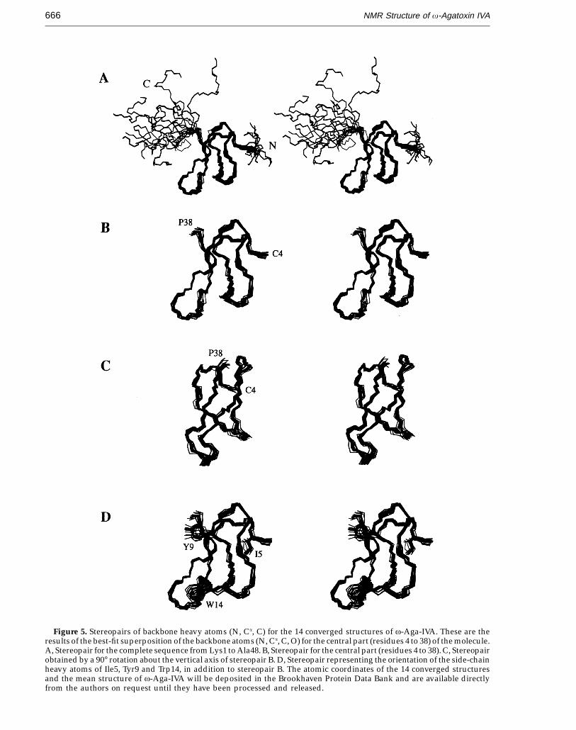
Figure 5 shows a stereopair representation ofthe best-fit superposition of the backbone atoms(N, Ca, C) for the 14 converged structures. Analysisof the 14 converged structures indicates that themolecular structure of v-Aga-IVA consists of threeshort b-strands, three b-turns, and the disordered N-and C-terminal segments. The three b-strands areformed by residues 10 to 12 (b-strand 1), 24 to 27(b-strand 2), and 34 to 37 (b-strand 3).
We have identified the turns by using the standarddefinition that the distance between Ca(i ) andCa(i + 3) is less than 7 A (Lewis et al., 1973), and byevaluating the characteristic distance connectivities
JMB—MS 636
NMR Structure of v-Agatoxin IVA664
of the backbone protons (Wuthrich, 1986), andclassifying them into the types of b-turn proposed byWilmot & Thornton (1990). The three b-turns involveresidues 7 to 10 (type II), 13 to 16 (type I), and 20 to23 (type IV, a miscellaneous type). The averagedihedral angles for residues at position i + 1 and i + 2of these b-turns are as follows: f2 = −44°, c2 = 139°for Asp8 and f3 = 57°, c3 = 45° for Tyr9; f2 = −67°,c2 = −12° for Trp14 and f3 = −122°, c3 = 7° for Gly15;f2 = −36°, c2 = −29° for Arg21 and f3 = 121°, c3 = 31°for Gly22. The first b-turn, as numbered from theN terminus, presents in the loop (residues 4 to 10)between b-strand 1 and the N-terminal segment.
Interestingly, a tyrosine residue (Tyr9) with a positivef value is found in this b-turn. It is not common fora tyrosine residue to be found at position 3 of a typeII b-turn, but this showed a 3JHNa coupling constant ofapproximately 7 Hz and a very strong intraresidualNH–CaH NOE, consistent with the structuralcharacteristics of the positive f angle that wasobserved by NMR spectroscopy (Kline et al., 1988;Ludvigsen & Poulsen, 1992). The occurrence of apositive f angle for Tyr9 was also observed inv-Aga-IVB (Yu et al., 1993). The second and thirdb-turns present in the external long loop (residues 13to 23) between b-strands 1 and 2. The residues (28 to
Figure 3. A, Distribution of thenumber of experimental distanceconstraints as a function of thesequence position of v-Aga-IVA.Filled bars, intraresidue NOEs;hatched bars, sequential NOEs;stippled bars, medium-range NOEs;open bars, all long-range constraints.B and C, Distribution of the RMSdifference of backbone atoms (B) andall heavy atoms (C) for the 14 con-verged structures about the meanstructure as a function of the residuenumber. The bars indicate the stan-dard deviations in these values. Dand E, The angular order parametersfor F (D) and C (E) angles calculatedfrom the 14 converged structures.The S values define the two limits ofan exactly defined angle (S = 1) and acompletely random distribution ofthe angle (S = 0).
JMB—MS 636
NMR Structure of v-Agatoxin IVA 665
Figure 4. Ramachandran plot of the backbone confor-mational (F, C) angles for the central part (residues 4 to 38)of the 14 converged structures of v-Aga-IVA. The crosses(+) indicate glycine residues.
Structure-activity relationships ofv-agatoxin IVA
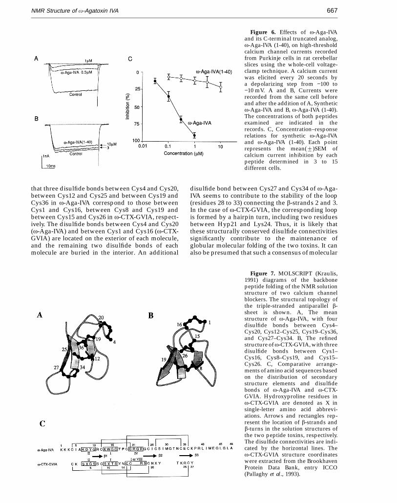
We synthesized a C-terminal truncated v-Aga-IVA, and examined its biological activity in order toassess the role of the C-terminal segment in theblocking action of v-Aga-IVA on P-type calciumchannels. Using the whole-cell voltage-clamptechniques, calcium channel currents were recordedfrom Purkinje cells in rat cerebellar slices. Appli-cation of intact v-Aga-IVA at a concentration of 1 mMblocked 9122% (mean2SEM, n = 11) of the high-threshold calcium channel current in Purkinje cells(Figure 6A). This is consistent with previous studies,suggesting that approximately 90% of the high-threshold calcium current is carried throughthe v-Aga-IVA–sensitive P-type calcium channel(Regan, 1991; Mintz et al., 1992a,b). In contrast to thepotent blocking action of v-Aga-IVA, v-Aga-IVA(1-40) exerted only a slight, or almost no, blockingaction on the P-type calcium channel current, evenat the relatively high concentration of 10 mM(Figure 6B).
Based on the concentration–response relationsshown in Figure 6C, it appears that v-Aga-IVA (1-40)is at least 100-fold less potent than intact v-Aga-IVAin the channel blocking activity. These resultssuggest that the C-terminal segment of v-Aga-IVA iscrucially important for its blocking action on theP-type calcium channel expressed in rat cerebellarPurkinje cells.
Discussion
In the present study, we have determined thethree-dimensional structure of the calcium channelantagonist v-Aga-IVA by 1H NMR spectroscopy andsimulated annealing calculations. The resultingsolution structure shows a similar molecular foldingto those of v-Aga-IVB, which is another 48mer pep-tide toxin sharing nearly 70% sequence homology(Yu et al., 1993), and the N-type calcium channelblocker v-CTX-GVIA. The common structural motifof these calcium channel antagonists is comprised ofthree antiparallel b-strands and several reverse turns.It can be classified as having a molecular topology of+2x, −1 (Richardson, 1981). Recently, this topologyhas also been found in other, functionally unrelated,cysteine-rich polypeptide inhibitors (Pallaghy et al.,1994). Below, we discuss the structure–activityrelationships of the v-agatoxins, together with thecomparison of the structural characteristics betweenv-Aga-IVA and v-CTX-GVIA.
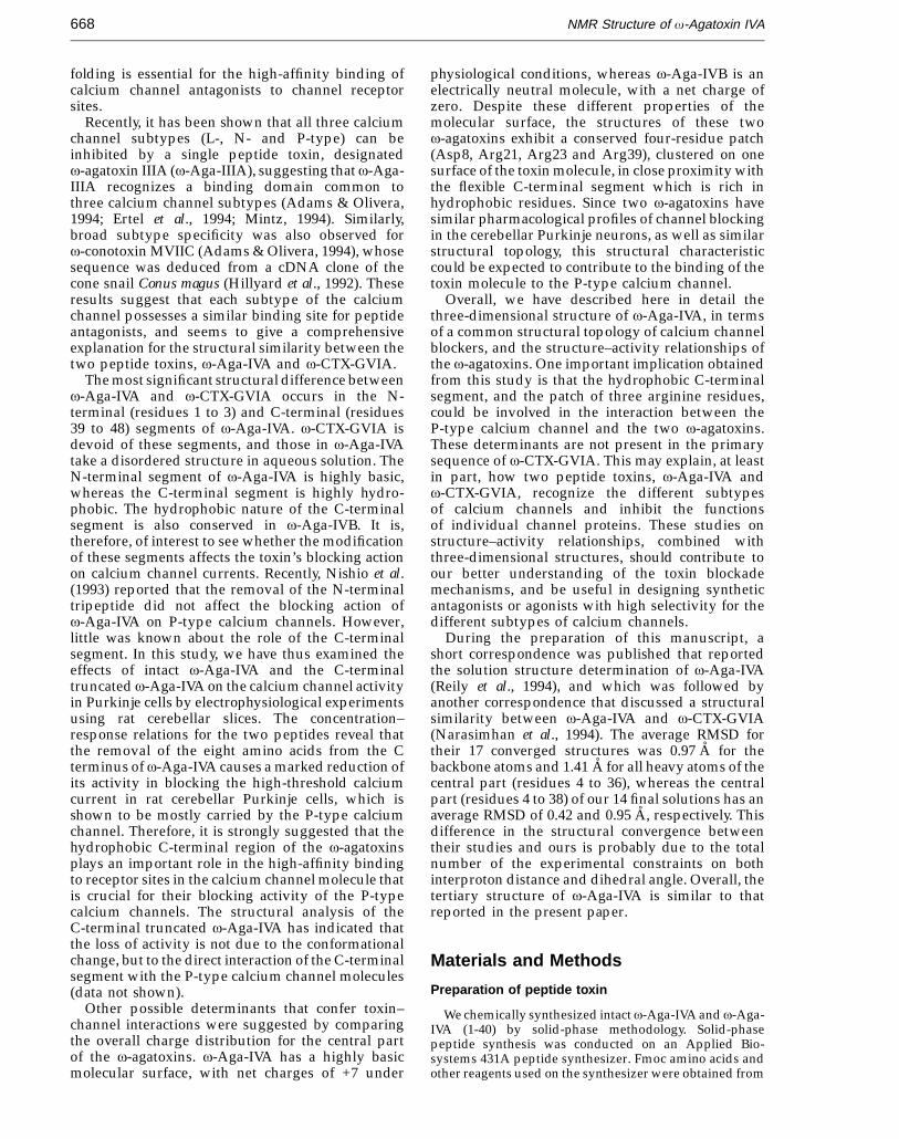
As shown in Figure 7A and B, the overall fold ofv-CTX-GVIA closely resembles the central part(residues 4 to 37) of v-Aga-IVA, which is the mostwell-defined region. Despite their very low hom-ology in the primary sequence, these two toxinsshare a very similar location and orientation ofsecondary structures in their three-dimensionalstructures (Figure 7C). The only conserved sequencemotif can be found in the spatial arrangement of thecysteine residues in three dimensions. It is apparent
33) of the loop between b-strands 2 and 3 did notshow the characteristic distance pattern of a tightreverse turn. In terms of RMSD, this loop was poorlydefined as compared with the rest of the central partof the molecule. The angular order parameters for thepeptide backbone of Trp14/Gly15 and Thr32/Asn33show relatively low S values (Figure 3D and E),which reflects the limited number of NOE distanceconstraints for Gly15 and Asn33, respectively(Figure 3A).
The four disulfide bonds appear to play animportant role in the formation and stabilization ofthe globular structure of v-Aga-IVA. The disulfidebond between Cys12 and Cys25 interconnectsb-strands 1 and 2, while the disulfide bonds betweenCys4 and Cys20 and between Cys19 and Cys36connect the external long loop with the N-terminalregion and b-strand 3, respectively. The disulfidebond between Cys27 and Cy34 interconnectsb-strands 2 and 3. Thus, the side-chain of Ile5 in theN-terminal region and the methyl group of Thr17 inthe external long loop form a hydrophobic core of themolecule, together with the side-chains of Cys12,Cys19, Cys25 and Cys36.
Figure 5D shows the arrangement of the side-chains of the aromatic residues together with that ofIle5 in the 14 final converged structures. v-Aga-IVAcontains only two aromatic residues. The side-chainof Tyr9 is located on the bottom of the b-sheet nearthe end position of b-strand 3, whereas the side-chain of Trp14 is positioned on the upper side of theb-sheet in the vicinity of the starting position of thesame strand. It should be noted that tyrosine andtryptophan are amphiphilic amino acids that caninteract with both the hydrophilic and hydrophobicenvironments.
JMB—MS 636
NMR Structure of v-Agatoxin IVA666
Figure 5. Stereopairs of backbone heavy atoms (N, Ca, C) for the 14 converged structures of v-Aga-IVA. These are theresults of the best-fit superposition of the backbone atoms (N, Ca, C, O) for the central part (residues 4 to 38) of the molecule.A, Stereopair for the complete sequence from Lys1 to Ala48. B, Stereopair for the central part (residues 4 to 38). C, Stereopairobtained by a 90° rotation about the vertical axis of stereopair B. D, Stereopair representing the orientation of the side-chainheavy atoms of Ile5, Tyr9 and Trp14, in addition to stereopair B. The atomic coordinates of the 14 converged structuresand the mean structure of v-Aga-IVA will be deposited in the Brookhaven Protein Data Bank and are available directlyfrom the authors on request until they have been processed and released.
JMB—MS 636
NMR Structure of v-Agatoxin IVA 667
Figure 6. Effects of v-Aga-IVAand its C-terminal truncated analog,v-Aga-IVA (1-40), on high-thresholdcalcium channel currents recordedfrom Purkinje cells in rat cerebellarslices using the whole-cell voltage-clamp technique. A calcium currentwas elicited every 20 seconds bya depolarizing step from −100 to−10 mV. A and B, Currents wererecorded from the same cell beforeand after the addition of A, Syntheticv-Aga-IVA and B, v-Aga-IVA (1-40).The concentrations of both peptidesexamined are indicated in therecords. C, Concentration–responserelations for synthetic v-Aga-IVAand v-Aga-IVA (1-40). Each pointrepresents the mean(2)SEM ofcalcium current inhibition by eachpeptide determined in 3 to 15different cells.
that three disulfide bonds between Cys4 and Cys20,between Cys12 and Cys25 and between Cys19 andCys36 in v-Aga-IVA correspond to those betweenCys1 and Cys16, between Cys8 and Cys19 andbetween Cys15 and Cys26 in v-CTX-GVIA, respect-ively. The disulfide bonds between Cys4 and Cys20(v-Aga-IVA) and between Cys1 and Cys16 (v-CTX-GVIA) are located on the exterior of each molecule,and the remaining two disulfide bonds of eachmolecule are buried in the interior. An additional
disulfide bond between Cys27 and Cys34 of v-Aga-IVA seems to contribute to the stability of the loop(residues 28 to 33) connecting the b-strands 2 and 3.In the case of v-CTX-GVIA, the corresponding loopis formed by a hairpin turn, including two residuesbetween Hyp21 and Lys24. Thus, it is likely thatthese structurally conserved disulfide connectivitiessignificantly contribute to the maintenance ofglobular molecular folding of the two toxins. It canalso be presumed that such a consensus of molecular
Figure 7. MOLSCRIPT (Kraulis,1991) diagrams of the backbonepeptide folding of the NMR solutionstructure of two calcium channelblockers. The structural topology ofthe triple-stranded antiparallel b-sheet is shown. A, The meanstructure of v-Aga-IVA, with fourdisulfide bonds between Cys4–Cys20, Cys12–Cys25, Cys19–Cys36,and Cys27–Cys34. B, The refinedstructure of v-CTX-GVIA, with threedisulfide bonds between Cys1–Cys16, Cys8–Cys19, and Cys15–Cys26. C, Comparative arrange-ments of amino acid sequences basedon the distribution of secondarystructure elements and disulfidebonds of v-Aga-IVA and v-CTX-GVIA. Hydroxyproline residues inv-CTX-GVIA are denoted as X insingle-letter amino acid abbrevi-ations. Arrows and rectangles rep-resent the location of b-strands andb-turns in the solution structures ofthe two peptide toxins, respectively.The disulfide connectivities are indi-cated by the horizontal lines. Thev-CTX-GVIA structure coordinateswere extracted from the BrookhavenProtein Data Bank, entry ICCO(Pallaghy et al., 1993).
JMB—MS 636
NMR Structure of v-Agatoxin IVA668
folding is essential for the high-affinity binding ofcalcium channel antagonists to channel receptorsites.
Recently, it has been shown that all three calciumchannel subtypes (L-, N- and P-type) can beinhibited by a single peptide toxin, designatedv-agatoxin IIIA (v-Aga-IIIA), suggesting that v-Aga-IIIA recognizes a binding domain common tothree calcium channel subtypes (Adams & Olivera,1994; Ertel et al., 1994; Mintz, 1994). Similarly,broad subtype specificity was also observed forv-conotoxin MVIIC (Adams & Olivera, 1994), whosesequence was deduced from a cDNA clone of thecone snail Conus magus (Hillyard et al., 1992). Theseresults suggest that each subtype of the calciumchannel possesses a similar binding site for peptideantagonists, and seems to give a comprehensiveexplanation for the structural similarity between thetwo peptide toxins, v-Aga-IVA and v-CTX-GVIA.
The most significant structural difference betweenv-Aga-IVA and v-CTX-GVIA occurs in the N-terminal (residues 1 to 3) and C-terminal (residues39 to 48) segments of v-Aga-IVA. v-CTX-GVIA isdevoid of these segments, and those in v-Aga-IVAtake a disordered structure in aqueous solution. TheN-terminal segment of v-Aga-IVA is highly basic,whereas the C-terminal segment is highly hydro-phobic. The hydrophobic nature of the C-terminalsegment is also conserved in v-Aga-IVB. It is,therefore, of interest to see whether the modificationof these segments affects the toxin’s blocking actionon calcium channel currents. Recently, Nishio et al.(1993) reported that the removal of the N-terminaltripeptide did not affect the blocking action ofv-Aga-IVA on P-type calcium channels. However,little was known about the role of the C-terminalsegment. In this study, we have thus examined theeffects of intact v-Aga-IVA and the C-terminaltruncated v-Aga-IVA on the calcium channel activityin Purkinje cells by electrophysiological experimentsusing rat cerebellar slices. The concentration–response relations for the two peptides reveal thatthe removal of the eight amino acids from the Cterminus of v-Aga-IVA causes a marked reduction ofits activity in blocking the high-threshold calciumcurrent in rat cerebellar Purkinje cells, which isshown to be mostly carried by the P-type calciumchannel. Therefore, it is strongly suggested that thehydrophobic C-terminal region of the v-agatoxinsplays an important role in the high-affinity bindingto receptor sites in the calcium channel molecule thatis crucial for their blocking activity of the P-typecalcium channels. The structural analysis of theC-terminal truncated v-Aga-IVA has indicated thatthe loss of activity is not due to the conformationalchange, but to the direct interaction of the C-terminalsegment with the P-type calcium channel molecules(data not shown).
Other possible determinants that confer toxin–channel interactions were suggested by comparingthe overall charge distribution for the central partof the v-agatoxins. v-Aga-IVA has a highly basicmolecular surface, with net charges of +7 under
physiological conditions, whereas v-Aga-IVB is anelectrically neutral molecule, with a net charge ofzero. Despite these different properties of themolecular surface, the structures of these twov-agatoxins exhibit a conserved four-residue patch(Asp8, Arg21, Arg23 and Arg39), clustered on onesurface of the toxin molecule, in close proximity withthe flexible C-terminal segment which is rich inhydrophobic residues. Since two v-agatoxins havesimilar pharmacological profiles of channel blockingin the cerebellar Purkinje neurons, as well as similarstructural topology, this structural characteristiccould be expected to contribute to the binding of thetoxin molecule to the P-type calcium channel.
Overall, we have described here in detail thethree-dimensional structure of v-Aga-IVA, in termsof a common structural topology of calcium channelblockers, and the structure–activity relationships ofthe v-agatoxins. One important implication obtainedfrom this study is that the hydrophobic C-terminalsegment, and the patch of three arginine residues,could be involved in the interaction between theP-type calcium channel and the two v-agatoxins.These determinants are not present in the primarysequence of v-CTX-GVIA. This may explain, at leastin part, how two peptide toxins, v-Aga-IVA andv-CTX-GVIA, recognize the different subtypesof calcium channels and inhibit the functionsof individual channel proteins. These studies onstructure–activity relationships, combined withthree-dimensional structures, should contribute toour better understanding of the toxin blockademechanisms, and be useful in designing syntheticantagonists or agonists with high selectivity for thedifferent subtypes of calcium channels.
During the preparation of this manuscript, ashort correspondence was published that reportedthe solution structure determination of v-Aga-IVA(Reily et al., 1994), and which was followed byanother correspondence that discussed a structuralsimilarity between v-Aga-IVA and v-CTX-GVIA(Narasimhan et al., 1994). The average RMSD fortheir 17 converged structures was 0.97 A for thebackbone atoms and 1.41 A for all heavy atoms of thecentral part (residues 4 to 36), whereas the centralpart (residues 4 to 38) of our 14 final solutions has anaverage RMSD of 0.42 and 0.95 A, respectively. Thisdifference in the structural convergence betweentheir studies and ours is probably due to the totalnumber of the experimental constraints on bothinterproton distance and dihedral angle. Overall, thetertiary structure of v-Aga-IVA is similar to thatreported in the present paper.
Materials and Methods
Preparation of peptide toxin
We chemically synthesized intact v-Aga-IVA and v-Aga-IVA (1-40) by solid-phase methodology. Solid-phasepeptide synthesis was conducted on an Applied Bio-systems 431A peptide synthesizer. Fmoc amino acids andother reagents used on the synthesizer were obtained from
JMB—MS 636
NMR Structure of v-Agatoxin IVA 669
Applied Biosystems Japan (Chiba, Japan). Fmoc-Ala-and Fmoc-Leu-p-oxybenzyloxybenzyl alcohol resinswere obtained from Watanabe Chemical Industries Ltd.(Hiroshima, Japan). Other reagents of peptide synthesisgrade were obtained from the Peptide Institute (Osaka,Japan) or Kokusan Chemical Works Ltd. (Tokyo, Japan).
Linear precursors of v-Aga-IVA and v-Aga-IVA (1-40)were assembled by solid-phase methodology of Fmocchemistry, starting with Fmoc-preloaded resin, using atrityl group for the protection of the SH groups of thecysteine residues. The oxidative folding reaction wascarried out by a procedure similar to that reported byNishio et al. (1993). After trifluoroacetic acid (TFA)cleavage, crude linear peptides were extracted with 2 MAcOH, and diluted to a peptide concentration of 0.02 mMin the presence of reduced/oxidized glutathione (molarratio of peptide:GSH:GSSG was 1:100:10) and 0.5 Mguanidine hydrochloride. The solutions were adjusted topH 7.8 with aqueous NH4OH, and stirred slowly at 4°C forthree days. The crude cyclized peptides were purified bysuccessive chromatographic procedures with SephadexG-50F and preparative HPLC. The purities of the syntheticpeptides were confirmed by analytical HPLC andelectrospray ionization mass spectrometry (ESI–MS)measurement.
Electrophysiological experiments
The whole-cell voltage-clamp recording was made fromPurkinje cells in rat cerebellar slices as previouslydescribed (Edwards et al., 1989; Mitoma et al., 1994). Brainswere removed from five- to eight-day-old Wistar rats underanesthesia with sodium pentobarbital. The cerebellumwas sagittally sliced at a thickness of 250 mm. Afterincubation with oxygenated saline for more than one hour,slices were transferred to a recording chamber on the stageof a microscope, and continuously perfused with artificialcerebrospinal fluid (ACSF) of the following composition:NaCl 139 mM, KCl 3.4 mM, CaCl2 2.5 mM, MgCl2 1 mM,NaHCO3 21 mM, NaH2PO4 0.6 mM, glucose 10 mM. ThepH of the ACSF was maintained at 7.4 by gassing with 95%(v/v) O2–5% (v/v) CO2. Recordings were made at roomtemperature (20 to 25°C) using patch electrodes filled withan internal solution that contained Na-GTP 0.4 mM(pH 7.4), CsCH3SO3 150 mM, KCl 5 mM, K-ethylene glycolbis(b-aminoethyl ether)-N,N,N',N '-tetraacetic acid (K-EGTA) 0.1 mM, Na-Hepes 5 mM, Mg-ATP 3 mM, and hada tip resistance of 3 to 8 MV.
Purkinje cells were viewed under Nomarski optics witha Zeiss 40 × water immersion objective. After achievingwhole-cell recording mode, the external solution wasswitched to ACSF containing BaCl2 3 to 5 mM and tetrodo-toxin 1 mM. Membrane currents were recorded with a ListEPC-7 patch-clamp amplifier, and digitized for transfer tocomputer disk for later analysis. A series resistancecompensation of 60 to 85% was used. The membranepotential of Purkinje cells was held at −70 mV. High-threshold calcium channel currents were elicited by adepolarizing step to a potential of −20 to 0 mV from apre-holding potential of −100 mV. Leak and capacitativecurrents were cancelled by subtraction using the P/nmethod (Armstrong & Bezanilla, 1974).
Synthetic v-Aga-IVA and v-Aga-IVA (1-40) were dis-solved in the external solution supplemented with cyto-chrome C (1 mg/ml) to prevent binding to containers bysaturation. The synthetic peptides were applied either byperfusion or pressure pulses from micropipettes to thevicinity of the recorded neurons.
NMR measurements
NMR spectra were recorded on a Bruker AMX 500spectrometer operating at 500 MHz for the protonfrequency. All two-dimensional NMR experiments, i.e.DQF-COSY (Rance et al., 1983), PE-COSY (Mueller, 1987),HOHAHA (Bax & Davis, 1985), and NOESY (Jeener et al.,1979; Macura et al., 1981), were performed using standardpulse sequences and phase cycling. Quadrature detec-tion in the t1 dimension was achieved with the time-proportional phase incrementation (TPPI) method (Marion& Wuthrich, 1983).
HOHAHA spectra were recorded with mixing times of60 ms and 80 ms. NOESY spectra were recorded withmixing times of 100 ms, 200 ms, and 300 ms. In all exper-iments, 512 increments of 2 K data points were recordedwith 64 to 128 transients, and were zero-filled once alongthe t1 dimension. In the case of the PE-COSY experiment,the acquired data were further zero-filled once along thet2 dimension. Suppression of undesirable t2 ridges arisingfrom the strong solvent resonance was achieved by linearbase-line correction of the F2 cross section, prior to Fouriertransformation in the t1 dimension. The Gauss function wasused for the apodization function. The solvent resonancewas suppressed by selective irradiation during the relax-ation delay, which was set to 1.2 seconds. A complete setof the two-dimensional spectra were recorded at 37°C andpH 2.5 (uncorrected meter readings). Slowly exchangingbackbone amide protons were identified by the analysis ofHOHAHA spectra recorded at time scales of 0.5 hours,3 hours and 15 hours. The backbone amide protonresonances originating from the b-sheet region were stillobserved in a HOHAHA spectrum recorded after 15 hoursin 2H2O solution. The resonance assignments were alsomade at 30°C and pH 4.5.
The 1H chemical shifts were referenced to internalsodium 3-(trimethylsilyl)propionate-2, 2, 3, 3-2H4 (TSP).For the NMR experiments, samples were prepared bydissolving the synthetic v-Aga-IVA in 0.4 mL of 2H2O orH2O containing 10% (v/v) 2H2O. The peptide concen-tration used in all NMR measurements was about 7 mM.
Distance constraints and structure calculations
Interproton distance constraints were obtained from theNOESY spectra observed with mixing times of either100 ms or 200 ms. Quantitative determination of thecross-peak intensities was based on the counting of theexponentially spaced contour levels. Observed NOE datawere classified into three distance ranges, 1.8 to 2.5 A, 1.8to 3.5 A, and 1.8 to 5 A, corresponding to strong, medium,and weak NOEs, respectively. Pseudo-atoms were used forthe methyl protons or non-stereospecifically assignedmethylene protons (Wuthrich et al., 1983). Correctingfactors for the use of pseudo-atoms were added to thedistance constraints. In addition, 0.5 A was added to thedistance constraints involving methyl protons (Clore et al.,1987). Twelve additional constraints were added to definethe four disulfide bonds that are present in v-Aga-IVA. Thepairing of cysteines has recently been determined by thecombination of amino acid analysis, gas-phase sequencing,and mass spectrometry of proteolytic fragments (Nishioet al., 1993). For each disulfide bond, we used threedistance constraints, S(i )–S( j ), S(i )–Cb( j ) and S( j )–Cb(i ),whose target values were set to 2.02(20.02) A,2.99(20.5) A and 2.99(20.5) A, respectively (Nilges et al.,1988b).
All calculations were carried out on a HP 9000/720workstation with the X-PLOR 3.1 program (Brunger, 1993).
JMB—MS 636
NMR Structure of v-Agatoxin IVA670
The three-dimensional structure was calculated on thebasis of the experimentally derived distance and torsionangle constraints by using the dynamical simulatedannealing protocols. We started the calculation from atemplate structure with randomized backbone f and ctorsion angles. For the dynamical simulated annealing, thestandard forcefield parameter set (parallhdg.pro) andtopology file (topallhdg.pro) in the X-PLOR 3.1 programwere used. The starting structures with a random array ofatoms (Nilges et al., 1988a) were regularized by thedgsa.inp protocol, and the refine.inp protocol was usedfor final optimization of the structures obtained fromthe dgsa.inp protocol. NOE distance and torsion angleconstraints were applied with 50 kcal mol−1 A−2 and200 kcal mol−1 rad−2 force constants, respectively. The meanstructure was obtained by the restrained minimization ofthe averaged coordinate of the final converged structures.For structural comparison, the coordinates of v-CTX-GVIA were extracted from the Brookhaven Protein DataBank, entry ICCO (Pallaghy et al., 1993).
AcknowledgementsThe authors express their thanks to Dr Terutoshi Kimura
for his advice in the synthesis of v-Aga-IVA, and DrKatsuyoshi Masuda for ESI-MS measurement of syntheticv-Aga-IVA. Thanks are also due to Dr Tadakazu Maeda forhelpful discussion of the structural calculation.
ReferencesAdams, M. E. & Olivera, B. M. (1994). Neurotoxins:
Overview of an emerging research technology. TrendsNeurosci. 17, 151–155.
Armstrong, C. M. & Bezanilla, F. (1974). Charge movementassociated with the opening and closing of the acti-vation gates of the Na channels. J. Gen. Physiol. 63,533–552.
Bax, A. & Davis, D. G. (1985). MLEV-17–based two-dimensional homonuclear magnetization transferspectroscopy. J. Magn. Reson. 65, 355–360.
Brooks, B. R., Bruccoleri, R. E., Olafson, B. D., States, D. J.,Swaminathan, S. & Karplus, M. (1983). CHARMm: aprogram for macromolecular energy, minimization,and dynamics calculations. J. Comput. Chem. 4,187–217.
Brunger, A. T. (1993). X-PLOR Manual, Version 3.1. YaleUniversity, New Haven, CT, USA.
Clore, G. M., Gronenborn, A. M., Nilges, M. & Ryan, C. A.(1987). Three-dimensional structure of potato car-boxypeptidase inhibitor in solution. A study usingnuclear magnetic resonance, distance geometry, andrestrained molecular dynamics. Biochemistry, 26,8012–8023.
Davis, J. H., Bradley, E. K., Miljanich, G. P., Nadasdi, L.,Ramachandran, J. & Basus, V. J. (1993). Solutionstructure of v-conotoxin GVIA using 2-D NMRspectroscopy and relaxation matrix analysis. Biochem-istry, 32, 7396–7405.
Edwards, F. A., Konnerth, A., Sakmann, B. & Takahashi, T.(1989). A thin slice preparation for patch clamp record-ings from neurones of the mammalian central nervoussystem. Pflugers Arch. 414, 600–612.
Ertel, E. A., Warren, V. A., Adams, M. E., Griffin, P. R.,Cohen, C. J. & Smith, M. M. (1994). Type IIIv-agatoxins: a family of probes for similar binding
sites on L- and N-type calcium channels. Biochemistry,33, 5098–5108.
Gouda, H., Torigoe, H., Saito, A., Sato, M., Arata, Y.& Shimada, I. (1992). Three-dimensional solutionstructure of the B domain of staphylococcal protein A:comparisons of the solution and crystal structures.Biochemistry, 31, 9665–9672.
Gray, W. R., Olivera, B. M. & Cruz, L. J. (1988). Peptidetoxins from venomous conus snails. Annu. Rev.Biochem. 57, 665–700.
Hillyard, D. R., Monje, V. D., Mintz, I. M., Bean, B. P.,Nadasdi, L., Ramachandran, J., Miljanich, G.,Azimi-Zoonooz, A., McIntosh, J. M., Cruz, L. J.,Imperial, J. S. & Olivera, B. M. (1992). A new conuspeptide ligand for mammalian presynaptic Ca2+
channels. Neuron, 9, 69–77.Hyberts, S. G., Marki, W. & Wagner, G. (1987). Stereo-
specific assignments of side-chain protons andcharacterization of torsion angles in eglin c. Eur. J.Biochem. 164, 625–635.
Hyberts, S. G., Goldberg, M. S., Havel, T. F. & Wagner, G.(1992). The solution structure of eglin c based onmeasurements of many NOEs and coupling constantsand its comparison with X-ray structures. Protein Sci.1, 736–751.
Jeener, J., Meier, B. N., Bachmann, P. & Ernst, R. R.(1979). Investigation of exchange processes by two-dimensional NMR spectroscopy. J. Chem. Phys. 71,4546–4553.
Kim, J. I., Takahashi, M., Ogura, A., Kohno, T., Kudo, Y.& Sato, K. (1994). Hydroxyl group of Tyr13 is essentialfor the activity of v-conotoxin GVIA, a peptide toxinfor N-type calcium channel. J. Biol. Chem. 269,23876–23878.
Kline, A. D., Braun, W. & Wuthrich, K. (1988). Deter-mination of the complete three-dimensional structureof the a-amylase inhibitor tendamistat in aqueoussolution by nuclear magnetic resonance and distancegeometry. J. Mol. Biol. 204, 675–724.
Kraulis, P. J. (1991). MOLSCRIPT: a program to produceboth detailed and schematic plots of protein struc-tures. J. Appl. Crystallog. 24, 946–950.
Lewis, P. N., Momany, F. A. & Scheraga, H. A. (1973). Chainreversals in proteins. Biochim. Biophys. Acta, 303,211–229.
Ludvigsen, S. & Poulsen, F. M. (1992). Positive f-angles inproteins by nuclear magnetic resonance spectroscopy.J. Biomol. NMR, 2, 227–233.
Macura, S., Huang, Y., Suter, D. & Ernst, R. R. (1981). Two-dimensional chemical exchange and cross-relaxationspectroscopy of coupled nuclear spins. J. Magn. Reson.43, 259–281.
Marion, D. & Wuthrich, K. (1983). Application of phasesensitive two-dimensional correlated spectroscopy(COSY) for measurements of 1H–1H spin–spincoupling constants in proteins. Biochem. Biophys. Res.Commun. 113, 967–974.
McCleskey, E. W., Fox, A. P., Feldman, D. H., Cruz, L. J.,Olivera, B. M., Tsien, R. W. & Yoshikami, D. (1987).v-Conotoxin: direct and persistent blockade ofspecific types of calcium channels in neurons but notmuscle. Proc. Natl Acad. Sci. USA, 84, 4327–4331.
Mintz, I. M. (1994). Block of Ca channels in rat centralneurons by the spider toxin v-Aga-IIIA. J. Neurosci. 14,2844–2853.
Mintz, I. M., Adams, M. E. & Bean, B. P. (1992a). P-typecalcium channels in rat central and peripheralneurons. Neuron, 9, 85–95.
Mintz, I. M., Venema, V. J., Swiderek, K. M., Lee, T. D., Bean,
JMB—MS 636
NMR Structure of v-Agatoxin IVA 671
B. P. & Adams, M. E. (1992b). P-type calcium channelsblocked by the spider toxin v-Aga-IVA. Nature, 355,827–829.
Mitoma, H., Kobayashi, T., Song, S. Y. & Konishi, S.(1994). Enhancement by serotonin of GABA-mediatedinhibitory synaptic currents in rat cerebellar Purkinjecells. Neurosci. Lett. 173, 127–130.
Mueller, L. (1987). P.E. COSY, a simple alternative to E.COSY. J. Magn. Reson. 72, 191–196.
Narasimhan, L., Singh, J., Humblet, C., Guruprasad, K. &Blundell, T. (1994). Snail and spider toxins share asimilar tertiary structure and ‘cystine motif’. NatureStruct. Biol. 1, 850–852.
Nilges, M., Clore, G. M. & Gronenborn, A. M. (1988a).Determination of three-dimensional structures ofproteins from interproton distance data by dynamicalsimulated annealing from a random array of atoms.FEBS Letters, 239, 129–136.
Nilges, M., Gronenborn, A. M., Brunger, A. T. & Clore,G. M. (1988b). Determination of three-dimensionalstructures of proteins by simulated annealing withinterproton distance restraints: application to cram-bin, potato carboxypeptidase inhibitor and barleyserine proteinase inhibitor 2. Protein Eng. 2, 27–38.
Nishio, H., Yoshizawa-Kumagaye, K., Kubo, S., Chen,Y., Momiyama, A., Takahashi, T., Kimura, T. &Sakakibara, S. (1993). Synthesis of v-agatoxin IVA andits related peptides. Biochem. Biophys. Res. Commun.196, 1447–1453.
Olivera, B. M., McIntosh, J. M., Cruz, L. J., Luque, F. A. &Gray, W. R. (1984). Purification and sequence of apresynaptic peptide toxin from Conus geographusvenom. Biochemistry, 23, 5087–5090.
Pallaghy, P. K., Duggan, B. M., Pennington, M. W. & Norton,R. S. (1993). Three-dimensional structure in solution ofthe calcium channel blocker v-conotoxin. J. Mol. Biol.234, 405–420.
Pallaghy, P. K., Nielsen, K. J., Craik, D. J. & Norton, R. S.(1994). A common structural motif incorporating acystine knot and a triple-stranded b-sheet in toxic andinhibitory polypeptides. Protein Sci. 3, 1833–1839.
Pardi, A., Billeter, M. & Wuthrich, K. (1984). Calibrationof the angular dependence of the amide proton–Ca
proton coupling constants, 3JHNa, in a globular protein:Use of 3JHNa for identification of helical secondarystructure. J. Mol. Biol. 180, 741–751.
Rampe, D. & Triggle, D. J. (1989). 1,4-Dihydropyridineactivators and antagonists: Structural and functionaldistinctions. Trends Pharmacol. Sci. 10, 507–511.
Rance, M., So�rensen, O. W., Bodenhausen, G., Wagner, G.,Ernst, R. R. & Wuthrich, K. (1983). Improved spectralresolution in COSY 1H NMR spectra of proteinsvia double quantum filtering. Biochem. Biophys. Res.Commun. 117, 479–485.
Regan, L. J. (1991). Voltage-dependent calcium currents inPurkinje cells from rat cerebellar vermis. J. Neurosci.11, 2259–2269.
Reily, M. D., Holub, K. E., Gray, W. R., Norris, T. M. &Adams, M. E. (1994). Structure–activity relationshipsfor P-type calcium channel-selective v-agatoxins.Nature Struct. Biol. 1, 853–856.
Richardson, J. S. (1981). The anatomy and taxonomy ofprotein structure. Advan. Protein Chem. 34, 167–339.
Sato, K., Park, N. G., Kohno, T., Maeda, T., Kim, J. I.,Kato, R. & Takahashi, M. (1993). Role of basic residuesfor the binding of v-conotoxin GVIA to N-typecalcium channels. Biochem. Biophys. Res. Commun. 194,1292–1296.
Sevilla, P., Bruix, M., Santoro, J., Gago, F., Garcia, A. G.& Rico, M. (1993). Three-dimensional structure ofv-conotoxin GVIA determined by 1H NMR. Biochem.Biophys. Res. Commun. 192, 1238–1244.
Skalicky, J. J., Metzler, W. J., Ciesla, D. J., Galdes, A. &Pardi, A. (1993). Solution structure of the calciumchannel antagonist v-conotoxin GVIA. Protein Sci. 2,1591–1603.
Tsien, R. W., Lipscombe, D., Madison, D. V., Bley, K. R.& Fox, A. P. (1988). Multiple types of neuronalcalcium channels and their selective modulation.Trends Neurosci. 11, 431–438.
Uchitel, O. D., Protti, D. A., Sanchez, V., Cherksey, B. D.,Sugimori, M. & Llinas, R. (1992). P-type voltage-dependent calcium channel mediates presynapticcalcium influx and transmitter release in mammaliansynapses. Proc. Natl Acad. Sci. USA, 89, 3330–3333.
Wagner, G., Braun, W., Havel, T.F., Schaumann, T., Go, N. &Wuthrich, K. (1987). Protein structures in solution bynuclear magnetic resonance and distance geometry.The polypeptide fold of the basic pancreatic trypsininhibitor determined using two different algorithms,DISGEO and DISMAN. J. Mol. Biol. 196, 611–639.
Wilmot, C. M. & Thornton, J. M. (1990). b-Turns and theirdistortions: A proposed new nomenclature. ProteinEng. 3, 479–493.
Wishart, D. S., Sykes, B. D. & Richards, F. M. (1992). Thechemical shift index: a fast and simple method for theassignment of protein secondary structure throughNMR spectroscopy. Biochemistry, 31, 1647–1651.
Wuthrich, K. (1986). NMR of Proteins and Nucleic Acids.John Wiley, New York, USA.
Wuthrich, K., Billeter, M. & Braun, W. (1983). Pseudo-structures for the 20 common amino acids for use instudies of protein conformations by measurementsof intramolecular proton–proton distance constraintswith nuclear magnetic resonance. J. Mol. Biol. 169,949–961.
Wuthrich, K., Billeter, M. & Braun, W. (1984). Polypeptidesecondary structure determination by nuclear mag-netic resonance observation of short proton–protondistances. J. Mol. Biol. 180, 715–740.
Yu, H., Rosen, M. K., Saccomano, N. A., Phillips, D.,Volkmann, R. A. & Schreiber, S. L. (1993). Sequentialassignment and structure determination of spidertoxin v-Aga-IVB. Biochemistry, 32, 13123–13129.
Edited by P. E. Wright
(Received 13 January 1995; accepted 4 May 1995)