Embed Size (px)
Citation preview

RESEARCH Open Access
Tissue inhibitor of metalloproteinases-3 mediatesthe death of immature oligodendrocytes via TNF-a/TACE in focal cerebral ischemia in miceYi Yang1, Fakhreya Y Jalal1, Jeffrey F Thompson1, Espen J Walker5, Eduardo Candelario-Jalil1, Lu Li2,Ross R Reichard4, Chi Ben6, Qing-Xiang Sang6, Lee Anna Cunningham2 and Gary A Rosenberg1,2,3*
Abstract
Background and Purpose: Oligodendrocyte (OL) death is important in focal cerebral ischemia. TIMP-3 promotesapoptosis in ischemic neurons by inhibiting proteolysis of TNF-a superfamily of death receptors. Since OLsundergo apoptosis during ischemia, we hypothesized that TIMP-3 contributes to OL death.
Methods: Middle cerebral artery occlusion (MCAO) was induced in Timp-3 knockout (KO) and wild type (WT) micewith 24 or 72 h of reperfusion. Cell death in white matter was investigated by stereology and TUNEL. Mature orimmature OLs were identified using antibodies against glutathione S-transferase-π (GST-π) and galactocerebroside(GalC), respectively. Expression and level of proteins were examined using immunohistochemistry andimmunoblotting. Protein activities were determined using a FRET peptide.
Results: Loss of OL-like cells was detected at 72 h only in WT ischemic white matter where TUNEL showed greatercell death. TIMP-3 expression was increased in WT reactive astrocytes. GST-π was reduced in ischemic white matterof WT mice compared with WT shams with no difference between KO and WT at 72 h. GalC level was significantlyincreased in both KO and WT ischemic white matter at 72 h. However, the increase in GalC in KO mice wassignificantly higher than WT; most TUNEL-positive cells in ischemic white matter expressed GalC, suggesting TIMP-3deficiency protects the immature OLs from apoptosis. There were significantly higher levels of cleaved caspase-3 at72 h in WT white matter than in KO. Greater expression of MMP-3 and -9 was seen in reactive astrocytes and/ormicroglia/macrophages in WT at 72 h. We found more microglia/macrophages in WT than in KO, which were thepredominant source of increased TNF-a detected in the ischemic white matter. TACE activity was significantlyincreased in ischemic WT white matter, which was expressed in active microglia/macrophages and OLs.
Conclusions: Our results suggested that focal ischemia leads to proliferation of immature OLs in white matter and thatTIMP-3 contributes to a caspase-3-dependent immature OL death via TNF-a-mediated neuroinflammation. Futurestudies will be needed to delineate the role of MMP-3 and MMP-9 that were increased in the Timp-3 wild type.
BackgroundOligodendrocytes (OLs) undergo a complex pattern ofdeath in cerebral ischemia with an early loss from gluta-mate excitotoxicity and a slower death due to apoptosis[1-4]. The vulnerability of OLs to ischemia was shownmorphologically [5] and the molecular mechanism wasrelated to excitotoxicity via the glutamate AMPA
receptors [6,7]. Ischemia results in apoptosis of OLs, butthe mechanisms involved are less well understood. Thepro-inflammatory factor tumor necrosis factor-a (TNF-a) and TNF death receptors play an important role inOL death as shown in vitro and in vivo [1,8,9]. Cerebralischemia results in an inflammatory response consistingof microglia activation, gliosis, and cell death. Activatedmicroglia and reactive astrocytes produce TNF-a andinterleukin-1b (IL-1b), which are associated with peri-ventricular white matter damage in hypoxic neonatalbrain [10]. Extravasation of inflammatory cells into the
* Correspondence: [email protected] of Neurology, University of New Mexico Health SciencesCenter, Albuquerque, NM 87131, USAFull list of author information is available at the end of the article
Yang et al. Journal of Neuroinflammation 2011, 8:108http://www.jneuroinflammation.com/content/8/1/108
JOURNAL OF NEUROINFLAMMATION
© 2011 Yang et al; licensee BioMed Central Ltd. This is an Open Access article distributed under the terms of the Creative CommonsAttribution License (http://creativecommons.org/licenses/by/2.0), which permits unrestricted use, distribution, and reproduction inany medium, provided the original work is properly cited.

central nervous system (CNS) is facilitated by extracellu-lar activities of matrix metalloproteinases (MMPs) thatare regulated, in part, by the endogenous tissue inhibi-tors of metalloproteinases (TIMPs) [11].Cell surface sheddases, including TNF-a converting
enzyme (TACE) and stromelysin-1 (MMP-3) regulatethe TNF superfamily of death receptors by activatingthe ligands and removing the death receptors from thecell surface. Tissue inhibitor of metalloproteinases-3(TIMP-3) plays a central role in this process by inhibit-ing TACE and MMP-3. In an early study, TIMP-3blocked the release of the TNF death receptor byTACE, promoting apoptosis [12]. More recently, weobserved that TIMP-3 expression in ischemic neuronswas associated with APO-1 (Fas/CD95) and DNA frag-mentation [13]. Using neuronal cell cultures, we havedemonstrated that TIMP-3 promoted apoptosis by pre-venting the shedding of Fas receptors from the neuronalcell surface during oxygen-glucose deprivation, and thatthe Timp-3 knockout (KO) mouse showed reduced neu-ronal death after a middle cerebral artery occlusion(MCAO) with reperfusion compared to the wild type(WT) [14,15].Because of TIMP-3’s unique role as a cell surface inhi-
bitor of TACE and MMP-3, it could contribute to celldeath in OLs by promoting retention of TNF deathreceptors [16]. To investigate the role of TIMP-3 in OLcell loss, we induced a middle cerebral artery occlusion(MCAO) in Timp-3 knockout (KO) and wild type (WT)mice followed by 24 and 72 h of reperfusion. Wehypothesized that TIMP-3 and Fas/CD95 expressionoccurred early in neurons and preceded DNA fragmen-tation, but that in white matter TIMP-3 may contributeto OL death that is associated with TNF-a receptors.We report that TIMP-3 deficiency protects the imma-ture OLs from apoptotic death in white matter inducedby ischemia-reperfusion injury. The immature OL deathin the WT was associated with increased expression ofMMPs, TIMP-3, TNF-a/TACE in reactive astrocytesand/or infiltrating macrophages/microglia in whitematter.
MethodsMCAO with reperfusionThe study was approved by the University of New Mex-ico Animal Care Committee and conformed to theNational Institutes of Health Guide for the Care andUse of Animals in research. Eighty-nine male Timp-3KO (n = 44) and WT (n = 45) mice weighing 20-30 g(age 10-12 weeks) were employed in this study. Fifty-nine animals were subjected to 90 min MCAO usingthe intraluminal thread method followed by 24 or 72 hreperfusion. MCAO was performed as previouslydescribed [15,17]. Mice underwent reperfusion via the
circle of Willis. Thirty sham-operated animals wereanesthetized and had a suture introduced into the com-mon carotid artery and immediately removed.
Primary antibodies and dilutions used inimmunohistochemistryGlial fibrillary acidic protein (GFAP) (1:400; Sigma), Iba-1 (1:200; Wako Pure Chemical Industries), GST-π(1:600, Abcam), GalC (1:300, Millipore), Fas (1:400;Santa Cruz Biotechnology), Fas ligand (1:400; SantaCruz Biotechnology), MMP-2 (1:250; Chemicon), MMP-3 (1:1500; Chemicon), MMP-9 (1:1000; Chemicon), cas-pase-3 (1:1000, Cell Signaling), TIMP-3 (1:1000; Chemi-con), TNF-a (15 μg/ml; R&D Systems), TNF Receptor I(1:1000; Abcam), von Willebrand Factor (vWF) (10 μg/ml; Chemicon), myeloperoxidase (MPO; 1:250; SantaCruz Biotechnology), and several markers for oligoden-droglia; RIP (1:200,000; Chemicon), CNPase (15 μg/ml;Chemicon), and CC-1 (APC; 10 μg/ml; Calbiochem).
ImmunohistochemistryBrain tissues fixed with 2% paraformaldehyde, 0.1 Msodium periodate, 0.075 M lysine in 100 mM phosphatebuffer at pH 7.3 (PLP) were used for immunohistochem-ical analysis. For immunofluorescence, 10 μm PLP-fixedbrain sections were treated with acetone and blockedwith 5% normal serum. Primary antibodies were incu-bated for two nights at 4°C. Slides were incubated for 90min at room temperature with secondary antibodiesconjugated with FITC or Cy-3 (Jackson). 4’-6-diami-dino-2-phenylindole (DAPI; Molecular Probes) was usedto label cell nuclei.For brightfield immunolabeling, 10 μm PLP-fixed sec-
tions were rehydrated through alcohol series and incu-bated in phosphate buffered saline and 0.1% Tween-20pH 7.3 (PBT) prior to blocking with 5% normal serumin PBT. Slides were incubated with primary antibodiesfor 48 h at 4°C followed by 1 h incubation with biotiny-lated secondary antibodies, Vectastain Elite ABCreagents (Vector Laboratories) and reacted with 3,3’-dia-minobenzidine (DAB) was used for visualization ofimmunolabeling. Cresyl violet acetate (CVA, Sigma-Aldrich) was used for counterstaining.Immunohistochemistry negative controls were incu-
bated without the primary antibody or with normal(non-immune) IgGs and did not exhibit specific immu-nolabelling. All immunohistochemically stained slideswere viewed on an Olympus BX-51 bright field andfluorescence microscope (Olympus America Inc.)equipped with an Optronics digital camera and PictureFrame image capture software (Optronics). Dual immu-nofluorescence slides were also imaged using high reso-lution confocal microscopy to verify co-localization ofantigens (Zeiss LSM 510, Carl Zeiss Microimaging).
Yang et al. Journal of Neuroinflammation 2011, 8:108http://www.jneuroinflammation.com/content/8/1/108
Page 2 of 14

Immunofluorescence was quantified using ImageJ soft-ware (NIH software).
Stereological CountsThe cellular composition of the white matter is com-prised of > 90% OLs [18] consisting of OL progenitors,immature, and mature OLs. Antibodies used to identifymature OLs, including CC-1, RIP, and CNPase, eitherlabeled too few OL cell bodies or obscured the cellbodies in the processes. To measure white matterdamage in mouse focal ischemia, we first assessed lossof OL-like cells in white matter [2] using multiple-anti-body immunostaining [19]. Labeling cells with a Nisslsubstance stain, such as cresyl violet acetate (CVA), allcells are clearly observable and an assessment of OLdensity can be determined stereologically. To distinguishOLs from astrocytes and microglia, we stained slideswith GFAP for astrocytes and Iba-1 for microglia, usingthe DAB method. All slides were counterstained withCVA. By eliminating DAB-stained astrocytes and micro-glia, small CVA-positive cell bodies that were negativefor GFAP and Iba-1 immunoreactivity were identified asOL-like cells and quantified using unbiased stereologywith StereoInvestigator (Version 6, MBF Bioscience)software controlling a motorized stage equipped Olym-pus BX-51 microscope [19]. Employing the optical frac-tionator function of StereoInvestigator, we estimatedOL-like cell number in the medial corpus callosum (CC)and external capsule (EC) regions of white matter over atotal distance of 3.0 mm rostral and caudal to bregma.Tissue sections were blinded to the investigator as toanimal identity and reperfusion time. A subset of slideswas stained with myeloperoxidase or von Willebrandfactor to identify neutrophils and endothelial cells.These could be distinguished from the OL-like cellsbased on morphology (data not shown).
Western blotsWestern blot was performed to determine protein levelsin ischemic white matter. Proteins were extracted inRIPA buffer from punch biopsies of white matter inischemic and nonischemic external capsule regions ofTimp-3 KO and WT mice. 50 μg total protein was sepa-rated on 10% or 12% gels. The proteins were transferredto polyvinylidene fluoride (PVDF) membranes. Themembranes were then incubated with primary antibo-dies: MBP (1:2000, Millipore), GST-π (1:5000, Abcam),GalC (1:500, Millipore), MMP-3 (1:1000, Epitomics),both inactive (pro-) and active (cleaved) caspase-3(1:1000; Cell Signaling Technology), and TNFR1(1:1000; Abcam). The membranes were incubated withthe respective secondary antibodies and blots weredeveloped using the West Pico Detection System(Pierce). Protein bands were visualized on X-ray film.
Semiquantitation of target protein intensities was donewith the use of Scion image software (Scion), and Brilli-ant Blue R (Sigma-Aldrich) staining on the same PVDFmembranes was used to normalize protein loading andtransfer. The results are reported as normalized bandintensity for quantifying relative protein expression.
TUNEL assayTerminal deoxynucleotidyl transferase-mediated dUTPnick end labeling (TUNEL) assay was done to identifythe extent of DNA fragmentation. Using the Neuro-TACS II kit (Trevigen) brain sections were treated fol-lowing the procedure specified by the manufacturer. Fordual immunofluorescence, the brain sections were thenexposed to the primary antibodies and streptavidinfluorescein and Cy-3 secondary antibodies. For positivecontrols the nuclease provided with the kit was addedto the labeling reaction mix and induced TUNEL posi-tivity in all cells. As negative controls, slides were pre-pared in a labeling reaction mix without the TdTenzyme resulting in no TUNEL positivity (data notshown).
Enzymatic assay of MMP-3 activity using a fluorescenceresonance energy transfer (FRET) peptideMMP-3 enzymatic activity was assessed using a peptide-based FRET assay. In the intact FRET peptide, Mca-Arg-Pro-Lys-Pro-Val-Glu-Nva-Trp-Arg-Lys(Dnp)-NH2,obtained from Bachem (Torrance, CA) the fluorescenceof Mca (7-methoxycoumarin-4-acetyl) is quenched byDnp (2,4-dinitrophenyl). Upon cleavage into two sepa-rate fragments by the MMP-3 present in the sample, thefluorescence of Mca was recovered, and monitored atlex = 328 nm and lem= 393 nm using a spectrofluori-meter (Perkin-Elmer LS 50B).Brain tissue was homogenized in lysis buffer contain-
ing 50 mM Tris-HCl pH 7.6, 150 mM NaCl, 5 mMCaCl2, 0.05% Brij-35, 0.02% NaN3, and 1% Triton X-100, and centrifuged at 12,000 × g for 5 min at 4°C.Equal amount of total protein (6-10 μg) was mixed withassay buffer containing 50 mM HEPES buffer pH 7.5,200 mM NaCl, 10 mM CaCl2, 0.01% Brij-35, and fluoro-genic substrate (1 μM final concentration) to a finalvolume of 200 μl. Fluorescence was monitored for 15min. The relationship between fluorescence units andnM of product produced was determined from thefluorescence values obtained when all the substrate washydrolyzed.
Fluorometric assay of TNF-a converting enzyme (TACE)activityTACE activity was measured using the fluorometricSensoLyte 520 TACE (a-Secretase) Activity Assay Kit(AnaSpec) according to the manufacturer’s instructions.
Yang et al. Journal of Neuroinflammation 2011, 8:108http://www.jneuroinflammation.com/content/8/1/108
Page 3 of 14

SensoLyte™ 520 TACE Activity Assay Kit uses a 5-FAM (fluorophore) and QXL™ 520 (quencher) labeledFRET peptide substrate for continuous measurement ofenzyme activity. In the intact FRET peptide, the fluores-cence of 5-FAM is quenched by QXL™ 520. Upon clea-vage of the FRET peptide by the active enzyme, thefluorescence of 5-FAM is recovered, and can be con-tinuously monitored at lex = 490 nm/520 nm. TACEactivity was measured in the brain tissue homogenizedas described before for MMP-3 activity. Equal amountof total protein was mixed with assay buffer (AnaSpec)and TACE substrate to a final volume of 100 μl. Fluor-escence was monitored for 60 min with data recordedevery 5 min. Fluorescence was expressed as relativefluorescence units (RFU). Some brain samples from KOand WT mice had 10 μl of 10 μM TACE inhibitor,TAPI-0, added as a control.
Data analysisStatistical comparisons among groups were done usingANOVA with post-hoc analysis for multiple t- tests(Prism 4.0, GraphPad Software Inc.). All data are pre-sented as mean ± standard error of the mean (S.E.M.).Statistical significance was set at p < 0.05.
ResultsTimp-3 KO mice were protected from delayed immatureOL deathTwo regions of ischemic white matter were analyzed:the corpus callosum (CC) and the external capsule (EC).No OL-like cell loss was seen in ischemic CCs in bothKO and WT mice at 24 and 72 h (data not shown) orin ischemic EC at 24 h (Figure 1A, top two panels).Representative regions from the nonischemic and theischemic EC at 72 h showed the normal appearing OL-like cells in the nonischemic sides and the reactiveastrocytosis and/or microglia/macrophages in theischemic ECs in KO and WT (Figure 1A. bottom twopanels). The ischemic EC in KO showed no observableloss of OL-like cells, while the ischemic EC in WT hadmarked loss of OL-like cells (Figure 1A, bottom twopanels).Next, the loss of OL-like cells in ischemic white mat-
ter was quantified by unbiased stereology. At 24 h, KOand WT mice showed similar cell counts in theischemic and the nonischemic hemispheres (Figure 1B).By 72 h, there was a significant loss of OL-like cells inthe ischemic EC of WT compared to KO mice (p <0.01). Furthermore, there was a significant loss of cellsin the ischemic compared to the nonischemic hemi-sphere in WT only (p < 0.05). In the CC, no significantreduction of OL-like cells was detected in either WT orKO ischemic CC at 72 h (data not shown). Next, weinvestigated apoptotic cell death in ischemic EC at 72 h
of reperfusion by TUNEL assay. More TUNEL-positivecells were detected in ischemic WT hemisphere includ-ing the EC region compared with KO (Figure 1C). Thedifference in the distribution of TUNEL-positive cellsbetween the ischemic KO and WT hemispheres is con-sistent with the patterns of the neuronal degenerationrevealed by Fluoro-Jade staining (Figure 1D). This resultsuggested that ischemia-reperfusion injury induced adelayed cell loss in white matter in which TIMP-3 maybe involved.We then studied the expression of TIMP-3 in astro-
cytes and OLs in WT mice. At 24 h, very little TIMP-3was seen in the white matter. We observed a new popu-lation of GFAP-positive astrocytes closely surroundingthe core infarct areas at 72 h (data not shown), whichhas been reported by others [20]. Maximal TIMP-3expression occurred at 72 h in GFAP-positive astrocytesin ischemic EC (Figure 2A). Most of the astrocytes wereco-labeled with TIMP-3 and co-localization of TIMP-3with CC1, an OL marker, was also seen by 72 h (Figure2B). This temporal and spatial relationship of TIMP-3expression and the loss of OL-like cells in ischemic ECindicated the involvement of TIMP-3 in the white mat-ter damage in focal cerebral ischemia.To further investigate the role of TIMP-3 in OL death,
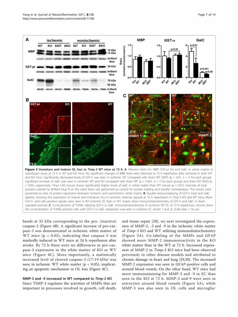
we examined the expression of myelin basic protein(MBP) and glutathione S-transferase π (GST-π, or GST-pi) [21], markers of mature OL, in ischemic ECs of KOand WT mice at 72 h using Western blot analysis (Fig-ure 3A). Unexpectedly, we found no significant changesof MBP level either in ischemic ECs between KO andWT mice or between ischemic ECs and sham ECs.However, we observed a significant decrease of GST-πlevel in WT ischemic EC compared with sham WTmice. Next, we tested the expression of galactocerebro-side (GalC), a marker of immature OL [22,23]. Wefound that a significant increase in GalC was seen inischemic ECs of WT and KO by 72 h reperfusion afterischemia compared with sham WT (p < 0.001) andsham KO (p < 0.05), respectively. Most importantly,TIMP-3 KO mouse shows significantly higher levels ofGalC expression in white matter than WT mouse (p <0.01). These findings suggested that TIMP-3-mediateddelayed OL death mainly occurs in immature OLs inwhite matter after ischemic stroke with reperfusioninjury.To confirm the observation that TIMP-3 contributed
to delayed immature OL death in ischemic white matter,we next performed immunohistochemistry of GST-πand GalC on brain tissue from KO and WT mice with90 min-MCAO and 72 h reperfusion (Figure 3B). Dou-ble-immunostaining of GST-π and GalC demonstratedstronger immunohistochemical signals of GST-π andGalC in ischemic KO EC than WT EC. Furthermore, we
Yang et al. Journal of Neuroinflammation 2011, 8:108http://www.jneuroinflammation.com/content/8/1/108
Page 4 of 14

Figure 1 White matter injury and OL-like cell loss in EC at 72 h in Timp-3 KO and WT mice. A. Representative regions from thenonischemic and the ischemic EC at 24 and 72 h. Iba-1 (microglia/macrophage) and GFAP (astrocyte) were both stained with DAB andcounterstained with cresyl violet acetate (CVA). The intact linear rows of dark-staining nuclei by CVA were formed by OL-like cells (arrows) innonischemic ECs at 24 and 72 h, as well as in ischemic ECs at 24 h. Ischemic EC shows massive DAB-positive glial cells at 72 h. A marked loss ofCVA positive cells was seen in ischemic EC in WT mouse compared with that in KO mouse. Scale bars = 50 μm. B. Stereological counting of OL-like cells in ischemic EC at 24 and 72 h reperfusion shows OL-like counts were similar at 24 h between Timp-3 KO and WT ischemic. A significantloss of OL-like cells occurred at 72 h in the WT ischemic EC compared to KO ischemic and to the WT nonischemic EC (p < 0.01 and 0.05,respectively, n = 6). From 24 to 72 h, OLs in ischemic KO EC remained unchanged. The brain cartoon shows location of regions used for OLcounting (shaded areas). CC: corpus callosum; EC: external capsule. Micrographic image shows Iba-1 and GFAP (arrows) visualized by DAB inhigher magnification. C. TUNEL assay demonstrates apoptotic cell death in ischemic EC at 72 h reperfusion. TUNEL-positive cells were seen inischemic WT EC. Scale bar = 100 μm. D. Distribution of neuronal degeneration in ischemic hemispheres in Timp-3 KO and WT mice at 24 and 72h of reperfusion following 90 min transient MCAO. Coronal sections were stained for degenerating neurons using Fluoro-Jade (FluoJ). Number ofFluoJ-positive cells was much greater in WT than in KO at both tiom points. La-ctx: lateral cortex, stri: striatum, pcf: piriform cortex. Scale bar =100 μm.
Yang et al. Journal of Neuroinflammation 2011, 8:108http://www.jneuroinflammation.com/content/8/1/108
Page 5 of 14

detected apoptotic death using TUNEL assay in OLs.We found TUNEL-positive cells in ischemic EC at 72 h,which mostly co-localized with GalC-positive OLs (Fig-ure 3C).
Caspase-3 increased in WT compared to Timp-3 KOTo study the possible mechanism of TIMP-3-mediatedcell death, we used immunohistochemistry to determinethe expression of caspase-3, since apoptosis has beenimplied in immature and mature OL death [1,10,24-26].In the ischemic EC of the WT, greater caspase-3
immunoreactivity was detected at 24 h of reperfusionthan in the KO. There was loss of DAPI stained nuclei(blue) in the WT EC as compared with the KO EC at72 h reperfusion (Figure 4A).Western blots were done with an antibody that recog-
nizes both pro- and cleaved-caspase-3. The activatedcaspase-3 often appears as multiple cleaved bandsmigrating variably in the 17-20 kDa range [27]. Therewas no detectable cleaved (active) caspase-3 (17/19 kDa)in white matter of KO and WT mice at 24 h afterstroke. Western blots exhibited strong immunoreactive
Figure 2 TIMP-3 expression in astrocytes and OLs in ischemic EC at 72 h in Timp-3 WT mice. A. Photomicrographs show doubleimmunofluorescent-labeling of TIMP-3 and GFAP in sham-operated and ischemic EC at 24 and 72 h of reperfusion. TIMP-3 was not expressed inGFAP-labeled astrocytes at 24 h, but by 72 h TIMP-3 was markedly expressed and co-localized with astrocytes (arrows). The co-localization ofTIMP-3 and GFAP was confirmed by Z-stack confocal image. B. CC1-positive OLs express TIMP-3 (arrows) in ischemic EC at 72 h. The co-localization of TIMP-3 and CC1 was confirmed by Z-stack confocal image. Scale bars = 50 μm.
Yang et al. Journal of Neuroinflammation 2011, 8:108http://www.jneuroinflammation.com/content/8/1/108
Page 6 of 14
bands at 32 kDa corresponding to the pro- (inactive)caspase-3 (Figure 4B). A significant increase of pro-cas-pase-3 was demonstrated in ischemic white matter ofWT mice (p < 0.05), indicating that caspase-3 wasmarkedly induced in WT mice at 24 h reperfusion afterstroke. By 72 h there were no differences in pro-cas-pase-3 expression in the white matter of KO or WTmice (Figure 4C). More importantly, a statisticallyincreased level of cleaved caspase-3 (17/19 kDa) wasseen in ischemic WT white matter (p < 0.05), implicat-ing an apoptotic mechanism in OL loss (Figure 4C).
MMP-3 and -9 increased in WT compared to Timp-3 KOSince TIMP-3 regulates the activities of MMPs that areimportant in processes involved in growth, cell death,
and tissue repair [28], we next investigated the expres-sion of MMP-2, -3 and -9 in the ischemic white matterof Timp-3 KO and WT utilizing immunohistochemistry(Figure 5A). Co-labeling of the MMPs and GFAPshowed more MMP-2 immunoreactivity in the KOwhite matter than in the WT at 72 h. Increased expres-sion of MMP-2 in Timp-3 KO mice had been observedpreviously in other disease models and attributed tochronic damage in heart and lung [29,30]. The increasedMMP-2 expression was seen in GFAP-positive cells andaround blood vessels. On the other hand, WT mice hadmore immunostaining for MMP-3 and -9 in EC thanseen in the KO at 72 h. MMP-3 and-9 were seen inastrocytes around blood vessels (Figure 5A), whileMMP-3 was also seen in OL cells and microglia/
Figure 3 Immature and mature OL loss in Timp-3 WT mice at 72 h. A. Western blots for MBP, GST-pi (π) and GalC in white matter inreperfusion injury at 72 h in WT and KO mice. No significant changes of MBP level were detected at 72 h reperfusion after ischemia in both WTand KO mice. Significantly decreased levels of GST-π was seen in ischemic WT compared with sham WT (SWT) (p < 0.01, n = 5 for each group).Significant increase of GalC was seen in ischemic WT and KO compared with sham WT (p < 0.001, n = 5 for each group) and sham KO (SKO) (p< 0.05), respectively. Timp-3 KO mouse shows significantly higher levels of GalC in white matter than WT mouse (p < 0.01). Intensity of totalproteins stained by Brilliant blue R on the same blots was performed as control for protein loading and transfer normalization. The results werepresented as ratio of protein expression between ischemic and nonischemic white matter. B. Double-immunostaining of GST-π (red) and GalC(green), showing the expression of mature and immature OLs in ischemic external capsule at 72 h reperfusion in Timp-3 KO and WT mice. MoreGST-π- and GalC-positive signals were seen in KO ischemic EC than in WT. Inserts show immunohistochemistry of GST-π and GalC in sham-operated animals. C. Co-localizarion of TUNEL labeling GST-π or GalC immunohistochemistry in ischemic WT EC at 72 h reperfusion. Arrows showthe co-localization of TUNEL-positive cells with GST-π or GalC expression was seen in ischemic EC (insert 1 and 2). Scale bars = 50 μm.
Yang et al. Journal of Neuroinflammation 2011, 8:108http://www.jneuroinflammation.com/content/8/1/108
Page 7 of 14

macrophages in the core infarcted white matter wherevery few astrocytes were detected (Figure 5A, B and 5C).The observation of higher expression of MMP-3 in glial
cells in WT white matter was unexpected since TIMP-3is supposed to inhibit MMP-3 on the cell surface [28].We quantified the MMP-3 protein in white matter byWestern blot and measured its activity with a fluorogenicactivity assay. We found increased MMP-3 (both pro-and active forms) in the ischemic hemisphere of the WTcompared to the KO (Figure 5D). The fluorogenic assay
showed a statistically significant increase in the activationof MMP-3 in the WT only (Figure 5E).
Increased microglia/macrophage expressing TNF-a andTACE activity in WT compared to Timp-3 KODeath of OLs can occur through death receptors of theTNF superfamily, including Fas/FasL and TNF-a/TNFR.Double immunostaining with Fas and FasL antibodiesdemonstrated greater Fas/FasL signal in the gray matterof KO mice at 24 h (Figure 6). Considering the neuronal
Figure 4 Caspase-3 expression in ischemic EC in Timp-3 KO and WT mice at 24 and 72 h. A. Immunostaining shows that expression ofcaspase-3 was detected in ischemic WT EC 24 h after reperfusion. The increased caspase-3 expression in WT was present in OL-like cells. Lesscaspase-3 immunostaining was detected in both ischemic WT and KO ECs at 72 h, while nuclear counterstaining with DAPI (blue) showedgreater loss of cells in ischemic WT EC. Insert 1 and 2 show caspase-3 immunostaing is associated with nuclei (arrows). Scale bars = 50 μm. B.Western blots for expression of caspase-3 in white matter at 24 h in Timp-3 KO and WT mice. The graph shows an increase in pro-caspase-3band at 32 kDa in the ischemic EC of the WT mice (p < 0.05, n = 4). Cleaved caspase-3 (17 kDa and 19 kDa) was not detected. Brilliant blue Rstaining on the same blots was performed as control for protein loading and transfer. C. Western blots for caspase-3 in white matter inreperfusion injury at 72 h showed no difference in expression of pro-caspase-3 between WT and KO, but increased cleaved caspase-3 (19 kDaand 17 kDa) in ischemic WT white matter (p < 0.05, n = 6 for each group).
Yang et al. Journal of Neuroinflammation 2011, 8:108http://www.jneuroinflammation.com/content/8/1/108
Page 8 of 14

degeneration occurs earlier and greater in WT gray matterthan KO (Figure 1D), the greater Fas/FasL signal in graymatter in KO mice at 24 h suggests delayed cell deathcompared with WT mice. However, Fas/FasL signal wasnot detected in white matter in KO or WT mice at both24 and 72 h reperfusion after focal ischemia (Figure 6).Since TNF-a and activated microglia and macrophagesare associated with white matter damage, we then
investigated the TNF-a expression in microglia/macro-phages. TNF-a was increased in white matter of Timp-3WT mice after 72 h reperfusion, and it was co-localizedwith Iba-1-positive microglia/macrophages (Figure 7A and7B). Furthermore, we observed greater Iba-1-positivemicroglia/macrophages that expressed TNF-a in WTwhite matter at 72 h. For quantification of Iba-1, we evalu-ated fields of the ischemic EC as well as cortical and
Figure 5 Expression of MMPs in ischemic EC in Timp-3 KO and WT mice at 72 h. A. Immunohistochemistry for MMPs in ischemic externalcapsule (EC) at 72 h reperfusion in Timp-3 KO and WT mice. The Timp-3 knockout had increased MMP-2 (red) immunoreactivity in ischemia,which co-localized with GFAP-expressing astrocytes (yellow). MMP-9 and MMP-3 were co-localized with astrocytes in the WT. Arrowheads showMMP-9 and -3 in astrocytes around vessels. Insert shows co-localization of MMP-3 and GFAP-positive astrocyte. Scale bar = 100 μm. B.Immunostaining shows MMP-3 expression in CC1-positive OLs in ischemic WT EC. C. Immunostaining shows MMP-3 expression in Iba-1 positivemicroglia/macrophages. Scale bar = 50 μm. D. Western blot of MMP-3 at 72 h reperfusion in Timp-3 KO and WT mouse brains. Western blots ofMMP-3 standard and mouse brain tissues show the proform of MMP-3 at 57 kDa and an active form at 45 kDa. A 60 kDa glycosylated form ofMMP-3 is also present in all brain samples. Brilliant blue R staining on the same blots was performed as control for protein loading and transfer.Quantification of pro and active MMP-3 forms together showed a significant increase in MMP-3 (57 kDa and 45 kDa) in ischemic WT comparedwith ischemic KO, sham KO and WT and nonischemic KO and WT (p < 0.05, n = 6 for each group). No significant difference was found betweenischemic and nonischemic hemispheres in the KO. E. Fluorogenic substrate assay for MMP-3 activity shows significant increase in MMP-3 relativeactivity in both ischemic and nonischemic WT compared with sham WT (p < 0.05, n = 6 for each group).
Yang et al. Journal of Neuroinflammation 2011, 8:108http://www.jneuroinflammation.com/content/8/1/108
Page 9 of 14

striatal tissue immediately bordering the EC in each sec-tion, quantifying the amount of fluorescent intensity ofIba-1 as assessed in five brains per group at each timepoint. We found significantly greater fluorescence of Iba-1in ischemic WT EC compared to KO at both 24 and 72 hafter ischemia (Figure 7C). These results suggest thatTIMP-3 facilitates the inflammatory response to ischemia-reperfusion injury.Since TIMP-3 inhibits TACE, which both generates
soluble TNF-a (17 kDa) from the 21 kDa cell surface formand cleaves the TNF receptors, we measured TACE activ-ity in white matter from Timp-3 KO and WT mice at 72 hreperfusion. Ischemia with 72 h reperfusion resulted in asignificant increase in activation of TACE in white matterof WT mouse compared with KO (Figure 7D). The doublestaining of TACE and Iba-1 or CC1 showed the expressionof TACE in Iba-1-positive microglia/macrophages andOLs in WT white matter (Figure 7E).The excess presence of TNF-a in white matter could
trigger cell apoptosis by ligation with the TNFR-1. Wedetermined the presence of this receptor and the celltype expressing it by immunohistochemistry. We foundthat the TNFR-1 co-localized with the OL marker, CC1(Figure 7F). Western blot showed no difference of TNFR-1 expression between KO and WT mice (Figure 7F).
DiscussionWe found that deletion of the Timp-3 gene reducedimmature OL death at 72 h following MCAO-inducedfocal ischemia with reperfusion. TUNEL assay demon-strated apoptotic death of immature OLs. There was anincrease in pro-caspase-3 at 24 h and cleaved caspase-3at 72 h in white matter, indicating apoptosis was
occurring at the time of delayed immature OL death.The presence of Timp-3 in the wild type led to an exag-gerated inflammatory response with an increase in bothinfiltration of microglia/macrophages at 72 h andexpression of MMP-3 and -9 in reactive astrocytesaround blood vessels in the white matter. We foundincreased activity of TACE and TNF-a in microglia/macrophages, while TNFR-1 was expressed on OLs.Finally, WT mice had an increase in active microglia/macrophages compared with KO mice. These resultsprovide the first evidence for TIMP-3 induction ofinflammation and promotion of delayed immature OLdeath via a TNF-a-mediated mechanism.An unexpected finding in this study was that TIMP-3
mainly promoted immature OL rather than mature OLdeath. Glutathione S-transferase π (GST-π) is a cytosolicisoenzyme in mature OLs, which is a marker of matureOLs in mammalian brain [21,31-33]. Western blot andimmunohistochemistry showed a significant decrease inGST-π protein in the ischemic EC of Timp-3 WT mice.Although less GST-π protein disruption was seen in theKO, MBP expression was similar in WT and KO mice.We found preferential damage in immature OLs, whichhas been reported in perinatal hypoxia-ischemia [4]. Oli-godendrocyte-specific galactosphingolipid (GalC) isexpressed during differentiation of OL lineage cells,including OL progenitor cells (OPCs), premyelinatingand myelinating OLs [34]. We used GalC as a markerfor immature OLs [35]. Our data demonstrated a signifi-cant increase in GalC expression in ischemic EC in bothKO and WT mice. We found that immature OLsundergo TIMP-3-mediated apoptosis associated withTIMP-3 expression in reactive astrocytes. These findingsare consistent with the previous observation that OLprogenitors are more susceptible to death due tohypoxia-ischemia than mature OLs in neonatal mice[25].Cell surface receptors and ligands of the TNF-a
superfamily play a fundamental role in apoptosis duringneuroinflammation and overproduction of the proin-flammatory cytokine, TNF-a, has been implicated in thepathogenesis of white matter damage [10,36]. TNF-adecreases the number of OL progenitors by causingtheir apoptosis [10,26,37]. Activated microglia and reac-tive astrocytes release TNF-a and other cytokines [38].We observed an increased infiltration of TNF-a- andTACE-expressing microglia/macrophages in ischemicwhite matter of WT mouse at 72 h. Timp-3 KO mousehad reduced TNF-a expression and TACE activity andshowed fewer microglia/macrophages in ischemic whitematter, which is consistent with TIMP-3’s role inenhancing inflammation in other tissues [39].Co-labeling of MMPs and GFAP indicated astrocytes
showed more MMP-2 immunoreactivity in the KO
Figure 6 A. Immunohistochemistry for Fas and FasL inischemic EC at 24 and 72 h in Timp-3 KO and WT mousebrains. No positive signal of Fas (green) and FasL (red) wasdetected in both KO and WT ECs. Scale bars = 50 μm.
Yang et al. Journal of Neuroinflammation 2011, 8:108http://www.jneuroinflammation.com/content/8/1/108
Page 10 of 14

white matter than WT, which is consistent with earlierstudies showing that Timp-3 KO mice have greaterMMP-2 activation during physiological and pathologicalevents [29,30]. On the contrary, WT mice showed moreimmunostaining for MMP-3 and MMP-9 in ischemicwhite matter than KO, and in reactive astrocytes aroundvessels. MMPs disrupt the blood-brain barrier by cleav-ing tight junction proteins at an early stage after ische-mia-reperfusion injury in the rat [40]. MMP-3 canproteolytically activate MMP-9 [41]. Therefore,increased MMP-3 and -9 in astrocytes in white mattermay play a role in BBB damage with facilitation of infil-tration of macrophages in WT mouse. In addition, wealso found increased active MMP-3 in OL-like cells andmicroglia/macrophages. MMP-3 is associated with theneuroinflammatory response. MMP-3 was shown to
activate microglia and increase the neuroinflammatoryresponse with expression of cytokines such as TNF-a[42]. In dopaminergic neurons MMP-3 may have anintracellular role in cell death [43,44]. We foundincreased MMP-3 (both pro- and active forms) in theischemic hemisphere of the WT compared to the KO. Afluorogenic assay showed a significant increase in theactivity of MMP-3 in the WT only. Although some stu-dies suggest that MMP-3 contributes to cell death,MMP-3 was protective to neurons in culture exposed todoxorubicin through its role in cleaving the death recep-tors and ligands [15]. Studies with the Mmp-3 knockoutmouse will be needed to resolve this issue.OLs have cell surface receptors for members of the
TNF-a death receptor family, including TNFR-1, TNFR-2, and Fas/CD95 [45]. TNF-R1 has an intracellular
Figure 7 Expression of TNF-a and TACE in ischemic EC in Timp-3 KO and WT mice at 72 h. A. Double-staining of TNF-a (red) and Iba-1(green), showing the expression of TNF-a in microglia/macrophages (Iba-1 positive cells) in ischemic EC at 72 h reperfusion in Timp-3 KO andWT mice. More TNF-a- and Iba-1-positive cells were seen in WT ischemic EC (arrows) than in KO. Scale bar = 40 μm. B. The three-dimension (3D)and Z-stack confocal images shows the co-localization of TNF-a in macrophage. Scale bar = 5 μm. C. Quantification of Iba-1 fluorescent intensityshowed greater Iba-1-positive macrophages/microglia in Timp-3 WT mice compared with KO at both 24 and 72 h (p < 0.05, n = 5 for eachgroup). D. TACE activity measured by a fluorometric assay in ischemic white matter at 72 h reperfusion in Timp-3 KO and WT mouse brains.Significant increase in TACE relative activity was seen in ischemic WT compared with ischemic KO (p < 0.05, n = 6) and sham WT (SWT) and KO(SKO) (p < 0.01). A greater level of TACE activity was seen in nonischemic WT compared with contralateral sham WT (p < 0.05). E. Top panel: Co-localization of TACE and Iba-1 in microglia/macrophages (arrows). Bottom: TACE and CC1 co-localization in OLs (arrows). Scale bars = 50 μm. F.Z-stack confocal images show TNFR-1 co-localized in CC1-positive OLs. Western blot analysis shows no significant changes ether betweenischemic and nonischemic hemispheres or between KO and WT mice (n = 6).
Yang et al. Journal of Neuroinflammation 2011, 8:108http://www.jneuroinflammation.com/content/8/1/108
Page 11 of 14
death domain and its activation leads to cell apoptosis[46]. Aberrant TNF-a/TNF-R1 signaling can have apotentially major role in the CNS pathologies in whichOL apoptosis and demyelination are primary pathologi-cal features [47]. TNF-a is toxic to OLs in culture andin periventricular white matter of hypoxic rats [26,48].TIMP-3 inhibits the action of TACE, which both acti-vates TNF-a to the mature 17-kDa form and releasesTNF death receptors from the cell surface [39]. BecauseTIMP-3 interacts with a number of cell surface pro-cesses, including formation of TNF-a by TACE, releaseof TNFR-1 from the cell surface, and other events,
promoting either cell death or remodeling, the effect ofTIMP-3 is difficult to predict [39].It should be noted that the MCAO produces damage
to the striatum, cortex, and the adjacent white matter.This could result in some of the molecules produced bythe injury of the striatum/cortex influencing the damagein the white matter. Other mechanisms of cell death arepossible in OLs where studies have shown that gluta-mate induces OL cell death both through an action onthe cell body and on the ion channels of the axon[6,49,50]. While the action of TIMP-3 in OL deathremains to be fully elucidated, it is possible to construct
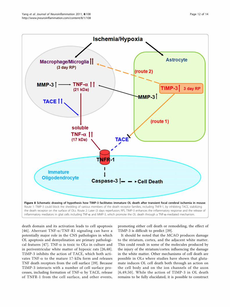
Figure 8 Schematic drawing of hypothesis how TIMP-3 facilitates immature OL death after transient focal cerebral ischemia in mouse.Route 1: TIMP-3 could block the shedding of various members of the death receptor families, including TNFR-1, by inhibiting TACE, stabilizingthe death receptor on the surface of OLs. Route 2: Later (3 days reperfusion; RP), TIMP-3 enhances the inflammatory response and the release ofinflammatory mediators in glial cells including TNF-a and MMP-3, which promote the OL death through a TNF-a-mediated mechanism.
Yang et al. Journal of Neuroinflammation 2011, 8:108http://www.jneuroinflammation.com/content/8/1/108
Page 12 of 14
a hypothetical mechanism to explain TIMP-3 involve-ment in the OL death that involves the activation ofmicroglia/macrophages by TIMP-3, which also inhibitsshedding of TNFR-1 by TACE. Figure 8 shows ahypothetical mechanism for TIMP-3-facilitated death ofimmature OLs in transient cerebral focal ischemia inmice.In conclusion, our results show that TIMP-3 contri-
butes to immature OL death in ischemic white matterby increasing TACE, TNF-a, cleaved caspase- 3, andactivated MMP-3 and -9. We observed a cellular prefer-ence with TIMP-3 strongly expressed in GFAP-positiveastrocytes, while TNF-a, TACE and MMP-3 appear inmicroglia/macrophages and TNFR-1 is found in OLs.These results are the first to implicate TIMP-3 in thedeath of immature OLs by apoptosis and they suggestthat an interaction occurs between astrocytes expressingTIMP-3, microglia/macrophages expressing TNF-a andMMP-3, and OLs expressing TNFR-1 that facilitates thedeath process. These results may have implications forthe delayed death of vulnerable OLs seen in a numberof hypoxic/ischemic neurological conditions.
List of abbreviationsOL: oligodendrocyte; CC: corpus callosum; EC: external capsule; TIMP-3:Tissue inhibitor of metalloproteinases-3; MMPs: matrix metalloproteinases;TNF-α: tumor necrosis factor-α; MCAO: middle cerebral artery occlusion; KO:knockout; WT: wild type; GST-π (pi): glutathione S-transferase π (pi); GalC:galactocerebroside; TACE; TNF-α converting enzyme; TNFR-1: TNF Receptor I;GFAP: glial fibrillary acidic protein; DAPI: 4’-6-diamidino-2-phenylindole; DAB:3,3’-diaminobenzidine; CVA: cresyl violet acetate; TUNEL: terminaldeoxynucleotidyl transferase-mediated dUTP nick end labeling; FRET:fluorescence resonance energy transfer; BBB: blood-brain barrier; OPCs:oligodendrocyte progenitor cells.
AcknowledgementsThis work was supported by grants from the National Institutes of Health(5RO1NS04547 and 2 R01 NS045847-05A2) to GAR, as well as AmericanHeart Association Beginning grants (0765473Z and 10BGIA4310034) to YY.Confocal images in this paper were generated in the University of NewMexico Cancer Center Fluorescence Microscopy Facility, supported asdetailed on the webpage: http://hsc.unm.edu/crtc/microscopy.
Author details1Department of Neurology, University of New Mexico Health SciencesCenter, Albuquerque, NM 87131, USA. 2Department of Neurosciences,University of New Mexico Health Sciences Center, Albuquerque, NM 87131,USA. 3Department of Cell Biology and Physiology, University of New MexicoHealth Sciences Center, Albuquerque, NM 87131, USA. 4Department ofPathology, University of New Mexico Health Sciences Center, Albuquerque,NM 87131, USA. 5Department of Anesthesia, University of California, SanFrancisco, San Francisco, CA 94110, USA. 6Department of Chemistry andBiochemistry, Florida State University, Tallahassee, FL 32306, USA.
Authors’ contributionsYY designed the experiments, carried out animal surgery and confocalmicroscopy, analyzed the data, and wrote the manuscript. YY, FYJ and JFTperformed the biochemical and histologic studies. EJW, JFT and RRR carriedout the stereological and morphological study. ECJ participated in thebiochemical studies. BC and QXS performed the MMP-3 activity assay. LLand LAC helped with the MCAO model in mouse. GAR who is the leader ofthe group conceived and coordinated the study. GAR, ECJ, FYJ, JFT, EJW and
LAC participated in writing and editing to the manuscript. All authors readand approved the final manuscript.
Competing interestsThe authors declare that they have no competing interests.
Received: 7 April 2011 Accepted: 29 August 2011Published: 29 August 2011
References1. Masumura M, Hata R, Nagai Y, et al: Oligodendroglial cell death with DNA
fragmentation in the white matter under chronic cerebralhypoperfusion: comparison between normotensive and spontaneouslyhypertensive rats. Neurosci Res 2001, 39:401-412.
2. Dewar D, Underhill SM, Goldberg MP: Oligodendrocytes and ischemicbrain injury. J Cereb Blood Flow Metab 2003, 23:263-274.
3. Park E, Velumian AA, Fehlings MG: The role of excitotoxicity in secondarymechanisms of spinal cord injury: a review with an emphasis on theimplications for white matter degeneration. J Neurotrauma 2004,21:754-774.
4. Ness JK, Romanko MJ, Rothstein RP, et al: Perinatal hypoxia-ischemiainduces apoptotic and excitotoxic death of periventricular white matteroligodendrocyte progenitors. Dev Neurosci 2001, 23:203-208.
5. Pantoni L, Garcia JH, Gutierrez JA: Cerebral white matter is highlyvulnerable to ischemia. Stroke 1996, 27:1641-1646.
6. Goldberg MP, Ransom BR: New light on white matter. Stroke 2003,34:330-332.
7. Tekkok SB, Goldberg MP: Ampa/kainate receptor activation mediateshypoxic oligodendrocyte death and axonal injury in cerebral whitematter. J Neurosci 2001, 21:4237-4248.
8. Selmaj K, Raine CS, Farooq M, et al: Cytokine cytotoxicity againstoligodendrocytes. Apoptosis induced by lymphotoxin. J Immunol 1991,147:1522-1529.
9. Selmaj KW, Raine CS: Tumor necrosis factor mediates myelin andoligodendrocyte damage in vitro. Ann Neurol 1988, 23:339-346.
10. Kaur C, Ling EA: Periventricular white matter damage in the hypoxicneonatal brain: role of microglial cells. Prog Neurobiol 2009, 87:264-280.
11. Crocker SJ, Whitmire JK, Frausto RF, et al: Persistent macrophage/microglial activation and myelin disruption after experimentalautoimmune encephalomyelitis in tissue inhibitor of metalloproteinase-1-deficient mice. Am J Pathol 2006, 169:2104-2116.
12. Smith MR, Kung H, Durum SK, et al: TIMP-3 induces cell death bystabilizing TNF-alpha receptors on the surface of human coloncarcinoma cells. Cytokine 1997, 9:770-780.
13. Wallace JA, Alexander S, Estrada EY, et al: Tissue inhibitor ofmetalloproteinase-3 is associated with neuronal death in reperfusioninjury. J CerebBlood Flow Metab 2002, 22:1303-1310.
14. Wetzel M, Rosenberg GA, Cunningham LA: Tissue inhibitor ofmetalloproteinases-3 and matrix metalloproteinase-3 regulate neuronalsensitivity to doxorubicin-induced apoptosis. Eur J Neurosci 2003,18:1050-1060.
15. Wetzel M, Li L, Harms KM, et al: Tissue inhibitor of metalloproteinases-3facilitates Fas-mediated neuronal cell death following mild ischemia. CellDeath Differ 2008, 15:143-151.
16. Cunningham LA, Wetzel M, Rosenberg GA: Multiple roles for MMPs andTIMPs in cerebral ischemia. Glia 2005, 50:329-339.
17. Kokovay E, Li L, Cunningham LA: Angiogenic recruitment of pericytesfrom bone marrow after stroke. J CerebBlood Flow Metab 2006, 26:545-555.
18. Bongarzone ER, Howard SG, Schonmann V, et al: Identification of thedopamine D3 receptor in oligodendrocyte precursors: potential role inregulating differentiation and myelin formation. J Neurosci 1998,18:5344-5353.
19. Walker EJ, Rosenberg GA: Divergent role for MMP-2 in myelin breakdownand oligodendrocyte death following transient global ischemia. JNeurosci Res 2010, 88:764-773.
20. Zhao BQ, Wang S, Kim HY, et al: Role of matrix metalloproteinases indelayed cortical responses after stroke. Nat Med 2006, 12:441-445.
21. Tamura Y, Kataoka Y, Cui Y, et al: Intracellular translocation of glutathioneS-transferase pi during oligodendrocyte differentiation in adult ratcerebral cortex in vivo. Neuroscience 2007, 148:535-540.
Yang et al. Journal of Neuroinflammation 2011, 8:108http://www.jneuroinflammation.com/content/8/1/108
Page 13 of 14

22. Baumann N, Pham-Dinh D: Biology of oligodendrocyte and myelin in themammalian central nervous system. Physiol Rev 2001, 81:871-927.
23. Buntinx M, Vanderlocht J, Hellings N, et al: Characterization of threehuman oligodendroglial cell lines as a model to study oligodendrocyteinjury: morphology and oligodendrocyte-specific gene expression. JNeurocytol 2003, 32:25-38.
24. Vinukonda G, Csiszar A, Hu F, et al: Neuroprotection in a rabbit model ofintraventricular haemorrhage by cyclooxygenase-2, prostanoid receptor-1 or tumour necrosis factor-alpha inhibition. Brain 2010, 133:2264-2280.
25. Skoff RP, Bessert DA, Barks JD, et al: Hypoxic-ischemic injury results inacute disruption of myelin gene expression and death ofoligodendroglial precursors in neonatal mice. Int J Dev Neurosci 2001,19:197-208.
26. Deng Y, Lu J, Sivakumar V, et al: Amoeboid microglia in theperiventricular white matter induce oligodendrocyte damage throughexpression of proinflammatory cytokines via MAP kinase signalingpathway in hypoxic neonatal rats. Brain Pathol 2008, 18:387-400.
27. Huesmann GR, Clayton DF: Dynamic role of postsynaptic caspase-3 andBIRC4 in zebra finch song-response habituation. Neuron 2006,52:1061-1072.
28. Cunningham LA, Wetzel M, Rosenberg GA: Multiple roles for MMPs andTIMPs in cerebral ischemia. Glia 2005, 50:329-339.
29. English JL, Kassiri Z, Koskivirta I, et al: Individual Timp deficienciesdifferentially impact pro-MMP-2 activation. J BiolChem 2006,281:10337-10346.
30. Leco KJ, Waterhouse P, Sanchez OH, et al: Spontaneous air spaceenlargement in the lungs of mice lacking tissue inhibitor ofmetalloproteinases-3 (TIMP-3). J ClinInvest 2001, 108:817-829.
31. Watanabe T, Zhang N, Liu M, et al: Cilostazol protects against brain whitematter damage and cognitive impairment in a rat model of chroniccerebral hypoperfusion. Stroke 2006, 37:1539-1545.
32. Kumar S, Biancotti JC, Yamaguchi M, et al: Combination of growth factorsenhances remyelination in a cuprizone-induced demyelination mousemodel. Neurochem Res 2007, 32:783-797.
33. Binder MD, Cate HS, Prieto AL, et al: Gas6 deficiency increasesoligodendrocyte loss and microglial activation in response to cuprizone-induced demyelination. J Neurosci 2008, 28:5195-5206.
34. Matsubayashi Y, Iwai L, Toda T, et al: Immunostaining for oligodendrocyte-specific galactosphingolipids in fixed brain sections using thecholesterol-selective detergent digitonin. J Neurosci Methods 2009,178:87-98.
35. See J, Zhang X, Eraydin N, et al: Oligodendrocyte maturation is inhibitedby bone morphogenetic protein. Mol Cell Neurosci 2004, 26:481-492.
36. Deng YY, Lu J, Ling EA, et al: Role of microglia in the process ofinflammation in the hypoxic developing brain. Front Biosci (Schol Ed)3:884-900.
37. Cammer W: Effects of TNFalpha on immature and matureoligodendrocytes and their progenitors in vitro. Brain Res 2000,864:213-219.
38. Kordek R, Nerurkar VR, Liberski PP, et al: Heightened expression of tumornecrosis factor alpha, interleukin 1 alpha, and glial fibrillary acidicprotein in experimental Creutzfeldt-Jakob disease in mice. Proc Natl AcadSci USA 1996, 93:9754-9758.
39. Smookler DS, Mohammed FF, Kassiri Z, et al: Tissue inhibitor ofmetalloproteinase 3 regulates TNF-dependent systemic inflammation. JImmunol 2006, 176:721-725.
40. Yang Y, Estrada EY, Thompson JF, et al: Matrix metalloproteinase-mediated disruption of tight junction proteins in cerebral vessels isreversed by synthetic matrix metalloproteinase inhibitor in focalischemia in rat. J Cereb Blood Flow Metab 2007, 27:697-709.
41. Candelario-Jalil E, Yang Y, Rosenberg GA: Diverse roles of matrixmetalloproteinases and tissue inhibitors of metalloproteinases inneuroinflammation and cerebral ischemia. Neuroscience 2009,158:983-994.
42. Kim YS, Kim SS, Cho JJ, et al: Matrix metalloproteinase-3: a novelsignaling proteinase from apoptotic neuronal cells that activatesmicroglia. J Neurosci 2005, 25:3701-3711.
43. Si-Tayeb K, Monvoisin A, Mazzocco C, et al: Matrix metalloproteinase 3 ispresent in the cell nucleus and is involved in apoptosis. Am J Pathol2006, 169:1390-1401.
44. Choi DH, Kim EM, Son HJ, et al: A novel intracellular role of matrixmetalloproteinase-3 during apoptosis of dopaminergic cells. J Neurochem2008, 106:405-415.
45. D’Souza SD, Bonetti B, Balasingam V, et al: Multiple sclerosis: Fas signalingin oligodendrocyte cell death. J ExpMed 1996, 184:2361-2370.
46. Nakazawa T, Nakazawa C, Matsubara A, et al: Tumor necrosis factor-alphamediates oligodendrocyte death and delayed retinal ganglion cell lossin a mouse model of glaucoma. J Neurosci 2006, 26:12633-12641.
47. Akassoglou K, Bauer J, Kassiotis G, et al: Oligodendrocyte apoptosis andprimary demyelination induced by local TNF/p55TNF receptor signalingin the central nervous system of transgenic mice: models for multiplesclerosis with primary oligodendrogliopathy. Am J Pathol 1998,153:801-813.
48. Jurewicz A, Matysiak M, Tybor K, et al: Tumour necrosis factor-induceddeath of adult human oligodendrocytes is mediated by apoptosisinducing factor. Brain 2005, 128:2675-2688.
49. Tekkok SB, Faddis BT, Goldberg MP: AMPA/kainate receptors mediateaxonal morphological disruption in hypoxic white matter. Neurosci Lett2005, 382:275-279.
50. Trapp BD, Stys PK: Virtual hypoxia and chronic necrosis of demyelinatedaxons in multiple sclerosis. Lancet Neurol 2009, 8:280-291.
doi:10.1186/1742-2094-8-108Cite this article as: Yang et al.: Tissue inhibitor of metalloproteinases-3mediates the death of immature oligodendrocytes via TNF-a/TACE infocal cerebral ischemia in mice. Journal of Neuroinflammation 2011 8:108.
Submit your next manuscript to BioMed Centraland take full advantage of:
• Convenient online submission
• Thorough peer review
• No space constraints or color figure charges
• Immediate publication on acceptance
• Inclusion in PubMed, CAS, Scopus and Google Scholar
• Research which is freely available for redistribution
Submit your manuscript at www.biomedcentral.com/submit
Yang et al. Journal of Neuroinflammation 2011, 8:108http://www.jneuroinflammation.com/content/8/1/108
Page 14 of 14