Embed Size (px)
Citation preview

Title A Follow-up Study of Closed Reduction of CongenitalDislocation of the Hip
Author(s) MORITA, SHIN; AKAHOSHI, YOSHIHIKO
Citation 日本外科宝函 (1968), 37(3): 333-355
Issue Date 1968-05-01
URL http://hdl.handle.net/2433/207457
Right
Type Departmental Bulletin Paper
Textversion publisher
Kyoto University

原著
A Follow-up Study of Closed Reduction
of Congenital Dislocation of the Hip
by
SHIN MORITA and Y os印 HIKOAKAHOSHI
From the Department of Orthpedic Surgery, Kyoto University Hospital, Kyoto
(Directors: Emeritus Prof. Eishi KoNOO and Prof. Tetsuo !To)
Received for Publication Feb. 15, 1968
333
Since Lorenz first reported his epoch-making method of conservative treatment of
congenital dislocation of the hip in 1895, more than seventy years have elaspsed, and
many reports on this problem have been published, some objecting to the original method
and some advocations of it. After Floelich9 discussed the end-results at the French Orth-
opaedic Congress in 1921, it turned out that the conservative treatment of the disorder
by Lorenz’s original method was not so satisfactory as had been expected at first. Since
then many authors have described various improved methods, and early conservative treat-
ment with later operative intervention for those not responding well has become the uni-
versa! methode of choice. Although early treatment of the disorder in the new-born has
recently met with fairly s:i.tisfactory results, secondary operative intervention in the pro-
phylactic sense is still needed in not a few cases.
We much appreciate Lorenz’s work as a great monument, but we cannot always
agree with such a formalized method of treatment, since the disorder must be treated in-
dividually. The most important aspect of individual treatment is to evaluate the present
state of the hip joint and also to estimate the fate of the reduced hip by roentgenograms
during and after the course of the treatment.
The chief aim of this paper is to evaluate the early and intermediate results of con-
servative treatment of congenital dislocation of the hip in reference to the age of the
patients. The fate of the reduced hip is estimated and the indications for prophylactic
operation in cases with unsatisfactory results are also discussed.
MATERIAL AND GENERAL STATISTICS
Of the 1054 patients with congenital dislocation of the hip who were treated con-
servatively in Kyoto University Hospital from 1940 to 1955, 600 were reviewed. How-
ever, patients with cerebral palsy, poliomylitis, serious congenital club-foot and early reluxa-
tion were excluded, 904 joints in 585 patients were investigated (Tables I and II).
The age of the patients at the time of initial treatment ranged from one month to
eleven years of age ; the majority (86. 1 % ) were under three years of age and more
334 日本外科宝函第37巻 第3号
than half ( 61 % ) under two years of age (Table III) .
The period between the initial treatment and the follow-up examination ranged from
one to nineteen years, and the patients were from one to twenty-four years of age at the
time of re-examination (Tables IV and V).
Table I Percentage of Patients surveyed in this Table II Sex, Side and Incidence
Study Males 87 (14. 9%)
Y田 rof first Tot山 of I No.川 e山 Sex Females 497 (85.1%) treatment patients re-exammed (%)
Total 585
217 94 (43. 3) 1940~1945 Unilateral 266 (45. 5%)
1946~1950 408 184 (45.0) One or both Bilateral 319 (54. 4%) 1951~1955 429 322 (76. 1) sides
585 Total
Total 1054 600 (56.9) Right 431 (47. 7%)
~~~ht or left Left 473 (52. 3%) s1 es Total 904
Table III Age of Patients at Initial Tr回 tment
Age in y白 rs I Under 1 J 1~2 2~3
No. of patients 35 147
Per cent 6.0 55.0 25.1
No. of joints 54 ¥ 487 238
Per cent 6.0 26.3
TREATMENT
Reduction:
Before 1950, manual reduction was done
under general anaesthesia to move the fem oral
head over the posterior rim of the acetabulum.
If the reduction was successful, a definite
click (“Einrenkungsgerausch”) was felt in
both hands. Some injury of the femoral head
during the repositioning was unavoidable by
this method. In order to minimize this injury,
3~5 5~10 10~12 Total
55 24 2 585
9.4 4.1 0.4 100
87 35 3 904
9.6 3.9 0.3 100
Table IV Follow-up Interval
Interval (years) I No(p~~ ~:~!rts I NCPe~fc~~!)ts 1~2 108 (18.5) 175 (19. 3)
2~3 101 (17. 3) 161 (17.8)
3~5 140 (23.9) 213 (23. 6)
5~10 148 (25.3) 219 (24. 2)
10~15 66 (11. 3) 102 (11. 3)
15~20 22 (3.8) 34 (3. 9)
Total 585 (100) 904 (100)
Table V Age of Patients at Re-examination
Age in years 1~2 2~3 3~5 日 ol恥 151恥 20I 20-25 [ Total
No. of patients 8 53 159
問 I" I 46 585
Per cent 1. 4 9.1 27.2 40.5 12.3 7.9 1. 7 100
No. of joints 85 250 360 I 113 I 69 904
Per cent 1. 4 9.4 27. 7 39.8 12.5 7.6 1. 5 100
CLOSED REDUCTION OF CONGENITAL DISLOCATION OF THE HIP 335
it was decided, thereafter, to employ a more gentle manipulation which had already been
suggested by Takagi. The femoral head is always conducted through the “furrow”
(termed “Gleitfurche”by Ludloff) into the acetabulum. In older children reduction was
carried out after preliminary traction, but after 1951 open reduction was employed in
these cases for the purpose of minimization of the head injury.
Fixation and Position :
The hip joint was immobilized for four to six months after reduction with a plaster
cast in the flexed and abducted position of 90° as adovocated by Lorenz. However, avas-
cular changes of the femoral head due to the unnatural frog-leg position and the long-
term fixation occurred so easily that after 1951 a modified position was employed; the
hip joint was flexed and abducted less than 90° and the thigh was rotated inwards 30°
for three months.
During the past several years, the majority of patients treated have been under three
months of age. In these cases, the hip joint was immobilized with a plaster cast for one
month, and then, a splint was applied for two to three months. In the new-born a splint
of von Rosen’s type was usually applied.
ASSESSMENT OF RESULTS
The point grading reported by Massie & Howorth and R. Merle d’Aubignる may
be adequate for the evaluation of end-results. However, it is of little aid in judging e恒 ly
or intermediate results, since the method does not evaluate the findings in young children
clearly enough to establish the prognosis in respect to the course or the degree of residual
deformity. The condition of the hip joint itself is the most important factor in determin-
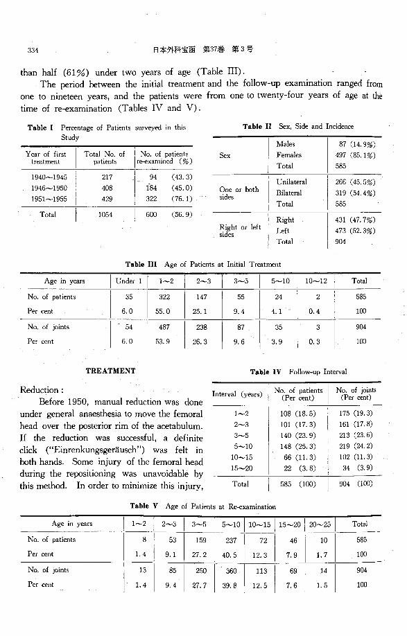
ing the indications for operation. Therefore, the simple criteria shown in Table VI were adopted.
Table VI Criteria for Assessment of Results
Results Excellent Good Fair Poor
Symptoms ; none |山田 after叫ぉme…movト IPain on …e叫walking ; none or , ment ; none at rest ; at rest slight Subjective
Full movement Full movement Slight impairment of I Impairment of move-movement ; none I ment ; fixed fixed Clinical
Slight or moderate I Moderate or 詑 verelimitation of func司 Ilimitation of func-tion I tion SI凶1tshortening of I Shorteni昭 ofthe leg the leg
Objective
h
b
L
問
g
r
t
・-
u
d
f
d
c
l
u
b
r
o
n
o
-- n
n
hに
1&
o
r
m凶
恥
町.
1
m
z
一
N
h山
N
h
一
ρlw
一
’h
一
m
d
一
U
E
一
c
n
一
nu
-Ea
一
u
n
一
f
民
一
rA
一
’E目・
nu
一
au
、a
一
m
d
一
TA
一
0
0
3
5
一
N
N
h
一
Roentgenological Almost normal
Dysplasia of the ace司
tabular roof without dislocation
j None or slight de-! formity of the femo-I 凶 head
Subluxation Dislocation
or
Slight or moderate deformity of the femoral head
Severe deformity of the femoral head
336 日本外科宝函第37巻 第3号
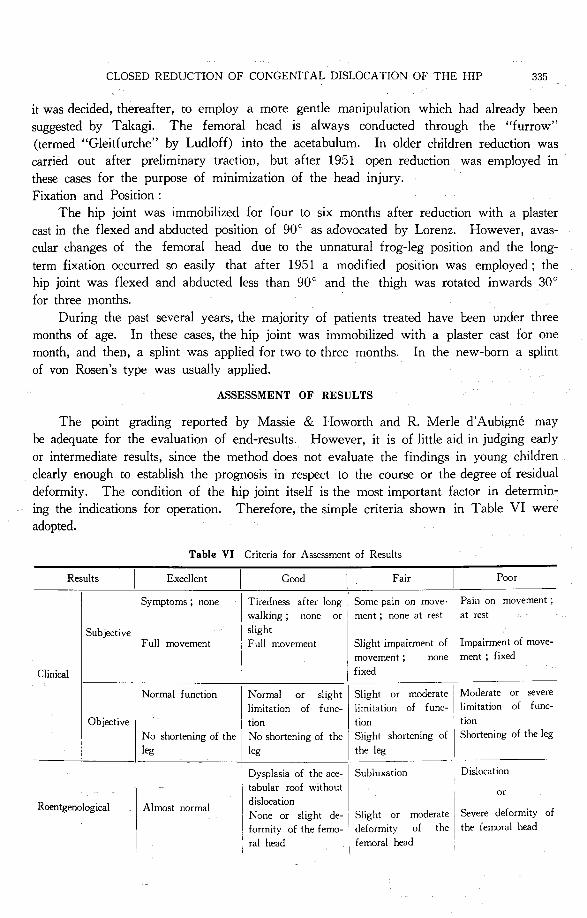
The roentgenological results were evaluated with the practical view of roentgeno・
grams in each cases. The results in the bilateral cases were evaluated on the basis of
the worse joint. Representative roentgenograms are shown in Figs. 1 3. A few patients
treated after the age of ten years excluded from the present study because of uniformaly
poor results.
Fig. 1 Excellent
3 y白 rsold
Fig. 2 Good
3 years and 4 months old
Fig. 3 Fair
3 years and 2 months old
8 years and 6 months old 12 y回 rsand 6 months old
8 years and 10 months old 18 years and 4 months old
A, B, C, D. E and G were reduced under 2 y白 1sof age ; between 14 and 20 months. F. H and I were reduced at the age of 2 years.
CLOSED REDUCTION OF CONGENITAL DISLOCATION OF THE HIP 337
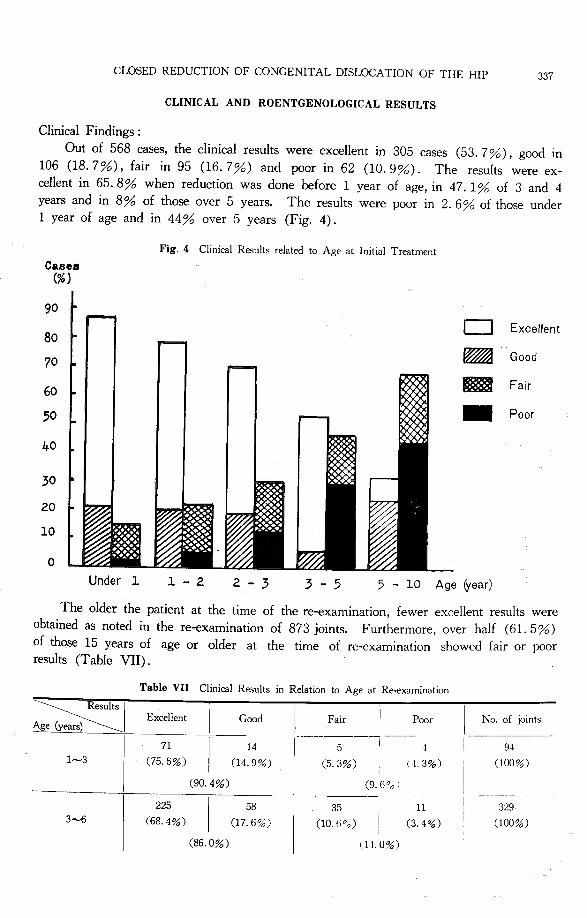
CLINICAL AND ROENTGENOLOGICAL RESULTS
Clinical Findings :
Out of 568四 ses,the clinical results were excellent in 305 cases (53. 7%), good in 106 (18. 7%), fair in 95 (16. 7%) and poor in 62 (10. 9%). The results were ex-cellent in 65. 8% when reduction was done before 1 year of age, in 47. 1% of 3 and 4 years and in 8 % of those over 5 years. The results were伊orin 2. 6% of those under 1 year of age and in 44% over 5 years (Fig. 4).
Cases (%)
90
80
70
60
50
40
30
20
10
。Under 1
Fig. 4 Clinical Results related to Age at Initial Tr白 tment
亡コ Excellent
m Good
匿盈 Fair .. Poor
1 -2 2 帽 3 3 -5 5 -10 Ageかear)
The older the patient at the time of the re-examination, fewer excellent results were obtained as noted in the re-examination of 873 joints. Furthermore, over half (61. 5%) of those 15 years of age or older at the time of re-examination showed fair or poor results (Table VII).
Table VII Clinical Results in Relation to Age at Re-examination
よ~ Excellent Good Fair Poor No. of joints
一一71 14 5 4 94
1~3 (75. 5%) 〔14.9%) (5. 3%) (.J. 3%) (100%〕
(90. 4%) (9. 6?o i
225 58 35 11 329 3~6 (68. 4%) (17. 6%) (10.6°o) (3. 4%) (100%)
(86. 0%) (l.J.0%)
338 日本外科宝函第37巻第3号
勾\二~;よ竺i Exce ent Good Fair Poor No. of joints
Total
512
(58.6%) 、‘,J%
ハU
qJ
nhv
・
唱
i
oE旬。A,,,‘、
、、,ノ%
nU
AU ヴ,
/
za‘、
136
(15. 6%)
、lノ%
円
unペU
ηL
’t、、
65
(7. 4%)
873
(100%)
Roentgenological Findings : The interpretation of roentgenograms of congenital dislocation of the hip is easy in
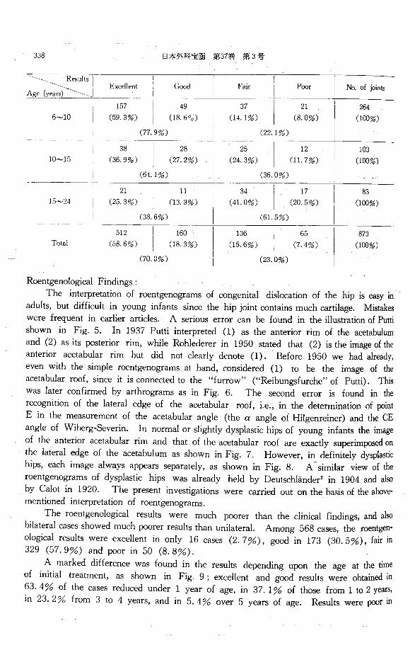
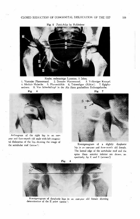
adults, but difficult in young infants since the hip joint contains much cartilage. Mistakes were frequent in earlier articles. A serious error can be found in the illustration of Putti shown in Fig. 5. In 1937 Putti interpreted (1) as the anterior rim of the a田tabulumand (2) as its posterior rim, while Rohlederer in 1950 stated that (2) is the image of the anterior acetabular rim but did not clearly denote (1). Before 1950 we had already, even with the simple roentgenograms at hand, considered (1) to be the image of the acetabular roof, since it is connected to the “furrow”(“Reibungsfurche”of Putti). This was later confirmed by arthrograms as in Fig. 6. The second error is found in the recognition of the lateral edge of the acetabular roof, i.e., in the determination of point E in the measurement of the acetabular angle (the αangle of Hilgenreiner) and the CE angle of Wiberg-Severin. In normal or slightly dysplastic hips of young infants the image of the anterior acetabular rim and that of the acetabular roof are exactly superimposed on the lateral edge of the acetabulum as shown in Fig. 7. However, in definitely dysplastic hips, each image always app白 rsseparately, as shown in Fig. 8. A similar view of the roentgenograms of dysplastic hips was already held by Deutschliinder2 in 1904 and also by Calot in 1920. The present investigations were carried out on the basis of the a加ve-mentioned interpretation of roentgenograms.
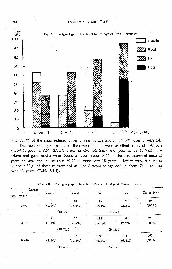
The roentgenological results were much poorer than the clinical findings, and also bilateral cases showed much poorer results than unilateral. Among 568 cases, the roentgen-ological results were excellent in only 16 cases (2. 7%), good in 173 (30. 5%), fair in 329 (57. 9%) and伊 orin 50 (8. 8%).
A marked difference was found in the results depending upon the age at the time of initial treatment, as shown in Fig. 9 ; excellent and good results were obtained in 63. 4 % of the cases reduced under 1 year of age, in 37. 1 % of those from 1 to 2 years, in 23. 2% from 3 to 4 y回 rs,and in 5. 4% over 5 years of age. Results were戸間 in
CLOSED REDUCTION OF CONGENITAL DISLOCATION OF THE HIP 339
Fig. 5 Putti・Atlasby Rohlederer
Knabe, rechtsseitige Luxation, 5 Jahre. 1. Ventraler Pfannenrand. 2. Dorsaler Pfannenrand. 3. Y・f凸rmigerKnorpel. 4. Mediale Halsecke. 5. Pfanner】h凸hie. 6. Tranenfigur CK凸hler). 7. Epiphy-配 nkern. 8. Von Schenkelkopf in der Ala iliaαgeschaffene Reibungsfurche.
Fig. 6 Fig. 7
Arthrogram of the right hip in an one-
y回 rand four-month old male with left congeni-
ta! dislocation of the hip, showing the image of
the acetabular roof (arrow 'J
Fig. 8
Roentgenogram of a slightly dysplastic
hip in an one-year and four-month old female.
The lateral edge of the acetabular roof and the
spma il1aca antenor mferior are shown, re-spectively, by E and S (arrows!)
Roentgenogram of dysplastic hips in an one-year old female showing determination of the E point (arrow ' 1
340
Cas巴
(%)
100
90
80
70
60
50
九O
30
20
10
。
日本外科宝函第37巻第3号
Fig. 9 Roentgenological R白 ultsrelated to Age of Initial Treatment
、
Under 1 1 ・3 3 -ラ
EコExcellent
~Good
医~Fair
- Poor
5 -10 Age (year)
only 2. 6% of the回 sesreduced under 1 y回 rof age and in 54. 5% over 5 y回 rsold.
The roentgenological results at the re-examination were excellent in 35 of 870 joints
(4. 0%), good in 323 (37. 1%), fair in 454 (52. 2%) and poor in 58 (6. 7%). Ex-
cellent and good results were found in over about 40% of those re-examined under 14
years of age and in less than 30 % of those over 15 years. Results were fair or伊or
in about 52% of those re-examined at 1 to 2 y伺 rsof age and in about 7 4% of those
over 15 years (Table VIII).
Table VIII Roentgenographic Results in Relation to Age at Re-examination
\恥suits!Excellent Good Fair Poor No. of joints
Age y
5 40 46 2 93
1~3 (5. 4%) (43. 0%) (49. 5%) (2. 2%) (100%)
(48. 4%〕 (51. 7%)
7 127 186 9 329
3~6 (2.1 %) (38. 6%) (56. 5%) (2. 7%) (100%)
(40. 7%) (59. 2%)
8 108 132 14 262
6~10 (3.1%) (41.2%) (50. 3%) (5. 4%) (100%)
「44.3%) (55. 7%〕
CLOSED REDUCTION OF CONGENITAL DISLOCA TIO>: OF THE HIP :m
10
1(9. 7%) I
(39. 8%)
5
(6. 0%)
31
(30. 0%)
44
(42. 7% I 10~15
17
(20. 5%) 15~24
323
(37. 1%) Total 35
(.t.0%)
46
(55. 4%)
454
(52. 2%)
(60. 2%)
r73. 5%)
is I 103
(17. !'i% I (100%)
15 ! 83
〔18.1%) ! (100%)
58 870
(fi.7%) I (100%J
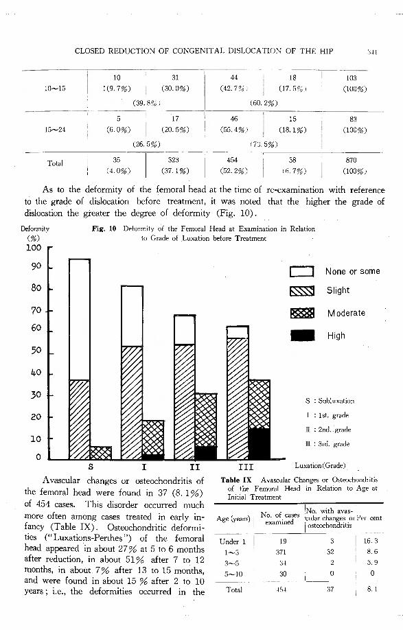
As to the deformity of the femoral head at the time of re-examination with reference
to the grade of dislocation before treatment, it was noted that the higher the grade of
dislocation the greater the degree of deformity (Fig. 10).
Deformity (%)
100
Fig. 10 Deformity of the Femoral Head at Examination in Relation to Grade of Luxation before Tr田 tment
90
Bo
70
60
50
与O
30
20
10
0 s 工
A vascular changes or osteochondritis of
the femoral head were found in 37 (8. 1%)
of 454伺 ses. This disorder occurred much
more often among cases treated in early in-fancy (Table IX). Osteochondritic deformi-ties (“Luxations-Perthes”) of the femoral head appeared in about 27% at 5 to 6 months after reduction, in about 51 % after 7 to 12 months, in about 7% after 13 to 15 months, and were found in about 15 % after 2 to 10 y田 rs;i.e., the deformities occurred in the
st
m
.,aヨU
炉、
’皐
FL
ム凶択
w
-
mA一店
、
z
r
d
O
F
V
o
s
wA
一
ait
k
l
一v民.口
h
x
一am0
.d
(
M
一
h
m凹
3
G
2
0
w
rh7uhh-
E
一5
日1
尚
一
wm蹴
一
一
e
一
LLH証
一
一
時・
m
一一N印
α
一
一
ヨU
1EBBIfi--haEE
--11
11111
1111111111111ifill』l
’hHila
-
-
c剖
一
雌d
一
MH
一
位
問
一
4
0
山
d
M
-d.m一
1幻
HJ3一品
a間
百
-
・由
一
一
;
;
一
ん
m則一
NE
pa白
n
-
xe,-
4
I・出
d-
町
E
t
:
MfxM
-b
a〈
1』-
e
T
L
叫
II
Under 1
1~3
3~5
5~10
Total
仁コ None or some
~ Slight
医罰 Moderate .. High
S : Subluxation
1 : 1st. grade
JI : 2nd. grade
][ : 3rd. grade
III Luxation(Grade)
16.3
8.6
5.9
0
8. I
342 日本外科宝函’第37巻第3号
majority of cas白 afterthe period of fixation or when the patients started to walk.
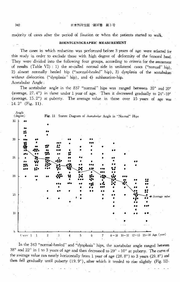
ROENTGENOGRAPHIC MEASUREMENT
The cas白 inwhich reduction was performed before 3 y伺 rsof age were selected for
this study in order to exclude those with high degree of deformity of the femoral head.
They were divided into the following four groups, according to criteria for the assessment
of results (Table VI) : 1) the so-called normal side in unilateral cas田(“normal”hip),
2) almost normally healed hip (“normal-healed”hip), 3) dysplasia of the acetabulum
without dislocation (“dysplasia”hip), and 4) subluxation-hip.
Acetabular Angle :
The acetabular angle in the 837“normal”hips was ranged between 35 ° and 20°
(average, 27. 4。) in those under 1 y白 rof age. Then it decreased gradually to 24 ° 10°
(average, 15. 2°) at puberty. The average value in those over 15 y田 rs of age was 14. 2° (Fig. 11).
35 ト ・・Angle (degree) Fig. 11 Scatter Diagram of Acetabular Angle in“N0tmal”Hips
30
25
20
15
10
.
.... ”
.... . . . . .. .. . -’・止、.
・・・M
剛・”・・“... , ....
-・・ . ... .. . . . . ::
. ・-. ・・. . . . . .. . ...
・"・ ・・・.
・・
.
.. . .. . . . ・・. . ’a
’ι甲
刷
He刷.. ..
u・. . . . ..
”符. u .. .. . .. ・・. ・・・ ・... . ... ...... . ・.. . . ~ . ・2・..・. . . . .
・-----. . ・...... l’nder I I 6
5
2 7 8 -10 10ー12 12 15 15ー20Age (year) 5
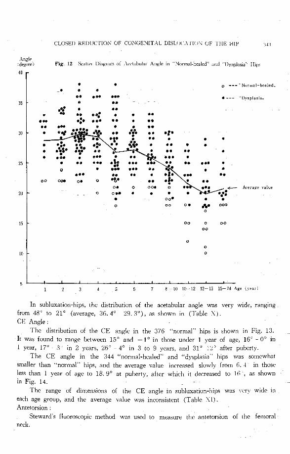
In the 343“normal-healed”and “dysplasia”hips, the acetabular angle ranged between
38° and 22° in 1 to 3 y田 rsof age and then decreased to 29° 10° at puberty. The curve of
the average value ran nearly horizontally from 1 y田 rof age (28. 8°) to 3 y白 rs(29. 8°) and
then fell gradually until puberty (19. 9°), after which it tended to rise slightly (Fig. 12)・
CLOSED REDUCTION OF CONGENITAL DISLOl、λT!Ul"OF THE HIP : l ~J:l
Angle Fig. 12 Scatter Diagram of Aceti!bular An巨lein “Normal-healed”and“Dyaplasia”Hrps I degree)
40
. . 。ーーー“Normal-healed.,. . . ・- ... ... ・・ーー “Dysplasia”
35卜 . .. ・:: ・. .. . .. ・4 . . ..
... -:・ ..・ .. . .. .. .... . . -・30ト . . ., .・.・a・飽・.且・y. .. . -.・. .. ・2・・. . . . . .. . .
. ・・.,~.・ ・も・ ・-.. . . ... . ... .
25十 . -・
... ・. -・・. 。 . . .
.・・ -・・ ・.。。00・ 0・ 。 .. 司. ・ゐ\・・空 /.← A川 agevalue 0・。
00・。20ト 。
0・・ . . . . oo• . . 。 。。 。. ,.. 。。。。
15卜 。。 。 。。。。。 。
10ト ロ
5 2 3 4 5 6 7 8 10 10ー12 12ー15 15 24 Age l、日r)
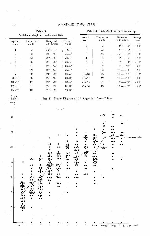
In subluxation-hips, the distribution of the acetabular angle was very wide, ranging
from 48° to 21° (average, 36. 4° 29. 3°), as shown in (Table X).
CE Angle:
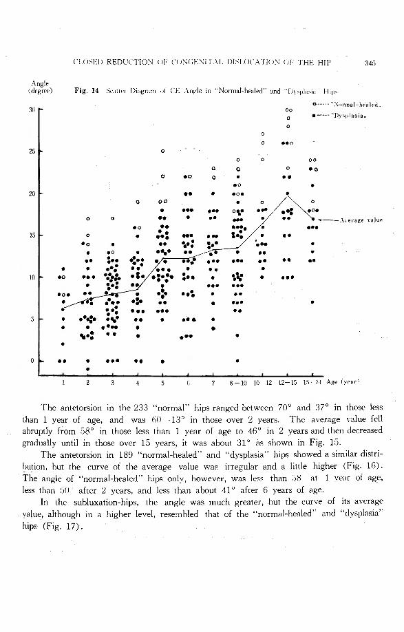
The distribution of the CE angle in the 376 “normal”hips is shown in Fig. 13.
It was found to range between 15° and -1° in those under 1 year of age, 16° -0° in
1 year, 17° 3〕 in2 years, 26° 4° in 3 to 9 years, and 31° l'.t' after puberty.
The CE angle in the 344“normal-healed" and “dysplasia" hips was somewhat
smaller than "normal”hips, and the average value increased slowly from 6. 4 in those
less than 1 year of age to 18. 9° at puberty, after which it decreased to lβ ’, as shown
in Fig. 14.
The range of dimensions of the CE angle in subluxation-hips was very wide in
each age group, and the average value was inconsistent (Table XI). Antetorsion :
Steward’s fluoroscopic method was used to m回 sure the antetorsion of the femoral
neck.
:¥-l-l 日本外科宝函第:17巻第3弓
Table X Table XI CE Angle in Subluxation-Hips
Acetabular Angle in Subluxation-Hips Age in Number of R~r7~~ of ミl'erage
Age in Number of ~~r7~~ of Aver<1ge 、ea" .JOmts dist tion value
years ド11nts di tion value -40~ー15°1 3 -9. 7。
3 :l2"~:;-1・ 33.3。 2 -13 K J~ー12° ' -1. 6'
つ -13 :5--lfi 3』.30 3 /<:l 11"~ー17。
3 83 己7'~」h 35. -1° 4 1)1〕 100~ー16。 -~ 0'
-I 66 :!80~-li'.i ·~ 36.4。 5 ’i」 7。~ー1I 0 ! -I 2。
5 :q 三日c~-IS乙 35.5° 59 IL"~ー22° 0. 4'
6 59 :!8ι~-1:) 36.4° I :l8 17°~ー1l'' 2. 8•
I 38 :!-I’~-1:!・' '.l-1. 5。 日~IO 25 10'~ー18° 1. 0。
H~l I) 25 c'i・~4り :i-1. 2° IO~! :! 17 J:l''~ー10。 3.2'
10~12 出也~-1:! 33.5° 1)~1:1 !:)τ~-22' !ー0.6
l三~15 :!fi~-l8' 33.3。 1九~21) 18 Iii'~-22° u。
IS~ゴ(} 18 21 ~-1:!♂ :!'!. 3。
Angle Fig. 13 Scatter Diagram of仁E Angle in“丸山nal'’Hips(degree)
3S
. ・切:ioト .
. . . ・- -・. . . . .
25ト . . z” ・・. . . . . . ・2・. ・::
!?ム. ・. . ... .
-・. ..
-・. . ・.
~II ト . .. ・」J・・A・2・-:・ . . ... ‘む -.. .. ・: .. .. . ••• ... :,•’ . . ... .
. ・. :-- .. . ..
・- ・.・-・ . . ISト . . ・.... . -:・.. . . ..
・-. -・ ・- -・・ ・- . . .1. -・・
. . . .:. ・- .. -・・JOト . ・.. 2・・ -・... . .
・. . ~Ii
. -・. . . -・・ ・-
. ・--
....:. . . ・WI持. ・2 . . ... .. ・-.- . -・・
. .. -・・.
。ト 'li . .
1
6
l r
f " 7 8ーIO IO 12 12-15 l'i-20 へ g•' ¥)・Par)
(、UJSEI J RED UC、,rION<J下(、<JNCト.NIT.¥LDJメ[()(、AT!<JN CJF THE HIP 345
Angle (degree〕 Fig. 14 Sc1ttn Diagram "' CE 人nglein“Normal-healed”and ・1入、pl‘";"、 Hl(J、
。一一-'':\。υrmalhraled” 30 ,. 。。。 ・--ーー“Dy勺'Iasi a” 。。
。 ..。。。 。 。。
Q 。 。 ・0。 ・0 Q . .. .。 . 20 I-
・-. ・0・。 。。 . 。
. ・-- .-- 。、- . .. ;~. 。 。
-・.
・- ... . - T一ーー一人veragevalue .。 .. ・・・. . ・・. 15ト 。 . ・. ... ,.: . .. ・・. . ..
・.’..・.. - .・4‘ . ・。 . .. .・・. .。 . -、- .. . - . . . .
・・ ・:・z・・p~・1・・・5・0: ・・ ・2 .・・ -・. ・-.. ..
. -...- -. . 10ト ・0 .. . . z’ . . ・・.. ・. =・・ . 色、r . .. .
. .. . ... ・》 ••\ .
・-.. . -・・.
・J ・.・- -.. ... -・.・5ト . .・4・・ぜ .. . .・・ .
. . .・.. . . 、・2 -・ . .-- .
. .. .
・--.. . .
. 2 3 5 7 8 10 10-12 12 15 15← :! I Age (vearl
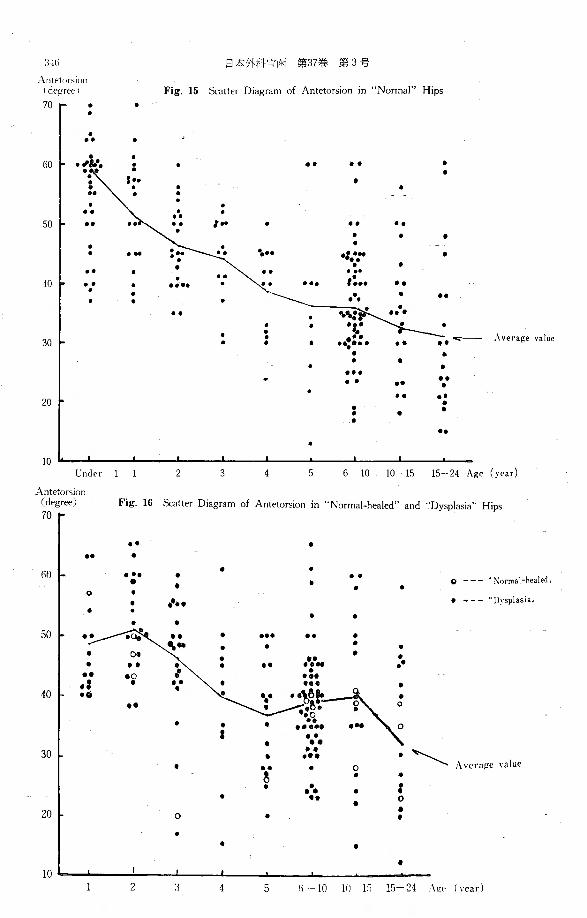
The antetorsion in the 233“normal”hips ranged between 70° and 37° in those less
than 1 year of age, and was 60 13° in those over 2 years. The average value fell
abruptly from 58° in those less than 1 year of age to 46° in 2 years and then decreased
gradually until in those over 15 years, it was about 31 ° as shown in Fig. 15.
The antetorsion in 189 "normal-healed" and “dysplasiaけ hipsshowed a similar distri-
bution, but the curve of the average value was irregular and a little higher (Fig. 16).
The angle of ・'normal-healed”hips only, however, was les5 than 三ぷ at 1 veけrof age,
less than 50 after 2 years, and less than about 41 ° after 6 years of age.
In the subluxation-hips, the angle was much greater, but the curve of its average
value, although in a higher level, resembled that of the “normal-healed門 and“dysplasia”hips (Fig. 17).
'.l-lti 日本外科‘I:商 第37巻第3号
Antet1•r>i•111 1 degree I Fig. 15 Scatter Diagram of Antetorsion m“Normal'’Hips
70「 . . . . ..
60 ト ·!~:·. . - . .. .. . . . - . . .. . ..
.. . .. -・. . . . . . も・・ ・: .. ・.” . .
-・ .. ・... .. ,:.. 40ト .. . ・・・. . -・ ・・. ・.. . . .
.. . . . ・-. 一--;.可- A附叩 value30卜 . . . ・4・・・. . . . . ... .. .. .. .
-・ .・20 I・ . - . . . .
・. . 10
lln<ler 1 1 2 3 5 6-10 10-15 15-24 A店e(year)
A ntetorsion 〔degr配) Fig. 16 Scatter Diagram of Antetorsion in“Normal-healed”and "Dysplasia”Hips
・- . ・-
. . .
60ト ・-. . .. 。--- ・・Normal-healed” . . . . . 。 . ・--ー” I〕~ splas1 a” . .・... . . . . .
50 ~ .. ~ -・
. ... .. .
ミ. . . . . ... . . ・.-.. . .. . -・.. ・0. .a ・.- 40ト ・6 .・
. . . I .
30 l.. ..
.、\、、、、 Averai(e、alue. -・・. ~
. 。. . . .・-.. . : . 。.
20ι . 。 . .
. . . .
10 2 4 5 Ii 10 If)ー l:i 15-24 ,¥g,. (year)
CLOSED REDUCTION OF CONGENITAL DISLOCATION OF THE HIP 347
Antetorsion (degree)
70
.- 60
50
. 40
.,
30
20
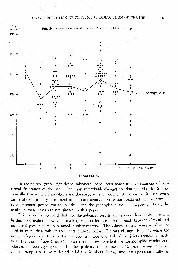
Cervical Angle :
Fig. 17 Scatter Diagram of Antetorsion in Subluxation-H1ps
. . ・司F . . . . .. . ・ . .. . . . ・.-・ .・ . ..・・ 畠陶 士、 .. . . . .. . . \ . ・. . . . . . -・. -・. .. . . ・.,,. . . . .
・--. .・・%-.-.
.. . . . . . . . . . .. ,.・-・-品 ・3・ー~~『一一 A、eragevalue
・-- . .. . ... . . .. . . . .・. .
. . . . . . . . .
・》 3 5 flー 10 10ー 15 15-24 A伊川町trI
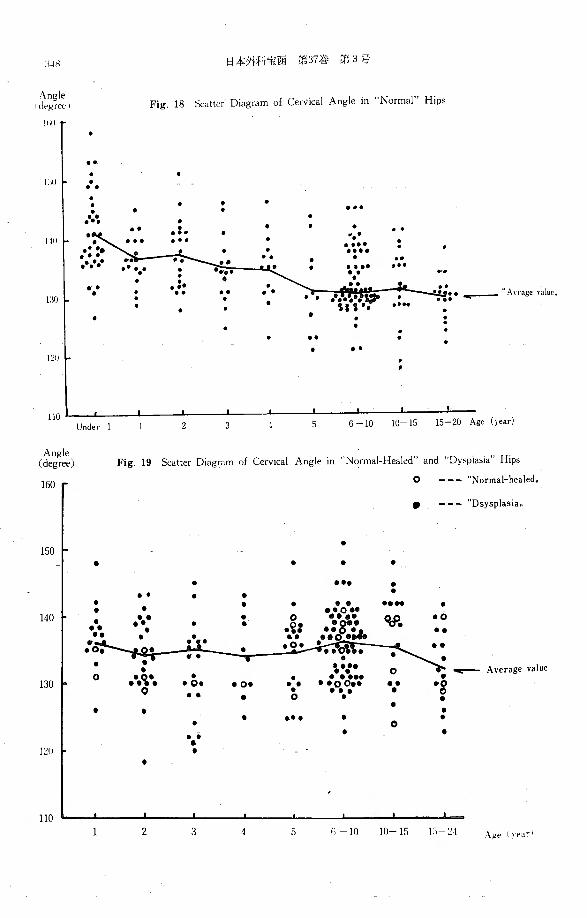
The cervical angle was measured in roentgenograms taken with the femur rotated
maximally inward.
The cervical angle in 218“normalηhips ranged between 158° and 127° in those
under 1 year of age, 151 ° and 128° in those from 1 to 2 years, 143° and 121° in 3 to
9 years, 141 ° and 118° in 10 to 14 years, and 138° and 122° in those over 15 years.
The curve of the average value fell considerably from 142° in those under 1 ye釘 of
age to 137° in 1 year, then fell gradually until 15 years, when it was 129°, as shown
in Fig. 18.
In the 209“normal-healed”and “dysplasia”hips, the cervical angle renged between
151° anid 118°, and the curve of the average value was nearly horizontal (Fig. 19). In “normal-h白 led'’ hips,the angle remained within a narrow range (140° 124°).
In the 149 subluxation-hips, the cervical angle had a wide range (155° 100 ・) and
the curve of the average value was very irregular (Fig. 20).
:i」H 日本外科宝函第37巻 tf)3号
Angle 1 degree I
li川
Fig. 18 氏《alterDiagram of Cεrvical Angle in "Normal”Hips
. .-
DJト. .. . . . . . . ... ・.. . . ・--. .. .- .. . .
1111ト ・3に . ・’.・
. . .-- . .. ... .. . . ..
.・・. ... 、v ~-· 品凡ーー・::
J:lOド . ・.. . . ー・・ ’ . . . . . . . . . . . .. . . L~ll
. ・-
110 15-20 Age (year) Under I 2 3 5 6 10 10 15
Angle (degr配) Fig. 19 Scatter Diagram of Cervical Angle in”Normal-Healed”and “Dysplasia”Hips
。ーー『“Normal-healed”. ーー『“Dsysplas1a”
150「.
. . . . .
・-- . ・- . . . . . .
・・・・.・~・.・・・・・・... . . .
140ト .. . 。明
.。. . 凸.・. .
. .- ・'=~・..
\ト• ---A川 agevalue . 。。.·.~。~'・- .
130ト. .
・0・ ・0・ .. ・・oo・・ ・- •g .. . ・・. . . 。 . . . . . .
. . ・・. . . 。... . .
l:!IJト . .
ハU1
1 2 3 4 5 れ 10 10-15 Li ~I 人区" I、earI
CLOSED REDUCTION OF L・UNGENITALDISLOCATION <JF ・1・HEHIP 349
Angle (degree) Fig. 20 Scatter Diagram of Cervical Angle in Sublu入dtl<川ーHip'
]Ii()
. . .
!50ト . . ・. -・ . .. . . . . . .. . . . . . ・-
.・ト
. . 1111ド . -・・・・・・- .・・・・ ....・ . . .
-・. .. .. -・. .-- . ..
-・. 130ト
. .. ..司守一一- Average value \・ ・・. .. .・・・. . . . .. .
・--. . . . . . . .
120ト. .. ・-
. . . . . ’
. .
llOト. .
. 100 .
2 3 5 6 -10 10-15 15 24 Age (year)
DISCUSSION
In recent ten years, significant advances have been made in the treatment of con-
genital dislocation of the hip. The most remarkable changes are that the disorder is now
generally treated in the new-born and the surgery, as a prophylactic measure, is used when
the results of primary treatment are unsatisfactory. Since our treatment of the disorder
in the neonatal period started in 1962, and the prophylactic use of surgery in 1954, the
results in these cases are not shown in this paper.
It is generally accepted that roentgenological results are poorer than clinical results.
In this investigation, however, much greater differences were found between clinical and
roentgenological results than noted in other reports. The clinical results were excellent or
good in more than half of the joints reduced before 5 y回 rsof age (Fig. 4), while the
roentgenological results were fair or poor in more than half of the joints reduced as early
as at 1-2 years of age (Fig. 9). Moreover, a few excellent roentgenographic results were
achieved in each age group. In the patients re-examined at 15 刊日r~ of age or o、er,unsatisfactory results were found clinically in about 臼 So, and roentgenographically in
350 日本外科宝函第37巻第3号
about 72% of the joints (Tables VII and VIII). Therefore, the early or intermediate clinical results are of very little value, and roentgenograms should be evaluated more strictly
than advocated by Severin. It may also be noted that the majority of hips treated conser-
vatively will show some roentgenographic disorders in the future and that both the clinical
and roentgenological results will become rapidly worse after puberty.
According to our investigation, the upper limit for closed reduction is 4 years of age,
since almost no cases of roentgenographic h回 lingwere found among those in whom re-
duction was performed after 5 years of age (Fig. 9). Although the problem has become
mainly theoretical because of the popularization of treatment in the neonatal period, we simply suggest the upper age limit of 4 y回 rsfor prophylactic surgery. Our investigation
also shows that indication for the prophylactic surgery will be considered in more than
half of even those treated before 2 y白 rsof age, except for new-barns.
A vascular changes or osteochondritis of the femoral head were of ten found in casEs in which reduction was performed in early infancy, as was reported by Petit. On the
other hand, osteochondritic deformities (“Luxations-Perthes”) of the femoral head developed
in the majority of cases after pericd of fixation or when the patients started to walk.
The data mentioned above suggest that avascular changes caused by the unnatural frog-
leg position and long-term fixation are primary factors responsible for the osteochondritic
deformity, and that secondary factors are traumatic injuries, in the wide sense, due to
incomplete reduction because of so・calledinterposition, high acetabular obliquity, excessive
antetorsion, etc.
Evaluation of the tendency to develop an acetabular roof during and afttr treatment
is now the most important problem. Schede in 1925 suggested that the lower age limit for the shelf operation is 7 years since the acetabular roof could be expected to develop adequately in young children. However, this is a classical consideration prior to the intro-
duction of the early shelf operation as a prophylactic procedure to prevent the develop-
ment of the disorders which followed“dysplasia" and subluxation-hips. It was already
proposed by Gill in 1935 that the shelf operation should be done at 2 years of age. Then
M. Lange, in 1951, stated that the lower age limit for the shelf operation should be 4
years, since natural formation of the acetabulum could be expected up to 3 years of age.
The natural formation of the acetabulum mentioned earlier articles, however, would be
that of the 1st false cotyloid cavity depicted by Calot, and not the formation of the
original cotyle. In 1962 M. Lange revised his opinion and stated that the shelf operation
should generally be carried out at 3-4 years of age and the operation is definitely indicated
when sclerosis appears in the upper acetabular roof. The “sclerosis" of the acetabular
roof, a term used since Schede, is not sclerosis in the true sense of the term, but indicates
a regular sharp contour of the image of the acetabular roof. If genuine sclerosis appears,
it indicates already the development of osteoarthritic changes, by which time it would be too late to perform the prophylactic shelf operation.
To determine the indications for the prophylactic shelf operation, it is necess釘yto
ascertain the present state of the osseous acetabular roof and its future development. For
that evaluation, it is very helpful to measure the acetabular angle and the CE angle peri-
odically. In “normal”hips, our investigation showed that the acetabular angle decreased
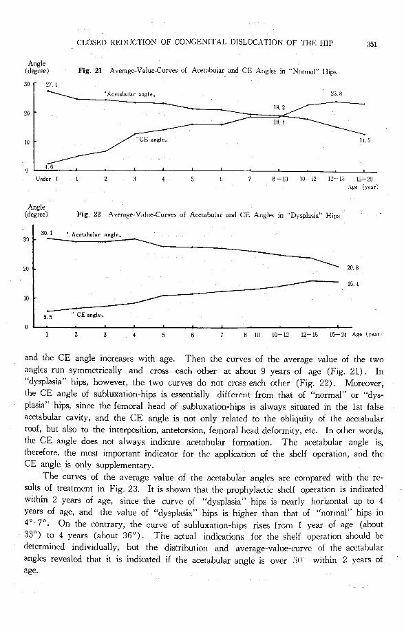
CLOSED REDUCTION OF CONGENITAL DISLOCATION OF THE HIP 351
Ane:le (degree) Fig. 21 Average-Value-Curves of Acetabular and CE Angb m“Normal”Hips
30「 27.4
2:J. 8
20
10 “Cιangle., 11. G
Under 1 2 3 4 8 -10 10 12 12ーl:J 15 20 ¥ge (year)
Angle (degree) Fig. 22 Average-Value-Curves of Acetabular and CE Angles in "Dysplasia”Hips
30 30目 1 “Acetabularangle”
20 20.8
15. 4
10
5目5。2 3 4 6 8 -10 10ー12 12 15 15-24 Age (year I
and the CE angle increases with age. Then the curves of the average value of the two
angles run symmetrically and cross each other at about 9 years of age (Fig. 21). In
“dysplasia”hips, however, the two curves do not cross each other (Fig. 22). Moreover,
the CE angle of subluxation-hips is essentially different from that of “normal”or“dys-plasia”hips, since the femoral head of subluxation-hips is always situated in the 1st false
acetabular cavity, and the CE angle is not only related to the obliquity of the acetabular
roof, but also to the interposition, antetorsion, femoral head deformity, etc. In other words,
the CE angle does not always indicate acetabular formation. The acetabular angle is,
therefore, the most important indicator for the application of the shelf operation, and the
CE angle is only supplementary.
The curves of the average value of the acetabular angles are compared with the re-
suits of treatment in Fig. 23. It is shown that the prophylactic shelf operation is indicated
within 2 ye釘 sof age, since the curve of “dysplasia”hips is nearly horizontal up to 4
years of age, and the value of “dysplasia”hips is higher than that of“normal" hips in 4 ° -7° ・ On the contrary, the curve of subluxation-hips rises from 1 year of age (about
33°) to 4 years (about 36°). The actual indications for the shelf operation should be
determined individually, but the distribution and average-value-curve of the acetabular
angles revealed that it is indicated if the acetabular angle is over ;)() within 2 years of age.
:;s2 日本外科宝函第37巻 第3号
Angle Fig. 23 Average-Value-Curve of Acetabular Angle related to Age at Examination (degr田)
36. 4
35
30
25
20
15
2 3 5 6 7 8-10 10ー12 12 15 15 24 Ag向!目付
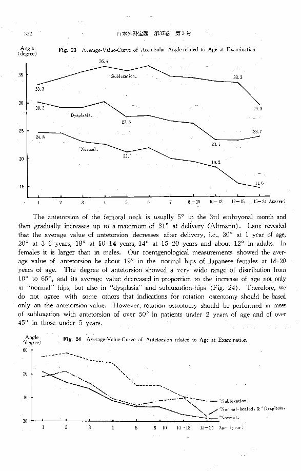
The antetorsion of the femoral neck is usually 5° in the 3rd embryonal month and
then gradually increases up to a maximum of 31° at delivery (Altmann). Lanz revealed
that the average value of antetorsion decreases after delivery, i.e., 30° at 1 year of age,
20° at 3 6 years, 18° at 10-14 ye沼rs,14 ° at 15-20 years and about 12° in adults. In
females it is larger than in males. Our roentgenological measurements showed the aver-
age value of antetorsion be about 19° in the normal hips of Japanese females at 18-20
years of age. The degree of antetorsion showed a very wide range of distribution from
10° to 65°, and its average value decreased in proportion to the increase of age not only
in "normal門 hips,but also in “dysplasia”and subluxation-hips (Fig. 24). Therefore, we
do not agree with some othors that indications for rotation osteotomy should be based
only on the antetorsion value. However, rotation osteotomy should be performed in cases
of subluxation with antetorsion of over 50° in patients under 2 years of age and of over
45 ° in those under 5 years.
会;~:) Fig. 24 Av叫 e司Value
60
50
Ill
30
、、、.,喝 、、-、、司、、、-、、、、、、
\ ー-ーー田ー、
ミ\\、、む~ _ . ....:.干、ーー一-、.』-・-・- ·,『h・h 『~"Subluxatwn”
一~-~·- ・・. 、" /'白:\ ormal-healed”&“Dysplasia..
\こ-":¥ormal”
2 3 5 6-10 10 15 15 ~I A!(e I ¥earl

Angle (degree)
140 、、、、
CLOSED REDl lCTION OF CON(;ENI’L¥L DIペLOCATIONOF THE HIP 353
Fig. 25 Average-Value-Curve of Cervical Angle related to Age at i九<1mination
-~·-·一三『F・ーー・・』’ーーー-ー・-::-_・--=ニヨヲ雪Eマ・一・ー・ーー--τ’ \ ・、\
, ’ 一一一一‘、ヘ .-' 、\ \ J “:\ormal healed”
, \ミミ:・’, .司~~司 、ー &”υ; aplas1a,, 130ト \〆 • -----ベ二\ Subluxation,
、 \ ι :--;,’1mal ”
2 3 5 6 10 10ー I~ 15 24 Age (year)
The average value of the cervical angle is 144 ° in 1 2 years of age and gradually
decreases to 126° in adults (Lanz). In our investigation, the average value in “normal" hips was 136° in 1 year of age and 130° in those over 15 years of age; its distribution
showed a range of 158つ-118°. Although the cervical angle of subluxation-hips was widely
distributed from 155° to 100°, its average value showed only a little deviation from 141
to 131 ° (Fig. 25). Therefore, the operative correction of the cervical angle is not necessarily
performed as a single porcedure in young infants. However, it would be preferable to
combine it with rotation osteotomy for the prevention of valgus deformity developing after
surgery.
SUMMARY
The results of conservative treatment of 585 patients with congenital dislocation of
904 hips which had been treated by closed reduction at Kyoto University Hospital during
the period from 1940 to 1955 have been reviewed, and the indications for prophylactic
surgery in cases with unsatisfactory results are discussed.
1) In 504 (86.3%) of the c前 田 町viewedreduction was performed before 3 vears
of age, in 322 (55%) before 2 years, and in only 35 (6%) before 1 year. The period
from initial treatment to re-examination was 1-19 years, and the patients were 1 ~ 1 years
old at the time of re-examination; the majority (90.5%) were under 15 years of age.
2) Clinical results were excellent and good over 64.1 % of those under 15 y田 rsof
age, but were fair and poor in 61.5% of those over 15 years of age.
3) Roentgenological results were much poorer than clinical. Although it is generally
accepted that the younger the age of primary treatment the better the results, fair and
poor results were found in 36 % of those treated under 1 year of age and in 62 % of those 1-2 years of age. Moreover, re-examination revealed subluxation, dislocation or
severe deformity of the femoral head in 52%, already, at 1 2 years of age, and in 73%
after 15 y白 rsof age. Therefore, prophylactic surgery is indicated in half of the treated
cases in order to prevent later development of the osteoarthritis of the hips.
4) The indications for prophylactic shelf operation can be determined already at the
age of 1 2 years, when we take into the consideration the distribution of the acetabular
angle and the transition of its mean value. If the acetabular angle is 30° or more ope-
ration is indicated. The indication for rotation osteotonw in younger infantベcannotbe
decided on the basis of the antetorsion value alonゼ Theoperation should be performed
only for subluxation with antetorsion of more than ;)0° at the age of 1 2 year宍 andof
354 日本外科宝函第37巻第3号
over 45 ° in those under 5 y白 rsof age. The operative correction of the cervical angle
is not necessarily performed as a single procedure in young infants. However, it is pre-
ferable to combine it with rotation osteotomy for the prevention of valgus deformity de-
veloping after surgery.
5) Avascular changes or osteochondritis of the femoral head were frequent after
reduction in early infancy, and develop after the period of fixation in the majority of
伺 ses. Therefore, avascular changes due to unnatural frog-leg position and long-term forn-
tion are probably as primary factors for osteochondritic deformity (“Luxations-Perthes"),
while traumatic injuries, in the wide sense, are secondary factors.
REFERENCES
1) Altmann, Fr. : Untersuchung i.iber die Torsio femoris und damit in Zu回 mmenhangstehende Frage. Anal.
Record, 75. 82, 1925.
2) Brandes, M. : Ueber die Einstellung des Femurkopfes bei der Luxatio coxae congenita. Z. Orth., 44, 497,
1924.
3) Calot, F. : Ueber neuere Anschauungen in der Pathologie der Hi.ifte auf Grund der Arbeiten der letzten
Jahre. Z. Orth., 51. 134, 1929.
~ i Chiari. K. : Ergebnis担 derFri.ihestbehandlung der angeborenen Hi.iftgelenkverrenkung. Arch. ・orth. Unfall-
Chir., 45. 644, 1953.
5) Forgen, M. : Zu einem atiologi配 hen F acktor des 回 genannten“LuxationトPerthes”. Z. Orth., 102, 305,
1966. 6) Gaugele, K. : Beitrag zur Diagnose der Kopfとtellungbei der angeborenen Hi.iftgelenkverren'kung. Z. Orth.,
44. 569, 1924.
7) Gi』II.A. B. : Plastic Construct』onof an Acetabulum in Congenital Dislo白 tionof the Hip-the Shelf Ope-
ration. J. B. J. S .. 17, 48, 1935.
8) G凹 ht,H.: Die田 genannteAngeborene Hi.iftverrenkung, ihre Pathologie und Therapie von Adorf Lorenz.
Deut配 heOrthopadie 3, Ferdinand Enke, Stuttgart 1920.
9) Lange. M. : Endresult der Behandlung der Angeborener Hi.iftluxation. Verh. dtsch. Orthop., 24. Kongress.
119, 1930.
10) Lange, M. ; Orthopadisch-chirurgische Op.-lehre. 2. Aufl .. Bergmann, Mi.inchen 1962.
11) Lanz, T. von : Anatomie und Entwicklung des menschlichen Hi.iftgelenkes. Verh. dtsch. Orthop., 37.
Kong re田, 5,1950.
12) Martin, R. : Lehrbuch der Anthropologie. II. 2. Aufl., Gustav Fischer, Jena 1924.
13) Ma,;,ie, W. D., and Howorth. M. B. : Congenital Dislocation of the Hip.-Method of Grading Results
J. B. J. S .. 32 A. 519, 1950.
14) Muller, G. M .. and Seddon, E. T.: Late Results of Treatment of Congenital Dislocation of the Hip Joint.
J. B. J. S .. 35-B. 342, 1953.
15) Penners, R.: Fri.idiagno田 undSofortbehandlung der angeborenen Hi.iftluxation. Arch. orthop. Unfall-Chir.,
47. 391, 1955.
16) Petit, P. : Osteochondritis in Congenital Dislocatonりithe Hip. J. B. J. S., 37-B, 513. 1955.
17) Pitzen P. : F ruhdiagnc同 derangeborene Hi.iftgelenkverrenkung. Verh. dtsch. Orthop., 37. Kongress, 78,
1950.
18) Platou, E. : Rotat旧nO錦町tomyin the Treatment of Congenital Dislocation of the Hip. J. B. J. S .. 35-
A. 48, 1953.
19) Rohlederer, 0. Zur R凸ntgendiagnostikder Hi.iftluxation. Verh. dtsch. Orthop., 37. Kongress, 95, 1950.
20) Ro担n.S. von : Early Diagnos i ~ and Treatment of Congenital D店locationof the Hip Joint. Acta oロhop.Scandinavica 26, 136, 1956.
21) Schede, Fr. : Die Behandlung der veralteten Subluxation der Hi.ifte. Z. Orth., 43, 284. 1925.
22) Seddon, H. T. : Congenital Di』sl促:ationof the Hip lntermedi凶te・lateResults of Standat Method of Treat・
ment. J. B. J.日, 33-B,281, 1951.
23) Severin. E. : Congenital Dislocation of the Hip-Development of the Hip after Clo担 dReduction. J. B. J.
?合, 32-A.507, 19'.lO.
24) Thomao, G. : Die L町stungsgrenzender Detorsionsosteotomie am proximalen Femurende. Z. Orth., 101, 388,
1966.
CLOSED REDUCTION OF CONGENITAL DISLOCATION OF THE HIP 355
先天股脱に対する非観血的治療の成績
京都大学医学部整形外科学教室
森田 信・赤星義彦
1940年から1955年にわたる15年間lζ京大整形外科に
おいて保存的療法を受けた先天性股脱児について,治
療終了後l~22年における成績を系統的に調査し,治
療実数の56.9%に相当する585例 ・904関節において臨
床並びにレ線学的検査を実施することができた.また
股関節のレ線学的計測値を中心l乙検討を行L3,成績不
良の症例lζ対する予防的補正手術の適応範囲およびそ
の適応年令の決定lζ関し考察を加えた.
!. 調査例中504(86. 3%)は 3才未満の整復例,ま
た322(55%)は 1才台における整復例であり, lJ未満
の整復例はわずかに35( 6 %)であった.初療から調
査までの経過年数は 1~22年,また調査時の患者の年
令は 1~24才で,その中の90.5%は15才未満である.
2. 臨床的成績は,14才までは優・ 良例が過半数を
占めるが,思春期前後から自覚的および他覚的lζ何ら
かの障害を来す症例が増加し, 15才以後になるとその
比率は逆転して可 ・不可例が過半数lζ達する.
3. レ線学的成績は臨床的成績に比し著明lζ悪く,
一般的lζ初療時年令の若いものほど良好な成績を示す
が,すでに 1才未満の整復例においても36%. 1およ
び2才の整復例では62劣lζ可・不可例が見出された.
また調査時年令別にみても, 1および 2才ですでに
可・不可例すなわち骨頭の転位と変形を伴う臼蓋形成
不全が25%に見出され, 15才以後になるとそれが73%
lζ達した.したがって,早期治療例においても,その
約半数において, 2次的変形性関節症防止を目的とす
る予防的補正手術について適応の有無を慎重に考慮す
べきである.
4. 臼蓋角, CE角,前捻角および頚体角は, いわ
ゆる正常側関節においては,いずれもその分布および
平均値にそれぞれの年令lζ応じた規則正しい変化が示
され,また骨頭転位を伴わぬ臼蓋形成不全例において
も,それぞれその平均値は異なるが,大体において正
常側に近いパタンを現わす.とれに反し,亜脱臼例に
おいてはとれと大いに趣きを異にし,分布の範囲は広
しその平均値曲線はきわめて不規則であった.
5. 予防的臼蓋形成術ば. α角の分布範囲およひ’そ
の平均値の年令的推移からみて,その適応決定はすで
に 1~2才の時期において可能であり,乙の時期にお
ける手術の応用は30。以上のα角を示すものから選ぶ
べきである.減捻骨切り術の適応は,前捻角値のみに
よって決定する乙とは無謀であり,幼若児においては
前捻角45。以上の亜脱臼例lζ限定すべきである. また
頚休角の矯正手術は,これを単独に幼年児l乙行う必要
は一般的には認め難 く,転子間あるいは転子下骨切り
術後lζ外反股の発生を見るζとが少くないので,減捻
骨切り術を施す場合.同時IC頚体角を120。程度lζ減ず
るζとが望ましい.
6. 阻血性骨頭変化ないし脱臼性骨軟骨炎は,整復
年令の若いものにより多く見出され,かっその大多数
は固定終了後lζ発生した.したがって,脱臼性骨軟骨
炎(Luxations-Perthes〕は整復による機械的障害より
もむしろ長期間の画一的固定肢位による阻血性骨頭変
化を 1次的因子とし,固定期間中ないしその後におけ
る広義の外傷を 2次的因子として発生するものと考え
たll.
![Title Highly selective photocatalytic reduction of carbon ......6 [12]. In these photocatalytic reaction systems, the reduction of CO 2 to CO (eq. 1) and reduction of proton to the](https://img.pdfslide.tips/doc/110x75/5f8de0d57434da41ef7ddd89/title-highly-selective-photocatalytic-reduction-of-carbon-6-12-in-these.jpg)