Embed Size (px)
Citation preview

Title CANCER SPREAD VIA EXTRAVASCULAR FLUID PATH
Author(s)
YAMAMOTO, MASAKATSU; MATSUOKA, SCHOZO;FUKUKSHIMA, NOBUKO; KIDO, MARIKO; MIYAMOTO,TSUNEKO; MAEDA, KIMIKO; AOYAGI, HAJIME;INOUE, YASUKO; SATO, YURIKO; ITANI, KANICHI
Citation 日本外科宝函 (1960), 29(6): 1456-1473
Issue Date 1960-11-01
URL http://hdl.handle.net/2433/207185
Right
Type Departmental Bulletin Paper
Textversion publisher
Kyoto University

1456
CANCER SPREAD VIA EXTRAVASCULAR FLUID PATH
MAsAKATsu YAMAMOTO, ScHozo MATSUOKA,
NoBUKO FUKUSHIMA, MARIKO Kmo, TsuNEKO
MIYAMOTO, KIMIKO MAEDA, HAJIME AoY AGI,
y ASUKO INOUE, y URIKO SA TO
From the Department of Surgery, the Kansai Medical School, Osaka, Japan.
(Director . Prof. Dr MuNEO OKA)
KA NI CHI h ANI
From the Second Surgical Clinic: of Kyoto University Hospital.
(Director : Prof. Dr YAsu;iAsA AoYAGr)
Received for publication Aug. 29. 1960.
Since a local tissue invasion is a prerequisite to the formation of lymphogenic
metastasis, knowledge of the pr・e-l~'mphovascular fluid path is essential for a踊 tis-
factory explanation of the spread of cancer.
This approach was initial!γrcprt:sented by Kiharas’report.
It is the purpose of the present report to review our concepts concerning
lymphogenic metastasis in the light of knowledge gained from our experiments
during the past four years.
NEWER CONCEPTS ON THE MECHANISM OF
INTRAPERITONEAL ABSORPTION
Since v. REcKLINGHAUsEN (1863) discovered that intraperitoneally injected part-
iclcs were rapidly absorbed through the diaphragm of rabbits, numerous studies
have been made on this problem.
Despite these efforts a problem still remains unsolved : Is there any structure
existent between the diaphragmatic peritoneal endothelium and the diaphragmatic
lymphocapillary may be found.
However, KrnARA and his colleagues have systematically investigated this pro-
blem and discovered the fact that the particles absorbed through diaphragmatic
peritoneum travel through the sievelike constitution, and enter the diaphragmatic
lymph capillary. This sieve-like constitution, that is formed by both collagen and
i et'cular自民rsin the suoendothelial connective tissues of the diaphragm, gives a
~ p3'Jike distribution over the diaphragm. This w日日 termed by KrnARA as l¥IAcULA
cribriformis. Thereafter, this structure was discovered on parietal pleura, pericar-
dium, mediastinal pleura, and omentum, in which were found absorbed particles
into lymphatics. In other words, macula cribriformis constitutes the prelympho・
vascular fluid path. This pt・e-l~·mphovascular tluid path was brought to light mor-
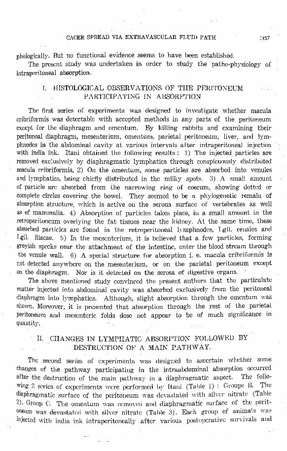
CACER SPREAD VIA EXTRAVASCULAR FLUiD PATH 1451
phologically. But no functional evidence seems to have been established.
The present study was undertaken in order to study the patho-ph~アsiology of
intraperitoneal absorption.
I. HISTOLOGICAL OBSERVATIONS OF THE PERITONEUM
PARTICIPATING IN ABSORPTION
The first series of experiments was designed to investigate whether macula
cribriformis was detectable with accepted methods in any parts of the peritoneum
except for the diaphragm and omentum. By killing rabbits and examining their
peritoneal diaphragm, mesenterium, omentum, parietal peritoneum, liver, and lym-
phnodes in the abdominal cavity at various intervals after intraperitoneal injection with india ink. Itani obtained the following results : 1) The injected particles are
removed exclusively by diaphragmatic lymphatics through conspicuously distributed macula cribriformis, 2) On the omentum, some particles are absorbed into venules
and I:iァmphatics,being chiefly distributed in the milky spots. 3) A small amount
of particle are absorbed from the narrowing ring of coecum, showing dotted or
complete circles covering the bowel. They seemed to be a phylogenetic remain of
absorption structure, which is active on the serous surface of vertebrates as well as of mammalia. 4) Absorption of particles takes place, in a small amount in the
retroperitoneum overlying the fat tissues near the kidney. At the same time, these absorbed particles are found in the retroperitoneal lymphnodes, Lgll. renales and
Lgll. iliacae. 5) In the mesenterium, it is believed that a few particles, forming
greyish sp巴cksnear the attachment of the intestine, enter the blood stream through
the venule wall. 6) A special structure for absorption i. e. macula cribriformis is
not detected anywhere on the mesenterium, or on the parietal peritoneum except on the diaphragm. Nor is it detected on the serosa of digestive organs.
The above mentioned study convinced the present authors that the particulate
matter injected into abdominal cavity was absorbed exclusively from the peritoneal
diaphragm into lymphatics. Although, slight absorption through the omentum was shown. Moreover, it is presented that absorption through the rest of the parietal
peritoneum and mesenteric folds dose not appear to be of much significance in quantity.
II. CHANGES IN LYMPHATIC ABSORPTION FOLLOWED BY
DESTRUCTION OF A MAIN PATHWAY.
The second series of experiments was designed to ascertain whether some
changes of the pathway participating in the intraabdominal absorption occurred
after 社ledestruction of the main pathway in a diaphragmatic aspect. The folio-
wing 2 series of experiments were performed b~· Itani (Table 1) : Groupe B. The diaphragmatic surface of the peritoneum was devastated with silve1・nitrate (Table
2) ・ Group C. The omen tum was 1・cmovecland diaphragmatic surface of the perit司
oneum was devastated with silver nitrate (Table 3). Each group of animals wa出
injected with india ink intraperitoneally after various postυperativc survivals and

第6号第29巻日本外科宝函1458
- I 件
! + i + i
I + I + i
Notes : C. L. ; Lg!!. cardiacae. P. L. ; Lgll. art. pancreatico-lienalis. L. L. ; Lg!!. art. hepaticae. M. L. ; Lg!!. mesentericae craniales. R. L. ; Lg!!. renales. I. L. ; Lg!!. iliacae. A. L. ; Lgll. axillares. Pp. L. ; Lg!!. poplitae.
Group A : Control animals
Rabbit ヘい
32
31
84
83
30
49
85
34
35
33
36
I. L. I A. L. I Pp.L.
,J
+
R. L. i
+ +
M. L. j L. L.1
一
+
+
#
一
+
朴
+
+
+十
++
+件
+
* + +
Table
Intervals fromi :Injection to I Liver Death '
u H }-2
2}-2
3
4
24
48
一
+
+
骨
骨
mw川廿冊川柑掛川甘
hr. hr. hr. hr. hr. hr. hr. hr. hr. hr.
hr. + +
TU n-
P
L
A
I. L.
Group B : Devastation of Diaphragm by Silver Nitrate
p L I L.L.1 M.L.
+
+
++
+
+
+
R.L.
一
+
仲
+
AH
+
H
廿+H
廿H
廿H
廿+
++
++
骨
骨
+
+
+
+ +
C.L.
+
++
2
Intervals • Intervals I
Rabbit from I from I
Operation I Injection i Liver k仏 to ! to i
一一一」_!!jectionI Death I
76 ! 3}-2 hr. [ 1 hr. 43 ' 3 ds. i H hr. 59 ' 3 ds. ~ 4 hr. 40 I 5 ds. i 1 hr. 44 5 ds. I 4}-2 hr. 57 7 ds. 1 }-2 hr. 74 i 14 ds. ! 1 hr. 77 I 23 ds. i 1 hr. 90 i 24 ds. i 1 hr. 62 j 31 ds. ! 3 hr.
+
+
+
+
Table
判+
then bled to death at 1/2 to 4 hours. Histological studies on intraabdominal lym-
phnodes, pa1匂talperitoneum and mesenterium were performed.
A microscopic study also reveals marked di百erencesbetween normal and treated
animals subたctedto intraabclominal absorption a日 follows: 1. The lymphnodes in
the epiga日tricreεion, Lg!!. cardiacae, Lgll. art. pancreaticcトlienalisand Lgll. h巴pati-
cae, were containing carbon particles from the early stage in both normal and
treated animals. However, it seems to be of a great significance that the carbon
particles were detected most markedly and most rapidly in the medullary sinuses
of Lgll. mesentericae craniales of the treated group (B and C.). A comparison of
Group B and Group C with coloured superior mesenteric nodes shows a less extent
of carbon particles in the Group B. These differences can not be attributable to
+ + + +

1459 CACER SPREAD VIA EXTRAVASCULAR FLUID PATH
Pp.L.
Group (' : Devastation of Diaphragm and extirpation of Omentum.
A.L. I L
+
++
+
+
R.L
++
一+川廿山廿
L
I
司、μ
+
+
+
H
廿日廿
++
+
L.L
+
+
一
+
+
++
Table
L
P
L
-
rtt r
一
刊
一件
+
1
一
|九円
Ltll
h
n
一
日
l
m
h一rI
1
1
1
1
1
1
I
1
1
vrtt一h
h
h
h
h
h
h
h
h
h
h
rHcnMa一
11
ぬ
wuampth11131311131
一則
f
n
I
I
I
同
町
n
一
an・k
m
t
i
S
E
E
s
s
a
s
s
E
示
問
叫
ω批一仇
E
d
d
d
d
d
d
d
d
d
ロパ
f引
-
N
L’1
4
4
7
7
2
6
9
0
7
一洲町・
n
2
1
2
2
3
5
T
O
T-
t
-
-- h
u
o
’DM川
a
nn
* +
一
+
+
U
廿け廿H
廿川廿
一
件
+
+
+
一件山W
H廿
3
+
+
一nBn3capo
ハU
守
tqupb凋性
ponU
-7a
守’
aqAUZRuda・7’
戸
OE1uponhO 山廿
the effect of the extirpated omentum. It is explained h:・: the fact that since onl;i.・
small amounts of deposits of carbon particles in the liver were detectable in both
B and C Groups, the amounts of absorption through the omentum is out of the
question. Moreover, in the retroperitoneal lymphnodes such as Lgll. renales and
Lgll. iliacae of the treated one, more rapidly and markedly the d~·e was found
than the control. Brieflyァ speaking,the more perfectly blockage of a diaphragmatic
aspect continues, the more evidently colouring of nodes and lymphatics is evidenced.
We are of the opinion that these features are analogous to those of Lgll. mesen-
tericae craniales.
In both Group B and Group C, absorption was observed, though in a small
degree through the peritoneum, attachment of the intestine,. over fat tissues near
the kidney, and the narrowing ring of the co配 um.Such absorption is more evident
and rapid than in control animals.
From the foregoing data he emphasizes a compensative function resulting from
a diaphragmatic closure in which Lgll. mesentericae craniales are found containing
free carbon particles in their medullaryア sinusesrapidly and evidently.
+++ ++
SOME DETECTABLE CHANGES OF THE PRE-LYMPH-\'人 SC‘UL九R
PA TH UNDER VARIOUS TYPES OF PERITONITIS.
III.
Many authors including Opie, Menkin and Bangham, have adovocated
inflammatory reaction in the peritoneal cavity induced b:--some irritants
be localized in situ. Under such conditions, however, no evident histology
pr・e-lymphovascularpathway such as macula cribriformis of the diaphragm was
obtained. The present study was undertaken to ascertain whether or not macula
cribriformis of the diaphragm, acting as a main intraabdominal absorption route,
may su仇rsome morphological changes under such peritonitis. The followi昭 two
series of experiments with albino rabbits were performed by Itani. The animals
that an
tends to of the

1 Iiitervals I Ii-iterrnls ' Rabbit! from I from
!Operation I Injection ' No. 1 to I to
Injection I Death
1 hr. I 1 hr.
4 hr. I 1 hr.
10 hr. I 1 hr.
24 hr. I 1 hr.
24 hr. I 3 hr.
28 hr. I 1 hr.
4 ds. I l}-ihr. 7 ds. I 1 hr.
14 ds. ! I hr.
勺
t
つOTよ
RvnwuFhiuoonunU
一6
6
8
5
5
5
6
7
8
1460
Table 4
白木外科宝函第29巻 第6号
T
’・ap
DA
L
A
* + + +
+
+ + +十+ *
Table 5 Group E : Extirpation of Omentum.
Interヤals' Intervals Habbitl from I from
/Operation I Injection λ'o. I to I t刀
Injection I Death
3 hr. I 1 hr.
1 d. I 1 hr.
3 ds. I 1 hr.
3 ds. I 5 hr.
5 ds. I H hr. 5 ds. I 3 hr.
7 ds. I 1 hr.
26 ds. I 1 hr.
一Fb
つムゥ,ヮ“
1AQU14内
d
一づ’勺,。osqAaqontkd
L
M
L
L
L
P
L
c
r
e
yv ・1L
A.L. r Pp.L. R.L. I.L.
*
+
+
++ ++ 一+H廿
H廿
+
+
件
+
++ 一件
H
廿+
++
+ト
++
件
+
+ ++ + ++ +
belonging to Group D (DT) were injected intraperitoneally with terpentine oil (Table 4). In Group E, extirpation of the omentum was performed under ether
anaesthesia (Table 5). At various intervals following such procedure, india ink
was injected intraperitoneally. Then they were sacrificed 1/2旬 5hours later.
While making a morphological study on the streched diaphragm, omentum, and
other parts of peritoneum, he compared the amounts of carbon particles through the diaphragm with those in retrosternal lymphnodes and liver. He obtained the
following results : In Group D showεd acute inflammatory changes, such as marked
ascites, and abnormally dull colored peritoneum were found. Approximately, 1 or 2
hours after intraperitoneal injection with terpentine oil. DT rabbits disclosed a
normal-shaped macula cribriformis which was covered partly with a fibrous mesh.
The retrosternal lymphnodes, liver, spleen were accompained with conspicuous carbon
deposits as control animals. The particles which had been injected 5 hours after
injection of an irritant, were detained in these organs. This detaining was markedly
demonstrated in cases which had terpentine peritonitis for two weeks. Nevertheless,
the ten hours or more elapsed cases after injection of an irritant, could rcYcal

CACER SPREAD VIA EXTRA VA只「TlLARFLUID PATH 1461
marked changes on the diaphragm i. c. so-called concentric luminal. narrowing res-
ulting from the thickend and tortuous collagen fibers with the replacement of fine reticulum fibrils to thick fibers.
IV. AN EXTRA VASCULAR LEAKAGE PHENOMENON OF
RETROSTERN AL LYMPH,¥ TICS.
A recent comprehensive anatomical report on intraabdominal !~'mph drainage
is presented by KIHARA and his co『 workers. C~arbon particles absorbed into the
lymphocapillary of the diaphragm through macula cribriformis from the peritoneal
cavity are removed mainlyア viaretrosternal ly,mphatics or the thoracic duct. However,
th~y discovered an extravascular leakage phenomenon of retrosternal lymphatic
ve坦selsthat was attributable to a special function of these lymphatics to excrete
foreign bodies outwards. And they concluded that the constant exsistence of the
reticulum fibers at the leakage site was definitely located.
A confirmat~on of this pl明 iologicalphenomenon is establi山 clin the study of
a series dealing with pericostal tub2rculosis by AoYAGI and his colaborator YAMA-MOTO (1957). They emphasize that constant occurrence of various sized and extents
of the tuberculous lesion is observed in the adipose tissues around the internal
mammary lymphatics. This occurrence seems attributable to leakage phenomenon
of internal mammary lymphatics.
Physiologically, the present result was in conformity with that of the above
mentioned interpretation、.
AN EXPERifyIENT AL STUDY ON LYMPHATIC METASTASIS
The appearance of spontaneous metastasis is quite different from a metastatic
growths produced by an intravascular injection method in which tumor cells are
injected into lymphatics using excessive pressure. For the purpose of obtaining the
easily demonstrable site for the. spontaneous metastasis, the authors performed the
foregoing studies. In consequence of this, since various aspects of the lymphatic
drainage system from the abdominal cavities become evident, the authors select the
lymphatic path from the peritoneal diaphragm to the upper retrosternal lymphnodes
as a favourable site for studying a lymphatic spread of cancer.
It was obvious, also, that metastasis of tumor when tumor cells we1℃ inje-cted
intraperitoneally, occurred chiefly in sternal l~γmphnodes. However, whether or not
in anywhere along these paths intervening between diaphragmatic endothelium and
their regional superior •. sternal lymphnodes further tumor metastasis may occur, is
the subject of the present study.
AN EXPERIMENT AL STUDY ON THE SPREAD OF CλNCER
IN THE LYMPHATIC SYSTEM
The tumors used were• -the transplantable YosHIDA ascites hepatoma in rats,
and Ehrlich ascites tumor!in mice. For each experiment 0.1 1.0 cc of tumor cell

1462 日本外科宝函第29巻第6号
suspension was used. Counts of tumor cells ranged from 190,000 to 250,000/cmm.
By killing the animals and examining histologically the diaphragm, surrounding
fattγtissues encircle internal mammary b・mphatics, sternal lymphnodes, and the
thoracic duct at various intervals from one to ten ct句アsafter intraperitoneal inocu-
lation, we have obtained the following results: 1) The site of arrest of tumor cell
emboli in diaphragm. The first problem is to ascertain whether or not tumor cells, inoculated in the
abdominal cavit~· , may pass immediately through macula cribriformis of the dia-
phragm. One to ten days after injection, the diaphragmatic peritoneum was peeled
out from the underlying submesothelial connective tissues as thinly possible ; stret-ched preparations were prepared for silver impregnation stain or MayアーGiemsastain.
Earlyア changesof these diaphragm revealed the most prominent features: Each
sieve of the fenestrated macula cribriformis were densely packed with embolic tumor cells (Fig 5 and 6). Some of them disclosed an evident mitotic division.
These embolic deposits gradually developed being accompanied by proliferative
reticulin fibrils and confluent with each other. In these cases, on any spot of the
diaphragm there are evidently observed remnants of small embolic deposits of tumor
cells in the preexistent macula cribriformis. When these foci became older, they
tended to be encapsulated by the proliferated reticulum and collagen fibers. A
strong conviction for the morphogenesis of these lesions was obtained from serial sections of the diaphragm.
When cells of Ehrlich ascites tumor were used, the result was the same as that obtained with Y osHrnA ascites hepatoma (Fig. 8).
In the experiment just described, it seems conceivable that part of the absorbed
tumor cells may stopped at the first filter station, i. e. macula cribriformis, which
works as an effective barrier to a further spread of cancer.
2) Metastases resulting from cancer cells leaking through retrosternal lymp-ha tics.
The second problem is to ascertain whether or not tumor cells may leak imm-
吋iatelythrough the internal mammary lymphatics to the surrounding adipose
tissue. For this purpose, the anterior portion of the chest wall in the just described
experiments which at necropsy appeared to be normal was removed as part of a
block resection including the internal mammary chain, intercostal muscles, ribs, costal cartilages, and pleura, but the resection stopped at the sternal margin and was studied by serial section.
Microscopic sections, stained with hematoxylin eosin, revealed in most of the
cases, an early tumor growth invariablv an~·where in and around the fatty tissues
around retrosternal lymphatics. Gradually, these become more remarkable, and
occasionally many cancer‘cells permeated the interstitial tissue of the intercostal
muscles and transversus thoracic muscles (Fig. 7). Since we often disclosed vigorous
tumor cells in mitosis within these fatty tissues, we pointed out that these results
indicated a successful metastasis of the absorbed tumor cells. In early stages,
cancer cells involve the perivascular area of the retrosternal lymphatics. However,

CACER SPREAD VIA EXTRAVASCULAR FLUID PATH 1463
they tend to increase in number. It was conceivable, also, that the involvement
may result in a leakage through the wall of retrosternal lymphatics, disclosing physiologicall_yア theirspecial function i. c. leakage phenomenon, since th巳sclymphatics
in the early cases occasionally contained tumor cells showing no direct invasion.
Sections with silver stain revealed that cancer cells have a tendency to leak at the internal mammary lymphatics, in which reticulum fibers are arranged.
A HISTOLOGICAL STUDY ON HUMANλUTOPSY M人TERIALS
A histological study was made on 5 fatal cases of tumor including intraabdo-
minal tumors. Their entire anterior portion of the chest wall including internal
mammary lymphatics, the thoracic duct, parasternal nodes, and the diaphragm
were removed and dissected. Multiple blocks representing cross sections were pre-
pared for microscopic study. Those preparations which were peeled at the plan of
submesothelial connective layer and stained with 1\fa~· Giemsa method, Bielschowsky-
Maresch silver impregnation method and haematoxylin eosin showed clearly the
pattern between cancer cells and macula cribriformis. The most prominent feature
of human autopsy materials was that macula cribriformis was packed with cancer
cells. This fact was ascertained also with sections cut in a plane perpendicularly
to the surface of the diaphragmatic peritoneum (Fig. 9).
The cancer cells in the anterior portion of the chest wall of human autopsy
materials occupy the same sites and show the same relationship to internal mam-
mary lymphatics as observed in animals (Fig. 10). The larger parts of leaked
cancer cells into adipose tissues around internal mammary lymphatics were not
affected by the destructive and degenerative changes in cytoplasms as well as in nuclei.
We emphasized that the incidence of such involvement in sub-diaphragmatic tumors is not unusual and that the impression of rarity persists because of neglect
of study of the diaphragm and internal mammary lymphatics.
A DEFENSIVE RESPONSE OF MACULA CRIBRIFORMIS TO A
CANCER INVASION.
Macula cribriformis of the diaphragm of the mice 24 hours after intraperitoneal injection with Ehrlich ascites tumor cells is ncarlγalways found to inhibit slightly
a further passage of tumor cells through them by a luminal narrowing followed
Table 6 Morphological Changes of Macula Cribriformis induced by Intraperitoneal Injection with Ehrlich Ascites Tumor Cells.
After I Collagen Fibers I Reticular Fibers I Concentric Coiled Cancer Cells I申出∞ 卜 |一- 1一一 1 p 1 f I Luminal j一雨函石市石函ご一一 ISwelling I Tortuous I Swelling Tortuo叫マi~era-j Narrowing!一一旦塑L
The First 尚一三-γ一千 子 工-I L 「_,.. I Sli帥 I -
The Thi
The Fifth Day I I I I I I I TheS的問 DayI> 件 | 件 ! tt i tt I ー | 件 I Many ; +

1464 日本外科宝函第29巻第6号
h~· a mγa\守〉
The adjacent fat tissues alon広 the course of I℃trosternal l~’mphatics or retrost巴司
rnal ly’mr〕hno〔lesw巳rethe site of the metastasis. However, these leaked cancer
cells spread more vigorously toward the interstitial conne-ctive tissue 5 days after
inoculation. These results were summarized in Table 6. From the above mentioned
data, the impression was that there was a slight interference with a further spread
of cancer through macula cribriformis under a normal condition.
THE RESPONSE OF HY ALURONIDASE ON
INTRAABDOMIN AL ABSORPTION.
Some workers have postulated a possibilit~· that cancers liberate hyaluronidase
and that the pathway for invading cancer cells is opened in consequence of hydro-
tvzation of h~·aluronic acid in connective tissues. However, some workers denied
this possibility. Quite apart from the problem whether cancers contain hyaluro-
nidase, or not, in the hope that chemical components in macula cribriformis may
be detectable the authors tried to ascertain the effects of hyaluronidase upon the
passage of carbon rarticles through macula cribriformis. Thirtyア minutesprior to intraabdominal injection of india ink, rabbits intraper-
itoneal injected with various units of hyaluronidase. Rabbits were bled to death
one hour after intraperitoneal injection with india ink. A histological comparison of the diaphragm and the anterior portion of the chest wall in each group was
performed in reference to the absorption quantity of india ink.
Table 7 The Response of Hyaluronidase on Intraabdominal Absorptio恒.
一一「一一一一一-IT ;sue along the Retro蜘 nal[山{:;~inοsum j 占:;:u
Hyalur刀ni【lase 100 Unit i 掛 !僻|朴
グ 200 グ|掛|柵 |排
// 1000 グ|+|÷|+
Control (D凶 llP.dWa伽)|掛|冊|件
The results as illustrated in Table 7. were obtained. In spite of the author》
anticipation, however, the results suggest that high units of hyaluronidase cause the concentric luminal narrowing resulting from the swelling and torsion of the
collagen fibers in macula cribriformis, and consequently intraabdominal absorption
was unexpectedly reduced.
DISTRIBUTION OF THE ABSORBED TUMOR CELLS IN THE
INTERNAL MAMMARY LYMPHATICS AND THORACIC DUCT
FOL LO羽TINGINTRAPERITONEAL ADMINISTRATION.
To determine if tumour cells passed through the macula cribriformis of the
diaphragm, a suspension of Ehrlich ascites tumour cells was injected into the abdo-
minal cavity of rabbits directly upon the inferior surface of the diaphragm. Thirty

CACER SPREAD VIA EXTRAVASCULAR FLUID PATH 1465
minutes later a drop of !j・m11h \γas obtained from main excreting collecting l~·mphatic channels (the starting point of the internal mammar.Jマ lymphatic)on either
side adjacent to the sternum by exposing the pleural surface of the diaphragm,
and simultaneously from the thoracic duct just above the level of the VIIIth ver-
tebrae.
This lymph was then observed under a phase contrast microscope. Simultane-
ously, lymph was put into a R. B. C. pipette and diluted to 1 : 100 concentration
with acet-gentianaviolett solution and transferred to a counting chamber. The result was given in table 8. Table 8. strongly suggests a higher absorptive distribution
of tumour cells in the internal mammary lymphatics than in the thoracic duct.
Furthermore, it becomes evident that the tumour cells in the inter・nalmammary
lymphatics indicated no degenerative changes in shape of mitochondria. On the
contrary, the tumour cells contained in the thoracic duct in【Iicatedyarious types of
cellular degeneration ; degenerating cells with vesicular mitochondria or semilunar
type of mitochondrial degeneration. From this fact, it is strongly suggestcc1 that
the internal mammary lymph seemed to be a more suitable living medium for
tumour cells than the thoracic lymph which contained various factors related to
the digestion. As stated by TAKEDA, the reason to choose the changes of the
mitochondrial shape a月 asusceptible sign of the cellular degeneration is that the
changes in the mitochondrial shape are in parallel with the cellular degenerative changes.
Next, the impression was gained that there was some interference with absor-
ption by macula cribriformis. This impression was reinforced lη’ the histological
evidence that the areas of macula cribriformis of this rabbits were packed with
cancer cells.
Table 8 Shows the Distribution of Tumour Cells According to Number in Lymph.
i Peritoneal Cavity
TumorαH Number/cmr
Features of Phase-Contras t I 出1crosc泊pe I
Interna!Mammary Lymphatics
54,000
No degeneration
PREVENTING AGAINST METASTASIS.
Duct. Thoracicus
10,000
Degeneration Remarkable
A complete prevention against metastasis is the crux of the problem of survival.
Before the cause and cure of cancer can be determined, it necessary to use methods
available at present to prevent metastasis. The authors had the following cxper-
iment to ascertain whether or not an immediate passage of tumor cells through
the macula cribriformis and the spread followed by the leaked tumor cells through
the retro蜘 ・nallyrr could be prevented. Mice, weighing approximately 20g. were used. The series o士
experiments shown in Table 9. were performed. The tumors used were the
transplantable Ehrlich ascites tumors in mice. Bγkilling the animals at various

1466 日本外科宝函第四巻 第6号
intervals, histological studies were performed.
Table 9 Method of Proceeding.
Anti-Cancer Agent ! Group Nitromin ' h示示二五ご-, -~二三ご一一一一一ー一一一- · Colcemid
~hiit~1 'υI ~~·. Ehrlich Ascites
Tumor
2
3
1略/勾 j lOOU/惚 Io.02g1匂 I1略/匂 Io同 /日 |
2 グ I200 II I 0.04 II I 3 II • I 0.02 II i 3 II i 300 II I 0 .06 II I 5 /I I州 II I
4 [ S~~l~;_]:~~nt. r Solv町一i可 亡一一一一一, ! I !Tumor cells In…~
5 I 2 mg/同 I200U/同 i0.04 g/kg I 1 mg/kg I O.O!rng/kg !Hours after Final Injection with / i / !Anti-Cancer Agents
6 II II ! II ! 3 グ J 0.02 11 j 48 Hours 7 I/ II I // I 5 // I O.Q4ρ I 72 Hours
Intervals 7 Days I 7 D…加r…RESULTS.
In the first group of experimentation, animals under normal condition are
submitted to intraperitoneal injection with anti-cancer agents of various degrees,
and the following results were obtained : There were no appreciable differences of
diaphragmatic macula cribriformis between control mice and mice treated with the
following dose : Sarcomycin 0.02 gr./kg., Nitromin lmg./kg., Carzinophilin 200
units/kg., Thio・TEPA (Trietylene Thio・phosphoramide( 1 mg/kg., Colcemid (Colchi-
tin) lmg/kg. However, Sarcomycin 0.04gr /kg. or more, Carzinophilin 300 units/kg.
or more, Nitromin 2mg/kg. or more, Tespamin 5mg/kg. or more. Colcemid O.Olmg/
kg. or more, showed a typical response to a further spreads of cancers. The
structural elements of the macula cribriformis changed by these drugs more or less
consisted of severely altered collagen fibers. The collagen fibers disclosed the fibrinoid
swellinglike changes, i. e., very thick.end and somewhat tortuous fibrils. The most
pronounced feature was the various degrees of the overproliferation of fine reticular
fibrils in all series, except for Carzinophilin. These tendencies were most remarkably
shown in Colcemid and Sarcomycin and less weak in Nitromin and Thio・TEPA.
Carzinophilin had no such response (Table 10 and 11)・ Suchfeatures suggest an
extreme inhibition of intraabdominal absorption through the diaphragm. In fact,
the inhibition of cancer cell passage through macula cribriformis was essentially
due to the concentric luminal narrowing followed by these fibrosis.
When the intraabdominal pr・e-administrationof anticancer agents is superimpo・
sed on intraabdominal inoculation with Ehrlich ascites tumor cells, the lodged cancer
cells in macula cribriformis are coiled around by proliferating reticular白brilsand
then fallen into degeneration. Moreover, such cases failed to yield leaked metastasis

1467
through the retrostcrnal lymphatics. The results of X-ray irradiation on rats were in accord with the response of Nitromin.
CACER SPREAD VIA EXTRAVASCULAR FLUID PATH
Morphological Changes of the Diaphragmatic l¥Iacula Cribriformis Induced by the Anti-Cancer Agents.
10 Table
Diaphragm
Gro山由/ほ|一一型la内山r I Reticulum日ber
竺空ling I Tortuous I Dis阿 onJ Swelling To此uous Dispersioー示ムing
|-!+ー|ー!ー
I - I + 1 + I - 件一 I + ・ + I ー|件ー . +十 I - j 一
件! - : - !
件 一|件
特;一|件
一 | 朴++!-
I - ,ー!
ート
+
+十+
#t
+H廿
H廿
一+++
一朴一++++
++++ H
廿H
廿H
廿H
廿一++++
主I IOOU.
:g_ 2 200U. 。.S 3 300U. N
話4Solvent. ! ω
.5 I 0.02g.
~ 2 0.04g. !
§ 3 0.06g.
~ 4 Solvent. 1
I mg.
2略.
3mg.
Solvent.
咽Ea
。,M内t
u
d佳
=τ=。HJ担問【向
Inhibitory Effects to the Passage of India Ink through Macula Cribriformis
+十+
#t
++
++
+ +
+
Morphological Changes of Fiber of Macula Cribriformis
Induced byけieAnti-Cancer Agents
I…ing I Sw山
11 Table
Tortuous
H
廿+++土土
+++++件
#t
++
+
+
+
土
Colee mid
Sarcomycin
Nitro min
X-Ray Radiation
Thio・TEPA
Carzinophilin
DISCUSSION
A recent comprehensive concepts on metastasis of neoplasms is presented by
Zeidman and Coman et al. They emphasized that the development of a blood-born metastasis involves three major aspects i. e. (I) invasion, (II) embolism, (III) de-
velopment of arrested emboli. ;\只 the~· described, it is conceivable that at least a
major factor responsible for invasion is dec1℃ased adhesiveness, ameboid motility
and penetrability of cancer cells against the vascular wall. However, it is the authors’opinivn that a successful resolution of fundamental changes that occur in
the primary tumor cells and metastatic cells is quite necessary. But, quite apart
from this consioeration, it is necessary to st ucl)’ the mode of tumor cells which
invade the prelymphovascular fluid path or extravascular fluid path. No experim-
ental work on the invasiveness of the cancer cells to the pre-lymphovascular 日uid

1468 日本外科宝函第29巻第6号
path does not seem to have been so far reported. The present experimental results
indicate that cancer cells in part are caught by prelymphovascular fluid path,
where macula cribriformis or reticular fibers were arranged. They凹 cupyan intr-
aluminal position, where they come into close relationship with reticular and collagen
fibers. At least part of them had the degenerative changes. It was considered that
the factors responsible for retrogression of the embolic cancer cell deposits are
attributable to inhibitory agents of the proliferating reticular白bers. This result
ma~· suggest the e古田tivenessof the macula cribriformis as a temporary barrier
to a further spread of cancers.
vVhile, the paramount interesting thing was that .the cancer cells discharged
into lymphatics leak into the surrounding environment at a definite site in the
course of l:>ァmphaticpassage. Sometimes, these leaked cells or embolic deposits cancer
cells in the macula cribriformis seem to have remained dormant in the adipose
tissue or in the macula without ever producing remarkable symptom. Sometimes after such a resting period the tumor cells may regain their vigor and flare up
anew with widerspread through out the bαly. However, leaked cancer cells or de-
posited cancer cells in the macula cribriformis in its relation to surrounding tissues, may be more complex than hitherto imagined. Still left entirely unexplained, ho・wever, is the problem in this field.
The soil hypothesis seems to have been widely acceptanced. If cited the soil
hypothesis, it is conceivable that the adipose tissue encircle internal mammary
lymphatics may be the fertile soil suitable for growing the cancer cell. But, still left enti1℃b・ unresolved.
Since these extravascular leaked implants were observed, an attempt was made
to carry out the foregoing experiment to prevent such metastatic mechanism. Any
reasonable, practical measm℃ which may reduce the incidence of cancer metastasis
is suggested for use, until the cause and cure of cancer can be determined. But,
complete inhibitor~’ response the ultimate aim of the authors’attempt, were not
obtained from our data. In an~· w町, itis interesting that these anti-cancer agents that are regarded only as a radiornimctica responsible for cancer cell destruction,
show considerable e古田tson the praelJ・mphovascular日uidpath or lymphatics.
Hm'℃\Tr, concerning the passage of tumor cells through macula cribriformis,
it is considered desirable to make a quantitative measurement of the di百erences
in the discharged agent into lymphatics between animals treated with anti-cancer
agents and controls. The above mentioned experiments strongly suggest that these
anti『 canceragents may inhibits such form of metastasis to some extent although not completely.
SUMMARY
1¥: study is made of the development of metastasis regarding the pas団 geof
tumor cells through the pr℃l~·mphovascular fluid path in reference to the l】henome・non of their extrava日c;.ilarleakage.
l'al't ol" the tumor c℃I ls which a1℃ intrapcrito11eall~· transplanted depo日iton the日]lecifical1:-・ featuredぉitcs,i. c., macula cribriformis and the area of lymphatic

CACER SPREAD VIA EXTRAVASCULAR FLUID PATH 1469
leakage. It is considered that such involvement may be found more widely on a
more careiul observation. We emphasize that the extravascular fluid path may
participate in cancer spread.
羽Teare much indebted to Emeritus Prof. Dr. Takusaburo Kihara and Prof. Dr. Fukuzo Osh-
1111a, for their kind guidance throughouf this experimental study.
Literature
J J Balazs-Euler : Cancer Res. 12. 326, 1952.
ZJ Bangham A. D .. Brit. J. Exp. Path. 34, l, 195:1.
3) Boyland et al : J. Path. Bact. 41. 55:0. 1935.
4J Coman D. R. . Cancer Res, 11, 648, 1951.
5J Coman D. R. . Cancer Res, 13, 397, 1953.
6J Crile Jr. C : Surg. Gyn. Obst, 103, 312, 1956. 7J Drinker C. K. : Lymphatics, Lymph and Lymphoid Tissue., Havard Uni,・ersity Press 1941
SJ Duran-Reynals : Am. J. Ca, 15, 2790, 1931.
9) Itani K Arch. Jap. Chir (kyoto) 28, 802, 1959. 10) Kihara T . Ketsuekigaku-togikai-hokoku (Sympoium on Ha巴matatology)3, 118, 1950.
11) Matsuoka. S : Arch. Jap. Chir (Kyoto) 28, 2351, 1959.
121 Menkin : Dynamics of Inflammation. Mac '.¥Iil!ian Comp. :¥'cw York 1940.
13) Opie : J. Immunol., 17, 329, 1929.
14) Osaya : Hokaido-igakukai-zashi, 29. 1310, 1954. 15) Ota, K ・ Gan Kenkyu-no-shinpo, 387, Igakushoin. Japan 1956.
16) Takeda : Shuyosaibo, Nagaishoten, Japan, 1956.
17) Teshima, G : Arch. Jap. Chir. 9, 3, 1932.
18) Tsubouchi, M : Kaibogakukkaizashi, 24, 2, 1949.
19) Tsubouchi. M , Kaibogakukkaizashi, 25, 1, 1950. 20) Yamamoto, M : Arch. Jap. Chir. (Kyoto) 25, 495, 1956. 21) Yamamoto, J.¥I : Jour. Jap. Ass. Thorac. Sm、g:~ 5, 313, 1957.
22) Walther, H. E : Krebsmetastasen, 1948.
23) Willis, R. S. ・ Spread of tumour 1952.
24) Zeideman, I Cancer Res. 17, 157, 1957.

1470 日本外科宝函第29巻第6号
Fig. I Normal appearance of the diaphrag-matic Macula Cribriformis (Rabbit) ×JOO
Fig. 3 Normal appearance of the diaphrag-matic Macula Crib1吋ormis(Human)× 100
Fig. 5 Each sieve of the fenestrate<l Macula cribriformis were densely packed with embolic Yoshida ascites hepatoma cells 1 sikcr impr・e-gnation stain) x 100
Fig. 2 Normal appearance of the diaphrag-matic Macula Cribriformis (Rabbit)×400
Fig. 4 Normal appearance of the diaphrag-matic Macula Cribriformis (Mouse)×200
Fig. 6A Each sieve of the fenestrated Macula cribriformis were densely packed with embolic Yoshida ascites hepatoma ’cells (May-Giemsa
Stain) x200

CACERISPREAD VIA EXTRAVASCULAR FLUID PATH 1471
Fig. GB Each sieve of the fenestrated macula
cribriformis were deusely packed with embolic
Yoshida ascites hepatoma cells (May-Giemsa
stain) x 400
Fig. 7B Leakinεtumour cells in and around the fatty tissues around the internal mammary
lymphatics (Yoshida sarcoma Rat)×200
Fig. 9A Leakin且・ cancer cells through the in司
ternal mammary lymphatics (Gastric cancer
human materials) x 100
Fig. 7 A Leaking tumour cells in and around
the fatty tissues around the internal mammary
lymphatics (Yoshida sarcoma Rat)×100
Fig. 8 Macula cribriformis of the human autopsy materials were packed with cancer
cells. (extension preparatJ
Fig. 9B Leaking cancer cells through the
internal mammary lymphatics(Gastric cancer
human materials) x 200

1472 日本外外宝函第29券第6号
Fig. 10 The'. phasecontrast microscopic feat-ures of the internal mammary lymph contai-
ning the tumour cells. No degeneration
.~
・... .デ a.. -・ -・d, ’' "' ‘-. - •. ’・
. . -~" .
,.
4
’’
4
. -
‘’ ・a . 咽, ・ a血・”..’ “ . ,,
・-‘ .・
. -
q’a・酔.
~. . ロ , ,. ,,
,,. ロFig. 12 The !uminal narrowing of the macula
cribriformis of the mouse treated with Colcemid
These changes were mainly induced by the
overproliferation of the reticular fibrils.
Fig. 11 The phasecontrast microscopic features
of the thoracic duct lymph containig the tumour
cells. Cellular degeneration remarkable.
-
6
,
・4旬、dvt,
岨--
e-fa
司
R
-a
司、.t
.
、,.,.
,
--
-te--i
-v
,・ae
---‘
.,
..... ‘‘
-h
.,.、,‘.. 、・
1
-
i
・7Lve--
‘‘・2
:sum恥
J
’’g
・
ペ.‘
3
.. ‘,Aq
・‘
iha
’
tiArtIre
--
A
-E--v
a
‘,.
,
.
-e
M岬
. , 咽『・、.h、
4・
ー、Fig. 13 The luminal narrowing of the ma-
cula cribriformis of the mouse treated with
Colcemid. These chan畠・eswere mainly induced
by the overproliferation of the reticular fibrils.
with Sarcomycin.
Fig. 14 The luminal narrowing of the ma-cula cribriformis of the mouse treated with
Colcemid. These changes were mainly induced
by the overproliferation of the reticular fibr-ils. with Nitromin.
Fig. 15 The luminal narrowing of the ma-
cula cribriformis of the mouse tr巴atedwith
Colcemid. These changes were mainly induced by the overproliferation of the reticular日br」
ils目、.vithCarzinophilin.

CACER SPREAD VIA EXTRAVASCULAR FLUID PATH 1473
和文抄録
脈管外通液路系による癌の拡がり方
関西医科大学外科学教室(指導悶宗夫教授)
山本政勝・松岡昇三・福島信子
城戸摩利子・宮本恒子・ 前 田 公 子
青柳 一・佐藤百合子・井上康子
京都大学医学部外科学教室(指導青柳安誠教授)
井ノj、l口.
吾々は,木原名誉教授の発見された脈管外通液路系
が癌細胞の移動に際し如何なる態度をとるかを追求し
た.先づ井谷の腹腔内吸収に関する基礎実験の結果前
リンパ管通液路系を形成する横隔膜筒状斑の病態生理
面を知り得た.次いで吉田腹水癌p マウスのエールリ
ツヒ腹水癌,等による実験の結果横隔膜筒状斑が抑留
増殖の場を形成し得る事実や内胸リンパ管から周囲組
織内に癌細胞漏出の見られる事実を確認し得た.同時
幹
に腹腔内癌腫症の剖検例に際し上記同様の個所を研索
せる結果上記動物実験と全く同ーの結果を得た.同時
に筒状斑の抑留機構の解明やその機能に関する実験を
行い殊に制癌剤に対する筒状斑の態度を検討し 2.3の
興味ある新知見を得た.以上により癌細胞は脈管外通
液路就中前リンパ管通液路にて抑留増殖の場を形成し
得る事p 更には傍リンパ管通液路系による癌細胞の拡
大進展の起り得る新事実を解明し得た.


![ETAPS 2019 AWARDyoshida/slides/Shonan2019.pdf• [POPL19] Alceste Scalas , Nobuko Yoshida: Less Is More: Multiparty Session Types Revisited • [POPL19] Bernardo Toninho, NY: Interconnectability](https://img.pdfslide.tips/doc/110x75/60b2bdc24701a2055369049c/etaps-2019-yoshidaslidesshonan2019pdf-a-popl19-alceste-scalas-nobuko-yoshida.jpg)