Embed Size (px)
Citation preview

Title Carcinoma of the Peripapillary Portion of the Duodenumwithout Jaundice
Author(s) MINAMI, AKIRA
Citation 日本外科宝函 (1975), 44(5): 444-448
Issue Date 1975-09-01
URL http://hdl.handle.net/2433/208089
Right
Type Departmental Bulletin Paper
Textversion publisher
Kyoto University

Arch. Jap. Chir., (44) 151, 444~448, Sept,. 1975
症 例
Carcinoma of the Peripapillary Portion of the
Duodenum without Jaundice
by
AKIRA MrNAMI
Osaka Kita Teishin Hospital Department of Surgery
(Received for Publication, July 10, 1975.)
Th巴 presenceof obstructive jaundice is usually helpful in the diagnosis of a tumor at
the peripapillary portion of the duodenum, but I exp己rienced a case of the peripapillary
carcinoma without jaundice. The purpose of this paper is to report this case and to discuss
about the classification of the duodenal carcinoma.
Case Report
A 63 year-old man was admitted to the
Saiseikai Suita Hospital with a 2 month
history of nausea, loss of appetite and he-
artburn. At first the patient had fever
ranging from 38° to 39' C, but the tempera-
ture had dropped to normal a month before
admission. He had no history of jaundice,
vomiting, abゴominal pain or tarry stools.
On examination there was no evidence of
jaundice. Abdomen was flat and no tumor
was felt. Th2 gallbladder was not palpable.
An X ray film of the chest was normal.
Roentgenologis examination of the stomach
and duod2num rとvealed an obstructing
lesion at the j~mction of the se::ond and
third p1rt of the duodenum. Hypotonic
duodenography disclosed an irregular filli口g
defect (Fig. 1), which had the appearanc巴
of a peripapillary carcinoma of the duode-
nuπl.
Laboratory studies r巴vealed; R. B. C.
3,410,000, hemoglobin 7. 7g/dl, hematocrit
28九, W.B. C. 12, 100, serum amylase 16
Fig. 1. I 1、potonic duodenoi(raphv sho¥¥ s .lll it 1egular filling dl'iじLt at the junじt1onof the second and and third part of the duodenum
Key words・ Classitication, Ducdenal carcinoma, Jaundice, Peripapillary carcinoma Present address ・ Osaka Kita Teishin Hospital, Department of Surgery, Kita-ku, Osaka, Japan. 〒530
CARCINOMA OF THE PERIPAPILLARY PORTION OF THE JAUNDICE 445
units, urine amylase 16 units, Meulengracht
8, total serum bilirubin 1. 2mg p巴r cent,
thymol turbidity tests 0. 7, GOT 49噌 (;ドT43,
alkaline phosphatase 60 Bodansky units, LDH
170, BU i¥ 15mg per cent, blood glucose 76
mg per lOOml, urinaly~is negative.
At operation an egg-sized mass was
found in the second part of the duodenum.
The common bile duct was dilated to a
maximum diameter of 1. Scm. ;'\’either en-
largement of the gallbhdder nor dilatation of
the pancreatic duct was seen. The liver
appeared healthy. Radical pancreatoduode-
nectomy was carried out. Anastomosis was
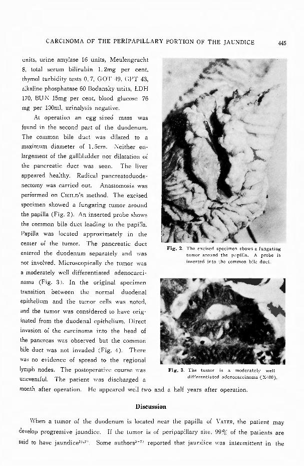
performed on CHILD’s method. The excised
specimen showed a fungating tumor around
the papilla (Fig. 2). An inserted probe shows
the common bi!εduct leading to the papilla.
Papilla was located approximat巴ly in the
center of the tumor. The pancreatic duct
entered the duodenum separately and was
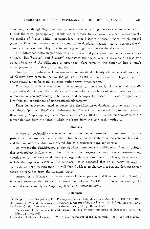
not involved. Microscopically the tumor was
a moderately well-cliff巴rentiated adenocarci-
noma (Fig. 3). In the original specimen
transition between the normal duodenal
epithelium and the tumor cells was noted,
and the tumor was considered to have orig-
inated from the duodenal epithelium. Direct
invasion of the carcinoma into the head of
the pancreas was observed but the common
bile duct was not invaded (Fig. 4). There
was no evidence of spread to the regional
lymph nodes. The postoperative course was
uneventful. The patient was discharged a
Fig. 2. The excised specimen shows a fungatmg tumor around the papilla. A probe is inserted into the common bile duct
Fig. 3. The tumor is a moderatelv well-differentiated adenocarcinoma (X400).
month after operation. He appeared well two and a half years after operation.
Discussion
When a tumor of the duodenum is located near the papilla of ¥'ATER, the patient may
develop progressive jaundice. If the tumor is of peripapillary site, 99 ~1)' of the patients are
said to have jaundice1 l•21 Some authorsト 7> reported that jaundice was intermittent in the

446 日・外・宝第44巻 第5号(昭和50年9月)
’r
ョ、 V勘
Fig. 4. The common bile duct is not invaded by the tumor cells (XZOO).
case of peripapillary carcinoma. 1'¥ccording
to MoNGE6l, jaundice fluctuated in half the
patients of 65 ""ith carcinoma of the papilla,
but in only 2 of the 28 with carcinoma of
the pancreatic head.
In the case of the patient presented
here, there was no jaundice and the icterus
index was normal, although the level of the
alkaline phosphatase was elevated and the
common bile duct was dilated. HoFFMAN8'
stated that the jaundice may entirely dis-
appear or be intermittent for the following
reasons : (1) a soft, friable tumor in this
location may ulcerate and slough away, ac-
companied by a temporary or lasting relief
of the jaundice ; (2) increased intrabiliary
pressur巴 may forc巴 the bile through the
obstructed papilla (BRILL) ; (3) the sub-
sidence of papillary巴demamay release the
obstruction and afford relief (MEYER and
ROSENBERG). On the other hand, MONGE6'
stated that variation in the jaundice was not
related to ulceration and sloughing of the surface of carcinoma of the papilla, but may
perhaps have been due to the soft, polypoid, and some,,・hat mobile nature of these tumors.
I suppose that the patient was not jaundiced because there was no infiltration to the
common bile duct. It is said that carcinoma of the stomach will stop short at the pylorus
and carcinoma of the caecum will stop at the ileocaecal valve. Similarly it is suppos巴dthat
duodenal carcinoma stop巴dat th巴 papillaof \λTER, but the common bile duct was dilated
presumably due to a transient papillary edema. That the tumor had originated from the
duodenal mucosa is evident pathologically for the following reasons : (1) macroscopically the
tumor protruded from duodenal mucosa into the duodenal lumen, (2) microscopically transition
from the normal duodenal epithelium to the tumor cells was noted, (3) the common bile
duct was not infiltrated by the tumor cells. But clinically this case should not be included
in duodenal carcinoma for the present as indicated in my previous paper9'.
恥1ostof th巴 peripapillarylesions arise in the biliary or pancreatic ducts rather than
from the duodenal mucosa. But in the literature many peripapillary carcinomas have been
included in the duodenal neoplasms because of the difficulty in deciding the tru巴 siteof
the origin of these carcinomas. This accounts for the high percentage of peripapillary
lesions in the older literature. I believe that the pei-ipapillary lesions should be in a sepa-
rate category. The t巴rms・・peri papillary" and “periampullary" are occasionally used indis-
CARCINOMA OF THE PERIPAPILLARY PORTION OF THE JAUNDICE 447
criminately, as though they were synonymous words indicating the same anatomic structure.
I think the term ・・peripapillary”should indicate those tumors which invade macroscopically
the papilla of V ATER while “periampullary” should indiG:tc those tumors 11・hich spread
submucosally 11・ithout macroscopical changes in the duodenal mucosa. As to V巴riampulla1y”
th巴reis a far less possibility of a tumor originating from the duodenal mucosa.
The difference between periampullary carcinoma and pancreatic carcinoma is sometimes
difficult. But WARREN7> and Suzu1<110> emphasize the importance of division of these two
cancers because of the difference of prognosis. Carcinoma of the pancreas has a much
worse prognoEis than that of the ampulla.
However, the problem still remains as to how we should classify a far advanced carcinoma
which may have come to include the papilla of ¥"ATER or the pユncreas. I hope an appro-
priate classification be made by some authoritative organization.
Relatively little is known about the anatomy of the ampulla of VATER. MIYAZAl<I11'
expressed a doubt upon the existence of the ampulla on the basis of his experiences in the
intraoperative cholangiography (362 cases) and necropsy (11 cases). I 11・ish to agree with
him from my experiences of pancreaticoduodenectomy.
From the above mentioned evidence, the classification of duodenal carcinoma as ・・supra-
ampullary",ヤ eriampullary”and“infraampullary”is not recommended. I propose to classify
them simply "'suprapapillary”and “infra papillary”as RESl¥!K12l, since embryologically the
former derived from the foregut while the latter from the yolk sack (midgut).
Summary
A case of peripapillary cancer without jaundice is presented. I supposed that the
patient had no jaundice because there had been no infiltration to the common bile duct,
and the common bile duct was dilated due to a transient papillary edema.
A.t present the classification of the duodenal carcinoma is ambiguous. I am of opinion
that peripapillary lesions should be in a separate category, although there remains some
question as to how we should classify a large extensive carcinoma which may have come to
include the papilla of VATER or the pancreas. It is expected that an authoritative organi-
zation decides the classification. Untill then I wish to emphasize that peripapillary carcinoma
should be excluded from the duodenal cancer.
According to MIYAZAKI11l, the existence of the ampulla of ¥.ATER is doubtful. Therefore
it is not recommended to use the word “ampulla of YATERぺ Ipropose to classify the
duodenal cancer simply as "suprapapillary”and "infra papillary”・
References
1) Berger, L. and Koppelman, H. Primary carcinoma of the duodenum. Ann. Surg., 116 : 738, 1942. 2) Iovine, V. M. and Tsangaris, N. : Primary carcinoma of the duodenum. Am. ]. Surg., 27 : 744, 1961. 3) Lunn, G. M .. Carcinoma of the duodenum. Brit. ]. Surg., 40 : 5, 1952. 4) Kleinerman, ]., Yardumian, K. and Tamaki, H. T. : Primar)' carcinoma of duodenum. Ann. Int.
Med., 32 : 451, 1952. 5) Mateer, ]. G. and Hartman, F. W.: Primary carcinoma of the duodenum. JAMA、99: 1853, 1932.

448 日・外・宝第44巻第5号(昭和50年9月)
6) Monge, ]. ]. et al ・ Clinopathologic observations on radical pancreatoduodenal resection for peri-
papillary carcinoma. Surg. Gyn. & Obst., 118 : 275, 1964.
7) Warren, K. W. et al A long-term appraisal of pancreaticoduodenal resection for periampullary
carcinoma. Ann. Surg., 155 653, 1962.
8) Hoffman, W. ]. and Pack, G. T.・Cancerof the duodenum. Arch. Surg., 35 : 9, 1937.
9) Minami, A. and Shibata, G. Primary carcinoma of the duodenum. Jap. ]. Surg., 1 . 232, 1971.
10) Suzuki, T. et al : Correlation between clinical aspects and location of lesion in carcinoma of the
head of the pancreas. Am. ]. Surg., 125 : 546, 1973.
11) Miyazaki, I., Nagakawa, T. and Kito, M .. ・Concerningthe so-called ampulla of Yater. Geka (Tokyo),
35 : 1097, 1973.
12) Resnik, H. L. P. and Cooper, D. R .. Carcinoma of the duodenum. Am. ]. Surg., 95 : 946, 1958.
和文抄録
乳頭部癌の無黄 痘 症 例
大阪北逓信病院外科
南
|二指腸乳頭部周囲IC発育した癌でありながら,業
痘を中、たさなかった 1例を報告した.文献には,乳頭
部癌を I_:_指腸癌lζ含めているのが多く見られるが,
乳頭部総で十二指IJl,J1tM葉より発生するのは少なく,乳
ロ'TC
頭部は除くべきだと恩われる.そして十二指腸癌は,
乳頭上部癌と乳頭下部癌とに分類し,乳頭部癌につい
ては,膨大部癌, E孝頭部癌等と共lζ別の分類法を考え
るべきだと思われる.