Embed Size (px)
Citation preview
DMD #65037
1
TITLE
Novel Bioactivation Pathway of Benzbromarone
Mediated by Cytochrome P450
Yumina Kitagawara, Tomoyuki Ohe, Kumiko Tachibana, Kyoko Takahashi,
Shigeo Nakamura, and Tadahiko Mashino
Faculty of Pharmacy, Keio University, 1-5-30 Shibakoen, Minato-ku, Tokyo, Japan (Y.K.,
T.O., K.T., T.M.); Department of Chemistry, Nippon Medical School, 1-7-1 Sakaiminami-
cho, Musashino-shi, Tokyo, Japan (S.N.)
This article has not been copyedited and formatted. The final version may differ from this version.DMD Fast Forward. Published on June 23, 2015 as DOI: 10.1124/dmd.115.065037
at ASPE
T Journals on A
pril 3, 2019dm
d.aspetjournals.orgD
ownloaded from
DMD #65037
2
RUNNING TITLE
Novel metabolites of benzbromarone and their cytotoxicity
Correspondence to: Tomoyuki Ohe and Tadahiko Mashino, Faculty of Pharmacy, Keio
University, 1-5-30 Shibakoen, Minato-ku, Tokyo 105-8512, Japan
Tel: +81-3-5400-2691, Fax: +81-3-5400-2691
E-mail: [email protected] (T. Ohe) and [email protected] (T. Mashino)
The number of text pages: 15
The number of tables: 2
The number of figures: 2
The number of references: 16
The number of words in Abstract: 101
The number of words in Introduction: 541
The number of words in Discussion: 574
Non-standard abbreviations
BBR, benzbromarone; BSTFA, N,O-bis(trimethylsilyl)trifluoroacetamide; CYP, Cytochrome
P450; DBBQ, 2,6-dibromobenzoquinone; CAT, 2-ethyl-3-(3-bromo-4,5-
dihydroxybenzoyl)benzofuran; DBH, 2,6-dibromohydroquinone; G6P, glucose-6-phosphate;
G6PDH, glucose-6-phosphate dehydrogenase; 1’-OH-BBR, 1’-hydroxybenzbromarone; 6-
OH-BBR, 6-hydroxybenzbromarone; SIM, selected ion monitoring
This article has not been copyedited and formatted. The final version may differ from this version.DMD Fast Forward. Published on June 23, 2015 as DOI: 10.1124/dmd.115.065037
at ASPE
T Journals on A
pril 3, 2019dm
d.aspetjournals.orgD
ownloaded from
DMD #65037
3
ABSTRACT
Benzbromarone (BBR) is a hepatotoxic drug but the detailed mechanism of its toxicity
remains unknown. We identified 2,6-dibromohydroquinone (DBH) and mono-debrominated
catechol (2-ethyl-3-(3-bromo-4,5-dihydroxybenzoyl)benzofuran; CAT) as novel metabolites
of BBR in rat and human liver microsomal systems by comparison with chemically
synthesized authentic compounds, and also elucidated that DBH is formed by cytochrome
P450 (CYP) 2C9 and that CAT is formed mainly by CYP1A1, 2D6, 2E1 and 3A4.
Furthermore, CAT, DBH and the oxidized form of DBH are highly cytotoxic in HepG2,
compared with BBR. Taken together, our data demonstrate that DBH, a novel reactive
metabolite, may be relevant to BBR-induced hepatotoxicity.
This article has not been copyedited and formatted. The final version may differ from this version.DMD Fast Forward. Published on June 23, 2015 as DOI: 10.1124/dmd.115.065037
at ASPE
T Journals on A
pril 3, 2019dm
d.aspetjournals.orgD
ownloaded from
DMD #65037
4
INTRODUCTION
Benzbromarone (BBR), a potent human uric acid transporter I (hURAT1) inhibitor, is
effective in the treatment of hyperuricemia and gout, particularly when allopurinol, an
international first-line treatment, is ineffective. It has been used as a quite useful drug in
Japan for a long time (Masseoud et al., 2005). However, BBR has been associated with
hepatotoxicity, including fulminant or fatal liver injury (van der Klauw et al., 1994). Thus,
the use of BBR is not approved in the United States and has been withdrawn from the market
in Europe (Lee et al., 2008).
Adverse reactions are one of the most important issues in drug development and in the
therapeutic usage of drugs during the post-approval stage. Specifically, idiosyncratic adverse
drug reactions occur only in a small group of patients who are treated with certain drugs at
therapeutic does and are unpredictable (Ulrich, 2007). It is widely accepted that drug-
induced idiosyncratic hepatotoxicity is often associated with cytochrome P450 (CYP)-
mediated bioactivation (Kalgutkar et al., 2012; Stephens et al., 2014). Thus, BBR could be
metabolized to reactive metabolites that cause liver injury.
Early metabolic studies identified that the major metabolites of BBR are 6-
hydroxybenzbromarone (6-OH-BBR) formed by CYP2C9 and CYP2C19, and 1’-
hydroxybenzbromarone (1’-OH-BBR) produced by CYP3A4 (Kobayashi et al., 2012;
McDonald and Rettie, 2007). It has been reported that human hepatocarcinoma HepG2 cells
infected with adenovirus vector expressing CYP2C9 significantly increase the cytotoxicity of
BBR, suggesting that CYP2C9 is associated with BBR-induced cytotoxicity (Imamura et al.,
2011). Thus, the formation of 6-OH-BBR catalyzed by CYP2C9 was thought to play a key
role in hepatic toxicity. McDonald et al. proposed that 5,6-dihydroxybenzbromarone, the
secondary metabolite from 6-OH-BBR by CYP2C9, and the o-quinone form may be reactive
This article has not been copyedited and formatted. The final version may differ from this version.DMD Fast Forward. Published on June 23, 2015 as DOI: 10.1124/dmd.115.065037
at ASPE
T Journals on A
pril 3, 2019dm
d.aspetjournals.orgD
ownloaded from
DMD #65037
5
metabolites of BBR, although they could not show the formation of such reactive metabolites
when BBR was used as a substrate in liver microsomal systems (McDonald and Rettie,
2007). Conversely, Kobayashi et al. reported that 6-OH-BBR was not cytotoxic but that BBR
and 1’-OH-BBR exhibited cytotoxicity in human hepatocarcinoma FLC4 cells cultured in
micro-space cell in which the expression levels of CYP2C9 and CYP3A4 had been enhanced
(Kobayashi et al., 2013). They proposed that 1’-OH-BBR might be the key metabolite that
causes liver injury induced by BBR. However, 1’-OH-BBR is formed predominantly by
CYP3A4 and it remains unknown why CYP2C9 is primarily responsible for BBR-induced
hepatotoxicity.
In previous research, we discovered ipso-substitution as a unique metabolic reaction of a
variety of substituted phenols by CYP (Ohe et al., 1994; Ohe et al., 1995; Ohe et al., 1997;
Ohe et al., 2000; Tezuka et al., 2007; Nakamura et al., 2011). We hypothesized that BBR,
which contains a phenolic hydroxyl group, could be oxidized to 2,6-dibromohydroquinone
(DBH) and/or mono-debrominated catechol (2-ethyl-3-(3-bromo-4,5-
dihydroxybenzoyl)benzofuran; CAT) through ipso-substitution. The formation of DBH and
CAT from BBR might be linked to the mechanism for BBR-induced hepatotoxicity because
catechols, hydroquinones, and their oxidized forms are known to be toxic via the formation
of covalent bonds with biological macromolecules and the catalytic reduction of oxygen to
superoxide and other reactive oxygen species.
The purpose of this study is to elucidate the formation of ipso-substitution metabolites in
liver microsomal systems and their cytotoxicity. To this end, we report that these metabolites
may be relevant to the hepatotoxicity induced by BBR.
This article has not been copyedited and formatted. The final version may differ from this version.DMD Fast Forward. Published on June 23, 2015 as DOI: 10.1124/dmd.115.065037
at ASPE
T Journals on A
pril 3, 2019dm
d.aspetjournals.orgD
ownloaded from
DMD #65037
6
MATERIALS AND METHODS
Materials. NADP+ was obtained from Wako Pure Chemical Industries, Ltd. (Osaka, Japan).
Glucose-6-phosphate (G6P) and G6P dehydrogenase (G6PDH) were obtained from Roche
Diagnostics (Basel, Switzerland). Hydroquinone and N,O-
bis(trimethylsilyl)trifluoroacetamide (BSTFA) were obtained from Tokyo Chemical Industry
Co., Ltd. (Tokyo, Japan). All other reagents used for metabolism and toxicity experiments
were of analytical grade.
Synthesis of expected metabolites. We chemically synthesized ipso-substituted metabolites,
CAT, DBH and the oxidized form of DBH (DBBQ), and the known main metabolite, 6-OH-
BBR, as authentic standards for metabolite identification and/or cytotoxicity studies. Briefly,
for the synthesis of CAT, vanillin was brominated at the 3-position with Br2 followed by
methylation and oxidation to form 3-bromo-4,5-dimethoxybenzoic acid. After treatment with
SOCl2, Friedel-Crafts acylation with 2-ethylbenzofuran was directed to the 3-position of the
benzofuran ring. Then, the demethylation reaction of the two methoxy groups with BBr3
produced CAT. DBH was obtained via regioselective bromination of 4-methoxyphenol with
benzyltrimethylammonium tribromide (Kajigaeshi et al., 1987). DBBQ was directly
prepared by the oxidation of DBH using Oxone® (potassium peroxymonosulfate). 6-OH-BBR
was synthesized according to the procedure reported by McDonald et al. with minor
modifications (McDonald and Rettie, 2007). More detailed procedures and analytical data for
synthesized compounds are shown in Supplemental Methods.
Liver microsomal incubation. Phenobarbital-treated SD rat and pooled human (200 donors)
liver microsomes were purchased from XenoTech, LLC (Kansas, US). The incubation
mixture containing rat or human liver microsomes (1 mg protein/mL), BBR (100 μM),
MgCl2 (3 mM), G6P (10 mM), and G6PDH (1 U/mL) with or without tienilic acid (10 μM)
This article has not been copyedited and formatted. The final version may differ from this version.DMD Fast Forward. Published on June 23, 2015 as DOI: 10.1124/dmd.115.065037
at ASPE
T Journals on A
pril 3, 2019dm
d.aspetjournals.orgD
ownloaded from
DMD #65037
7
in 1.5 mL of 0.1 M K-Pi buffer (pH 7.4) was preincubated for 3 min at 37˚C. The reaction
was initiated by adding NADP+ (1 mM) and incubated for 30 min (rat) or 120 min (human)
at 37˚C. One milliliter of the incubation mixture was treated with 2 mL of ice-cold ethyl
acetate to stop the reaction and to extract the products, and the organic phase was separated.
The organic phase was dried over anhydrous Na2SO4 and concentrated by N2 flushing. The
products formed were trimethylsilylated with BSTFA and pyridine. After the removal of the
excess BSTFA by N2 flushing, the residue was dissolved in a small amount of acetone and
analyzed by GC-MS (SHIMADZU GC-MS-QP5050A, capillary column HP-1 0.32 mm × 30
m; J & W Scientific). The injection temperature was 260˚C. The initial column temperature
was 90˚C for 3 min; then the temperature was raised at intervals of 10˚C/min to 250˚C,
followed by an isothermal hold at this temperature. On the other hand, 0.5 mL of the
incubation mixture was treated with 1 mL of acetonitrile : methanol (2:1) to the stop the
reaction. All samples were centrifuged at 10,000 × g for 10 min, and the supernatant was
analyzed by LC-MS (Agilent Technologies 6120) Chromatographic separations were
performed on XBride™Shield RP18 column (Waters, 3.5 μm 2.1 × 150 mm) at a flow rate
0.2 mL/min under liner isocratic conditions of acetonitrile/water/HCOOH (60/40/0.1). The
eluent was introduced directly into the mass spectrometer via electrospray ionization in the
negative ion mode.
Incubation with CYP isozymes. Recombinant human CYP isozymes (EasyCYP
Bactosomes) were purchased from Cypex, Ltd. (Dundee, UK). Incubation was performed
under the same conditions used for liver microsomes described above, except that the
incubation mixture contained 0.1 μM CYP isozymes, 0.1 mM BBR, 4 mM MgCl2, 4 mM
G6P, 1 U/mL G6PDH, and 0.4 mM NADP+.
Cell culture. Human hepatocarcinoma HepG2 cells were obtained from ATCC (Virginia,
This article has not been copyedited and formatted. The final version may differ from this version.DMD Fast Forward. Published on June 23, 2015 as DOI: 10.1124/dmd.115.065037
at ASPE
T Journals on A
pril 3, 2019dm
d.aspetjournals.orgD
ownloaded from
DMD #65037
8
US). The HepG2 cells were maintained in Minimum Essential Media (GIBCO) containing
5% fetal bovine serum (GIBCO) and 1% penicillin-streptomycin (SIGMA) in a 5% CO2
incubator with a humidified atmosphere at 37˚C.
Cytotoxicity assay. Cell toxicity was evaluated by a trypan blue dye exclusion test. HepG2
cells were seeded (1×10⁶ cells/mL/well) on a 96-well plate (IWAKI). After 24 hr of
incubation, the cells were treated with BBR, 6-OH-BBR, CAT, DBH, and DBBQ at 0.4 μM.
After being incubated with the drugs for 24 hr, the viable cells were counted according to the
dye exclusion method using Vi-Cell™ (Beckman Coulter).
This article has not been copyedited and formatted. The final version may differ from this version.DMD Fast Forward. Published on June 23, 2015 as DOI: 10.1124/dmd.115.065037
at ASPE
T Journals on A
pril 3, 2019dm
d.aspetjournals.orgD
ownloaded from
DMD #65037
9
RESULTS
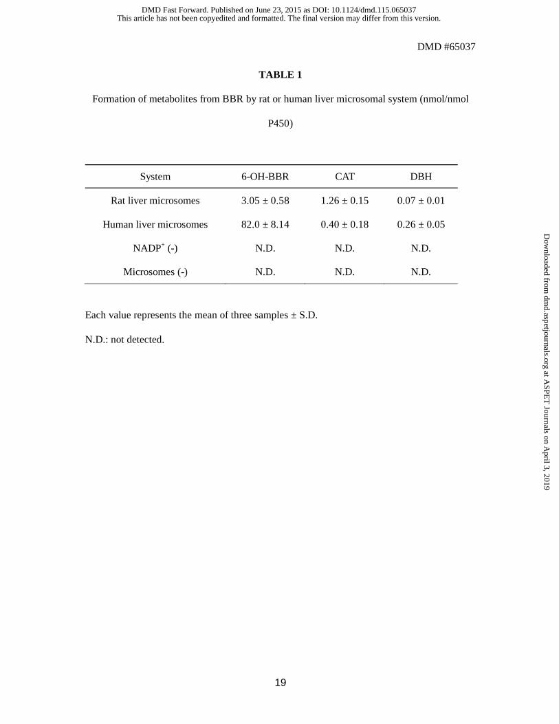
Metabolism of BBR by rat or human liver microsomes. BBR was incubated with
phenobarbital-treated rat liver microsomes or pooled human liver microsomes. The
metabolite extracts were analyzed by LC-MS for 6-OH-BBR and CAT and analyzed by GC-
MS after trimethylsilylation for DBH. All of the metabolites were identified on the basis of
retention time and the m/z peak ratio in selected ion monitoring (SIM) mode, compared with
the synthesized chemical standards (6-OH-BBR, CAT, and DBH). The quantification results
including control incubations (NADP+ (-) and microsomes (-) ) are shown in Table 1. The
results demonstrate that the novel metabolites CAT and DBH were formed with rat and
human hepatic microsomes. Not all of the metabolites were detected when NADP+ or
microsomes were omitted from the complete system.
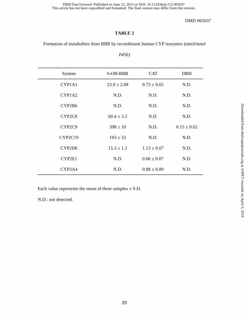
Metabolism of BBR by isozymes of human CYP. BBR was incubated with various
human CYP isozymes expressed in Escherichia coli (bactosomes). The metabolite extracts
were analyzed in a similar manner to the metabolic study with liver microsomes, and the
amounts of formed metabolites were determined (Table 2). It was found that the formation of
6-OH-BBR is catalyzed by CYP1A1, CYP2C8, CYP2C9, CYP2C19 and CYP2D6. CYP1A1,
CYP2D6, CYP2E1 and CYP3A4 were found to catalyze the formation of CAT, while DBH
was formed by only CYP2C9.
Effect of CYP2C9 inhibitor on the BBR metabolite formation. Additionally, we
investigated the effect of tienilic acid, a mechanism-based inhibitor specific for CYP2C9, on
the formation of three metabolites from BBR in human liver microsomes to confirm the
contribution of CYP2C9. BBR was incubated with human liver microsomes in the presence
of 10 μM tienilic acid, and the amounts of formed metabolites were determined. As shown in
Supplemental Figure S1, the formation of 6-OH-BBR was markedly inhibited by tienilic acid,
This article has not been copyedited and formatted. The final version may differ from this version.DMD Fast Forward. Published on June 23, 2015 as DOI: 10.1124/dmd.115.065037
at ASPE
T Journals on A
pril 3, 2019dm
d.aspetjournals.orgD
ownloaded from
DMD #65037
10
and the formation of DBH was also significantly inhibited. In contrast, tienilic acid had no
inhibitory effect on the formation of CAT from BBR.
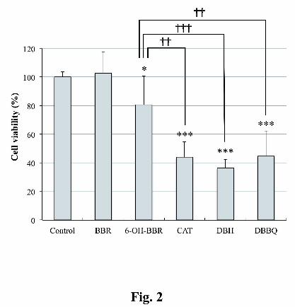
Cytotoxicity of BBR and BBR metabolites in HepG2 cells. The cell toxicities of BBR, 6-
OH-BBR, CAT and DBH in the hepatocyte cell line (HepG2 cells) were assessed by the use
of the trypan blue dye exclusion method after 24 hr of drug exposure. In addition, based on
the possible significance of DBH for the bioactivation process of BBR, the toxicity of the
oxidized form of DBH, DBBQ, was also evaluated because DBH is thought to be easily
oxidized to DBBQ during incubation, although DBBQ can never be detected in the extracts
from the microsomal system because of its susceptibility to bind to macromolecules. The cell
toxicities of the compounds at various concentrations are demonstrated in Supplemental
Figure S2. As shown in Fig. 2, the ipso-substituted metabolites CAT, DBH and DBBQ
induced significant cytotoxicity at the lowest concentration (0.4 μM) and, they exhibited
more potent toxicity than did 6-OH-BBR. On the other hand, BBR was not cytotoxic at this
concentration.
This article has not been copyedited and formatted. The final version may differ from this version.DMD Fast Forward. Published on June 23, 2015 as DOI: 10.1124/dmd.115.065037
at ASPE
T Journals on A
pril 3, 2019dm
d.aspetjournals.orgD
ownloaded from
DMD #65037
11
DISCUSSION
The exact mechanisms leading to the hepatocellular toxicity of BBR, a therapeutically useful
uricosuric drug, are not yet understood. The present study is an attempt to elucidate the
cause of BBR-induced liver toxicity by proposing a novel bioactivation mechanism for BBR.
First, we hypothesized that BBR, which contains a phenolic hydroxyl group, could be
subject to an ipso-substitution metabolism, which we had previously discovered as a unique
metabolic pathway catalyzed by CYP, and oxidized to DBH and/or CAT.
As shown in Table 1, DBH and CAT were formed in rat and human microsomal systems in
NADPH- and microsome-dependent manners, as determined from comparison with the
synthetic standards, although the formation amounts were much lower than that of 6-OH-
BBR. The production of CAT and DBH may be underestimated because the recovery of
these metabolites from the microsomal incubation mixtures was shown to be low (data not
shown) probably due to the covalent binding of the oxidized forms (quinone forms) with
macromolecules such as proteins.
Isozyme profiling with recombinant CYP bactosomes clarified that the formation of 6-OH-
BBR is catalyzed by CYP1A1, CYP2C8, CYP2C9, CYP2C19 and CYP2D6. This result is
consistent with the previous reports that CYP2C9 and CYP2C19 are the major isozymes
capable of catalyzing the formation of 6-OH-BBR (Kobayashi et al., 2012; McDonald and
Rettie, 2007). CYP2C9 was also responsible for the formation of DBH from BBR. The
additional analysis using the CYP2C9 specific inhibitor further confirmed that CYP2C9
contributes to 6-hydroxylation of BBR and ipso-substitution metabolism to generate DBH.
There have been some reports that CYP2C9 is primarily responsible for BBR-induced liver
toxicity. Iwamura et al. reported that HepG2 cells infected with adenovirus vector expressing
CYP2C9 significantly increase the cytotoxicity of BBR (Imamura et al., 2011). McDonald et
This article has not been copyedited and formatted. The final version may differ from this version.DMD Fast Forward. Published on June 23, 2015 as DOI: 10.1124/dmd.115.065037
at ASPE
T Journals on A
pril 3, 2019dm
d.aspetjournals.orgD
ownloaded from
DMD #65037
12
al. suggested that a catechol metabolite, 5,6-dihydroxybenzbromarone, which is formed via
6-OH-BBR, and its o-quinone form may be reactive metabolites and that the sequential
metabolism is catalyzed by CYP2C9 (McDonald and Rettie, 2007), although they did not
show the formation of such reactive metabolites directly from BBR in liver microsomal
systems, and their cytotoxicity has not been elucidated.
The current study demonstrated that 6-OH-BBR was cytotoxic to HepG2 cells but the
toxicity was not remarkable. Conversely, novel metabolites, DBH and CAT showed more
potent cytotoxicity than BBR and 6-OH-BBR, suggesting that the ipso-substituted
metabolites may possibly be reactive metabolites. In addition, the cytotoxicity of the quinone
form of DBH, DBBQ, was also examined because it is thought that DBH is easily oxidized
to DBBQ in liver microsomal systems. As a result, DBBQ also exhibited potent cytotoxicity
that is comparable to that of DBH and CAT. Taking into consideration that DBH is mainly
formed by CYP2C9, the contribution of DBH and DBBQ to BBR-induced hepatotoxicity, in
particular, might be important.
As described above, Kobayashi et al. proposed that the metabolic activation via 1’-OH-BBR
formed by CYP3A4 might be responsible for BBR-induced hepatotoxicity (Kobayashi et al.,
2013), but the mechanism is not yet clear and it remains unknown why CYP2C9 enhances
the cytotoxicity of BBR. The current study clearly presents an alternative mechanism for
BBR-induced hepatotoxicity activated by CYP2C9.
In conclusion, we have demonstrated the formation of reactive metabolites in liver
microsomal systems via an ipso-substitution metabolism of BBR, which is a novel metabolic
pathway for BBR. We consider that the formation of DBH or DBBQ might be one of the
reasons for hepatocellular toxicity induced by BBR. Additional studies to investigate the
detailed mechanism for BBR-induced hepatotoxicity are in progress.
This article has not been copyedited and formatted. The final version may differ from this version.DMD Fast Forward. Published on June 23, 2015 as DOI: 10.1124/dmd.115.065037
at ASPE
T Journals on A
pril 3, 2019dm
d.aspetjournals.orgD
ownloaded from
DMD #65037
13
AUTHORSHIP CONTRIBUTIONS
Participated in research design: Ohe, Kitagawara, Takahashi, Nakamura, and Mashino.
Conducted experiments: Kitagawara and Tachibana.
Contributed new reagents or analytic tools: Kitagawara, Ohe, and Tachibana.
Performed data analysis: Kitagawara and Ohe.
Wrote or contributed to the writing of the manuscript: Ohe, Kitagawara, and Mashino.
This article has not been copyedited and formatted. The final version may differ from this version.DMD Fast Forward. Published on June 23, 2015 as DOI: 10.1124/dmd.115.065037
at ASPE
T Journals on A
pril 3, 2019dm
d.aspetjournals.orgD
ownloaded from
DMD #65037
14
REFERENCES
Iwamura A, Fukami T, Hosomi H, Nakajima M, and Yokoi T (2011) CYP2C9-mediated
metabolic activation of losartan detected by a highly sensitive cell-based screening assay.
Drug Metab Dispos. 39: 838-846.
Kalgutkar A, Dalvie D, Obach S, and Smith D (2012) Methods and Principles in
Medicinal Chemistry: Reactive Drug Metabolites, Wiley-VCH, Weinheim, Germany.
Kobayashi K, Kajiwara E, Ishikawa M, Oka H, and Chiba K (2012) Identification of
CYP isozymes involved in benzbromarone metabolism in human liver microsomes.
Biopharm Drug Dispos. 33: 466-473.
Kobayashi K, Kajiwara E, Ishikawa M, Mimura H, Oka H, Ejiri Y, Hosoda M, and
Chiba K (2013) Cytotoxic effects of benzbromarone and its 1'-hydroxy metabolite in human
hepatocarcinoma FLC4 cells cultured on micro-space cell culture plates. Drug Metab
Pharmacokinet. 28: 265-268.
Lee MH, Graham GG, Williams KM, and Day RO (2008) A benefit-risk assessment of
benzbromarone in the treatment of gout. Was its withdrawal from the market in the best
interest of patients?. Drug Saf. 31: 643-665.
Masseoud D, Rott K, Liu-Bryan R and Agudelo C (2005) Overview of hyperuricaemia
and gout. Curr Pharm Des. 11: 4117-4124.
This article has not been copyedited and formatted. The final version may differ from this version.DMD Fast Forward. Published on June 23, 2015 as DOI: 10.1124/dmd.115.065037
at ASPE
T Journals on A
pril 3, 2019dm
d.aspetjournals.orgD
ownloaded from
DMD #65037
15
McDonald MG and Rettie AE (2007) Sequential metabolism and bioactivation of the
hepatotoxin benzbromarone: formation of glutathione adducts from a catechol intermediate.
Chem Res Toxicol. 20: 1833-1842.
Nakamura S, Tezuka Y, Ushiyama A, Kawashima C, Kitagawara Y, Takahashi K, Ohta
S, and Mashino T (2011) Ipso substitution of bisphenol A catalyzed by microsomal
cytochrome P450 and enhancement of estrogenic activity. Toxicol Lett. 203: 92-95.
Ohe T, Mashino T, and Hirobe M (1994) Novel metabolic pathway of arylethers by
cytochrome P450: cleavage of the oxygen-aromatic ring bond accompanying ipso-
substitution by the oxygen atom of the active species in cytochrome P450 models and
cytochrome P450. Arch. Biochem. Biophys. 310: 402-409.
Ohe T, Mashino T, and Hirobe M (1995) Novel oxidative pathway of para-substituted
phenols in cytochrome P450 chemical model: substituent elimination accompanying ipso-
substitution by the oxygen atom of the active species. Tetrahedron Lett. 42: 7681-7684.
Ohe T, Mashino T, and Hirobe M (1997) Substituent elimination from p-substituted
phenols by cytochrome P450: ipso-substitution by the oxygen atom of the active species.
Drug Metab Dispos.25: 116-122.
Ohe T, Hirobe M, and Mashino T (2000) Novel metabolic pathway of estrone and 17β-
estradiol catalyzed by cytochrome P-450. Drug Metab Dispos.28: 110-112.
This article has not been copyedited and formatted. The final version may differ from this version.DMD Fast Forward. Published on June 23, 2015 as DOI: 10.1124/dmd.115.065037
at ASPE
T Journals on A
pril 3, 2019dm
d.aspetjournals.orgD
ownloaded from
DMD #65037
16
Stephens C, Andrade RJ, and Lucena MI (2014) Mechanisms of drug-induced liver
injury. Curr Opin Allergy Clin Immunol. 14: 286-292.
Tezuka Y, Takahashi K, Suzuki T, Kitamura S, Ohta S, Nakamura S, and Mashino T
(2007) Novel metabolic pathways of p-n-nonylphenol catalyzed by cytochrome P450 and
estrogen receptor binding activity of new metabolites. J Health Sci. 53: 552-561.
Ulrich RG (2007) Idiosyncratic toxicity: A convergence of risk factors. Annu Rev Med.
58: 17-34.
van der Klauw MM, Houtman PM, Stricker BH, and Spoelstra P (1994) Hepatic injury
caused by benzbromarone. J Hepatol. 20: 376-379.
This article has not been copyedited and formatted. The final version may differ from this version.DMD Fast Forward. Published on June 23, 2015 as DOI: 10.1124/dmd.115.065037
at ASPE
T Journals on A
pril 3, 2019dm
d.aspetjournals.orgD
ownloaded from
DMD #65037
17
FOOTNOTES
This work was supported by Keio Gijuku Academic Development Funds; a grant from the
Science Research Promotion Fund of the Japan Private School Promotion Foundation; and
Platform for Drug Discovery, Informatics; and Structural Life Science from the Ministry of
Education, Culture, Sports, Science and Technology of Japan (MEXT).
This article has not been copyedited and formatted. The final version may differ from this version.DMD Fast Forward. Published on June 23, 2015 as DOI: 10.1124/dmd.115.065037
at ASPE
T Journals on A
pril 3, 2019dm
d.aspetjournals.orgD
ownloaded from
DMD #65037
18
FIGURE LEGENDS
Fig. 1. Proposed metabolic pathway of benzbromarone.
Fig. 2. Cytotoxicity of BBR and BBR metabolites at 0.4 μM in HepG2 cells. Cell viability
was assessed by the use of the trypan blue dye exclusion method after 24 hr of drug exposure.
The data are expressed relative to control incubations containing 0.1% DMSO alone. Each
value represents the mean ± S.D. of six determinations from two separate experiments. ***P
< 0.001 and *P < 0.05 compared with the control incubations. †††P < 0.001 and ††P < 0.01
versus the incubations with 6-OH-BBR.
This article has not been copyedited and formatted. The final version may differ from this version.DMD Fast Forward. Published on June 23, 2015 as DOI: 10.1124/dmd.115.065037
at ASPE
T Journals on A
pril 3, 2019dm
d.aspetjournals.orgD
ownloaded from
DMD #65037
19
TABLE 1
Formation of metabolites from BBR by rat or human liver microsomal system (nmol/nmol
P450)
System 6-OH-BBR CAT DBH
Rat liver microsomes 3.05 ± 0.58 1.26 ± 0.15 0.07 ± 0.01
Human liver microsomes 82.0 ± 8.14 0.40 ± 0.18 0.26 ± 0.05
NADP+ (-) N.D. N.D. N.D.
Microsomes (-) N.D. N.D. N.D.
Each value represents the mean of three samples ± S.D.
N.D.: not detected.
This article has not been copyedited and formatted. The final version may differ from this version.DMD Fast Forward. Published on June 23, 2015 as DOI: 10.1124/dmd.115.065037
at ASPE
T Journals on A
pril 3, 2019dm
d.aspetjournals.orgD
ownloaded from
DMD #65037
20
TABLE 2
Formation of metabolites from BBR by recombinant human CYP isozymes (nmol/nmol
P450)
System 6-OH-BBR CAT DBH
CYP1A1 23.9 ± 2.08 0.75 ± 0.02 N.D.
CYP1A2 N.D. N.D. N.D.
CYP2B6 N.D. N.D. N.D.
CYP2C8 60.4 ± 3.3 N.D. N.D.
CYP2C9 398 ± 10 N.D. 0.15 ± 0.02
CYP2C19 193 ± 33 N.D. N.D.
CYP2D6 15.3 ± 1.2 1.13 ± 0.07 N.D.
CYP2E1 N.D. 0.66 ± 0.07 N.D.
CYP3A4 N.D. 0.88 ± 0.00 N.D.
Each value represents the mean of three samples ± S.D.
N.D.: not detected.
This article has not been copyedited and formatted. The final version may differ from this version.DMD Fast Forward. Published on June 23, 2015 as DOI: 10.1124/dmd.115.065037
at ASPE
T Journals on A
pril 3, 2019dm
d.aspetjournals.orgD
ownloaded from
This article has not been copyedited and formatted. The final version may differ from this version.DMD Fast Forward. Published on June 23, 2015 as DOI: 10.1124/dmd.115.065037
at ASPE
T Journals on A
pril 3, 2019dm
d.aspetjournals.orgD
ownloaded from
This article has not been copyedited and formatted. The final version may differ from this version.DMD Fast Forward. Published on June 23, 2015 as DOI: 10.1124/dmd.115.065037
at ASPE
T Journals on A
pril 3, 2019dm
d.aspetjournals.orgD
ownloaded from