Embed Size (px)
Citation preview

Title 後腹膜気管支原性嚢胞の1例
Author(s) 近藤, 秀明; 藤本, 清秀; 青木, 勝也; 趙, 順規; 平尾, 佳彦; 夏目, 修
Citation 泌尿器科紀要 (2005), 51(1): 25-29
Issue Date 2005-01
URL http://hdl.handle.net/2433/113533
Right
Type Departmental Bulletin Paper
Textversion publisher
Kyoto University

Acta Urol. Jpn. 51: 25-29, 2005 25
A CASE OF RETROPERITONEAL BRONCHOGENIC CYST
Hideaki KOND01, Kiyohide FU]IMOT01, Katsuya AOKI1,
Masaki CH01, Yoshihiko HIRA01
and Osamu NATSUME2
1 The Department 01 Urology, Nara Medical Univeηiり2The Department 01 Urology, Nara Rehabilitation Center for Psychologically
and Physiologically Handicapped Persons
A 59-year幽 oldhypertensive man was referred to our hospital with a retroperitoneal cystic tumor, measuring 6 cm in diameter that was detected by an ultrasound examination during routine check-up 2
years before coming to our department. During the 2-year follow-up, the cystic tumor gradually increased in size. The patient also became hypertensive with slightly elevated urine levels of
noradrenaline and dopamine, while the plasma catecholamines and their metabolites in the urine were within the normal range. Computed tomographic scanning and magnetic resonance imaging revealed
a dumbbell-shaped retroperitoneal cyst with dense ftuid, measuring 7 X 3.5 X 3 cm in diameter, in the left supra-adrenal and sub-diaphragmatic regions. He underwent extirpation ofthe cystic tumor with
suspicion ofadrenal endocrine cystic tumor. The histopathological diagnosis was a bronchogenic cyst, which is an extremely rare developmental anomaly in the retroperitoneal space. We herein report this
rare case of retroperitoneal bronchogenic cyst and present a brief review of the previously reported 30
]apanese cases.
Key words: Bronchogenic cyst, Retroperitoneum
INTRODUCTION
A bronchogenic cyst is an unusual developmental
anomaly and very rarely presents as a retroperitoneal
cystic tumorl,2) Broncogenic cysts usually develop
on the posterior side of the mediastinum along the
right paratracheal wall or in the pulmonary
parenchyma, and are occasionally found in unusual sites such as the skin, subcutaneous tissue, pericardium, diaphragm, and intra-dural region.
This anomaly is genetically considered as a
developmental abnormality of the primitive foregut, which arises commonly from an accessory lung bud in
the posterior part of the mediastinum after the third
week of embryonic development, but it rarely
migrates into the retroperitoneum.
This cystic tumor is unusually encountered in the
retroperitoneal space, and there have been only 30
case reports inJapan since 1978 as well as only about
20 case reports in the English literature. 、ヘ1eherein
report this extremely rare case of a retroperitoneal
bronchogenic cyst in an adult man and present a brief
review of the previously reported 30 J apanese cases
including the present casel,3-9)
CASE REPORT
A 59-year-old hypertensive man was referred to our
department for a left retroperitoneal cystic tumor,
which possibly was derived from the adrenal gland or
pancreas. This cystic tumor was disclosed without
any symptoms by ultrasound examination during a
(Hinyokika Kiyo 51: 25-29, 2005)
routine health checkup 2 years ago, and was followed up without treatment. However, the cystic tumor increased slightly in size along with an increase in his
blood pressure to around 150/90 mmHg. The
plasma and urine catecholamines and their
metabolites, aldosterone and cortisol were measured to determine whether or not this cystic tumor had
hormone-producing activity. The urinary cate-
cholamines measurement showed increased oaily
output ofnor-adrenaline (152μg/day) and dopamine
(1,200μg/day). On the other hand, the plasma
concentrations of these catecholamines and their
metabolites in urine were within the normallevels as
well as aldosterone and cortisol. The other
biochemical examinations revealed mildly elevated
serum levels of GOT (83 IU/l), GPT (91 IU/l), y-GTP (185 IU/l) and LDH (235 IU/l) because he had
a past history of liver dysfunction of unknown
etiology
Abdominal computed tomography (CT) scanning
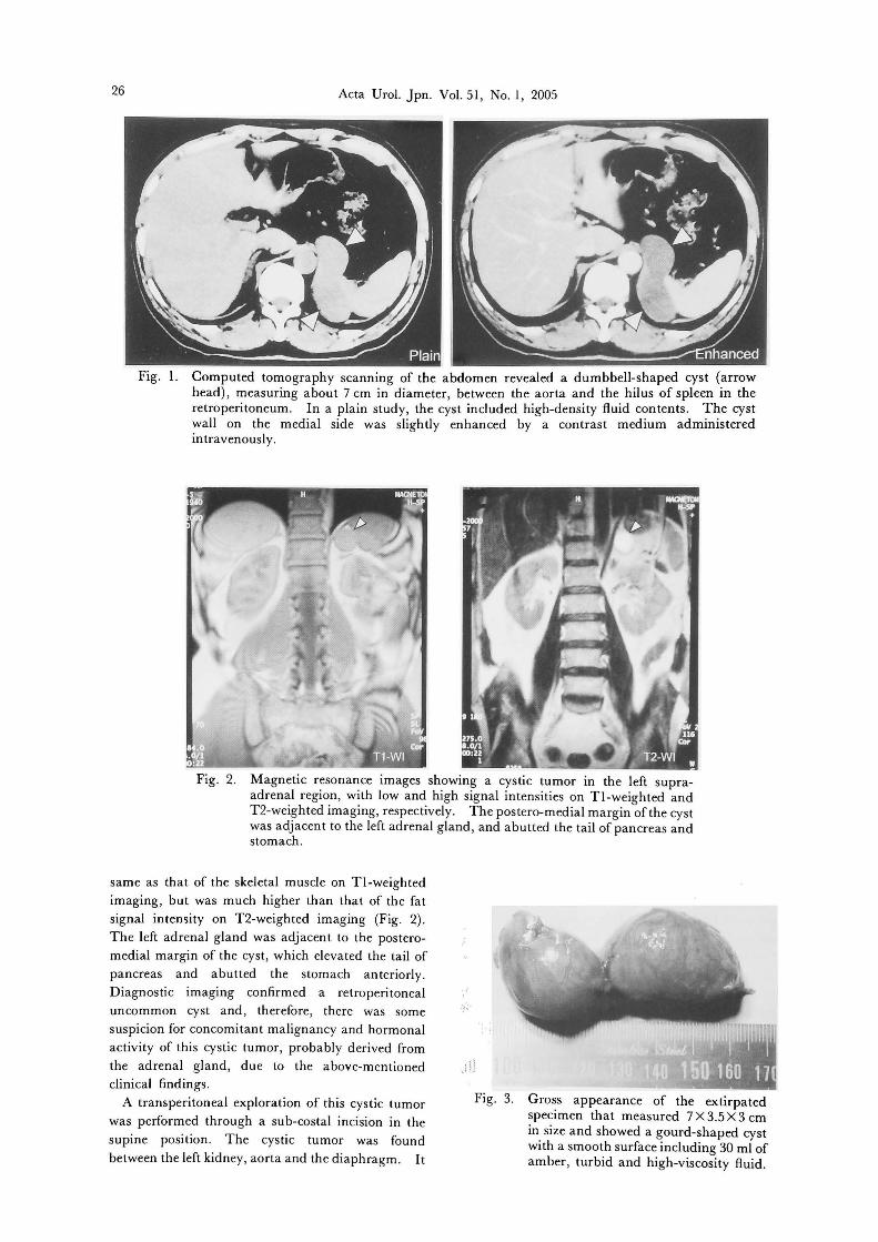
revealed a dumbbell-like cystic tumor, measuring about 7 cm in diameter, between the aorta and splenic hilus in the retroperitoneal space (Fig. 1). In a plain
study, the cystic tumor showed high-density ftuid contents, as if the ftuid seemed to include mineral
deposits or highly concentrated proteins. Contrast
enhancement was seen slightly in the parenchymal
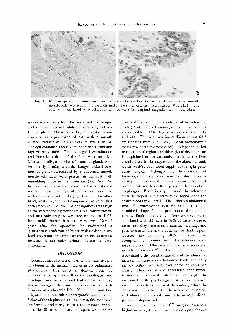
portion on the medial side ofthe cyst wall. Magnetic
resonance imaging (MRI) showed a cystic tumor in
the left supra-adrenal region, the intensity of which
was slightly lower than that ofthe liver and almost the
26 Acta Urol. Jpn. Vol. 51, No. 1, 2005
Fig. 1. Computed tomography scanning of the abdomen revealed a dumbbell-shaped cyst (arrow head), measuring about 7 cm in diameter, between the aorta and the hilus of spleen in the retroperitoneum. In a plain study, the cyst included high-density f¥uid contents. The cyst wall on the medial side was slightly enhanced by a contrast medium administered intravenously.
Fig. 2. Magnetic resonance images showing a cystic tumor in the left supra-~drenal region, with low and high signal intensities on TI-weighted and T2・weightedimaging, respective1y. The postero-medial margin ~ofthe cyst was adjacent to the le仇adrenalgland, and abutted the tail ofpancreas and stomach.
same as that of the skeletal muscle on Tl-weighted
imaging, but was much higher than that of the fat
signal intensity on T2-weighted imaging (Fig. 2)
The left adrenal gland was adjacent to the postero-
medial margin of the cyst, which e1evated the tail of pancreas and abutted the stomach anteriorly.
Diagnostic imaging confirmed a retroperitoneal
uncommon cyst and, therefore, there was some
suspicion for concomitant malignancy and hormonal
activity of this cystic tumor, probably derived from the adrenal gland, due to the above-mentioned
clinical findings
A transperitoneal exploration of this cystic tumor
was performed through a sub-costal incision in the
supine position. The cystic tumor was found
between the left kidney, aorta and the diaphragm. It
!
《〉di
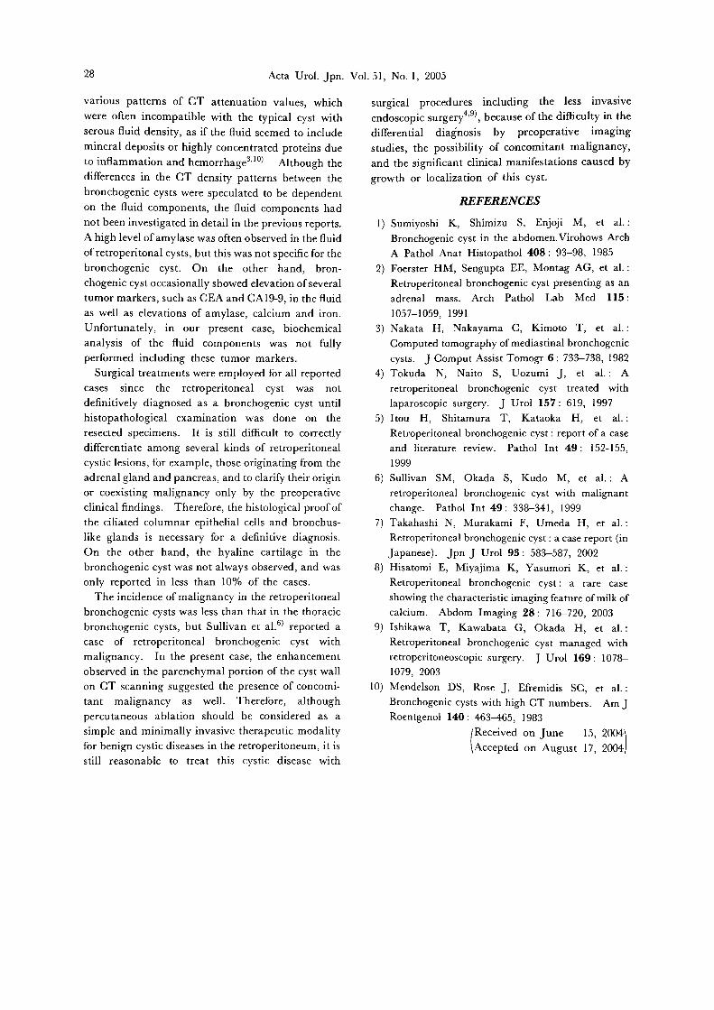
Fig. 3. Gross appearance of the extirpated specimen that measured 7 X 3.5 X 3 cm in size and showed a gourd-shaped cyst with a smooth surface including 30 ml of amber, turbid and high-viscosity f¥uid.
KONDO, et al.: Retroperitoneal bronchogenic cyst 27
was dissected easily from the aorta and diaphragm, and was solely excised, while the adrenal gland was
left in place. Macroscopically, the cystic tumor
appeared as a gourd-shaped cyst with a smooth
surface, measuring 7X3.5X3 cm in size (Fig. 3).
The cyst contained about 30 ml of amber, turbid and high-viscosity fluid. The cytological examination
and bacterial culture of .the fluid were negative.
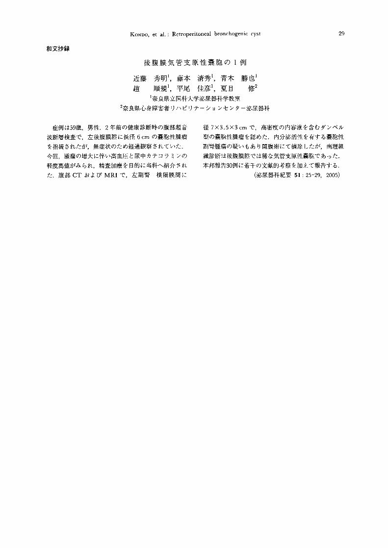
Microscopically, a number of bronchial glaRds were
seen partly forming a cystic change. Mixed sero-
mucous glands surrounded by a thickened smooth
muscle cell layer were present in the cyst wall, resembling those in the bronchus (Fig. 4a). No
hyaline cartilage was 0 bserved in the histological
sections. The inner layer of the cyst wall was lined
with columnar ciliated cells (Fig. 4b). On the other
hand, analyzing the fluid components revealed that each catecholamine level was not significantly as high
as the ∞rresponding normal plasma concentration, and that only amylase was elevated to 456 IU /U, being mildly higher than the serum level. Now, 3
years after the operation, he maintained a
spontaneous remission of hypertension without any
local recurrence or complications, or any abnormal increase in the daily urinary output of cate-
cholamines.
DISCUSSION
Bronchogenic cyst is a congenital anomaly usually
developing in the mediastinum or in the pulmonary
parenchyma. This entity is derived from the
endodermal foregut as well as the esophagus, and develops from an abnormal bud of the primitive
tracheal anlage or the bronchial tree during the日rst3-
6 weeks of embryonic life. If the abnormal bud
migrates into the sub-diaphragmatic region before
fusion of the diaphragm's components, this cyst exists incidentally and rarely in the retroperitoneal space.
In the 30 cases reported, in Japan, we found no
gender difference in the incidence of bronchogenic
cysts (15 of men and women, each). The patient's
age ranged from 17 to 74 years with a peak in the 40's
and 50's. The mean maximum diameter was 6土3
cm (ranging from 3 to 14 cm). Most bronchogenic
cysts (86% ofthe reviewed cases) developed in the left
retroperitoneal region, and this regional deviation can be eXplained on an anatomical basis as the liver
usually disturbs the migration of the abnormal bud,
which receives poor blood supply in the right para-
aortic region. Although the localizations of
bronchogenic cysts have been described using a
variety of anatomical representations, the most
common site was basically adjacent to the crus of the
diaphragm. Exceptionally, several bronchogenic
cysts developed in the retrovesical region or in the
gastro-esophageal wall. The thoraco-abdominal
type of bronchogenic cyst represents a unique
dumbbell shape for its penetration through the
narrow diaphragmatic slit. There were symptoms
associated with this cyst in 69% of these reviewed
cases, and they were mainly nausea, vomiting, and
pain or discomfort in the abdomen or ftank region, whereas the remammg 31 % of cases had
asymptomatic incidental cysts. Hypertension was a
rare symptom and the catecholamines were measured
in only a few cases2,7) including the present case.
Accordingly, the possible causality of the abnormal increase in plasma catecholamine levels and daily
urinary output was not investigated or eXplained
clearly. However, it was speculated that hyper-
tension and elevated catecholamines might be
associated with psychological stress or physical
symptoms, such as pain and discomfort, before the
operation. Therefore, the hypertensive symptom
and abnormal catecholamine data actually disap-
peared postoperatively
In our present case, plain CT imaging revealed a
high-density cyst, but bronchogenic cysts showed
28 Acta Urol. jpn. Vol. 51, No. 1, 2005
various patterns of CT attenuation values, which were often incompatible with the typical cyst with
serous fluid density, as if the fluid seemed to include mineral deposits or highly concentrated proteins due
to inflammation and hemorrhage,IO) Although the
differences in the CT density patterns between the
bronchogenic cysts were speculated to be dependent
on the fluid components, the fluid components had
not been investigated in detail in the previous reports.
A high level of amylase was often observed in the fluid
ofretroperitonal cysts, but this was not specific for the
bronchogenic cyst. On the other hand, bron-chogenic cyst occasionally showed elevation ofseveral
tumor markers, such as CEA and CAI9-9, in the fluid as well as elevations of amylase, calcium and iron.
Unfortunately, in our present case, biochemical
analysis of the fluid components was not fully
performed including these tumor markers.
Surgical treatments were employed for all reported
cases since the retroperitoneal cyst was not
definitively diagnosed as a bronchogenic cyst until
histopathological examination was done on the
resected specimens. It is still difficult to correctly
differentiate among several kinds of re仕operitoneal
cystic lesions, for example, those originating from the adrenal gland and pancreas, and to clarify their origin
or coexisting malignancy only by the preoperative
clinical findings. Therefore, the histological proof of the ciliated columnar epithelial cells and bronchus-
like glands is necessary for a definitive diagnosis.
On the other hand, the hyaline cartilage in the
bronchogenic cyst was not always observed, and was only reported in less than 10% of the cases
The incidence of malignancy in the retroperitoneal
bronchogenic cysts was less than that in the thoracic
bronchogenic cysts, but Sullivan et a1.6) reported a
case of retroperitoneal bronchogenic cyst with
malignancy. In the present case, the enhancement observed in the parenchymal portion of the cyst wall
on CT scanning suggested the presence of concomト
tant malignancy as well. Therefore, although percutaneous ablation should be considered as a
simple and minimally invasive therapeutic modality
for benign cystic diseases in the retroperitoneum, it is still reasonable to treat this cystic disease with
surgical procedures including the less invasive
endoscopic surgerl,9)事 becauseof the difficuIty in the
differential diagnosis by preoperative imaging
studies, the possibility of concomitant malignancy, and the significant c1inical manifestations caused by
growth or localizatIon of this cyst.
REFERENCES
1) Sumiyoshi K, Shimizu S, E吋oji M, et al.:
Bronchogenic cyst in the abdomen.Virohows Arch
A Patho1 Anat Histopatho1 408: 93-98, 1985
2) Foerster HM, Sengupta EE, Montag AG, et al.:
Retroperitonea1 bronchogenic cyst presenting as an
adrena1 mass. Arch Patho1 Lab Med 115 :
1057-1059, 1991 3) Nakata H, Nakayama C, Kimoto T, et al.:
Computed tomography ofmediastina1 bronchogenic
cysts. j Comput Assist Tomogr 6: 733-738, 1982
4) Tokuda N, Naito S, Uozumi j, et al.: A
retroperitonea1 bronchogenic cyst treated with
1aparoscopic surgerγ. j Uro1 157・619,1997
5) Itou H, Shitamura T, Kataoka H, et al.:
Retroperitonea1 bronchogenic cyst : report of a case
and 1iterature review. Patho1 Int 49: 152-155,
1999
6) Sullivan SM, Okada S, Kudo M, et al.: A
retroperitonea1 bronchogenic cyst with ma1ignant
change. Patho1 Int 49・33ι341,1999
7) Takahashi N, Murakami F, Umeda H, et al.:
Retroperitoneal bronchogenic cyst : a case report (in
japanese). jpn j Urol 93: 583-587, 2002
8) Hisatomi E, Miy司jimaK, Yasumori K, et al.:
Retroperitoneal bronchogenic cyst: a rare case
showing the characteristic imaging feature of mil孟of
calcium. Abdom Imaging 28: 716ー720,2003
9) Ishikawa T, Kawabata G, Okada H, et al.:
Retroperitoneal bronchogenic cyst managed with
retroperitoneoscopic surgery. J Urol 169: 1078-1079, 2003
10) Mendelson DS, Rose j, Efremidis SC, et al.・
Bronchogenic cysts with high CT numbers. Am j
Roentgenol 140: 463-465, 1983
(RE印刷 onjune 15,吋Accepted on August 17, 2004}
KONDO, et al.: Retroperitoneal bronchogenic cyst 29
和文抄録
後腹膜気管支原性嚢胞の l例
近藤秀明藤本清秀青木勝也i
越 順規平尾佳彦夏目 修2
l奈良県立医科大学泌尿器科学教室
2奈良県心身障害者リハビリテーションセンター泌尿器科
症例は59歳,男性. 2年前の健康診断時の腹部超音
波断層検査で,左後腹膜膝に長径 6cmの嚢胞性腫癒
を指摘されたが,無症状のため経過観察されていた.
今回,腫癌の増大に伴い高血圧と尿中カテコラミンの
軽度高値がみられ,精査加療を目的に当科へ紹介され
た腹部 CTおよび MRIで,左副腎横隔膜聞に
径 7X3.5X3cmで,高密度の内容液を含むダンベル
型の嚢胞性腫癒を認めた.内分泌、活性を有する嚢胞性
副腎腫蕩の疑いもあり開腹術にて摘除したが,病理組
織診断は後腹膜腔では稀な気管支原性嚢胞であった.
本邦報告30例に若干の文献的考察を加えて報告する.
(泌尿器科紀要 51: 25-29, 2005)

![AtletIQ.com — тренируйся с умом! [версия для печати]Bent-Arm Barbell Pullover Seated Cable Rows Dumbbell Flyes Weighted Bench Dip Pushups (Close and](https://img.pdfslide.tips/doc/110x75/60156b459506ba2adf41cacf/a-f-f-bent-arm.jpg)


![SplenicInfarctioninAcuteCytomegalovirusandHuman … · 2019. 7. 30. · [9]S. Naviglio, M. V. Abate, M. Chinello, and A. Ventura, “Splenic infarction in acute infectious mononucleosis,”](https://img.pdfslide.tips/doc/110x75/613ec40eb946476b8b530f56/splenicinfarctioninacutecytomegalovirusandhuman-2019-7-30-9s-naviglio-m.jpg)