Embed Size (px)
Citation preview
Title Silk as the Nidus for the Formation of Gallstones
Author(s) AOKI, YOZO; KATSUMI, MASAHARU
Citation 日本外科宝函 (1979), 48(1): 85-91
Issue Date 1979-01-01
URL http://hdl.handle.net/2433/208320
Right
Type Departmental Bulletin Paper
Textversion publisher
Kyoto University
Arch Jap Chir 48 (1), 85~91, Jan., 1979
Silk as the Nidus for the Formation of Gallstones
Yozo AoKr and MASAHARU KATSUMI
Department of Surgery (Gastroenterological Division) Wakayama Medical College
Received for Publication Sept. 13, 1978
Introduction
Gallstones sometimes develop around the foreign body such as silk ligatures. This type
of silk ligature is mostly derived from the first operation, especially on the biliary tract
diseases, and may migrate into the duct and lead to calculus formation.
The purpose of the present paper is to report one case in which silk ligatures migrated
into the common bile duct and symptoms resembling biliary colic developed and 3 cases
of gallstone formation secondary to a silk ligature, and describe an importance in selection
of suture material in the biliary tract surgery.
Case Reports
Case 1. In December, 1974, a 30 year-old married woman underwent cholecystectomy,
choledochotomy and transduodenal sphincteroplasty with T-tube drainage for gallstone
disease at the other hospital. In this operation, the cystic duct and common bile duct
were ligated or closed with silk. Her recovery was uneventful but half a year after the
operation, the patient had experienced repeated attacks of upper abdominal pain and fever.
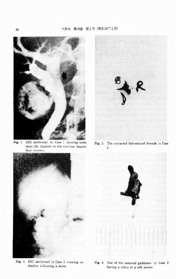
Endoscopic retrograde cholangiography (ERC) showed stone like shadow within the common
hepatic duct. She was admitted for treatment to our clinic in July, 1975. The serum bilirubin,
SGOT, SGPT and serum alkaline phosphatase were in normal ranges. ERC revealed almost
the same findings as performed before her admission (Fig. 1). At the second operation,
5 bilestained threads of silk sutures as well as biliary mud were removed from the common
bile duct (Fig. 2).
Case 2. A 36 year-old man was admitted to our clinic in October, 1975, complaining
of nausea, vomiting, fever and right hypochondralgia of 6 month duration. One year earlier,
he had undergone a cholecystectomy for cholecystolithiasis at the other hospital. The cystic
duct had been tied with silk. Laboratory data were normal except a slight elevation of
serum alkaline phosphatase (11. 8 King-Armstrong unit). Drip infusion cholangiography
(DIC) showed slightly dilated common bile duct (1. 1 cm in diameter) and the cystic duct
remnant (2. 0 cm in length and 1. 0 cm in width), but no shadow indicating a stone (Fig.
3). ERC failed to reveal the common bile duct. The preoperative diagnosis of the remnant
Key words : Silk nidus, Gallstone formation, Absorbable suture material, Biliary tract surgery, Present Address : Department of Surgery (Gastroenterobgical Division), Wakayama Medical College, Wakayama City, Wakayama 640, Japan.
86 日外宝 第48巻第1号(昭和54年 1月)
Fig. 1. ERC performed in Case 1 showing some Fig. 2. The er.tracted bile-stained threads in Case stone like shadows in the common hepatic 1.
duct (arrow)ー
Fig. 3. DlC performed in Case 2 showing no
shadow indicating a stone. Fig. 4. One of the removed gallstones in Case 2
having a nidus of a silk suture.

SILK AND GALLSTONE FORMATION 87
of cystic duct and retained gallstones was made. At reoperation, the common bile duct
contained 10 round stones consisted of calcium bilirubinate ranging from 0. 5 to 1. 0 cm in
diameter and, in addition, one irregular shaped stone. The latter stone was brown in
colour, and measured 1. 8×1. 0×0. 3 cm (Fig. 4). It was composed of a nidus of a silk
suture.
Case 3. A 68 year-old man was admitted to our clinic in May, 1976, with chief
complaints of upper abdominal pain, slight jaundice and fever. In August, 1973, a cholecysto-
lithotomy had been performed. The incised gallbladder had been closed with interrupte♂ silk sutures. After his admission, DIC and ERC revealed a little finger tip sized stone
shadow within the common bile duct (Fig. 5). At reoperation, a stone was extracted from
the common bile duct. The stone was found to have a nucleus of silk ligature (Fig. 6).
Case 4. A 30 year-old man was admitted to our clinic in June, 1977, complaining of'
right hypochondralgia and jaundice of 4 month duration. The attacks of pain resembled
biliary colic. Laboratory data included : serum bilirubin 3. 8 mg/dl, SGOT 71 Karmen unit
and SGPT 40 Karmen unit. No other abnormality was seen. His gallbladder had been
removed for cholecystolithiasis about 6 years ago, and the cystic duct had been tied with
Fig. 5. ERC performed in Case 3 indicating a round shadow in the common bile duct (arrow〕.
Fig. 6. Common duct stone formed around silk ligature in Case 3. Portion of the stone crushed and the silk thread.
88 日外宝第48巻第1号(昭和54年1月)
民〆
/
Fig. 7. ERC performed in Case 4 demonstrating a common duct stone (arrow〕, butno cystic duct remnant.
Fig. 8.
‘ The extracted stone from the cystic duct remnant containing a nidus of sillく
sutu r巴
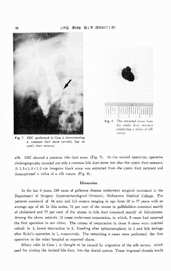
silk. ERC showed a common bile duct stone (Fig. 7). At the second operation, operative
cholangiogra phy士evealednot only a common bile duct stone but also the cystic duct remnant.
A 1. 5×1. 0×1. O cm irregular black stone was extracted from the cystic duct remnant and
demonstrated a nidus of a silk suture (Fig. 8).
Discussion
In the last 5 years, 199 cases of gallstone disease underwent surgical treatm巴ntin the
Department of Surgery (Gastroenterological Division), Wakayama Medical College. The
patients consisted of 84 men and 115 women ranging in age from 16 to 77 years with an
average age of 48. In this series, 72 per cent of the stones in gallbladders consisted mainly
of cholesterol and 77 per cent of the stones in bile duct consisted mainly of bilirubinate.
Among the above patients, 13 cases underwent reoperation, in which, 9 cases had received
the first operation in our clinic. The causes of reoperation in these 9 cases were retained
calculi in 5, bowel obstruction in 2, bleeding after sphincteroplasty in 1 and bile leakage
after Kehr’s operation in 1, respectively. The remaining 4 cases were performed the first
operation in the other hospital as reported above.
Biliary colic in Case 1 is thought to be caused by migration of the silk suture, which
used for closing the incised bile duct, into the ductal system. These migrated threads would
SILK AND GALLSTONE FORMATION 89
either flow out to the duodenum via Vater’s papilla or remain within the common bile duct
and lead to the formation of stones.
In Case 2 and 4, the cystic duct had been tied with silk in the primary operation. In
Case 2, the silk ligature migrated into the common bile duct, and retained there, resulting
in formation of a calculus around it. In Case 4, the silk entered into the cystic duct remnant
and led to stone formation.
In Case 3, a silk ligature used for closing the incised gallbladder fell into the common
bile duct via the cystic duct, and there, gave rise to a calculus.
A gallstone formed around a silk ligature was first reported by HoMANs3> in 1897. BAN
et al.21 reviewed 63 cases of foreign objects in the biliary tract seen from 1897 through
1971, including their own 2 patients. According to the report, their signs and symptoms
were essentially the same as those from simple biliary calculus disease without foreign
bodies. They classified foreign bodies found in the biliary tract into three main categories :
1) operative residuals, 2) missiles, and 3) ingestions. Among them, the most frequently
encountered foreign bodies were those which came within the first category, that is, silk
ligature used for ligating the cystic duct, for closing the incised biliary syst巴m,and for
reconstructing the biliary way. They stated that 89 per cent of patients with suture material
residuals in the bile ducts had stones around the foreign body nidi.
MILLBOURN4> found out that in the 22 patients already reported in the literature with
positive calculus findings at reoperation, 2 instances had shown common duct stones formed
around silk ligatures. Hence, he pointed out that silk ligatures used in the biliary tract
surgery may lead to the formation of a stone.
AHLBERG1> mentioned that, while the cystic duct and cystic artery were ligated with
silk as a rule at that time, 3 cases out of 493 cholecystectomies developed calculus formation
around silk ligature. ToLAND7>, SILVENNOINEN et al6> and SIGLER et al5> separately reported
similar experiences. The exact mechanism concerning a migration of nonabsorbable suture
materials into the biliary ductal system is not clarified yet, but it is probable that local
infection secondary to, for instance, spillage of infected bile plays an important role6>
The incidence of silk ligature as a cause of gallstone fermation after biliary tract
surgery also remains unknown. The stone with silk nidus may pass into the duodenum via
the papilla of Vater or cause no symptom. Many cases presumably remain undiagnosed,
and even when such a calculus is removed at the second operation, a silk ligature nucleus
may well remain undetected unless a special search is made5>. Figures 4, 6 and 8 vividly
indicate that one of the most important characteristics of this type of gallstone is irregular
in shape. Thus, stones of unusual shape or irregular surface should be investigated for a
nidus of nonabsorbable suture material if the patient has the history of biliary surgery.
On the basis of our experience, together with the review of the literature1-7>, suture or
ligature using nonabsorbable materials should not be done in the biliary tract surgery
since they may migrate into the duct and lead to calculus formation. In this viewpoint, it R
is advisable to use absorbable suture materials such as cat gut or Dexon (polyglycolic acid

90 日外宝第48巻第1号(昭和54年 1月)
polym巴r)for ligating the cystic duct, for suturing or ligating the incised bile duct, for
reconstructing the biliary way and for suturing or ligating adjacent to the bile ducts.
Summary
In this paper, a case of migration of silk ligatures into the biliary way and 3 cases of
gallstone formation caused by using silk sutures or ligatures in the primary surgery for
gallstone dis巴asesare reported.
It is advisable that sutures or ligatures with nonabsorbable material should not be
done in the biliary tract surgery since they may migrate into the biliary ductal system and
signs and symptoms resembling biliary colic develop or lead to calculus formation.
References
1〕 AhlbergA Silk Ligatures as a cause of choledocholithiasis after cholecystectomy. Acta Chir Scandinav 118: 22-24, 1959.
2) Ban JL Hirose F恥fet al : Foreign bodies of the biliary tract : Report of two patients and a review of the literature. Ann Surg 176: 102 107, 1972.
3) Homans J ・ Gall-stones formed around silk sutures twenty months after recovery from cholecy-stectomy. Ann Surg 26: 114-119, 1897.
4) Millbourn E : On re-operation for choledocholithiasis. Experience from and comments on 34 operated cases. Acta Chir Scandinav 99:・ 285-312,1949.
5〕 SiglerL and Sahler CO : Silk; The nidus of a common bile duct calculus. Surgery 65: 276 279, 1969.
6〕 SilvennoinnenE and Asp K : Clinical ob3ervation on complications from nonabsorbable suture material in gall bladder surgery. Acta Chir Scandinav 132:587 589, 1966.
7) Toland CG: Foreign bodies in the biliary tract. Ann Surg 98:・ 904-908,1933.

SILK AND GALLSTONE FORMATION 91
和文抄録
絹糸に起因する胆石形成について
和歌山県立医科大学消化器外科学教室
青木洋三,勝見正治
初回胆道手術時lζ使用した絹糸を核として胆石の再
発をみるととが稀ながらある.私達の教室でもとの 3
年聞に 4例経験した.何れも初回手術は他施設で行な
われたものが,恐らく胆道の縫合や結主主lζ絹糸を用い
た結果がとれが胆道内lζ脱落し,再発の因となったも
のであろう. 1例目は31才の女性で, 7カ月前総胆管
切開術を受け,との際縫合lζ用いられた絹糸が胆管内
lζ脱落したものであり,とれが長期胆管内lととどまれ
ばとれを核として結石が再発するものと考えられた.
2例目は36才の男性で 1年前lζ胆褒粘膜抜去術を受け
ており,胆嚢管断端の閉鎖lζ用いた絹糸が胆管内IC::脱
落しとれが核となったもの, 3例目は68才の男性で 3
年前lζ胆嚢切開設石術を受けた際,胆嚢の縫合閉鎖lζ
用いた絹糸が胆管内lζ脱落し核となって再発したも
の, 4例目は30才の男性で, 6年前胆嚢摘除術を受け
た時の胆嚢管断端処理lζ用いた絹糸が脱落し,遺残胆
嚢管内で再発したものであった. 4症例共IC::通常の胆
石症と同様の臨床症状を呈し,術前lζ乙れが絹糸lこ起
因せるものであるか否かの判定は不可能であった.
絹糸結石の報告は1897年 Homansが行なって以来
既lζ久しいが,その発生機序IC::ついては明らかにされ
ておらず,感染胆汁による局所の汚染の如き限局性の
感染が一因ではなし、かと説明されているが推定の減を
出ない.
絹糸結石の発生頻度が不明である原因の lつに摘出
した結石の検索が十分になされていない乙とが挙げら
れる 再手術例で,しかも結石の形状,表面が不規則
な際は必ず割面を入れ,十分に観察する必要があろ
っ.とのような絹糸結石の発生を未然IC::予防するために
は,胆褒管断端の結主主,胆道の縫合閉鎖,胆管消化管
吻合などの際,吸収性の縫合糸を用いる ζ とが最も肝
要である.
![Dr. Yusif Shukurlua in...Книга1. — ОЛМАМедиаГрупп. — 328 с. — ISBN 9785948496450.]. One of the centers of the silk industry of the South Caucasus, in the](https://img.pdfslide.tips/doc/110x75/5edc0b6fad6a402d66668aa4/dr-yusif-shukurlua-in-1-a-oeoef-a-328.jpg)


![Azerbaijan's Foreign Policy and the New Silk Road [Essay]](https://img.pdfslide.tips/doc/110x75/5529dc684a79590e778b45dd/azerbaijans-foreign-policy-and-the-new-silk-road-essay.jpg)