Embed Size (px)
Citation preview
Title Studies on Foreign Gene Transfer into Fish( Dissertation_全文)
Author(s) Inoue, Koji
Citation Kyoto University (京都大学)
Issue Date 1994-09-24
URL https://doi.org/10.11501/3079269
Right
Type Thesis or Dissertation
Textversion author
Kyoto University
" \",
:'''', . . ~'\~
"- i,'
~ * .... '
',~,'-.. ~' ". '</ I .
, ..... :.
, ~,. ( " . " .'-, , , "~ r
",r'
J:- -, , , , "',,
$iJ- l *ii Jill I
I
689 \'

Studies on Foreign Gene Transfer into Fish
Koji Inoue
1994
Contents
Abbreviations ......... ..................... ........ .......................... .............. II
Introduction ................................................................................ .
Chapter 1 Development of methods for introducing foreign genes into fish ......................................................................... . 1.1 Microinjection into oocytes ..................................................... . 1.2 Microinjeclion into fertilized eggs ........................................... . 1.3 Electroporation ., ................................................................... .
Chapter 2 Estimation of activities of various promoters in fish ........................................................................................... . 2.1 Activities of various promoters in fish culture cells ................... . 2.2 Activities of various promoters in fish embryos and fry ............ .
Chapter 3 Stage-dependent expression of the chicken 0-crystallin gene in transgenic medaka embryos ...................... .
Chapter 4 Application of foreign gene transfer techniques to fish breeding ..................................................................... .. 4.1 The method for introducing foreign genes into rainbow trout
4.2 Activity of miw in rainbow trouL ............................................ . 4.3 Introduction of miw-rtGH cDNA construct into rainbow trout .. ..
4 5
16 21
28 28 36
45
59
59 64 71
Chapter 5 Comprehensive discussion ..... ................. .............. 8 I
Chapter 6 Summary................................................................ 84
Acknowledgements ........................................................................ 87
References ................................................................................... 88
Abbreviations
BSA CAT eDNA DNA EDTA PCS FLV GH GYBD HBS LTR MLY mMT-1 mRNA PBS PCR PMS RNA RSV rtGH rtMT:A RT-PCR SDS SV40 TLC X-gal
bovine serum albumin chloramphenicol acetyllransferase complementary DNA deoxyribonucleic acid ethylenediaminetetraacetic acid fetal cal f serum Friend leukemia virus growth hormone germinal vesicle breakdown HEPES-buffered saline long tenninal repeat Moloney leukemia virus mouse metallothionein I messenger RN A phosphate-buffered saline polymerase chain reaction pregnant mare serun gonadotropin ribonucleic acid Rous sarcoma virus rainbow trout growth hormone rainbow trout metallothionein A reverse transcription-PCR sodium dodecylsulfate Simian Virus 40 thin-layer chromatography 5-bromo-4-chloro-3-indolyI-~-Dgalactopyranoside
ii
Introduction
Explosive progress in genetic engineering in the [970's made it possible to isolate eukaryotic genes, and is resulting in the accumulation of an enormous amount of information on their structure and function through in vitro experiments. The ill vitro study alone, however, can not be the basis for conclusions, because genes exist and function in living individuals. Early in the 1980's revolutionary technique was developed to introduce isolated genes into living animals and investigate their function
ill vivo. This technique is microinjection of foreign genes into eggs and
production of animal lines carrying the injected genes. Individuals carrying such foreign genes are called "transgenic" animals and have been
produced ill various species including Caellorhabditis eiegans (Stinchcomb
et al. 1985), Drosophila (Spradling and Rubin 1982; Rubin and Spradling
1982), sea urchin (McMahon et al. 1984; 1985; Flytzanis el al. 1985;
1987; Colin et al. 1987; Hough-Evans et al. 1987; Katula el aJ. 1987;
Franks et al. 1988a, b; Vitelli et al. 1988), amphibians (Rusconi and Schaffner 198[; Etkin 1982; Bendig and Wiliams 1983; Elkin and Peannan 1984, Etkin el a1. 1987; Wilson et al. 1986), mice (Gordon et aI. 1980; Palmiter and Brinster 1986), and farm mammals (Hammer et al. 1985; Brem et al. 1985). This technique has been particularly well established in mice and Drosophila, and used as a practical tool for studying genetic processes in such fields as developmental biology,
immunology, neurobiology and oncology, and for producing new
experimental animals useful in biological and medical research.
Application of transgenic technique to fish began in the middle of
1980' s (Maclean et al. 1987), about 5 years later after the first report of
transgenic mice (Gordon el al. 1980). Fish is the most diversed group of
vertebrates. An enormous number of fish species are distributed in a wide
variety of environment. Each species has variety of features to adapt to
environmental conditions including physical, geographical and ecological factors. Such diversed features Qf various fish species offer useful models
for studying various biological phenomena (Powers 1989). In addition,
some species such as medaka (O,yzias talipes) and zebrafish (Brachydallio rerio) have been recognized as excellent experimental animals in
verebrates (Yamamoto 1975; Kimmel 1989). Thus development of transgenic systems is expected to contribute to the advance of molecular
biological studies of vertebrates. Fish is important not only as experimental animals but also as food
resources. In recent years, fish culture has received much attention because traditional fisheries depending only on natural populations may calise the decrease in fish resources. Unlike farm animals and plants, however, few special strains for aquaculture has been produced by traditional breeding in spite of the increasing importance of fish culture.
Establishment of the foreign gene transfer system in fish is expected to have significant impact upon commercial aquacullure because this technique is expected to become a novel method to produce useful strains
for aquaculture. The first successful introduction of foreign genes into fish was
reported in 1986 by two groups. Chourrout el al. (1986) microinjected
the foreign growth hormone gene into rainbow trout eggs and showed the existence of the transgene in embryos. Ozato ef al. (1986) microinjected
the chicken o-crystallin gene into medaka oocytes and showed the evidence of foreign gene expression. Although these pioneering studies indicated that foreign gene transfer is possible in fish, their systems have
not been proved to be entirely successful. The result by Chourrout et al. e 1986) in which the transgene was detected only by dot blotting was
somewhat preliminary and they failed to detect the expression. Ozato et al. (1986) clearly showed the expression of the transgene by
immunological methods but the tissue specificity of the transgene
expression seemed to be lost. In addition, inheritance of transgenes into
offspring which is an essential step in transgenic studies has not been
examined in these studies.
This study has been performed to establish the system for foreign
gene transfer which enable to achieve introduction, expression and germ
line transmission of transgenes in fish. The author established three
different methods for foreign gene transfer, microinjection into oocytes,
microinjection into fertilized eggs and electroporation, using medaka as
an experimental animal. Succeedingly, activities of various promoters derived from fish and other animals in fish cells were estimated using
2
cultured cells derived from the rainbow troul liver. In vivo activities of promoters which were found to be active in fish cells were also examined using medaka embryos. By these experiments, information about the choice of the promoter to achieve appropreate expression of transgenes was obtained. Another objective of this study is to evaluate the potential of transgenic technique as a tool for basic science and genetic engineering. As a model experiment of util ization of transgenic fish in basic studies of genes, the regulation of expression of the chicken bcrystallin gene during the embryonic development was examined using
medaka embryos. By this experiment, the potential of the transgenic fish as an effective experimental system to study gene regulation ill vivo was
shown. To evaluate the potential of the transgenic technique as a method
for genetic engineering of fish, growth acceleration of rainbow trout
(Oncorhynchus mykiss) by introducing the growth hormone cDNA linked
to a heterologous promoter was also attempted. It was shown that introduced gene was expressed and the growth of injected fry was accelerated. Thus, the transgenic technique became applicable to breeding
of economically important species of fish.
Chapter 1 Development of methods for introducing foreign genes into fish
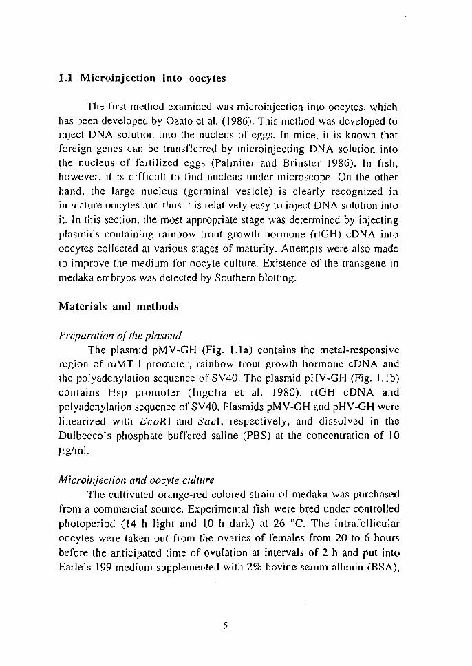
Medaka (Oryzias talipes) is a small egglaying freshwater teleost which is widely lIsed as a laboratory animal (Yamamoto 1975). Il is the only fish species whose several inbred strains have been established (Hyodo-Taguchi and Egami 1985). The oogenesis (lwamatslI et a!. \988),
fertilization (Iwamatsu 1992), and embryonic development (Yamamoto 1975) of this species have been extensively studied. Table 1.1 summerizes biological characters of medaka in relation to transgenic experiments. The 3 cm long size medaka falls into the smallest group of vertebrates known so far. The generation time is short, 2 to 3 months, compared with
that of mice. The spawning is daily and the timing of spawning can be
controlled by light conditions during a 24-hr period. In addition, the
transparency of eggs is distinct advantage for embryological
manupilations. Three different methods. microinjection into oocytes, microinjection into fertilized eggs and eleclroporalion each of which has
unique features were established by a series of experiments lIsing medaka as a model fish in this chapter.
Table 1.1 Biological characters of medaka
Sirain
Inbred slrain Life span Sex maluration Total length Spawning Spawning cycle Days for hatching Diameter of eggs Transparency of eggs Chorion Diameter of oocytes Diameter of genninal vescle Culture of oocyles Chromosome number Genome size
4
Orange-red-colored Wild type Present 2 years 3 months 3cm year-round 24 h 8-10 days lmm Transparent Slightly hard <lmm 100 Jlm Possible 48 20% of mammals
1.1 Microinjection into oocytes
The first method examined was microinjection into oocytes, which
has been developed by Ozato et al. (1986). This method was developed to inject DNA solution into the nucleus of eggs. [n mice, iL is known that
foreign genes can be transflerred by microinjecting DNA solution into the nucleus of fertilized eggs (Palmiter and Brinster 1986). In fish, however. it is difficult to rind nucleus under microscope. On the other
hand, the large nucleus (germinal vesicle) is clearly recognized in
immature oocytes and thus it is relatively easy to inject DNA solution into
it. In this section, the most appropriate stage was determined by injecting
plasmids containing rainbow trout growth hormone (rtGH) eDNA into
oocytes collected at various stages of maturity. Attempts were also made
to improve the medium for oocyte culture. Existence of the transgene in
medaka embryos was detected by Southern blotting.
Materials and methods
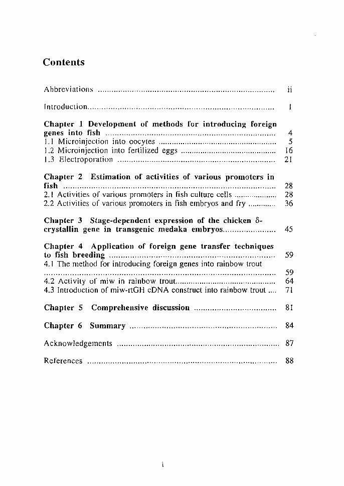
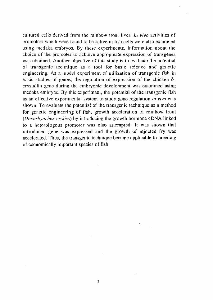
Preparation of the plasmid The plasmid pMV-GH (Fig. I. [a) contains the metal-responsive
region of m MT -I promoter, rai nbow trouL growth hormone cD N A and
the polyadenylation sequence of SV40. The plasmid pHV-GH (Fig. l.lb)
contains Hsp promoter (Ingolia et al. 1980), rtGH cDNA and
polyadenylation sequence of SV40. Plasmids pMV-GH and pHV-GH were
linearized with EcoRI and SacI, respectively, and dissolved in the
Dulbecco's phosphate buffered saline (PBS) at the concentration of 10
Jlg/ml.
Microilljectioll alld oocyte culture The cultivated orange-red colored strain of medaka was purchased
from a commercial source. Experimental fish were bred under controlled
photoperiod (14 h light and 1.0 h dark) at 26°C. The intrafollicuIar
oocytes were taken oul from the ovaries of females from 20 to 6 hours
before the anticipated lime of ovulation at intervals of 2 h and put into
Earle's 199 medium supplemented with 2% bovine serum albmin (BSA),
.')
17.8 mM NaHC03, 25 mg/l penicillin a and 15 mg/l streptomycin sulfate. In some experiments, 50 IU pregnant mare serum gonadotropin (PMS,
Teikoku Hormone Mfg. Co.) was added into the medum (lwamatsu et al.
1974, 1978). About 20 pi DNA solution was microinjecled into the oocyte nuclei immediately after the isolation 0[' oocytes. Some of the oocytes were left uninjected as the control. Injected and uninjecled oocytes were
cultured at 26°C in the same medium until the anticipated time of ovulation, i. e., the beginning of the light period. Oocytes in which
germinal vesicle breakdown (aYEO) were counted as mature oocytes,
which were inseminated according to azato et al. (1986). Eggs in which
peri vitellin space was formed were regarded as fertilized. After rinsing
in distilled water, fertilized eggs were cultured individually until stage 32 in the Matsui stage (Matsui 1949; Yamamoto 1975) or the hatch-out stage
in 96-well culture plates filled with distilled water.
a
pMV-GH 5.2kb
BamHI
b
Pvull
Pvul1
Pvull Socl
pHV~GH
6.7kb
PsI!
Pvul1
RTGH
Pvull
Fig. 1.1 The structure of pMV-GH (a) and pHV-GH (b). Hatched boxes represent the mouse metallothionein promoter and the Drosophila Hsp70 promoter. Open boxes represent the rainbow trout growth hormone eDNA. Dotted boxes represent the SV40 sequence containing the polyadenyla(ion site. Thin lines indicate the plasmid sequences.
6
Detect iOIl of foreign sequences
Genomic DNA was extracted from whole bodies of 7-day-old embryos dechorionized with fine forceps or hatchlings. After incubation in 300 J.l.1 lytic buffer containing 50 mM Tris-HCI (pH7.5), 10 mM EDTA, 0.5% SDS, 500 I-lg/ml Proteinase K at 55°C for 4 h, samples were extracted once with an equal volume of phenol and then twice with phenol: chloroform: isoamyl alcohol (24: 24: I). The aqueous phase was then treated with ethanol and the precipitate was dissolved in TE (l0 mM Tris-HCI, I mM EDTA). The DNA sample for slot blotting was denatured in 0.2 M NaOH for 10 min, neutralized by adding an equal volume of 2 M NH 40Ac, and immediately fixed onto a nylon membrane. The DNA sample for Southern blotting was digested with PstI, electrophoresed on 0.7% agarose gels, and transferred onto a nylon membrane. Mambranes were hybridized with rtGH eDNA labelled with [J2P]dCTP and exposed to X-ray film.
Results
Table 1.2 Numbers of oocytes used for microinjection at various stages of maturity
Hours before Microinjecled Control ovulation oocytes oocyles
20 36 30
18 50 24
16 75 38 [4 88 36
12 72 25 10 130 30
8 78 53
6 48 23
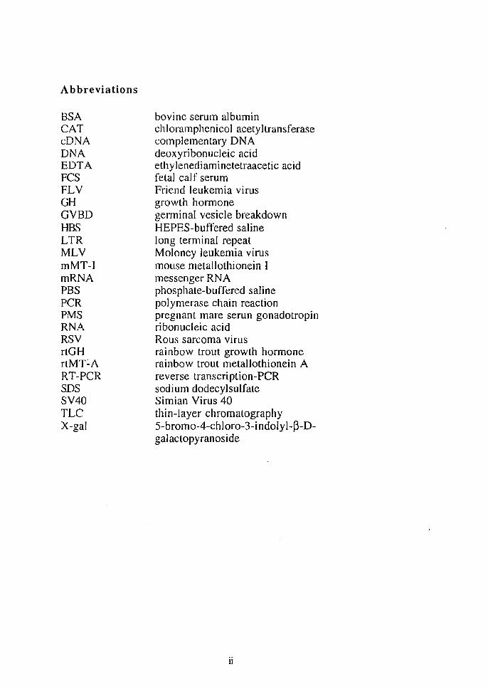
Appropreate stages of oocyte maturity for foreign gene transfer The outline of the procedure of the microinjection into oocytes was described in Fig. 1.2. In the original method, oocytes collected 9 h before
7
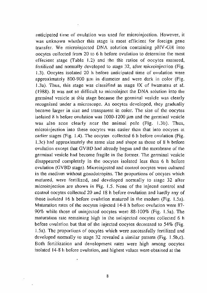
anticipated lime of ovulation was used for microinjection. However, it was unknown whether this stage is most efficient for foreign gene transfer. We microinjected DNA solution containing pHV-GH into oocytes collected from 20 to 6 h before ovulation to determine the most effecient stage (Table 1.2) and the the ralios of oocytes matured, fertilized and normally developed to stage 32, after microinjection (Fig. 1.3). Oocytes isolated 20 h before anticipated time of ovulation were approximately 800-900 /-un in diameter and were dark in color (Fig. 1.3a). Thus, this stage was classified as stage IX of Iwamalsu et at. (1988). It was not so difficult to microinjecl the DNA solution into the
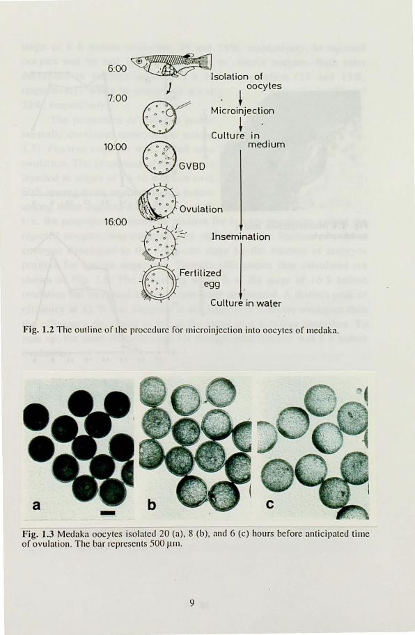
germinal vesicle at this stage because the germinal vesicle was clearly recognized under a microscope. As oocytes developed, they gradually became larger in size and transparent in color. The size of the oocytes isolated 8 h before ovulation was 1000- I 200 J.1m and the germinal vesicle was also seen clearly near the animal pole (Fig. 1.3b). Thus, microinjection into these oocytes was easier than that into oocytes at earlier stages (Fig. 1.4). The oocytes collected 6 h before ovulation (Fig.
l.3c) had approximately the same size and shape as those of 8 h before ovulation except that GV BD had already begun and the membrane of the germinal vesicle had become fragile in the former. The genninal vesicle
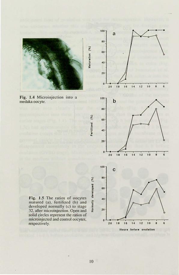
disappeared completely in the oocytes isolated less than 6 h before ovulation (OVBD stage). Microinjected and control oocytes were cultured in the medium without gonadotropins. The proportions of oocytes which matured, were fertilized, and developed normally to stage 32 after microinjection are shown in Fig. J .5. None of the injeced control and control oocytes collected 20 and 18 h before ovulation and hardly any of those isolated 16 h before ovulation matured in the medum (Fig. 1.5a). Maturation rates of the oocytes injected J 4-8 h before ovulation were 87-
90% while those of uninjected oocytes were 88-100% (Fig. 1.5a). The maturation rate remaining high in the uninjected oocytes collected 6 h before ovulation but that of the injected oocytes decreased to 54% (Fig.
J .5a). The proportions of oocytes which were successfully fertilized and developed nomlally to stage 32 revealed a similar pattern (Fig. 1.5b,c).
Both fertilization and development rates were high among oocytes isolated 14-8 h before ovulation, and highest values were obtained at the
8
~. ~ 6:00 ~ .' . ~
7:00
10:00
16:00
Isolation of ) oocytes
+ Microinjection
~ Culture in
medium
GVBD
Ovulation
Insemination
Fertilized egg
Culture in water
Fig. 1.2 The outline or the procedure ror ll1icroinjection into oocytes or l11edaka.
Fig. 1.3 Medaka oocytes isolated 20 (a). 8 (b), and 6 (c) hours berore anticipated lime or ovulation. The bar represents .'100 ~nl.
9
100 a
eo
t GO
c 0
~ :> ~o . ::li
20 -
0 20 1 e 16 1 ~ 12 10 0 6
Fig. 1.4 Microinjection into a 100
Illedaka oocyte. b eo
)~ GO -
" • !! ~o -
• u.
20
0 20 18 16 14 12 10 8 6
100 C
t eo
" • Q. 60 0 .. > .
Fig. 1.5 The ratios of oocytes " 40
Illat ured (a), fertilized (b) and ~ co
developed normally (c) to stage E
:n. after llIicroinjectioll. Open and (; 20 z
solid circles represent the ratios of microinjected and control oocytes, 0 respectively. 20 1 e 16 14 12 10 e 6
Ifou, • bolo,. ovul.lIon
• 10
stage of 8 h before ovulation: 79 and 58%, respectively, in injected oocyles and 96 and 75%, respectively, in control oocytes. Both rates decreased in oocytes injected 6 h before ovulation (21 and 13%, respectively) while in uninjected oocytes they remained high (83 and 52%, respectively).
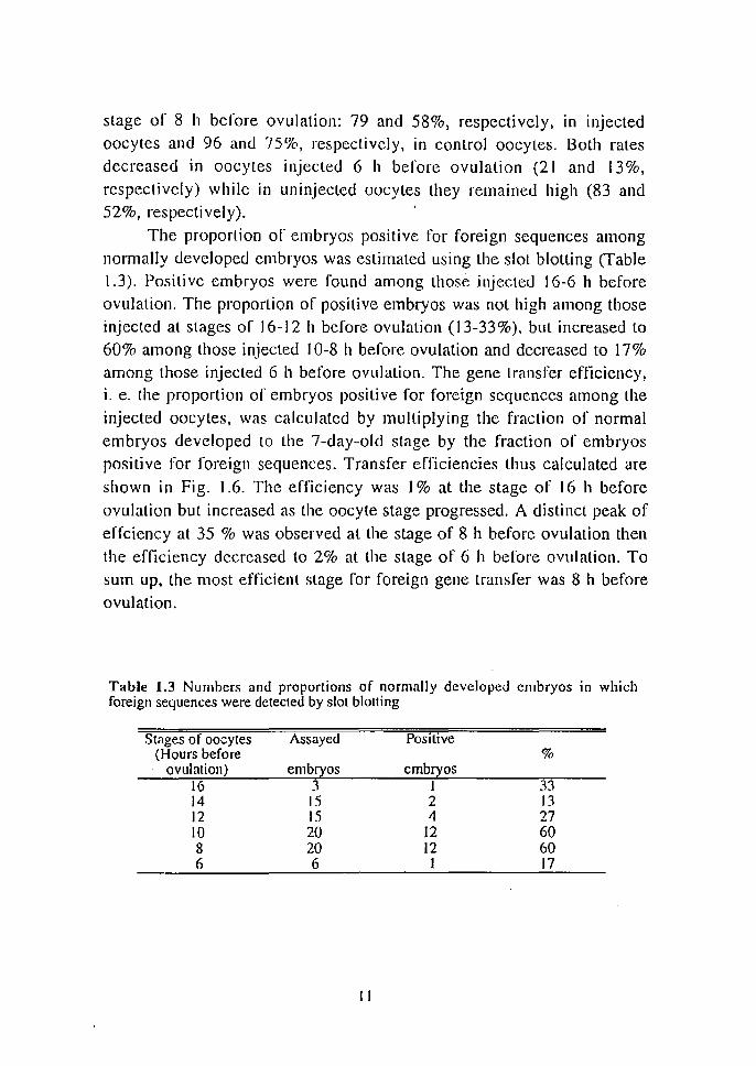
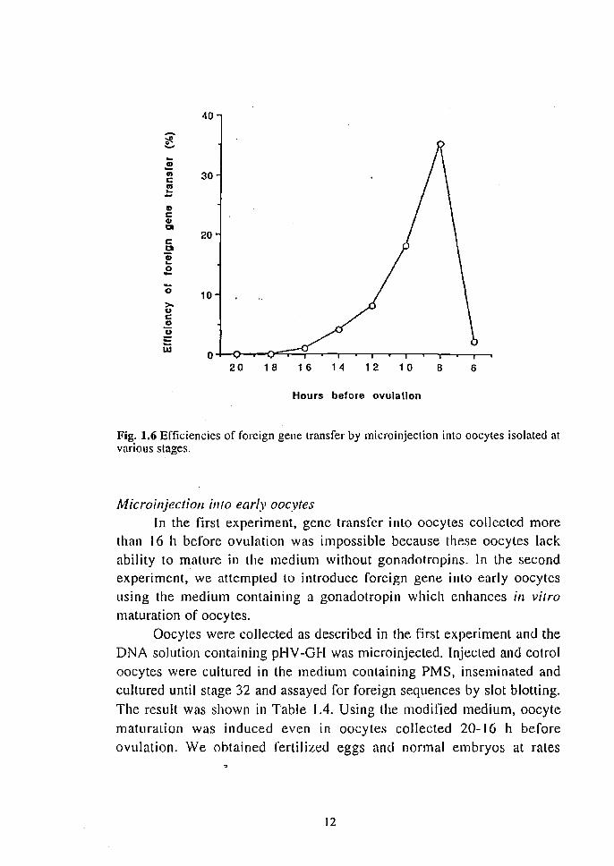
The proportion of embryos positive for foreign sequences among nonnally developed embryos was estimated using the slot blotting (Table 1.3). Positive embryos were found among those injected 16-6 h before ovulation. The proportion of positive embryos was not high among those injected at stages of 16-12 h before ovulation (13-33%), but increased to 60% among those injected 10-8 h before ovulation and decreased to 17% among those injected 6 h before ovulation. The gene transfer efficiency, i. e. the proportion of embryos positive for foreign sequences among the injected oocytes, was calculated by multiplying the fraction of normal embryos developed to the 7-day-old stage by the fraction of embryos positive for foreign sequences. Transfer efficiencies thus calculated are shown in Fig. 1.6. The efficiency was I % at the stage of 16 h before ovulation but increased as the oocyte stage progressed. A distinct peak of effciency at 35 % was observed at the stage of 8 h before ovulation then the efficiency decreased to 2% at the stage of 6 h before ovulation. To sum up, the most efficient stage for foreign gene transfer was 8 h before ovulation.
Table 1.3 Numbers and proportions of normally developed embryos in which foreign sequences were detected by slot blotting
Stages of oocytes Assayed Positive (Hours before %
ovulation) embryos embryos 16 3 1 33 14 15 2 13 12 15 4 27 10 20 12 60 8 20 12 60 6 6 1 17
II
40.
'j: ...... ... .! 1/1 30 c III ... -CD C CD til
20 c CI Iii L-
a -0 10 >-u c CD U = Ui
0 20 18 1 6 14 12 10 B 6
Hours before ovulallon
Fig. 1.6 Efficiencies of foreign gene transfer by micro injection into oocytes isolated at various stages.
Microilljeclioll into early oocytes In the first experiment, gene transfer into oocytes collected more
than 16 h before ovulation was impossible because these oocyles lack
ability to mature in the medium without gonadotropins. In the second experiment, we attempted to introduce foreign gene into early oocytes
using the medium containing a gonadotropin which enhances in virro maturation of oocytes.
Oocytes were collected as described in the first experiment and the
DNA solution containing pHV-GH was microinjected. Injected and cotrol oocytes were cultured in the medium containing PMS, inseminated and
cultured until stage 32 and assayed for foreign sequences by slot blotting.
The result was shown in Table 1.4. Using the modified medium, oocyte
maturation was induced even in oocytes collected 20-16 h before
ovulation. We obtained fertilized eggs and normal embryos at rates
12
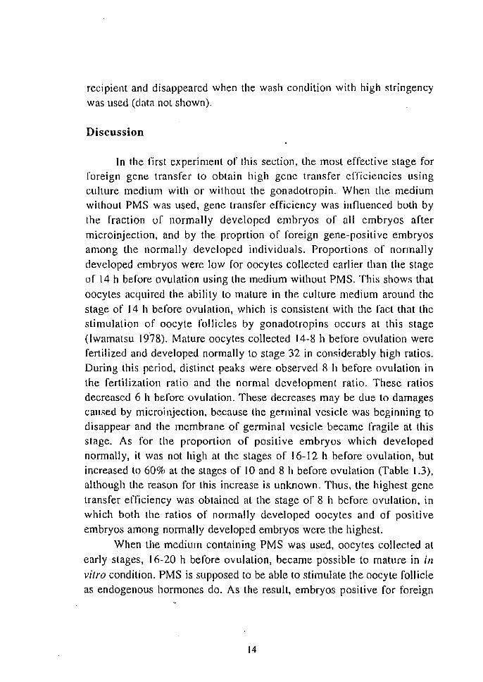
nearly as high as those found in oocytes collected at latcr stages (Table J .4). Embryos which were positive for the foreign sequences were obtained in normal 7-day-old embryos derived from oocytes at every
stagcs. even in embryos from oocytes more than 16 h before ovulation in which no positive embryos was obtained in the first experiment. Only the rate of positive embryos in oocytes collccted 8 h before ovulation was lower than in the first experiments.
Table 1.4 Efficiencies of fertilization, normal development until stage 32, and gene transfer, after microinjeclion.
Hours Injected Control before No. of Fertilized Normal Trans- No. of Fertilized Normal
ovulation oocyles (%) embryos genic (%) oocytes (%) embryos (%) (%)
20 16 38 19 6 14 71 50 18 27 78 70 7 15 7-:' 73 16 35 66 51 II 27 78 74 14 25 48 24 16 19 74 74 [2 31 68 42 13 27 85 63 [0 22 73 36 18 7 [00 86 8 31 42 26 10 26 81 65 6 23 43 13 9 16 81 63
Introduction of pMV-GH The plasmid pMV -GH containing mMT -I promoter and rtGI-l
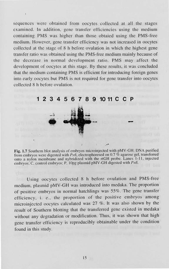
cDNA was introduced into 420 oocytcs collccted 8 h before ovulation. Oocytes were cultured in the PMS-free medium and 207 (49%) fertilized eggs wcre obtained and 104 hatched out. Twenty-two hatchlings were analyzed for the existence of introduced sequences by Southern blotting and positive signal was detected in 12 (55%) hatchlings. As shown in Fig.
1.7, a signal of 2.4 kb corresponding to the Pstl fragment of pMV-GH was detected in positive individuals. This result indicates the existence of the introduced sequences without receiving degradation. A signal at higher molecular weight was also observed in both injected and control
hatchlings when the wash condiLion with low-stringency was used (Fig.
1.7). This band is supposed to correspond to the genomic GH gene of the
13
recipient and disappeared when the wash condition with high stringency was used (data not shown).
Discussion
In the l1rst experiment of this section, the most effective stage for foreign gene transfer to obtain high gene transfer efficiencies using culture medium with or without the gonadotropin. When the medium without PMS was used, gene transfer efficiency was influenced both by
the fraction of normally developed embryos of all embryos after microinjection, and by the proprtion of foreign gene-positive embryos among the normally developed individuals. Proportions of normally
developed embryos were low for oocytes collected earlier than the stage of 14 h before ovulation using the medium without PMS. This shows that
oocytes acquired the ability to mature in the culture medium around the stage of 14 h before ovulation, which is consistent with the fact that the stimulation of oocyte follicles by gonadotropins occurs at this stage (lwamatsu 1978). Mature oocytes collected 14-8 h before ovulation were fertilized and developed normally to stage 32 in considerably high ratios. During this period, distinct peaks were observed 8 h before ovulation in the fertilization ratio and the normal development ratio. These ratios decreased 6 h before ovulation. These decreases may be due to damages caused by microinjection, because the germinal vesicle was beginning to disappear and the membrane of germinal vesicle became fragile at this stage. As for the proportion of positive embryos which developed normally, it was not high at the stages of 16-12 h before ovulation, but increased to 60% at the stages of 10 and 8 h before ovulation (Table 1.3), although the reason for this increase is unknown. Thus, the highest gene
transfer efficiency was obtained at the stage or 8 h before ovulation, in which both the ratios of normally developed oocytes and of positive embryos among normally developed embryos were the highest.
When the medium containing PMS was used, oocytes collected at early stages, 16-20 h before ovulation, became possible to mature in ill vitro condition. PMS is supposed to be able to stimulate the oocyte follicle as endogenous hormones do. As the result, embryos positive for foreign
14
· equence were ob tain ed from oocy te coli cted at all the tage examined. In additi n gene tran fer ef fic iencie using the medium
contaInIng PMS wa higher th an th o e ob taied using the PMS- free
medi um. However, gene tran fer effi ciency wa not increa ed in oocy te.
co llec ted at the stage of h be fore ovul ati on in which the highe t gene
tran f r rati wa obtained u ing the PM S- free medium mainl y becau e of th e decrea e in norm al deve lopment rati o. PMS may affec t th e
dev lopment of oocy te at thi tage. By these re ult it wa concluded
that the medi um containing PM S i effi cient for introducing for ign genes into earl y oocy tes but PMS i. not required for gene tran fer into oocytes
coll ected 8 h before ovulation.
1 2 3 4 5 6 7 8 9 10 11 C C P
.... -.'
Fig.1.7 oUl h rn blot analy i orembryo micro injecled with pMV-GH. 0 A purified from mbryo \ cr~ dige ted with PsJl , clectrophoresed on 0.7 % agaro e gel. tran felTed onto a nylon mcmbrane and nybridi zed \ ith the rtGH pr be. L ane I - II , injecled embryos: C. contr I el1lbryos: P. 10pg pia mid pMV-GH dige tcd with PSI\.
IIl g oocy t co ll ec ted 8 h before ovul ati on and PMS-free
m dium, pi a mid pM V-GH \Va introduced into medaka. The proporti on
of p iti ve embryo in norm al hatchling wa 55%. The gene tran fer
effi c iency . i . e., th proporti on of th e po ili ve embryo among
microinjec ted ocy te ca lcul ated wa 27 %. It wa al hown by the
re ult of S uthern bl ttin g that the tran ferred gene ex isted in medaka
without any degradati on r modifica ti on. Thu it wa hown that high
gene tran fer effi ciency i. reproducibly obtainable und r the conditi on
found in thi s study.
15
1.2. Microinjection into fertilized eggs
MicroinjecLion into oocyte nuclei was shown to be an effective
method for introducing foreign genes into medaka. This method is,
however, applicable only to species in which the technique for in vitro
culture of oocytes has already been established and the oocyte culture has not established in most of fish species. As a more general method, we attempted to introduce transgenes by microinjetion into the cytoplasm of
fertilized eggs. The microinjection into fertilized eggs has been attempted on other fish species (for review see Maclean 1987) but has never been reported on medaka.
Materials and Methods
Preparation of the plasmid
The plasmid pHV~GH (Fig. 1.1a) and the plasmid pMV-GH (Fig.
1.1 b) were linearized with Sacl and EcoRI, respectively, and dissolved in the Dulbecco's phosphate buffered saline (PBS) at the concentration of 100j.lg/ml.
Egg collection and microinjectioll
Medaka was maintained under controlled photoperiod (14 h light
and 10 h dark). Females and males were separated one day before
experiment using the tank separater (Nisso Kogyo) and kept separately during light period. They are mated at the beginning of the light period.
Eggs were spawned within 5 min after mating. Egg clusters were taken from the abdomen of females 20 min after mating and eggs were separated from the cluster by cutting off the attaching filaments with a fine scissors. In some experiments, eggs were kept at 4 °C until microinjection. Then 100-500 pI of DNA solution was microinjected into
the geml disk before the first cleavage. Injected eggs were cultured in 96-
well microplates filIed with distilled water at 26 °C until stage 32
(Yamamoto 1975) or hatch-out.
16

Detection of foreign sequences Genomic DNA was extracted from whole bodies or 7-day-old
embryos dechorionized with rine forcep or hatchling .. After incubation
in 300 pi lytic burfer containing SO 111M Tris-HCl (pH7.S) 10 111M
EDTA, 0.5% SDS, 500 pg/ml Proteinase K at 55°C f r 4 h, samples
wcre extractcd once with an equal volume of phenpl and then twice with
phcnol: ch 101'01'01'111: isoamyl alcohol (24: 24: I). The aqueous phase was
thcn precipilateu with ethanol and dis o!ved in TE (10 111M Tris-IICI, 1
mM EDTA). The DNA sample for slot blotting wa denatured in 0.2 M
NaO!1 ror 10 min, neutralized by adding an equal volume of 2 M
N H 40Ac, anu iml11ediately fixed onto a nylon membrane. The DNA
sample for Southern blotting was digested with Pstl, electrophoresed on
0.7% agarose gels, and transferred onlo a nylon membrane. Mal11branes
were hybridized with rtGH cDNA labelled with l:l2P]uCTP and exposed to
X-ray fi 1m.
Fig. 1.8 Microinjcclion inlo lhe cylOplaSlll or Il1cuaka rCi1ililcd eggs.
Results and Discussion
Appropriate 'i(oges for foreign gene transfer
The DNA solulion was Illicroinjecled into 37 eggs at Ihe early 1-
cell slage (slages I and 2) and 39 eggs althe late I-cell stage (stages 3 and
17
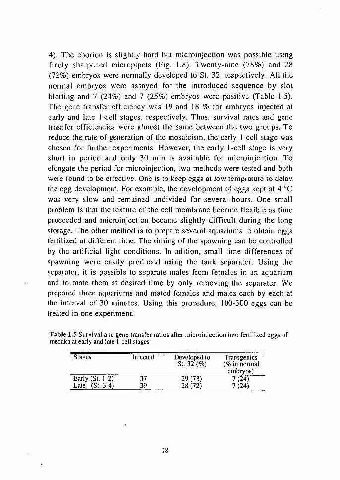
4). The chorion is slightly hard but microinjection was possible using finely sharpened micropipets (Fig. 1.8). Twenty-nine (78%) and 28 (72%) embryos were normally developed to St. 32, respectively. All the normal embryos were assayed for the introduced sequence by slot blotting and 7 (24%) and 7 (25%) embryos were positive (Table 1.5). The gene transfer efficiency was 19 and 18 % for embryos injected at early and late I-cell stages, respectively. Thus, survival rates and gene lrasnfer efficiencies were almost the same between the two groups. To reduce the rate of generation of the mosaicism, the early I-cell stage was chosen for further experiments. However, the early I-cell stage is very short in period and only 30 min is available for microinjection. To elongate the period for microinjection, two methods were tested and both were found to be effective. One is to keep eggs at low temprature to delay the egg development. For example, the development of eggs kept at 4 DC was very slow and remained undivided for several hours. One small problem is that the texture of the cell membrane became flexible as time proceeded and microinjection became slightly difficult during the long storage. The other method is to prepare several aquariums to obtain eggs fertilized at different time. The timing of the spawning can be controlled
by the artificial light conditions. In adition, small time differences of spawning were easily produced using the tank separater. Using the separater, it is possible to separate males from females in an aquarium and to mate them at desired time by only removing the separater. We prepared three aquariums and mated females and males each by each at the interval of 30 minutes. Using this procedure, 100-300 eggs can be treated in one experiment.
Table 1.5 Survival and gene transfer ratios after microinjection into fertilized eggs of medaka at early and late I-cell stages
Stages
Early (SI. 1-2) Late (St. 3-4)
Injected
37 39
18
Developed to SI. 32 (%)
29 (78) 28 (72)
Transgenics (% in nonnal
embryos) 7 (24) 7 (24)
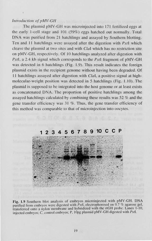
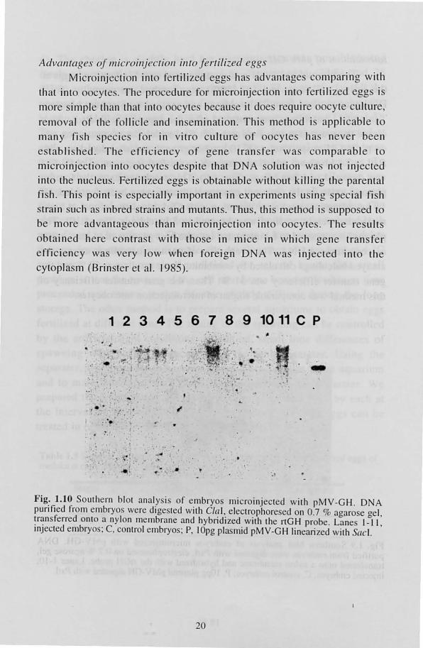
Inlrodll 'Iion oj pMV-CH
The pia mid pMV-GH was microinjected int 17 1 fertili zed egg at
the ea rl y I-ce ll stage and 10 1 (59%) eggs hat hed out norm all y. Total
D A wa purifi ed from 2 1 hatchling and a ayed by S uthern blolling.
T en and II hatchlings were a. sayed after the digesti on with PSII which
cleave the pia mid at two ite and with CIa I which ha no re tri clion ite
on pMV -GH, re pecti vely. Of 10 hatchling analyzed after dige ti on w ith
PSI I, a 2.4 kb ignal which correspond to the PSII fragment of pMV-GH
wa detected in 6 hatchlings (Fig. 1.9). Thi s re ult indi cate the foreign
pia mid ex i t in the rec ipient genome with ul hav ing been degraded. O f
I I hatchling a. ayed aft er dige tion with Cla l a po iti ve ignal at high
molecul ar- weight po iti n wa det cted in 5 hatchling ' (Fig. 1.1 0) . The
pi a mid i supposed to be integrated into the ho t genome or at lea t ex i t
a oncal nated D A. The prop rti on of po iti ve hatchling among the
a ayed hatchling calculated by combining the e re ulls wa 52 % and the
gene tran fer effi ciency wa. 3 1 %. Thu the gene tran f r effi ciency o f
thi method was c mparab le t that of micro injecti on into 0 cy te .
1 2 3 4 5 6 7 8 9 10 C C P
Fig. 1.9 outhern blot analy.s i. of e ' ~1b ryo microinjcc tcd with p~V -GH . DN A purificd from cmbryo. were dl gc ted wllh P.I·!I.. electf(?phore ed n 0.7 1l agaro e gel , transfcrrcd onto a nylon ll1 emb r~lI1 e and hybndlzed. \ IIh the nG I~ I I' bc. ~anes 1- 10. injectcd cmbr 0 : C. c ntr I cmbryo.: P. 10pg Ilas lmd pMV-GH dlgc tcd With PSI!.
19
Ad\ (llIlages oj lIIicroilljN.;fioll illfO Jerfili-ed egg.
Micr inj cti on int r rtili z d egg ha advantage comparing with
th at into 00 yte . . Th proc dure for microinjection into fertili zed egg i
m r imple than that into oocy t b cau e it do requir 0 cy t culture,
remova l r the folli cl and in mination. Thi m th d i ' appli ab le to
many fish sp cie for in vitro culture r 0 cy te ha.' n er been
es tabli hed. The ffi ci ncy of g ne tran fer wa co mparab le to
micro injec ti on into ocy tes de pite that D A so l uti n was not injec ted
into the Ilucl us. Fertiliz d eggs i btainable withoUl killing the par ntal
fi h. Thi s point is e pec ially important in experiment u ing pe ial fi h train such a inbred trains and mUlant . Thu , thi method is uppo ed to
be more advantag LI S than mi cro injec ti on into oocy te ' . The re LIlt
obtain d her contras t with tho e in mi ce in which oene tran fer
efrici ncy wa v ry low when foreign DNA wa injec ted int o th
cy topla m (Brin t I' et al. 1985).
1 2 3 4 5 6 7 8 9 10 11 C P
. ' ..
• ~~t. :4 .. , , . . .. .
: .:. '. . 1 . " .. .. ' . .
'.'
J" , "
',' '0
J "
. ,-
• I ' • . : .
•
.. . '
' . 1
Fig: 1.10 Soulhcrn biOI ana ly i of embryo microinjecled wilh pMV -GH . DNA punfied from embryo, were dige led wilh CIa) , eleclrophore ed on 0.7 % agaro e gel , ~r~n sferred onlO a nylon mcmbrane and hybrid ized wilh the nGH probe. Lane I_II , 1I1Jecled embryo: C, control embryo; P, 10pg pia mid pMV-GH linearized wi th (leI.
20
1.3. Electroporation
The microinjection into oocytes and fertilized eggs was proved to
be an effective method for introducing foreign genes into fish. The latter is more advantageous in most purposes as described in the previous
section. However, ,even microinjection into fertilized eggs involves certain difficulties. One problem is microinjection itself is a complicated operation under the microscope which requires a great deal of skill. The
other problem is the chorion of fertilized eggs. Eggs of many fish species other than medaka are covered with a Lough and/or opaque chorion which prevents insertion of the micropipette. To overcome this problem, special methods have been developed in each species, e. g., penetration of micropipetes through the chorion by two-step injection in salmonids (Chourrout et al. 1986; McEvoy et al. 1988); injection through the micropile in lilapia (Brem et al. 1988; Fletcher et al. 1988); dechorionization in goldllsh (Zhu el al. 1986), 10ach (Zhu et al. 1985) and zebrafish (Stuart et aJ. 1988). To avoid lhe difficulties accompanying microinjeclion, we applied electroporation which is often used for gene transfer into culture cells (Neumann et al. 1982; Potter et al. 1984) to the
production of transgenic fish.
Materials and Methods
Preparatioll of fhe plasmid The plasmid pM V -GH (Fig. 1.1 a) contains the metal-responsive
region of mMT-1 promoter, rainbow trout growth hormone cDNA and the polyadenylalion sequence of SV40. After linearization with EcoRI. the plasmid was dissolved in the mannitol buffer containing 0.25 M mannitol, 0.1 mM CaCI2, 0.1 111M MgCh. 0.2 mM Tris-HCl (pH 7.5) at
the concentration of 100 J.Lg/ml.
Egg collection Medaka was maintained under controlled photoperiod (14 h light
and 10 h dark). Females and males were separated one day before
21
experiment using the tank separator (Nisso Kogyo) and kept separately during light period. They are mated at the beginning of the light period. Eggs were spawned wilhin 5 min after mating. Egg clusters were taken
from the abdomen of females 20 min after mating and eggs were separated from the cluster by clItting off the atlaching filaments with a fine scissors.
Elect roporatioll Isolated eggs were put between the electrodes of a transfection
chamber, Shimadzu FfC~03 (electrode distance, 2 mm), filled with 800 111 of the DNA solution. With this chamber, about 120 medaka eggs could be treated at a time. Before flrst cleavage, electric pulses were applied by a gene transfer equipment (Shimadzu GTE-I) under the following condition: pulse height, 750 V fern; pulse interval, I s; pulse length, SOils; pulse number,S. Treated eggs were rinsed in distilled water several times and incubated separately in 96-well plastic microplates filled with distilled water until hatching at 26°C.
Slot blot and Southern analysis
Genomic DNA was extracted from whole bodies of hatchlings or tail fin pieces of adult fish. After incubation in 300 III lytic buffer containing 50 mM Tris-HCI (pH7.5), 10 mM EDT A, 0.5% SDS, 500
Jlg/ml Proteinase K at 55°C for 4 h, samples were extracted once with an equal volume of phenol and then twice with phenol: chloroform: isoamyl
alcohol (24: 24: I). The aqueous phase of was then precipitated with ethanol and dissolved in TE (10 mM Tris-HCI, I mM EDTA). The DNA sample for slot blotting was denatured in 0.2 M NaOH for 10 min, neutralized by adding an equal volume of 2 M N H40Ac, and immediately fixed onto a nylon membrane. The DNA sample for Southern blotting was digested with Pstl or EcoRI and HilldlJl, electrophoresed on 0.7% agarose gels, and transferred onto a nylon membrane. Membranes were hybridized with rtGH cDNA leveled with [32P]dCTP and exposed to xray film.
22
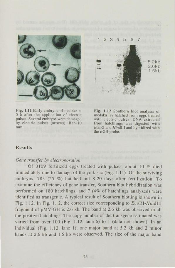
Fig. 1.11 Early embryos of medaka at .') h after the application of electric flulseS. Several ell1bryos were daillaged by electric pulses (arrows) . Bar= I 0 rnm.
Results
Gene transfer by electroporatiol/
12345
.. -
I ,
I ••• t
6 7
- 5.21<b - 2.61<b - 1.51<b
Fig. 1.12 Southern blot analysis of Illedakn r ry hatched rrom eggs treated with electric pulses . DNA extracted frofl1 hatchlings wa digested with EcuRI and f-lilldlll and hybridized with the rtGH probe.
Of 3109 fertilized eggs treated with pulses, about 10 % died
immediately due to damage of the yolk sac (Fig. 1.11). Of the surviving embryos 783 (25 %) hatched out 8-20 days arter fertilization . To
examine the clTiciency of gene transfer Southern blot hybridization was perrormed on 180 hatchlings, and 7 (4% or hatchlings analyzed) were identified as transgenic. A typical result of Southern blolling is shown in Fig. 1.12. In Fig. 1.12, the correct size corresponding to EcoRI-Hil/dlll fragment of pMV -GH is 2.6 kb. The band at 2.6 kb was observed in all
the positive hatchlings. The copy number of the transgene estimated was
varied frolll over 100 (Fig. 1.12, lane 6) to I (data not hown). In an
individual (Fig. 1.12, lane I), Olle major band at 5.2 kb and 2 minor
bands at 2.6 kb and 1.5 kb were observed. The size of the major band
23
corresponds 10 that of the whole pMV -GH. This may be caused by
integrat ion of several copies of transgene in a head- to- tail manner after deletion of the EcoRI site lIsed to linearize the pIa mid. The minor band
at 1.5 kb may be an end f the tran genes . Only the 2.6-kb band
maintained the correct size. No band was detected in untrea ted control
(data not shown).
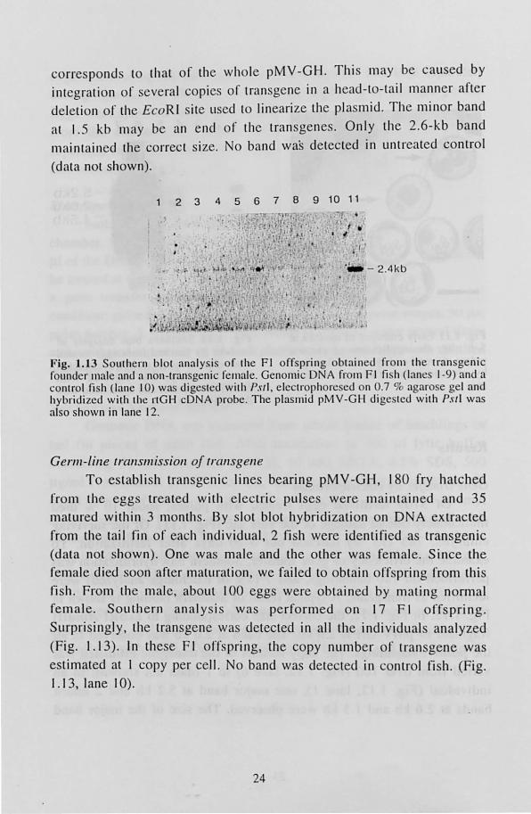
2 3 4 5 6 7 8 9 10 11
Fig. 1.13 SOllthern blot analysis o r the FI orrspring obtained rrom the transgenic rounder male and a non-transgenic female. GenomiL: DNA rrom F I fi h ( lanes 1-9) and a control fish (Ialle 10) was dige ted with PSII, electrophorc ed on 0.7 % agarose gel and hybriui zed with the rtGH cDNA probe. The plasmid pMV-GH dige ted wi th Pst! was also shown in lane 12.
Germ-line transmissioll oj Irall sgene To establish transgenic lines bearing pMV -GH, 180 fry hatched
from the eggs treated with electric pulses were maintained and 35
matured within 3 months. By slot blot hybridi zati on on DNA extracted
from the tail fin or each individual , 2 fish were identifi ed as transgenic
(data not shown). One was male and the other was female. Since the
female died soon after maturation, we failed to obtain offspring from this
fish . From the male, about 100 eggs were obtained by mating normal female. South ern analysis was performed on t 7 F I o ffsprin g.
Surprisingly, the transgene was detected in all the individual s analyzed
(Fig. I . t 3). In these I~ I offspring, the copy number of tran gene was
estimated at I copy per cell. No band was detected in control fish. (Fig. 1.13, lane to).
24
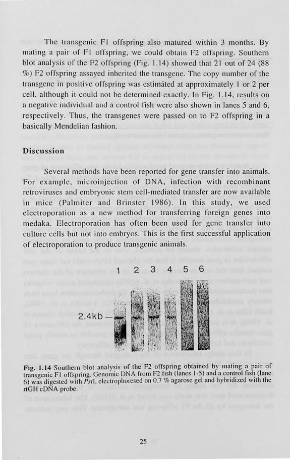
The transgenic FI offspring also matured within 3 months. By mating a pair of FI offspring we could obtain F2 offspring. Southern
blot analy is of the F2 offspring (Fig. 1.14) showed that 21 out of 24 (88
%) F2 offspring assayed inherited the transgene. The copy number of the
transgene in positive offspring wa estimated at approximately I or 2 per
cell , although it could not be determined exactly. In Fig. 1.14, re ults on
a negative individual and a control fi h were also shown in lanes 5 and 6,
respectively. Thus, the tran sgenes were passed on to F2 offspring in a
basically Mendelian fashion.
Discussion
Several methods have been reported for gene transfer into animals. For example, microinjection of DNA, infection with recombinant retroviruses and embryonic stem cell-mediated transfer are now available
in mice (Palmiter and Brin ter 1986). In this study, we used electroporation as a new method for transferring foreign genes into
medaka. Electroporation has often been used for gene transfer into
culture cells but not into embryos. This is the first successful application
of electroporation to produce transgenic animals.
1 2 3 4 5 6
2.4kb - '
.. '
Fig. 1.14 Southern blot analysis or the F2 orrspring obtained by mating a pair or lransgenic FI orrspring. Genoll1ic DNA rrom F2 fish (lanes 1-5) and a CO~ll~ol fisl~ (lane 6) was digested with PSII, electrophoresed on 0.7 % agarose gel and hybndl zed Wllh the rtGH cDNA probe.
25
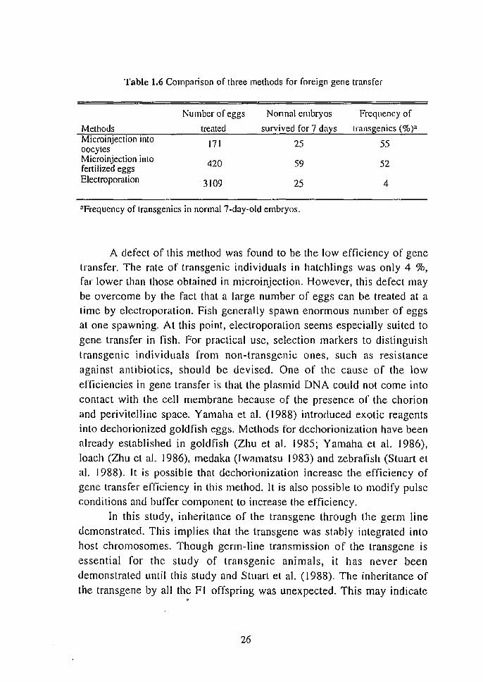
Table 1.6 Comparison of three methods for foreign gene transfer
Number of eggs Nonna) embryos Frequency of
Methods treated survived for 7 days transgenics (%)a Microinjection into 171 25 55 oocytes Microinjection into 420 59 52 fertilized eggs Electroporation 3109 25 4
aFrequency of transgenics in normal 7-day-old embryos.
A defect of this method was found to be the low efficiency of gene transfer. The rate of transgenic individuals in hatchlings was only 4 %, far lower than those obtained in microinjection. However, this defect may be overcome by the fact that a large number of eggs can be treated at a time by electroporation. Fish generally spawn enormous number of eggs at one spawning. At this point, electroporation seems especially suited to gene transfer in fish. For practical use, selection markers to distinguish transgenic individuals from non-transgenic ones, such as resistance against antibiotics, should be devised. One of the cause of the low
efficiencies in gene transfer is thallhe plasmid DNA could not come into contact with the cell membrane because of the presence of the chorion and perivitelline space. Yamaha et al. (1988) introduced exotic reagents into dechorionized goldfish eggs. Methods ror dechorionization have been already established in goldfish (Zhu et al. 1985; Yamaha et a\. 1986), loach (Zhu et al. 1986), medaka (Iwamatsu 1983) and zebrafish (Stuart et al. 1988). It is possible that dechorionization increase the efficiency of gene transfer efficiency in this method. It is also possible to modify pulse conditions and buffer component to increase the efficiency.
In this study, inheritance of the transgene through the germ line demonstrated. This implies that the transgene was stably integrated into host chromosomes. Though germ-line transmission of the transgene is
essential for the study of transgenic animals, it has never been demonstrated until this study and Stuart et al. (1988). The inheritance of the lransgene by all the FI offspring was unexpected. This may indicate
,
26
that the transgene was integrated into both sets of chromosomes independently although it remains to be proved.
To sum up the results obtained in this chapter, three different methods for introducing foreign genes into fish have been established using medaka as a model fish. Surv'ivai rates and gene transfer efficiencies of the three methods were summarized in Table [.6. Of these methods, eiectroporation is the most simple, but its gene transfer
efficiency is the lowest, although it may be increased by improving the procedure. Microinjection into oocytes is the best with regard to the gene transfer efficiency although it involves complicated procedures and
applicable only to limited species. Microinjection into fertilized eggs is Jess difficult than into oocytes but is obstructed by the chorion in some species. It is important to choose a method for gene transfer according to the purpose of the experiment and the species used.
27
Chapter 2 Estimation of' activities of various promoters in fish
To establish the complete experimental system for transgenic fish,
achievement of three steps, introduction, germ-line transmission and expression of transgenes is essential. We nave established the methods for introducing transgenes into fish and the transmission of the foreign gene to offspring in the first chapter. The next step to be achived is the
expression of introduced genes. Foreign gene expression of transgenes has never been reported in early studies on transgenic fish except for the report by Ozato et al. (1986) (for review see Maclean et al. 1987). To
obtain appropriate expression of transgenes, the choice of regulatory elements is important. A variety of promoters and enhancers derived
from genes and viruses or higher vertebrates are available at present and
several promoters have also cloned recently from fish. However, information on activities of such regulatory elements in fish is still
largely insufficient. In this chapter, activities of various promoters and enhancers in fish were examined in ill vitro experiments using a fish cell
line and in in vitro experiments using medaka embryos.
2.1. Activities of various promoters in fish culture cells
For estimating activities of promoters and enhancers, in vitro experiments using culture cells are generally performed prior to ill vivo experiments because the ill vitro experiments are considered to be more simple than the ill vivo system. We examined activities of some wellknown promoters and enhancers derived from fish and other animals in a
series of experiments using a cell line derived from the rainbow troul liver as the recipient of transgenes and the chloramphenicol
acetyltransferase (CAT) gene as a reporter.
Materials and methods
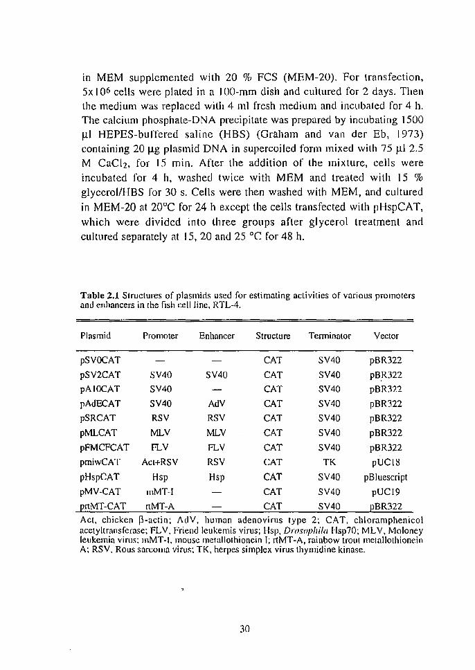
Plasm ids Structures of pJasmids lIsed in this section were summerized in Table 2.1.
The pSV2CAT is a plasmid containing the promoter-enhancer region of
28
SV40 in front of the CAT gene (Gorman et a!. 1982a). Plasmid pSVOCAT is derived from pSV2CAT by removing the enhancerpromoter (Gorman el al. 1982a). Plasmid pA I OCAT was an enhancerless derivative, but possessed a promoter region, or pSV2CAT (Laimins et al. 1982, 1983). Plasmid pAdECAT was constructed by adding the enhancer sequence of adenovirus type 2 to pA 1 OCAT (Hasegawa ct al. 1990). Plasmids, pSRCAT, pMLCAT and pFMCFCAT were constructed by inserting the long terminal repeat (LTR) sequences of Rous sarcoma virus
(RSV), Moloney murine leukemia virus (MLV) and Friend leukemia
virus (FLV) into the HilldIIl site of pSVOCAT, respectively (Hasegawa et
al. 1990). Plasmid pmiwCAT was constructed by replacing a parl of the
S' upstream region of chicken p-actin sequence of plasmid ppact-CAT9
(Fregien and Davidson 1986) with a RSV-LTR sequences containing the
promoter and the enhancer regions (Kalo et al. 1990). Plasmid pHspCAT was constructed as follows: First, the Bglil-Pstl fragment of the 5' franking sequence of the Drosophila Hsp70 gene (lngolia et al. 1980) was subcloned into the BamHI-Pstl site of pUC19 (pUC-Hsp). Secondly, The EcoRI site of pUC-Hsp was cleaved and blunt-ended, and HilldlIl linker was added (pUC-H-Hsp). Then the Drosophila sequence was isolaled using Hindlll and inserted into the HilldllI site of pSVOCAT. Plasmid prtMTCAT was constructed by inserting blunt-ended Spel-BamHI fragment of the rainbow trout metallothionein A (rtMT-A) promoter (Murphy et a1.
1990) into the blunt-ended Hilldlll site of pSVOCAT. Plasmid pMV-CAT was constructed as follows: Plasmid pMK containing mouse
metallothionein I (mMT-I) promoter (Glanville et al. 1981) was cleaved
with BglIl, treated with SI nuclease and then cleaved with BgllL This
fragment was ligated to the BgllI-EcoRI fragment of pSV2CAT
containing the terminator region. Resulted BglI-EcoRI fragment was
inserted into the SmaI-EcoRl site of pUCl9 (pM V). Then the BamH I fragment of pCM4 containing CAT gene was inserted into the BglII site
at the junction of mMT-I promoter and SV40 tenninator, of the pMV.
Cell Culture alld Trarzsjection The cell line RTL-4, derived from the liver of rainbow trout
(Watanabe et al. J 987), was used as the recipient. It was grown at 20°C
29
in MEM supplemented with 20 % FCS (MEM-20). For transfection, 5x 10⁶ cells were plated in a IOO-mm dish and cultured for 2 days. Then the medium was replaced with 4 ml fresh medium and incubated for 4 h. The calcium phosphate-DNA precipitate was prepared by incubating 1500 III HEPES-buiTered saline (HBS) (Graham and van der Eb, 1973) containing 20 Ilg plasmid DNA in supercoiled form mixed with 75 J.l1 2.5 M CaCI2, for 15 min. After the addition of the mixture, cells were
incubated for 4 h, washed twice with MEM and treated with 15 % glycerollHBS for 30 s. Cells were then washed with MEM, and cultured
in MEM-20 at 20°C for 24 h except the cells transfected with pHspCAT,
which were divided into three groups after glycerol treatment and
cultured separately at 15, 20 and 25°C for 48 h.
Table 2.1 Structures of plasmids used for estimating activities of various promoters and enhancers in the fish cell line, RTL-4.
Plasmid Promoter Enhancer Structure Tenninator Vector
pSVOCAT CAT SV40 pBR322
pSV2CAT SV40 SV40 CAT SV40 pB.R322
pAIOCAT SY40 CAT SY40 pBR322
pAdECAT SV40 AdV CAT SV40 pBR322
pSRCAT RSV RSV CAT SV40 pBR322
pMLCAT MLY MLV CAT SV40 pBR322
pFMCFCAT R...V R...V CAT SY40 pBR322
pmiwCAT Act+RSY RSV CAT TK pUC18
pHspCAT Hsp Hsp CAT SV40 pBluescript
pMV-CAT rnMT-I CAT SY40 pUCI9
~rtMT-CAT rtMT-A CAT SV40 2BR322
Act, chicken p-actin; AdV, human adenovirus type 2; CAT, chloramphenicol acetyltransferase; FL V, Friend leukernis virus; Hsp, Drosophilll Hsp70; MLY, Moloney leukemia virus; mMT-I, mouse melallothionein I; rtMT-A, rainbow troul melallothionein A; RSV, Rous sarcoma virus; TK, herpes simplex virus thymidine kinase.
30
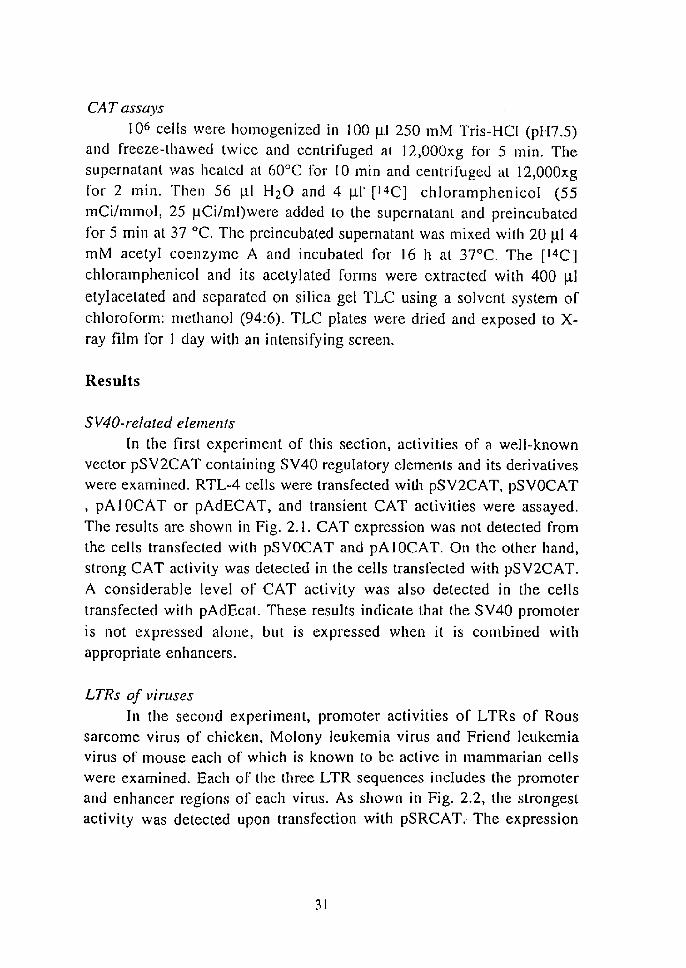
CAT assays
106 cells were homogenized in 100 111250 mM Tris-HCI (pH7.5) and freeze-thawed twice and centrifuged at 12,000xg for 5 min. The supernatant was heated at GO°C for 10 min and centrifuged at 12,OODxg
for 2 min. Then 56 III H20 and 4 Ill' [14C] chloramphenicol (55 mCi/mmol, 25 IlCi/ml)were added to the supernatant and preincubated
for 5 min at 37°C. The preincubated supernatant was mixed with 20 III 4 mM acetyl coenzyme A and incubated for 16 h at 37°C. The [14C]
chloramphenicol and its acetylated forms were extracted with 400 III
etyIacetated and separated on silica gel TLC using a solvent system of
chloroForm: methanol (94:6). TLC plates were dried and exposed to Xray film for I day with an intensifying screen.
Results
SV40-re/ated elements
In the IIrs1 experiment of this section, activities of a well-known vector pSV2CAT containing SV40 regulatory elements and its derivatives were examined. RTL-4 cells were transfected wiLh pSV2CAT, pSVOCAT
, pA 1 DCA T or pAdECAT, and transient CAT activities were assayed.
The results are shown in Fig. 2.1. CAT expression was not detected from the cells transFected with pSVOCAT and pAIOCAT. On the other hand,
strong CAT activity was detected in the cells transrected with pSV2CAT. A considerable level of CAT activity was also detected in the cells
transfected with pAdEcat. These results indicate that the SV40 promoter
is not expressed alone, but is expressed when it is combined with appropriate enhancers.
LTRs of viruses In the second experiment, promoter activities of LTRs of Rous
sarcome virus of chicken, Molony leukemia virus and Friend leukemia virus of mouse each of which is known to be active in mammarian cells were examined. Each of the three LTR sequences includes the promoter and enhancer regions of each virus. As shown in Fig. 2.2, the strongest activity was detected upon transfection with pSRCAT. The expression
31
level of pSRCAT was a high as that of pSV2CAT. pMLCAT was also expressed, although only at a low level. The transl"ection 01" pFMCFCAT
showed only trace activity.
A chicken chimel/ic proll/oter lIlilV
The activity of the chicken promoter miw (Suemori et al. 1990;
Kato et al. 1990) was examined. This promoter con ists of the chicken ~
actin promoter and the RSV -L TR both of which are expected to be very
active in variety types of cells. As shown in Fig. 2.3, pmiwCAT was
expressed at a considerable level in RTL-4 although somewhat weaker
than pSV2CA T.
Hsp promoters
In addition to constitutive elements examined in experiments above,
activities 01" some inducible promoters were also examined. The Drosophila Hsp70 promoter is expected to be activated by the heat shock.
Cells transrected with pHspCAT were cultured at three di fferent temperatures and assayed for CAT activity. As expected, CAT activity
was detected in the cells cultured at 25°C, a higher temperature than that
used nOnllally ror culturing, while the cells incubated at 15 and 20°C exhibited no detectable CAT activity (Fig. 2.4) . Thus, the Hsp70
promoter was regulated in a heat-responsive manner in RTL-4.
1 2 3 4
Fig. 2.1 Expression of plasmids derived from pSV2CAT in RTL-4. Cells were transfected with plaslllids, pS VOCAT (I), pA I OCAT (2), pSV2CAT (]) and pAdECA T (4) and assayed for CAT activities.
32
12345
Fig. 2.2 Expression directed by L TR of viruses of higher vertebrates in RTL-4. Cells were tran s fected with plas rnid s. pSVOCAT (I) , pSV2CAT (2). pMLCAT (3) , pFMCFCAT (4) and pSRCAT (5) and assayed for CAT activities.
1 :1 .. / • ~ . ~,.
1 2 3
Fig. 2.3 Express ion of pmiwCA T in RTL-4. Cells were transfected with pSVOCAT (I), pSV2CAT (2) and pmiwCAT (3) and assayed for CAT activities.
" . ~ .
. :,,'"'
III 123
Fig. 2.4 Heat-inducible expression Dro.wphif{/ Hsp70 promoter in RTL-4 . Cells were transfected with pHspCAT cultured for 48 hat 15°C (I), 20°C (2) and 25 °C (3) and assayed for CAT activities .
33
a b c
• . , •. ,
.....
R 0 2 ZnCuCd -ZnCuCd
Fig. 2.5 M etal-inducible activities of mctallothionein promoters in RTL-4 . a. controls: Cells without transfec tion (R) and Ihose Iransfecled wilh pSVOCAT (0) and pSV2CAT (2). h. rIMT-A: Ct!1I lransfec ted with pr1MT-CAT and cultured in MEM-20 (-) r same medium containing I 00 ~M zinc chloride (Zn), I 00 ~M copper chloride (Cu) or I 00 ~M cadmium chloride (Cd). c. rnMT-I: Cells were tfansfected with pMV -CAT and cultured in the same manner as given in b.
Metal/othioll ein promoters
Metallothionein promoters are expected to be inducible by treatment with heavy metals. To examine activities of mMT-1 and rtMT
A promoters, plasm ids pMV-CAT and prlMT-CAT were introduced into
RTL-4 cells. The results or transfection with prtMT-CAT are shown in
Fig. 2.5. CAT activity was low without exposure to metals, but after addition of zinc, expres ion increased to a level comparable to that of
pSV2CA T. Copper and cadmium had no effect. The result of transfection with pMV-CAT are also shown in Fig. 2.5 . Only a trace level of activity
was observed without induction, but expression was timulated by adding
either zinc or cadmium to the medium. Thus both metallothionein promoters are metal-inducible in RTL-4 cells .
34
Discussion
In this section, activities of various promoters and enhancers in the trout cell line were examined as summarized in Table 2.2. Some of the promoters and the enhancers derived I"rOl11 viruses of higher vertebrates exhibited constitutive expression. Among them, the promoter-enhancer of SV40 and RSV -LTR directed the expression at high levels. These results are reasonable because both of them are known as strong promoters in mammarian cells. The activity of the RSV LTR has also been shown in
other fish cell lines (Friendenreich and Schartl, 1988). Thus RSV -L TR
and SV 40 may be used as constitutive regulatory elements to achieve high
level expressions of transgenes in fish. The chimeric promoter miw was also shown to be active in RTL-4. The chicken f)-actin promoter itself is expected to be active in various types of cells because J)-actin is a cytoskeletal actin. In addition, the RSV -L TR which is another component of miw has been shown Lo be active in this study. Therefore, the result obtained in this experiment is reasonable.
Table 2.2 Activities of various promoters and enhancers in a trout cell line, RTL-4.
Plasmid Promoter Enhancer Activity Inducibility
pSVOCAT
pAIOCAT SV40
pSV2CAT SV40 SV40 ++ Constitutive
pAdECAT SV40 AdV + Constitutive
pSRCAT RSV RSV ++ Constitutive
pMLCAT MLV MLV + Constitutive
pFMCFCAT FLV FLV Constitutive
pmiwCAT Acl+RSV RSV ++ Constitutive
pHspCAT Hsp + Inducible
pMV-CAT mMT-I + Inducible
ErtMT-CAT rtMT-A + Inducible
Act. chicken p-aclin: AdV. human adenovirus lype 2; FLV, Friend leukemis virus; Hsp, Drosophil(/ Hsp70; MLV. Moloney leukemia virus; mMT-I. mouse l11etallothionein I; rtMT-A, rainbow trout metallothionein A; RSV, Rous sarcoma virus.
35
In contrast with these constitutive elements, the Drosophila Hsp70 promoter and two metallothionein promoters were expressed in faithful response to heat shock and metal-treatments, respectively. Inducible
promoters and enhancers. which can be modulated by environmental factors. are desirable when the transgene products are anticipated to be harmful to host animals. Hsp70 promoter was activated by simple heat
treatment without any harmful substances. Metallothionein promoters is also advantageous in transgenic fish experiment because expression can be
induced by adding metals to aquarium water without injection. Therefore. these promoters were supposed to be especially useful in transgenic fish
experiments.
Since the activities of promoters are influenced by cell types. the state of differentiation and many other factors, more information is
required to elucidate the mechanism of gene regulation in fish. It is yet unknown whether the results of this study are reproducible in in vivo experiments. If constitutive and inducible expression systems are established, they will be powerful tools for both genetic engineering of culture fish and basic studies of genes.
2.2. Activities of various promoters in fish embryos and fry
In experiments using the cultured cells, several promoters were
found to be active. Among them, activities of two constitutive promoter.
the SV2 and the chimeric promoter miw and two inducible promoters.
mMT-I and rtMT-A were examined by in vivo experiments using medaka
embryos to confirm the results obtained in ill vitro experiments. The activities of the miw and pSV2 were estimated using the ~-galactosidase reporter and the CAT reporter, respectively. The activities of the both
metallothionein promoters were estimated using the CAT reporter and the induction of the expressiol,l was attempted by adding zinc into the aquarium water.
36
pmiwZ
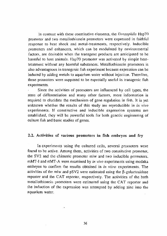
RSV act cry lacZ tk SV pUC18
E B E B BX BK 1kb
Fig. 2.6 Structure of pmiwZ. Chicken l3-actin sequences (act) including promoler region are indicated by boxes with wavy stripes. RSV L TR sequence is indicated by dotted box. The solid box represents chicken o-crystallin sequence (cry) containing a methionine codon which functions as an iniliator codon. The open box represents the bacterial l3·galactosidase gene (lacZ). The termination regions derived from herpes virus thymidirne kinase gene (tk) and SV40 (SV) are indicated by boxes with vertical and horizontal stripes, respectively. The thin line indicates plasmid vector sequence. Restriction sites: B, B(//IlHI; E, EcoRI; K, Kpll[; X, XhaI.
Materials and Methods
Plasm ids The pSV2 CAT is a plasmid contammg the promoter-enhancer
region of SV 40 in front of the CAT gene (Gonnan et al. 1982a). Plasmid pSVOCAT is derived from pSV2CAT by removing the enhancer
promoter (Gorman el al. 1982a). Plasmid pmiw Z was constructed by replacing the CAT-coding region of pmiwCAT with the Splz I fragment of p8Ztk (Ueno ct al. 1988) which has a part of the chicken 8-crystallin
sequence as a translation initiator, the coding sequence of the bacterial ~galactosidase and the termination sequence of herpes simplex virus thymidine kinase gene (Fig. 2.6) Plasmid prtMT-CAT was constructed by inserting blunt-ended Spel-BamHI fragment of the rainbow trout metallothionein A (rtMT-A) promoter (Murphy et al. 1990) into the
blunt-ended Hind II I site of pSVOCAT. Plasmid pMV-CAT was constructed as follows: Plasmid pMK containing mouse metallothionein I CmMT-1) promoter was cleaved with Bglll. treated with S I nuclease and then cleaved with BglIl. This fragment was ligated to the BgllI-EcoRI fragment of pSV2CAT containing the terminator region. Resulted Bgll-
37
EcoRI fragment was inserted into the SmaI-EcoRI site of pUC 19 (pMV). Then the Ballzi-II fragment of pCM4 containing CAT gene was inserted into the BglII site at the junction of mMT-1 promoter and SV40
tenninator, of the pMV.
M ic roilljeclioll Microinjection into fertilized eggs of medaka was performed as
follows: The orange-red type of medaka maintained for more than 4 weeks at 26°C under controlled light (14 h light and 10 h dark), was
used. On the day before microinjection, males were separated from females using the tank separator made of a thin plastic board. On the day of microinjection, males and females were mated by removing the tank
separator at the onset of the light. Spawning started within several
minutes after mating. Clusters of fertilized eggs were collected from the abdomen of the females 20 min after mating and put into distilled water. Each egg was isolated from egg clusters by cutting the attachment filament. Then about 100 pi of DN A solution was microinjected into the germinal disk before the first cleavage. Injected eggs were washed in distilled water and cullured separately in 96-well microplates at 26°C until hatching. Embryos were staged according to the Matsui stages (Matsui 1949; Yamamoto 1975).
Metal-treatment
Medaka hatchlings were collected 10 days after fertilization, and maintained in distilled water or exposed to 100 JlM ZnCI2 solution for 6 h. Then hatclings were rinsed in distilled water and stored at -80°C until
analysis.
CAT assays Embryos and hatchlings injected with prtMT-CAT or pMV-CAT
were homogenized in 100 JlI 250 mM Tris-HCI (pH 7.5), disrupted by
two cycles of freezing and thawing, and centrifuged at 12,000xg for 5 min. The supernatant was incubated at 60°C for 10 min and centrifuged
at 12,OOOxg for 2 min. Then, the supernatant was preincubated for 5 min at 37°C with addition or 56 Jll H20, 4 JlI [14C] chloramphenicol (55
38
mCi/mmol, 25 ~Cilml), then 20 ~l 4 rnM acetyl coenzyme A was added to the supernatant, and was Further incubated for I h at 37°C. [14C]chloramphenicol and its acetylated form extracted with 400 ~I
ethylacelate were spotted and separated on silica gel thin-layer chromatography (TLC) plates with chloroform: methanol (94 : 6). The plates were dried and exposyd to X-ray film ror I day with an intensifying screen.
Histochemical staining
Medaka embryos injected with pmiwZ were fixed with 1.25 %
glutaraldehyde in PBS for 3 hand dechorionized with fine forceps. Hatchlings were fixed with the same fixLative For 30 min. Fixed embryos and hatchlings were rinsed in PBS and incubated in PBS containing 1.2 mM 5-bromo-4-chloro-3-indolyl-~-D-galactopyranoside (X-gal), 0.1 % TritonX-100, I mM MgCI2. 6 mM K4[Fe(CN)6], 6 mM K3[Fe(CN)6] at 37 °C for 3 h. The embryos and hatchlings were transferred to 1 mM
EDT NPBS to stop the staining reaction.
Results
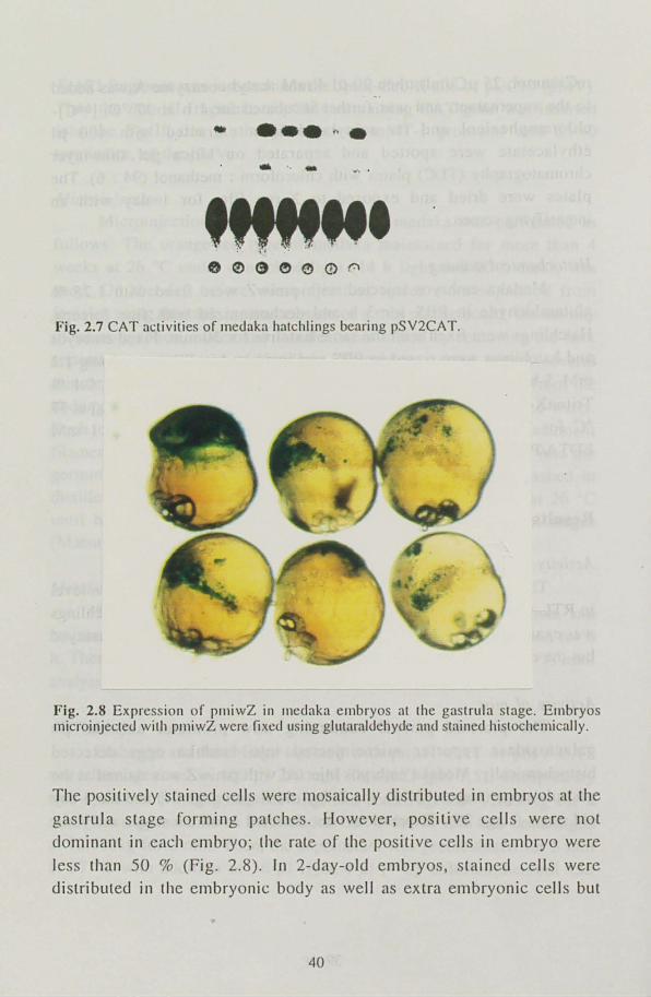
Activity of pSV2 vector The pSV2CAT which was the revealed the highest expression level
in RTL-4, was injected into medaka eggs and the expression in hatchlings
was examined. The expression was detected in most of hatchlings assayed but the expression level was rather low (Fig. 2.7)
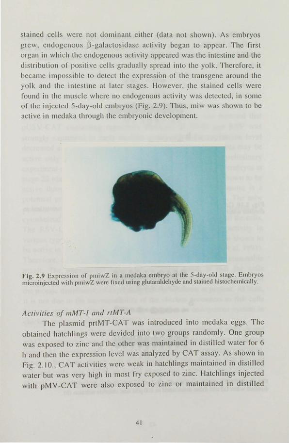
Activity of miw The plasmid pmiwZ contalllll1g miw promoter and the ~
galactosidase reporter microinjecLed into medaka eggs detected histochemically. Medaka embryos injected with pmiwZ was stained at the gastrula stage (stages 15-17), the 2-day-old stage (stage 24) and the 5-day old stage (stage 29). Expression was detected at all the stages analyzed.
39
- .... , . ... '. -
'"""' Fig. 2.7 CAT activities of medaka hatchlings bearing pSV2CA T.
Fig. 2.8 Expression or pll1iwZ in Il1cdaka embryos at the gastrula stage. Elllbryos rnicroinje<.:led with pJIliwZ wcre fixed using glutaraldehyde and stained histochemically.
The positively stained cells were mosaically distributed in embryos at the
gastrula stage forming patches. However, positive cells were not
dominant in each embryo; the rate of the positive cells in embryo were
less than SO % (Fig. 2.8). In 2-day-old embryos, stained cells were distributed in the embryonic body as well as extra embryonic cells but
40
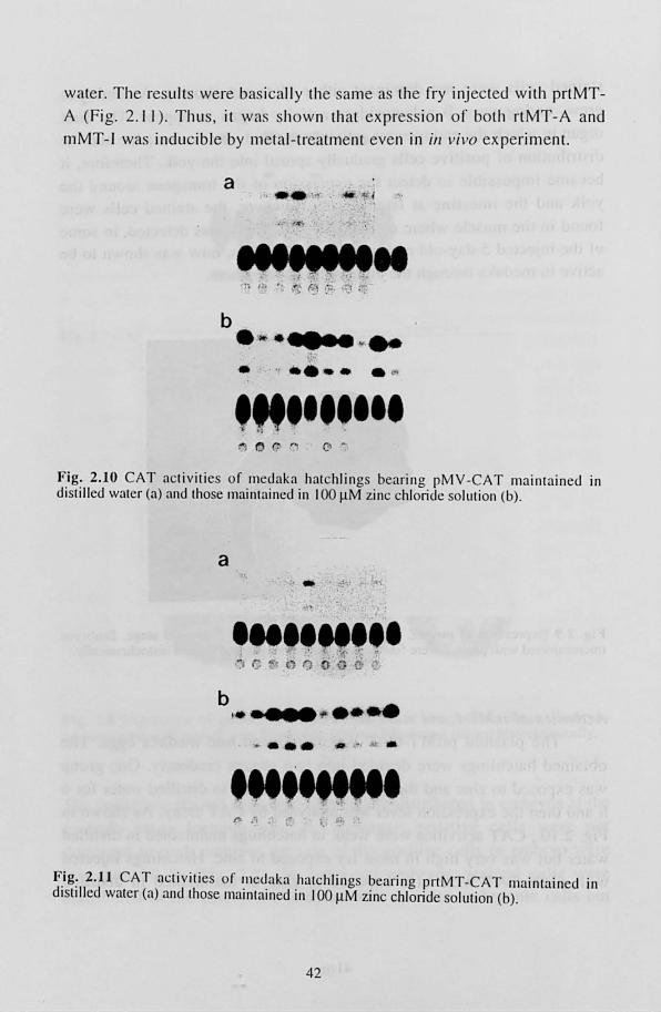
stained cells were not dominant either (data not shown). As embryos
grew, endogenous ~-galactosidase activity began to appear. The first
organ in which the endogen us activity appeared was the intestine and the
distribution of positive cells gradually spread into the yolk. Therefore, it
became impossible to detect the expression of the transgene around the
yolk and the intestine at later stages. However, the stained cells were
,"ound in the muscle where no endogenous activity was detected in some of the injected 5-day-old embryos (Fig. 2.9). Thus, miw was shown to be
active in medaka through the embryonic development.
Fig. 2.9 Expression or p11liwZ in a 11lc<.Iaka embryo at thc 5-<.Iay-ol<.l stagc. Embryos microinjected with pllliwZ were fixeu using glutamldehyue anu stained histochemically.
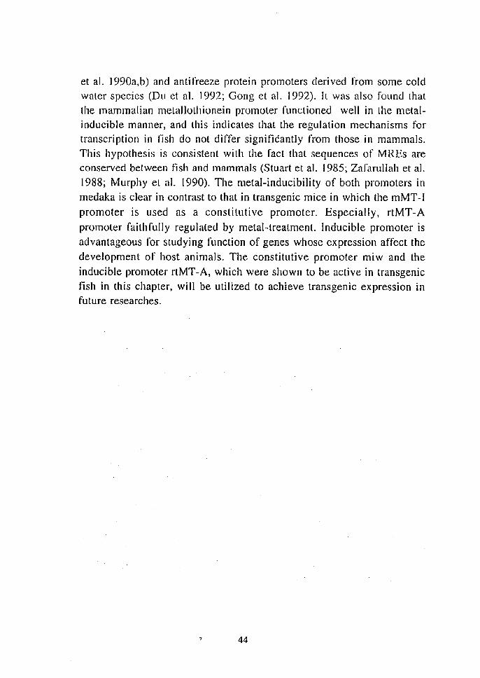
Activities oj IIIMT-I alld rtMT-A The plasmid prtMT-CAT was introduced into mcdaka eggs. The
obtained hatchlings were devidecJ into two groups randomly . One group
was exposed to zinc and the other was maintained in distilled water for 6
h and then the expression level was analyzed by CAT a 'ay. As shown in
Fig. 2.10., CAT activitie were weak in hatchlings maintained in distilled
water but wa' very high in Illost fry expo ed to zinc. Hatchlings injected
with pMV-CAT were also exposed to zinc or maintained in distilled
41
water. The results were bas ically the same as the fry injected with prtMT
A (Fig. 2. 11 ). Thus, it was hown lhat ex press ion o f both rlMT-A and
mMT-1 was inducible by metal-treatment even in ill vi vo ex periment.
a "-.'., ~ . /
. ~ ,JI!.. '. wi
b . s.- ..•.. " ... ~ ... . . , ..... '" ,"",,111
Fig. 2.10 CAT activiti es or medaka halchlings bearill g pMY -CAT maint ained In distilled water (a) and those maintained in I 00 ~M zinc chloride Soluli on (b).
a
.. INIIM" :' . . '".'... ) (; "' . n ') C v dr
b I ••••• .,. ....
. ... .. . ' .. '''''''''' • " 1 .. ,_, •
F.ig: 2.ll CAT a<.:tivities or lIledaka hatchlillgs bearing prtMT-CAT maintained III dlslilled waler (a) and those mainlained in 100 11M zinc chloride Solulion (b).
42

Discussion
In this section, ill vivo activities of SV2, miw, rtMT-A and mMT-I were examined using medaka embryos. The SV2 is the most popular expression vector in mammal cells. It was' also the most active element in RTL-4 cells as shown in the previous section. Thus, the low activity of the SV2 in medaka hatchlings was unexpected. Winkler et a!. (199 J) reported the expression of SV40 promoter-l3-galactosidase construct in
early medaka" embryos. Chong and Vielkind (1989) also reported that
pUSV -CAT containing regulatory elements of SV40 and RSV was
strongly expressed in early medaka embryos but the expression level decreased as embryos developed. Therefore, such viral elements may be
active only in early embryonic stages in fish. In fact, our preliminary experiment showed that pSV2CAT has considerable activity in embryos at
stage 22 (data not shown). In constrast to the SV2, miw was shown to be active through the embryonic development Thus this promoter is a potential promoter for constitutive expression of transgenes. The miw
contains the chicken l3-actin promoter and the RSV-LTR. The l3-actin is a cytoskeletal actin which is expected to be expressed in almost all the cells. The RSV -LTR is also known to have a strong promoter activity in various types or cells (Gorman et a!. 1 982b) and has also been shown to be active in goldfish and medaka (Yoon et al. 1990; Winkler et al. 1991).
Therefore, the strong activity of miw in fish embryos is a reasonable result. The unexpected resull, on the other hand, is that the cells expressing l3-galactosidase was not dominant in embryos. The reason of
the mosaic distribution of expressing cells is unknown at present. At least,
it is not due to the incompatibility of the chicken promoters to fish cells because similar results have been reported even in endogenous system in
chicken embryos (Naito et al. 1991). In contrast to these constitutive elements, expression of rtMT-A and
mMT-1 promoters was inducible by addition of zinc into the aquarium water. At present, rtMT -A is one of the few fish promoters available. Since it has been shown lhat it is active and inducible in fish fry and
cultured cells, this will be an useful expression system in transgenic fish as well as constitutive fish promoters such as carp ~~actin promoter (Liu
43
et al. 1990a,b) and antifreeze protein promoters derived from some cold water species (Du et al. 1992; Gong et al. 1992). Il was also found that the mammalian metallothionein promoter functioned well in the metalinducible manner, and this indicates that the regulation mechanisms for transcription in fish do not differ significantly from those in mammals. This hypothesis is consistent with the fact that sequences of MREs are conserved between fish and mammals (Stuart et a1. 1985; Zafarullah et al. 1988; Murphy et al. 1990). The metal-inducibility of both promoters in
medaka is clear in contrast to that in transgenic mice in which the mMT-1
promoter is used as a constitutive promoter. Especially, rtMT-A
promoter faithfully regulated by metal-treatment. Inducible promoter is
advantageous for studying function of genes whose expression affect the
development of host animals. The constitutive promoter miw and the
inducible promoter rlMT-A, which were shown to be active in transgenic fish in this chapter, will be utilized to achieve transgenic expression in future researches.
44
Chapter 3 Stage-dependent expression of the chicken 8-crystallin gene in transgenic medaka embryos
Transgenic technique is a powerful tool to study gene regulation ill vivo. In this chapter, expression of the chicken 8-crystallin gene during the embryonic development was examined using the microinjeclion system established in Chapter I.
Crystallins are major proteins of the lens of vertebrates and are
divided into four major subclasses: a-, ~-, 'Y- and 8-crystallins (Clayton 1974; Piatigorsky 1984). 8-Crystallin is present in birds and reptiles but in no other vertebrate classes. To study vertebrate crystallin genes, it is of
interest to detennine the consequence of introducing 8-crystallin genes into a vertebrate species which lacks this particular subclass. Extensive
studies in which the chicken 8-crystallin gene is introduced into mice have
shown that the chicken 8-crystallin gene is expressed in a tissue-specific manner in cell cultures of mouse embryos (Kondoh et a1. 1983; Hayashi
et al. 1985), transgenic mice (Kondoh et al. 1987), and chimeric mice (Takahashi et al. 1988). The introduction of the 8-crystallin gene into medaka has been auempted by Ozato et al. (1986). They detected expression in 7-day-old embryos but the detailed analysis of developmental regulation was not performed. In this chapter. the 8-crystallin gene was again introduced into medaka embryos and expression
was examined immunohistologically at several stages of development, from the lens formation stage to one day before hatching. Expression of
the gene was observed in the central fiber cells in the lens at the retina
pigmentation stage and in non-lens tissues at stages when the tissues
underwent differentiation.
Materials and methods
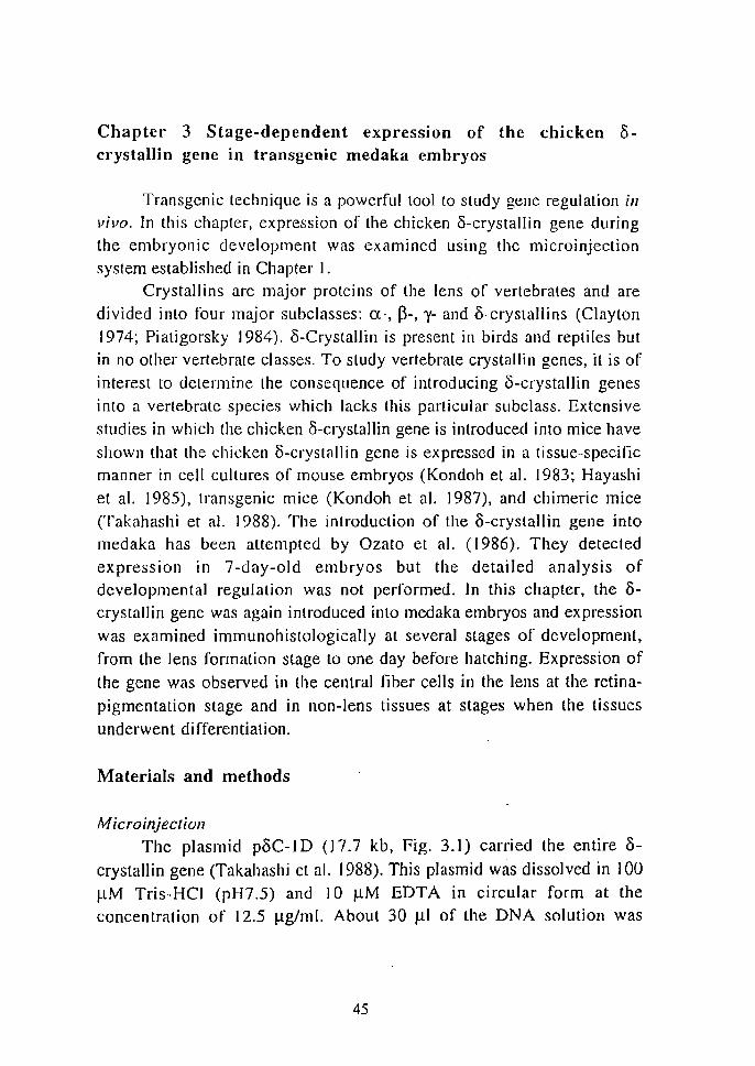
M icroinjeclioll The plasmid p8C-ID (J7.7 kb, Fig. 3.1) carried the entire 8-
crystallin gene (Takahashi ct al. 1988). This plasmid was dissolved in 100
~M Tris-HCI (pH7.5) and 1 0 ~M EDTA in circular form at the concentration of 12.5 ~g/ml. About 30 ~I of the DNA solution was
45
microinjected into the germinal vesicle of medaka oocytes 9 h before ovulation. Injected oocytes were cultured in the medium without gonadotropins, inseminated and incubated in distilled water as described
. in the chapter I. Embryos were staged according to Matsui stages (Matsui 1949; Yamamoto 1975) Normally developed embryos were sampled at 36 h (stage 22), 60 h (stage 27), 5 days (stage 29) and 7 days (stage 32) after fertilization. Stages 22 and 27 are characterized by lens formation and increasing pigmentation in the retina. The embryonic body encircles the yolk sac entirely at stage 29. Embryos at stage 32 are at one day before
hatching.
Xhol
EcoRI
EcoR Xbal
~~--- pAT153
Xhol ~
Fig. 3.1 Structure of the recombinant plasmid, pdC-1 D, injected into medaka oocytes. Solid bars represent exon DNA sequences of the chicken 15-crystallin gene. Open bars represent nanking and intron DNA sequences of the chicken o-clysiallin gene. Dolted bar represents the G4l8-resislance gene, STneo. Line indicates plasillid vector sequence derived from pAT! 53 including telra-cycline resistance gene (Tetr). The direction of transcription is indicated by arrows.
ImmWlOhistological staining
Sampled embryos were fixed with 4% (w/v) paraformaldehyde in phosphate buffered saline, embedded in paraffin and sectioned
longitudinally at a 5 ~m thickness. Sections were stained by avidin-biotinperoxidase complex (ABC) methods according to Hsu el al. (1981).
Briefly, sections were deparaffinized, hydrated and treated with 0.3 % H202 in methanol to block endogenous peroxidase activity and with normal goat serum, diluted 1:50 Lo reduce non-specific staining. Then sections were incubated sequentially with rabbit antiserum against 0-
46
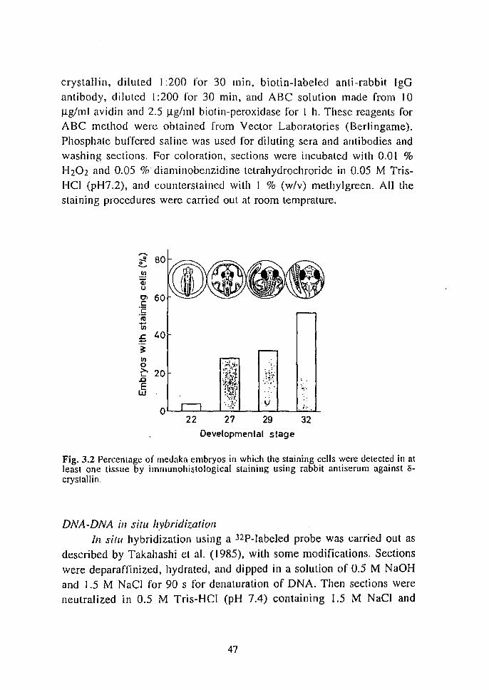
crystallin, diluted I :200 for 30 mIn, biotin-labeled anti-rabbit IgO
antibody, diluted 1 :200 for 30 min, and ABC solution made from 10 Ilg/ml avidin and 2.5 Mg/ml biotin-peroxidase for I h. These reagents for ABC method were obtained from Vector Laboratories (Berlingame).
Phosphate buffered saline was used for diluting sera and antibodies and
washing sections. For coloration, sections were incubated with 0.01 %
H202 and 0.05 % diaminobenzidine tetrahydrochroride in 0.05 M Tris
HCI (pH7.2), and countersLaincd with I % (w/v) methylgrecn. All the
staining procedures were carried out at room temprature.
iQ <II U
01 t:
:f nI -III .c 40 ::::
22
'.'~~: "
''''J " :7;!... ~!~;t-. :t.'t. 'Y"~ ~ ..
.>t V " ; ...
27 29 32 Developmental stage
Fig. 3.2 Percentage of lI1edaka embryos in which the staining cells were detected in at least one tissue by immunohistological staining using rabbit antiserulll against 0-crystallin.
DNA-DNA ill situ hybridization In sifLi hybridization using a 32P-labeled probe was carried out as
described by Takahashi et al. (1985), with some modifications. Sections
were deparaffinized, hydrated, and dipped in a solution of 0.5 M NaOH
and 1.5 M Nae] for 90 s for denaturation of DNA. Then sections were
neutralized in 0.5 M Tris-HCl (pH 7.4) containing 1.5 M NaCI and
47
transferred to 2xSSC (I xSSC is 0.15 M NaCI and 15 mM sodium citrate). After prehybrizidalion with 100 J.1g/ml denatured salmon DNA for 2 h at 68°C in 6xSSC containing 0.012% (w/v) heparin, 0.12 % (w/v) FicoH and 0.12 % (w/v) polyvinylpyrrolidone, sections were hybridized with 70 ng/ml p8C- I D probe in the same solution supplemented by 50 % formam ide. The p8C- I D probewas labeled with [32P]dCTP by nick translation. Hybridized sections were washed at 50°C with several changes of 2xSSC for I h and with O.lxSSC for 30 min and dryed. Then sections were exposed to X-ray film overnight for autoradiography.
Results
Outline of the expression in whole embryos Of 443 microinjected oocytes, 269 (61 %) were successfully
fertilized and 222(50%) survived until one day after fertilization. Among those surviving, 181 embryos developed normally until the time of sampling, while 41 were morphologically abnormal. Normall developed embryos, which were injected with the 8-crystallin gene, were sampled and stained immunohistologically at stages 22, 27, 29 and 32. The number of sampled embryos where 3-crystallin was detected in at least one tissue was I of26 (4%), 7 of 25 (28%), 8 of25 (32%) and 12 of 23 (52%) at stages 22, 27, 29 and 32, respectively (Fig. 3.2). The percentage of 3-crystalJin~positive embryos increased markedly with development. Embryos from stage 22; showing minimum number of tissues in which 3-crystallin was detected, were obtained from the same parental group ,as the embryos from stage 32, which showed the maximum number of them. No 8-crystallin was detected in non-injected embryos.
Expression. in the lens
8-Crysta11in in the lens was detected in three embryos at stage 27 (Nos.
27-5,27-12 and 27-]7) and not detected at any other stages (Fig 3.3).0-Crystallin was localized in the central fiber cells of the lens but not detected in' the epithelium (Fig. 3.4). This distribution pattern of the staining cells in the lens was common to these three embryos. The lens at
48
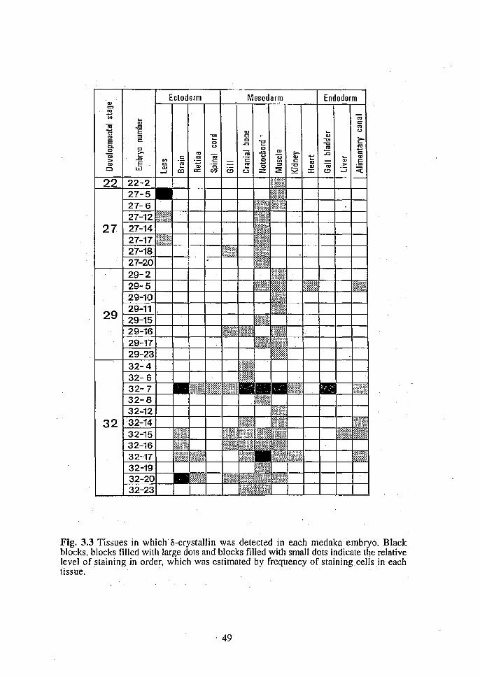
Ectoderm Mesoderm Endo rm Q)
C>
'" .... '" .... '" -;a 111 0::
..c '" c: E 111 .... U
111 => 1: 0:: '" ~ E c 0 0 -0 -0
C. u -'" 0 -0
'" 0 0 '" --~ '" -;; ..a::: '" ::- :c c a; ..c .5 . = -;a .2: u u '" 1::: .... '" > '" .s 0 <II 0:: CD .§ '" E t: E .... E "0 :::J -0 '" <ii >
CJ UJ 111 '" c. :2! S2
111 ::::; ::( ....J co a: en (!J u z ::c (!)
Fig. 3.3 Tissues in which a-crystallin was detected in each medaka embryo. Black blocks, blocks filled with large dots and blocks filled with small dots indicate the relative level of staining in order, which was estimated by frequency of staining cells.in each tissue;
. 49
this stage had features distinct from those at stages 29 and 32: many nuclei were observed in the central fiber cells at stage 27, while distribution of nuclei was confined to the epithelium at later stages.
Expression ill the non-lells tissues (i-Crystallin was detected for the first time in presumptive muscle
cells of the posterior parl of the embryonic body of one embryo (No. 22-2) at stage 22 (Figs. 3.3 and 3.5). At later stages, (i-crystallin was detected
in various types of tissues, which differed from embryo to embryo. At stage 27, (i-crystallin was detected in a few mesodermal tissues, such as
the notochord, muscle, and gill (Fig. 3.3), though the staining cells were small in number in each tissue. It was noted that all three embryos expressing the exogenous gene in the lens also exhibited expression in the notochord or muscle. At stage 29, (i-crystallin was detected in the muscle, notochord, heart, cranial bone, skeleton of the gill, and alimentary canal (Fig. 3.3). Thus, 8-crystallin in these two stages was detected mainly in mesodermal tissues, especially in the muscle and notochord. At stage 32, number of staining tissues increased dramatically (Fig. 3.3). 8-Crystallin was detected in the brain (Figs. 3.6 and 3.7a), retina (Fig. 3.6), spinal cord (Figs. 3.6 and 3.7b), gill, cranial 1;>ones (fig. 3.7c), notochord (Fig. 3.6), muscle (Figs 3.6 and 3.7d), kidney (Figs. 3.6 and 3.7e), gallbladder (Fig. 3.6), liver (Fig. 3.7f) and alimentary canal (Fig. 3.6). [n the brain, the staining cells were found in the prosencephalon, mesencephalon, including the optic lobe, and rhombencephalon, including the cerebellum and medulla oblongata. Thus, at stage 32, the central nervous system was one of the major tissues of expression. The staining cells in the gill were
confined to the mesodermal part, the skeleton and mesenchymal tissues. In the muscle, the rate of the staining cells to non-stainng cells varied
from embryo to embryo. For example, the staining cells accounted for
about 20 % of all muscle tissues in embryo No. 32-7 (Fig. 3.6) while only one muscle fiber was stained in embryo No. 32-14 (Fig. 3.7d). The
increase in the number of 8-crystallin-positive embryos at later stages can
be ascribed to an increase in the staining tissues of non-lens types.
To sum up these results in the lens and non-lens tissues: i) in the lens, 8-crystallin was detected in the central fiber cells at stage 27 in a
50
stage dependent manner; ii) in non-lens tissues, o-crystallin was rarely
detected at stage 22, while it was detected mainly in mesodermal tissues such as the notochord and muscle at stage 27 and 29, and in many ectodermal and endodermal tissues at stage 32. This indicated that the expression in non-lens tissues was also stage-dependent and the stage of the expression was variable depending on the tissue type.
Expression ill abnormal embl)'os Of 41 abnormally developed embryos,S and 9 embryos were
sampled 5 and 7 days after fertilization, respectively. Thirteen of these 14 embryos developed a dwerfish embryonic body, and 1 formed an
undifferentiated cell mass without forming the embryonic body. Three of 13 dwerfish embryos Failed to form eye vesicles. 8-Crystallin was detected in various tissues of all 14 embryos examined. In the lens, it was
detected in 2 of the 5-day-old embryos (Fig. 3.8) and 2 of the 7-day-old
embryos. Thus, 4 of 10 abnormal embryos which developed lenses expressed the 8-crystallin gene in them. The lenses of these 4 embryos
had many nuclei in inner part, as found in the normal embryos at stage 27, but Lhey were not isolated well from the retina; the one exception was
a 5-day-old embryo which exhibited normal-shaped lenses, similar to those found in a stage 27 embryo (Fig. 3.8). Abnormal embryos that were not sampled failed to hatch and died within 20 days after
fertilization.
Detectioll of the DNA sequence To confirm the existence of the introduced DNA sequence, DNA
DNA in situ hybridization was carried out on several sections of four embryos at stage 32. One was an injected embryo in which 8-crystallin was detected (No. 32-7). Two were injected embryos in which 0-crystallin was not detected (Nos 32-1 and 32-3) and one was a nOI1-
injected control. The DNA sequence was detected in the 8-crystallinpositive embryo, but not in the other three. In the former embryo, the
injected sequence was ditributed throughout the section (Fig 3.9).
51
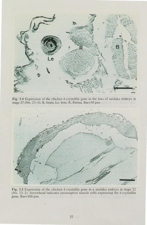
Fig. 3.4 Expression or the chicken I)-crystallin gene in the lens of Illeuaka embryo at stage 27 (No. 27-5). 13, brain; Le, lens; R, Retina. Oar=50 Illll .
Fig. 3.5 Expression of the chicken I)-crystallin gene in a meunkn embryo at stnge 22 (No. 22-2). Arrowheau indicates presllmptive muscle cells expressing the I)-crystallin gene. Bar=IOO j..lIn .
52
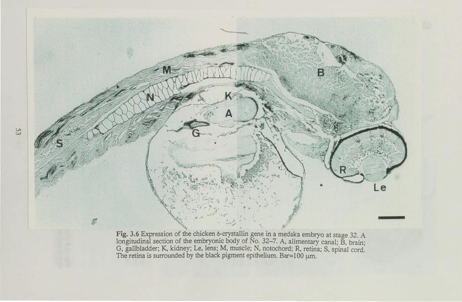
V1 :..,.)
"'------..
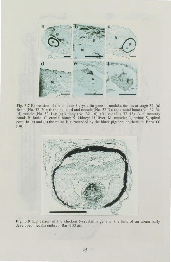
c ·
--, --- -
A r
F ig. 3.7 Expression of the chicken Ii-crystallin gene in medaka tissues at stage J2. (a) Brain (No. J2-20); (b) pinal cord and Illuscle (No. J2-7); (c) cranial bone (No. J2-6); (d) muscle (No. l2-14); (e) kidney (No. J2-16); U) liver (No. J2-15). A. alimentary canal; B. brain; C. cranial bone ; K, kidney; Li . liver; M, muscle; R. retina; S, spinal cord. In (a) and (e) the retina is surrounded by the black pigment epilherium. Bar=IOO ~1l1.
Fig. 3.8 Expression of the chicken Ii-crystallin gene in the lens of an abnormally developed mcdaka embryo. Bar= I 00 ~Jl1.
54
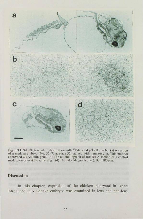
a
Fig. 3.9 DNA-DNA ill ,filII hybridization with nP-labeled poC-1D probc. (a) A scction of a Il1cdaka cmbryo (No. :12- 7) at stage 32, staincd with hcmatoxylill. This embryo exprcssed o-crystallill gene: (b) The autoradiograph of (a); (e) A section of a control Illcdaka el1lbryo at the same stage: (d) The aUloradiograph of (c). Bar=J 00 JlI1l.
Discussion
In lhi chapter exprcslon of thc chicken 8-cryslallin gene
introduced into medaka cmbryo was cxamined in IClls and non-lens
55
tissues at different stages of development. In the lens, a-crystallin was detected in 3 of 25 embryos at stage 27 but not in embryos at other stages. The protein was also detected in the lens in 4 of 10 abnormal embryos. The lenses of these embryos were morphologically similar to those of a normal embryos at stage 27. It'is, thererore, likely that the acrystallin expression in the lens is stimulated in a stage-dependent manner
when the lens reaches a certain state of differentiation. Interestingly, that a-crystallin was localized in the central fiber cells and not detected in the newly differentiating epitherial cells. In the chicken, a-crystallin synthesis
occurs mainly in the embryonic lens. With maturation of the lens, synthesis of a-crystallin first decreases in the epithelial cells and subsequently is confined to the central regions of the fiber mass (Piatigorsky, 1981; Treton et al. 1982). The localized distribution of acrystallin within the medaka lens may be due to the fact that the a
crystallin expression had occured in the medaka lens shortly before stage 27 and ceased in the newly differentiating epitherial cells. A similar phenomenon was reported in transgenic mice carrying the y2-crystallin gene promoter fused to the coding region of the bacterial ~-galactosidase gene (Goring et al. 1987). The enzyme activity was detected in the central
nuclear fiber cells of the lens in these mice when they were examined cylochemically at the age of 6 weeks. It is noted that y2-crystallin is also an embryonic type of crystallin, similar to a-crystallin. At the present time, it is impossible to make a comparison between the exogenous crystallin gene and endogenous ones, with regard to the developmental pattern of expression in the medaka lens, because there have been no developmental investigations of fish crystallins. Our preliminary observations that crystallin polypeptides were detected from 5 days after fertilization in the normal medaka lens may shed some light on this problem. No a-crystallin was detected in any region of the medaka lens at stages 29 and 32, although it was detected in various other tissues of many
embryos. It is assumed that the 8-crystallin accumulated in the central fiber cells at earlier stages is decomposed during these stages. a-Crystallin may be unstable in the heterologous lens which naturally lacks it,
although it remains in the chicken lens at all stages of development and maturation (Piatigorsky 1984).
56
The finding that the o-crystallin gene expression in non-lens tissues is stage-dependent, as is found in the lens tissue, was unexpected. 0-Crystallin in non-lens tissue was first detected in a small region of undifferentiated mesodermal tissues in one embryo at stage 22. This low level of expression cannol be due to the absence of the exogenous gene,
. because the specimens althis stage were sampled randomly from the same
experimental groups as those at stage 32 which showed the highest level of expression. Similarly, restricted localization of the staining cells is not due to localization of the injected sequences, because the homogenous distribution of the injected sequences throughout the tissues was shown by DNA-DNA in situ hybridization (Fig. 3.9; see also Ozalo et al. 1986). Subsequently, o-crystallin was detected mainly in the notochord and muscle, and later in various tissues including ectodermal and endodennal tissues. This stage-dependent expression in non-lens tissues implies that a
cellular condition inducing gene expression appears at different stages in each tissue. The most probable condition for this may be tissue differentiation, and it is likely that the stage-dependent paLLern of the gene
reflects the temporal order in tissue differentiation among the non-lens tissues. Thus, the condition which induces the non-lens type gene
expression may rarely be present in undifferentiated tissues at stage 22, while it may occur with progress of tissue differentiation first in mesodermal tissues at stages 27 and 29, and successively in ectodermal and endodermal tissues at stage 32. In fact, mesodermal tissues, such as notochord and muscle, are the earliest tissues undergoing differentiation in medaka embryos (Hiraki and Iwamatsu 1979). However, correlation between o-crystallin expression and tissue differentiation must be confirmed more exactly by further experiments at the cellular level.
The differentiation-dependent expression of the exogenous 0-crystalli n gene has been found in mouse teratocarcimoma cells carrying this gene. In solid tumors formed by injection of these cells into the mouse peritoneal cavity, the gene expression is not detected as long as the cells are in undifferentiated state, but is detected in a differentiated
popUlation of cells such as skeletal muscle cells and secreting epitherial cells (Kondoh eL al. 1984; Takahashi et aI. 1988). A similar phenomenon has been reported in endodermal cells which differentiated from
57
teratocarcinima cells in the presence of retinoic acids (Goto et at. 1988). Thus, the idea lhat o-crystallill expression in non-lens tissue of medaka embryos depends on di fferentiation is supported by these experiments on
mouse teratocarcinoma cells. It is an interesting question whether'there are any differences in the
regulation of o-crystallin expression between the lens and non-lens tissues in medaka embryos. A low level of the 8-crystallin expression has been observed in various non-lens tissues of chick embryos, as well as in heterologous experimental systems of mouse carrying this gene. Hayashi et al. (1985) demonstrated, by line deletion analysis in mouse lens cells and fibroblasts, that the o-crystallin expression could be classified into lens and non-lens types. For instance, dependence of o-crystallin expression on the 5' franking sequence of the gene differs slightly between lens and non-lens cells of the mouse (see also Kondoh and Okada 1986). An enhancer was identified in an intron of the o-crystallin gene which has both lens-specific and apparently non-specific components (Hayashi et al. 1987). It may thus be reasonable to assume that 0-crystallin expression in the lens and non-lens tissues of medaka embryos
is due to the activation of lens-specific and non-specific components of the enhancer, respectively, and that the activation of the non-specific components is differentiation-dependent.
In this chapter, stage-specific expression of the chicken o-crystallin
gene was elucidated. Results obtained in this experiment indicated that detailed studies on gene regulation is possible using medaka. Among the diversed species of fish, medaka is especially advantageous as a host of foreign gene transfer. This medaka system has the potential of becoming one of the most promising materials for in vivo studies of gene regulation in vertebrates.
58
Chapter 4 Application of foreign gene transfer techniques to fish breeding
The fish cultivation has grown prosperous in recent years. Unlike
farm animals and plants, however, few special strains for aquaculture have been developed through traditional breeding because fisheries have
largely been dependent upon the natural population for a long time. Efforts are now being made to produce domestic fish strains which have advantageous features for aquaculture using not only traditional means, but also novel methods such as hormone treatment, chromosome
manipulation and interspecific hybridization (Donaldson 1986). Techniques for foreign gene transfer are also expected as new methods to breed economically important species. In this chapter, the author attempted to enhance the growth of rainbow trout by introducing growth
hormone gene (eDNA) as a model experiment of fish breeding by foreign
gene transfer. Rainbow trout is one of the most popular species of fish for aquaculture and is also Llsed as an experimental animal.
4.1 The method for introducing foreign genes into rainbow trout
Eggs of rainbow trout are enough large for embryonic manipulation but are covered with the thick chorion which prevent the insertion of the micro pipettes for microinjection. To penetrate the
chorion, micropipets should be more than 15 11m in diameter. To solve this problem, several methods have been reported. Chourrout et a1. (1986) devised the "two-step" method. In this method, the micropipets are
inserted through the window on the chorion made by thick needles. By this method, foreign genes can be transferred into rainbow trout but it
requires much time and skilled technique. Another method has been reported by Yoshizaki et al. (1989): the hardening of chorion is prevented by the glutathione treatment after insemination. It is also
possible to penetrate the chorion using micropipets which are more than
10 J.lm in diameter (direct microinjection). As the first step of the
59
experiment, foreign gene transfer was attempted lIsing the glutathione methods and its gene transfer efficiency compared with that of the direct microinjection without glutathione treatment.
Materials and methods
Preparation of plasmid The plasmid pMV-GH (Fig. 1.1) containing the mMT-I promoter,
rainbow trout growth honnone eDNA and the po\yadenylation sequence of SV40 was linearized with EcoRI and dissolved in phosphate-buffered saline at the concentration of 100 J..lg/ml.
Gamete collection, insemination and microinjection Trout eggs and milt were collected in January 1989 from 2-year
old fish. About 500 eggs were mixed with 5 ml milt, 300 ml 120 mM NaHC03 at 10°C was added immediately and allowed to stand for I min. The eggs were gently rinsed with water several times to remove excess milt and allowed to swell for 3 h at LO °C in water or in 0.5 mM glutathione (reduced form) the pH of which was adjusted to 10 with I N NaOH. After swelling, about 10 01 of DNA solution was microinjected
into the cytoplasm of fertilized eggs with micropipets which are about 5 J..lm or 15 J..lm in external diameter. Injected eggs and uninjected control were rinsed with water and maintained in circulating water at about 10 DC until hatching.
DNA analysis Hatchlings were anesthetized with 2-phenoxyethanol. The yolk sac
was torn with fine forceps in Dulbecco's phosphate-buffered saline to remove the yolk. Genomic DNA was prepared using Proteinase K-SDS treatment followed by phenol extraction and ethanol precipitation. The
existence of foreign DNA was detected using polymerase chain reaction (PCR) as follows: One hundred nanograms of DNA was dissolved in Tth buffer containing 6 ~g primer GH7 (5 '-GGe TCT TCA ACA TeG CGG
TC-3'), 6 flg primer GH8 (5 '-TCT TGT TCA GCT GTC TGC GT-3 '),
60
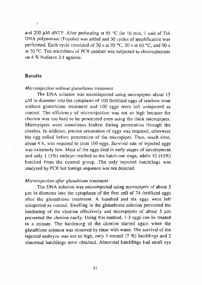
and 200 ~M dNTP. After preheating at 95 °C for 10 min, I unit of Tth DNA polymerase (Toyobo) was added and 30 cycles of amplification was
performed. Each cycle consisted of 30 s at 95 °C, 30 s at 60 °C, and 90 s
at 70 0c. Ten microliters of PCR product was subjected to electrophoresis on 4 % NuSieve 3: 1 agarose.
Results
Microinjectioll without glutathione treatment The DNA solution was microinjected using micropipets about 15
~M in diameter into the cytoplasm of IOO fertilized eggs of rainbow trout without glutathione treatment and 100 eggs were left uninjected as
control. The efficiency of microinjection was not so high because the chorion was too hard to be penetrated even using the thick micropipets. Micropipets were sometimes broken during penetration through the
chorion. In addition, precise orientation of eggs was required, otherwise the egg rolled before penetration of the micropipet. Thus, much time,
about 4 h, was required to treaL 100 eggs. Survival rate of injected eggs
was extremely low. Most of the eggs died in early stages of development
and only I (I %) embryo reached to the hatch-out stage, while 10 (IO%)
hatched from the control group. The only injected hatchlings was
analyzed by PCR but foreign sequence was not detected.
Microinjeclioll after glutathione treatment The DNA solution was microinjected using micropipets of about 5
~m in diameter into the cytoplasm of the first cell of 74 fertilized eggs after the glutathione treatment. A hundred and six eggs were left uninjected as control. Swelling in the glutathione solution prevented the
hardening of the chorion effectively and micropipets of about 5 ).lm prevented the chorion easily. Using this method, 1-3 eggs can be treated
in a minute. The hardening of the chorion started again when the
glutathione solution was removed by rinse with water. The survival of the injected embryos was not so high, only 5 normal (7 %) hatchlings and 2
abnormal hatchlings mere obtained. Abnonnal hatchlings had small eye
61
and b dies. Fr m control egg 23 (22 '}( ) hatched out norm all y. The
glutathi ne . luti on did not . e m to a f~ ct the d el pment f embryo al
lea I for til contr I group. 0 A analY' i wa p rl" rmed on 5 norm al
and 2 abnormal hatchling injec ted and 2 hatchling uninjected (Fig. 4.1 ) .
The GH7 and GH 8 primer. u ed would amplify nGH cD A from th
porti on of + 0 to + 193 r lati ve to th e point o f the tran lati n initi ati on
and gen rat a 11 4-bp fragment. In th genomi GH equen e, GH7 and
GH 8 are 10 at d in second and third exon , r pec ti vely, and a 138-bp
il1lron ex i I between th em (A g II n et al. 1988a). Th refor , the e
primer g nerat a 2S2- bp fragment when they amplify genomic GH
g ne. The for ign s quence can be di tingui h d ea il y from the crenomic
GH equence of the rec ipient. In Fig. 4. 1, th 11 4-bp band corre ponding
to the tran gen wa det ct d only in th .4 hatchling while the 2S2-bp
band corre ponding t the genomic GH gene wa bserved in all th
injected and ontrol ample '. Thi ev idence indi ca te th at I o f 5 inj cled
hatchlings wa ' found to have the tran gene. Thu it wa ' hown that
foreign gene tran. fer is po' ible u ing the glutathion meth d.
123456789P
Fig. 4.1 DNA analy i of rainbow trout fry microinjected with pM V-GH . Total DNA wa extracted from :> normal injected fry (Lane t -5),2 abnormal injected fry (lane 6, 7) and control fry ( lane 8, 9) were amplified by polymera e chain reaction u ing primer GH7 and GH 8. Amplified products were electrophoresed on a 4 % agaro e gel.
62
Discussion
In this section, two methods; direct microJnjeClion and the
glutathione method was examined. The microinjection into the cytoplasm itself is possible by both methods. However, the results obtained by direct
microinjection was quite poor and no positive hatchling was obtained. The glutathione J'nethod, on the other hand, worked beller; the high survival rate and a positive hatchling was obtained. Although, the gene transfer efficiency was far [rom satisfaction. The gene transfer efficiency is affected by the survival rate and the proportion of the positive embryos among survived embryos. In this case, it seems that the problem is the survi val rate rather than the proportion of positive embryos. Generally the survival of the embryo is largely dependent upon the quality of gametes. And it is said that differences in survival rate between the eggs of high and low qualities are more remarkabIly recognized after microinjection (G. Yoshizaki, personal communication). Quality of fertilized eggs used in this study did not seem to be so high because the rates of hatch-out in uninjected control were relatively low (10 and 22 %
for the two experiments) for rainbow trout which has been adapted to artificial culture conditions. Therefore, the poor rate in this experiment
may be due to the egg quality. The quality of eggs are affected by various
factors such as the genetic back grou.nd of parental fish and the health but most important factor is the timing to collect the unfertilized eggs. The
egg quality is the best just after ovulation and is lost day by day. During the spawning season selection of ovulated females should be carried out frequently. In this experiment, two abnormal hatchlings also appeared. The abnonna1 phenotype is often caused the insertion of transgenes into the functional genes of not only by the expression products of transgenes but also by the host (Palmiter and Brinster, 1986). In this case, however, transgene was not detected from the DNA of abnormal hatchlings. Dwarfish phenotype is often observed in embryos reared in commercial hatcheries. Thus, it was concluded that these abnormality was not due to the transgene at least in this case although it may be due Lo the embryonic
manipulation with micropipets.
63
4.2. Activity of miw in rainbow trout
Rainbow trout has been often used for transgenic experiments
(Chourrout el al. 1986; Rokkones et al. 1989; Guyomard et al. 1989;
Penman et al. 1990). However, none' of them have achieved the
expression except for Rokkones et al. (1989) in which mRNA derived from the exogehous GH gene was detected by northern blot hybridization. The lack of expression is supposed to be due to inappropriate regulatory elements used. The choice of regulatory element to drive the coding region is the most important point to achieve the expression of the introduced gene. In Chapter 2, several active regulatory elements were described in medaka. Among them, the chimeric promoter miw was chosen to drive the GH eDNA in rainbow trout because it is a
constitutive element which requires no induction for expression and its
activity is expected to continue for longer period than that of the pSV2 vector. Before the use of this promoter to express the growth hormone,
its activity in rainbow trout was examined using the ~-galactosidase gene
as a reporter.
Materials and methods
Gamete collection, insemination and microinjection The microinjection experiment was carried out in April, 1990, out
of the normal spawning season of rainbow trout of a winter spawning
strain in December, 1989 and cryopreserved according to Stoss and
Refstie (1983). The collected milt was diluted with 3 ml of the extender containing 0.3 M glucose and 10 % dimethylsulfoxide. The sperm extender suspension was frozen as described by Stoss and Holts (1981). Frozen pellets were stored in liquid nitrogen until insemination. For
fertilization, the pellets were dissolved in 120 mM NaHCO] solution at 10
°C and immediately added to 350 unfertilized eggs which were obtained
from a female trout of a spring spawning strain produced by being kept under the artificially controlled photoperiod. To prevent hardening of the
chorion, inseminated eggs were rinsed and swelled 3 h at 10°C in 0.5
64
mM glutathione (reduced form) adjusted to pH 10 with I N NaOH. The fertilization rate estimated by fixing a part of the inseminated eggs with Bouin's fixtalive 12 h after fertilization and counting eggs at the cleavage
stage, was 80 %. About 10 nl of the DNA solution containing the plasmid
pmiwZ (Fig. 2.7) linearized with KpnI (l00 ~g/ml) in phosphatebuffered saline was microinjecled into the cytoplasm of eggs with a
micropipet (5 ~m in external diameter) attached to a micromanipulator (Narishige Model M-152, Narishige Scientific Instrument Lab., Tokyo
Japan) under a dissecting microscope, 3-5 h after fertilization. Injected
eggs were rinsed with distilled water and maintained in circulating water at 15°C.
Southern blot hybridization Genomic DNA was prepared from whole bodies of individual fry
30 days after fertilization as follows: Fry were homogenized in the lytic solution containing 50 mM Tris-HCI (pH 7.5), 10 mM EDTA, 0.5 %
SDS, 500 ~g/ml Proteinase K and incubated for 5 hat. 55°C. Then samples were extracted twice with equal volume of saturated phenol and then several times with phenol: chloroform: isoamyl alcohol (24 : 24 : I).
The aqueous phase was precipitated with ethanol and dissolved in TE (l0 mM Tris, I mM EDTA). Isolated DNA was digested with Xbal,
electrophoresed on 0.7 % agarose gels, and transferred onto nylon
membranes. The DNA on membranes was hybridized with the 3 kb BamHI fragment of pmiwZ containing the coding region of the pgalactosidase gene labeled by random priming with [32P] dCTP, and
exposed to X-ray film.
Histochemical detectioll oj j3-galactosidase activity Embryos were fixed with 1.25 % glutaraldehyde in PBS for 3 h
and dechorionized with fine forceps. Hatchlings were fixed with the same
fixtative for 30 min. Fixed embryos and hatchlings were rinsed in PBS and incubated in PBS containing 1.2 ruM 5-bromo-4-chloro-3-indolyl-f3-
D-galactopyranoside (X-gal), 0.1 % TritonX-lOO, I mM MgCh, 6 ruM
K4 [Fe(CN)6}, 6 ruM K3[FeCN)6] at 37°C for 3 h. The embryos and
65
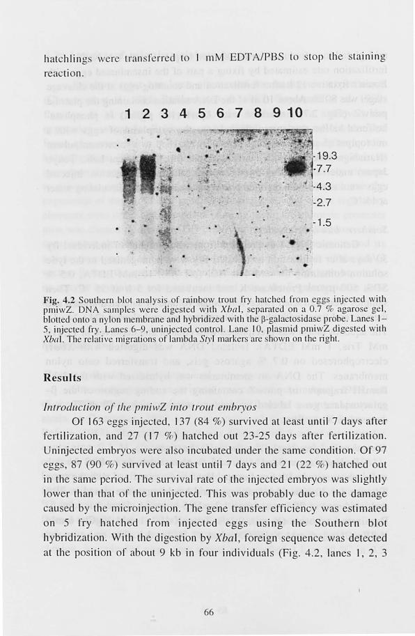
hatchling ' weI' transferred t 1 mM EDTA/PBS to stop th e tainin g
r ac ti n.
1 2 3 4 5 6 7 8 9 10
• • 19.3 ••
-7.7
-4.3
. -1.5 • .. . !I
•
Fig. 4.2 Southern blot analy is or rainbow troll t fry hatched fr m egg injected with pmiwZ. DNA '''lInp les were digested with Xbal, eparated on a 0.7 % agarose gel , blotted onto a nylon membrane and hybridized wi th the p-galacto ida e probe. Lane 1-5, injected fry. Lane 6-9, uninj cted con tI' I. Lane 10, plasmid pmiwZ dige ted with Xb{/ 1. The relati ve migrations of lambda 51) I marker are . hown on the right.
Results
Introduction of til e pll/illZ into trOllt ell/br.)Os or 163 gg. injected, 137 (84 %) 'urvived at I a t until 7 day aft r
r rtili za tion and 27 ( 17 %) hatched out 23-25 days after r rtili za ti on.
Uninjected mbryo. were al 0 incubated under the. ame condition. or 97
egg. , 87 (90 ~) urvived at lea t until 7 days and 2 1 (22 ~) hatch d out
in the ame period. The urvi va l rate of the inject d embryo. was lightly
lower th an th at of th uninj cted. This wa probably due to the damage
cau ed by the microinj ec ti on. The gene tran fer effi ciency wa e timated
on 5 rry hatched from inj ected egg u in g the Southern blot
hybridi zation. With the dig tion by Xbal, foreign sequence wa detect d
at the po ition or about 9 kb in four individual (Fig. 4.2, lanes I , 2, 3
66
and 5). No signal was detected in uninjected fry. These resulls indicate the existence of the injected sequences in the each positive fry. However, it is unknown whether the foreign sequences were integrated into the
chromosome or not because no junction fragment was detected in positive individuals. The copy number of the lrahsgene were less than one per genome in all the positive fry. This may suggest the mosaic distribution of introduced sequences in each fry. The mosaic distribution of transgene has been reported in previous studies (Ozalo et al. 1986; Stuart ct aJ. 1989, 1990; Guyomard et al. 1989).
Histochemical detectioll oj f3-galactosidase in el1lbl),os alldfry
To detect the expression of the p-galactosidase gene, ) 0 embryos and 10 fry were sampled 7 and 30 days after fertilization, respectively, and stained histochemically. Of the 7-day-old embryos, 6 were morphologically normal, forming the embryonic body. p-Galactosidase activity was detected in all the nonnal embryos (Fig. 4.3a). Positively
stained cells were distributed mosaically in different types of cells in the embryonic body and rarely in the cells on the yolk sac. The frequency
and the distribution pattern of the expressing cells were different from embryo to embryo. The other four embryos were morphologically abnormal and failed to form the embryonic body (Fig. 4.3c). Since the
similar type of abnormality was frequently observed in control embryos, it is considered that the abnormality was due to the egg quality, but not to the effect of transgenes. Of the four abnormal embryos, one embryo was positively stained (Fig. 4.3c), in which stained cells formed masses in the embryo. No staining was observed in normal and abnormal embryos from un injected eggs (Fig. 4.3b, c) Thus, positive staining in the embryos at this stage is considered due to the expression of the transgene. Of the 10 fry sampled, 9 were morphologically normal while I has a slightly curved body (No.3 in Fig. 4.4). Upon the histochemical staining, the yolk sac of 3 normal hatchlings (Nos. I, 2 in Fig. 4.4) and the abnormal
hatch1ing (No.3 in Fig. 4.4) were intensely stained. No staining was
observed in any other organs except the intestine which was weakly stained in both the injected and uninjected hatchlings. This slaining
67
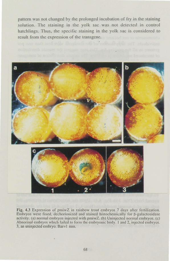
pattern was not changed by the prolonged incubation of fry in the staining
solution. Tile staining in the yolk sac wa not detected in control
hatchlings. Tllus, the specif'ic staining in the yolk sac is considered to
result from the expression or the transgene.
Fig. 4.3 Exprcssion or pmiwZ in rainbow trout cmbryos 7 days artcr fertilization. Embryos wcrc fixcd. dccilOrionizcd and staincd histochcll1ically for p-galaclosidase activity. (a) normal cmbryos injcctcd wilh pllliwZ. (b) Uninjccted normal cmbryos. (c) Abnormal embryos which railcd to rOrJIlthc embryonic body. J and 2, injected embryos. 1, an uninjeclcd cmbryo. Bar=1 1111ll .
68
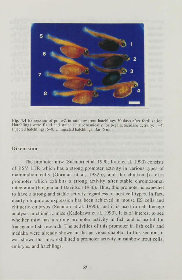
Fig. 4.4 Expression of pllliwZ in rainbow trOllt hatchlings 30 days after fertilization. Hatchlings were fixed and stained histochemically for p-galactosi<.lasc aClivity . 1-4, Injected hatchlings. 5-8, Uninjcc\e<.l hatchlings. Bar=5 111 Ill.
Discussion
The promoter miw (Suemori el al. 1990; Kalo et al. 1990) consists
of RSV L TR which has a strong promoler activity in various types of
mammalian cells (Gorman et aI, 1982b), and the chicken ~-actin
promoter which exhibits a strong activity after stable chromosomal integration (Fregien and Davidson 1986). Thus, this promoter is expected
to have a strong and stable activity regardless of ho t cell types. In facl,
nearly ubiquitous expression has bcen achieved in mousc ES cells and
chimcric cmbryos (Suemori et a!. 1990), and it is uscd in cell lineage
analysis in chimeric mice (Kadokawa et al. 1990). It is or interest to see
whether l1liw has a strong promoter activity in fish and is useful for
transgenic fish research. The activities of this promoter ill lish cells and
mcdaka were already shown in the perviolls chapter. In this section, it
was shown that miw exhibited a promoler activity in rainbow trout cells,
embryos, and hatchlings.
69
To analyze the ill vivo activity of miw in rainbow trout, we used bacterial p-galactosidase gene as a reporter. The p-galactosidase reporter is widely used in studies on developmental gene regulation, because the expression can be visualized by histochemical staining. The histochemical method has been used mainly in mouse arid Drosophila embryos which is relatively small in size. Though the rainbow trout embryos and hatchlings are far larger in size, they were effectively stained. The endogenous pgalactosidase activity was not so high to give a serious obstacle for analysis of gene expression. Thus, the p-galactosidase gene is also useful in trout embryos and hatchlings. This reporter is considered to be a powerful tool in characterizing various promoter in fish.
The microinjection of pmiwZ was carried out in April, out of the nonnal spawning season of rainbow trout in Japan. The limited spawning season is a serious obstacle for gene transfer experiments. To overcome this, we used cryopreserved milt in the present study. The technique for the cryopreservation of the salmonid milt has already been established (Stoss and Refstie 1983). On the other hand, since it is impossible to storage eggs by freezing at present, we used eggs obtained from a female trout matured under the artificially controlled photoperiod. Induction of the maturation by an artificial photoperiod is commonly carried out in
Japan and other countries. Using these methods, it become possible to obtain fertilized eggs out of the spawning season. The problem is the
quality of the fertilized eggs. Survival rates of the eggs obtained using such methods are sometimes lower than those of eggs naturally obtained.
In fact, the hatchling rate of both injected and uninjected embryos in this study was not high, 17 % and 22 %, respectively.
After microinjection, embryos and hatchlings were sampled 7 and 30 days after fertilization, respectively. In 7-day-old embryos, miw showed a strong promoter activity. The staining which showed pgalactosidase activity was detected in various types of cells, almost ubiquitously. The stained cells were distributed mosaically. Although the mosaic distribution of the introduced sequences in transgenic hatchlings
was suggested by the fact that the copy number of the transgene in the positive individuals was less than one per genome as shown in Fig. 4.2, it is unknown whether the expression patterns in the present study renects
70
distribution of the transgene or difference in its expression among
different ce[ls. [n hatchlings, the expression was detected in the yolk sac
and not in any other tissues. Since miw is constructed from the promoters
and an enhancer which arc active regardless of host cell types, the tissue
specific expression was unexpected. Similar phenomenon has been also in
a homologous system, i. e., the transgenic chicken system (Naito et a!.
1991). A[though the reason of the tissue specificity is unknown at present,
the promoter activity of miw was shown in rainbow trout in this section. This promoter will contribute to gene transfer studies as one of the
promoters for the expression of genes of interest.
4.3. Introduction of miw~rtGH eDNA construct into rainbow trout
The rate at which fish grow from egg to market size is an impOrlant component of the economics of commercial aquaculture.
Especially the growth rate in fist several months after hatch-out is a key
factor in the successful culture of fish. During this period, fish hatchlings
generally complete absorption of the yolk, start to swim and feed. This
also a susceptive period to infectious diseases (Wolf, 1988). Thus,
shortening of this sensitive phase of the fish life-cycle would be
economically beneficial. Administration of growth hormone into fry is a
possible method for the purpose. Growth hormone cDNA has been
isolated from several fish species and expressed in bacteria (Sekine et al.
1985, Agelion and Chen 1986 Sato et al. 1988, Saito et al. 1988, Hew et
al. 1989, Rentier-Delrue el al. 1989, Kosugi et al. 1992). However, it is
difficult to administrate growth hormone into hatch-out fry, which are
sensitive to the handling. Transgenic technique is a potential method to deliver growth
hormone to fish in commercial culture condition as shown in mice
(Pa[miter et al. 1982). Using this technique, it is expected that growth
hormone can be administrated to fish fry without any damages during
handling. As a model experiment to accelerate the growth of fish fry
using foreign growth hormone gene, we introduced rainbow trout growth
71
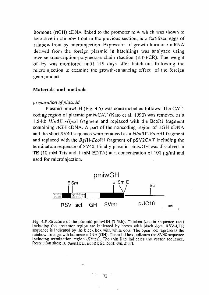
hormone (rLGH) cDNA linked Lo the promoter miw which was shown to be active in rainbow trout in the previous section, into fertilized eggs of
rainbow trout by microinjection. Expression of growth hormone mRNA derived from the foreign plasmid in hatchlings was analyzed using reverse transcription-polymerase chain reaction (RT-PCR). The weight
of fry was monitored until 149 days after hatch-out following the microinjection to 'examine the growth-enhancing effect of the foreign
gene product
Materials and methods
preparation of plasmid Plasmid pmiwGH (Fig. 4.5) was constructed as follows: The CAT
coding region of plasmid pmiwCAT (Kato et al. 1990) was removed as a 1.5-kb Hilldl! I-Hpal fragment and replaced with the EcoRI fragment containing rtGH cDNA. A part of the noncoding region of rtGH cDNA and the short SV40 sequence were removed as a Hilldlll-BamHI fragment and replaced with the BgIlI-EcoRI fragment of pSV2CAT including the
termination sequence of SV40. Finally plasmid pmiwGH was dissolved in TE (10 mM Tris and I mM EDT A) at a concentration of J 00 Ilg/ml and
used for microinjection.
pmiwGH ESm 8 Sm E
I \/ Sc
~~,L;~:;:':\I'-----~_-l..I~ RSV act GH SVter pUC18
lkb
Fig. 4.5 Structure of the plasmid pmiwGH (7.5kb). Chicken l3-actin sequence (act) including the promoter region are indicated by boxes with black dots. RSY-LTR sequence is indicated by the black box with white dots. The open box represents the rainbow trout growth hormone cDNA (GI-I). The solid box indicates the SV40 sequence including termination region (SYter). The thin line indicates the vector sequence. Restriction sites: B, BamHI; E, £CoR I; Sc, Seal; Sm, SmaI.
72

Gamete collectiofl, illSellliJ1(/fiOIl, alld microilljecfioll
Rainbow trout eggs and kilt were collected in January 1993 from 2-year-old fish. After about 400 eggs were mixed with 5 1111 of mill, 300
ml 120 m NaHC03 at 12°C was added immediately and allowed to stand for 1 min. The eggs were gently rinsed several times with water at 12°C to remove excess milt and allowed to swell for 3 h at 12°C in 0.5 mM glutathione (reduced form) the pH of which was adjusted to 10 with I N NaOH to prevent hardening of the chorion (Yoshizaki et al. 1991). After
swelling in the glutathione solution, about 10 nl of DNA solution was microinjected into the cytoplasm of fertilized eggs with a micropipet. Injected and control eggs wee rinsed with distilled water to remove glutathione and maintained in circulating water at about 12°C until hatching.
DNA alld RNA analysis
A part of the trout hatchlings was sampled 32 days after fertilization and anesthetized with 2-phenoxyethanol. The yolk sac was torn with fine forceps in sterilized Dulbecco's phosphate buffered saline (PBS) to remove the yolk. Then, each hatchling was divided into two parls: the head part, anterior to the operculum, and the posterior part. The fonner was used for DNA analysis and the latter for RNA analysis.
All samples were immediately frozen in liquid nitrogen and stored at -80
°C until analyzed. Genomic DNA was prepared using proteinase K-SDS treatment
followed by phenol extraction and ethanol precipitation. The existence of foreign nucleic acid sequences was detected using PCR as follows: One hundred nanograms of DNA was dissolved in 1 x Tlh buffer containing 6 ~g primer GH7 (5'-GGC TCT TCA ACA TCG CGG TC-3'), 6 Jlg primer GHS (5'-TCT TGT TCA GCT GTC TGC GT-3'), and 200 JlM dNTP. After preheating at 95°C for 10 min, I unit of Ttlz DNA polymerase (Toyobo) was added and 30 cycles of amplification was performed. Each cycle consisted of 30 s at 95°C, 30 s at 60°C, and 90 s at 70°C. Ten microliters of PCR product was subjected to electrophoresis on 4 % NuSieve 3: I agarose.(FMC), transferred to a nylon filter, and
hybridized with an oligonucleotide probe (5'-CAG AAA ATG TIC AAT
73
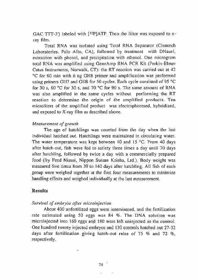
GAC TTT-3') labeled with [32P]ATP. Then the filler was exposed to x
ray film. Total RNA was isolated using Total RNA Separator (Clontech
Laboratories, Palo Alto, CA), followed by treatment with DNasel, extraction with phenol, and precipitation with ethanol. One microgram
total RNA was amplified using GeneAmp RNA PCR Kit (Perkin-Elmer Cetus Instruments, Norwalk, CT): the RT reaction was carried out at 42
°C for 60 min with 6 ng GH8 primer and amplification was performed using primers GH7 and GH8 for 50 cycles. Each cycle consisted of 95°C
for 30 s, 60°C for 30 s, and 70°C for 90 s. The same amount of RN A
was also amplified in the same cycles without performing the RT
reaction to determine the origin of the amplified products. Ten microliters of the amplified product was electrophoresed. hybridized,
and exposed to X-ray film as described above.
Measurement of growth
The age of hatchlings was counted from the day when the last
individual hatched out. Hatchlings were maintained in circulating water.
The water temperature was kept between 10 and 15°C. From 40 days after hatch-out, fish were fed to satiety three times a day until 70 days
after hatchling, followed by twice a day with a commercially prepared
food (fry Feed Nissui, Nippon Sui san Kaisha, Ltd.). Body weight was
measured five times from 39 to 140 days after hatchling. All fish of each
group were weighed together at the first four measurements to minimize
handling effects and weighed individually at the last measurement.
Results
Survival of embryos after microinjecfioll
Abollt 400 unfertilized eggs were inseminated, and the fertilization rate estimated using 50 eggs was 84 %. The DNA solution was
microinjected into 160 eggs an,d 180 were left uninjected as the control.
One hundred twenty injected embryos and 130 controls hatched out 27-32 days after fertilization giving hatch-out rates of 75 % and 72 %, respectively,
74
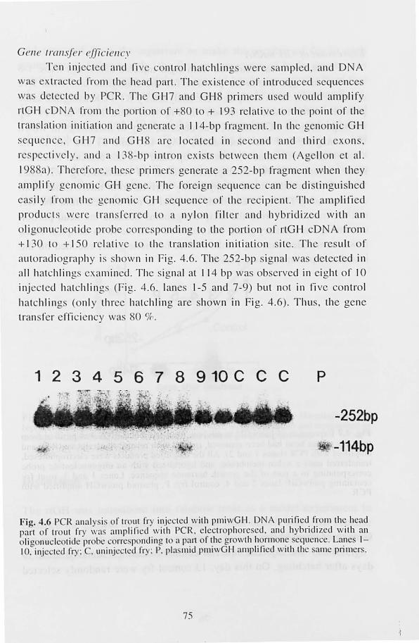
Cene transfe r (~fJic ien 'V
T n injec ted and ri v conLr I hatchlin us wer . ampled, and 0 A was ex tracted rrom the head parI. The xistence or inLroduc cI sequences
wa det cted I y peR. T he GH7 and GH primer: u cd would amplify
rtGH cD A 1'1' 111 the porti on or +80 to + 193 relati e to the pint of the
tran lati on initiati on and g n rat e a 11 4-bp fragment. In the genomic GH
. eq uence, GH7 and GH8 ar l oca ted in . econd and third exon ,
res l ec ti ve ly . and a 138-bp inlron ex i t between them (Ag li on el al.
1988a). T il refor . til e primer ' gen rate a 252-bp fragment when they
amplify genomic GH g lie. The foreign sequence ca n be di tingui hed
ea il y 1'1' nl th genomic GH equell ce or the rec ipienl. T he amplified
products we I' transrerr d to a ll y l n rilt r and hybr idi zed w ith an
li g nucleotid prob corresponding to the portion of rtGH cDNA from
+ 130 10 + 150 r "Hi ve t Ih tran lati on initi ati on sit e. Th result o f
autoradi ograph y i . how n ill Fig. 4.6. The 252-bp . ignal wa detected in
all h a t chlin ~s xamilled. The signal at I 14 bp was ob rvecl in eight of 10
injected hatchlin ,..,s (F ig. 4.6 . lanes \-5 and 7-9) but n t in ri ve con tr I
hatchlings (onl y thr e hatchlin e are hown in Fi 2. 4.6) . Thu . the gene
tran rer elli cienc wa' 80 Yc .
1 2 3 4 5 6 7 8 9 10 C C C P
-252bp
. 1 ,\ -114bp
Fig. 4.6 PCR nn:! ly is f tr lit fr injected \ ith pmi \ GH . DN A purified. f~'om Ih . head part f tr ut fry \\":1 amplifi ed with PCR. electrophoresed . and hybn dlzed \ nh an oli g lIucle tide prob' corre pOllding t a P~11 oft.h or wth I~ rmoll. equ II C . La!leS 1-10. injected fry: . unilljcclcd fry: P. Ila. mId pl11l\ GH amplified \ nh th allle prllner..
75
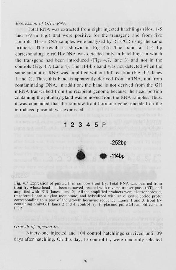
£: 'fJressiol/ (~/ Gf-! IIIRNA
TOlal R \Va. xlrac l d rrom eighl inj cled hat hlin gs ( 0 . . 1-5
and 7-9 in Fi " .) Ihal \ ere pos ili 1'0 1' Ih IranS"t;ne and rro m fi v
, nlrols. T h Sl,; R samll s w I' analyz cI by RT- P R using Ihe sa ml,;
prim er '. Th I' sull is show n in F ig 4. 7. Th band al I 14 br
CO lT . P nding 10 rlGH cD wa d teet d nl y in hatchlin !}. in which
th tran gl,;n had b n inlr dll eq (Fig. 4.7 l ane 3 and nOI in th
conl ro ls (Fi t">. 4.7, L ane 4). T h 11 4-bp band was not ddl,;CI d \ hen Ih
ame am lint or R wa ' amplified w ith lit RT r ac ti n (Fi '">. -+.7 lane
I and 2), T hLl ', Ihi s band i ' appal' nll y d ri v d from mR A, n t fr m
contaminating 0 . In additi on, Ih band i n ( d ri ved rr m th G H
mR tran 'crib d 1'1' m th r cipi nt genom b ca LI . e the h ad I rti on . ntaining th piluitary gland wa. r mov d from Ih R N sample . T hu "
it \Va' concluded Ihal Ih rainbow troul h rm one 0 n , n od d on the
intI' du d pi a 'mid , was xpr s d.
1 2 3 4 5 P
-252bp
-114bp
Fig. 4.7 Ex rr~ ion f prni wG I-I ill rainbow trout fry. T tal RN \Va puri fied fr rn trout fry who. e head had been n.: 111 vl:d, reacted w ith rl:ver. e tmn. cri pta t: (RT), and amplified wilh PCR Ian ~ I and 2). A ll the amplified prod uct weI' l;kctroph re e I, tran. ferred Ol1t o a nylon Jll~ l11 b ra l1 , and hybridi zed w ith an oligonucleotide probe corre. ponding to a pa rt f the gr wth hormone sequence. Lane I and 1. tr ut fry ontaining pmiwG I-I : lane :2 and 4, control fry; P, pia mid pJl1 iwG H ampli fied with
PCR.
Gra ll (Ii oj il/jee/eel fr.' in ly-on inj cled and 104 contr I hatchling : urv i ed until 39
days art r halchling. On thi s day, 13 c ntr I fry were rand ml y elect d
76
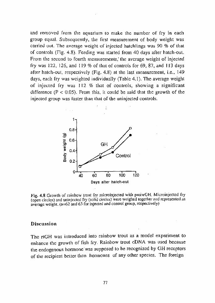
and removed from the aquarium lO make the number of fry in each
group equal. Subsequently, the nrst measurement of body weight was carried oul. The average weight of injected hatchlings was 90 % of that
of controls (Fig. 4.8). Feeding was started from 40 days after hatch-out.
From the second to fourth measurements: the average weight of injected
fry was 122, 126, and 119 % of thal of controls for 69, 87, and 1 J3 days
after hatch-out, respectively (Fig. 4.8) at the last measurement, i.e., 149
days, each fry was weighted individually (Table 4.1). The average weight
of injected fry was I 12 % that of contro]s, showing a significant
difference (P < 0.05). From this, it could be said that the growth of the
injected group was faster than that of the uninjected controls.
0.8 :§ 1: 0.6 Q) Q) ~ 0.4 >-"0 0 m 0.2
0 40 60 80 100 120
Days aller hatch-oul
Fig. 4.8 Growth of rainbow trout fry microinjecled with pmiwGH. Microinjected fry (open circles) and uninjccLcd fry (solid circles) were weighed together and represented as average weigh!. (n=62 and 63 for injected and control group, respectively)
Discussion
The rtGH was introduced into rainbow trout as a model experiment to
enhance the growth of fish fry. Rainbow trout cDNA was used because
the endogenolls hormone was supposed to be recognized by GH receptors
of the recipient belter than hormones of any other species. The foreign
77
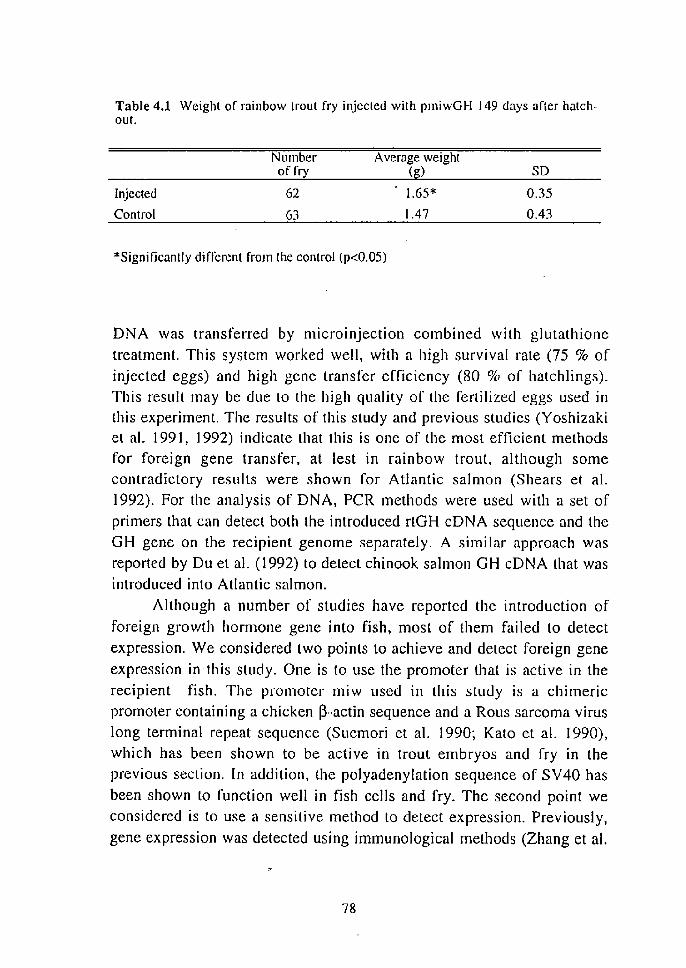
Table 4.1 Weight of rainbow trout fry injected with pmiwGI-I 149 days after halchout.
Number A verage weight of fry (g) so
Injected 62 1.65* 0.35
Control 63 1.47 0.43
*Significantly different from the control (p<0.05)
DNA was transferred by microJllJection combined with glutathione treatment. This system worked well, with a high survival rate (75 % of injected eggs) and high gene transfer efficiency (80 % of hatchlings). This result may be due to the high quality of the fertilized eggs used in this experiment. The results of this study and previous studies (Yoshizaki el al. 1991, 1992) indicate that this is one of the most efficient methods for foreign gene transfer. at lest in rainbow trout, although some contradictory results were shown for Atlantic salmon (Shears et al. 1992). For the analysis of DNA, peR methods were used with a set of
primers that can detect both the introduced rtGB cDNA sequence and the GH gene on the recipient genome separately. A similar approach was reported by Du et al. (1992) to detect chinook salmon GH cDNA that was introduced into Atlantic salmon.
Although a number of studies have reported the introduction of
foreign growth hormone gene into fish, most of them failed to detect expression. We considered two points to achieve and detect foreign gene
expression in this study. One is to use the promoter that is active in the recipient fish. The promoter miw used in this study is a chimeric promoter containing a chicken ~-actin sequence and a Rous sarcoma virus long terminal repeat sequence (Suemori et al. 1990; Kalo et al. 1990), which has been shown to be active in trout embryos and fry in the previous section. In addition, the polyadenylation sequence of SV40 has
been shown to function well in fish cells and fry. The second point we considered is to use a sensitive method to detect expression. Previously,
gene expression was detected using immunological methods (Zhang et al.
78
1990; Du et al. 1992); however, these methods did not distinguish the hormone derived from transgenes from the native hormone of the recipient when homogeneous GH sequence was introduced. Rokkones et al. (1989) detected III RNA of the transgene using northern blot hybridization. As the hormones generally· function allow concentrations, more sensitive detection methods are required. The RT-PCR (Rappolee et
al. 1988) technique used lor the detection of the trallsgelle expression is a
sensitive method for the rnRNA detection (Kawasaki 1990). The expression of transgenc was detected using RT-PCR combined with
hybridization. It will be a powerful method to detect low-level expression
of transgene in fish. The RT-PCR method used in this study is not designed for quantitative analysis. Several reports have shown that
quantitative analysis is possible using RT-PCR (Gilliland et al. 1990; Wang and Mark, 1990).
The injected and control fry were weighed once before the first feeding and four times about the start of feeding. Injected fry were smaller than controls at the first measurement but became larger with
subsequent measurements and a significant difference between the injected group and the control group was apparent at the forth measurement.
Thus, the effect of foreign GH expression on growth enhancement became clear after the start of feeding. Since the amount of the food consumed by each group was not determined in this study, it is not known whether the difference in weight was due to a difference in the amount of
food consumed or differences in the food conversion ratio between the
two groups. It has been reported that administration of recombinant GH
induced aggressive feeding behavior in rainbow lrout (Agellon et al.
1988b) and increased food conversion efficiency in red sea bream
(Ishioka et al. 1992). Such possibilities could not be ruled out here. The
average weight at four measurement after the start of feeding was 112-126 % of that of controls. The growth rate acceleration is not as high in
this study as in those previously reported (Zhang et al. 1990; Du et al. 1992). However, direct comparison is difficult because the fish used in
this study were quite young. The young fry stage is a critical stage in the life cycle of fish.
During this period, fish hatchlings generally complete absorption of the
79

yolk and start to swim and· feed. This is also a period where fish are prone to infectious diseases (Wolf 1988). We have demonstrated in this study enhanced growth of young fry using a foreign plasmid including a promoter that is active in embryonic and fry stages. Results of this study indicate the possibility of accelerating the growth of fish fry at a critical stage in the life cycle using lransgenictechniques.
80
Chapter 5 Comprehensive discussion
Transgenic animals have mainly been used to investigate gene regulation and function in studies of genes. The regulation of genes is generally examined by introducing the 'gene of interest into the host animal and monitoring the expression. To monitor the expression, the product of the introduced gene can be detected by immunological methods as shown in Chapter 3. It is also possible to detect the expresion
at the RNA level as shown in chapter 4. In addition, reporter genes are useful for easy detection of expression. As reporter genes, the pgalactosidase gene and the chloramphenicol acetyItransferase (CAT) gene have been often used. Since both reporters were also shown to be effective in fish as described in Chapters 2 and 4, they will be utilized in transgenic researches in the future. Even deletion analysis of regulatory elements is also possible using fish embryos instead of culture cells. Microinjection into ferlilized eggs of medaka is not so difficult and rather easier than transfection on culture cells once the microinjection or electroporatioll system was set up. In fact, Gong et al. (1991) successfully identified the functional regulatory elements of the fish antifreeze protein
promoter using medaka embryos. The function of the gene is generally studied by introducing the
coding region of the gene of interest linked to suitable promoters. For
this purpose, several promoters active in fish cells and embryos were identified in the chapter 2. As shown in the chapter 4, constitutive
expression of transgenes became possible using constitutive promoters. Moreover, it was shown that some promoters were inducible. Using inducible promoters, expression can be stimulated at desired lime. Using
these promoters, it is possible to examine function of genes which may affect the development of the hosl. Thus, most of materials required for studying gene regulation and function have already been prepared in the
process of this study. Transgenic fish system is expected to be applicable not·only to these
standard experiments, but also to special experimental methods e. g., the "enhancer trap" and the "insertional mutagenesis", both of which are known to be excellent methods Lo isolate the genes regulating the
81
development. The former is a method to detect tisslIe- and stage -specific enhancers by introducing reporter genes linked to a weak promoter. By determining the position of the insertion of transgelle in the genome of individuals revealing strong transgcne expression in the target tissue, tissue specific enhancers can be isolated. The latter is to isolate genes which participate in morphogenesis by introducing a foreign gene. Morphogenic genes can be identi lied by detrmi ning the sequence around the point of the transgene insertion into the genome of the mutant. These methods have been used on mice but significant effort is required to treat
a large number of transgenic mice. Fish which spawn large number of eggs is supposed Lo be more suitable for such experiments.
In the lield of medical sciences, transgenic mice have been used as desease models. It is impossible to replace mice by fish entirely because most of human deseases are not common with fish. However, fish is a member of vertebrates and it seems that some of des eases including cancer and some genetic deseases can be reproducible in fish. Concerning cancer, an exellent model is available in tropical fish, platty and swordtail. Hybrids of these species develop meranoma reproduciblly (Anders and Anders, 1978; Ozato and Wakamatsu 1983; Schwab, 1987).
Such system combined with transgenic technique may bring us valuable
information on tumorgenesis. Fish is also a potent model to study neural
deseases because neural development in vertebrates is being studied
actively using zebrafish (Kimmel, 1989). In fact, efforts to produce
mutant strains in zebrafish to study neural development has already
started in several countries. In relation to these projects, transgenic
system in zebrafish is also .being actively studied (Stuart et al. 1988, 1990; Culp et al. 1991; Westerfield et al. 1992). If desease models were
established in fish species such as medaka and zebrafish, they would be varuable because such species are easy to maintain under laboratory conditions and not so expensive. In addition, fish is easier to be enclosed
than mice because fish cannot escape from aqualium to environment without enough water.
Aquaculture of fish is thought to be indispensable to maintain fish resourses. However, the history of fish culture is shorter than that of plants and farm animals because fisheries has been dependent upon only
82
natural resourses for a long time. At present, few special strains· for
aquaculture is available. Thus breeding of economically important species is required. However, breeding through traditional methods takes too
much time because generation lime of most of fish species is more than 2 years. One possible technique to improve fish in relatively short time is the chromosome sets manipulation. Chromosome sets manipuration is possible by Uv- or y-ray irradiation and applying hydrostatic pressure or heat shock to gamete. Using this technique, genetically uniform
population can be produced within several generations. However,
chromosomes were manipulated as a haploid set and it is impossible to
manipulate a single trait using this technique.
The other technique expected to become a novel method for fish breeding is foreign gene transfer examined in this study. The potential of the transgenic technique as a method for breeding was partly shown in this study. Growth of rainbow trout fry was successfully accelerated by introducing foreign growth hormone cDNA (chapter 4). It is apparent that fish phemotype can be modified by foreign gene transfer. However, many hurdles are remaining before practical breeding of fish by foreign gene transfer. The most serious problem is that few genes are available to
introduce into fish at present because molecular-biological studies in fish started only recently. Only growth hormone genes and the antifreeze
protein gene have been introduced for improvement of fish. The former
is partially successful (Zhang et a!. 1989; Du et al. 1992) but the latter has
not succeeded yet unlil now (Hew el a!. 1992). More information about
fish genes should be accumulated for more progress. Transgenic fish
reseaches have just been started. Further progress depends on the
progress of molecular-biological studies of fish. Molecular-biological studies in fish will be accelerated using the transgenic techniques
established in this study.
83
Chapter 6 Summary
The technique for transferring foreign genes into animals is a powerful method to study gene regulation and function of genes ill vivo. Successful foreign gene transfer has been reported in various species including nematodes, Drosophila. sea urchin, amphibians, mice and farm animals. The application of foreign gene transfer technique to fish began about 5 years later after the tirst report in mice in spite of the importance of fish not only as experimental animals but also as food resources. This
study has been performed to establish the system for foreign gene transfer which enable to achieve introduction, expression and germline
transmission of transgenes in fish. Another objective of this study is to evaluate the potential of the foreign gene transfer technique as a tool for basic studies of genes as well as genetic engineering of fish.
In Chapter I, three different methods for foreign gene transfer into fish were described using medaka (Oryzias [aripes) as a model animal. In Section 1.1, microinjection into oocyte nuclei was examined. Foreign genes were successfully introduced by injecting foreign DNA into oocytes collected 14--8 h before ovulation and the most appropriate stage was 8 h before ovulation (Section 1.1). By adding pregnant mere gonadotropin to the culture medium for oocytes, it became possible to introduce oocytes collected 20~ 16 h before ovulation. In Section 1.2, microinjection into
fertilized eggs was examined (Section 1.2). It was shown that foreign genes can be introduced by injecting foreign DNA into the cytoplasm of
fertilized eggs at the I-cell stage. In the section 1.3, foreign gene transfer
by electroporation was examined. It was shown that foreign genes can be transferred by immersing fertilized eggs in DNA solution and applying
electric pulses to them. It was also shown that introduced genes are inherited to F I and F2 generations. Efficiencies of foreign gene transfer of the three methods were compared by introducing the same plasmid
pMV-GH. Among the three methods, electroporation was most simple but its gene transfer efficiency was the lowest. Microinjection into gene
oocytes is the best with regard to the gene transfer efficiency although it involves complicated procedures and applicable only to limited species. Microinjection into fertilized eggs is less difficulL than into oocytes but is
84
supposed to be obstructed by the chorion in some species. Thus, it seemed important to choose a method according to the purpose of the experiment and the species used.
Activities of various promoters derived from fish as well as other animals in cultured fish cells and fish embryos were examined to achieve transgene expression as described in Capter 2. In the cell line RTL-4, several viral clements such as pSV2 vector and the long terminal repeat
of Rous sarcoma virus, and a chicken chimeric promoter miw were
constitutively active. The Drosophila Hsp70 promoter and metallothionein
promoters derived from rainbow trouL and mice were active when cells
were stimulated by heat shock and metal-treatment, respectively (Section 2.1). III vivo activities or pSV2, miw and metallothionein promoters were
also examined using medaka embryos (Section 2.2). As the result, it was
shown that pSV2 and miw are also constitutively active and metallothionein promoters are active after exposure to zinc. These constitutive and inducible promoters were used to express transgenes in transgenic fish; the basic techniques to achieve introduction, expression and germ-line transmission of transgenes were thus established.
Expression of chicken o-crystallin gene during cmbryonic development was examined using a microinjeclion system in medaka as
described in Chapter 3. In the lens, the expression was detected only in the lens-formation stage. In non-lens tissues, expression was barely
dctectable only in mesodermal tissues in early stages but became
deLectable in greater varieties of tissues including ectodermal,
mesodermal and endodermal tissues. These result suggest the dual
regulation of 8-crystallin gene expression by lens-specific and non
specific regulatory elements. It was shown that the transgenic system established in this study is useful for ill vivo studies of genes.
In Chapter 4, potential of transgenic techniques as a method to improve economically important fish species was examined using rainbow troul as a model. Methods for introducing foreign genes into rainbow
trout were examined (Section 4.1). It was found that foreign genes can be transferred by microinjection into fertilized eggs after the glutathione treatment. Activity of miw. one of the most active promoters in medaka
embryos, was examined in rainbow trout (Section 4.2). It was shown that
85
miw is active in trout embryos and fry. In 7-day-old embryos, the expression was detected in various types of tissues. In hatchlings, the expression was observed in the yolk sac. Since it was found that miw is aClive in rainbow trout, the rainbow trout growth hormone eDNA was linked to miw and microinjected into rainbow trout eggs (Section 4.3). The expression in hatchlings was detected by reverse lranscriptionpolymerase chain· reaction. Injected and control fry were weighed from 39 to 149 days after hatch-out and it was shown that the growth of injected fry was significantly faster than that of the control group
(p<O.05). Results of this study indicate the potential of transgenic techniques as methods for genetic engineering of fish.
86

Acknowledgments
The author wishes La express the sincere gratitude to Professor Moribiko Sakaguchi, Department of Fisheries, Faculty Agriculture, Kyoto University for helpful discussions and encouragement. The aULhor is grateful to Drs. Kenjiro Ozato and Yuko Wakamatsu for their continuous guidance and advice. Most of this work was performed at Central Research Laboratory, Nippon Suisan Kaisha, Ltd. The author thank Drs. Mikio Satake and Shinya Yamashita for supporting this study.
87
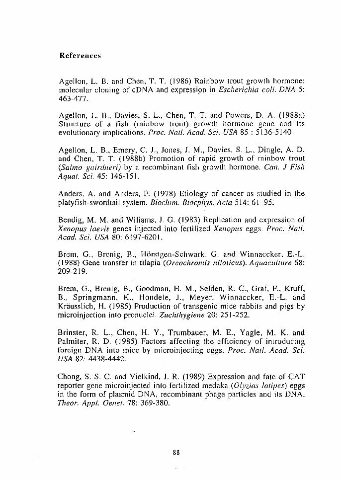
References
Agellon, L. B. and Chen, T. T. (1986) Rainbow trout growth hormone: molecular cloning of cDNA and expressi~n in Escherichia coli. DNA 5: 463-477.
Agellon, L. B., Davies, S. L., Chen, T. T. and Powers, D. A. (J988a) Structure of a fish (rainbow troul) growth hormone gene and its evolutionary implications. Proc. Nat!. A cad. Sci. USA 85 : 5136-5140
Agellon, L. B., Emery, C. J., Jones, J. M., Davies, S. L., Dingle, A. D. and Chen, T. T. (1988b) Promotion of rapid growth of rainbow trout (Sa/mo gairdlleri) by a recombinant fish growth hormone. Call. J Fish Aquat. Sci. 45: 146-151.
Anders, A. and Anders, F. (1978) Etiology of cancer as studied in the platyfish-swordtail system. Biochim. Biocplzys. Acta 5 [4: 61-95.
Bendig, M. M. and Wiliams, J. G. (1983) Replication and expression of Xenopus !aevis genes injected into fertilized Xenopus eggs. Proc. Nat!. Acad. Sci. USA 80: 6 [ 97 -620 [ .
Brem, G., Brenig, B., Horstgen-Schwark, G. and Winnaccker, E.-L. (1988) Gene transfer in tilapia (Oreochromis !lilOtjCllS). Aquaculture 68: 209-219.
Brem, G., Brenig, B., Goodman, H. M., Selden, R. c., Graf, F., Kruff, B., Springmann, K., Hondele, J., Meyer, Winnaccker, E.-L. and Krausslich, H. (1985) Production of transgenic mice rabbits and pigs by microinjection into pronuclei. Zuchthygiene 20: 251-252.
Brinster, R. L., Chen, H. Y., Trumbauer, M. E., Yagle, M. K. and Palmiter, R. D. (1985) Factors affecting the efficiency of introducing foreign DNA into mice by microinjecting eggs. Proc. Natl. Acad. Sci. USA 82: 4438-4442.
Chong. S. S. C. and Vielkind, J. R. (1989) Expression and fate of CAT reporter gene microinjected into fertilized medaka (Olyzias laripes) eggs in the form of plasmid DNA, recombinant phage particles and its DNA. Thear. Appl. Gellet. 78: 369-380.
88
Chourrout, D., Guyomard, R. and Houdebine L. M. (1986) High efficiency gene transfer in rainbow trout (Salmo gairdneri Rich.) by microinjectiun into egg cytoplasm. Aquaculture, 51: [43-150.
Clayton, R. M. ([974) Comparative aspec.ts of lens proteins. In : Davson, H. and Graham, L. T. (eds.) The Eye, Vol. 5. Academic Press London pp399-494
Colin, A. M., Cal[in, T. L., Kidson, S. H. and Maxson, R. (1987) Closely linked early and [ate H2B histone genes are differentially expressed after microinjection into sea urchin egg. Proc. Natl. Acad. Sci. USA 85: 507-510.
Donaldson, E. M. (1986) The integrated development and application of controI1ed reproduction techniques in Pacific salrnonid aquaculture. Fish Physiol. Biochell1. 2: 9-24.
Du, S. l, Gong, Z., Fletcher, G. L., Shears, M. A., King, M. J., Idler, D. R. and Hew, C. L. (1992) Growth enhancement in transgenic Atlantic salmon by the use of an "all fish" chimeric growth hormone gene construct. Bioitecllllology [0: 176-181.
Etkin, L. D. (1982) Analysis of the mechanisms of involved in gene regulation and ce[1 differentiation by microinjection of purified genes and somatic cell nuclei into amphibian oocytes and eggs. Differentiation 21: 149-159.
Elkin, L. D. and Pearman, B. (1984) Distribution, expression and germline transmission of exogenous DNA sequences following microinjection into Xenopus laevis eggs. Develop11lellt-99: 15-23.
Etkin, L. D. and Pearman, B., Roberts, M. and Bektesh, S. L. (1987) Replication, integration and expression of exogenous DNA injected into fertilized eggs of Xenopus laevis. Differentiation 26: 194-202.
Fletcher, G. L., Shears, M. A., King, M. J., Davies, P. L., and Hew, C. L. (1988) Evidence for antifreeze protein gene transfer in Atlantic salmon (Sa/Illo safar). Call. 1. Fish Aquat. Sci. 45: 352-357.
Flytzanis, C. N, McMahon, A. P., Hough-Evans, B. R., Katula, K. 5., Britten, R. J. and Davidson, E. H. (1985) Persistence and integration of cloned DNA in postembryonic sea urchins. Dev. BioI. 108: 431-442.
89
Flytzanis, C. N, Britten, R. 1. and Davidson, E. H. (1987) Ontogenic activation of a fusion gene intoroduced into the sea urchin eggs. Proc. Natl. A cad. Sci. USA 84: 151-155.
Franks, R. R., Hough-Evans, B. R., Britten, R. J. and Davidson, E. H. (1988a) Spatially degraded though temporally correct expression of a Strollgylocebtrolltus purpura/us actin gene fusion in transgenic embryos of a different sea urchin family. Gelles & Del'. 2: 1-12.
Franks, R. R., Hough-Evans, B. R., Britten, R. 1. and Davidson, E. H. (l988b) Direct introduction of cloned DNA into the sea urchin zygote nucleus, and fate of injected DNA. Development 102: 287-299.
Fregien, N. and Davidson, N. (1986) Activating elements in the promoter region of the chcken p-actin gene. Gene 48: 1-11.
Friedenreich. H. and Schartl. M. (1990) Transient expression directed by homologous and heterologous promoter and enhancer sequences in fish cells. Nuc. Acids Res. 18: 3299-3305.
Gilliland, G., Perrin, S. and Bunn. H. F., (1990) Competitive PCR for quantitation of mRNA. In : Innis, M. A .• Gelfand, D. H., Sninsky. J. J., White, T. J. (eds.) peR protocols. Academic Press, San Diego, CA, pp 60-69.
Glanville, N., Durnam, D. M. and Palmiter, R. D. (1981) Structure of mouse metallothionein-I gene and its mRNA. Nature 292: 267-269.
Gong, Z., Hew. C. L. and Vielkind, 1. R. (1991) Functional analysis and temporal expression of promoter regions from fish antifreeze genes in Lansgenic Japanese medaka embryos. Mol. Mar. BioI. Bioteclmol. 1: 64-72.
Gordon. J. W., Scangos. G. A .. Plotkin. D. J., Barbosa. J. A. and Ruddle. F. H. (1980) Genetic transformation of mouse embryos by microinjeclion of purified DNA. Proc. Natl. Acad. Sci. USA 77: 7380-7384.
Goring, D. R., Rossant, 1., Clapoff. S., Breitman. M. L. and Tsui, L.-C. (1987) In situ detection of p-galactosidase in lenses of transgenic mice with a y-crystallinllacZ gene. Science 235: 456-458.
90
Gorman, C. M., Moffat, L. F. and Howard, B. H. (1982a) Recombinant gcnomes which express chloramphenicol acetylLransrerasc in mammalian cells. Mol. Cell. Bioi. 2: 1044-1051.
Gorman, C. M., Merlino, T. G., Willirgham, M. c., Paslan, I. and Howard, B. H. (1982b) The Rous sarcoma virus long terminal repeat is a strong promoter when introduced in to a variety of eukaryoLie cells by DNA-mediated trallsfection. Proc. Nat/. Acad. Sci. USA 79: 6777-6781.
Goto, K., Hayashi, S .. Shirayoshi, Y., Takeichi, M. and Kondoh, H. (1988) Exogenous b-crystallin gene expression as probe for differentiation of teratocarcinoma stem cells. Cell Differ. 24: 139-148.
Graham, F. L. and van der Eb, A. J. (1973) A new technique for the assay of infeclivity of human adenovirus 5 DNA. Virology 52: 456-467.
Guyomard, R., Chourrout, D., Leroux, C., Houdebine, L. M., Pourrain, F. (1989) Integration and germ line transmission of foreign genes microinjected into fertilized trout eggs. Bioclzilllie 71: 857-863.
Hammer, R.E., Pursel, V. G., Rexroad, C. E. Jr., Wall, R. J. Bolt, D. J., Ebert, K. M., Palmiter, R. D. and Brinster, R. L. (1985) Production of transgenic rabbits, sheep and pigs by microinjection. Nature 315: 680-683.
Hasegawa, T., Nakada, S., Nakajima, T., Oda, K., Kawata, M., Kimura, H. and Sekiya, S. (1990) Expression of various viral and cellular enhancer-promoters during differentiation of human embryonal carcinoma cells. Differelltiatioll 42: J 91-198.
Hayashi, S., Kondoh, H., Yasuda, K., Soma, G., Ikawa, Y. and Okada, T. S. (1985) Tissue-specific regulation of a chicken b-crystallin gene in mouse cells: involvement of the 5' end region. EMBO 1.4: 2201-2207.
Hayashi, S., Goto, K. Okada, T. S. and Kondoh, H.( 1987) Lens-specific enhancer in the third inlron regulates expression of the chicken 8 J -
crystallin gene. Gelles Dev. I: 818-828.
Hew, C. L., Trinh, K. Y., Du, S. J. and Song, S. (1989) Molecular cloning and expression of salmon pituitary hormones. Fish Physiol. Bioc!zem. 7: 375-380.
91
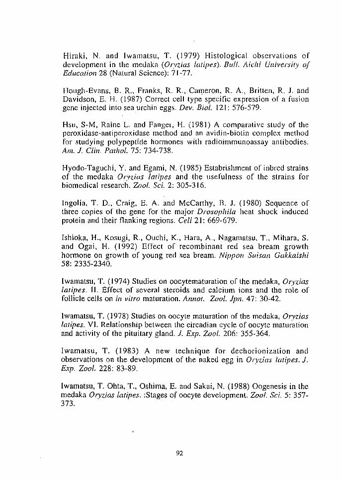
Hiraki, N. and Iwamatsu, T. (1979) Histological observations of development in the medaka (Oryzias /atipes). BlII/. Aichi University of Education 28 (Natural Science): 71-77.
Hough-Evans. B. R., Franks, R. R., Call1erOn, R. A., Britten, R. J. and Davidson, E. H. (1987) Correct cell type specific expression of a fusion gene injected into sea urchin eggs. Dev. BioI. 121: 576-579.
Hsu. S-M, Raine L. and Fanger, H. (1981) A comparative study of the peroxidase-anti peroxidase method and an avidin-biotin complex method for studying polypeptide hormones with radioimmunoassay antibodies. Am. 1. Clill. Pallzol. 75: 734-738.
Hyodo-Taguchi. Y. and Egami, N. (1985) Estabrishment of inbred strains of the medaka Oryzias tatipes and the usefulness of the strains for biomedical research. Zoo!. Sci. 2: 305-316.
Ingolia. T. D., Craig, E. A. and McCarthy, B. J. (1980) Sequence of three copies of the gene for the major Drosophila heat shock induced protein and their nanking regions. Cell 21: 669-679.
Ishioka, H., Kosugi, R., Ouchi, K., Hara, A., Nagamatsu, T., Mihara, S. and Ogai, H. (1992) Effect of recombinant red sea bream growth hormone on growth of young red sea bream. NippolZ 5t1isan Gakkaishi 58: 2335-2340.
Iwamatsu, T. (1974) Studies on oocytematuration of the medaka, Oryzias laripes. II. Effect of several steroids and calcium ions and the role of follicle cells on ill vitro maturation. Annal. Zool. lpn. 47: 30-42.
lwamatsu, T. (1978) Studies on oocyte maturation of the medaka. Oryzias latipes. VI. Relationship between the circadian cycle of oocyte maturation and activity of the pituitary gland. J. Exp. Zoo!. 206: 355-364.
Iwamatsu, T. (1983) A new technique for dechorionization and observations on the development of the naked egg in Oryzias talipes. 1. Exp. Zool. 228: 83-89.
Iwamatsu, T. Ohta, T., Oshima, E. and Sakai, N. (1988) Oogenesis in the medaka Oryzias talipes. :Stages of oocyte development. Zool. Sci. 5: 357-373.
92
Iwamatsu, T. (1992) Egg activation. Fish BioI. 1. MEDAKA 4: 1-9.
Kadokawa, Y., Suemori, H. and Nakatsuji, N. (1990) Cell lineage analyses of epithelia and blood vessels in chimeric mouse embryos by use of an embryonic stem cell line expressing the ~-galactosidase gene. Cell Differ. Del!. 29: 187-194.
Kato, K., Kanamori, A., Kondoh, H. (1990) Rapid and transient decrease of N-myc expression in retionic acid-induced differentiation of OTF9 teratocarci noma cells. M ul. Cell. BioI. 10: 486-491 .
Katula, K. S., Hough-Evans, B. R., Britten, R. J. and Davidson, E. H. (1987) Ontogenic expression of a Cyl: actin fusion gene injected into sea urchin eggs. Development 101: 437-447.
Kawasaki, E.( 1990) Ampli fication of RNA. In : Innis, M. A., Gelfand, D. H., Sninsky, J. J., White, T. J. (eds.) PCR protocols. Academic Press, San Diego, CA, pp 21-27.
Kimmel, C. B. (1989) Genetics and early development of zebrafish. Trends Geller. 5: 283-288.
Kondoh, H., Yasuda, K. and Okada. T. S. (1983) Tissue-specific expression of a cloned chick 8-crystallin gene in mouse cells. Nature 301: 440-442.
Kondoh, H., and Okada. T. S. (1986) Dual regulation of expression of exogenous 8-crystallin gene in mammalian cells: A serch for molecular background or instability in differentiation. Curl'. Top. Dev. BioI. 20: 153-164.
Kondoll, H., Katoh, K., Takahashi, Y., Fujisawa, H., Yokoyama, M., Kimura, S., Katsuki, M., Saito, M., Nomura, T., Hiramoto, Y. and Okada. T. S. (1987) Specific expression of the chicken 8-cryslallin gene in the lens and the pyramidal neurons of the piriform cortex in transgenic mice. Dev. BioI. 120: 177-185.
Kosugi, R., MOlllota, H., Kawai, S., lshioka, H. and Hara, A. (1992) Production of recombinant red sea bream growth hormone using a secretion plasmid of Escherichia coli and its binding activity to the membranes of red sea bream liver. Nippon Suisall Gakkaislzi 58: 1115-
1122.
93
Laimins, L. A., Khoury, G., Gorman, C., Howard, B. and Gruss, P. (1982) Host-specific activation of transcription by tandem repeats from simian virus 40 and Moloney murine sarcoma virus. Pro. Natl. A cad. Sci. USA 79: 6453-6457.
Laimins, L. A., Kessel, M., Rosenthal, N. and Khoury, G. (1983) Viral and cellular enhancer elements. In: Gluzman, Y. and Shenk, T. (eds.) Enhancers and eukaryotic gene expression. Cold Spring Harbor Laboratory, New York, pp. 28-37.
Liu, Z., Moav, B., Foras, A. J., Guise, K. 5., Kapuscinski, A. R. and Hackelt, P. B. (I990a) Development of expression vectors for transgenic fish. BiolTecllllOlogy 8: 1268-1272.
Liu, Z., Moav, B., Foras, A. J., Guise, K. 5., Kapuscinski, A. R. and Hackett, P. B. (1990b) Functional analysis of elements affecting expression of the ~-actin gene of carp. Mol. Cell. Bio!. 10: 3432-3440.
Maclean, N., Penman, D. and Zhu, Z. (1987) Introduction of novel genes into fish. Bio/leelmol. 5: 257-261.
Matsui,K. (1949) Illustration of the normal course of development in the fish, Olyzias latipes. lpll. l. Exp. Morp/z. 5: 33-42.
McEvoy, T. G., Stack, M., Keane, B., Barry, T., Sreenan, J. M. and Gannon, F. (\988) The expression of a foreign gene in salmon embryos. Aquaculture 68: 27-38.
McMahon, A. P., Novak T. J., Britten, R. J. and Davidson, E. H. (1984) Inducible expression of a cloned heat shock fusion gene in sea urchin embryos. Proc. Nat!' Acad. Sci. USA 81; 7490-7494.
McMahon, A. P., Flytzanis, C. N., Hough-Evans, B. R., Katula, K. 5., Britten, R. J. and Davidson, E. H. (1985) Introduction of cloned DNA into sea urchin egg cytoplasm: replication and persistence during embryogenesis. Dev. Bioi. 108: 420-430.
Murphy, M. F., Collier, l, Koutz, P. and Howard, B. (1990) Nucleotide sequence of the trout metallothionein A gene 5' regulatory region. Nuc. Acids Res. 18: 4622.
94
Naito, M., Agata, K., Otsuka, K., Kino, K., Ohta, M., Hirose, K., Perry, M. M. and Eguchi, G. (1991) Embryonic expression or ~-aclin-Iac Z hybrid gene injected into the J"erLilized ovum of the domestic fowl. lilt. 1. Dev. Bioi. 35: 69-75.
Neumann, E., Schaefer-Ridder, M., Wa~g, Y. and Hofschneider, P. H. (1982) Gene transJ"er into mouse Iyoma cells by electroporation in high electric fields. EMBO 1. 1: 841-845.
Ozato, K. and Wakamatsu, Y. (1983) Multi-step genetic regulation of oncogene expression in fish hereditary melanoma. Differentiation 24: 181-190.
Ozato, K., Kondah, H., Inohara, H., lwamatsu, T., Wakamatsu, Y. and Okada, T. S. (1986) Production of transgenic fish : introduction and expression of chicken 8-crystallin gene in medaka embryos. Cell Differ. 19: 237-244.
Palmiter, R. D. and BrinsLer, R. L. (1986) Germline transformation of mice. AIlIl. Rev. Gellet. 20: 465-499.
Palmiter, R. D. Brinster, R. L., Hammer, R. E., Trumbauer, M. E., Rosenfeld, M. G., Birnberg, N. C. and Evans, R. M., (1982) Dramatic growth of mice that develop form eggs microinjected with metallothionein-growlh hormone fusion genes. Nature 300: 611-615.
Penman, D., Beedling, A. J., Penn, S. and Maclean, N. (1990) Factors affecting survival and integration following microinjection of novel DNA into rainbow troul eggs. Aquaculture 85: 35-50.
Piatigorsky, J. (1981) Lens differantiation in vertebrates: A review of cellular and molecular features. DifferentiatioH 19: 134-153.
Piatigorsky, J. (1984) Lens crystallins and their gene families. Cell 38: 620-621.
Potter, H., Weir, L. and Lader, P. (1984) Enhancer-dependent expression of human K-immunogloblin genes introduced into mouse pre-~lymphocytes by eleclroporation. Pro. Nall Acad. Sci. USA 81: 7161-7165.
Powers, D. A. (1989) Fish as model systems. Science 246: 352-358.
95
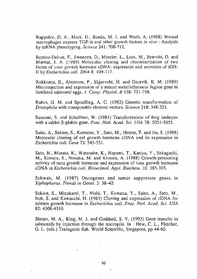
'Rappolee, D. A" Mark, D., Banda, M. J. and Werb, A. (1988) Wound macrophages express TGF-a and other growth factors in vivo: Analysis by mRNA phenolyping. Sciellce 241: 708-712.
Rentier-Delrue, F., Swennen, D., Mercier, L., Lion, M., Benrubi, O. and Martial. J. A. (1989) Molecular cloning and characterization of two forms of trout growth hormone eDNA: expression and secretion of tGHII by Esche rich ia coli. DNA 8: 109-1 17.
Rokkones, E., Alestrom, P., Skjervold, H. and Gautvik, K. M. (1989) Microinjection and expression of a mouse metallothionein fugion gene in fertilized salmonid eggs. 1. Compo Physiol. B 158: 751-758.
Rubin, G. M. and Spradling, A. C. (1982) Genetic transformation of Drosophila with transposable element vectors. Science 218: 348-353.
Rusconi, S. and SchatTner, W. (1981) Transformation of frog embryos with a rabbit p-globin gene. Proc. Nat!. Acad. Sci. USA 78: 5051-5055.
Saito, A., Sekine, S., Komatsu, Y., Sato, M., Hirano, T. and Ito, S. (1988) Molecular cloning of eel growth hormone cDNA and its expression in Escherichia coli. Gene 73: 545-551.
Sato, N., Murata, K., Watanabe, K., Hayami. T., Kariya, Y., Sakaguchi, M., Kimura, S., Nonaka. M. and Kimura. A. (1988) Growth-promoting activity of tuna growth hormone and expression of tuna growth hormone cDNA in Escherichia coli. Biolechllo!. Appl. Biochem. 10: 385-393.
Schwab, M. (1987) Oncogenes and tumor suppressor genes in Xiphophorus. Trends in Gellet. 3: 38-42.
Sekine, S., Mizukami, T., Nishi, T., Kuwana, Y., Saito, A., Sato, M., Itoh, S. and Kawauchi, H. (1985) Cloning and expression of cDNA for salmon growth hormone in Escherichia coli. Proc. Nat!. A cad. Sci. USA 82: 4306-4310.
Shears, M. A., King, M. J. and Goddard, S. V. (1992) Gene transfer in salmonids by injection through the micropile. In : Hew, C. L., Fletcher, G. L. (eds.) Transgenic fish. World Scientific, Singapore, pp 44-60.
96
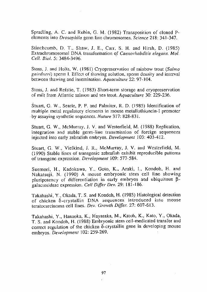
Spradling, A. C. and Rubin, G. M. (1982) Transposilioll of cloned Pelements into Drosophila germ line chromosomes. Sciellce 218: 341-347.
Stinchcomb, D. T., Shaw, 1. E., Carr, S. H. and Hirsh, D. (1985) Extrachromosomal DNA transfonnation C!f Caell.orlzabdiris elegalls. Mol. Cell. Bioi. 5: 3484-3496.
SLOSS, J. and Holls, W. (1981) Cryopreservation of rainbow trout (Salmo gairdller;) sperm I. Effect of thawing solution, sperm density and interval between thawing and insemination. Aquaculture 22: 97-104.
Stoss, J. and Refstie, T. (1983) Short-term storage and cryopreservation of milt from Atlantic salmon and sea trout. Aquaculture 30: 229-236.
Stuart, G. W., Searle, P. F. and Palmiter, R. D. (1985) Identification of multiple metal regulatory elements in mouse metallothionein-I promoter by assaying synthetic sequences. Nature 317: 828-831.
Stuart, G. W., McMurray, J. V. and Westerfield, M. (1988) Replication, integration and stable germ-line transmission of foreign sequences injected into early zebrafish embryos. Development 103: 403-412.
Stuart, G. W., Vielkind, J. R., McMurray, J. V. and Westerfield, M. (1990) Stable lines of transgenic zebrafish exhibit reproducible patterns of transgene expression. Developmem 109: 577-584.
Suemori, H., Kadokawa, Y., Goto, K., Araki, I., Kondoh, H. and N akatsuji, N. (1990) A mouse embryonic stem cell line showing p[uripotency of differentiation 'in early embryos and ubiquitous ~galactosidase expression. Cell Differ Dev. 29: 181-186.
Takahashi, Y., Okada, T. S. and Kondoh, H. (1985) Histologica[ detection of chicken 8-crystallin DNA sequences introduced into mouse teratocarcinoma cell lines. De". Growth Differ. 27: 607-613.
Takahashi, Y., Hanaoka, K., Hayasaka, M., Katoh, K., Kato, Y., Okada, T. S. and Kondoh, H. (1988) Embryonic slem cell-medicated transfer and correct regulation of the chicken o-crystallill gene in developing mouse embryos. Development 102: 259-269.
97
Treton, J. A., Shinohara, T. and Piatigorsky, J. (1982) Degradation of 8-crystallin mRNA in the lens fiber cells of the chicken. Dev. BioI. 92: 60-65.
Ueno, K., Hiramoto, Y., Hayashi, Sand Kondoh, 1-1. (1988) Introduction and expression of recombinant ~-galactosidase genes in cleavage stage mouse embryos. Dev., Growth Differ. 30: 61-73.
Vitelli, L., Kemler, 1., Lauber, B., Birnstiel, M. L. and Busslinger, M. (1988) Developmental regulation of microinjected histone genes in sea urchin embryos. Dev. BioI. 127: 54-63
Wang, A. M. and Mark, D.F. (1990) Quantitative PCR. In : Innis, M. A., Gelfand, D. H., Sninsky, J. J., White, T. J. (eds.) peR protocols. Academic Press, San Diego, CA, pp 70-75.
Watanabe, T., Nakano, M., Asakawa, H. and Morimoto, T. (1987) Cell culture of rainbow trout liver. Nippon Suisan Gakkaishi 53: 537-542.
Wilson, c., Cross, G. S. and Woodland, H. R. (1986) Tissue specific expression of actin genes injected into Xenopus embryos. Cell 47: 589-599.
Winkler, c., Vielkind, J. R. and Scharll, M. (1991) Transient expression of foreign DNA during embryonic and larval development of the medaka fish (Oryzias [atipes). Mol. Gen. Genet. 226: 129-140.
Wolf, K. (1988) Fish viruses and fish viral diseases. Cornell University Press, Ithaca, NY.
Yamaha, E., Usui, K., Onozato, H. and Hamada, K. (1986) A methods for dechorionization in gold fish, Carassius auratus. Nippon Suiscm Gakkaishi 52: 1929-1934.
Yamaha, E., Matsuoka. M. and Yamazaki, F. () 988) Introduction of exotic reagents into denuded eggs of goldfiss and crucian carp by electroporation. Nippon Suisan Gakkaishi 54: 2043.
Yamamoto, T. (1975) Medaka (Killfish). Biology and strains. Keigaku Pub!. Co, Tokyo.
98
Yoon, S. 1., Hallerman, E. M., Gross, M. L., Liu, Z., Schneider, J. F., Faras, A.,J. Hackett, P. B., Kapuscinski, A. R. and Guise, K. S. (1990) Transfer of the gene for neomycin resistance into goldfish, Carassius auralus. Aquaculture 85: 21-33.
Yoshizaki, G., Oshiro, T. and Takashima, F. (1989) Prevention of hardening of chorion and dechorionation for microinjection into fish eggs. Nippon Suiswz Gakkaishi 55: 369.
Yoshizaki, G., Oshiro, T. and Takashima, F. (1991) Introduction of carp a-globin gene inlo rainbow trouL. Nippon Suisan Gakkaishi 57: 819-824.
Yoshizaki, G., Kobayashi, S., Oshiro, T. and Takashima, F. (1992) Introduction and expression of CAT gene in rainbow trout. Nippon Suisall Gakkaislzi 58: 1659-1665.
Zafarullah, M., Bonham, K. and Gedamu, L. (1988) Structure of the rainbow trout mclallolhionein B gene and characterization of its metalresponsive region. Mol. Cell. BioI. 8: 4469-4476.
Zhang, P., Hayat, M., Joyce, C., Gonzales-Villasenol, L. I., Lin, C. M., Dunham, R. A., Chen, T. T. and Powers, D. A. (1990) Gene transfer, expression and inheritance of pRSV-rainbow trout -GH cDNA in the common carp, Cyprinus carpio (Linnaeus). Mol. Reprod. Dev. 25: 3-13.
Zhu, Z., Li, G., He, L. and Chen, S. (1985) Novel gene transfer into the fertilized eggs of goldfiss (Carassius aura/us L. 1785). Z. Angew. Iclzthyol. I: 3 1-34.
Zhu, Z., Xu, K. Li, G., Xie, Y. and He, L. (1986) Biological effects of human growth hormone gene microinjected into the fertilized eggs of loach Misglll'lltlS allgui/licauda/us (Cantor). Kexue Tongbao 3 I: 988-990.
99
Author's subjects
Inoue, K., Gzalo, K., Kondoh, H" Iwamat5u, T., Wakamalsu, T., Fujita, T. and Okada, T. S. (1989) Stage-dependent expression of the chicken 8-crystallin gene in transgenic fish embryos: Cell Differ. Dev. 27: 57-68.
Inoue, K., Yamashita, S., Hala, 1., Kabeno, S., Asada, S., Nagahisa, E. and Fujita, T. (1990) Electroporalion as a new technique for producing transgenic fish. Cell Differ. Dev. 29: 123-128.
Inoue, K., Akita, N., Yamashita, S., Shiba, T. and Fujita, T. (1990) Constitutive and inducible expression of a transgene directed by heterologous promoters in a trout liver cell line. Biochem. Biophys. Res. Commull. 173: 1311-1316.
Inoue, K., Yamashita, S., Akita, N., Mitsuboshi , T., Nagahisa, E., Shiba, T. and Fujita, T. (1991) Histochemical detection of foreign gene expression in rainbow trout. Nippon Suisan Gakkaishi 57: 1511-1517.
Inoue, K., Akita, N., Shiba, T., Satake, M. and Yamashita, S. (1992) Metal-inducible activities of metallothionein promoters in fish cells and fry. Biochem. Biophys. Res. ComnllUl. 185: 1108-1114.
Inoue, K., Asada, S., Kabeno, S., Yamashita, S., Nagahisa, E. and Satake, M. (1992) Specific stages of oocyte maturity in medaka to obtain efficient foreign gene transfer by microinjection. Nippon Suisall Gakkaishi 58: 1251-1255.
Inoue, K., Yamada, S. and Yamashita, S. (1993) Introduction, expression, and growth-enhancing effect of rainbow trout growth hormone cDNA fused to an avian chimeric promoter in rainbow trout fry. 1. Mar. Biotechnol. I: 131-134.
Inoue, K. and Yamashita, S. (1993) Successful foreign gene transfer into medaka by microinjection into oocytes at early stages of maturity. Nippon Suisan Gakkaishi 59: 2091.
100