Embed Size (px)
Citation preview
ORIGINAL ARTICLE
Topography of coronary arteries and their ramifications in the goat
Karolina Barszcz1 & Olga Szaluś-Jordanow2& Michał Czopowicz3 & Marcin Mickiewicz3 & Agata Moroz3 &
Jarosław Kaba3 & Michał Polguj4 & Grzegorz Wysiadecki5 & Robert Haładaj5 & Halina Purzyc-Orwaszer6,7
Received: 12 August 2018 /Accepted: 7 February 2019 /Published online: 21 February 2019# The Author(s) 2019
AbstractThe aim of this study was to investigate the morphology of the coronary arteries of the goat’s heart. The study was carried out on36 hearts of adult females dairy goats, belonging to two breeds, aged 7–12 years, with an average bodyweight of 37 kg. A distinctview of coronary arteries and their branches was obtained by filling them with dyed synthetic latex (LBS 3060) or Batson’s No.17. In all studied goats the common trunk of the left coronary artery was divided into the interventricular paraconal branch and thecircumflex branch. The branch of the interventricular septum originated in the interventricular paraconal branch. In 25 individuals(69%) the circumflex branch ended with small ramifications on the atrial surface of the heart. In 11 individuals (31%), the vesselextended in the subsinuosal interventricular groove into the subsinuosal interventricular branch. The right coronary artery wasless developed than the left coronary artery. In 35 individuals (97%) the right coronary artery ended with small ramifications onthe atrial surface of the heart. In one goat (3%) the vessel reached the subsinuosal interventricular groove and extended into thesubsinuosal interventricular branch.
Keywords Coronary arteries . Corrosion castings . Goat . Heart . Vascular casting
Introduction
Coronary vessels have been studied in domestic and wildruminants including Angora and Akkamaran goats (Besolukand Tipirdamaz 2001), roe deer (Capreolus capreolusLinnaeus, 1758) (Frąckowiak et al. 2007), Bactrian camel(Camelus bactrianus Linnaeus, 1758) (Yuan et al. 2009),one-humped camel (Camelus dromedarius Linnaeus, 1758)(Ghazi and Tadjalli 1993), European bison (Bison bonasusLinnaeus, 1758) (Kupczyńska et al. 2015). However, theavailable literature provides little information on the topogra-phy of coronary arteries and their ramifications in the goats
(Capra hircus Linnaeus, 1758) (Nickel et al. 1981; Barone1996; Besoluk and Tipirdamaz 2001). More information onits anatomy is needed. Because of the anatomical similaritybetween the goats and other ruminants, the results of this studycan be applied also to the other species.
Goats are a commonly accepted species for biomedicalstudy. The similarity of organ size between goats and humansmakes these animals widely used. They are used in humanmedicine in research such as cardiac, orthopedic, model forinfectious diseases or many others (Shiraishi et al. 2012; Chenet al. 2015; Zhang et al. 2015; Lukovsky-Akhsanov et al.2016; Du et al. 2018; Vandersteene et al. 2018).
* Halina [email protected]
1 Department of Morphological Sciences, Faculty of VeterinaryMedicine, Warsaw University of Life Sciences, Nowoursynowska159, 02-776 Warsaw, Poland
2 Department of Small Animal Diseases with Clinic, Faculty ofVeterinary Medicine, Warsaw University of Life Sciences,Nowoursynowska 159c, 02-776 Warsaw, Poland
3 Laboratory of Veterinary Epidemiology and Economics, Faculty ofVeterinary Medicine, Warsaw University of Life Sciences,Nowoursynowska 159c, 02-776 Warsaw, Poland
4 Department of Angiology, Interfaculty Chair of Anatomy andHistology, Medical University of Łódź, Narutowicza 60,90-136 Łódź, Poland
5 Department of Normal and Clinical Anatomy, Interfaculty Chair ofAnatomy and Histology, Medical University of Łódź, Łódź, Poland
6 The Koret School of Veterinary Medicine, Hebrew University ofJerusalem, P.O. Box 12, 76100 Rehovot, Israel
7 Faculty of Veterinary Medicine, Department of Biostructure andAnimal Physiology, Wroclaw University of Environmental and LifeSciences, Kożuchowska 1/3, 51-631, Wroclaw, Poland
Biologia (2019) 74:683–689https://doi.org/10.2478/s11756-019-00208-z
The aim of this study was therefore to investigate the mor-phology of the coronary arteries of the goat’s heart.
Material and methods
The study was carried out on 36 hearts of adult females dairygoats aged 7–12 years, belonging to two breeds – Polish FawnImproved (n = 20) and PolishWhite Improved (n = 16), close-ly related to French Alpine and Saanen, respectively. Bodyweight ranged from 20 to 49 kg (mean 37 kg). Pathologicalexamination of the whole body was performed before thedissection of the hearts. None of the animals included in thestudy had any pathological changes in the thoracic cavity.Nomenclature from the Nomina Anatomica Veterinaria(2017) was used.
Coronary arteries were visualized by filling them throughthe aorta with Latex (LBS 3060) (Synthos Dwory Sp. z o.o,Poland) with additional dye (INCHEM) or Batson’s No. 17(Polyscience, Incorporation, Warrington, US). The hearts (n =26) filled with the latex injection mass were placed in a 4%formaldehyde solution. Next, the proximal segments of the leftand right coronary arteries were dissected. Corrosion castingswere prepared by filling selected hearts (n = 10) with coloredresin. The specimens were then left at room temperature(>18 °C) until the casts hardened. Next, to dissolve the organictissue, the materials were placed in 40%KOH solution at 50 °Cfor approximately 24 h. The remnants of the tissue were re-moved by continuous flushing with water for 38 h and thenthe material was cleaned by water and a small amount of stan-dard washing liquid. The cast was later dried at room temper-ature for 48 h. These methods of injection were successfullyused in our previous studies (Polguj et al. 2009, 2011; Barszczet al. 2014, 2016a, 2017; Kupczyńska et al. 2015).
Results
Morphology of the left coronary artery
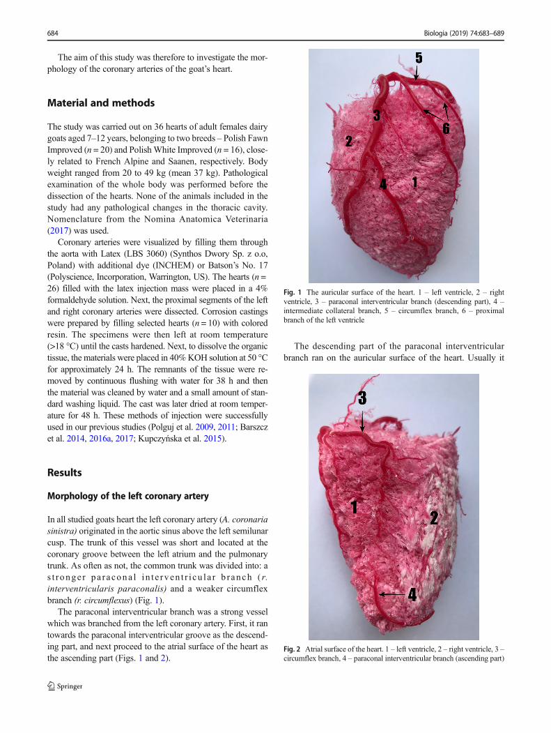
In all studied goats heart the left coronary artery (A. coronariasinistra) originated in the aortic sinus above the left semilunarcusp. The trunk of this vessel was short and located at thecoronary groove between the left atrium and the pulmonarytrunk. As often as not, the common trunk was divided into: as t r onge r pa r acona l i n t e rv en t r i cu l a r b r anch ( r.interventricularis paraconalis) and a weaker circumflexbranch (r. circumflexus) (Fig. 1).
The paraconal interventricular branch was a strong vesselwhich was branched from the left coronary artery. First, it rantowards the paraconal interventricular groove as the descend-ing part, and next proceed to the atrial surface of the heart asthe ascending part (Figs. 1 and 2).
The descending part of the paraconal interventricularbranch ran on the auricular surface of the heart. Usually it
Fig. 1 The auricular surface of the heart. 1 – left ventricle, 2 – rightventricle, 3 – paraconal interventricular branch (descending part), 4 –intermediate collateral branch, 5 – circumflex branch, 6 – proximalbranch of the left ventricle
Fig. 2 Atrial surface of the heart. 1 – left ventricle, 2 – right ventricle, 3 –circumflex branch, 4 – paraconal interventricular branch (ascending part)
684 Biologia (2019) 74:683–689
divides into the left conal branch, (r. coni arteriosi sinister),the proximal, intermediate and distal collateral branches, (r.collateralis proximalis, intermedius et distalis), also the septalbranches, (rr. septales) and lots of small vessels to the lateralwalls of both ventricles on the auricular heart’s surface. Weobserved also the branch of the interventricular septum whichoriginated from the paraconal interventricular branch.
In all studied goats, we observed that:
– the left conal branch, originated from the right side of theparaconal interventricular branch,
– the proximal collateral branch was the smallest lateralvessel,
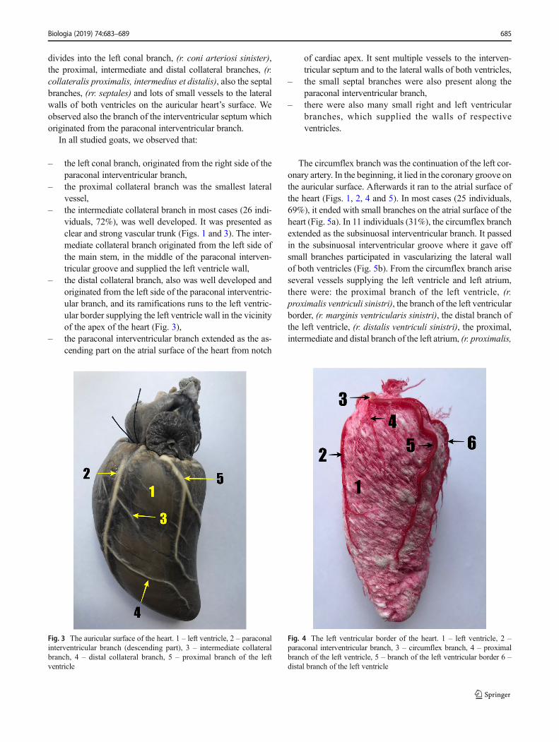
– the intermediate collateral branch in most cases (26 indi-viduals, 72%), was well developed. It was presented asclear and strong vascular trunk (Figs. 1 and 3). The inter-mediate collateral branch originated from the left side ofthe main stem, in the middle of the paraconal interven-tricular groove and supplied the left ventricle wall,
– the distal collateral branch, also was well developed andoriginated from the left side of the paraconal interventric-ular branch, and its ramifications runs to the left ventric-ular border supplying the left ventricle wall in the vicinityof the apex of the heart (Fig. 3),
– the paraconal interventricular branch extended as the as-cending part on the atrial surface of the heart from notch
of cardiac apex. It sent multiple vessels to the interven-tricular septum and to the lateral walls of both ventricles,
– the small septal branches were also present along theparaconal interventricular branch,
– there were also many small right and left ventricularbranches, which supplied the walls of respectiveventricles.
The circumflex branch was the continuation of the left cor-onary artery. In the beginning, it lied in the coronary groove onthe auricular surface. Afterwards it ran to the atrial surface ofthe heart (Figs. 1, 2, 4 and 5). In most cases (25 individuals,69%), it ended with small branches on the atrial surface of theheart (Fig. 5a). In 11 individuals (31%), the circumflex branchextended as the subsinuosal interventricular branch. It passedin the subsinuosal interventricular groove where it gave offsmall branches participated in vascularizing the lateral wallof both ventricles (Fig. 5b). From the circumflex branch ariseseveral vessels supplying the left ventricle and left atrium,there were: the proximal branch of the left ventricle, (r.proximalis ventriculi sinistri), the branch of the left ventricularborder, (r. marginis ventricularis sinistri), the distal branch ofthe left ventricle, (r. distalis ventriculi sinistri), the proximal,intermediate and distal branch of the left atrium, (r. proximalis,
Fig. 3 The auricular surface of the heart. 1 – left ventricle, 2 – paraconalinterventricular branch (descending part), 3 – intermediate collateralbranch, 4 – distal collateral branch, 5 – proximal branch of the leftventricle
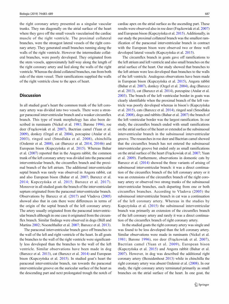
Fig. 4 The left ventricular border of the heart. 1 – left ventricle, 2 –paraconal interventricular branch, 3 – circumflex branch, 4 – proximalbranch of the left ventricle, 5 – branch of the left ventricular border 6 –distal branch of the left ventricle
Biologia (2019) 74:683–689 685

intermedius et distalis atrii sinistri) and also numerous smallramifications (Figs. 1, 3 and 4).
Our study showed that:
– the proximal branch of the left ventricle in 19 goats(53%) was very well developed (Fig. 3), in 16 in-dividuals (44%) was poorly developed, while onegoat (3%) had two well-developed proximalbranches (Fig. 1). It arose from the ventral side ofthe circumflex branch and passed caudo-ventrallythrough the auricular surface to the cardiac atrialsurface were gave off several branches to the lateralwall of the left ventricle and to the apex of heart,
– the branch of left ventricular border originated from theleft circumflex branch near the left auricle (Fig. 4). It gaveoff small branches to the auricular surface of heart,
– the distal branch of the left ventricle originated from theventral side of the circumflex branch and lied ventrallyclose to the left ventricular border. Their ramificationssupplied the walls of the left ventricle on the atrial surfaceof the heart (Figs. 4 and 5a),
– the branches for the left atrium were very small and orig-inated from the medial wall of the circumflex branch,
– the proximal branch of the left atrium, passed on the leftatrium near to the base of heart,
– the intermediate branch of the left atrium, originated closeto the branch of the left ventricular border,
– the distal branch of the left atrium, was the weakestbranch of the circumflex branch,
– apart from the described vessels, we observed also thenumerous weakly developed arteries originating fromthe circumflex branch and vascularizing the left ventricleand left atrium. There were the left ventricular branchesand the left atrial branches.
Morphology of the right coronary artery
The right coronary artery (A. coronaria dextra) was less de-veloped than the left coronary artery (Fig. 6). In all studiedgoat’s heart it arose from aortic sinus above to the right semi-lunar cusp. The vessel was located into the coronary groovebetween the right auricle and the pulmonary trunk. Next itreached the right ventricular border and gave off ramificationsto the right atrium and right ventricle. In 35 individuals (97%)the right coronary artery ended as a small ramifications on theatrial surface of the heart. Only in one case (3%) the rightcoronary artery prolongated as the subsinuosal interventricu-lar branch and ran in the subsinuosal interventricular groove.
In our goat’s studies the several vessels arise from the rightcoronary artery. There were: the right conal branch (r. coniarteriosi dextra), the proximal, intermediate and distal collat-eral branch (r. collateralis proximalis, intermedius et distalis)and also numerous small ramifications to the lateral walls ofthe right ventricle on the atrial surface of the heart (Fig. 6). Inall specimens the right conal branch, was continuation of theright coronary artery from its right side and surrounded thearterial cone. We observed also that all collateral branches of
Fig. 5 The atrial surface of the heart (a and b). 1 – left ventricle, 2 –circumflex branch,− 3 – distal branch of the left ventricle, 4 – final branchof the circumflex branch, 5 – subsinuosal interventricular branch insubsinuosal interventricular groove
Fig. 6 The atrial surface of the heart. 1 – right coronary artery, 2 – rightconal branch, 3 – proximal collateral branch, 4 – intermediate collateralbranch, 5 – distal collateral branch
686 Biologia (2019) 74:683–689
the right coronary artery presented as a singular vasculartrunks. They ran diagonally on the atrial surface of the heartwhere they gave off the small vessels vascularised the cardiacmuscle of the right ventricle. The proximal collateralbranches, were the strongest lateral vessels of the right coro-nary artery. They generated small branches running along thewalls of the right ventricle. However the intermediate collat-eral branches, were poorly developed. They originated fromthe stem vessels, approximately half-way along the length ofthe right coronary artery and lied along the walls of the rightventricle.Whereas the distal collateral branches, ran from bothside of the stem vessel. Their ramifications supplied the wallsof the right ventricle close to the apex of heart.
Discussion
In all studied goat’s heart the common trunk of the left coro-nary artery was divided into two vessels. There were a stron-ger paraconal interventricular branch and a weaker circumflexbranch. This type of trunk morphology has also been de-scribed in ruminants (Nickel et al. 1981; Barone 1996), roedeer (Frąckowiak et al. 2007), Bactrian camel (Yuan et al.2009), donkey (Ozgel et al. 2004), porcupine (Atalar et al.2003), ringed seal (Smodlaka et al. 2008), chinchilla(Ozdemir et al. 2008), cat (Barszcz et al. 2014, 2016b) andEuropean bison (Kupczyńska et al. 2015). Whereas Baharet al. (2007) reported that in the Angora rabbit, the commontrunk of the left coronary artery was divided into the paraconalinterventricular branch, the circumflex branch and the proxi-mal branch of the left atrium. The additional interventricularseptal branch was rarely was observed in Angora rabbit, catand also European bison (Bahar et al. 2007; Barszcz et al.2014; Kupczyńska et al. 2015; Barszcz et al. 2017).Moreover in all studied goats the branch of the interventricularseptum originated from the paraconal interventricular branch.Observations by Barszcz et al. (2017) and Vladova (2005)showed also that in cats there were differences in terms ofthe origin of the septal branch of the left coronary artery.The artery usually originated from the paraconal interventric-ular branch although in one case it originated from the circum-flex branch. Similar findings were observed in dogs (Büll andMartins 2002; Noestelthaller et al. 2007; Barszcz et al. 2013).
The paraconal interventricular branch gave off branches tothe wall of the left and right ventricle of the heart. In all goatsthe branches to the wall of the right ventricle were significant-ly less developed than the branches to the wall of the leftventricle. Similar observations have been made in dog(Barszcz et al. 2013), cat (Barszcz et al. 2014) and Europeanbison (Kupczyńska et al. 2015). In studied goat’s heart theparaconal interventricular branch, ran towards the paraconalinterventricular groove on the auricular surface of the heart asthe descending part and next prolongated trough the notch of
cardiac apex on the atrial surface as the ascending part. Theseresults were observed also in roe deer (Frąckowiak et al. 2007)and European bison (Kupczyńska et al. 2015). Additionally, inour study the proximal collateral branch was the smallest ram-ification of the paraconal interventricular branch in contractwith the European bison were observed two or three welldeveloped lateral vessels (Kupczyńska et al. 2015).
The circumflex branch in goats gave off ramifications tothe left atrium and left ventricle and also small branches on theatrial surface of the heart. Our study showed that branches tothe left atrium were less developed than branches to the wallsof the left ventricle. Analogous observations have been madein European bison (Kupczyńska et al. 2015), Angora rabbit(Bahar et al. 2007), donkey (Ozgel et al. 2004), dog (Barszczet al. 2013), cat (Barszcz et al. 2014), porcupine (Atalar et al.2003). The branch of the left ventricular border in goats wasclearly identifiable when the proximal branch of the left ven-tricle was poorly developed whereas in bison’s (Kupczyńskaet al. 2015), cats (Barszcz et al. 2014), ringed seal (Smodlakaet al. 2008), dogs and rabbits (Bahar et al. 2007) the branch ofthe left ventricular border was the largest ramification. In ourstudy, the circumflex branch ended with small ramificationson the atrial surface of the heart or extended as the subsinuosalinterventricular branch in the subsinuosal interventriculargroove. The researches in roe deer and Bactrian camel showedthat the circumflex branch has not entered the subsinuosalinterventricular groove but ended only as small ramificationson the atrial surface of the heart (Frąckowiak et al. 2007; Yuanet al. 2009). Furthermore, observations in domestic cats byBarszcz et al. (2014) showed the three variants of arising ofsubsinuosal interventricular branch: it was a direct continua-tion of the circumflex branch of the left coronary artery or itwas an extensions of the circumflex branch of the right coro-nary artery or observed two strong trunks of the subsinuosalinterventricular branches, each departing from one or bothcircumflex branches. According to Vladova (2005) thesubsinuosal interventricular branch in cat was a continuationof the left coronary artery. Whereas in the studies byKupczyńska et al. (2015) the subsinuosal interventricularbranch was primarily an extension of the circumflex branchof the left coronary artery and rarely it was a direct continua-tion of the circumflex branch of right coronary artery.
In the studied goats the right coronary artery was single andwas found to be less developed than the left coronary artery.Similar observations were made in ruminants (Nickel et al.1981; Barone 1996), reo deer (Frąckowiak et al. 2007),Bactrian camel (Yuan et al. 2009), European bison(Kupczyńska et al. 2015) and Angora rabbit (Bahar et al.2007). However, in dog was described the additional rightcoronary artery (Bezuidenhout 2013) while in chinchilla theright coronary artery was absent Ozdemir et al. (2008). In ourstudy, the right coronary artery terminated primarily as smallbranches on the atrial surface of the heart. In one goat, the
Biologia (2019) 74:683–689 687
right coronary artery reached the subsinuosal interventriculargroove and extended into the subsinuosal interventricularbranch. Similar findings were made in European bison byKupczyńska et al. (2015). Observations in bison showed thatthe right coronary artery usually ended in subsinuosal inter-ventricular groove as a few small vessels or rarely itprolongated as the subsinuosal interventricular branch.
Numerous studies descriptions of the blood supply to theheart in different species of domestic and wild animals. Alsoin the arterial supply of the goats heart we have observedindividual differences.
Compliance with ethical standards
Conflict of interest The authors declare that they have no conflict ofinterest.
Ethical approval The study were carried out in accordancewith the localethical committee.
Open Access This article is distributed under the terms of the CreativeCommons At t r ibut ion 4 .0 In te rna t ional License (h t tp : / /creativecommons.org/licenses/by/4.0/), which permits unrestricted use,distribution, and reproduction in any medium, provided you give appro-priate credit to the original author(s) and the source, provide a link to theCreative Commons license, and indicate if changes were made.
Publisher’s note Springer Nature remains neutral with regard to jurisdic-tional claims in published maps and institutional affiliations.
References
Atalar Ö, Yilmaz S, İlkay E, Burma O (2003) Investigation of coronaryarteries in the porcupine (Hystrix cristata) by latex injection andangiography. Ann Anat 185:373–376
Bahar S, Ozdemir V, Eken E, Tipirdamaz S (2007) The distribution of thecoronary arteries in the angora rabbit. Anat Histol Embryol 36:321–327. https://doi.org/10.1111/j.1439-0264.2007.00770.x
Barone R (1996) Anatomie comparée des mammifères domestiques.Tome 5. Angiologie. VIGOT, Paris
Barszcz K, Kupczyńska M, Janczyk P, Dzierzęcka M, Jańczak D (2016a)Venous drainage of the heart in the domestic cat. Med Weter 72:186–190
Barszcz K, Kupczyńska M, Klećkowska-Nawrot J, Janczyk P, KrasuckiK, Wąsowicz M (2014) Arterial coronary circulation in cats. MedWeter 70:373–377
Barszcz K, Kupczyńska M, Klećkowska-Nawrot J, Skibniewski M,Janczyk P (2016b) Morphology of coronary ostia in domestic short-hair cat. Anat Histol Embryol 45:81–87. https://doi.org/10.1111/ahe.12174
Barszcz K, Kupczyńska M, Polguj M, Klećkowska-Nawrot J, JaneczekM, Goździewska-Harłajczuk K, Dzierzęcka M, Janczyk P (2017)Morphometry of the coronary ostia and the structure of coronaryarteries in the shorthair domestic cat. PLoS One 12(10):e0186177.https://doi.org/10.1371/journal.pone.0186177
Barszcz K, Kupczyńska M, Wąsowicz M, Czubaj N, Sokołowski W(2013) Patterns of the arterial vascularization of the dog’s heart.Med Weter 69:531–534
Besoluk K, Tipirdamaz S (2001) Comparative macroanatomic investiga-tions of the venous drainage of the heart in Akkaraman sheep andangora goats. Anat Histol Embryol 30:249–252
Bezuidenhout A (2013) The heart and arteries. In: Evans HE, de LahuntaA (ed) Miller’s anatomy of the dog, 4th edn. Elsevier Saunders, St.Louis, pp 428–504
Büll ML, Martins MR (2002) Study of the arterial coronary circulation inthe dog (Canis familiaris). Rev Chil Anat 20:117–123. https://doi.org/10.4067/S0716-98682002000200001
Chen X, Chu GJ,Wang FY, Zhu YF, Zhang B, Zhao XX, Qin YW, Ge JB(2015) Transcatheter aortic valve implantation assisted withmicrocatheter: a new method to avoid coronary artery obstruction.Chin Med J 128:740–744. https://doi.org/10.4103/0366-6999.152473
Du Z, Zhu Z, Yue B, Li Z, Wang Y (2018) Feasibility and safety of acemented PEEK-on-PE knee replacement in a goat model: a prelim-inary study. Artif Organs. https://doi.org/10.1111/aor.13101
Frąckowiak H, Jasiczak K, Pluta K, Godynicki S (2007) Coronary arter-ies of the roe deer (Capreolus capreolus; Linnaeus 1758) heart. Pol JVet Sci 10:105–108
Ghazi SR, Tadjalli M (1993) Coronary arterial anatomy of the one-humped camel (Camelus dromedarius). Vet Res Commun 17:163–170. https://doi.org/10.1007/BF01839161
Kupczyńska M, Barszcz K, Olbrych K, Polguj M, Wysiadecki G, TopolM, Klećkowska-Nawrot J (2015) Coronary arteries of the Europeanbison (Bison bonasus). Acta Vet Scand 57(82). https://doi.org/10.1186/s13028-015-0173-4
Lukovsky-Akhsanov N, Keating MK, Spivey P, Lathrop GW Jr, PowellN, Levin ML (2016) Assessment of domestic goats as models forexperimental and natural infection with the north American isolateof Rickettsia slovaca. PLoS One 11(10):e0165007. https://doi.org/10.1371/journal.pone.0165007
Nickel R, Schummer A, Seiferle E (1981) The anatomy of the domesticanimals. Volume 3. The circulatory system, the skin, and the cuta-neous organs of the domestic mammals. Verlag Paul Parey, Berlinand Hamburg
Noestelthaller A, Probst A, König HE (2007) Branching patterns of theleft main coronary artery in the dog demonstrated by the use ofcorrosion casting technique. Anat Histol Embryol 36:33–37.https://doi.org/10.1111/j.1439-0264.2006.00711.x
Nomina Anatomica Veterinaria (2017) International Committee onVeterinary Gross Anatomical Nomenclature (I.C.V.G.A.N.), 6thedn. Editorial Committee Hanover (Germany), Ghent (Belgium),Columbia, MO (U.S.A.), Rio de Janeiro (Brazil) with permissionof the World Association of Veterinary Anatomists (W.A.V.A)
Ozdemir V, Çevik-Demirkna A, Türkmenoğlu I (2008) The right coro-nary artery is absent in the chinchilla (Chinchilla lanigera). AnatHistol Embryol 37:114–117. https://doi.org/10.1111/j.1439-0264.2007.00803.x
Ozgel O, Haligur (Ç)A, Dursun N, Karakurum E (2004) Themacroanatomy of coronary arteries in donkeys (Equus asinus L.).Anat Histol Embryol 33:278–283. https://doi.org/10.1111/j.1439-0264.2004.00548.x
Polguj M, Jędrzejewski KS, Dyl Ł, Topol M (2009) Topographic andmorphometric comparison study of the terminal part of human andbovine testicular arteries. Fol Morphol (Warsz) 68:271–276
Polguj M, Jedrzejewski KS, Topol M (2011) Angioarchitecture of thebovine spermatic cord. J Morphol 272:497–502. https://doi.org/10.1002/jmor.10929
688 Biologia (2019) 74:683–689

Shiraishi Y, Yambe T, Yoshizawa M, Hashimoto H, Yamada A, Miura H,Hashem M, Kitano T, Shiga T, Homma D (2012) Examination ofmitral regurgitation with a goat heart model for the development ofintelligent artificial papillary muscle. Conf Proc IEEE EngMed BiolSoc 2012:6649–6652. https://doi.org/10.1109/EMBC.2012.6347519
Smodlaka H, Henry RW, Schumacher J, Reed RB (2008) Macroscopic anat-omy of the heart of the ringed seal (Phoca hispida). Anat HistolEmbryol 37:30–35. https://doi.org/10.1111/j.1439-0264.2007.00791.x
Vandersteene J, Baert E, Schauvliege S, Vandevelde K, Dewaele F, DeSomer F, Van Roost D (2018) A non-hydrocephalic goat experimental
model to evaluate a ventriculosinus shunt. Lab Anim 52:504–514.https://doi.org/10.1177/0023677217753976
Vladova D (2005) Ventricular coronary pattern in the cat. TJS 3:44–49Yuan G, Ma J, Ye W, Bai Z, Wang J (2009) Macroanatomy of coronary
arteries in Bactrian camel (Camelus bactrianus). Vet Res Commun33:367–377. https://doi.org/10.1007/s11259-008-9185-0
Zhang Y, Wang YT, Shan ZL, Guo HY, Guan Y, Yuan HT (2015) Role ofinflammation in the initiation and maintenance of atrial fibrillationand the protective effect of atorvastatin in a goat model of asepticpericarditis. Mol Med Rep 11:2615–2623. https://doi.org/10.3892/mmr.2014.3116
Biologia (2019) 74:683–689 689