Embed Size (px)
Citation preview
Small Molecule Therapeutics
Trastuzumab-Based PhotoimmunotherapyIntegrated with Viral HER2 Transduction InhibitsPeritoneally Disseminated HER2-Negative CancerMichihiro Ishida1, Shunsuke Kagawa1, Kyoko Shimoyama1, Kiyoto Takehara1,Kazuhiro Noma1, Shunsuke Tanabe1, Yasuhiro Shirakawa1, Hiroshi Tazawa1,2,Hisataka Kobayashi3, and Toshiyoshi Fujiwara1
Abstract
Peritoneal dissemination is the most frequent metastasis ingastric cancer and is associated with poor prognosis. The lack ofparticular target antigens in gastric cancer other than HER2 hashampered the development of treatments for peritoneal dissem-inationof gastric cancer.Wehypothesized thatHER2-extracellulardomain (HER2-ECD) gene transduction combined with trastu-zumab-based photoimmunotherapy (PIT) might provide excel-lent and selective antitumor effects for peritoneal disseminationof gastric cancer. In vitro, adenovirus/HER2-ECD (Ad/HER2-ECD)efficiently transduced HER2-ECD into HER2-negative gastric can-cer cells. Trastuzumab-IR700 (Tra-IR700)–mediated PIT inducedselective cell death of HER2-ECD–transduced tumor cells. Ad/HER2-ECD also induced homogenous expression of HER2 in
heterogeneous gastric cancer cells, resulting in uniform sensitivityof the cells to Tra-IR700–mediated PIT. Anti-HER2 PIT integratedwith adenoviral HER2-ECD gene transfer was applied in micebearing peritoneal dissemination ofHER2-negative gastric cancer.Intraperitoneal administration of Ad/HER2-ECD and Tra-IR700with PIT inhibited peritoneal metastasis and prolonged the sur-vival of mice bearing MKN45. Furthermore, minimal side effectsallowed the integrated therapy to be used repeatedly, providingbetter control of peritoneal dissemination. In conclusion, thenovel therapy of molecular-targeted PIT integrated with genetransfer technology is a promising approach for the treatment ofperitoneal dissemination in gastric cancer.Mol Cancer Ther; 15(3); 1–10. �2016 AACR.
IntroductionGastric cancer is one of themost commonmalignancies world-
wide,making it the third leading cause of cancer-related death (1).Although the localized stage of gastric cancer can be treatedcuratively with surgical resection, it is frequently diagnosed asfar advanced disease (2). In addition, even after an optimalcurative surgery, cancer recurrence is sometimes inevitable (3, 4).
Peritoneal dissemination, which is the most frequent mode ofmetastasis in gastric cancer (2, 4), is associated with poor prog-nosis (5, 6). The lack of treatment success is due to the difficulty ofselectively delivering anticancer drugs to peritoneal lesions whilesparing normal tissue. Therefore, a novel therapeutic strategy thatcan provide highly selective antitumor effects against peritonealdissemination is desired.
Photoimmunotherapy (PIT) is a novel cancer therapyemploying amAb conjugated to a photosensitizer, IRDye700DX(mAb-IR700). PIT provides a highly specific cytotoxicity totumor cells expressing particular antigens (7). PIT could workonly where mAb-IR700 binds to the targeted cell membranefollowing the irradiation of near-infrared (NIR) light. Therefore,its ability to target tumors fully depends on the nature ofantibody conjugated to IRDye700DX. Trastuzumab, a human-ized mAb for HER2, is the only molecular-targeting anticancerdrug that improves the survival of patients with HER2-positiveadvanced gastric cancer (8, 9). Hence, trastuzumab-based PITmight be an ideal candidate as a novel targeted therapy even forperitoneally disseminated gastric cancer (10).
Mitsunaga and colleagues showed that Tra-IR700–mediatedPIT has promising antitumor effects on HER2-positive cancer(7). However, before Tra-IR700–mediated PIT is widely appliedto the treatment of peritoneal dissemination of gastric cancer,some problems remain to be addressed. Only 12.2% to 22.1%of gastric cancers are HER2 positive (9, 11–13). Moreover,peritoneal dissemination of gastric cancer has an extremelylow HER2 expression rate of 2.9% as HER2-positive status ismore common among intestinal-type cells than diffuse-typecells that often cause peritoneal dissemination (14, 15). Inaddition, gastric cancer itself exhibits intratumoral and inter-tumoral HER2 heterogeneity (16–18). Thus, even a HER2-postive tumor contains cells with different levels of HER2expression, which leads to the inaccurate assessment of HER2status, leading to therapeutic resistance of HER2-targeted ther-apy (19–21). The lack of particular target antigens in gastric
1Department of Gastroenterological Surgery, Okayama UniversityGraduate School of Medicine, Dentistry and Pharmaceutical Sciences,Okayama, Japan. 2Center for Innovative Clinical Medicine, OkayamaUniversity Hospital, Okayama, Japan. 3Molecular Imaging Program,Center for Cancer Research, National Cancer Institute, National Insti-tutes of Health, Bethesda, Maryland.
Note: Supplementary data for this article are available at Molecular CancerTherapeutics Online (http://mct.aacrjournals.org/).
Corresponding Author: Shunsuke Kagawa, Okayama University GraduateSchool of Medicine, Dentistry and Pharmaceutical Sciences, 2-5-1 Shikata-cho,Kita-ku, Okayama 7008558, Japan. Phone: 81-86-235-7257; Fax: 81-86-221-8775;E-mail: [email protected]
doi: 10.1158/1535-7163.MCT-15-0644
�2016 American Association for Cancer Research.
MolecularCancerTherapeutics
www.aacrjournals.org OF1
Research. on September 14, 2018. © 2016 American Association for Cancermct.aacrjournals.org Downloaded from
Published OnlineFirst February 1, 2016; DOI: 10.1158/1535-7163.MCT-15-0644
cancer also hampers the success of molecular-targeted therapy(22). Consequently, cancer-targeted therapy cannot be simplyapplied as a therapy of choice for peritoneal dissemination ofgastric cancer.
To overcome these limitations, we previously developed anadenoviral vector that expresses the HER2-extracellular domain(HER2-ECD) on the cancer cell membrane, Ad/HER2-ECD.Ad/HER2-ECD induced exogenous HER2-ECD overexpressionon HER2-negative cancer cells and successfully sensitized themto trastuzumab (23). In addition, we demonstrated that theintegrated therapy of Ad/HER2-ECD and Tra-IR700–mediatedPIT effectively and selectively killed HER2-negative breast cancercells in vitro, suggesting that the integration of gene transductionwith PIT expands molecular-targeted therapy even for target-negative cancer (24).
Here, we investigated the therapeutic effects of this integrationtherapy for peritoneal dissemination of HER2-negative gastriccancer in vivo. The approach used in the present study overcomesthe limitations of HER2-mediated therapy and provides a noveltherapeutic strategy for the peritoneal dissemination of gastriccancer.
Materials and MethodsCell lines and cell cultures
The human gastric adenocarcinoma cell lines MKN1 andMKN45 were obtained from Human Science Research ResourcesBank (Osaka, Japan). The HER2-expressing human gastric ade-nocarcinoma cell N87 was obtained from the American TypeCulture Collection. MKN45/Luc and SKOV-3/Luc, a human ovar-ian cancer cell line, both stably express luciferase and werepurchased from the Japanese Collection of Research BioresourcesCell Bank (Osaka, Japan) and Cell Biolabs, Inc., respectively. Theauthentication was not performed by the authors. All gastriccancer cell lines were cultured in RPMI 1640 medium (Sigma-Aldrich), and the ovarian cancer cell line was cultured in DMEM(high glucose) with 0.1 mmol/L nonessential amino acids (MPBiomedicals). The media were supplemented with 1% penicillin/streptomycin (Sigma-Aldrich) and 10% FBS. Cells were main-tained at 37�C in ahumidified incubator at an atmosphere of 95%air and 5% CO2.
Recombinant adenovirusA replication-deficient adenoviral vector expressing the
extracellular and transmembrane domains of HER2 (Ad/HER2-ECD) was constructed, expanded, and purified asdescribed previously (23). The titer of virus vector was deter-mined by a plaque assay using HEK 293 cells. Adenovirus/GFP(Ad/GFP), which expressed GFP, was used as control adeno-virus (25).
Synthesis of IR700-conjugated trastuzumabA water-soluble, IRDye 700DX NHS ester was obtained from
LI-COR Bioscience. Trastuzumab was purchased from ChugaiPharmaceutical Co. Conjugation of IR700 with trastuzumabwas performed according to previous reports (7, 10). Briefly,trastuzumab was incubated with IR700 in Na2HPO4 at roomtemperature for 15 minutes. The mixture was purified with aSephadex G50 column (PD-10; GE Healthcare UK Ltd.). Theprotein concentration was determined with a BIO-RAD proteinassay kit (Bio-Rad).
Western blotting analysisMKN1, MKN45, and N87 cells (2� 105 cells) were seeded into
6-well plates. Cells were washed with cold PBS and lysed with theSDSbuffer. Equivalent amounts of protein fromwhole-cell lysateswere loaded into each lane of an 8% SDS–polyacrylamide geland electrophoretically transferred to Hybond-polyvinylidenedifluoride transfer membranes (GE Healthcare UK Ltd.). Mem-branes were incubated with primary antibodies against HER2-ECD(ThermoScientific.) overnight at 4�Cand visualized using anAmersham ECL chemiluminescence system (GE Healthcare UKLtd.) according to the manufacturer's protocol. Equal loading ofsamples was confirmed by stripping each blot and reprobing withanti–b-actin antibody (Sigma-Aldrich). In this experiment,MKN1and MKN45 cells were infected with Ad/HER2-ECD at a differentmultiplicity of infection (MOI; 0, 20, 50, and/or 100) for 24and/or 48 hours. Ad/GFP was used as a control adenovirus.
Flow cytometric analysisCells were fixed with 4% paraformaldehyde for 10 minutes
without permeabilization and labeled with the primary antibo-dies at room temperature for 45 minutes. APC-conjugated anti-human ErbB2 (HER2) monoclonal mouse antibody (R&D Sys-tems Inc.) and phycoerythrin (PE)-conjugated anti-human epi-thelial cell adhesion molecule (EpCAM) monoclonal mouseantibody (BD Biosciences) were used for the confirmation ofHER2-ECD expression and the identification of MKN45 cells,respectively. APC-conjugated mouse IgG antibody (Miltenyi Bio-tec, K.K.) and PE-conjugated mouse IgG antibody (BD Bios-ciences) were also used as each isotype control antibody. Aftercells were washed with PBS and trypsinized, flow cytometry wasperformed with a FACS instrument (BD Biosciences). The inten-sity of staining was calculated by using the BD-FACS Software(Flow Jo 7.6.1; BD Biosciences). MKN1 and MKN45 cells wereinfected with Ad/HER2-ECD at an MOI of 50 for 48 hours.
ImmunohistochemistryCells were fixedwith 4%paraformaldehyde for 30minutes and
then washed with PBS. After blocking with blocking one reagent(Nacalai Tesque, Inc.), cells were labeled with the primary anti-bodies for 1 hour. The secondary antibodies were also reacted for1 hour. To detect HER2 expression, the anti-HER2 extracellulardomain monoclonal mouse antibody (R&D Systems Inc.) withFITC-conjugated polyclonal goat secondary antibody to mouseIgG (abcam) was used. APC-conjugated mouse monoclonal anti-human HER2-ECD antibody (R&D Systems Inc.) was also usedfor HER2 detection. PE-conjugated anti-human EpCAM mono-clonal mouse antibody (BD Biosciences) was used to identifygastric cancer cells. DAPI was used for nuclear and chromosomalcounterstaining. Tra-IR700 could be detected by IR700 fluores-cence. Cells were subsequently photographed, and the mergedimages were overlaid using a confocal laser scanning biologicalmicroscope (FV10i; Olympus).
In vitro PIT with adenovirus/HER2-ECDMKN1 and MKN45 cells were seeded on plates (4 plates
or wells per group) for 24 hours. The cells were infected withAd/HER2-ECD or Ad/GFP at an MOI of 50 for 48 hours.Trastuzumab and Tra-IR700 (10 mg/mL) were added for6 hours, and cells were irradiated with NIR light at 10 J/cm2
(20 mW/cm2, 500 s). The irradiation was performed by anirradiator using a light emitting diode (LED) light with a peak at
Ishida et al.
Mol Cancer Ther; 15(3) March 2016 Molecular Cancer TherapeuticsOF2
Research. on September 14, 2018. © 2016 American Association for Cancermct.aacrjournals.org Downloaded from
Published OnlineFirst February 1, 2016; DOI: 10.1158/1535-7163.MCT-15-0644
690 nm. The irradiation power density was measured by energymeter console, PM100D (Thorlabs, Inc.). The morphologic cellchanges after treatment were observed under a fluorescencemicroscope (IX71; Olympus). The time-lapse movies weretaken serially at hourly intervals for 80 hours just after PITusing a confocal laser scanning biological microscope withbuilt-in culture incubator (FV10i; Olympus).
Cell death and viability assayTo assess theHER2 target selectivity cell death,N87 cells labeled
with Cell Tracker Blue CMAC dye (Life Technologies) were cocul-tured with unlabeled MKN1 cells. Cocultured cells were infectedwith Ad/HER2-ECD at an MOI of 50 for 48 hours and incubatedwith Tra-IR700 (10mg/mL) for 1 hour. The irradiation ofNIR lightwas performed at 5 J/cm2. After the irradiation, cells were stainedwith propidium iodide (PI; 1 mg/mL) to identify dead cells. Cellviability for quantitative evaluation was determined using an XTTCell Proliferation Kit II (Roche Life Science) according to themanufacturer's protocol.
Ex vivo experimentsAthymic female BALB/c nu/nu nudemice were purchased from
CLEA. The animal care and experimental procedures were con-ducted in accordance with the regulations of the Animal Care andUse Committee of Okayama University. Normal mouse perito-neal cells were collected from 6-week-old nude mice. Briefly, 5 ccof RPMI1640 medium (Sigma-Aldrich) was injected into theabdominal cavity, and then the abdomen was massaged. Theascites was collected, and cells were isolated by a centrifugalseparator. The cells collected from normal mice were seeded intotwo culture conditions, a single culture (normalmouse peritonealcells, 2 � 105 cells) and a coculture (mixed normal mouseperitoneal cells with MKN45 cells, 1 � 105 cells of each type).We analyzed the cells using two-color flow cytometry (BD Bios-ciences) with APC-conjugated anti-human HER2 monoclonalmouse antibody (R&D Systems Inc.) and PE-conjugated anti-human EpCAM monoclonal mouse antibody (BD Biosciences).To confirm the HER2 expression and Tra-IR700 conjugation onfloating tumor cells in the abdominal cavity of peritoneal dis-semination xenografted mice, immunohistochemistry was per-formed with APC-conjugated anti-HER2 antibody and PE-con-jugated anti-EpCAM antibody, as mentioned above. Ad/HER2-ECD at a dose of 1 � 108 plaque-forming units (pfu) and Tra-IR700 (80 mg) were injected into the peritoneal cavity of mice ondays 5 and 7 after tumor injection, respectively. The free-floatingcells in the peritoneal cavity were collected by lavage washmethods.
Evaluation of antitumor effects in the peritoneal disseminationmouse model
We established the peritoneal dissemination xenograftedmouse model by i.p. administration of MKN45 cells (1 � 107
cells) into 6- to 8-week-old nude mice using a 22-gauge catheterneedle. To assess the efficiency of adenoviral gene transfer to theperitoneally disseminated tumors, mice were injected withAd/HER2-ECD at a dose of 1 � 108 pfu in 500 mL PBS into theperitoneal cavity 14 days after injection of MKN45 cells. Thesemice were sacrificed 48 hours later. Immunohistochemical anal-ysis of paraffin-embedded tissues was performed using HER2/ErbB2 (D8F12) XP rabbit monoclonal antibody (Cell SignalingTechnology, Inc.), according to the manufacturer's protocol.
To evaluate the antitumor effects, the mice were randomlydivided into the following four groups (each group: n ¼ 5): notreatment (control), treated with Tra-IR700–meditated PIT(IRþPIT), treated with Ad/HER2-ECD–mediated PIT (AdþPIT),and treated with Ad/HER2-ECD with Tra-IR700–mediatedPIT (AdþIRþPIT). Ad/HER2-ECD was i.p. administered 5 daysafter tumor cell injection into the peritoneal cavity at a dose of1 � 108 pfu in 500 mL PBS. Tra-IR700 was also i.p. adminis-tered at 80 mg in 500 mL PBS 48 hours after Ad/HER2-ECDadministration. One day after Tra-IR700 administration,irradiation was performed with 50 J/cm2 of NIR light usingan LED light source (L690-66-60 with Lens550; EPITEX, Inc.)at 690 nm as the peak wavelength. Subcutaneous anesthesiawas used for all procedures. All mice were sacrificed, and thedisseminated peritoneal tumors were resected. The totalweight of tumors per mouse was measured on day 28 aftertumor cell injection.
Evaluation of luciferase activity in peritoneal disseminationmouse model
We assessed the tumor-inhibitory effects of PIT by using theperitoneal dissemination mouse model bearing luciferase-expressing cells, SKOV-3/Luc cells, and MKN45/Luc cells.SKOV-3/Luc and MKN45/Luc cells (5 � 106 of each cell type)were injected into the peritoneal cavity of 6- to 8-week-oldnude mice. Seven days after tumor injection, the fluorescenceintensities of the peritoneal dissemination in the mice weremeasured. The mice bearing SKOV3/Luc cells that exhibitedfluorescence were randomly assigned to three groups: no treat-ment (control), only Tra-IR700 (IR), and Tra-IR700–mediatedPIT (IRþPIT). After the randomization, Tra-IR700 (80 mg) wasi.p. administered, and irradiation with NIR light was performed2 days after Tra-IR700 administration. The mice bearingMKN45/Luc cells were also randomly assigned to one of 7groups as follows: no treatment (control); a single treatmentof Tra-IR700–meditated PIT (IRþPIT�1), Ad/HER2-ECD–
mediated PIT (AdþPIT�1), or Ad/HER2-ECD with Tra-IR700–mediated PIT (AdþIRþPIT�1); and three repeated treatments ofTra-IR700–meditated PIT (IRþPIT�3), Ad/HER2-ECD–mediatedPIT (AdþPIT�3), or Ad/HER2-ECD with Tra-IR700–mediatedPIT (AdþIRþPIT�3). The schedule of the integrated therapyregimen is shown in Fig. 5A. For analyzing fluorescence intensi-ties, the mice were intraperitoneally injected with XenolightRediject D-luciferin (Caliper Life Sciences) at 150mg and imagedunder isoflurane anesthesia after 4 minutes. The in vivo fluores-cence and bioluminescence images were obtained with an IVISLumina imaging system (Xenogen IVIS Lumina II; Caliper LifeSciences), and the image analysis and bioluminescent quantifi-cation were performed by using Living Image software. InMKN45/Lucmice, the survival rate at 50days after tumor injectionwas also assessed.
Statistical analysisData analysis was performed using the two-sided Student t test
or one-way repeatedmeasures ANOVA followed by aDunnett testfor multiple comparisons. Survival curves were estimated usingthe Kaplan–Meier method, and survival differences betweensubgroups were analyzed using the log-rank test. All statisticalanalyses were performed with SPSS for Windows, version 19.0(SPSS, Inc.). P values <0.05 were considered to indicate a statis-tically significant difference.
PIT with Viral HER2 Transduction for HER2-Negative Cancer
www.aacrjournals.org Mol Cancer Ther; 15(3) March 2016 OF3
Research. on September 14, 2018. © 2016 American Association for Cancermct.aacrjournals.org Downloaded from
Published OnlineFirst February 1, 2016; DOI: 10.1158/1535-7163.MCT-15-0644
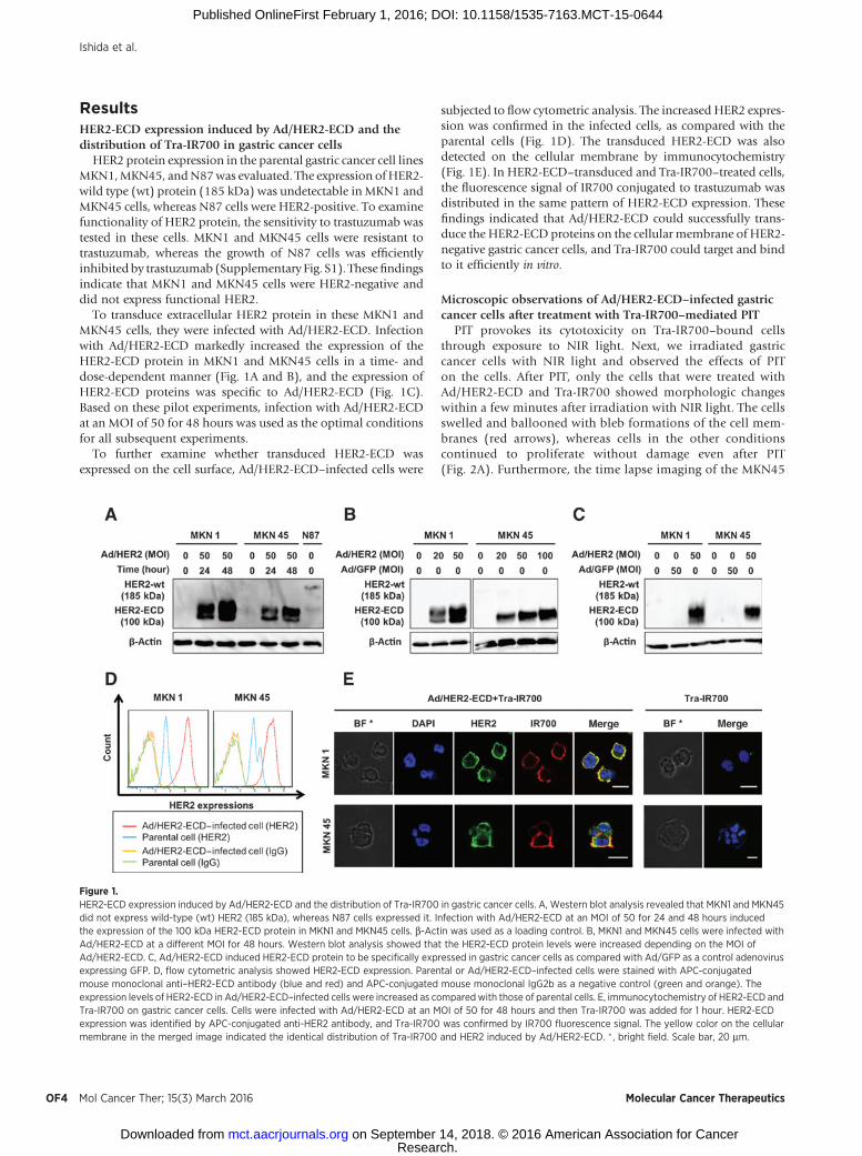
ResultsHER2-ECD expression induced by Ad/HER2-ECD and thedistribution of Tra-IR700 in gastric cancer cells
HER2 protein expression in the parental gastric cancer cell linesMKN1,MKN45, andN87was evaluated. The expression ofHER2-wild type (wt) protein (185 kDa) was undetectable in MKN1 andMKN45 cells, whereas N87 cells were HER2-positive. To examinefunctionality of HER2 protein, the sensitivity to trastuzumab wastested in these cells. MKN1 and MKN45 cells were resistant totrastuzumab, whereas the growth of N87 cells was efficientlyinhibited by trastuzumab (Supplementary Fig. S1). Thesefindingsindicate that MKN1 and MKN45 cells were HER2-negative anddid not express functional HER2.
To transduce extracellular HER2 protein in these MKN1 andMKN45 cells, they were infected with Ad/HER2-ECD. Infectionwith Ad/HER2-ECD markedly increased the expression of theHER2-ECD protein in MKN1 and MKN45 cells in a time- anddose-dependent manner (Fig. 1A and B), and the expression ofHER2-ECD proteins was specific to Ad/HER2-ECD (Fig. 1C).Based on these pilot experiments, infection with Ad/HER2-ECDat an MOI of 50 for 48 hours was used as the optimal conditionsfor all subsequent experiments.
To further examine whether transduced HER2-ECD wasexpressed on the cell surface, Ad/HER2-ECD–infected cells were
subjected to flow cytometric analysis. The increased HER2 expres-sion was confirmed in the infected cells, as compared with theparental cells (Fig. 1D). The transduced HER2-ECD was alsodetected on the cellular membrane by immunocytochemistry(Fig. 1E). In HER2-ECD–transduced and Tra-IR700–treated cells,the fluorescence signal of IR700 conjugated to trastuzumab wasdistributed in the same pattern of HER2-ECD expression. Thesefindings indicated that Ad/HER2-ECD could successfully trans-duce the HER2-ECD proteins on the cellular membrane of HER2-negative gastric cancer cells, and Tra-IR700 could target and bindto it efficiently in vitro.
Microscopic observations of Ad/HER2-ECD–infected gastriccancer cells after treatment with Tra-IR700–mediated PIT
PIT provokes its cytotoxicity on Tra-IR700–bound cellsthrough exposure to NIR light. Next, we irradiated gastriccancer cells with NIR light and observed the effects of PITon the cells. After PIT, only the cells that were treated withAd/HER2-ECD and Tra-IR700 showed morphologic changeswithin a few minutes after irradiation with NIR light. The cellsswelled and ballooned with bleb formations of the cell mem-branes (red arrows), whereas cells in the other conditionscontinued to proliferate without damage even after PIT(Fig. 2A). Furthermore, the time lapse imaging of the MKN45
Figure 1.HER2-ECD expression induced by Ad/HER2-ECD and the distribution of Tra-IR700 in gastric cancer cells. A, Western blot analysis revealed that MKN1 and MKN45did not express wild-type (wt) HER2 (185 kDa), whereas N87 cells expressed it. Infection with Ad/HER2-ECD at an MOI of 50 for 24 and 48 hours inducedthe expression of the 100 kDa HER2-ECD protein in MKN1 and MKN45 cells. b-Actin was used as a loading control. B, MKN1 and MKN45 cells were infected withAd/HER2-ECD at a different MOI for 48 hours. Western blot analysis showed that the HER2-ECD protein levels were increased depending on the MOI ofAd/HER2-ECD. C, Ad/HER2-ECD induced HER2-ECD protein to be specifically expressed in gastric cancer cells as compared with Ad/GFP as a control adenovirusexpressing GFP. D, flow cytometric analysis showed HER2-ECD expression. Parental or Ad/HER2-ECD–infected cells were stained with APC-conjugatedmouse monoclonal anti–HER2-ECD antibody (blue and red) and APC-conjugated mouse monoclonal IgG2b as a negative control (green and orange). Theexpression levels of HER2-ECD in Ad/HER2-ECD–infected cells were increased as comparedwith those of parental cells. E, immunocytochemistry of HER2-ECD andTra-IR700 on gastric cancer cells. Cells were infected with Ad/HER2-ECD at an MOI of 50 for 48 hours and then Tra-IR700 was added for 1 hour. HER2-ECDexpression was identified by APC-conjugated anti-HER2 antibody, and Tra-IR700 was confirmed by IR700 fluorescence signal. The yellow color on the cellularmembrane in the merged image indicated the identical distribution of Tra-IR700 and HER2 induced by Ad/HER2-ECD. � , bright field. Scale bar, 20 mm.
Ishida et al.
Mol Cancer Ther; 15(3) March 2016 Molecular Cancer TherapeuticsOF4
Research. on September 14, 2018. © 2016 American Association for Cancermct.aacrjournals.org Downloaded from
Published OnlineFirst February 1, 2016; DOI: 10.1158/1535-7163.MCT-15-0644
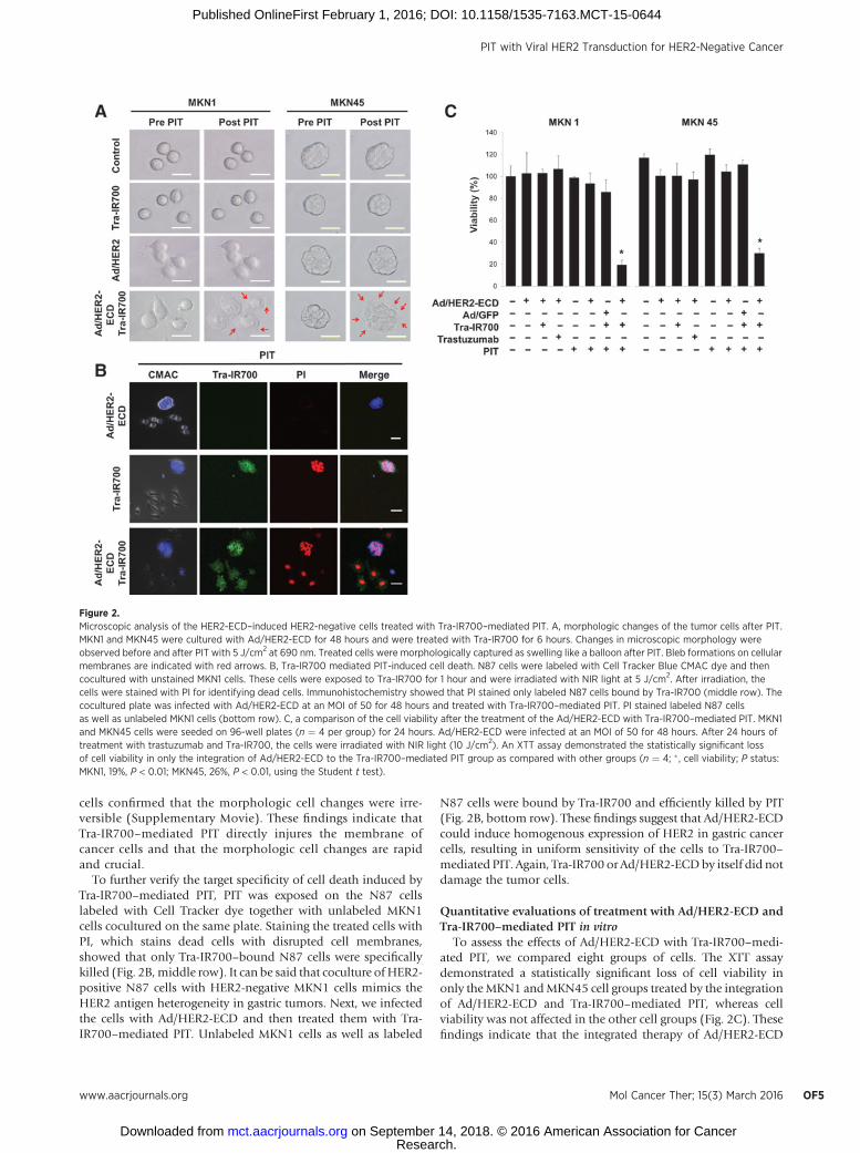
cells confirmed that the morphologic cell changes were irre-versible (Supplementary Movie). These findings indicate thatTra-IR700–mediated PIT directly injures the membrane ofcancer cells and that the morphologic cell changes are rapidand crucial.
To further verify the target specificity of cell death induced byTra-IR700–mediated PIT, PIT was exposed on the N87 cellslabeled with Cell Tracker dye together with unlabeled MKN1cells cocultured on the same plate. Staining the treated cells withPI, which stains dead cells with disrupted cell membranes,showed that only Tra-IR700–bound N87 cells were specificallykilled (Fig. 2B,middle row). It can be said that coculture of HER2-positive N87 cells with HER2-negative MKN1 cells mimics theHER2 antigen heterogeneity in gastric tumors. Next, we infectedthe cells with Ad/HER2-ECD and then treated them with Tra-IR700–mediated PIT. Unlabeled MKN1 cells as well as labeled
N87 cells were bound by Tra-IR700 and efficiently killed by PIT(Fig. 2B, bottom row). These findings suggest that Ad/HER2-ECDcould induce homogenous expression of HER2 in gastric cancercells, resulting in uniform sensitivity of the cells to Tra-IR700–mediated PIT. Again, Tra-IR700 or Ad/HER2-ECDby itself did notdamage the tumor cells.
Quantitative evaluations of treatment with Ad/HER2-ECD andTra-IR700–mediated PIT in vitro
To assess the effects of Ad/HER2-ECD with Tra-IR700–medi-ated PIT, we compared eight groups of cells. The XTT assaydemonstrated a statistically significant loss of cell viability inonly theMKN1 andMKN45 cell groups treated by the integrationof Ad/HER2-ECD and Tra-IR700–mediated PIT, whereas cellviability was not affected in the other cell groups (Fig. 2C). Thesefindings indicate that the integrated therapy of Ad/HER2-ECD
Figure 2.Microscopic analysis of the HER2-ECD–induced HER2-negative cells treated with Tra-IR700–mediated PIT. A, morphologic changes of the tumor cells after PIT.MKN1 and MKN45 were cultured with Ad/HER2-ECD for 48 hours and were treated with Tra-IR700 for 6 hours. Changes in microscopic morphology wereobserved before and after PIT with 5 J/cm2 at 690 nm. Treated cells were morphologically captured as swelling like a balloon after PIT. Bleb formations on cellularmembranes are indicated with red arrows. B, Tra-IR700 mediated PIT-induced cell death. N87 cells were labeled with Cell Tracker Blue CMAC dye and thencocultured with unstained MKN1 cells. These cells were exposed to Tra-IR700 for 1 hour and were irradiated with NIR light at 5 J/cm2. After irradiation, thecells were stained with PI for identifying dead cells. Immunohistochemistry showed that PI stained only labeled N87 cells bound by Tra-IR700 (middle row). Thecocultured plate was infected with Ad/HER2-ECD at an MOI of 50 for 48 hours and treated with Tra-IR700–mediated PIT. PI stained labeled N87 cellsas well as unlabeled MKN1 cells (bottom row). C, a comparison of the cell viability after the treatment of the Ad/HER2-ECD with Tra-IR700–mediated PIT. MKN1and MKN45 cells were seeded on 96-well plates (n ¼ 4 per group) for 24 hours. Ad/HER2-ECD were infected at an MOI of 50 for 48 hours. After 24 hours oftreatment with trastuzumab and Tra-IR700, the cells were irradiated with NIR light (10 J/cm2). An XTT assay demonstrated the statistically significant lossof cell viability in only the integration of Ad/HER2-ECD to the Tra-IR700–mediated PIT group as compared with other groups (n ¼ 4; � , cell viability; P status:MKN1, 19%, P < 0.01; MKN45, 26%, P < 0.01, using the Student t test).
PIT with Viral HER2 Transduction for HER2-Negative Cancer
www.aacrjournals.org Mol Cancer Ther; 15(3) March 2016 OF5
Research. on September 14, 2018. © 2016 American Association for Cancermct.aacrjournals.org Downloaded from
Published OnlineFirst February 1, 2016; DOI: 10.1158/1535-7163.MCT-15-0644
and Tra-IR700–mediated PIT has the potential to eradicate HER2-negative gastric cancer cells with low cytotoxicity.
In vivo experiments with Ad/HER2-ECD and Tra-IR700To apply the integrated therapy for the treatment of peritoneal
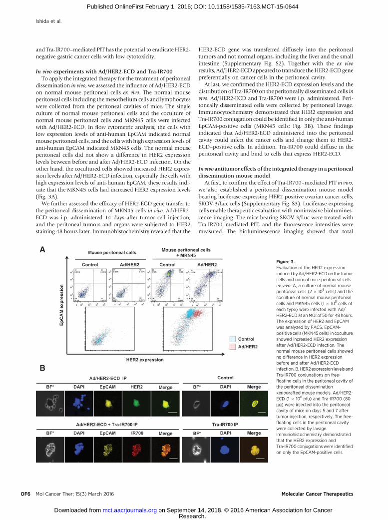
dissemination in vivo, we assessed the influence of Ad/HER2-ECDon normal mouse peritoneal cells ex vivo. The normal mouseperitoneal cells including themesothelium cells and lymphocyteswere collected from the peritoneal cavities of mice. The singleculture of normal mouse peritoneal cells and the coculture ofnormal mouse peritoneal cells and MKN45 cells were infectedwith Ad/HER2-ECD. In flow cytometric analysis, the cells withlow expression levels of anti-human EpCAM indicated normalmouse peritoneal cells, and the cells with high expression levels ofanti-human EpCAM indicated MKN45 cells. The normal mouseperitoneal cells did not show a difference in HER2 expressionlevels between before and after Ad/HER2-ECD infection. On theother hand, the cocultured cells showed increased HER2 expres-sion levels after Ad/HER2-ECD infection, especially the cells withhigh expression levels of anti-human EpCAM; these results indi-cate that the MKN45 cells had increased HER2 expression levels(Fig. 3A).
We further assessed the efficacy of HER2-ECD gene transfer tothe peritoneal dissemination of MKN45 cells in vivo. Ad/HER2-ECD was i.p. administered 14 days after tumor cell injection,and the peritoneal tumors and organs were subjected to HER2staining 48 hours later. Immunohistochemistry revealed that the
HER2-ECD gene was transferred diffusely into the peritonealtumors and not normal organs, including the liver and the smallintestine (Supplementary Fig. S2). Together with the ex vivoresults, Ad/HER2-ECDappeared to transduce theHER2-ECDgenepreferentially on cancer cells in the peritoneal cavity.
At last, we confirmed the HER2-ECD expression levels and thedistribution of Tra-IR700on the peritoneally disseminated cells invivo. Ad/HER2-ECD and Tra-IR700 were i.p. administered. Peri-toneally disseminated cells were collected by peritoneal lavage.Immunocytochemistry demonstrated that HER2 expression andTra-IR700 conjugation could be identified in only the anti-humanEpCAM-positive cells (MKN45 cells; Fig. 3B). These findingsindicated that Ad/HER2-ECD administered into the peritonealcavity could infect the cancer cells and change them to HER2-ECD–positive cells. In addition, Tra-IR700 could diffuse in theperitoneal cavity and bind to cells that express HER2-ECD.
In vivo antitumor effects of the integrated therapy in aperitonealdissemination mouse model
At first, to confirm the effect of Tra-IR700–mediated PIT in vivo,we also established a peritoneal dissemination mouse modelbearing luciferase-expressing HER2-positive ovarian cancer cells,SKOV-3/Luc cells (Supplementary Fig. S3). Luciferase-expressingcells enable therapeutic evaluation with noninvasive biolumines-cence imaging. The mice bearing SKOV-3/Luc were treated withTra-IR700–mediated PIT, and the fluorescence intensities weremeasured. The bioluminescence imaging showed that total
Figure 3.Evaluation of the HER2 expressioninduced byAd/HER2-ECDon the tumorcells and normal mice peritoneal cellsex vivo. A, a culture of normal mouseperitoneal cells (2 � 105 cells) and thecoculture of normal mouse peritonealcells and MKN45 cells (1 � 105 cells ofeach type) were infected with Ad/HER2-ECD at an MOI of 50 for 48 hours.The expression of HER2 and EpCAMwas analyzed by FACS. EpCAM-positive cells (MKN45 cells) in cocultureshowed increased HER2 expressionafter Ad/HER2-ECD infection. Thenormal mouse peritoneal cells showedno difference in HER2 expressionbefore and after Ad/HER2-ECDinfection. B, HER2 expression levels andTra-IR700 conjugations on free-floating cells in the peritoneal cavity ofthe peritoneal disseminationxenografted mouse models. Ad/HER2-ECD (1 � 108 pfu) and Tra-IR700 (80mg) were injected into the peritonealcavity of mice on days 5 and 7 aftertumor injection, respectively. The free-floating cells in the peritoneal cavitywere collected by lavage.Immunohistochemistry demonstratedthat the HER2 expression andTra-IR700 conjugationswere identifiedon only the EpCAM-positive cells.
Ishida et al.
Mol Cancer Ther; 15(3) March 2016 Molecular Cancer TherapeuticsOF6
Research. on September 14, 2018. © 2016 American Association for Cancermct.aacrjournals.org Downloaded from
Published OnlineFirst February 1, 2016; DOI: 10.1158/1535-7163.MCT-15-0644
fluorescence of themice treatedwith Tra-IR700–mediated PITwaslower than that of other mouse groups (Supplementary Fig. S4).These findings indicated that the NIR light applied by externalLED light irradiation could reach themouse peritoneal cavity andthat Tra-IR700–mediated PIT could control the progression of theperitoneal dissemination of HER2-positive cells.
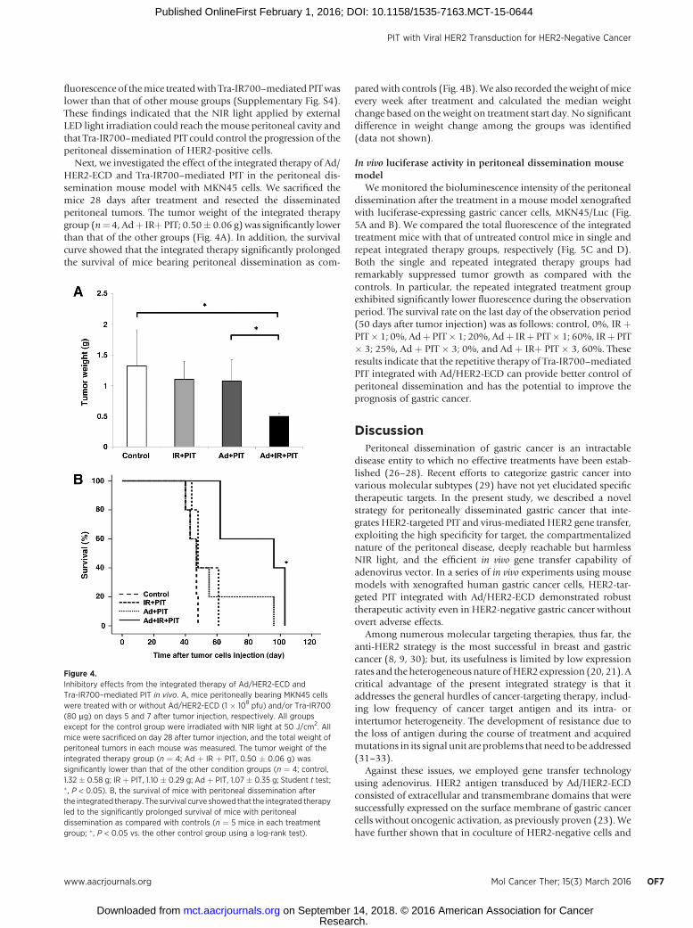
Next, we investigated the effect of the integrated therapy of Ad/HER2-ECD and Tra-IR700–mediated PIT in the peritoneal dis-semination mouse model with MKN45 cells. We sacrificed themice 28 days after treatment and resected the disseminatedperitoneal tumors. The tumor weight of the integrated therapygroup (n¼ 4, Adþ IRþ PIT; 0.50� 0.06 g) was significantly lowerthan that of the other groups (Fig. 4A). In addition, the survivalcurve showed that the integrated therapy significantly prolongedthe survival of mice bearing peritoneal dissemination as com-
paredwith controls (Fig. 4B).We also recorded theweight ofmiceevery week after treatment and calculated the median weightchange based on the weight on treatment start day. No significantdifference in weight change among the groups was identified(data not shown).
In vivo luciferase activity in peritoneal dissemination mousemodel
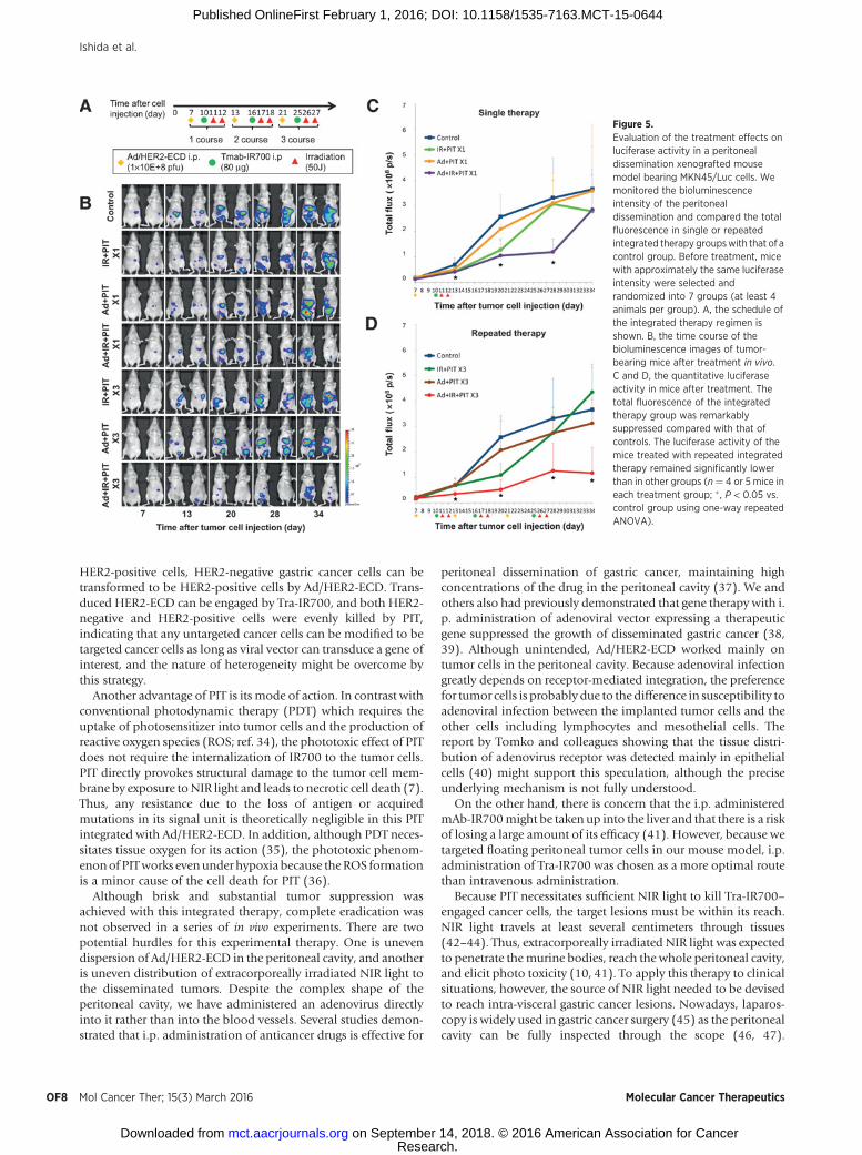
Wemonitored the bioluminescence intensity of the peritonealdissemination after the treatment in a mouse model xenograftedwith luciferase-expressing gastric cancer cells, MKN45/Luc (Fig.5A and B). We compared the total fluorescence of the integratedtreatment mice with that of untreated control mice in single andrepeat integrated therapy groups, respectively (Fig. 5C and D).Both the single and repeated integrated therapy groups hadremarkably suppressed tumor growth as compared with thecontrols. In particular, the repeated integrated treatment groupexhibited significantly lower fluorescence during the observationperiod. The survival rate on the last day of the observation period(50 days after tumor injection) was as follows: control, 0%, IR þPIT� 1; 0%, Adþ PIT� 1; 20%, Adþ IRþ PIT� 1; 60%, IRþ PIT� 3; 25%, Ad þ PIT � 3; 0%, and Ad þ IRþ PIT � 3, 60%. Theseresults indicate that the repetitive therapy of Tra-IR700–mediatedPIT integrated with Ad/HER2-ECD can provide better control ofperitoneal dissemination and has the potential to improve theprognosis of gastric cancer.
DiscussionPeritoneal dissemination of gastric cancer is an intractable
disease entity to which no effective treatments have been estab-lished (26–28). Recent efforts to categorize gastric cancer intovarious molecular subtypes (29) have not yet elucidated specifictherapeutic targets. In the present study, we described a novelstrategy for peritoneally disseminated gastric cancer that inte-grates HER2-targeted PIT and virus-mediated HER2 gene transfer,exploiting the high specificity for target, the compartmentalizednature of the peritoneal disease, deeply reachable but harmlessNIR light, and the efficient in vivo gene transfer capability ofadenovirus vector. In a series of in vivo experiments using mousemodels with xenografted human gastric cancer cells, HER2-tar-geted PIT integrated with Ad/HER2-ECD demonstrated robusttherapeutic activity even in HER2-negative gastric cancer withoutovert adverse effects.
Among numerous molecular targeting therapies, thus far, theanti-HER2 strategy is the most successful in breast and gastriccancer (8, 9, 30); but, its usefulness is limited by low expressionrates and the heterogeneousnature ofHER2expression (20, 21). Acritical advantage of the present integrated strategy is that itaddresses the general hurdles of cancer-targeting therapy, includ-ing low frequency of cancer target antigen and its intra- orintertumor heterogeneity. The development of resistance due tothe loss of antigen during the course of treatment and acquiredmutations in its signal unit are problems that need to be addressed(31–33).
Against these issues, we employed gene transfer technologyusing adenovirus. HER2 antigen transduced by Ad/HER2-ECDconsisted of extracellular and transmembrane domains that weresuccessfully expressed on the surface membrane of gastric cancercells without oncogenic activation, as previously proven (23). Wehave further shown that in coculture of HER2-negative cells and
Figure 4.Inhibitory effects from the integrated therapy of Ad/HER2-ECD andTra-IR700–mediated PIT in vivo. A, mice peritoneally bearing MKN45 cellswere treated with or without Ad/HER2-ECD (1 � 108 pfu) and/or Tra-IR700(80 mg) on days 5 and 7 after tumor injection, respectively. All groupsexcept for the control group were irradiated with NIR light at 50 J/cm2. Allmice were sacrificed on day 28 after tumor injection, and the total weight ofperitoneal tumors in each mouse was measured. The tumor weight of theintegrated therapy group (n ¼ 4; Ad þ IR þ PIT, 0.50 � 0.06 g) wassignificantly lower than that of the other condition groups (n ¼ 4; control,1.32 � 0.58 g; IR þ PIT, 1.10 � 0.29 g; Ad þ PIT, 1.07 � 0.35 g; Student t test;� , P < 0.05). B, the survival of mice with peritoneal dissemination afterthe integrated therapy. The survival curve showed that the integrated therapyled to the significantly prolonged survival of mice with peritonealdissemination as compared with controls (n ¼ 5 mice in each treatmentgroup; � , P < 0.05 vs. the other control group using a log-rank test).
PIT with Viral HER2 Transduction for HER2-Negative Cancer
www.aacrjournals.org Mol Cancer Ther; 15(3) March 2016 OF7
Research. on September 14, 2018. © 2016 American Association for Cancermct.aacrjournals.org Downloaded from
Published OnlineFirst February 1, 2016; DOI: 10.1158/1535-7163.MCT-15-0644
HER2-positive cells, HER2-negative gastric cancer cells can betransformed to be HER2-positive cells by Ad/HER2-ECD. Trans-duced HER2-ECD can be engaged by Tra-IR700, and both HER2-negative and HER2-positive cells were evenly killed by PIT,indicating that any untargeted cancer cells can be modified to betargeted cancer cells as long as viral vector can transduce a gene ofinterest, and the nature of heterogeneity might be overcome bythis strategy.
Another advantage of PIT is its mode of action. In contrast withconventional photodynamic therapy (PDT) which requires theuptake of photosensitizer into tumor cells and the production ofreactive oxygen species (ROS; ref. 34), the phototoxic effect of PITdoes not require the internalization of IR700 to the tumor cells.PIT directly provokes structural damage to the tumor cell mem-brane by exposure toNIR light and leads to necrotic cell death (7).Thus, any resistance due to the loss of antigen or acquiredmutations in its signal unit is theoretically negligible in this PITintegrated with Ad/HER2-ECD. In addition, although PDT neces-sitates tissue oxygen for its action (35), the phototoxic phenom-enonof PITworks evenunder hypoxia because theROS formationis a minor cause of the cell death for PIT (36).
Although brisk and substantial tumor suppression wasachieved with this integrated therapy, complete eradication wasnot observed in a series of in vivo experiments. There are twopotential hurdles for this experimental therapy. One is unevendispersion of Ad/HER2-ECD in the peritoneal cavity, and anotheris uneven distribution of extracorporeally irradiated NIR light tothe disseminated tumors. Despite the complex shape of theperitoneal cavity, we have administered an adenovirus directlyinto it rather than into the blood vessels. Several studies demon-strated that i.p. administration of anticancer drugs is effective for
peritoneal dissemination of gastric cancer, maintaining highconcentrations of the drug in the peritoneal cavity (37). We andothers also had previously demonstrated that gene therapy with i.p. administration of adenoviral vector expressing a therapeuticgene suppressed the growth of disseminated gastric cancer (38,39). Although unintended, Ad/HER2-ECD worked mainly ontumor cells in the peritoneal cavity. Because adenoviral infectiongreatly depends on receptor-mediated integration, the preferencefor tumor cells is probably due to the difference in susceptibility toadenoviral infection between the implanted tumor cells and theother cells including lymphocytes and mesothelial cells. Thereport by Tomko and colleagues showing that the tissue distri-bution of adenovirus receptor was detected mainly in epithelialcells (40) might support this speculation, although the preciseunderlying mechanism is not fully understood.
On the other hand, there is concern that the i.p. administeredmAb-IR700might be taken up into the liver and that there is a riskof losing a large amount of its efficacy (41). However, because wetargeted floating peritoneal tumor cells in our mouse model, i.p.administration of Tra-IR700 was chosen as a more optimal routethan intravenous administration.
Because PIT necessitates sufficient NIR light to kill Tra-IR700–engaged cancer cells, the target lesions must be within its reach.NIR light travels at least several centimeters through tissues(42–44). Thus, extracorporeally irradiated NIR light was expectedto penetrate themurine bodies, reach the whole peritoneal cavity,and elicit photo toxicity (10, 41). To apply this therapy to clinicalsituations, however, the source of NIR light needed to be devisedto reach intra-visceral gastric cancer lesions. Nowadays, laparos-copy is widely used in gastric cancer surgery (45) as the peritonealcavity can be fully inspected through the scope (46, 47).
Figure 5.Evaluation of the treatment effects onluciferase activity in a peritonealdissemination xenografted mousemodel bearing MKN45/Luc cells. Wemonitored the bioluminescenceintensity of the peritonealdissemination and compared the totalfluorescence in single or repeatedintegrated therapy groupswith that of acontrol group. Before treatment, micewith approximately the same luciferaseintensity were selected andrandomized into 7 groups (at least 4animals per group). A, the schedule ofthe integrated therapy regimen isshown. B, the time course of thebioluminescence images of tumor-bearing mice after treatment in vivo.C and D, the quantitative luciferaseactivity in mice after treatment. Thetotal fluorescence of the integratedtherapy group was remarkablysuppressed compared with that ofcontrols. The luciferase activity of themice treated with repeated integratedtherapy remained significantly lowerthan in other groups (n¼ 4 or 5 mice ineach treatment group; � , P < 0.05 vs.control group using one-way repeatedANOVA).
Ishida et al.
Mol Cancer Ther; 15(3) March 2016 Molecular Cancer TherapeuticsOF8
Research. on September 14, 2018. © 2016 American Association for Cancermct.aacrjournals.org Downloaded from
Published OnlineFirst February 1, 2016; DOI: 10.1158/1535-7163.MCT-15-0644
Accordingly, as long as the disseminated cancerous lesions arecompartmented in the peritoneal cavity, NIR light can reach themthrough an endoscope.
Based on the advantages of HER2-targeted PIT integrated withviral gene transfer, including its tremendous specificity, fast-actingproperty, and minimal side effects, repetitive administration isexpected to enhance the therapeutic activity of a single treatment.In the present study, we have tested up to three courses oftreatment, which significantly improved the survival rate at 50days after tumor inoculation. Although the treatment schedulemay need more optimization, repetitive administration of PITintegrated with Ad/HER2-ECD appeared to be a feasible andeffective option.
Even after multiple courses of PIT, however, eradication ofperitoneally disseminated gastric cancer remained incomplete;thus, we still need further improvement of the system. In thepresent study, we have employed an adenoviral vector that wasengineered not to replicate after infection. However, to addressincomplete gene transduction by adenovirus, a conditionallyreplicating viral vector might be a solution as an alternativeapproach. We had previously developed a telomerase-targetedreplicating adenovirus, which can replicate only in telomerase-active malignant cells (48). Kishimoto and colleagues previouslydemonstrated that, when intraperitoneally administered, repli-cating adenovirus carrying the GFP gene efficiently infected thedisseminated cancer nodules and expressed GFP in the lesions inthe peritoneal cavity of mice (49). These data suggested that viralvectorswith cancer-specific replicationwould facilitate the expres-sion of the transduced gene and improve transduction efficiencyby further infection of viral progeny to the uninfected adjacentcancer cells in the peritoneal cavity.
In conclusion, our data demonstrated that PIT integrated withadenovirus-mediated HER2-ECD gene transfer could overcomethe lack of tumor-targeted antigen and its heterogeneity andefficiently inhibit the growth of peritoneal dissemination ofgastric cancer and prolong mouse survival. The novel integrationtherapy of gene transfer technology and antibody-based PIT is a
promising approach for breaking the limitations of and resistanceto cancer therapy.
Disclosure of Potential Conflicts of InterestT. Fujiwara is a consultant/advisory board member for Oncolys BioPharma,
Inc. No potential conflicts of interest were disclosed by the other authors.
Authors' ContributionsConception and design: M. Ishida, S. Kagawa, H. Kobayashi, T. FujiwaraDevelopment of methodology: M. Ishida, K. Shimoyama, K. Takehara,K. Noma, H. Tazawa, H. KobayashiAcquisition of data (provided animals, acquired and managed patients,provided facilities, etc.): M. IshidaAnalysis and interpretation of data (e.g., statistical analysis, biostatistics,computational analysis): M. Ishida, S. Kagawa, Y. Shirakawa, H. Tazawa,T. FujiwaraWriting, review, and/or revision of the manuscript: M. Ishida, S. Kagawa,H. Kobayashi, T. FujiwaraAdministrative, technical, or material support (i.e., reporting or organizingdata, constructing databases): S. Kagawa, S. Tanabe, H. KobayashiStudy supervision: S. Kagawa, H. Kobayashi, T. Fujiwara
AcknowledgmentsThe authors thank Tomoko Sueishi and Tae Yamanishi for technical
support.
Grant SupportThis study was supported by grants-in-aid from the Ministry of Education
Culture, Sports, Science and Technology, Japan (to T. Fujiwara, H. Tazawa,S. Tanabe, and S. Kagawa) and grants from the Ministry of Health, Labor andWelfare, Japan (to T. Fujiwara). H. Kobayashi was supported by the IntramuralResearch Program of the National Institutes of Health, National Cancer Insti-tute, Center for Cancer Research.
The costs of publication of this articlewere defrayed inpart by the payment ofpage charges. This article must therefore be hereby marked advertisement inaccordance with 18 U.S.C. Section 1734 solely to indicate this fact.
Received August 3, 2015; revised November 13, 2015; accepted December 9,2015; published OnlineFirst February 1, 2016.
References1. Torre LA, Bray F, Siegel RL, Ferlay J, Lortet-Tieulent J, Jemal A. Global cancer
statistics, 2012. CA Cancer J Clin 2015;65:87–108.2. Nashimoto A, Akazawa K, Isobe Y, Miyashiro I, Katai H, Kodera Y, et al.
Gastric cancer treated in 2002 in Japan: 2009 annual report of the JGCAnationwide registry. Gastric Cancer 2013;16:1–27.
3. YooCH,Noh SH, ShinDW,Choi SH,Min JS. Recurrence following curativeresection for gastric carcinoma. Br J Surg 2000;87:236–42.
4. Katai H, Maruyama K, Sasako M, Sano T, Okajima K, Kinoshita T, et al.Mode of recurrence after gastric cancer surgery. Dig Surg 1994;11:99–103.
5. Roviello F, Caruso S, Neri A, Marrelli D. Treatment and prevention ofperitoneal carcinomatosis from gastric cancer by cytoreductive surgery andhyperthermic intraperitoneal chemotherapy: overview and rationale. EurJ Surg Oncol 2013;39:1309–16.
6. Imano M, Okuno K. Treatment strategies for gastric cancer patients withperitoneal metastasis. Surg Today 2014;44:399–404.
7. Mitsunaga M, Ogawa M, Kosaka N, Rosenblum LT, Choyke PL, KobayashiH. Cancer cell-selective in vivo near infrared photoimmunotherapy target-ing specific membrane molecules. Nat Med 2011;17:1685–91.
8. Holden J, Garrett Z, Stevens A. NICE guidance on trastuzumab for thetreatment of HER2-positive metastatic gastric cancer. Lancet Oncol 2011;12:16–7.
9. Bang YJ, Van Cutsem E, Feyereislova A, Chung HC, Shen L, Sawaki A, et al.Trastuzumab in combination with chemotherapy versus chemotherapy
alone for treatment of HER2-positive advanced gastric or gastro-oesopha-geal junction cancer (ToGA): a phase 3, open-label, randomised controlledtrial. Lancet 2010;376:687–97.
10. Sato K, Choyke PL, Kobayashi H. Photoimmunotherapy of gastric cancerperitoneal carcinomatosis in a mouse model. PLoS One 2014;9:e113276.
11. GordonMA, Gundacker HM, Benedetti J, Macdonald JS, Baranda JC, LevinWJ, et al. Assessment of HER2 gene amplification in adenocarcinomas ofthe stomach or gastroesophageal junction in the INT-0116/SWOG9008clinical trial. Ann Oncol 2013;24:1754–61.
12. KimKC,KohYW,ChangHM,KimTH,Yook JH, KimBS, et al. EvaluationofHER2 protein expression in gastric carcinomas: comparative analysis of1,414 cases of whole-tissue sections and 595 cases of tissue microarrays.Ann Surg Oncol 2011;18:2833–40.
13. Katai H, Ishida M, Yamashita H, Ohashi M, Morita S, Katayama H, et al.HER2 expression in carcinomas of the true cardia (Siewert type II esopha-gogastric junction carcinoma). World J Surg 2014;38:426–30.
14. Imano M, Satou T, Itoh T, Yasuda A, Kato H, Shinkai M, et al. Peritonealmetastatic lesions of gastric cancer exhibit low expression of humanepidermal growth factor receptor 2. Target Oncol 2012;7:213–6.
15. ShengWQ, Huang D, Ying JM, Lu N, WuHM, Liu YH, et al. HER2 status ingastric cancers: a retrospective analysis from four Chinese representativeclinical centers and assessment of its prognostic significance. Ann Oncol2013;24:2360–4.
PIT with Viral HER2 Transduction for HER2-Negative Cancer
www.aacrjournals.org Mol Cancer Ther; 15(3) March 2016 OF9
Research. on September 14, 2018. © 2016 American Association for Cancermct.aacrjournals.org Downloaded from
Published OnlineFirst February 1, 2016; DOI: 10.1158/1535-7163.MCT-15-0644
16. Ruschoff J, HannaW, Bilous M, HofmannM, Osamura RY, Penault-LlorcaF, et al. HER2 testing in gastric cancer: a practical approach. Mod Pathol2012;25:637–50.
17. Hofmann M, Stoss O, Shi D, Buttner R, van de Vijver M, Kim W, et al.Assessment of a HER2 scoring system for gastric cancer: results from avalidation study. Histopathology 2008;52:797–805.
18. Ishida M, Sekine S, Taniguchi H, Fukagawa T, Katai H, Kushima R.Consistent absence of HER2 expression, regardless of HER2 amplificationstatus, in neuroendocrine carcinomas of the stomach. Histopathology2014;64:1027–31.
19. Warneke VS, Behrens HM, Boger C, Becker T, Lordick F, Ebert MP, et al.Her2/neu testing in gastric cancer: evaluating the risk of sampling errors.Ann Oncol 2013;24:725–33.
20. Lee HE, Park KU, Yoo SB, Nam SK, Park do J, Kim HH, et al. Clinicalsignificance of intratumoral HER2 heterogeneity in gastric cancer. Eur JCancer 2013;49:1448–57.
21. Yang J, Luo H, Li Y, Li J, Cai Z, Su X, et al. Intratumoral heterogeneitydetermines discordant results of diagnostic tests for human epidermalgrowth factor receptor (HER) 2 in gastric cancer specimens. Cell BiochemBiophys 2012;62:221–8.
22. Kothari N, Almhanna K. Current status of novel agents in advancedgastroesophageal adenocarcinoma. J Gastrointest Oncol 2015;6:60–74.
23. Yoshida R, Tazawa H, Hashimoto Y, Yano S, Onishi T, Sasaki T, et al.Mechanism of resistance to trastuzumab and molecular sensitization viaADCC activation by exogenous expression of HER2-extracellular domainin human cancer cells. Cancer Immunol Immunother 2012;61:1905–16.
24. ShimoyamaK, Kagawa S, IshidaM,Watanabe S, Noma K, Takehara K, et al.Viral transduction of the HER2-extracellular domain expands trastuzu-mab-based photoimmunotherapy for HER2-negative breast cancer cells.Breast Cancer Res Treat 2015;149:597–605.
25. Umeoka T, Kawashima T, Kagawa S, Teraishi F, Taki M, Nishizaki M, et al.Visualization of intrathoracically disseminated solid tumors in mice withoptical imaging by telomerase-specific amplification of a transferred greenfluorescent protein gene. Cancer Res 2004;64:6259–65.
26. Isobe Y, Nashimoto A, Akazawa K, Oda I, Hayashi K, Miyashiro I, et al.Gastric cancer treatment in Japan: 2008 annual report of the JGCAnationwide registry. Gastric Cancer 2011;14:301–16.
27. YamaguchiH, Kitayama J, IshigamiH, Emoto S, Yamashita H,Watanabe T.A phase 2 trial of intravenous and intraperitoneal paclitaxel combinedwithS-1 for treatment of gastric cancer with macroscopic peritoneal metastasis.Cancer 2013;119:3354–8.
28. Imano M, Peng YF, Itoh T, Nishikawa M, Satou T, Yasuda A, et al. Apreliminary study of single intraperitoneal administration of paclitaxelfollowed by sequential systemic chemotherapy with S-1 plus paclitaxel foradvanced gastric cancer with peritoneal metastasis. Anticancer Res 2012;32:4071–5.
29. Wang K, Yuen ST, Xu J, Lee SP, Yan HH, Shi ST, et al. Whole-genomesequencing and comprehensive molecular profiling identify new drivermutations in gastric cancer. Nat Genet 2014;46:573–82.
30. Dawood S, Broglio K, Buzdar AU, Hortobagyi GN, Giordano SH.Prognosis of women with metastatic breast cancer by HER2 status andtrastuzumab treatment: an institutional-based review. J Clin Oncol2010;28:92–8.
31. Nagata Y, LanKH,ZhouX, TanM, Esteva FJ, SahinAA, et al. PTENactivationcontributes to tumor inhibition by trastuzumab, and loss of PTEN predictstrastuzumab resistance in patients. Cancer Cell 2004;6:117–27.
32. Muthuswamy SK. Trastuzumab resistance: all roads lead to SRC. Nat Med2011;17:416–8.
33. Zhang S, Huang WC, Li P, Guo H, Poh SB, Brady SW, et al. Combatingtrastuzumab resistance by targeting SRC, a common node downstream ofmultiple resistance pathways. Nat Med 2011;17:461–9.
34. Dougherty TJ, Gomer CJ,Henderson BW, JoriG, Kessel D, KorbelikM, et al.Photodynamic therapy. J Natl Cancer Inst 1998;90:889–905.
35. Henderson BW, Dougherty TJ. How does photodynamic therapy work?Photochem Photobiol 1992;55:145–57.
36. Shirasu N, Yamada H, Shibaguchi H, Kuroki M. Potent and specificantitumor effect of CEA-targeted photoimmunotherapy. Int J Cancer2014;135:2697–710.
37. Soma D, Kitayama J, Konno T, Ishihara K, Yamada J, Kamei T, et al.Intraperitoneal administration of paclitaxel solubilized with poly(2-methacryloxyethyl phosphorylcholine-co n-butyl methacrylate) forperitoneal dissemination of gastric cancer. Cancer Sci 2009;100:1979–85.
38. Tsunemitsu Y. Molecular therapy for peritoneal dissemination of xeno-transplanted human MKN-45 gastric cancer cells with adenovirus medi-ated Bax gene transfer. Gut 2004;53:554–60.
39. Ueda K, IwahashiM,Matsuura I,NakamoriM,NakamuraM,Ojima T, et al.Adenoviral-mediated gene transduction of the hepatocyte growth factor(HGF) antagonist, NK4, suppresses peritoneal metastases of gastric cancerin nude mice. Eur J Cancer 2004;40:2135–42.
40. Tomko RP, Johansson CB, Totrov M, Abagyan R, Fris�en J, Philipson L.Expression of the adenovirus receptor and its interaction with the fiberknob. Exp Cell Res 2000;255:47–55.
41. Sato K, Hanaoka H, Watanabe R, Nakajima T, Choyke PL, Kobayashi H.Near infrared photoimmunotherapy in the treatment of disseminatedperitoneal ovarian cancer. Mol Cancer Ther 2015;14:141–50.
42. WhelanHT, Buchmann EV,Dhokalia A, KaneMP,WhelanNT,Wong-RileyMT, et al. Effect of NASA light-emitting diode irradiation on molecularchanges for wound healing in diabetic mice. J Clin Laser Med Surg2003;21:67–74.
43. Otberg N, GroneD,Meyer L, Schanzer S, Hoffmann G, AckermannH, et al.Water-filtered infrared-A (wIRA) can act as a penetration enhancer fortopically applied substances. Ger Med Sci 2008;6:Doc08.
44. Eells JT, Wong-Riley MT, VerHoeve J, Henry M, Buchman EV, Kane MP,et al. Mitochondrial signal transduction in accelerated wound and retinalhealing by near-infrared light therapy. Mitochondrion 2004;4:559–67.
45. Nakamura K, Katai H, Mizusawa J, Yoshikawa T, Ando M, Terashima M,et al. A phase III study of laparoscopy-assisted versus open distal gastrec-tomy with nodal dissection for clinical stage IA/IB gastric Cancer(JCOG0912). Jpn J Clin Oncol 2013;43:324–7.
46. de Graaf GW, Ayantunde AA, Parsons SL, Duffy JP, Welch NT. The role ofstaging laparoscopy in oesophagogastric cancers. Eur J Surg Oncol2007;33:988–92.
47. Muntean V, Mihailov A, Iancu C, Toganel R, Fabian O, Domsa I, et al.Staging laparoscopy in gastric cancer. Accuracy and impact on therapy.J Gastrointestin Liver Dis 2009;18:189–95.
48. Kawashima T, Kagawa S, Kobayashi N, Shirakiya Y, Umeoka T, Teraishi F,et al. Telomerase-specific replication-selective virotherapy for human can-cer. Clin Cancer Res 2004;10:285–92.
49. Kishimoto H, Zhao M, Hayashi K, Urata Y, Tanaka N, Fujiwara T, et al. Invivo internal tumor illumination by telomerase-dependent adenoviralGFP for precise surgical navigation. Proc Natl Acad Sci U S A 2009;106:14514–7.
Mol Cancer Ther; 15(3) March 2016 Molecular Cancer TherapeuticsOF10
Ishida et al.
Research. on September 14, 2018. © 2016 American Association for Cancermct.aacrjournals.org Downloaded from
Published OnlineFirst February 1, 2016; DOI: 10.1158/1535-7163.MCT-15-0644
Published OnlineFirst February 1, 2016.Mol Cancer Ther Michihiro Ishida, Shunsuke Kagawa, Kyoko Shimoyama, et al. HER2-Negative CancerViral HER2 Transduction Inhibits Peritoneally Disseminated Trastuzumab-Based Photoimmunotherapy Integrated with
Updated version
10.1158/1535-7163.MCT-15-0644doi:
Access the most recent version of this article at:
Material
Supplementary
http://mct.aacrjournals.org/content/suppl/2016/01/30/1535-7163.MCT-15-0644.DC1
Access the most recent supplemental material at:
E-mail alerts related to this article or journal.Sign up to receive free email-alerts
Subscriptions
Reprints and
To order reprints of this article or to subscribe to the journal, contact the AACR Publications
Permissions
Rightslink site. (CCC)Click on "Request Permissions" which will take you to the Copyright Clearance Center's
.http://mct.aacrjournals.org/content/early/2016/02/22/1535-7163.MCT-15-0644To request permission to re-use all or part of this article, use this link
Research. on September 14, 2018. © 2016 American Association for Cancermct.aacrjournals.org Downloaded from
Published OnlineFirst February 1, 2016; DOI: 10.1158/1535-7163.MCT-15-0644