Embed Size (px)
Citation preview
Trypanosoma brucei metabolite indolepyruvatedecreases HIF-1α and glycolysis in macrophagesas a mechanism of innate immune evasionAnne F. McGettricka,1,2, Sarah E. Corcorana,1, Paul J. G. Barrya, Jennifer McFarlanda, Cécile Crèsa, Anne M. Curtisb,Edward Franklina, Sinéad C. Corrc, K. Hun Moka, Eoin P. Cumminsd, Cormac T. Taylord, Luke A. J. O’Neilla,3,and Derek P. Nolana,2,3
aSchool of Biochemistry and Immunology, Trinity Biomedical Sciences Institute, Trinity College Dublin, Dublin 2, Ireland; bFaculty of Medicine and HealthSciences, Royal College of Surgeons in Ireland, Dublin 2, Ireland; cDepartment of Microbiology, Trinity College Dublin, Dublin 2, Ireland; and dSchool ofMedicine and Medical Science, The Conway Institute, University College Dublin, Dublin 4, Ireland
Edited by Edward J. Pearce, Max Planck Institute of Immunobiology and Epigenetics, Freiburg im Breisgau, Germany, and accepted by Editorial Board MemberRuslan Medzhitov October 14, 2016 (received for review May 23, 2016)
The parasite Trypanasoma brucei causes African trypanosomiasis,known as sleeping sickness in humans and nagana in domesticanimals. These diseases are a major burden in the 36 sub-SaharanAfrican countries where the tsetse fly vector is endemic. Untreatedtrypanosomiasis is fatal and the current treatments are stage-dependent and can be problematic during the meningoencepha-litic stage, where no new therapies have been developed in recentyears and the current drugs have a low therapeutic index. Thereis a need for more effective treatments and a better understandingof how these parasites evade the host immune response will helpin this regard. The bloodstream form of T. brucei excretes signifi-cant amounts of aromatic ketoacids, including indolepyruvate, atransamination product of tryptophan. This study demonstratesthat this process is essential in bloodstream forms, is mediated bya specialized isoform of cytoplasmic aminotransferase and, impor-tantly, reveals an immunomodulatory role for indolepyruvate. Indo-lepyruvate prevents the LPS-induced glycolytic shift in macrophages.This effect is the result of an increase in the hydroxylation anddegradation of the transcription factor hypoxia-inducible factor-1α(HIF-1α). The reduction in HIF-1α levels by indolepyruvate, followingLPS or trypanosome activation, results in a decrease in production ofthe proinflammatory cytokine IL-1β. These data demonstrate an im-portant role for indolepyruvate in immune evasion by T. brucei.
immunometabolism | innate immunity | immune evasion |Trypanosoma brucei
African trypanosomes, such as Trypanasoma brucei (T. brucei),are extracellular protozoan parasites of the mammalian
hemolymphatic and CNSs that are transmitted by tsetse flies.These parasites cause persistent infections in humans (sleepingsickness) and animals (nagana) that are fatal unless treated (1).Because parasites are continuously exposed to the attention oftheir host’s immune response, but are reliant on an insect vector(tsetse flies) for transmission, trypanosomes must evade theseresponses while also prolonging host survival to ensure life cyclecompletion. The primary strategy of immune evasion by the try-panosomes involves the antigenic variation of the variant surfaceglycoprotein (VSG) coat on the surface of the parasite (2–4). Thisprocess gives rise to characteristic waves of parasitemia as thenumber of trypanosomes in circulation rises and falls, as thepredominant VSG variants in the population expand and aresubsequently cleared by antibody-mediated lysis. Before clearance,parasite loads at the peak of the parasitemia can be >108 cells permilliliter of blood (5). The innate immune response also plays arole in host defense against T. brucei. Exposure of macrophages totrypanosome-derived components, including released VSGs, re-sults in an IFN-γ–dependent proinflammatory response that isrequired to control the initial parasitemia. However, macrophagesfrom chronically infected animals appear to be impaired in their ability
to produce proinflammatory cytokines (6–8). In the case of IL-1βlevels, there have been conflicting reports following T. brucei experi-mental infections, with some studies reporting levels were elevated (9,10), and other studies showing that production of IL-I was severelydepressed in infected mice (11). Taken together, the data in the lit-erature suggest that modulation of the innate immune response isextremely important in the progression of trypanosomiasis (8).Trypanosomiasis is also associated with a profound disturbance
in aromatic amino acid metabolism (12–14). Serum levels of ar-omatic amino acids (e.g., tryptophan) are significantly depressedin infected animals (15). This decrease is accompanied by theexcretion of abnormal amounts of aromatic ketoacids, such asindolepyruvate, phenylpyruvate, and hydroxyphenylpyruvate atlevels that correlate with the parasitemia (16–19). Indeed thepresence and subsequent oxidation of these ketoacids explains thepungent odor and reddish brown color characteristic of the urineof camels infected with Trypanosoma evansi, a feature that hasbeen used as a traditional diagnostic tool by camel herders toidentify infected animals (20). These aromatic ketoacids arethought to be derived from the metabolic activities of the parasiterather than the host (19, 21, 22). The functional significance ofthese abnormal levels of aromatic ketoacids in the circulation
Significance
The parasite Trypanosoma brucei causes African sleeping sick-ness. This disease, which lacks effective treatments, affects mil-lions of humans and livestock in sub-Saharan Africa. This paperreveals a mechanism by which the parasite can evade our im-mune response. Indolepyruvate is a metabolite produced by theparasite and it manipulates the immune cells, called macro-phages, preventing them from becoming fully active. The se-lective advantage for the parasite of excretion of indolepyruvateis possible modulation of host inflammatory responses to pro-long host survival, thereby potentiating transmission of theparasite to the tsetse fly vector and ensuring completion of thelife cycle. This discovery could lead to new drug targets andbetter treatments.
Author contributions: A.F.M., S.E.C., J.M., K.H.M., L.A.J.O., and D.P.N. designed research;A.F.M., S.E.C., P.J.G.B., J.M., C.C., A.M.C., E.F., S.C.C., and D.P.N. performed research; E.P.C.and C.T.T. contributed new reagents/analytic tools; A.F.M. and S.E.C. analyzed data; andA.F.M., L.A.J.O., and D.P.N. wrote the paper.
The authors declare no conflict of interest.
This article is a PNAS Direct Submission. E.J.P. is a Guest Editor invited by theEditorial Board.1A.F.M. and S.E.C. contributed equally to this work.2To whom correspondence may be addressed. Email: [email protected] or [email protected]. and D.P.N. contributed equally to this work.
This article contains supporting information online at www.pnas.org/lookup/suppl/doi:10.1073/pnas.1608221113/-/DCSupplemental.
E7778–E7787 | PNAS | Published online November 15, 2016 www.pnas.org/cgi/doi/10.1073/pnas.1608221113
Dow
nloa
ded
by g
uest
on
June
6, 2
020
remains obscure, although it has been suggested that they maycontribute to the pathogenesis of trypanosomiasis, and specificallythe neuropsychiatric symptoms, through alterations in the biogenicamine pool (15).In this study we have examined whether these aromatic
ketoacids might have a role in the modulation of the host innateresponse. Macrophage activation in innate immunity causes a shiftin metabolism toward glycolysis (23, 24). This event is hypoxia-inducible factor-1α (HIF-1α)–dependent, with HIF-1α also beingneeded for induction of HIF-1α–dependent genes, including thoseencoding IL-1β. Here we demonstrate directly that aromaticketoacids are transamination products constitutively and obligatorilyexcreted by bloodstream forms of T. brucei and that indolepyruvatespecifically reduces HIF-1α levels, preventing the induction ofHIF-1α–dependent genes, such as IL-1β, and metabolic reprog-ramming in macrophages. Our study suggests a potential role forindolepyruvate in the modulation of the host immune responseby African trypanosomes.
ResultsAromatic Ketoacids Are Produced by Transamination Catalyzedby an Essential Cytoplasmic Aspartate Aminotransferase Activity inBloodstream Forms of T. brucei. The high consumption of trypto-phan and phenylalanine by bloodstream form trypanosomes (19,21, 22, 25) suggested that the abnormal levels of aromaticketoacids present in infected animals are transamination productsgenerated by the parasites, but this has never been directly dem-onstrated. Therefore, aromatic ketoacid production by T. bruceiincubated with tryptophan, tyrosine, or phenylalanine was mea-sured directly using NAD-linked aromatic α-hydroxy acid dehy-drogenase (26). Significant production rates of 50.2 ± 4.6, 77.3 ±11.0, and 84.8 ± 12.7 nmols/5 × 107cells per hour were measuredfor indolepyruvate, hydroxyphenylpyruvate, and phenylpyruvate,respectively (Fig. 1A). Interestingly, cellular production of aro-matic keto acids increased linearly with the concentration of thearomatic amino acid even above physiological levels (Fig. S1A).No significant aromatic ketoacid production was detected when
A
B
E
H
F G
C D
Fig. 1. Aromatic ketoacids production in bloodstreamforms of T. brucei involves an essential cASAT activity.(A) Trypanosomes (5–6 × 10⁷ cells/mL) were incubated inCreek’s Minimal Medium (CMM) supplemented withtryptophan, phenylalanine, or tyrosine (2 mM). At theindicated times, samples of the extracellular mediumwere assayed for aromatic ketoacid content usingAHADH, as described in Materials and Methods. Theresults represent the mean ± SD of three determina-tions and the rates were estimated by linear regressionanalysis and expressed as nanomoles per 5 × 10⁷cells perhour. (B) Serum was prepared from infected rat bloodand concentration of aromatic ketoacids was deter-mined using AHADH, as described in Materials andMethods. In each case the level of the parasitemia wasalso determined. (C) The total ASAT activity of non-induced and induced bloodstream TbcASAT RNAi cellswas measured as described in Materials and Methods.The results were expressed as relative to the activity ofthe wild-type cells (46.7 ± 2.3 nmol·min−1·mg−1) andrepresent the mean ± SD of three determinations.(D) Total aromatic ketoacid production by noninducedand induced bloodstream TbcASAT RNAi cells wasmeasured as described in Materials and Methods. Theresults are expressed as relative to wild-type productionand represent the mean ± SD of three determinations.(E) The growth of the parental wild-type cells (▼) anda bloodstream form TbcASAT RNAi cell line cultured inthe absence (●, noninduced) or presence (○, induced)of tetracycline was monitored and expressed as log-cumulative number of cells per milliliter. (F) Thegrowth of the parental wild-type cells (▼) and aprocyclic form TbcASAT RNAi cell line cultured in theabsence (●, noninduced) or presence (○, induced) oftetracycline was monitored and expressed as log cu-mulative number of cells per milliliter. (G) The totalASAT activity of noninduced and induced procyclicTbcASAT RNAi cells were measured as described inMaterials and Methods. The results were expressed asrelative to the activity of the wild-type cells (203.3 ±10.4 nmol·min−1·mg−1) and represent the mean ± SDof three determinations. (H) Bloodstream form wild-type (WT) MITat 1.1 and TbcASAT RNAi cells cul-tured in the presence (in. RNAicASAT) or absence (nonin.RNAicASAT) of tetracycline for 48 h were incubated inHMI-9 containing L-(indole-2-13C)-tryptophan for 5 hand the excreted end products were analyzed. Thechemical shift of indolepyruvate (∼127.97 ppm) and L-tryp-tophan (∼127.86 ppm) are clearly distinguishablein a 13C NMR spectra. Molecular structure of (indole-2-13C) tryptophan, indicating the position of the 13C-labeled atom in the indole ring that was used formetabolite identification.
McGettrick et al. PNAS | Published online November 15, 2016 | E7779
IMMUNOLO
GYAND
INFLAMMATION
PNASPL
US
Dow
nloa
ded
by g
uest
on
June
6, 2
020
aromatic amino acids were omitted from the medium. The sameassay also demonstrated that aromatic ketoacids are produced invivo in infected animals and were in the 0.2- to 0.5-mM range
when the parasitemia was ∼108 parasites per milliliter (Fig. 1B).The levels of aromatic ketoacids produced did not correlate pre-cisely with the number of parasites present in the rat. This varia-tion may be because of differences in the ability of the rats toprocess indolepyruvate from the serum and the metabolic or cell-cycle status of parasites in the bloodstream. Nevertheless the datashow that circulating levels of aromatic ketoacids are significantclose to the peak of parasitemia. Production and excretion of ar-omatic ketoacids is a bloodstream-stage–specific phenomenonbecause there was no detectable production of aromatic ketoacidsby the procyclic or insect gut form of the parasite (not shown).Thus, following uptake, aromatic amino acids are deaminated andthe ketoacid product is excreted at sufficiently high rates thatcirculating levels of aromatic ketoacids in infected animals ap-proach millimolar levels at the peak of parasitemia (i.e., >108
parasites per milliliter of blood).Several studies indicate that T. brucei cytoplasmic aspartate
aminotransferase (TbcASAT) can efficiently catalyze transami-nation of aromatic amino acids (27, 28). Therefore, the func-tional role of TbcASAT in aromatic ketoacid production wasinvestigated by conditional interference RNA (RNAi). Inductionof the TbcASAT double-stranded RNA (dsRNA) resulted in adecrease of ∼70% in transcript levels within 24 h as demon-strated by quantitative RT-PCR (qRT-PCR), which gave a rel-ative expression ratio (induced/noninduced) of 0.37 ± 0.05(mean ± SD of three separate determinations). There was asimilar reduction in cellular ASAT activity in the induced cells(Fig. 1C). Significantly, this decrease in activity was also ac-companied by a proportionate decrease in aromatic ketoacidsecretion by the induced cells (Fig. 1D). Knockdown of TbcASATalso had a rapid and deleterious effect on the growth ofbloodstream forms (Fig. 1E). Cell division essentially ceased 24 hafter induction of the TbcASAT dsRNA and was subsequentlyfollowed by cell death. A slight growth effect was also observedfor the noninduced cells, which can be attributed to a smallknockdown (∼20%) of ASAT activity in noninduced cells be-cause of leaky expression of the dsRNA (Fig. 1C). Knockdown ofTbcASAT had no effect on the growth of the procyclic form (Fig.1F) despite specific knockdown of the TbcASAT transcript inprocyclic forms, confirmed by qRT-PCR, which gave a relativeexpression ratio (induced/noninduced) of 0.35 ± 0.06 (mean ±SD of three separate determinations) 24 h after induction of thedsRNA. A significant ASAT activity (∼80%) remained, however,in the induced procyclic form (Fig. 1G), which can be attributedto the presence of the mitochondrial form (mASAT). This ac-tivity is found in procyclic but not bloodstream forms and ishighly specific for aspartate (28).To demonstrate that aromatic ketoacids were produced by
transamination, the metabolism of (indole-2-13C)-tryptophanby trypanosomes was investigated by solution-state NMR. Thecorresponding ketoacid, indolepyruvate (chemical shift ∼127.97ppm), was easily distinguished from tryptophan (at ∼127.86ppm) in a 13C spectra (Fig. 1H and Fig. S2). Indolepyruvatederived from tryptophan accumulated in the extracellular me-dium of wild-type and the noninduced TbcASAT RNAi cellsover time, demonstrating that both wild-type and noninducedRNAi cells maintained similar continuous production rates ofthe ketoacid, whereas that of the induced RNAi cells was lower(Fig. S2). Importantly, indolepyruvate was the sole excretedmetabolite produced from tryptophan under these conditions.Production of indolepyruvate from tryptophan decreased sig-nificantly following knockdown of TbcASAT [Fig. 1H, Top,compared with Middle (noninduced) and Bottom (wild-type)].Taken together, these data demonstrated that bloodstreamforms of T. brucei excrete significant amounts of aromaticketoacids that are produced by transamination catalyzed byTbcASAT, and that this is an essential process during this stageof the life cycle.Berger et al. (22, 27) have suggested that TbcASATmay play a key
role in the transfer of amino groups from aromatic amino acids toketothiobutyric acid as the final step in the recycling of methionine
0
0.2
0.4
0.6
0.8
1
1.2
1.4
Untreated IP
Fo
ld o
ver
un
tre
ate
d
Glucose consumption
ns
0
50
100
150
200
250
Untreated IP
- LPS + LPS
Pro
ton
prod
uctio
n ra
te(p
mol
H+/
min
)Glycolysis
0
2
4
6
8
10
12
Untreated IP
- LPS + LPS
Tota
l glu
cose
con
sum
ed (
mol
es)
A
B
C
Fig. 2. Indolepyruvate inhibits the ability of LPS to induce glycolysis. BMDMwere preincubated with indolepyruvate at a final concentration of 1 mM for30 min before stimulation with LPS (100 ng/mL) for 24 h. (A) The protonproduction rate (PPR) was measured as described in Materials and Methods.***P < 0.001. (B) The levels of glucose consumed from the supernatant wasmeasured as described in Materials and Methods and (C) an LDH assay wasperformed to ensure indolepyruvate was not toxic at this concentration. **P <0.01. One millimolar indolepyruvate was added to BMDM for 25 h and then thelevels of cell death were measured as described in Materials and Methods. Thelevel of cell death is presented as fold over untreated. These are representative ofat least three independent experiments. IP, indolepyruvate; NS, not significant.
E7780 | www.pnas.org/cgi/doi/10.1073/pnas.1608221113 McGettrick et al.
Dow
nloa
ded
by g
uest
on
June
6, 2
020
from methylthioadenosine, produced from S-adeosylmethionineduring essential polyamine synthesis. However, methionine sup-plementation did not rescue the growth phenotype in cASATknockdown cells (Fig. S1B), a result that is consistent with metab-olomic data showing that a methionine recycling pathway does notappear to operate in bloodstream forms (29). Neither increasing nordecreasing extracellular levels of the aromatic amino acids amelio-rated the growth defect in the knockdown cells, which indicated thatthe defect was not related to a detoxification role for TbcASAT (Fig.S1C). To investigate the acceptor ketoacid specificity of TbcASATusing tryptophan as the amino donor, the enzyme was expressed inEscherichia coli, purified to homogeneity, and subject to a kineticanalysis (Fig. S1D and Table S1). Significantly, TbcASAT cata-lyzed amino group transfer from tryptophan to oxaloacetate orα-ketoglutarate, whereas pyruvate could also act as a poor ac-ceptor substrate. The highest affinity (lowest Km) was for oxalo-acetate (0.1 mM), whereas the Vmax/Km ratio indicated that thecatalytic efficiency was also greatest with the tryptophan/oxaloac-etate pair (Table S1).
Indolepyruvate Alters the Glycolytic State of the Cell Following LPSStimulation. Because T. brucei clearly excrete aromatic ketoacids,we examined whether indolepyruvate, phenylpyruvate, or hydrox-yphenylpyruvate would affect macrophage activation. We specu-lated that, like the ketoacid α-ketoglutarate, they might act topromote HIF-1α degradation via prolyl hydroxylase activation.We first used LPS to activate macrophages, because LPS is a
potent inducer of proinflammatory cytokines. LPS is a Gram-negative bacterial product and the agonist for Toll-like receptor4 (TLR4). LPS is also relevant during a natural T. brucei infec-tion, because levels of circulating LPS are increased in bothexperimental and human African trypanosomiasis (29–31) and itis thought that this LPS contributes to pathology in trypanoso-miasis by increasing the proinflammatory environment of thehost (31, 32). The alteration in glycolytic rate (which is HIF-1α–dependent) in murine bone marrow-derived macrophages(BMDM), following LPS stimulation was examined. Circulatinglevels of aromatic ketoacids are likely to be in the millimolarrange, so concentrations of up to 1 mM were used in these ex-periments. LPS increased glycolysis as predicted and indolepyr-uvate at a concentration of 1 mM inhibited this response (Fig.2A). Indolepyruvate also reduced the maximum glycolytic ca-pacity of the cells along with the glycolytic reserve (Fig. S3 A–C);0.2 mM and 0.5 mM indolepyruvate didn’t appear to affect theglycolytic rate of these cells following LPS stimulation. However,the overall glycolytic state of the cell was altered as the maxi-mum glycolytic capacity and the spare glycolytic capacity of thecells was reduced (Fig. S3 D and E). Phenylpyruvate andhydroxyphenylpyruvate had no effect on the glycolytic state ofcells stimulated with LPS, even at a concentration of 1 mM (Fig.S3 F and G). We also examined the rate of glucose consumptionfrom the media, an indicator of the level of glycolysis occurringwithin the cell. LPS increased the rate of glucose consumptionand indolepyruvate again prevented this increase (Fig. 2B), thus
0
5
10
15
20
25
30
35
0
0.5
1
1.5
2
2.5
3κB ISRE
I-κB
0 15 30 45 60 0 15 30 45 60
β-actin
Indolepyruvate
0 15 30 45 60 120 0 15 30 45 60 120
β-actin
Pp38
β-actin
HIF-1α
0 0.2 0.5 1 0 0.2 0.5 1 mM IP
LPS
0
0.5
1
1.5
2
2.5HRE
Unstimulated Unstim + IP LPS LPS + IP
Fo
ld lu
c/c
on
tro
l
Indolepyruvate
min LPS
00.5
11.5
22.5
33.5
44.5
Unstimulated LPS
1 2 3 4 5 6 7 8
hHIF-1α
HIF-1α
0 0.5 1 0 0.5 1 mM IP
+MG132
hHIF-1α
HIF-1α
β-actin
0 0.5 1 0 0.5 1 mM IP
Normoxia Hypoxia
1 2 3 4 5 6 1 2 3 4 5 6
LDHa
β-actin
0
100
200
400
500
600
700
Unstimulated LPS
**ns ns
TNFα** ns
controlIndolepyruvate
controlIndolepyruvate
300
0
1
2
3
4
5
6
7
Unstimulated LPS
GLUT1 *
Rel
ativ
e m
RN
A ex
pres
sion control
Indolepyruvate
min LPS
Unstimulated Unstim + IP LPS LPS + IP Unstimulated Unstim + IP LPS LPS + IP
C DB
A
E F
G
H I
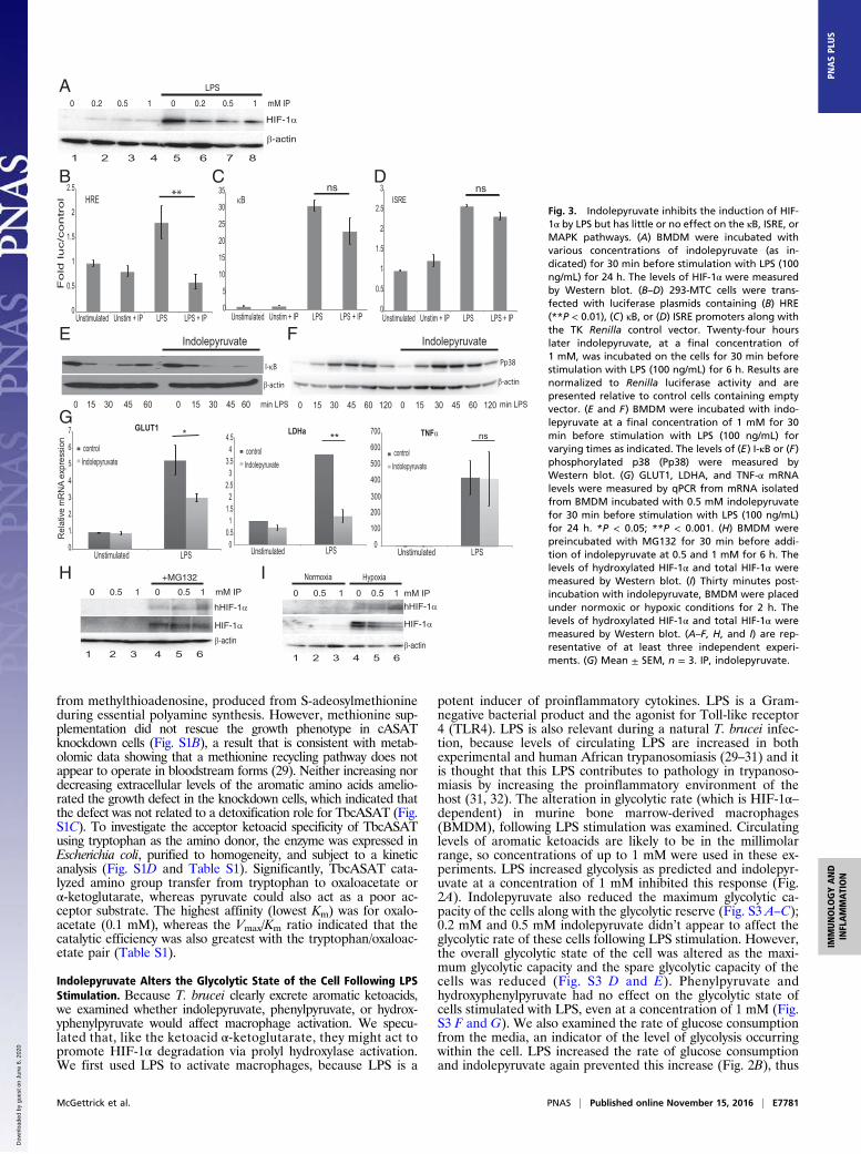
Fig. 3. Indolepyruvate inhibits the induction of HIF-1α by LPS but has little or no effect on the κB, ISRE, orMAPK pathways. (A) BMDM were incubated withvarious concentrations of indolepyruvate (as in-dicated) for 30 min before stimulation with LPS (100ng/mL) for 24 h. The levels of HIF-1α were measuredby Western blot. (B–D) 293-MTC cells were trans-fected with luciferase plasmids containing (B) HRE(**P < 0.01), (C) κB, or (D) ISRE promoters along withthe TK Renilla control vector. Twenty-four hourslater indolepyruvate, at a final concentration of1 mM, was incubated on the cells for 30 min beforestimulation with LPS (100 ng/mL) for 6 h. Results arenormalized to Renilla luciferase activity and arepresented relative to control cells containing emptyvector. (E and F) BMDM were incubated with indo-lepyruvate at a final concentration of 1 mM for 30min before stimulation with LPS (100 ng/mL) forvarying times as indicated. The levels of (E) I-κB or (F)phosphorylated p38 (Pp38) were measured byWestern blot. (G) GLUT1, LDHA, and TNF-α mRNAlevels were measured by qPCR from mRNA isolatedfrom BMDM incubated with 0.5 mM indolepyruvatefor 30 min before stimulation with LPS (100 ng/mL)for 24 h. *P < 0.05; **P < 0.001. (H) BMDM werepreincubated with MG132 for 30 min before addi-tion of indolepyruvate at 0.5 and 1 mM for 6 h. Thelevels of hydroxylated HIF-1α and total HIF-1α weremeasured by Western blot. (I) Thirty minutes post-incubation with indolepyruvate, BMDM were placedunder normoxic or hypoxic conditions for 2 h. Thelevels of hydroxylated HIF-1α and total HIF-1α weremeasured by Western blot. (A–F, H, and I) are rep-resentative of at least three independent experi-ments. (G) Mean ± SEM, n = 3. IP, indolepyruvate.
McGettrick et al. PNAS | Published online November 15, 2016 | E7781
IMMUNOLO
GYAND
INFLAMMATION
PNASPL
US
Dow
nloa
ded
by g
uest
on
June
6, 2
020
confirming that indolepyruvate inhibits the ability of LPS to in-crease glycolysis. Indolepyruvate, at a concentration of 1 mM,was not toxic to the cells as assessed by LDH release (Fig. 2C).
Indolepyruvate Inhibits the LPS-Induced Signaling Pathway to HIF-1αbut Has No Effect on the NF-κB, MAPK, or IFN-Stimulated ResponseElement Signaling Pathways. We next tested whether indolepyr-uvate affected HIF-1α activation. LPS increased HIF-1α proteinlevels and indolepyruvate inhibited this increase in a dose-dependent manner (Fig. 3A, compare lanes 6–8 to lane 5).Indolepyruvate also significantly inhibited the ability of LPS toinduce a reporter gene, luciferase, under the control of a HIF-response element (HRE) (Fig. 3B) but not luciferase under thecontrol of NF-κB (Fig. 3C) or the IFN response element (whichbinds IRF3) (Fig. 3D). Indolepyruvate also had no effect on LPS-induced I-κB degradation (Fig. 3E) or another LPS signal, p38phosphorylation (Fig. 3F), even at the highest concentration of1 mM. To confirm the role of HIF-1α in the mechanism of actionof indolepyruvate, we examined two HIF-1α target genes, glu-cose transporter 1 (GLUT1) and lactate dehydrogenase A(LDHA) (33, 34). LPS induced the mRNA of both genes and thisinduction was inhibited in the presence of indolepyruvate(Fig. 3G). Indolepyruvate had no effect on the induction of theNF-κB–dependent gene TNF-α (Fig. 3G, Right). These resultssuggest that indolepyruvate specifically inhibits the signaling
pathway leading to HIF-1α activation, while having little or noimpact on the NF-κB, MAPK, or IFN-stimulated response ele-ment (ISRE) pathways.We next investigated the mechanism of inhibition of HIF-1α
by indolepyruvate. Several studies suggest tryptophan catabo-lites, including breakdown products of indolepyruvate, act asendogenous ligands for the aryl hydrocarbon receptor (AhR), aligand-activated transcription factor (35, 36). AhR was of par-ticular interest because AhR dimerizes with aryl hydrocarbonreceptor nuclear translocator (ARNT) (36, 37), the bindingpartner of HIF-1α; thus, an increase in AhR activation couldcause a decrease in HIF-1α–ARNT complexes, reducing theproduction of HIF-1α–dependent target genes. Indolepyruvatewas still able to inhibit LPS-induced pro–IL-1β production inBMDM from AhR knockout mice (Fig. S4). The AhR receptoris therefore unlikely to be required for the inhibition of LPS-induced pro–IL-1β by indolepyruvate.Because indolepyruvate is a ketoacid, it might act in a similar
way to αKG, which is a cofactor for the prolyl hydroxylases thathydroxylate HIF-1α and promote its degradation, so we nexttested whether indolepyruvate could induce HIF-1α hydroxyl-ation. The proteasomal inhibitor MG132 was used in these ex-periments as HIF-1α hydroxylation is difficult to detect undernormal conditions because of the speed at which it is degradedonce hydroxylated. The presence of indolepyruvate increased the
IL-1β 0 0.2 0.5 1 0 0.2 0.5 1 0 0.2 0.5 1 mM IP
10 ng/ml LPS 100 ng/ml LPS
β-actin
0
10
20
30
40
50
60
70
80
90
- LPS 10 ng/ul LPS 100 ng/ul LPS
Re
lative
mR
NA
exp
ressio
n
Untreated0.2 mM IP0.5 mM IP1 mM IP
IL-1β
- - - + + + LPS
0 0.5 1 0 0.5 1 mM IP
pro-IL-1β
β-actin
pro IL-1β
HIF-1α
0 LPS LPS+IP
β-actin
******
***
******
***
0
500
1000
1500
2000
2500
3000
- LPS 10 ng/ml LPS 100 ng/ml LPS
Untreated
0.2 mM IP
0.5 mM IP
1 mM IP
pg/m
l TN
F
TNFα
0
1000
2000
3000
4000
5000
6000
7000
- LPS 10 ng/ml LPS 100 ng/ml LPS
Untreated 0.2 mM IP 0.5 mM IP 1 mM IP
pg/m
l IL-
6
IL-6ns ns ns ns
0
200
400
600
800
1000
1200
1400
1600
1800
2000
- LPS 10ng LPS 100ng LPS
untreated
0.5 mM IP
1 mM IP
pg/m
l IL-
1β
*****
******
A B
C
D E
F
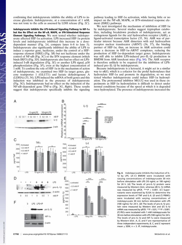
Fig. 4. Indolepyruvate inhibits the induction of IL-1β by LPS. (A–C ) BMDM were incubated withvarying concentrations of indolepyruvate 30 minbefore stimulation with LPS (10 ng/mL or 100 ng/mL)for 24 h. (A) The levels of pro-IL-1β protein weremeasured by Western blot, whereas (B) IL-1β mRNAwas measured by qPCR. ***P < 0.001. (C ) Super-natants were examined by ELISA to determine theexpression levels of TNF and IL-6. (D and E ) PBMCswere incubated with varying concentrations ofindolepyruvate 30 min before stimulation with LPS(100 ng/mL) for 24 h. (D) The levels of pro–IL-1β pro-tein were measured by Western blot and (E) IL-1βmRNAwas measured by qPCR. **P < 0.01; ***P < 0.001.(F) PECs were incubated with 1 mM indolepyruvate for30min before stimulationwith LPS (100 ng/mL) for 24 h.The levels of pro–IL-1β and HIF-1α were measuredby Western blot. A, D, and F are representative ofthree independent experiments; B, C, and E representmean ± SEM, n = 3. IP, indolepyruvate.
E7782 | www.pnas.org/cgi/doi/10.1073/pnas.1608221113 McGettrick et al.
Dow
nloa
ded
by g
uest
on
June
6, 2
020
levels of HIF-1α hydroxylation in BMDM (Fig. 3H, Upper,compare lanes 5 and 6 to lane 4). Under hypoxic conditions,which result in HIF-1α stabilization, the presence of indolepyr-uvate increased the levels of HIF-1α hydroxylation (Fig. 3I,Upper, compare lanes 5 and 6 to lane 4) and decreased the levelsof HIF-1α protein (Fig. 3I,Middle, compare lanes 5 and 6 to lane4). These results strongly suggest that indolepyruvate is pro-moting HIF-1α hydroxylation, leading to its degradation.
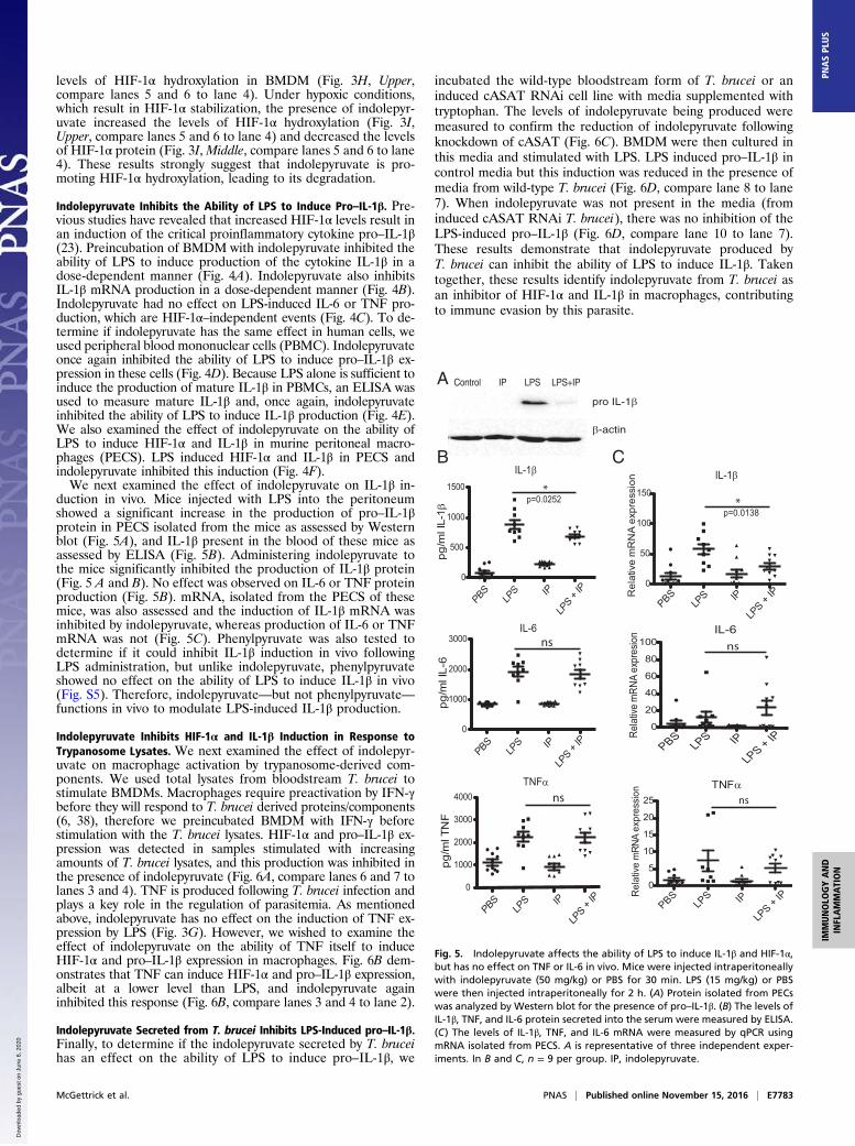
Indolepyruvate Inhibits the Ability of LPS to Induce Pro–IL-1β. Pre-vious studies have revealed that increased HIF-1α levels result inan induction of the critical proinflammatory cytokine pro–IL-1β(23). Preincubation of BMDM with indolepyruvate inhibited theability of LPS to induce production of the cytokine IL-1β in adose-dependent manner (Fig. 4A). Indolepyruvate also inhibitsIL-1β mRNA production in a dose-dependent manner (Fig. 4B).Indolepyruvate had no effect on LPS-induced IL-6 or TNF pro-duction, which are HIF-1α–independent events (Fig. 4C). To de-termine if indolepyruvate has the same effect in human cells, weused peripheral blood mononuclear cells (PBMC). Indolepyruvateonce again inhibited the ability of LPS to induce pro–IL-1β ex-pression in these cells (Fig. 4D). Because LPS alone is sufficient toinduce the production of mature IL-1β in PBMCs, an ELISA wasused to measure mature IL-1β and, once again, indolepyruvateinhibited the ability of LPS to induce IL-1β production (Fig. 4E).We also examined the effect of indolepyruvate on the ability ofLPS to induce HIF-1α and IL-1β in murine peritoneal macro-phages (PECS). LPS induced HIF-1α and IL-1β in PECS andindolepyruvate inhibited this induction (Fig. 4F).We next examined the effect of indolepyruvate on IL-1β in-
duction in vivo. Mice injected with LPS into the peritoneumshowed a significant increase in the production of pro–IL-1βprotein in PECS isolated from the mice as assessed by Westernblot (Fig. 5A), and IL-1β present in the blood of these mice asassessed by ELISA (Fig. 5B). Administering indolepyruvate tothe mice significantly inhibited the production of IL-1β protein(Fig. 5 A and B). No effect was observed on IL-6 or TNF proteinproduction (Fig. 5B). mRNA, isolated from the PECS of thesemice, was also assessed and the induction of IL-1β mRNA wasinhibited by indolepyruvate, whereas production of IL-6 or TNFmRNA was not (Fig. 5C). Phenylpyruvate was also tested todetermine if it could inhibit IL-1β induction in vivo followingLPS administration, but unlike indolepyruvate, phenylpyruvateshowed no effect on the ability of LPS to induce IL-1β in vivo(Fig. S5). Therefore, indolepyruvate—but not phenylpyruvate—functions in vivo to modulate LPS-induced IL-1β production.
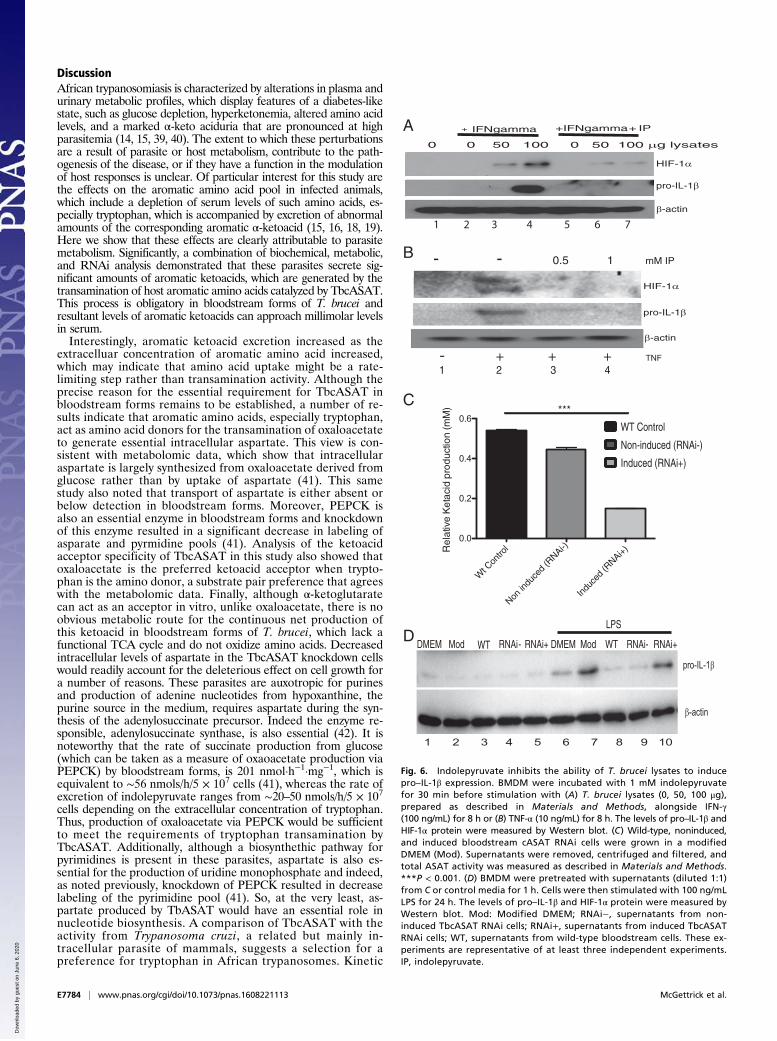
Indolepyruvate Inhibits HIF-1α and IL-1β Induction in Response toTrypanosome Lysates. We next examined the effect of indolepyr-uvate on macrophage activation by trypanosome-derived com-ponents. We used total lysates from bloodstream T. brucei tostimulate BMDMs. Macrophages require preactivation by IFN-γbefore they will respond to T. brucei derived proteins/components(6, 38), therefore we preincubated BMDM with IFN-γ beforestimulation with the T. brucei lysates. HIF-1α and pro–IL-1β ex-pression was detected in samples stimulated with increasingamounts of T. brucei lysates, and this production was inhibited inthe presence of indolepyruvate (Fig. 6A, compare lanes 6 and 7 tolanes 3 and 4). TNF is produced following T. brucei infection andplays a key role in the regulation of parasitemia. As mentionedabove, indolepyruvate has no effect on the induction of TNF ex-pression by LPS (Fig. 3G). However, we wished to examine theeffect of indolepyruvate on the ability of TNF itself to induceHIF-1α and pro–IL-1β expression in macrophages. Fig. 6B dem-onstrates that TNF can induce HIF-1α and pro–IL-1β expression,albeit at a lower level than LPS, and indolepyruvate againinhibited this response (Fig. 6B, compare lanes 3 and 4 to lane 2).
Indolepyruvate Secreted from T. brucei Inhibits LPS-Induced pro–IL-1β.Finally, to determine if the indolepyruvate secreted by T. bruceihas an effect on the ability of LPS to induce pro–IL-1β, we
incubated the wild-type bloodstream form of T. brucei or aninduced cASAT RNAi cell line with media supplemented withtryptophan. The levels of indolepyruvate being produced weremeasured to confirm the reduction of indolepyruvate followingknockdown of cASAT (Fig. 6C). BMDM were then cultured inthis media and stimulated with LPS. LPS induced pro–IL-1β incontrol media but this induction was reduced in the presence ofmedia from wild-type T. brucei (Fig. 6D, compare lane 8 to lane7). When indolepyruvate was not present in the media (frominduced cASAT RNAi T. brucei), there was no inhibition of theLPS-induced pro–IL-1β (Fig. 6D, compare lane 10 to lane 7).These results demonstrate that indolepyruvate produced byT. brucei can inhibit the ability of LPS to induce IL-1β. Takentogether, these results identify indolepyruvate from T. brucei asan inhibitor of HIF-1α and IL-1β in macrophages, contributingto immune evasion by this parasite.
Control IP LPS LPS+IP
pro IL-1β
β-actin
PBSLP
S
IP0
500
1000
1500
PBSLP
S
IP0
1000
2000
3000
PBSLP
S
IP
LP
S + IP
0
1000
2000
3000
4000
pg/m
l TN
F
p=0.0252
pg/m
l IL-
6
IL-1β
IL-6
TNFα
pg/m
l IL-
1β
ns
ns
*
LP
S + IP
LP
S + IP
PBS
LPS
IP
LP
S + IP
0
50
100
150
Rel
ativ
e m
RN
A e
xpre
ssio
n
p=0.0138
IL-1β
*
TNFα
PBSLP
S
IP
LPS + IP
0
5
10
15
20
25
Rel
ativ
e m
RNA
expr
essi
on
ns
IL-6
PBSLPS
IP
LPS + IP
0
20
40
60
80
100Re
lativ
e m
RNA
expr
esio
nns
A
B C
Fig. 5. Indolepyruvate affects the ability of LPS to induce IL-1β and HIF-1α,but has no effect on TNF or IL-6 in vivo. Mice were injected intraperitoneallywith indolepyruvate (50 mg/kg) or PBS for 30 min. LPS (15 mg/kg) or PBSwere then injected intraperitoneally for 2 h. (A) Protein isolated from PECswas analyzed by Western blot for the presence of pro–IL-1β. (B) The levels ofIL-1β, TNF, and IL-6 protein secreted into the serum were measured by ELISA.(C) The levels of IL-1β, TNF, and IL-6 mRNA were measured by qPCR usingmRNA isolated from PECS. A is representative of three independent exper-iments. In B and C, n = 9 per group. IP, indolepyruvate.
McGettrick et al. PNAS | Published online November 15, 2016 | E7783
IMMUNOLO
GYAND
INFLAMMATION
PNASPL
US
Dow
nloa
ded
by g
uest
on
June
6, 2
020
DiscussionAfrican trypanosomiasis is characterized by alterations in plasma andurinary metabolic profiles, which display features of a diabetes-likestate, such as glucose depletion, hyperketonemia, altered amino acidlevels, and a marked α-keto aciduria that are pronounced at highparasitemia (14, 15, 39, 40). The extent to which these perturbationsare a result of parasite or host metabolism, contribute to the path-ogenesis of the disease, or if they have a function in the modulationof host responses is unclear. Of particular interest for this study arethe effects on the aromatic amino acid pool in infected animals,which include a depletion of serum levels of such amino acids, es-pecially tryptophan, which is accompanied by excretion of abnormalamounts of the corresponding aromatic α-ketoacid (15, 16, 18, 19).Here we show that these effects are clearly attributable to parasitemetabolism. Significantly, a combination of biochemical, metabolic,and RNAi analysis demonstrated that these parasites secrete sig-nificant amounts of aromatic ketoacids, which are generated by thetransamination of host aromatic amino acids catalyzed by TbcASAT.This process is obligatory in bloodstream forms of T. brucei andresultant levels of aromatic ketoacids can approach millimolar levelsin serum.Interestingly, aromatic ketoacid excretion increased as the
extracelluar concentration of aromatic amino acid increased,which may indicate that amino acid uptake might be a rate-limiting step rather than transamination activity. Although theprecise reason for the essential requirement for TbcASAT inbloodstream forms remains to be established, a number of re-sults indicate that aromatic amino acids, especially tryptophan,act as amino acid donors for the transamination of oxaloacetateto generate essential intracellular aspartate. This view is con-sistent with metabolomic data, which show that intracellularaspartate is largely synthesized from oxaloacetate derived fromglucose rather than by uptake of aspartate (41). This samestudy also noted that transport of aspartate is either absent orbelow detection in bloodstream forms. Moreover, PEPCK isalso an essential enzyme in bloodstream forms and knockdownof this enzyme resulted in a significant decrease in labeling ofasparate and pyrmidine pools (41). Analysis of the ketoacidacceptor specificity of TbcASAT in this study also showed thatoxaloacetate is the preferred ketoacid acceptor when trypto-phan is the amino donor, a substrate pair preference that agreeswith the metabolomic data. Finally, although α-ketoglutaratecan act as an acceptor in vitro, unlike oxaloacetate, there is noobvious metabolic route for the continuous net production ofthis ketoacid in bloodstream forms of T. brucei, which lack afunctional TCA cycle and do not oxidize amino acids. Decreasedintracellular levels of aspartate in the TbcASAT knockdown cellswould readily account for the deleterious effect on cell growth fora number of reasons. These parasites are auxotropic for purinesand production of adenine nucleotides from hypoxanthine, thepurine source in the medium, requires aspartate during the syn-thesis of the adenylosuccinate precursor. Indeed the enzyme re-sponsible, adenylosuccinate synthase, is also essential (42). It isnoteworthy that the rate of succinate production from glucose(which can be taken as a measure of oxaoacetate production viaPEPCK) by bloodstream forms, is 201 nmol·h−1·mg−1, which isequivalent to ∼56 nmols/h/5 × 107 cells (41), whereas the rate ofexcretion of indolepyruvate ranges from ∼20–50 nmols/h/5 × 107cells depending on the extracellular concentration of tryptophan.Thus, production of oxaloacetate via PEPCK would be sufficientto meet the requirements of tryptophan transamination byTbcASAT. Additionally, although a biosynthethic pathway forpyrimidines is present in these parasites, aspartate is also es-sential for the production of uridine monophosphate and indeed,as noted previously, knockdown of PEPCK resulted in decreaselabeling of the pyrimidine pool (41). So, at the very least, as-partate produced by TbASAT would have an essential role innucleotide biosynthesis. A comparison of TbcASAT with theactivity from Trypanosoma cruzi, a related but mainly in-tracellular parasite of mammals, suggests a selection for apreference for tryptophan in African trypanosomes. Kinetic
TNF
pro-IL-1β
- - 0.5 1 mM IP
β-actin
pro-IL-1β
β-actin
DMEM Mod WT RNAi- RNAi+ DMEM Mod WT RNAi- RNAi+
LPS
Rel
ativ
e K
etac
id p
rodu
ctio
n (m
M)
0 0 50 100 0 50 100 μg lysates
+ IFNgamma + IFNgamma + IP
HIF-1α
pro-IL-1β
β-actin
1 2 3 4 5 6 7
1 2 3 4
1 2 3 4 5 6 7 8 9 10
WT Control
Non-induced (RNAi-)
Induced (RNAi+)
HIF-1α
pro-IL-1β
β-actin
- + + + TNF
Wt C
ontro
l
Non in
duce
d (R
NAi-)
Indu
ced
(RNAi+)
0.0
0.2
0.4
0.6***
A
B
C
D
Fig. 6. Indolepyruvate inhibits the ability of T. brucei lysates to inducepro–IL-1β expression. BMDM were incubated with 1 mM indolepyruvatefor 30 min before stimulation with (A) T. brucei lysates (0, 50, 100 μg),prepared as described in Materials and Methods, alongside IFN-γ(100 ng/mL) for 8 h or (B) TNF-α (10 ng/mL) for 8 h. The levels of pro–IL-1β andHIF-1α protein were measured by Western blot. (C) Wild-type, noninduced,and induced bloodstream cASAT RNAi cells were grown in a modifiedDMEM (Mod). Supernatants were removed, centrifuged and filtered, andtotal ASAT activity was measured as described in Materials and Methods.***P < 0.001. (D) BMDM were pretreated with supernatants (diluted 1:1)from C or control media for 1 h. Cells were then stimulated with 100 ng/mLLPS for 24 h. The levels of pro–IL-1β and HIF-1α protein were measured byWestern blot. Mod: Modified DMEM; RNAi−, supernatants from non-induced TbcASAT RNAi cells; RNAi+, supernatants from induced TbcASATRNAi cells; WT, supernatants from wild-type bloodstream cells. These ex-periments are representative of at least three independent experiments.IP, indolepyruvate.
E7784 | www.pnas.org/cgi/doi/10.1073/pnas.1608221113 McGettrick et al.
Dow
nloa
ded
by g
uest
on
June
6, 2
020
studies have demonstrated that TbcASAT has a greater prefer-ence (lower Km) and a significantly higher Vmax/Km ratio fortryptophan as the amino group donor compared with tyrosine,phenylalanine, or aspartate, whereas the T. cruzi activity is lessselective and transaminates aromatic amino acids and aspartatewith equal efficiency (28).Here we show that indolepyruvate, the transamination product
of tryptophan, acts as a metabolic modulator of the innate im-mune response in vitro and in vivo. Indolepyruvate prevents theglycolytic shift in response to LPS, a critical response for mac-rophage function, by promoting the hydroxylation of the tran-scription factor HIF-1α, which targets HIF-1α for degradation.HIF-1α is known to bind to, and promote expression of multipleinflammatory genes, notably pro–IL-1β. This study demonstratesthat indolepyruvate inhibits the ability of LPS to induce pro–IL-1β. Although indolepyruvate promotes HIF-1α degradation andIL-1β production at 0.2 mM, it shows more consistent inhibitionat 0.5 and 1 mM, suggesting that the inhibition of the innateimmune response by indolepyruvate is most important at thepeak of the parasitemia, when levels of indolepyruvate in theblood are close to millimolar levels. At the peak of parasitemia,the levels of pathogen-associated molecular patterns and damage-associated molecular pattern molecules activating the innate im-mune response would be at their highest, leading to a potentialoveractivation of the innate immune response. This would bedetrimental for the host and the well-being of the host is crucialfor the survival of the parasite. Therefore, the ability of indole-pyruvate to dampen down the immune response at the peak of theparasitemia would be very beneficial to the parasite.Modulation of the innate immune response by T. brucei has
previously been demonstrated. Garzón et al. (43) used LPS tostimulate an innate immune response and demonstrated thatproteins secreted from T. brucei gambiense affected the hostimmune response. The secretome impaired LPS-induced matu-ration of murine dendritic cells as well as inhibiting the pro-duction of the cytokines TNF-α, IL-6, and IL-10. Mitchell et al.(11) demonstrated that LPS-induced IL-1 activity was pro-foundly depressed in cells isolated from both A/J and C57BL/6Jmice following Trypanosoma congolense—another extracellularAfrican trypanosome—infection. Day 7 of infection found no IL-1activity in supernatants from either mouse strain. This effectmay have been a result of indolepyruvate being present inthe bloodstream of these mice. Aromatic ketoacids have alsobeen shown to elicit an anti-inflammatory response. Aoki et al.(44) demonstrated that indolepyruvate, phenylpyruvate, andhydroxyphenylpyrvate, at concentrations similar to those used inthis study, protected the skin of hairless mice from UVB-induceddamage. Interestingly, indolepyruvate proved to be the mosteffective and topical application of indolepyruvate to the dorsalskin of hairless mice reduced the severity of UVB-induced skinlesions; it also suppressed the production of IL-1β and IL-6 inresponse to UVB radiation in these mice.Macrophage activation occurs following infection with African
trypanosomes but what causes this activation is poorly under-stood. It is likely that macrophages are exposed to several dif-ferent activating agents. Some of these agents, such as VSG andtrypanosome DNA, originate from the parasite; others willoriginate from the host, such as IFN-γ. The levels of circulatingLPS also increase during African trypanosomiasis (30, 31, 29).This increase is likely because of an increase in gut permeability(45) and it is thought that this increase in LPS contributes to thepathology of the disease by increasing the proinflammatory en-vironment of the host (30–32). Nyakundi et al. (9, 45) demon-strated that increases in cytokine production, including IL-1β,TNF-α, IL-6, and IFN-γ, following T. brucei infection, stronglycorrelated with the increase in LPS levels observed during in-fection. T. brucei-infected mice become hyperresponsive to LPS(6). The release of IFN-γ from several cell types, includingT cells, is thought to prime macrophages making them moresusceptible to LPS activation. Levels of LPS binding protein alsoincrease in the plasma of infected mice, which would enhance
the cellular response to LPS (46). Because of the increase in LPSpresent in the blood of infected patients, indolepyruvate mayplay a role in reducing the susceptibility of these patients to LPSor may be involved in the down-regulation of macrophage acti-vation by all activating agents.Indolepyruvate reduces HIF-1α protein levels. Because HIF-
1α is a transcription factor, this reduction will lead to the re-duction in several HIF-1α target genes, including GLUT1,VEGF, LDHA, and IL-1β. This study has concentrated on theimpact of indolepyruvate on the proinflammatory cytokine, IL-1β. The exact role for IL-1β in T. brucei infection is not yet fullyunderstood. IL-1β levels increase following T. brucei infection (9,10) but Drennan et al. (38) demonstrated no significant differ-ence between wild-type and IL-1R1−/− mice in T. brucei infec-tion. A study by Quan et al. (47), however, demonstrated a rolefor the IL-1R1 in T. brucei infection. Intracerebral infusion of theIL-1R1 antagonist (IL-1ra) prevented the weight loss, or lack ofweight gain, associated with infection. Infusion of IL-1ra andsoluble type 1 TNF receptor (sTNFr1) reduced trypanosome-induced neurodegeneration, to a greater extent than TNFr1 onits own and reduced the expression of IL-1β and I-κB with noeffect on TNF-α. Additional studies will be needed to determineif the impact of IL-1β during T. brucei infection would be fargreater if the trypanosomes were not producing indolepyruvate.IL-1β is a potent pyrogen, and so inhibiting its production mayindeed prevent fever and seizures during infection, but furtherstudies will be needed to investigate this. Indolepyruvate mayalso enhance host survival by inhibiting endogenous trypano-some-induced gene expression, notably those targeted by HIF-1α, to lessen systemic pathologies associated with a persistentinfection, such as anemia and cachexia, to increase the proba-bility of parasite transmission. Although the physiological role ofindolepyruvate during an infection requires further investigation,it is tempting to speculate that is has a function in regulating hostinflammatory responses, particularly at the peak of the para-sitemia when circulating levels of the ketoacid are likely to behighest. It is known that a proinflammatory response appears tobe required to control the initial parasitemia (8) but indolepyr-uvate may limit the extent or systemic nature of this responseduring the clearance of parasites. Interestingly, the neurologi-cal symptoms associated with the late stage of the disease canoccur in the absence of a CNS inflammatory response (48).Perhaps, as originally suggested by Newport et al. (15), dis-ruption of the biogenic amine pool through metabolism oftryptophan and excretion of indolepyruvate contributes to theneurological effects, while simultaneously ameliorating a CNSinflammatory response.Our results therefore confirm that indolepyruvate is produced
by the bloodstream form T. brucei and can modulate the innateimmune response. The levels of indolepyruvate present in apatient could therefore be an important diagnostic tool for thepresence, and stage of disease, of T. brucei. Indolepyruvate mightalso have potential as an inhibitor of IL-1β in inflammatorydiseases.
Materials and MethodsReagents. Reagents used were LPS (E. coli, serotype EH100, Alexis), CpG,Pam3Cys, R848 (Invitrogen), TNF (peprotech), and IFN-γ (R+D systems); invivo LPS agonist (E. coli, serotype 055:B5); indolepyruvate and kynurenic acid(Sigma-Aldrich). ELISA kits used were IL-1β (DuoSet DY401), IL-6 (DuoSetDY406), TNF-α (Duoset DY410), and IL-1β (Quantikine, MLB00C). Antibodiesused: β-actin (4267), IκB (44D4), phospho-p38 (9211), HydroxyHIF (3435) (CellSignaling Technologies), anti–IL-1β (R&D, AF401-NA), anti–HIF-1α (Novus,NB100-449).
Mice and Cell Culture.All BMDMs and PECs were isolated from C57BL/6 HarlanUK mice as previously described (23). The in vivo experiments were carriedout in C57BL/6 Harlan UK mice. All experiments were carried out with priorethical approval from the Trinity College Dublin Animal Research EthicsCommittee. Each “n” represents BMDM/PECs from individual mice.
McGettrick et al. PNAS | Published online November 15, 2016 | E7785
IMMUNOLO
GYAND
INFLAMMATION
PNASPL
US
Dow
nloa
ded
by g
uest
on
June
6, 2
020
Trypanosome Cell Lines and Culture. Trypanosomes were cultured in stan-dard HMI-9 (bloodstream form) or SDM79 (procyclic form) medium con-taining 10% (vol/vol) FCS (49). The parental bloodstream (strain 13.90) andprocyclic (29.13) conditional RNAi cell lines were maintained by selectionin G418 and hygromycin, as described by Wirtz et al. (50). These cell linesexpress a T7 polymerase and tetracycline repressor. Monomorphic MITat1.1 bloodstream cells were isolated from infected rats as described else-where (39).
NMR. Bloodstream form wild-type and cASAT RNA1 cells were cultured inHMI-9 media for 24 h in the absence and presence of tetracycline (1 μg/mL).Cells were collected by centrifugation at 1,500 × g for 10 min, washed oncein phosphate-buffered saline-glucose buffer (pH 7.4), centrifuged, andresuspended at 5 × 10⁷ cells/mL in HMI-9 media supplemented with 150 μMsingle-labeled (indole-2-13C)-tryptophan for a 5-h incubation at 37 °C with5% CO2. Cell viability was actively monitored via microscopy during in-cubation, and media samples were collected at 1-h intervals during the 5-hincubation period by removing cells using centrifugation at 14,000 × g for5 min. Media samples were snap-frozen and stored at −80 °C until use forNMR. Next, 600-μL samples, consisting of 540 μL media and 60 μL D2O (lock),were run on an 800-MHz Agilent DD2/4.2 K premiumCOMPACT spectrom-eter. All 13C-NMR spectra were recorded at 25 °C, and samples were refer-enced to internal glucose at ∼98.64 ppm. Data were acquired with activeproton decoupling and NOE, 7.35 s 90° pulse width, 1-s relaxation delay,spectral width −15.0 ppm to 235.2 ppm, with 3,200 scans performed. Post-acquisition Line Broadening was 1-Hz exponential.
LDH Cytotoxicity Assay. BMDM were seeded at 200,000 cells per well of a 96-well plate. Indolepyruvate was added to experimental wells at a final con-centration of 1 mM for 25 h. Cytotox 96 nonradioactive cytotoxity assay(Promega) was carried out as per the manufacturer’s instructions.
RT-PCR. Cells were lysed and total RNA was extracted using the RNAeasy kit(Qiagen) and was reverse-transcribed using High-Capacity cDNA ReverseTranscription Kit (Applied Biosystems) according to the manufacturers’ in-structions. This cDNA served as a template for amplification of target genesby real-time PCR to determine the relative amounts of IL-1β (primersF: GGAAGCAGCCCTTCATCTTT, R: TGGCAACTGTTCCTGAACTC), LDHA (primersF: ATCTTGACCTACGTGGCTTGGA, R: CCATACAGGCACACTGGAATCTC), GLUT1(primers F: GATCACTGCAGTTCGGCTATAA R: GTAGCGGTGGTTCCATGTT), TNF(primers F: GCCTCTTCTCATTCCTGCTT, R: TGGGAACTTCTCATCCCTTTG), or IL-6(primers F: ACGATGATGCACTTGCAC R: ACTCCAGAAGACCAGAGGAA) mRNA.The cycling threshold method [2−(ΔΔCt)] was used for relative quantification bycomparative method after normalization to RPS18 (primer F: GGATGTGAAG-GATGGGAAGT, R: CCCTCTATGGGCTCGAATTT) or GAPDH expression (51).
Western Blotting. Cells were lysed in SDS sample buffer [0.125 M Tris pH 6.8,10% (vol/vol) glycerol, 0.02% SDS] and Western blot analysis was carried outas previously described (52). Western blots were developed using autora-diographic film or using a ChemiDoc MP gel imaging system (Bio-Rad).
ELISA. For ELISA, 1 × 10⁶ BMDMs per milliliter were pretreated for 30 min ±indolepyruvate, followed by 100 ng/mL LPS for 24 h. Supernatants werecollected and cytokines quantified by ELISA according to manufacturers’instructions (R&D Systems). Results are presented as mean ± SEM.
Reporter Gene Assays. HEK293-MD2-TLR4-CD14 cells seeded at 200,000 cellsper well into 96-well plates and transfected with expression vectors andluciferase reporter genes for NF-κB, ISRE, and HRE. phRL-TK reporter plasmidwas cotransfected to allow normalization of data for transfection effi-ciency. After 18 h, cells were treated with indolepyruvate followed by LPS(100 ng/mL) and 6-h later reporter gene activity was measured. All reporterassays were done in triplicate and data are expressed as “relative stimula-tion” (mean ± SD) over the nonstimulated empty vector control, for arepresentative experiment, a total of three separate experiments beingcarried out.
Isolation of Human PBMC. Human PBMC were isolated from human bloodusing Lymphoprep (Axis-Shield). Thirty milliliters of whole blood was layeredon 20mL lymphoprep and spun for 20min at 2,000 rpmwith no brake on. ThePBMCs were then isolated from the middle layer and washed twice in PBS.PBMCs were maintained in RPMI supplemented with 10% (vol/vol) FCS, 2 mML-glutamine, and 1% penicillin/streptomycin solution.
In Vivo Trial. Mice were injected intraperitoneally with indolepyruvate(50 mg/kg), phenylpyruvate (50 mg/mg), or PBS for 30 min. LPS (15 mg/kg) orPBS were then injected intraperitoneally. Two-hours postinjection the micewere humanely euthanized and serum was isolated from whole blood andPECs were harvested.
cASAT RNAi. A 446-bp fragment (from nuclotide 556–1002) from the cASAT(Tb927.10.3660) of T. brucei was amplified and cloned into the p2T7-177RNAi vector. The construct was linearized with NotI and transformed intothe RNAi parental cell lines using the Amaxa parasite nucleofection kit (51).The construct was targeted to the minichromosomes through homologousrecombination within the 177-bp repeats. The cASAT-RNAi cell lines wereselected by culturing in the presence of phleomycin and cloned by limitingdilution. Expression of the dsRNA was induced by addition of tetracycline(1 μg/mL). Total RNA was extracted using the Stratagene Absolutely RNApurification kit. The relative level of expression of the cASAT mRNA wasestimated by qRT-PCR using the Brilliant SYBR Green qRT-PCR Master Mix Kit(Stratagene) in an MxPro 3000 instrument (Stratagene). The cycling thresh-old method [2-(ΔΔCt)] was used for relative quantification by comparativemethod after normalization to actin expression (51).
ASAT Assay. Total ASAT specific activity was assayed at 340 nm in the directionof oxaloactate production using aspartate (12.5 mM) and α-ketoglutarate(10 mM) as substrates and malate dehydrogenase (5 units/mL) and NADH(0.2 mM) as a coupling enzyme/substrate as described in Bergmyer (53). Atvarious times the trypanosomes were harvested by centrifugation and theASAT specific activity (units per milligram) was determined in total cell ly-sates, prepared as described previously (54).
Aromatic Ketoacid Production. The full ORF (TcCLB.506937.10) for NAD-linkedaromatic α-hydroxy acid dehydrogenase (AHADH) was amplified and clonedinto the pNIC28-Bsa4 expression vector. Following induction, recombinantAHADH was purified from the soluble fraction by Ni2+ affinity chromatog-raphy. Purified AHADH was used to measure the amount of total aromaticketoacids in samples (up to 0.2 mL) using a standard assay (final volume of1 mL) containing Tris buffer (25 mM, pH 7.4), NaCl (50 mM), NADH (0.25 mM),and AHADH (850 U). Total aromatic ketoacid present in the sample wasdetermined by the decrease in absorbance at 340 nm. Appropriate blankcontrols were used to correct for sample volume and the assay was verifiedusing standard solutions of phenlpyruvate.
In Vitro Trypanosome Supernatant Transfer Experiment. Trypanosomes weregrown inmodified DMEM. Supernatants were harvested and used to pretreatBMDMs, followed by stimulation by LPS for 24 h. Cells were lysed in SDSsample buffer and analyzed by Western blot.
Glucose Consumption. The levels of glucose present in BMDM supernatantswere assayed enzymatically using a hexokinase/glucose-6-phosphate de-hydrogenase assay. The assay (final volume 1 mL) was performed in assaybuffer (Tris·Cl 50 mM, NaCl 50 mM, MgCl2 5 mM, pH 7.5) containing ATP(2.5 mM), NAD (2.5 mM), hexokinse (10 μ/mL) and glucose-6-phosphate de-hydrogenase from Leuconostoc mesenteroides (10 μ/mL). The amount ofglucose present was determined by the increase in absorbance at 340 nmfollowing addition of the sample (typically 10 μL). The assay was verifiedusing standard solutions of glucose.
Glycolysis Analysis Using Seahorse. BMDMs were plated at 200,000 cells perwell in XF24 plates overnight before 30-min pretreatment with varyingconcentrations of indolepyruvate, phenylpyruvate, or hydroxyphenylpyr-uvate, followed by stimulationwith 100 ng/mL LPS for 24 h. XF24 ExtracellularFlux analyzer (Seahorse Biosciences) was used to determine the protonproduction rate. Oligomycin (final concentration of 2 μM) and 2DG (finalconcentration of 50 mM) were used to determine the maximum glycolsis andgylcolytic reserve. Results were normalized to cell number and are repre-sented as mean ± SD.
Hypoxia Experiment. BMDMs were seeded in 24-well plates (1 × 10⁶ cells/mL),incubated with indolepyruvate for 30 min. Half the cells were then main-tained in normoxic conditions in a CO2 incubator, and half were placed inhypoxic conditions in a hypoxia chamber at 1% O2 (Coy Laboratories). Thecells were then lysed in SDS sample buffer [0.125 M Tris pH 6.8, 10% (vol/vol)glycerol, 0.02% SDS] and a Western blot was performed.
E7786 | www.pnas.org/cgi/doi/10.1073/pnas.1608221113 McGettrick et al.
Dow
nloa
ded
by g
uest
on
June
6, 2
020
In Vitro Trypanosome Supernatant Transfer Experiment. For the in vitro try-panosome supernatant transfer experiment, 3 × 10⁶ /mL MITat 1.1 wild-typeand cASAT RNA1 cells were grown for 5 h in modified DMEM [DMEM + 10%(vol/vol) FCS, 0.05 mM bathocuprione disulphonic acid, 1.5 mM L-cysteine,0.3 mM β-mercaptoethanol, 1 mM hypoxanthine, 3.0 g/L NaHCO3,, pH 7.5].Cells were then centrifugued and the supernatants harvested. Thesesupernatants were the used to pretreat BMDMs for 30 min, followed bystimulation by LPS at 100 ng/μL for 24 h. Cells were lysed in SDS sampleloading buffer and analyzed by Western blot.
Statistical Analysis. All statistical analysis was calculated using an unpairedtwo-tailed Student’s t test unless otherwise specified.
ACKNOWLEDGMENTS. We thank Paul Voorheis for advice and com-ments, and Brigitta Stockinger (Medical Research Council NationalInstitute for Medical Research) for access to bone marrow-derivedmacrophages from AhR− /− mice. This study was supported in part bythe Science Foundation Ireland, the Wellcome Trust, and the EuropeanResearch Council.
1. Stich A, Abel PM, Krishna S (2002) Human African trypanosomiasis. BMJ 325(7357):203–206.
2. Cross GA (1990) Cellular and genetic aspects of antigenic variation in trypanosomes.Annu Rev Immunol 8:83–110.
3. Pays E, Vanhamme L, Berberof M (1994) Genetic controls for the expression of surfaceantigens in African trypanosomes. Annu Rev Microbiol 48:25–52.
4. Morrison LJ, Marcello L, McCulloch R (2009) Antigenic variation in the African try-panosome: Molecular mechanisms and phenotypic complexity. Cell Microbiol 11(12):1724–1734.
5. Thuita JK, et al. (2008) Trypanosoma brucei rhodesiense transmitted by a single tsetsefly bite in vervet monkeys as a model of human African trypanosomiasis. PLoS NeglTrop Dis 2(5):e238.
6. Magez S, et al. (1998) The glycosyl-inositol-phosphate and dimyristoylglycerol moie-ties of the glycosylphosphatidylinositol anchor of the trypanosome variant-specificsurface glycoprotein are distinct macrophage-activating factors. J Immunol 160(4):1949–1956.
7. Magez S, Radwanska M, Beschin A, Sekikawa K, De Baetselier P (1999) Tumor necrosisfactor alpha is a key mediator in the regulation of experimental Trypanosoma bruceiinfections. Infect Immun 67(6):3128–3132.
8. Paulnock DM, Freeman BE, Mansfield JM (2010) Modulation of innate immunity byAfrican trypanosomes. Parasitology 137(14):2051–2063.
9. Nyakundi JN, Crawley B, Pentreath VW (2002) The relationships between endotoxins,nitric oxide and inflammatory cytokines in blood and intestinal tissues in experi-mental Trypanosoma brucei brucei infections. Parasitology 124(Pt 6):597–604.
10. Sileghem M, Darji A, Hamers R, De Baetselier P (1989) Modulation of IL-1 productionand IL-1 release during experimental trypanosome infections. Immunology 68(1):137–139.
11. Mitchell LA, Pearson TW, Gauldie J (1986) Interleukin-1 and interleukin-2 productionin resistant and susceptible inbred mice infected with Trypanosoma congolense.Immunology 57(2):291–296.
12. Stibbs HH, Seed JR (1973) Chromatographic evidence for the synthesis of possiblesleep-mediators in Trypanosoma brucei gambiense. Experientia 29(12):1563–1565.
13. Stibbs HH, Seed JR (1975) Short-term metabolism of (14-C) tryptophan in rats infectedwith Trypanosoma brucei gambiense. J Infect Dis 131(4):459–462.
14. Seed JR, Hall JE, Sechelski J (1982) Phenylalanine metabolism in Microtus montanuschronically infected with Trypanosoma brucei gambiense. Comp Biochem Physiol B71(2):209–215.
15. Newport GR, Page CR, 3rd, Ashman PU, Stibbs HH, Seed JR (1977) Alteration of freeserum amino acids in voles infected with Trypanosoma brucei gambiense. J Parasitol63(1):15–24.
16. Hall JE, Seed JR (1984) Increased urinary excretion of aromatic amino acid catabolitesby Microtus montanus chronically infected with Trypanosoma brucei gambiense.Comp Biochem Physiol B 77(4):755–760.
17. Hall JE, Seed JR, Sechelski JB (1985) Multiple alpha-keto aciduria in Microtus montanuschronically infected with Trypanosoma brucei gambiense. Comp Biochem Physiol B82(1):73–78.
18. el Sawalhy A, Seed JR, el Attar H, Hall JE (1995) Catabolism of tryptophan by Trypanosomaevansi. J Eukaryot Microbiol 42(6):684–690.
19. El Sawalhy A, Seed JR, Hall JE, El Attar H (1998) Increased excretion of aromatic aminoacid catabolites in animals infected with Trypanosoma brucei evansi. J Parasitol 84(3):469–473.
20. Hunter AG (1986) Urine odour in a camel suffering from surra (T. evansi infection).Trop Anim Health Prod 18(3):146–148.
21. Seed JR, Hall JE, Price CC (1983) A physiological mechanism to explain pathogenesis inAfrican trypanosomiasis. Contrib Microbiol Immunol 7:83–94.
22. Berger BJ, Dai WW, Wang H, Stark RE, Cerami A (1996) Aromatic amino acid trans-amination and methionine recycling in trypanosomatids. Proc Natl Acad Sci USA93(9):4126–4130.
23. Tannahill GM, et al. (2013) Succinate is an inflammatory signal that induces IL-1βthrough HIF-1α. Nature 496(7444):238–242.
24. McGettrick AF, O’Neill LA (2013) How metabolism generates signals during innateimmunity and inflammation. J Biol Chem 288(32):22893–22898.
25. Creek DJ, et al. (2013) Metabolomics guides rational development of a simplified cellculture medium for drug screening against Trypanosoma brucei. Antimicrob AgentsChemother 57(6):2768–2779.
26. Cazzulo Franke MC, Vernal J, Cazzulo JJ, Nowicki C (1999) The NAD-linked aromaticalpha-hydroxy acid dehydrogenase from Trypanosoma cruzi. A new member of thecytosolic malate dehydrogenases group without malate dehydrogenase activity. Eur JBiochem 266(3):903–910.
27. Berger LC, Wilson J, Wood P, Berger BJ (2001) Methionine regeneration and aspartateaminotransferase in parasitic protozoa. J Bacteriol 183(15):4421–4434.
28. Marciano D, et al. (2008) Biochemical characterization of stage-specific isoforms ofaspartate aminotransferases from Trypanosoma cruzi and Trypanosoma brucei. MolBiochem Parasitol 161(1):12–20.
29. Alafiatayo RA, Crawley B, Oppenheim BA, Pentreath VW (1993) Endotoxins and thepathogenesis of Trypanosoma brucei brucei infection in mice. Parasitology 107(Pt 1):49–53.
30. Pentreath VW, et al. (1997) Endotoxin antibodies in African sleeping sickness.Parasitology 114(Pt 4):361–365.
31. Pentreath VW, Alafiatayo RA, Crawley B, Doua F, Oppenheim BA (1996) Endotoxins inthe blood and cerebrospinal fluid of patients with African sleeping sickness.Parasitology 112(Pt 1):67–73.
32. Greenwood BM (1974) Possible role of a B-cell mitogen in hypergammaglobulinaemiain malaria and trypanosomiasis. Lancet 1(7855):435–436.
33. Chen C, Pore N, Behrooz A, Ismail-Beigi F, Maity A (2001) Regulation of glut1 mRNAby hypoxia-inducible factor-1. Interaction between H-ras and hypoxia. J Biol Chem276(12):9519–9525.
34. Hayashi M, et al. (2004) Induction of glucose transporter 1 expression through hypoxia-inducible factor 1alpha under hypoxic conditions in trophoblast-derived cells.J Endocrinol 183(1):145–154.
35. Bittinger MA, Nguyen LP, Bradfield CA (2003) Aspartate aminotransferase generatesproagonists of the aryl hydrocarbon receptor. Mol Pharmacol 64(3):550–556.
36. Nguyen LP, et al. (2009) D-amino acid oxidase generates agonists of the aryl hydro-carbon receptor from D-tryptophan. Chem Res Toxicol 22(12):1897–1904.
37. McGuire J, Whitelaw ML, Pongratz I, Gustafsson JA, Poellinger L (1994) A cellularfactor stimulates ligand-dependent release of hsp90 from the basic helix-loop-helixdioxin receptor. Mol Cell Biol 14(4):2438–2446.
38. Drennan MB, et al. (2005) The induction of a type 1 immune response following aTrypanosoma brucei infection is MyD88 dependent. J Immunol 175(4):2501–2509.
39. Voorheis HP (1969) The effect of T. brucei (S-42) on host carbohydrate metabolism:Liver production and peripheral tissue utilization of glucose. Trans R Soc Trop MedHyg 63(1):122–123.
40. Li JV, et al. (2011) Metabonomic investigation of single and multiple strain Trypanosomabrucei brucei infections. Am J Trop Med Hyg 84(1):91–98.
41. Creek DJ, et al. (2015) Probing the metabolic network in bloodstream-form Trypanosomabrucei using untargeted metabolomics with stable isotope labelled glucose. PLoS Pathog11(3):e1004689.
42. Mony BM, et al. (2014) Genome-wide dissection of the quorum sensing signallingpathway in Trypanosoma brucei. Nature 505(7485):681–685.
43. Garzón E, et al. (2013) The Trypanosoma brucei gambiense secretome impairslipopolysaccharide-induced maturation, cytokine production, and allostimulatorycapacity of dendritic cells. Infect Immun 81(9):3300–3308.
44. Aoki R, Aoki-Yoshida A, Suzuki C, Takayama Y (2014) Protective effect of indole-3-pyruvate against ultraviolet b-induced damage to cultured HaCaT keratinocytes andthe skin of hairless mice. PLoS One 9(5):e96804.
45. Nyakundi JN, Crawley B, Smith RA, Pentreath VW (2002) The relationships betweenintestinal damage and circulating endotoxins in experimental Trypanosoma bruceibrucei infections. Parasitology 124(Pt 6):589–595.
46. Ngure RM, et al. (2009) Lipopolysaccharide binding protein in the acute phase re-sponse of experimental murine Trypanosoma brucei brucei infection. Res Vet Sci86(3):394–398.
47. Quan N, He L, Lai W (2003) Intraventricular infusion of antagonists of IL-1 and TNFalpha attenuates neurodegeneration induced by the infection of Trypanosoma bru-cei. J Neuroimmunol 138(1-2):92–98.
48. MacLean L, Reiber H, Kennedy PG, Sternberg JM (2012) Stage progression and neu-rological symptoms in Trypanosoma brucei rhodesiense sleeping sickness: Role of theCNS inflammatory response. PLoS Negl Trop Dis 6(10):e1857.
49. Hirumi H, Hirumi K (1989) Continuous cultivation of Trypanosoma brucei bloodstream forms in a medium containing a low concentration of serum protein withoutfeeder cell layers. J Parasitol 75(6):985–989.
50. Wirtz E, Leal S, Ochatt C, Cross GA (1999) A tightly regulated inducible expression systemfor conditional gene knock-outs and dominant-negative genetics in Trypanosomabrucei. Mol Biochem Parasitol 99(1):89–101.
51. Livak KJ, Schmittgen TD (2001) Analysis of relative gene expression data using real-time quantitative PCR and the 2(-Delta Delta C(T)) Method. Methods 25(4):402–408.
52. Fitzgerald KA, et al. (2001) Mal (MyD88-adapter-like) is required for Toll-like receptor-4 signal transduction. Nature 413(6851):78–83.
53. Bergmeyer HU (1983) Methods of Enzymatic Analysis (Chemie, Weinheim, Germany),Vol 3.
54. Spitznagel D, et al. (2009) Alanine aminotransferase of Trypanosoma brucei—A keyrole in proline metabolism in procyclic life forms. FEBS J 276(23):7187–7199.
McGettrick et al. PNAS | Published online November 15, 2016 | E7787
IMMUNOLO
GYAND
INFLAMMATION
PNASPL
US
Dow
nloa
ded
by g
uest
on
June
6, 2
020