Embed Size (px)
Citation preview

Two HLA-DQ-Specific Human-Human Hybridoma Antibodies (TrG6;TrC5) Define Epitopes Also Expressed by a Transcomplementing Hybrid DQ Molecule (DQw7a/DQw4//)
Arne Kolstad, Bjarne Johansen, and Kristian Hannestad
A B S T R A C T : We have generated two IgG human-human hybridoma Abs, TrG6 and TrC5, that define subsets of HLA-DQ. TrG6 combined selectively with lymphoblastoid cell lines that ex- pressed DQwl or DQw4. By sequential immunoprecipitation, competition with other mAbs of defined specificity for binding to antigen, and experiments where HLA antigens from cell lysates crosslinked pairs of mAbs, it was established that TrG6 bound a DQ-molecule. mAb TrC5 specifically recognized DQw2+DR3 + and DQw7+DR5 + cells. The reaction pattern of TrC5 with HLA-loss mutants indicated that TrC5 bound to DQw2 of the DQw2+DR3 + haplotype. Antigens in lysate from DQw7+DR5 + cells crosslinked TrC5 to the murine mAbs Tii22 (anti- DQ monomorphic) and lVD-I2 (anti-DQw7 + DQw8 + DQw9), demonstrating that on these cells the TrC5 epitope is located on DQw7 molecules. Lysates from DQw7÷DR5+/ DQw4 + DRw8 + heterozygous cells crosslinked TrG6 and TrC 5 , and available evidence indicated that the epitopes defined by these two mAbs were expressed by the transcomplementing DQ-molecule DQw7a/DQw4~, where the DQw7c~ chain specifies epitope TrC5 and the DQw43 chain specifies epitope TrG6. Taken together with published nucleotide sequences of DQc~ and ~ genes, our data are consistent with the conclusion that the amino acids at positions 69 and~or 75 of the DQc~ chain of DQw2+DR3 ÷ and DQw7+DR5 + haplotypes are critical for epitope TrC5. The previously reported human-human hybridoma Ab TrB12 reacts with DQw6. DQwS, and DQw9. The specificity of the murine mAb IIB3 is similar to that of TrB12, but, unlike TrBI2, IIB3 also binds DQw4 ÷ cells.
A B B R E V I A T I O N S Ab, mAb
BSA EBV ELISA
FCS FITC
antibody, monoclonal H A T hypoxantine, antibody aminopterine,
bovine serum albumin thymidine Epstein-Barr virus IIF indirect immunofluore- enzyme-linked scence
immunosorbent assay PBMC peripheral blood fetal calf serum mononuclear cells fluorescein PBS 0.15 M NaCl, 0.01 M
isothiocyanate phosphate, 0.02% NAN,, pH = 7.3
From the Institute of Medical Biology, University of Troms~ School of Medicine, N-9001 Troms~, Norway.
Address reprint requests to." Arne Kolstad, Immunology Laboratory, Institute of Medical Biology, Box 977. N-9001 Troms¢, Norway.
Received May 2, 1988; revised July 8, 1988.
Human Immunology 24, 15-29 (1989) 15 © American Society for Histocompatibility and Immunogenetics, 1989 0198-8859/89/$3 50

16 A. Kolstad et al.
Pm probability of RIA radioimmunoassay monoclonal growth IIB3hi/IIB3 l'' cell lines that bind high
RFLP restriction fragment and low amounts of length polymorphism lIB3 in RIA
I N T R O D U C T I O N
At least three families of genes make up the HLA class II region: DR, DQ, and DP. Within the DQ subregion, three allelic "supertypes" have been identified by alloantisera: DQwl, DQw2, and DQw3 [1]. A fourth DQ subset has been desig- nated as DQblank, due to lack of serologic typing reagents, and among Cau- casoids DQblank has mainly been found together with some DRw8 + and DR4 (Dwl5) ÷ haplotypes [2,3]. Recently, a murine mAb specific for DQblank was reported (HU-46), and the specificity was named DQWa [4]. Population studies revealed that DQWa represents a fourth DQ specificity, in Hardy-Weinberg equilibrium with the DQwl, w2, and w3 alleles. An alloantiserum, TK2 [5], exhibits a reaction pattern very similar to HU-46 but cross-reacts with DR4.1" cells (Dr. Matsuki, personal communication). Murine mAbs have defined variants of HLA-DQ, other than the "supertypes" DQwl, w2 and w3 [6-11]. Human mAbs may prove to be even more useful in determining allogeneic differences. Thus, we recently reported a human-human hybridoma Ab (TrB12) reacting with subgroups of DQwl and DQw3 [12], and another human-human hybri- doma Ab (Tr7E2) detecting a variant of DQwl [13].
By the new nomenclature of the Tenth International Histocompatibility Con- ference [14] DQwl has been subdivided into DQw5 and DQw6, and the three splits of DQw3 (DQw3.1, DQw3.2, and DQw3.3) have been renamed as DQw7, DQw8, and DQw9, respectively. DQWa was designated as DQw4. We here report two human-human hybridoma Abs, of which one (TrG6) defines an epi- tope expressed by all DQwl + and DQw4 + cells, whereas the other (TrC5) proba- bly recognizes a DQce-chain-dependent determinant expressed by DQw7+DR5 + and DQw2+DR3 + cells.
MATERIALS AND METHODS
Cell lines and mAbs. EBV transformation and hybridization. Cell line T5-1 and the HLA-loss mutants 9.28.6 and 9.22 were generously provided by Dr. Pious (Uni-
TABLE 1 mAbs used in this study, and their specificities
H-chain Source mAbs isotype Specificity (reference)
W6/32 y2a Class 1 monomorphic ATCC [ 18] L243 y2a DR monomorphic ATCC [ 19] Tii22 y2a DQ monomorphic Dr. Pawelec [20] IVD-12 3,1 DQw3 (DQw7 + DQw8 + DQw9) ATCC [21] GENOX 353 y2a DQwl (DQw5 + DQw6) ATCC [22] I1B3 y2b DQwl (DQw6 + some DQw5); DQw4; Dr. Koning [v]
DQw8; DQw9 TrB 12 human y DQw6; DQw8; DQw9 This lab [ 12] TrG6 human y DQw4; DQwl (DQw5 + DQw6) This study TrC5 human 3, DQw2 on DR3 + cells; DQw7 on This study
DR5 ~ cells TK2 human DQw4; DR4.1 Dr. Matsuki [5]
antiserum
Human-Human Hybridoma Antibody-Defined Eptiopes 17
versity of Washington, Seattle, USA) [ 15,16]. Other cells and culture conditions have been described [12,17]. The mAbs used in this study are listed in Table 1; they were purified by affinity for Protein A Sepharose 4B and t25I-labeled by the Iodogen method [23]. PBMC from multiparous women with high titers of anti- HLA Abs in serum were EBV-transformed [ 17] and supernatants were screened for Ab-activity by an IIF assay [12]. EBV lines were established by transferring clusters of cells from wells with Ab activity separately to new wells [24]. Hybridi- zation of EBV-transformed cells with the human fusion partners KR-4 or KR-12 was done as reported by Kozbor et al. [25,26]; cloning and calculation of Pm has been described previously [17].
Assays for detection of antibody binding to cells. The IIF test on cells attached to bottom of poly-L-Lysine-coated Terasaki microtrays and RIA against cells dried in microtrays were reported previously [ 12]. The standard "NIH" microcytotoxi- city assay was used [27] and results were scored according to Mickey and Terasaki [28]. A rosette assay, where magnetic polymer beads coated with anti- HLA mAbs form rosettes with leukocytes was conducted as described previously [17,29].
Methods to determine the molecular localization of epitopes, t25I-labeling of mem- brane proteins, cell lysis by the detergent NP-40, immunoprecipitation, SDS- PAGE (12% gel), and autoradiography were conducted essentially as described earlier [17]. To clear the cell lysate of defined antigens, precipitation was done four successive times for 1 hr at room temperature with one mAb, before adding a second mAb.
An assay where 125I-labeled and unlabeled anti-HLA mAbs competed for binding to dried cells [30] was performed as described [12].
The crosslinking assay has been reported by others [31] and consists of the following steps: coating of mAb onto solid phase, blocking with PBS-BSA, incu- bation with cell lysate, washing, incubation with 125I-labeled mAb, washing, and measuring of radioactivity bound to solid phase.
RESULTS
Construct ion of H u m a n - H u m a n Hybridomas Producing Abs to HLA-DQ
The human-human hybridoma TrG6 was constructed by hybridizing an EBV line established by "cluster-picking" with the human fusion partner KR-4. After fusion, hybridoma growth occurred in 10% (Pro = 95%) of wells and one of them (TrG6) exhibited Ab activity by IIF. Hybridoma TrG6 was cloned three times by limiting dilution, and growth occurred in 17% (Pm = 91%), 43% (Pm = 75%), and 21% (Pm = 89%) of wells, respectively. It produces about 2 / , g IgG Ab per milliliter and both kappa (from KR-4) and lambda (from the EBV- line) light chains, suggesting that this is a hybridoma, and not an EBV clone. The hybridoma has now been stable in culture for 16 months. The TrG6 epitope is expressed by PBMC, as determined by the rosette assay (not shown). TrG6 is cytotoxic to EBV-transformed cells, but not to PBMC.
Another EBV line was cloned twice, expanded, and hybridized with the hu- man fusion partner KR-12. Several Ab-producing hybridomas were identified by IIF. One (called TrC5) was cloned three times, with growth occurring in 8% (Pm = 96%), 59% (Pm = 62%), and 17% (Pm = 9 1 % ) o f wells, respectively. The hybridoma TrC5 produces about 3 /zg/ml of IgG and has now been stable in culture for 20 months. Strong evidence that this is a hybridoma and not an EBV clone was provided by IIF, where TrC5 reacted with the fusion partner KR-12

18 A. Kolstad et al.
and the hybridoma itself, but not with EBV-transformed cells from the donor. TrC5 is cytotoxic neither to PBMC nor to lymphoblastoid cells and did not react with PBMC by IIF or rosette assay.
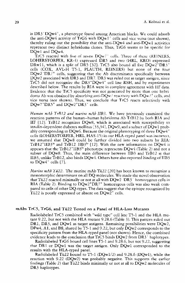
Immunoprecipitation Sequential immunoprecipitation experiments were performed using lysate from the D Q w l (DQw6) homozygous cell line H H K (Figure 1). Material precipitated by TrG6 and Tii22 (anti-DQ monomorphic) displayed two bands (MW of about 32 and 27 kD) by SDS-PAGE, consistent with the cz and/~ chains o f a HLA class II molecule. Bands with mAb L243 (anti-DR monomorphic) exhibited slightly higher molecular weights, indicating that TrG6 binds a non-DR class II antigen. Unlike L243, the mAb Tfi22 removed TrG6 antigen from the lysate, and vice versa. Thus, the epitopes defined by Tti22 and TrG6 are probably expressed on the same molecule, indicating that TrG6 combines with a DQ molecule. TrG6 also precipitates DQw4 antigens (not shown). Two weak bands (also about 32 and 27 kD) were observed when precipitating lysate of the DQw2÷DR3 ÷ cell line COX with mAb TrC5 (not shown).
Specificity defined by a Cell Panel (Table 2) Human mAbs TrG6 and TrC5. By RIA and IIF against a panel of 32 HLA-typed cell lines, TrG6 bound all DQwl + cells and did not discriminate between the two
FIGURE 1 Sequential immunoprecipitation of antigens in lysate of the DQwl homozy- gous cell line HHK analyzed by SDS-PAGE (12% gel) under reducing conditions. Lane A: L243 (anti-DR monomorphic), first precipitation. Lane B: L243, fourth precipitation. Lane C: TrG6, after preclearing four times with L243. Lane D: T/i22, (anti-DQ monomor- phic), first precipitation. Lane E: Tti22, fourth precipitation. Lane F: TrG6, after preclear- ing four times with Tti22. Lane G: TrG6, first precipitation. Lane H: TrG6, fourth precipi- tation. Lane I: Tii22, after preclearing four times with TrG6. Lane J: L243, after preclearing four times with TrG6.
A B C D E F G H I J
44kD~
25kD'~ f

Human-Human Hybridoma Antibody-Defined Eptiopes 19
TABLE 2 Specific(ties of the mAbs TrG6, TrCS, TrB12, Tii22, and IIB3 tested by a panel of HLA-typed cell lines
HLA" R I A
Cel ls D R D Q w T r G 6 TrC5 Ti i22 cpm x 10 ~ bound
11F h T O X'
T r G 6 TrC5 T r B I 2 l IB3
~2I w 1 5 / w 1 5 1/1(6) '1 128 0.6 103 3+ 0 3+ 8
T A B w 8 / w 8 6 /6 32 0.5 8.5 3+ 0 ' ,+ 8
K.RA. w 1 3 / w 1 3 I /1 (6) 91 0.7 84 3 + 0 3+ 8 _WDV w 1 3 / w 1 3 6 /6 122 0.6 106 3 + 0 3+ 8 H H K w 1 3 / w 1 3 6 /6 106 0.7 102 3+ 0 2,+ 8
H B S w 1 3 / w 1 3 1/116) 123 0.9 88 3+ 0 3+ 8 B V R - G 1/1 l / 1 (5 ) 118 0.6 92 3+ 0 1) 6 K A S O I I w 1 6 / w 1 6 1/1(5) nd ~ nd nd 3+ 0 0 6
C H N w 1 4 / w 1 4 1/1(5) nd 0.6 nd 3 + 0 l) 1
T 5 - I 3/1 2 / I ( 5 ) 29 4.4 20 3+ 2 + 0 l P L A I J T H 3/1 2 / I ( 5 ) 108 11 nd 3+ 2+ 0 1
C 'OX 3/3 2/2 nd I0 1.4 0 2 + 0 1
3 /3 2/2 0.5 4.1 1.5 0 2 + 0 1 B H - 1 3 7/7 2/2 0.3 0.4 4.6 0 0 0 1
PJ~ 7/7 2 /2 0.6 0.8 4.6 0 0 0 l B U P 7/7 2 /2 nd 0.5 nd nd 0 nd nd
M O U 7/7 2 /2 nd nd nd 0 0 l) 0 Bin-40 4 /4 8 /8 0.4 0.8 58 0 0 3+ 8
T S - I 0 4 /4 8 /8 0.6 0.4 85 0 0 3+ 8 J A H 4 /4 8 /8 nd nd nd 0 0 ~+ 8
D K B - E 9 /9 9 /9 0.5 0.5 73 0 0 3 + 8
PEA 4 /4 7/7 0.5 0.5 135 0 0 0 1 L U Y w 8 / w 8 7/7 nd nd nd 0 0 1) 1
R E I N E R S 3/5 2 /7 0.5 8.4 92 0 2 + 0 l
KR-4 2 /5 /7 nd 7.3 nd 0 2 + 0 1 K R O w l l / w l l 7/7 nd nd nd 0 2 + 0 l
H R L w 8 / w l 1 4 /7 62 12 115 3 + 2+ 0 6
L O H E R S T w8/5 4 /7 33 9.9 72 3+ 2 + 0 6 H A S - 15 4 /4 4 /4 53 0.6 19 .3 + 0 0 6
M A D U R A w 8 / w 8 4 /4 142 nd 50 3+ 0 0 8 B A E w 8 / w 8 4 /4 119 nd 42 3+ 0 0 8
R S H 3/3 4 /4 nd nd nd 3 + 0 () 6
" The 1987 HLA nomenclature has been adapted, as determined by the Tenth Histocompatibility Conference [ 14].
;' Indirect immunofluorecence, scored as follows: 0 = no fluorecence, + - weakly positive, 2+ positive, ~+ = strongly positive.
' Microcytotoxicity, scored as follows: 0 (invalid), 1 (negative), 2 (1()'74-19qd killing), 4 120°d-39(¥), 6 140%-79(;;.), 8 (80C4-100%).
u Numbers in parantheses state the most probable subset of DQwl (DQw5 or DQw6) deduced from the DR-type of the cell line.
nd not done. Sources and phenotypes of underlined cells have been published in "Histocompatibility Testing, 1984" [48,49] and by Koning et al. [50]. KRO, REINERS, and LOHERSTORFER originate from Ttibingen, FRG, BUP from John Hopkins University, USA, BVR-G from Leiden, Holland, RSH and OMW from du To(t, South Africa, and KASOI1 from Kastelan, Yugoslavia. T5-1 and KR-4 were described in Materials and Methods.
subtypes of DQwl ,DQw5 and DQw6. In addition, four DQw4 homozygous cells (HAS-15, MADURA, BAE, RSH) and two DQw4 heterozygous cells (HRL, LOHERSTORFER) were bound. To confirm the DQ phenotype of the DQw4 + cells, they were tested with the DQw4-specific antiserum TK2 in a microcytotoxic assay (not shown). TK2 killed all these cells, except RSH, which
20 A. Kolstad et al.
is DR3+DQw4 +, a phenotype found among American blacks. We could adsorb the anti-DQw4 activity of TrG6 with DQwl + cells and vice versa (not shown), thereby ruling out the possibility that the anti-DQw4 and anti-DQwl activities represent two distinct hybridoma clones. Thus, TrG6 seems to be specific for DQwl and DQw4.
TrC5 reacted with five of seven DQw7 ~ cells. Three of these (REINERS, LOHERSTORFER, KR-4) expressed DR5 and two (HRL, KRO) expressed DRwl l , which is a split of DR5 [32]. TrC5 also bound all five DQw2+DR3 + cells (COX, AVL-H T5-1, PLAUTH, REINERS) but none of the four DQw2+DR7 + cells, suggesting that the Ab discriminates specifically between DQw2 associated with DR3 and DR7. DR3 was ruled out as target antigen, since TrC5 did not recognize the DR3+DQw4 + cell line RSH, and by experiments described below. The results by RIA were in complete agreement with IIF data. Evidence that the TrC5 specificity was not generated by more than one hybri- doma Ab was obtained by absorbing anti-DQw7 reactivity with DQw2 + cells, and vice versa (not shown). Thus, we conclude that TrC5 reacts selectively with DQw7+DR5 + and DQw2+DR3 + cells.
Human mAb TrBI2 and murine mAb IIB3. We have previously examined the reaction patterns of the human-human hybridoma Ab TrB12 by both RIA and IIF [12]. TrB12 recognizes DQw8, which is associated with susceptibility to insulin-dependent diabetes mellitus [33,34], DQw9, and a subset of DQw 1 prob- ably corresponding to DQw6. Because the original phenotyping of three DQw4 + cells (LOHERSTORFER, HRL, HAS-15) in our HLA-typed panel was incorrect, we assumed that DQw8 could be further divided into two subsets by RIA: TrB12+IIB3 hi and TrB12 IIB3 l° [12]. With the new information on DQw4 it appears that the TrB 12-IIB3 l° phenotype represents DQw4 (Table 2) and not a subset of DQw8. Thus, the main difference between IIB3 and TrB12 is that IIB 3, unlike TrB 12, also binds DQw4. Others have also reported binding of IIB 3 to DQw4 + cells [7].
Murine mAb Tii22. The murine mAb Tii22 [20] has been known to recognize a monomorphic determinant on all DQ molecules. We made the novel observation that Tii22 reacted minimally or not at all with DQw2+DR3 + homozygous cells in RIA (Table 2). Binding to DQw2+DR7 + homozygous cells was also weak com- pared to cells of other DQ-types. The data suggest that the epitope recognized by Tfi22 is poorly expressed or absent on DQw2 + cells.
mAbs TrC5, TrG6, and Tii22 Tested on a Panel of HLA-Loss Mutants
Radiolabeled TrC5 combined with "wild type" cell line T5-1 and the HLA mu- tant 9.22, but not with the HLA mutant 9.28.6 (Table 3). This pattern ruled out DR1, DR3, and DQwl as target antigens. Remaining possibilities were DQw2, DPw4, A1, and B8, shared by T5-1 and 9.22, but only DQw2 corresponds to the specificity pattern from the HLA-typed panel (not shown). Hence, the combined evidence leads to the conclusion that TrC5 binds DQw2 from DR3 + haplotypes.
Radiolabeled TrG6 bound cell lines T5-1 and 9.28.6, but not 9.22, suggesting that DR1 or DQwl was the target antigen. Only DQwl corresponded to the results with the HLA-typed panel.
Radiolabeled Tfi22 bound to T5-1 (DQwl/2) and 9.28.6 (DQwl), while the reaction with 9.22 (DQw2) was probably negative. This supports the earlier findings (Table 2) that Tfi22 binds minimally or not at all to DQw2 molecules of DR3 haplotypes.

Human-Human Hybridoma Antibody-Defined Eptiopes 21
TABLE 3 The reaction pattern of Abs TrG6, TrC5, and Tii22 with a panel of HLA deletion mutants derived from cell line T5-1
HLA" Radioimmunoassay
(cpm x 10 5 b o u n d )
Cell line A B DR D Q w DPw T r G 6 T r C 5 Ti i22
T5-1 1/2 8 /27 3/1 2/1 4 /x 28.5 4.9 20.3
9 .28 .6 d /2 d /27 d/1 d/1 d /x 60.7 0.7 50.0
9.22 1/2 8 /27 d /d 2 /d 4 /x 0.5 6.8 0.8
d = deletion in the HLA locus, phenotype lost; x = untypable or homozygous.
TrG6 Binds Molecules That Express D Q w l
In a competition RIA against the DQwl (DQw6) cell lines HHK (not shown) and HBS (Table 4), the mAbs TrG6, TrB12, IIB3, and Tii22 all competed with each other for binding. Thus, the epitopes defined by these mAbs probably form an overlapping cluster on the surface of DQw6 molecules. The DQwl (DQw5 + DQw6)-specific mouse mAb Genox 353 G2a-5 did not compete with any of the other mAbs. Antigens contained in lysate from the DQwl (DQw6) homozygous
TABLE 4 The mAbs TrG6, TrB12, IIB3, and Tii22 compete with each other for binding to the DQwl (DQw6) homozygous cell line HBS
Ratio of leVI-labeled (hot) mAb Cold cold - to-hot
mAb mAb TrG6 TrBI2 l lB3 T622 G e n o x
Percent inhibition L24~
Anti-DQ T r G 6 30 98 97 97 95 - , i I)
0.5 58 61 65 57 ~ 2
TrBI2
I IB~
T022
60 76 - -# 95 76 - - - -
30 59 93 64 72 - 7 3
0.5 2 24 16 18 1 0
60 35 - - - - 65 - - - -
30 49 87 87 69 v 5
0.5 13 50 39 36 11 16
60 70 . . . . .
30 77 87 96 93 - 10 - 2
0.5 37 66 86 60 I,l 8
G e n o x 30 - 19 - 1 20 7 94
0.5 6 - 9 22 0 48
Anti-DR L243 30 - 6 1 5 12 --4 93
0.5 - 18 - 1 - 6 16 4 29
"--, not done.

22 A. Kolstad et al.
TABLE 5 Crosslinking of pairs of mAbs by antigen in detergent cell lysate from the DQwl (DQw6) homozygous cell line HHK
Soluble I-'~I-labeled m A b
C a p t u r e m A b G e n o x T r G 6 T r B 1 2 I IB3 Tt i22 L243 W()/~2
A n t i - D Q
G e n o x 1.1 ¢ 105.9 81.5 4 1.5 94 .6 2.7 1.1 T r G 6 57.2 2.1 1.0 I. l 0.8 I. 1 1.0
TrB 12 41.2 1.9 1.0 8.9 2.4 1.4 1.0
l I B 3 21.3 2.5 1.0 0.9 1.2 1.l 1.1 T~22 35.2 2.0 1.1 1.3 1.1 1.4 1.0
A n t i - D R L243 1.6 2.9 1.1 1.1 1.8 1.5 1.2
A n t i - c l a s s I
W 6 / 3 2 1.0 1.0 1.0 1.0 1.0 1.0 1.0
The degree of crosslinking (cpm bound) achieved with each pair of mAbs was related to the cprn bound using W6/32 (anti-class [) as capture mAb, according to the formula
cpm (capture mAb "x" + lysate + hot mAb "y") cpm (capture mAb W6/32 + lysate + hot mAb "y")
cell lines H H K (Table 5) and HBS (not shown) crosslinked Genox 353 with TrG6, TrB12, IIB3, and Tti22. Hence, the Genox 353 epitope is located on the same molecule, but is topographically unrelated to the epitope cluster defined by the other four mAbs. Competing pairs of mAbs were not crosslinked, except IIB3 and TrB12. This may be due to idiotypic interractions, because IIB3 and TrB 12 bound to each other in the absence of lysate (not shown). Genox 353 and TrG6 were also crosslinked by antigens in lysate from the DQwl + (DQw5 ÷) cell line PLAUTH (not shown). The data provide strong evidence that TrG6 interacts with molecules that express DQwl.
TrG6 Binds Molecules That Express DQw4 The mAbs TrG6, Tti22 and IIB3 all competed with each other for binding to the DQw4 homozygous cell line BAE (Table 6), and similar results were obtained with cell lines MADURA and HAS-15 (not shown). Antigens in lysate from cell lines BAE, MADURA and HAS-15 did not crosslink any of the pairwise combi- nations of mAbs (not shown). Thus, the data support the hypothesis that the epitopes defined by TrG6, IIB3, and Tii22 are overlapping on the surface of molecules expressing DQw4.
The Topographical Relationship Between Epitopes Defined by TrC5 and Other DQ-Specific mAbs
Unlabeled TrC5 did not compete with the 125I-labeled mAbs IVD-12 (anti- DQw7 + w8 + w9) or Tti22 (anti-DQ monomorphic) for binding to the DQw7+DRw11+/DQw4+DRw8 ÷ cell line HRL (Table 7). Hence, the TrCS- defined epitope is either located on a different molecule than the epitopes de- fined by the other two mAbs, or topographically apart on the same molecule. Similar data were obtained using the DQw7+DR5 + cells KR-4, REINERS, and LOHERSTORFER as target antigen (not shown). Unlabeled Tii22 inhibited binding of 125I-labeled IVD-12, and vice versa. Unlabeled T/i22 also competed

Human-Human Hybridoma Antibody-Defined Eptiopes
TABLE 6 The mAbs TrG6, IIB3, and Tii22 compete with each other for binding to the DQw4 homozygous cell line BAE
23
Ratio of
Cold cold-to-hot mAb mAb TrG6
125I-labeled (hot) mAb
IIB3 T022 L243 W6/32
Percent inhibition
Ant i -DQ
TrG6 50 97 94 73 10 6 l 70 85 64 14 14
l IB3 50 42 85 51 13 6
1 28 35 39 17 8
Tii22 50 62 80 82 10 5
1 25 45 34 18 9 Anti -DR
L243 50 23 16 32 97 13 1 20 20 23 52 - 6
Anti-c lass I W6/32 50 20 - 5 15 14 100
1 23 10 21 22 65
with radiolabeled TrG6 for binding to HRL, but unlabeled TrG6 failed to inhibit radiolabeled Tii22. This unilateral inhibition is probably due to Tfi22 binding both DQw4 and DQw7 molecules on HRL, and TrG6 only recognizing DQw4.
We further tested the ability of antigens in lysate from cell line HRL (Table 8)
TABLE 7 Competition between mAbs for binding to the DQw7/DQw4 heterozygous cell line HRL
Ratio of J>l-labeled (hot) mAb
Cold cold-to-hot mAb mAb TrG6 Tii22 IVD- 12 TrC5 L243
Percent inhibition
Ant i -DQ
TrG6
T622
30 98 2 - 4 4 3
1 67 5 - 2 3 - 9
60 52 ~ - - - - - - 30 36 92 95 10 8
1 13 70 86 9 1
IVD-12 60 - - 54 - - - - - - 30 1 42 96 1 7
1 13 21 40 20 4
TrC5 30 10 3 8 87 13 l 12 2 - 9 58 5
Ant i -DR L243 30 11 7 10 3 85
1 12 5 - 2 6 16 14
a - - , not done.

24 A. Kolstad et al.
TABLE 8 Crosslinking of pairs of mAbs by antigen in detergent cell lysate from the DQw7/DQw4 heterozygous cell line HRL
Solub le 125I-labeled m A b
C a p t u r e m A b T r C 5 T r G 6 Ti i22 I V D - 12 L243 W 6 / 3 2
A n t i - D Q T r C 5 1. l a 21 .3 55.7 9 .0 1.3 {).8
T r G 6 18.8 1.1 1.7 1.5 1.5 1.0
T/J22 6 1 . 9 1.5 1.5 1.1 3.2 1.0
I V D - 1 2 27 .4 1.1 3 . l 0 .9 1.9 1.0
A n t i - D R L243 1.2 1.6 3.8 1.2 1.9 1. 7
A n t i - c l a s s 1 W 6 / 3 2 1.0 1.0 1.0 1.0 1.0 1.0
" The degree of crosslinking (cpm bound) achieved with each pair of mAbs was related to the cpm bound using W6/~2 (anti-class I) as capture mAb, according to the fi)rmula given in the footnote to Table 4.
to crosslink TrC5 to other mAbs of known specificities. We found that the lysate crosslinked both mAb Tii22 and IVD-12 to TrCS. Similar data (not shown) were obtained with lysates from the DQw7+DR5 + cell lines KR-4, LOHERSTOR- FER, and REINERS. Thus, the combined evidence indicates that the epitope defined by TrC5 is expressed on DQw7 molecules but is topographically distant to the epitopes defined by IVD-12 and Tii22.
Interestingly, antigens contained in lysate from DQw7+DRwll+/DQw4 * DRw8 + heterozygous cell line HRL (and LOHERSTORFER--not shown) also crosslinked TrC5 and TrG6 (Table 8). No competition (Table 7) or crosslinking (Table 8) was noted between IVD-12 and TrG6, suggesting that the respective epitopes were located on different molecules of this lysate. Yet the lysate crosslinked both IVD-12 and TrG6 to TrC5. The observations can best be ac- counted for by assuming that TrC5 binds two distinct DQ antigens of the lysate, of which one expresses the TrG6 epitope and the other the IVD-12 epitope. The possibility that the TrCS+TrG6 + molecule may be a transassociated DQ mole- cule that derives its c~ and/3 chain from different haplotypes is discussed below.
Even though our data indicate that TrC5 binds DQw2 from DR3 + haplotypes, no competition or crosslinking was observed with T~i22 (anti-DQ monomorphic) or any of the other murine mAbs when applying DQw2+DR3 + cells as antigen (not shown). In as much as Tti22 binds minimally or not at all to DQw2+DR3 + cells (Table 2 and 3), this probably explains the inability of DQ-antigens of these cell lines to crosslink TrC5 and T/i22.
DISCUSSION
In this communication we report (1) a novel DQ-specific human-human hybri- doma Ab (TrG6) that binds cell expressing DQwl and DQw4, (2) that the previ- ously described [ 12] human-human hybridoma Ab TrB 12 is specific for DQw6, DQw8, and DQw9, and does not, as the murine mAb IIB3, recognize DQw4; (3) another novel human-human hybridoma mAb (TrC5) with apparent specificity for the DQ a chain (arguments in favor of this are presented later in the discus- sion) associated with the DQw2+DR3 + and DQw7+DR5 ÷ haplotypes; and (4) that the murine mAb Tii22 has minimal or no affinity for DQ molecules associ- ated with the DQw2+DR3 + haplotype.

Human-Human Hybridoma Antibody-Defined Eptiopes 25
The human-human hybridoma Ab TrG6 recognizes all DQw I * (both DQw5 + and DQw6 + cells) and DQw4 + lymphoblastoid cell lines, and the epitope is situated on DQ molecules. DQw4 is usually expressed by DRw8 ÷, DR4 (wl 5) +, and DR3 + haplotypes, and TrG6 combined with cells of all three by IIF and RIA (Table 2). A murine mAb apparently exhibiting a similar reaction pattern as TrG6 has recently been reported [11]. Interestingly, the DQw4-specific antiserum TK2 did not react with the DQw4+DR3 + cell line RSH by microcytotoxicity (not shown), suggesting that RSH might represent a structural variant of DQw4. When comparing DQc~ and DOff chain first-domain amino-acid sequences from various DR haplotypes (reviewed by Todd et al. [35]), no obvious clue was found as to which chain is involved in the interaction with TrG6. However, only DQ~- chains from DQwl + and DQw4 + haplotypes, and no others, have the amino-acid Arg in position 55, suggesting that this residue might be important for binding of TrG6.
Our data indicate that TrC5 recognizes DQ molecules of DQw7-DRS* and DQw2+DR3 + cells. Saiki and co-workers [36] synthesized a DQc~-specific oligo- nucleotide probe that hybridized selectively to DNA from DQw7+DR5 + and DQw2+DR3 + cell lines. Others have reported a human mAb specific for DQw2 [24] and a murine mAb recognizing a DQc~-chain-dependent determinant ex- pressed by DR3, DR5, DRw8, and DRwl2 cells [10]. To our knowledge no antiserum or mAb has been described before with the same specificity as TrCS. Nucleotide sequences of the gene segments encoding the first domains of several DQ~ and/3 chains have been determined [35,37-42]. These reports have dem- onstrated that the DQc~ chains of DQw2+DR3 + and DQw7+DR5 + haplotypes have identical first-domain sequences, which, however, are distinct from that of the DQw2+DR7 + haplotype. By contrast, the first domain of DQ,8 chains of DQw2+DR3 + and DQw2+DR7 + haplotypes are identical, and distinct from that of the DQw7 +DR5 + haplotype. Thus, the TrC 5 reaction pattern correlates strik- ingly with the first-domain amino-acid sequences of the DQc~ chains from the DQw7+DR5 ÷ and DQw2+DR3 + haplotypes, indicating that TrC5 probably identifies an epitope determined by a DQ~-chain.
An important set of observations were made with lysates of DQw7 + DR5 ~ / DQw4+DRw8 + heterozygous cells. Antigens in these lysates (1) crosslinked TrC5 and TrG6; (2) crosslinked TrC5 and IVD-12; but (3) did not crosslink TrG6 and IVD-12. Since TrG6 and IVD-12 did not compete for binding, these findings can be accounted for by assuming that TrC5 combines with two distinct species of DQ molecules, one that expresses epitopes TrC5 and IVD-12 and another that expressed epitopes TrC5 and TrG6. The former is found in lysates of DQw7+DR5 + cells, whereas the latter is selectively found in lysates from DQw7+DR5+/DQw4+DRw8 + heterozygous cells. The TrCS+TrG6 + species may arise by way of transcomplementation [43-45], creating DQw7~/DQw413 and DQw4~/DQw7/3 heterodimers. Which of these transcomplementing mole- cules are likely to be TrC5 +TrG6+? Strong clues may come from combining our observations with published nucleotide sequences of the first domains of DQ~ and DQ,8 genes [35]. These sequences reveal that the serologic specificities DQw2, DQw7, DQw8, and DQw9 are determined by amino acids encoded by DQ~ genes. Inasmuch as IVD-12 has been shown to be specific for DQw7, w8, and w9, it follows that its epitope is/~-chain-dependent. This, and the fact that TrG6 and IVD-12 neither compete for binding to DQw7+DQw4 + cells nor are crosslinked by DQw7+DQw4 + lysates imply that TrG6 does not recognize the putative transcomplementing species DQw7/3/DQw4cq leaving DQw7~/ DQw4/3 as the most likely TrG6+TrC5 + species. Furthermore, as argued above, the TrC5 epitope is probably specified by a DQc~ chain of the DQw7+DR5 +

26 A. Kolstad et al.
haplotype, and only the DQw7~/DQw4/3 species contains this ~ chain. Thus, we conclude that the molecule created by transassociation of DQw7~-chains and DQw4/3-chains is probably responsible for the crosslinking of TrC5 and TrG6. It follows that the T rG6 epitope is specified by the DQw4/3 chain.
DQw7~ (of DR5 ÷ haplotypes) and DQw4~ (of DRw8 ÷ haplotypes) first- domain deduced amino-acid sequences [35,38] only differ at positions 69 (Leu and Thr, respectively) and 75 (Ser and Ile, respectively). Nevertheless, TrC5 is apparently able to discriminate between the two antigens, suggesting that one or both of these amino acids are important for the TrC5 epitope. Since Ser in position 75 seems to be specific for DQw7÷DR5 ÷ and DQw2+DR3 ÷ haplotypes, this residue might be the most critical. Bjorkman and co-workers [46], who reported the tertiary structure of HLA-A2, suggested that the class I and class II molecules probably have a similar structure, and this was recently supported by the hypothetical model of the class II antigen binding site presented by Brown and co-workers [47]. If the model is correct, the DQ~ residues 69 and 75 correspond to an ~ helix and are thus probably available for interaction with TrC 5.
The deduced amino acid sequences of DQw4 (DRw8), DQw2, and DQw7 (DR5)/3 chains differ by as much as 12-18 of the first of 90 positions [35]. Nevertheless, all three/3 chains, when associated with the correct ~ chain, seem to be compatible with expression of the TrC5 epitope. Thus, this epitope appears to be relatively or maybe entirely independent of the /3-chain sequence. However , since we have not found that TrC5 reacts with free ~ chains (un- published observations), the /3 chain may be required to induce the correct a-chain conformation.
A C K N O W L E D G M E N T S
The technical assistance of Kjetil Andreassen is highly appreciated. We thank Dr. Pawelec and Dr. Koning for providing the mAbs Ti~22 and lIB3, Dr. Matsuki for providing antiserum TK2, and Dr. Pious for the gift of HLA-loss mutants. This work was supported by the Norwegian Research Council for Science and the Humanities and the Norwegian Society for Fighting Cancer (Kreftforeningen).
REFERENCES 1. Bodmer WF: The HLA-System, 1984. In Albert ED, Baur MP, Mayr WR (eds):
Histocompatibility Testing 1984. Berlin, Springer-Verlag, 1984, p 11.
2. Betuel H, Gebuhrer L, Schreuder GMT, Layrisse Z, Arnaiz Villena A, Goldmann SF: Antigen report: HLA-DRw8. In Albert ED, Baur MP, Mayr WR (eds): Histocompati- bility Testing 1984. Berlin, Springer-Verlag, 1984, p 198.
3. Grosse-Wilde H, Doxiadis I, Brandt H: Definition of HLA-D with HTC In Albert ED, Baur MP, Mayr WR (eds): Histocompatibility Testing 1984. Berlin, Springer- Verlag, 1984, p 249.
4. Ishikawe N, Kojima H, Nakayama T, Kunikane H, Hawkin S, Fukasawa Y, Ikeda H, Ogasawara K, Kasahara M, Tajima Y, Kakuta Y, Wakisaka A, Aizawa M: Detection of a novel HLA-DQ specificity: Serological and immunochemical analysis by a monoclo- nal antibody. Immunogenetics 26:143, 1987.
5. Matsuki K, Juji T, Satake M: A novel HLA class II alloantigen detected by an alloantiserum TK2. Hum Immunol 18:247, 1987.
6. Maeda H: Mouse monoclonal antibody detects a new polymorphic Ia determinant other than HLA-DR antigen: a possible allele of DC1. Tissue Antigens 23:163, 1984.

Human-Human Hybridoma Antibody-Defined Eptiopes 27
7. Koning F, Raghoebar J, Schreuder GMT, Schuurman R, Bruning H: A monoclonal antibody detecting an HLA-DQwl related determinant. Tissue Antigens 26:100, 1985.
8. Radka S, Stewart S, Smith SA: Analysis of HLA-DQ molecules with a monoclonal antibody detecting a DQ polymorphism absent from DQwl homozygous cells. Hum Immunol 14:206, 1985.
9. Shipp MA, Ahmed P, Kannapell CC, Ford JC, McCourt D, Leykam JF, Zacheis M, Bono C, Davie JM, Mustain E, Nahm MH, Schwartz BD: A new polymorphic deter- minant on HLA-DQ molecules. Hum Immunol 16:24, 1985.
10. Radka SF, Johnston JV, Schwarz D, Nepom BS: Biochemical analysis of DQ~ poly- morphism defined by a monoclonal antibody. Tenth International Histocompatibility Conference, November 18-23, 1987; New York, Abstract no. 351.
11. Nelson K, Radka SF, Nepom BS, Hurley C, Johnson AH, Colter M, Schwarz D, Johnston JV: DQ/3 epitopes expressed by both DQwl and DQ blank haplotypes in caucasian, american black and oriental populations. Tenth International Histocompat- ibility Conference, November 18-23, 1987, New York, Abstract no. 350.
12. Kolstad A, Hansen T, Hannestad K: A human-human hybridoma antibody (TrBI2) defining subgroups of HLA-DQwl and -DQw3. Hum Immunol 20:219, 1987.
13. Hansen T, Kolstad A, Mathisen G, Hannestad K: A human-human hybridoma (Tr7E2) producing cytotoxic antibody to HLA-DQwl. Hum Immunol 20:307, 1987.
14. Bodmer WF, Albert E, Bodmer JG, Dupont B, Mach B, Mayr W, Sasuki T, Schreuder GMT, Svejgaard A, Terasaki PI: Nomenclature for factors of the HLA system, 1987. Tenth International Histocompatibility Conference, November 18-23, 1987, New York.
15. Pious D, Dixon L, Levine F, Comer T, Johnson R: HLA class II regulation and structure. J Exp Med 162:1193, 1985.
16. Mellins E, Woelfel M, Pious D: Importance of HLA-DQ and -DP restriction elements in T-cell responses to soluble antigens: mutational analysis. Hum Immunol 18:211, 1987.
17. Hansen T, Kolstad A, Thorsby E, Hannestad K: A human-human hybridoma pro- ducing cytotoxic antibody to HLA-B15, crossreacting with B17, B5, B35, and B18. Tissue Antigens 29:246, 1987.
18. Barnstable CJ, Bodmer WF, Brown G, Galfre G, Milstein C, Williams AF, Ziegler A: Production of monoclonal antibodies to group A erythrocytes, HLA and other cell surface antigens--new tools for genetic analysis. Cell 14:9, 1978.
19. Lampson LA, Levy R: Two populations of Ia-like molecules on a human B-cell line. J Immunol 125:293, 1980.
20. Pawelec GP, Shaw S, Ziegler A, Miiller C, Wernet P: Differential inhibition of HLA-D- or SB-directed secondary lymphoproliferative responses with monoclonal antibodies detecting human Ia-like determinants. J Immunol 129:1070, 1982.
21. Giles RC, Nunez P, Katovich Hurley C, Nunez-Roldan A, Winchester R, Stasmy P, CapraJD: Structural analysis of a human I-A homologue using a monoclonal antibody that recognizes a MB3-1ike specificity. J Exp Med 157:1461, 1983.
22. Parham P, Kipps TJ, Ward FE, Herzenberg LA: Isolation of heavy chain class-switch variants of an anti-DC hybridoma cell line: Effective conversion of non-cytotoxic IgG 1 antibodies to cytotoxic IgG2 antibodies. Hum Immunol 8:141, 1983.
23. Fraker PJ, Speck JC: Protein and cell membrane iodination with a sparingly soluble chloramide, 1, 3, 4, 6, -tetrachloro-3~-6~-diphenylglycoluril. Biochem Biophys Res Commun 80:849, 1978.

28 A. Kolstad et al.
24. Pistillo MP, Haemmerling U, Dupont F, Ferrarra GB: In vitro production of human HLA alloantibody of restricted specificity (DQw2) via Epstein-Barr virus transfor- mation. Hum Immunol 15:109, 1986.
25. Kozbor D, Lagarde AE, RoderJC: Human hybridomas constructed with antigen-spe- cific Epstein-Barr virus-transformed cells. Proc Natl Acad Sci USA 79:6651, 1982.
26. Kozbor D, Triputti P, Roder JC, Croce CM: A human hybrid myeloma for produc- tion of human monoclonal antibodies. J Immunol 133:3001, 1984.
27. Mittal KK: Standarization of the HLA-typing method and reagents. Vox Sang 34:58, 1978.
28. Mickey MR, Terasaki PI: The serological data of the 8th Workshop and summary analysis. Histocompatibility Testing 1980. Los Angeles, CA, UCLA Press, 1980, p 21.
29. Kolstad A, Hansen T, Hannestad K: A cytotoxic human-human hybridoma antibody (TrH6) specific for HLA-DRw52. Tissue Antigens 31:90, 1987.
30. Effros LA, Zeller E, Dillard L, Walford RL: Detection of antibodies to cell surface antigens by a simplified cellular ELISA (CELISA). Tissue Antigens 25:293, 1985.
31. Lemonnier FA, Rebai N, le Bouteiller PP, Malissen 13, Caillol D, Kourilsky FM: Epitopic analysis of detergent-solubilized HLA molecules by solid-phase radioimmu- noassay. J Immunol Methods 54:9, 1982.
32. Betuel H, Gebuhrer L, Schreuder GMT, Goldmann SF, Arnaiz Villena A, Layrisse Z: Antigen report: HLA-DR5 and its subtypes HLA-DRwl 1 and DRwl2. In Albert ED, Baur MP, Mayr WR (eds): Histocompatibility Testing 1984. Berlin, Springer-Verlag, 1984, p 190.
33. Owerbach D, Lernmark A, Platz P, Ryder LP, Rask L, Peterson PA, Ludvigsson J: HLA-D region F3-chain DNA endonuclease fragments differ between HLA-DR iden- tical healthy and insulin-dependent diabetic individuals. Nature 303:815, 1983.
34. Nepom BS, Palmer J, Kim SJ, HansenJA, Holbeck SL, Nepom GT: Specific genomic markers for the HLA-DQ subregion discriminate between DR4 + insulin-dependent diabetes mellitus and DR4 ÷ seropositive juvenile rheumatoid arthritis. J Exp Med 164:345. 1986.
35. Todd JA, Bell JI, McDevitt HO: HLA-DQ~3 gene contributes to susceptibility and resistance to insulin-dependent diabetes mellitus. Nature 329:599, 1987.
36. Saiki RK, Bugawan TL, Horn GT, Mullis KB, Ehrlich HA: Analysis of enzymatically amplified /3-globulin with allele-specific oligonucleotide probes. Nature 324:163, 1986.
37. Boss JM, Strominger JL: Cloning and sequence analysis of the human major histo- compatibility complex gene DC-3/3. Proc Natl Acad Sci USA 81:5199, 1984.
38. Schenning L, Larhammar D, Bill P, Wiman K, Jonsson AK, Rask L, Peterson PA: Both c~ and f3 chains of HLA-DC class II histocompatibility antigens display extensive polymorphism in their aminoterminal domains. EMBO J 3:447; 1984.
39. Moriuchi J, Moriuchi T, Silver J: Nucleotide sequence of an HLA-DQ ce chain de- rived from a DRw9 cell line: Genetic and evolutionary implications. Proc Natl Acad Sci USA 82:3420, 1985.
40. Karr PW, Gregersen PK, Obata F, Goldberg D, MaccariJ, Alber C, Silver J: Analysis of DR/3 and DQ3 chain cDNA clones form a DR7 haplotype. J Immunol 137:2886, 1986.
41. Song Q-L, Gregersen PK, Karr RW, Silver J: Recombination between DQc~ and DQ3 genes generates human histocompatibility leukocyte antigen class II haplotype diver- sity. J Immunol 139:2993, 1987.

Human-Human Hybridoma Antibody-Defined Eptiopes 29
42. Schiffenbauer J, Didier DK, Klearman M, Rice K, Schuman S, Tieber VL, Kittelsen DJ Schwartz BD: Complete sequence of the HLA DQ~ and DQfl cDNA from a DR5/DQw3 cell line. J Immunol 139:228, 1987.
43. Kimoto M, Fathman GC: Antigen-reactive T cell clones. I. Transcomplementing hybrid I-A-region gene products function effectively in antigen presentation. J Exp Med 152:759, 1980.
44. Charron DJ, Lotteau V, Turmel P: Hybrid HLA-DC antigens provide molecular evidence for gene trans-complementation. Nature 312:157, 1984.
45. Giles RC, DeMars R, Chang CC, Capra D: Allelic polymorphism and transassociation of molecules encoded by the HLA-DQ subregion. Proc Natl Acad Sci USA 82:1776, 1985.
46. Bjorkman PJ, Saper MA, Samraoui B, Bennet WS, Strominger JL:, Wiley DC: The foreign antigen binding site and T cell recognition regions of class I histocompatibility antigens. Nature 329:512, 1987.
47. Brown JH, Jardetzky T, Saper MA, Samraoui B, Bjorkman PJ, Wiley DC: A hypo- thetical model of the foreign antigen binding site of Class II histocompatibility mole- cules. Nature 332:845, 1988.
48. Jaraquemada D, Reinsmoen NL, Oilier W, Bach FH, Festenstein H: First level testing of HLA-DR4-associated HLA-D specificities: Dwl3 (DB3), Dwl4 (LD-40), Dwl5 (DYT), and DKTe. Albert ED, Baur MP, Mayr WR (eds): Histocompatibility Testing 1984. Berlin, Springer-Verlag, 1984, p 270.
49. Schreuder GMT, Doxiadis I, Parlevliet J, Grosse-Wilde H: HLA-DR, DQ, LB, and TA10 specificities of Ninth Workshop homozygous typing cells. In Albert ED, Baur MP, Mayr WR (eds): Histocompatibility Testing 1984. Berlin, Springer-Verlag, 1984, p 243.
50. Koning F, Giphart M, Termijtelen A, Bruning H: Differential expression of DRw52- like determinants detected by monoclonal antibodies. Scand J Immunol 25:127, 1987.


![7 % )DQ &RLO 'LMLWDO 7HUPRVWDWÕ )DQ &RLO 'LJLWDO … · zzz ol]dugfrqwurov frp)dq &rlo 'lmlwdo 7huprvwdwÕ)dq &rlo 'ljlwdo 7khuprvwdw g]hoolnohu 6shflilfdwlrq idi idi di i (qhuml](https://img.pdfslide.tips/doc/110x75/5d4ec4ee88c993e16b8b5b15/7-dq-rlo-lmlwdo-7huprvwdwo-dq-rlo-ljlwdo-zzz-oldugfrqwurov-frpdq.jpg)