Embed Size (px)
Citation preview

Vol 5 | Issue 3 | Jul - Sep 2020 Eastern J Medical Sciences 61
Case Series
Association of antinuclear antibodies with toxic epidermal necrolysis – A novel manifestation
Meghna Dutta1, Suddhasatwya Chatterjee2, Sraboni Ghosh Zoha3, Suresh Ramasubban4,5
From 1Postgraduate Trainee, Department of Family Medicine, 2Senior Consultant, Department of Internal Medicine and Rheumatology, 3Senior Consultant, Department of Dermatology, 4Senior Consultant, Department of Critical Care, 5Senior Consultant, Department of Pulmonology, Apollo Gleneagles Hospital Limited, Kolkata, West Bengal, IndiaCorrespondence to: Meghna Dutta, Department of Family Medicine, Apollo Gleneagles Hospital Limited, Kolkata, West Bengal, India. E-mail: [email protected] - 24 October 2019 Initial Review - 03 March 2020 Accepted - 03 July 2020
Toxic epidermal necrolysis (TEN) is a potentially life-threatening dermatologic disorder characterized by widespread involvement of epidermis and mucous
membranes resulting in exfoliation and possible sepsis with morbidity or mortality [1]. TEN is a common yet one of the most dreaded dermatological emergencies. TEN-like rash of lupus is clinically indistinguishable from drug-induced TEN [2]. Therefore, a high index of suspicion is of prime importance. The clinician must watch out for the subtle features of systemic lupus erythematosus (SLE), and this must be balanced with laboratory findings such as hematological abnormalities, proteinuria, high positive antinuclear antibody (ANA) titer, and so on. Failing which, a sick patient presenting with vesiculobullous lesions with peeling of skin and screaming for urgent management maybe easily overlooked. Clinical outcome of these lesions will be influenced by the approach to the case and line of management [3]. We describe three female patients with widespread vesiculobullous lesions who were provisionally diagnosed as TEN, which later turned out to be the initial presentation of systemic LE.
CASE REPORT 1
A 25-year-old lady reported to our emergency department with severe erythematous blistering rashes over face and neck, mild rashes on buttocks, back, and abdomen (Fig. 1). The patient gave history of small joint polyarthralgia for 2 months. After more
than 50 days of suffering, she had visited a local physician and was started on Sulfasalazine and Hydroxychloroquine sulfate (HCQS). Almost 48 hours after intake of the medications, she developed high grade fever and sudden onset progressively increasing respiratory distress along with the rashes. She denied any similar episode in the past.
On examination, the patient was febrile (100.8℉) with tachycardia (102/min), blood pressure was 100/60 mmHg, respiratory rate 26/min, and adequate urine output. Oral mucosal ulcerations were also noted. She was admitted in the critical care unit, oxygen support was given, and barrier nursing was advised. Among her laboratory investigations, erythrocyte sedimentation rate (ESR) and C-reactive protein (CRP) were raised, C3 and C4 levels were low, ANA by Hep2 method (1:100 dilution) was 4+ with homogenous pattern and ANA panel was positive for anti-ds-DNA, antibody to nucleosomes, antibody to histones, and antibody to ribosomal P protein. She was diagnosed as drug-induced TEN in a newly diagnosed case of SLE. Thiopurine methyltransferase level was tested and found to have normal activity.
After a dermatology opinion, she was managed conservatively in monitor bed with a combination of steroids (Inj. methylprednisolone stat followed by Tab. methylprednisolone twice daily by Ryles tube), IV antibiotics (Inj. meropenem), eye care (carboxymethylcellulose sodium tear drops), and good care of skin and mucosa. She was discharged on day 14 with Tab.Prednisolone, carboxymethylcellulose sodium tear drops, Tab.
ABSTRACTToxic epidermal necrolysis (TEN) is a potentially life-threatening dermatologic disorder characterized by widespread erythema, necrosis, and bullous detachment of the epidermis of skin and mucous membranes, resulting in exfoliation and possible sepsis and/or morbidity/mortality. Exact diagnosis of lesions is very important in terms of therapeutic intervention as well as outcome. As cutaneous hypersensitivity reactions are known to occur with increased frequency in connective tissue disease, differentiating lesions of systemic lupus erythematosus that simulate drug-induced TEN is a challenge in the practical scenario. In this case series, we discuss about three patients aged between 24 and 67 years, who presented with vesiculobullous lesions all over the body and oral mucosal ulcerations. Each of them was initially admitted in the Critical Care Unit with a provisional diagnosis of TEN. Two of the three patients recovered well and were discharged. However, the third patient, who was the oldest and had multiple comorbidities, could not be saved and succumbed after 75 days of hospital stay.
Key words: Antinuclear antibodies, DMARDs, Systemic lupus erythematosus, Toxic epidermal necrolysis, Vesiculobullous lesions

Dutta et al. ANA and TEN
Vol 5 | Issue 3 | Jul - Sep 2020 Eastern J Medical Sciences 62
faropenem, Tab. HCQS, Tab. azathioprine, and other supportive measures (Tab. omeprazole, multivitamin supplements). To this day, she continues to do well and is currently on HCQS and azathioprine (Fig. 1).
CASE REPORT 2
A 24-year old nulliparous lady presented to our emergency at 14 weeks of gestation, with vesicullobullous lesions all over the body, oral ulceration, and worsening general condition for 7 days. She had recently started consuming doxylamine succinate for hyperemesis gravidarum. There was no history of recent infection or intake of other drugs. She gave a history of 2 abortions in the past 2 years. However, there was no history of similar lesions in the past.
On examination, besides the generalized vesiculobullous lesions and oral/mucosal ulceration, hypotension (80/50 mm of Hg) was noted (Fig. 2). Furthermore, she was afebrile, pulse rate was 100/min with a weak pulse, and respiratory rate was 26/min and poor urine output. She was admitted in the critical care unit and treatment started in the line of TEN. However, on account of
Type II respiratory failure experienced, she had to be intubated, ventilated, and started on vasopressors and total parenteral nutrition. On day 04, the patient’s left ventricle ejection fraction (LVEF) 25%, and she had developed bilateral pleural effusion. Under laboratory investigations, ANA by Hep2 method (1:100 dilution) was 3+ with fine granular pattern, anti-SS-A/anti-SS-B was strong positive, and immunofluorescence was strong positive for fibrinogen. She was negative for anti-phospholipid antibody. Histopathology images showed a full thickness epidermal necrosis, sub-cellular cell poor bulla containing fibrin with mild perivascular inflammation of dermis, suggesting a diagnosis of TEN-like acute cutaneous LE.
She was treated with IV steroids (Inj. methylprednisolone), IV antibiotics (Inj. meropenem), and other supportive measures (proton pump inhibitors and albumin supplementation). Good barrier nursing care for skin, eyes, and mucosa was provided. She gradually improved clinically and was shifted to ward on day10. On day12, she underwent a spontaneous abortion with no retained products of conception. As her LVEF improved to 63%, she was discharged on day 14 with Tab. HCQS, Tab. Prednisolone, and other supportive measures (Fig. 2).
CASE REPORT 3
A 67-year-old lady presented to us with generalized vesiculobullous lesions and maculopapular rashes on the face, chest, and trunk for 5 days. She was a known diabetic, hypertensive patient with history of hypothyroidism and ischemic heart disease. The patient did not give history of intake of any new medication and history of similar episode in the past. On examination, target lesions were noted all over the extremities (Fig. 3). She was afebrile, pulse rate was 84/min, blood pressure 130/90 mm of Hg, respiratory rate 24/min, and had adequate urine output. On the day of admission, patient had moderate anemia (8.8 g/dL), dyselectrolytemia, hypoalbuminemia, raised CRP, low procalcitonin (0.27 ng/mL), sterile blood culture, and no growth on urine culture. She was admitted in the critical care unit, conservative management started in the line of TEN, and good barrier nursing was advised.
Figure 1: Clinical picture of patient on day 1 of admission and on day 14
Figure 2: Clinical picture of patient on day 1, day 4, and day 14

Dutta et al. ANA and TEN
Vol 5 | Issue 3 | Jul - Sep 2020 Eastern J Medical Sciences 63
On day 3, skin biopsy was done which showed spongiosis, exocytosis, and hydropic degeneration of basal cell layer; edema and moderate perivascular chronic inflammation in the dermis consistent with erythema multiforme.
On day 7, her white blood cell count was 15,700/cumm, ESR and CRP were raised, procalcitonin was low (0.07 ng/mL), and blood/urine culture showed no growth. Although systemic bacterial infection was ruled out, leukocytosis could not be explained. We now tried to approach her case in the lines of an autoimmune disorder. ANA by Hep2 method (1:100 dilution) was 3+ with homogenous pattern and anti-SS-A was positive. C3 and C4 levels were low. Both direct and extended Coombs test were positive implying hemolytic anemia. She also had bilateral pleural effusion. She was finally diagnosed as a patient of TEN in a newly diagnosed case of SLE and started on IV methylprednisolone.
On the 25th day of her hospital stay, bleed from rectal mucosa was observed. At this time, she was found to have hypoalbuminemia, abnormal liver function tests but no growth on blood culture. On day 33, growth of Acinetobacter in blood and Klebsiella pneumoniae in sacral lesion was noted. On day 45 of stay, Acinetobacter and Candida non-albicans sp. were growing in her blood. In spite of our best efforts, she succumbed to nosocomial infection and multiorgan failure on day 75 of admission.
DISCUSSION
TEN is a severe adverse cutaneous reaction characterized by extensive full-thickness necrosis of the epidermis and mucous membrane involvement. It is an extreme form of Stevens-Johnson syndrome involving >30% of body surface area, clinically characterized by a prodrome of fever, malaise, diffuse erythema, and purpuric rash, with positive Nikolsky sign leading on to widespread blisters. The disorder usually follows a progressive course, often leading to dehydration, electrolyte imbalance, shock, secondary localized infections, and septicemia. Mucosal involvement presents as oral ulcers, crusted lips, dysphagia, genitourinary tract ulcers, and purulent conjunctivitis.
SLE is a prototypic heterogeneous autoimmune disease with patients often presenting with non-specific symptoms such as fever, weight-loss, and mild lymphadenopathy reflecting active inflammatory disease. Whereas, fatigue, malaise, and fibromyalgia-like symptoms are not necessarily associated with flares in disease activity. Mucocutaneous lesions including
TEN-like acute cutaneous SLE are a common occurrence among SLE patients [4].
Sontheimer classified vesiculobullous lesions in the setting of LE into following groups [5]• TEN-like acute cutaneous LE• TEN-like sub-acute cutaneous LE• TEN occurring in SLE patients not having conventional
LE-specific skin lesions.• Vesiculobullous changes occurring at the active border of
advancing annular sub-acute cutaneous LE• Vesiculobullous chronic cutaneous LE.
The first three entities clinically mimic drug-induced TEN, although an exacerbated activity of underlying lupus can be noticed on keen observation. The risk of drug-induced TEN is further increased in the patients of LE as they are often on chronic medications. Microscopically, a full thickness epithelial necrosis is observed in both conditions and they respond well to steroids. Each of our patients presented with typical clinical and histopathologic features of TEN and strongly positive ANA with diverse patterns. A plausible explanation for the cutaneous lesions and other major manifestations of LE seen in our patients could be the outcome of the exaggerated keratinocyte apoptosis due to increase in Fas expression [6,7].
According to the 2019 European League against Rheumatism/American College of Rheumatology criteria for classification of SLE, a serum titer of ANA of at least 1:80 on human epithelial-2-positive (Hep 2) cells or an equivalent positive test was the entry criterion for diagnosis of SLE. A distinctive serological profile, including a strongly positive ANA, a positive anti-dsDNA, a prolonged course, response to steroid treatment, histologic and immunofluorescence findings, and constitutional features such as fever, pleural or pericardial effusion, low C3 and low C4, point in favor of LE over, and above a diagnosis of TEN [8]. Our first patient was a patient of drug-induced (? Sulfasalazine) TEN in a newly diagnosed case of SLE, the second lady was diagnosed as TEN-like acute cutaneous LE, and the third patient was diagnosed to have TEN in a newly diagnosed case of SLE.
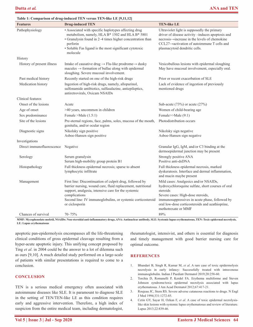
Table 1 describes the differences between drug-induced TEN versus TEN-like LE; however, these differences are to a large extent, subjective, and theoretical. TEN-like injury may be drug induced or due to other causes like LE such as acute graft versus host disease, pseudoporphyria, and so on. Acute syndrome of
Figure 3: Target-like lesions noted on extremities, face, and body
Dutta et al. ANA and TEN
Vol 5 | Issue 3 | Jul - Sep 2020 Eastern J Medical Sciences 64
Table 1: Comparison of drug-induced TEN versus TEN-like LE [9,11,12]Features Drug-induced TEN TEN-like LEPathophysiology • Associated with specific haplotypes affecting drug
metabolism, namely, HLA B* 1502 and HLA B* 5801• Granulysin found in 2–4 times higher concentration than
perforin• Soluble Fas ligand is the most significant cytotoxic
molecule
Ultraviolet light is supposedly the primary driver of disease activity –induces apoptosis and necrosis→increase in the levels of chemokine CCL27→activation of autoimmune T cells and plasmacytoid dendritic cells.
HistoryHistory of present illness Intake of causative drug → Flu-like prodrome→ dusky
macules → formation of bullae along with epidermal sloughing. Severe mucosal involvement.
Vesicobullous lesions with epidermal sloughingMay have mucosal involvement, especially oral.
Past medical history Recently started on one of the high-risk drugs Prior or recent exacerbation of SLEMedication history Ingestion of high-risk drugs, namely, allopurinol,
sulfonamide antibiotics, sulfasalazine, antiepileptics, antiretrovirals, Oxicam NSAIDs
Lack of evidence of ingestion of previously mentioned drugs
Clinical featuresOnset of the lesions Acute Sub-acute (73%) or acute (27%)Age of onset >40 years, uncommon in children Women of child-bearing age Sex predominance Female >Male (1.5:1) Female>>Male (9:1)Site of the lesions Pre-sternal regions, face, palms, soles, mucosa of the mouth,
genitalia, and/or ocular regionPhotodistribution occurs
Diagnostic signs Nikolsky sign positiveAsboe-Hansen sign positive
Nikolsky sign negativeAsboe-Hansen sign negative
InvestigationsDirect immunofluorescence Negative Granular IgG, IgM, and/or C3 binding at the
dermoepidermal junction may be presentSerology Serum granulysin
Serum high-mobility group protein B1Strongly positive ANAPositive anti-dsDNA
Histopathology Full thickness epidermal necrosis; sparse to absent lymphocytic infiltrate
Full thickness epidermal necrosis, marked dyskeratosis. Interface and dermal inflammation, and mucin maybe present
Management First line: Discontinuation of culprit drug, followed by barrier nursing, wound care, fluid replacement, nutritional support, analgesia, intensive care for the systemic complicationsSecond line: IV immunoglobulins, or systemic corticosteroid or ciclosporin
Mild cases: Analgesics and/or NSAIDs, hydroxychloroquine sulfate, short courses of oral steroidsSevere cases: High-dose steroids, immunosuppressives in acute phase, followed by oral low-dose corticosteroids and azathioprine, methotrexate or MMF
Chances of survival 70–75% 89%MMF: Mycophenolate mofetil, NSAIDs: Non-steroidal anti-inflammatory drugs, ANA: Antinuclear antibody, SLE: Systemic lupus erythematosus, TEN: Toxic epidermal necrolysis, LE: Lupus erythematosus
apoptotic pan-epidermolysis encompasses all the life-threatening clinical conditions of gross epidermal cleavage resulting from a hyper-acute apoptotic injury. This unifying concept proposed by Ting et al. in 2004 could be the answer to a lot of dilemma such as ours [9,10]. A much detailed study performed on a large-scale of patients with similar presentations is required to come to a conclusion.
CONCLUSION
TEN is a serious medical emergency often associated with autoimmune diseases like SLE. It is paramount to diagnose SLE in the setting of TEN/TEN-like LE as this condition requires early and aggressive intervention. Therefore, a high index of suspicion from the entire medical team, including dermatologist,
rheumatologist, intensivist, and others is essential for diagnosis and timely management with good barrier nursing care for optimal outcome.
REFERENCES
1. Bhandari B, Singh R, Kumar M, et al. A rare case of toxic epidermolysis necrolysis in early infancy: Successfully treated with intravenous immunoglobulin. Indian J Paediatr Dermatol 2019;20:258-60.
2. Torchia D, Romanelli P, Kerdel FA. Erythema multiforme and Steven Johnson syndrome/toxic epidermal necrolysis associated with lupus erythematosus. J Am Acad Dermatol 2012;67:417-21.
3. Roujeau JC, Stern RS. Severe adverse cutaneous reactions to drugs. N Engl J Med 1994;331:1272-85.
4. Cetin GY, Sayar H, Ozkan F, et al. A case of toxic epidermal necrolysis-like skin lesions with systemic lupus erythematosus and review of literature. Lupus 2013;22:839-46.

Dutta et al. ANA and TEN
Vol 5 | Issue 3 | Jul - Sep 2020 Eastern J Medical Sciences 65
5. Cisneros CG, Romiti R, Santi CG, et al. Toxic epidermal necrolysis-like cutaneous lupus erythematosus: A series of three patients. Acta Derm Venereol 2010;90:175-8.
6. Andrade F, Casciola-Rosen L, Rosen A. Apoptosis in systemic lupus erythematosus. Rheum Dis Clin North Am 2000;26:215-27.
7. Maldelcorn R, Shear NH. Lupus-associated toxic epidermal necrolysis: A novel manifestation of lupus? J Am Acad Dermatol 2003;48:525-9.
8. Baker MG, Cresce ND, Ameri M, et al. Systemic lupus erythematosus presenting as Stevens-Johnson syndrome/toxic epidermal necrolysis. J Clin Rheumatol 2014;20:167-71.
9. Brahmita M, Sangita G, Jain VK. Toxic epidermal necrolysis-like rash of lupus: A dermatologist’s dilemma. Indian J Dermatol 2014;59:401-2.
10. Ting W, Stone MS, Racila D, et al. Toxic epidermal necrolysis-like acute cutaneous lupus erythematosus and the spectrum of the acute syndrome of apoptotic pan-epidermolysis (ASAP): A case report, concept review and proposal for new classification of lupus erythematosus vesiculobullous skin lesions. Lupus 2004;13:941-50.
11. Romero LS, Bari O, Smith CJ, et al. Toxic epidermal necrolysis-like acute cutaneous lupus erythematosus: Report of a case and review of literature. Dermatol Online J. 2018;24:13030.
12. Walsh S, Lee HY, Creamer D. Severe cutaneous adverse reactions to drugs. In: Griffiths CE, Barker J, Bleiker T, Chalmers R, Creamer D, editors. Rook’s Text Book of Dermatology. 9th ed. Oxford: Wiley-Blackwell; 2016. p. 3361-4.
Funding: None; Conflicts of Interest: None Stated.
How to cite this article: Dutta M, Chatterjee S, Zoha SG, Ramasubban S. Association of antinuclear antibodies with toxic epidermal necrolysis – A novel manifestation. East J Med Sci. 2020;5(3):61-65.
Doi: 10.32677/EJMS.2020.v05.i03.004