Embed Size (px)
Citation preview
Ultrasound screening during pregnancy
Antepartum fetal testing
Attila Molvarec, MD, PhD
1st Department of Obstetrics and Gynecology
Semmelweis University, Budapest, Hungary
Ultrasound examinations in Hungary are performedunder the recommendations of the Hungarian Society of Ultrasound in Obstetrics and Gynecology
(Magyar Szülészeti és Nőgyógyászati Ultrahang
Társaság - MSZNUT)
MSZNUT
Established in 1992
Organizes four postgraduate courses every year,
with an average of 100 participants, and holds
the Society’s National Congress every 2 years
for almost 1000 registered participants.
INSTITUTIONAL CONDITIONS,
BOARD LEVELS
I. Basic level
II. Intermediate level
III. Upper level (special supply)
I. BASIC LEVEL
Screening or diagnostic ultrasound examination
performed in private practice, office,
city hospital.
In suspect or pathological cases, superior level
consultation is required.
Examinations are performed by specialists with
“A”, “B” and “C” proficiency certificates or
sonographers.
II. INTERMEDIATE LEVEL
“High risk” population screening or examination
of the previously screened pathological cases.
Place of examination: county or capital hospital
(regional center).
Expectation: diagnosis or differential diagnosis of
the abnormal cases, Doppler examination. If
special examination (fetal echocardiography,
genetic counseling, biochemistry) is needed,
higher level consultation is required.
Examinations are performed by specialists with
“B” and “C” proficiency certificates.
III. UPPER LEVEL
(SPECIAL SUPPLY)
Final estimation of the pathological cases.
Place of examination: prenatal diagnostic center
accredited for ultrasound-guided invasive
procedures, or institution appointed by
MSZNUT.
Examinations are performed by specialists with
“C” proficiency certificates.
TECHNICAL BACKGROUND AND
EXPECTATION
I. BASIC LEVEL
Fine resolution scanning, 2D real-time imagery with3.5-5.0 MHz convex, linear, transabdominal phased- or vector-array transducer and preferably 5-6.5-7.5-9 MHz convex, sector, phased-array transvaginal transducer.
The machine can measure distance and circumference.
Photo documentation
II. and III. LEVEL
High resolution scanning, 2D or 3/4D imagery,
Doppler technique, 3.5-5.0 MHz convex, linear,
transabdominal phased- or vector-array
transducer and preferably 5-6.5-7.5-9 MHz
convex, sector, phased-array transvaginal
transducer.
Photo documentation.
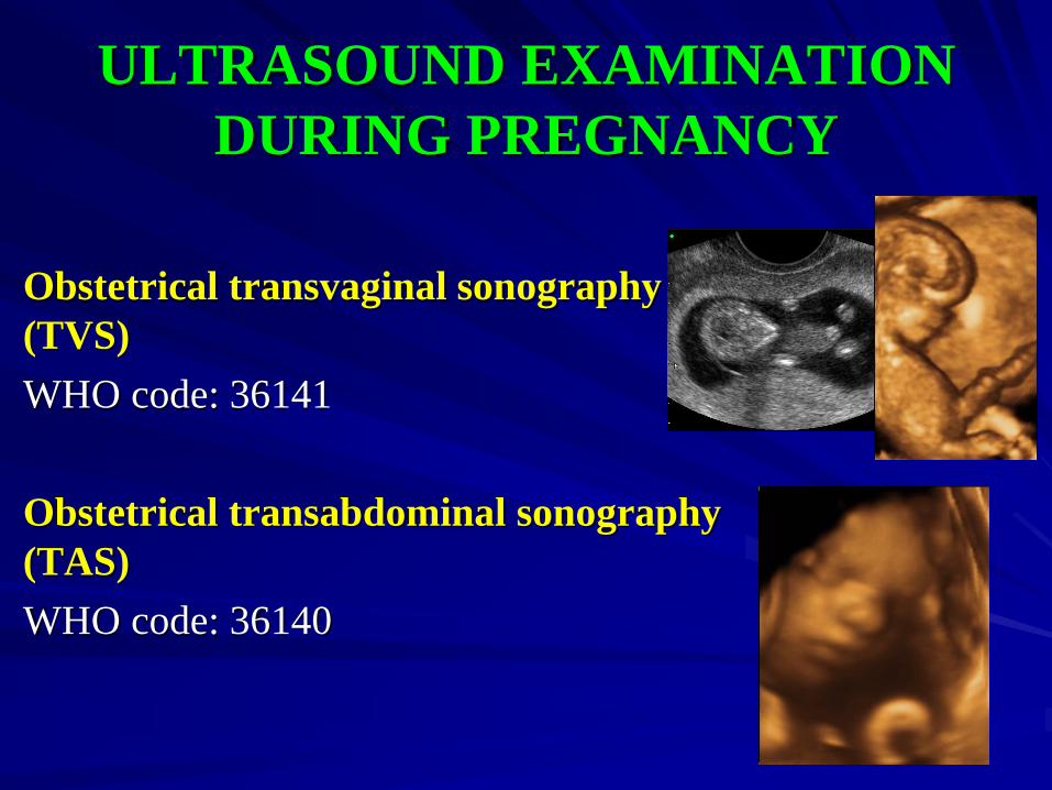
ULTRASOUND EXAMINATION
DURING PREGNANCY
Obstetrical transvaginal sonography
(TVS)
WHO code: 36141
Obstetrical transabdominal sonography
(TAS)
WHO code: 36140
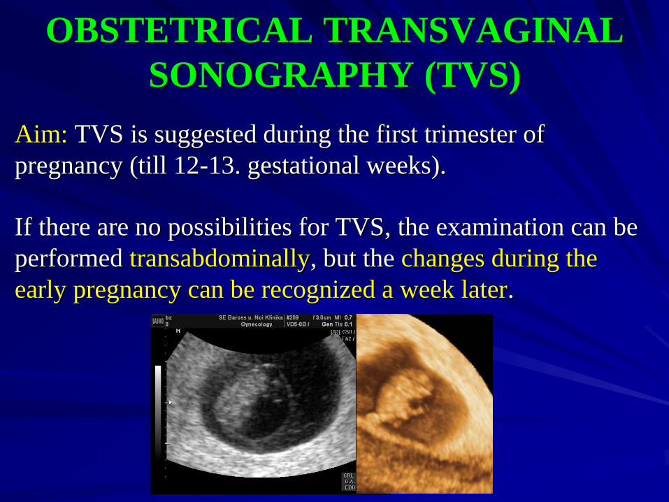
OBSTETRICAL TRANSVAGINAL
SONOGRAPHY (TVS)
Aim: TVS is suggested during the first trimester of
pregnancy (till 12-13. gestational weeks).
If there are no possibilities for TVS, the examination can be
performed transabdominally, but the changes during the
early pregnancy can be recognized a week later.
STRUCTURES WHICH CAN BE
EXAMINED
Gestational sac (format, number, dimension, localization)
Chorion frondosum
Yolk sac
Embryo/fetus: number, size – CRL, cardiac activity
Sonoanatomy, malformations (nuchal translucency, cystic hygroma, hydrops)
Pathological early gestation (subchorionic hematoma, mola hydatidosa, lost plural conception)
Uterus, adnexa (localization, size, structure, malformations)
Douglas cavity
Probable or assured sign of ectopic pregnancy
Pelvic pathology, mass (size, structure, localization, surface)
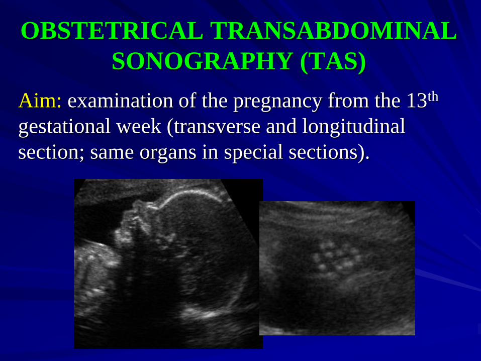
OBSTETRICAL TRANSABDOMINAL
SONOGRAPHY (TAS)
Aim: examination of the pregnancy from the 13th
gestational week (transverse and longitudinal
section; same organs in special sections).
STRUCTURES WHICH CAN BE
EXAMINED
Vital sign and position of the fetus
Amniotic fluid volume ( < 2 cm oligohydramnios,
> 8 cm hydramnios, or AFI – four quadrant method)
Placenta: localization, maturity (0-III), structure,
occurrent band(s)
Multiple pregnancy: absence or presence, layers and
thickness of the dividing membrane
Umbilical cord: blood vessels (two arteries, one
vein), structure
Skull: intracranial structures (falx cerebri, septum pellucidum, thalamus, ventricle, plexus chorioideus, cysterna magna), face
Spine: arch (longitudinal and transverse), vertebral ossification core
Thorax: configuration, size, lung’s echogenicity, breathing, diaphragm
Heart: rhythm, frequency, four chamber view, outflow tracts
Abdominal cavity, wall, umbilical ring: stomach, liver, bowels, free fluid accumulation, kidneys, bladder
Genitalia
Extremities: bone’s length, structure, absence, curve, deformity
Subcutaneous layer: signs of hydrops or fetopathy
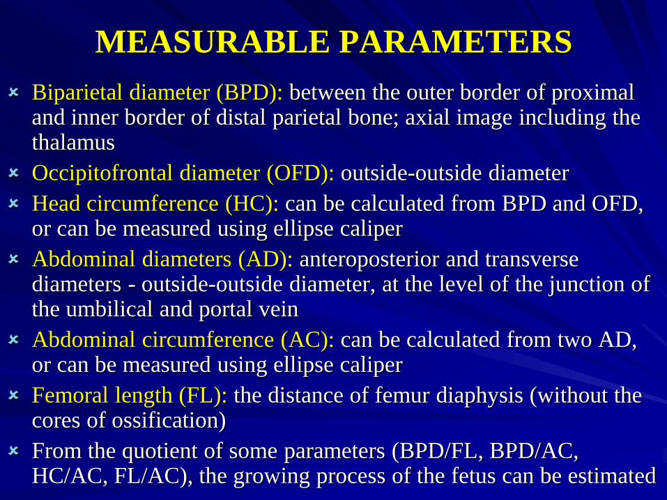
Biparietal diameter (BPD): between the outer border of proximal and inner border of distal parietal bone; axial image including the thalamus
Occipitofrontal diameter (OFD): outside-outside diameter
Head circumference (HC): can be calculated from BPD and OFD, or can be measured using ellipse caliper
Abdominal diameters (AD): anteroposterior and transversediameters - outside-outside diameter, at the level of the junction of the umbilical and portal vein
Abdominal circumference (AC): can be calculated from two AD, or can be measured using ellipse caliper
Femoral length (FL): the distance of femur diaphysis (without the cores of ossification)
From the quotient of some parameters (BPD/FL, BPD/AC, HC/AC, FL/AC), the growing process of the fetus can be estimated
MEASURABLE PARAMETERS
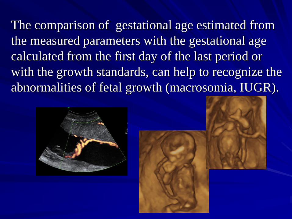
The comparison of gestational age estimated from
the measured parameters with the gestational age
calculated from the first day of the last period or
with the growth standards, can help to recognize the
abnormalities of fetal growth (macrosomia, IUGR).
RECOMMENDED ULTRASOUND
EXAMINATIONS DURING
PREGNANCY
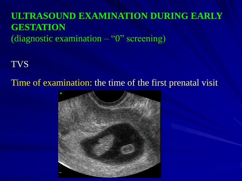
ULTRASOUND EXAMINATION DURING EARLY
GESTATION
(diagnostic examination – “0” screening)
TVS
Time of examination: the time of the first prenatal visit
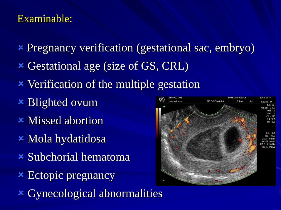
Examinable:
Pregnancy verification (gestational sac, embryo)
Gestational age (size of GS, CRL)
Verification of the multiple gestation
Blighted ovum
Missed abortion
Mola hydatidosa
Subchorial hematoma
Ectopic pregnancy
Gynecological abnormalities
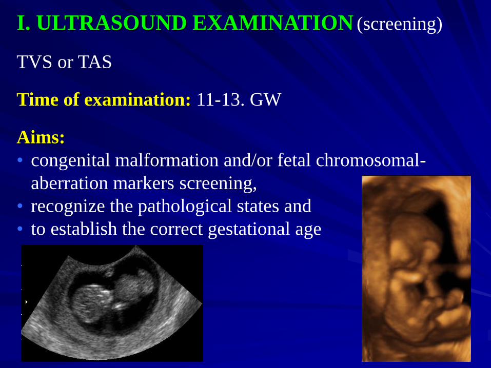
I. ULTRASOUND EXAMINATION (screening)
TVS or TAS
Time of examination: 11-13. GW
Aims:
• congenital malformation and/or fetal chromosomal-
aberration markers screening,
• recognize the pathological states and
• to establish the correct gestational age

Examinable:
Skull, nasal bone
Spine
Nuchal translucency
Heart (four chambers)
Diaphragm
Stomach
Abdominal wall
Kidneys, bladder
Extremities
Placenta, umbilical cord
Biometry (CRL, BPD, AC, FL)
Ductus venosus flow
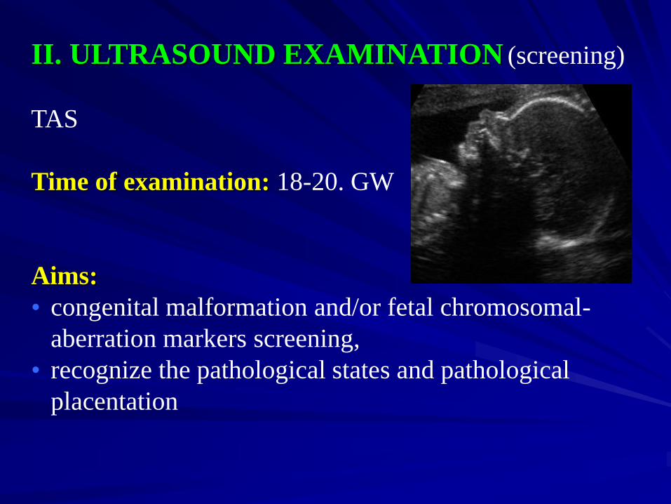
II. ULTRASOUND EXAMINATION (screening)
TAS
Time of examination: 18-20. GW
Aims:
• congenital malformation and/or fetal chromosomal-
aberration markers screening,
• recognize the pathological states and pathological
placentation
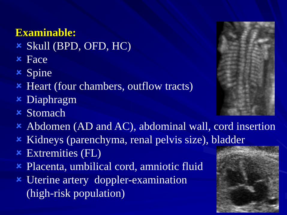
Examinable:
Skull (BPD, OFD, HC)
Face
Spine
Heart (four chambers, outflow tracts)
Diaphragm
Stomach
Abdomen (AD and AC), abdominal wall, cord insertion
Kidneys (parenchyma, renal pelvis size), bladder
Extremities (FL)
Placenta, umbilical cord, amniotic fluid
Uterine artery doppler-examination
(high-risk population)
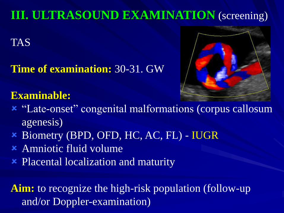
III. ULTRASOUND EXAMINATION (screening)
TAS
Time of examination: 30-31. GW
Examinable:
“Late-onset” congenital malformations (corpus callosum
agenesis)
Biometry (BPD, OFD, HC, AC, FL) - IUGR
Amniotic fluid volume
Placental localization and maturity
Aim: to recognize the high-risk population (follow-up
and/or Doppler-examination)
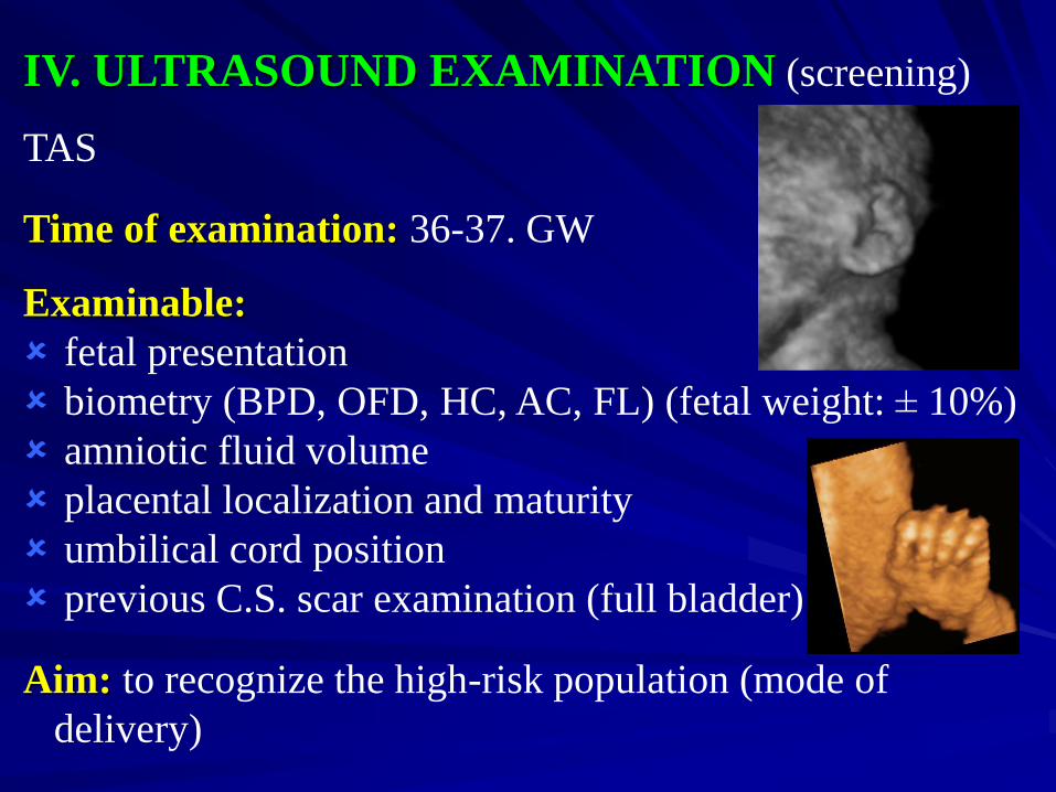
IV. ULTRASOUND EXAMINATION (screening)
TAS
Time of examination: 36-37. GW
Examinable:
fetal presentation
biometry (BPD, OFD, HC, AC, FL) (fetal weight: ± 10%)
amniotic fluid volume
placental localization and maturity
umbilical cord position
previous C.S. scar examination (full bladder)
Aim: to recognize the high-risk population (mode of
delivery)
FETAL ECHOCARDIOGRAPHY (WHO code: 3612G)
INDICATIONS positive history (maternal, previous child, family)
predisposing maternal diseases, states:
diabetes
isoimmunization
phenylketonuria
maternal age > 37 years
teratogenic or drug effects: phenytoin, lithium, isotretinoin, OC, rubella, antihypertensive drugs
screened anomalies during pregnancy:
proved or supposed fetal malformation
pathological fetal heart configuration
abnormal amniotic fluid volume
pathological fetal growth
multiple pregnancy
fetal arrhythmia
Ultrasound examinations in Hungary are performed
under the recommendations of the Hungarian Society of Ultrasound in Obstetrics and Gynecology
(MSZNUT) and there are five recommended
ultrasound examinations during the pregnancy (one
diagnostic and four screenings)
There are well-defined levels and protocols of
attendance
The different levels of attendance require proficiency
at different levels and this necessitates regular
training, which is ensured by MSZNUT
SUMMARY
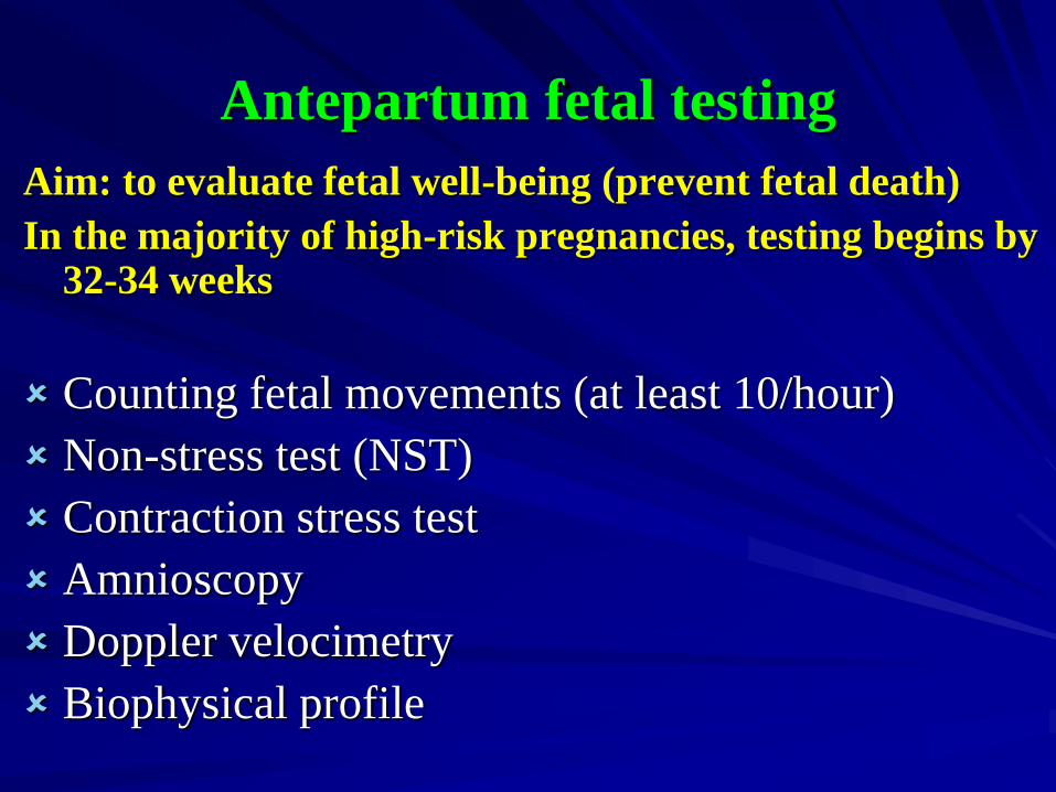
Antepartum fetal testing
Aim: to evaluate fetal well-being (prevent fetal death)
In the majority of high-risk pregnancies, testing begins by 32-34 weeks
Counting fetal movements (at least 10/hour)
Non-stress test (NST)
Contraction stress test
Amnioscopy
Doppler velocimetry
Biophysical profile
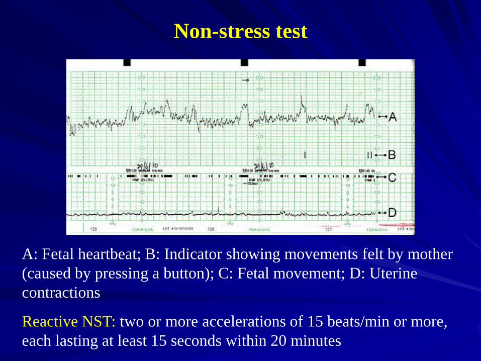
Non-stress test
A: Fetal heartbeat; B: Indicator showing movements felt by mother
(caused by pressing a button); C: Fetal movement; D: Uterine
contractions
Reactive NST: two or more accelerations of 15 beats/min or more,
each lasting at least 15 seconds within 20 minutes
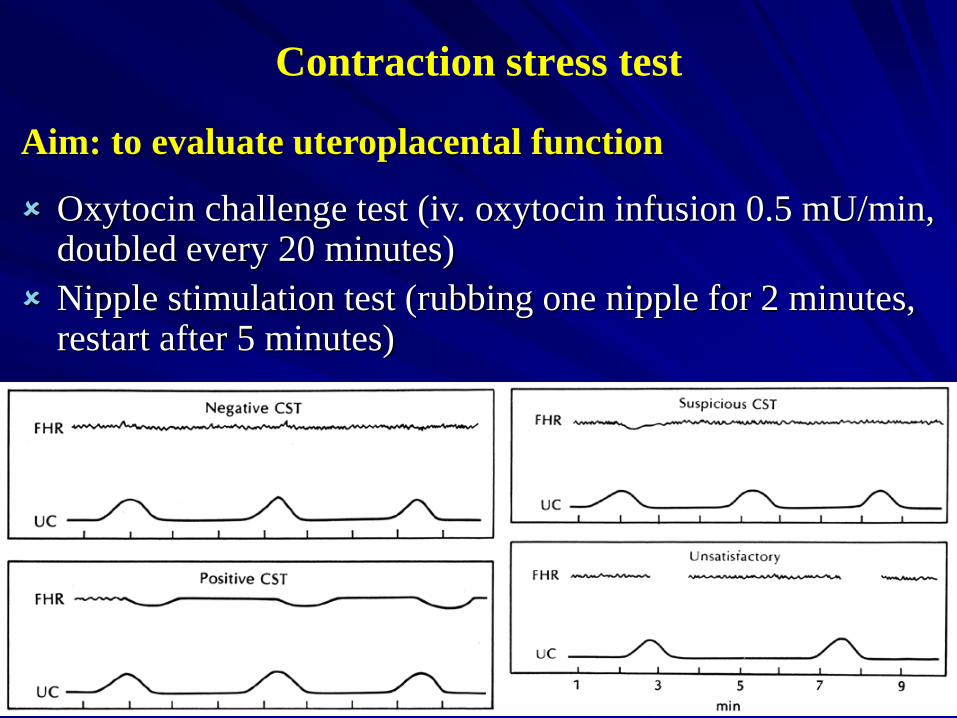
Contraction stress test
Aim: to evaluate uteroplacental function
Oxytocin challenge test (iv. oxytocin infusion 0.5 mU/min, doubled every 20 minutes)
Nipple stimulation test (rubbing one nipple for 2 minutes, restart after 5 minutes)
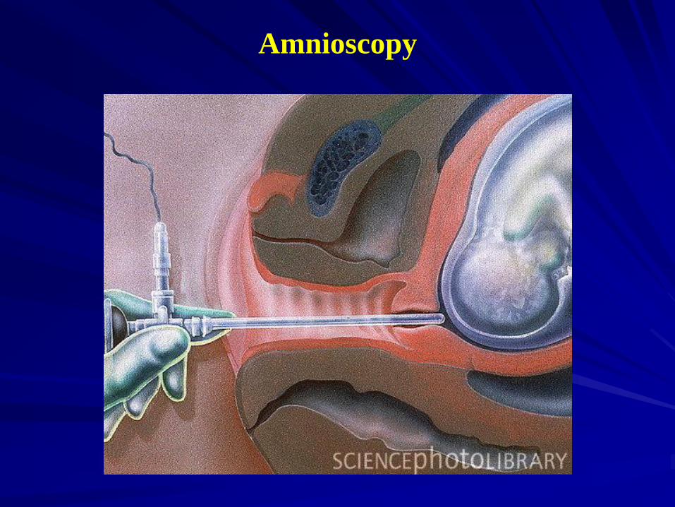
Amnioscopy
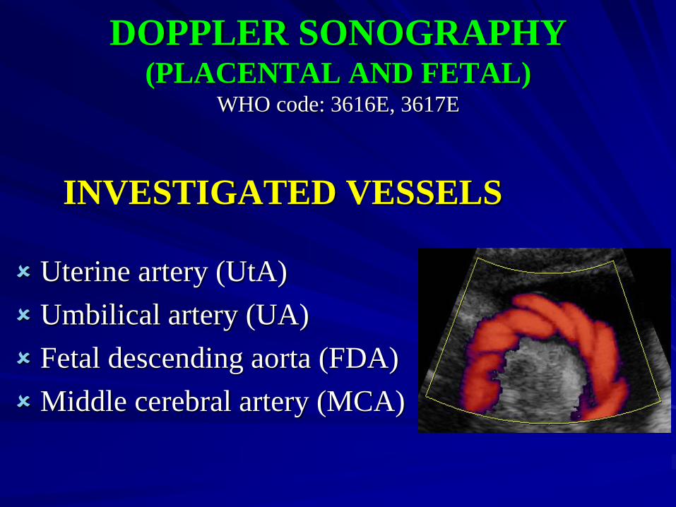
DOPPLER SONOGRAPHY(PLACENTAL AND FETAL)
WHO code: 3616E, 3617E
INVESTIGATED VESSELS
Uterine artery (UtA)
Umbilical artery (UA)
Fetal descending aorta (FDA)
Middle cerebral artery (MCA)
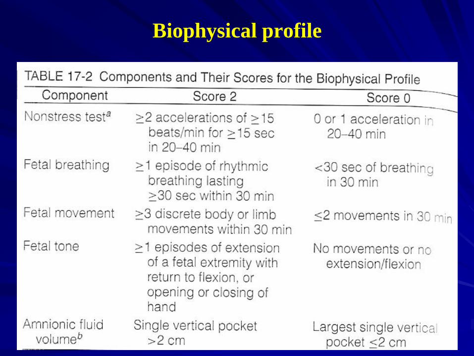
Biophysical profile

Thank you for attention!