Embed Size (px)
Citation preview
UNIVERSIDADE FEDERAL DO PAMPA
CAMPUS SÃO GABRIEL
PROGRAMA DE PÓS-GRADUAÇÃO EM CIÊNCIAS BIOLÓGICAS
RAQUEL SOARES OLIVEIRA
ATIVIDADE BIOLÓGICA DO VENENO DE RHINELLA ICTERICA (ANURA:
BUFONIDAE) SOBRE O SISTEMA NERVOSO DE VERTEBRADOS
DISSERTAÇÃO DE MESTRADO
SÃO GABRIEL, RIO GRANDE DO SUL, BRASIL
2016
ii
RAQUEL SOARES OLIVEIRA
ATIVIDADE BIOLÓGICA DO VENENO DE RHINELLA ICTERICA (ANURA:
BUFONIDAE) SOBRE O SISTEMA NERVOSO DE VERTEBRADOS
Dissertação apresentada ao Programa de Pós-
Graduação Stricto Sensu em Ciências Biológicas da
Universidade Federal do Pampa, como requisito
parcial para obtenção do Titulo de Mestre em
Ciências Biológicas.
Orientadora: Profª. Drª. Lúcia Vinadé
Coorientador: Prof. Dr. Cháriston André Dal Belo
São Gabriel
2016
iii
Ficha catalográfica elaborada automaticamente com os dados fornecidos pelo(a)
autor(a) através do Módulo de Biblioteca dos Sistema GURI (Gestão Unificada de
Recursos Institucionais).
O1206a
Oliveira, Raquel Soares
ATIVIDADE BIOLÓGICA DO VENENO DE RHINELLA ICTERICA
(ANURA: BUFONIDAE) SOBRE O SISTEMA NERVOSO DE
VERTEBRADOS / Raquel Soares Oliveira.
83 p.
Dissertação(Mestrado)-- Universidade Federal do
Pampa, MESTRADO EM CIÊNCIAS BIOLÓGICAS, 2016.
"Orientação:Drª. Lúcia Vinadé".
1. Qualidade Ambiental. 2. Neurobiologia. 3.
Farmacologia. I. Título.
iv
RAQUEL SOARES OLIVEIRA
ATIVIDADE BIOLÓGICA DO VENENO DE RHINELLA ICTERICA (ANURA:
BUFONIDAE) SOBRE O SISTEMA NERVOSO DE VERTEBRADOS
Dissertação apresentada ao Programa de Pós-
Graduação Stricto Sensu em Ciências Biológicas da
Universidade Federal do Pampa, como requisito
parcial para obtenção do Titulo de Mestre em
Ciências Biológicas.

v
Dedico essa dissertação à minha amada mãe Raimunda e meu amado Fabricio, por estarem ao
meu lado de modo incondicional e pela paciência que tiveram comigo, ao me ouvir falar
tantas horas sobre esse tal de Rhinella.
vi
AGRADECIMENTOS
Aos professores Lúcia Vinadé e Cháriston dal Belo agradeço imensamente os ensinamentos, a
paciência, a oportunidade e a confiança depositada em mim.
Aos professores Tiago Gomes, Leandro Lorentz e Cleci Menezes por toda ajuda para a
concretização deste trabalho.
Aos professores Carina Rodrigues Boeck, Thaís Posser e Dennis Reis de Assis que
cordialmente aceitaram participar da banca examinadora e ofereceram valiosas sugestões.
À UNIPAMPA e ao programa de pós graduação por esta oportunidade, e a CAPES pelo
suporte financeiro.
Aos colegas do LANETOX, por toda a ajuda nos dias longos de experimentos, pelos
momentos de descontração nas horas vagas e pelos quilos adquiridos com as idas ao
“Monstro”.
Aos colegas do PPGCB especialmente à Giulianna, Nathane e Matielo por toda a ajuda e
disponibilidade.
Ao Tio Carlos e Tio Douglas agradeço a toda ajuda profissional e pessoal, a paciência que
tiveram comigo nos dias de drama existencial e por todas as palavras de motivação.
À minha família pela confiança depositada e por todas as formas de auxílio. E a minha família
gaúcha, principalmente minha sogra Teresinha por me acolherem com tanto carinho.
Ao meu amado Fabricio por toda a paciência, carinho e apoio incondicional! Essa conquista
também é sua!
Às minhas amadas mãe Raimunda e minha tia Sandra por toda a paciência, pela confiança,
por sempre me apoiarem, e é claro, pelo suporte financeiro. Sem vocês nada disso seria
possível!
À todos vocês meu muito obrigada!
vii
E assim, depois de muito esperar, num dia como outro qualquer, decidi triunfar.
Decidi não esperar as oportunidades e sim, eu mesmo buscá-las.
Decidi ver cada problema como uma oportunidade de encontrar uma solução.
Decidi ver cada deserto como uma possibilidade de encontrar um oásis.
Decidi ver cada noite como um mistério a resolver.
Decidi ver cada dia como uma nova oportunidade de ser feliz.
Naquele dia descobri que meu único rival não era mais que minhas próprias
limitações e que enfrentá-las era a única e melhor forma de as superar.
Naquele dia, descobri que eu não era o melhor e que talvez eu nunca tivesse sido.
Deixei de me importar com quem ganha ou perde.
Agora me importa simplesmente saber melhor o que fazer.
Aprendi que o difícil não é chegar lá em cima, e sim deixar de subir.
Aprendi que o melhor triunfo é poder chamar alguém de "amigo".
Descobri que o amor é mais que um simples estado de enamoramento,
"o amor é uma filosofia de vida".
Naquele dia, deixei de ser um reflexo dos meus escassos triunfos passados
e passei a ser uma tênue luz no presente.
Aprendi que de nada serve ser luz se não iluminar o caminho dos demais.
Naquele dia, decidi trocar tantas coisas...
Naquele dia, aprendi que os sonhos existem para tornarem-se realidade.
E desde aquele dia já não durmo para descansar...
simplesmente durmo para sonhar.
Walt Disney
viii
RESUMO
Os venenos animais são fontes de compostos bioativos com aplicabilidade terapêutica. Os
anuros produzem através de glândulas paratóides, uma secreção venenosa rica em compostos
de diversas classes químicas, as quais apresentam uma série de atividades farmacológicas de
interesse biotecnológico. Os sapos da espécie Rhinella icterica (Spix, 1824), pertencem a um
grupo de animais venenosos presentes no bioma Pampa com carência de estudos
famacológicos e toxicológicos. Para os ensaios biológicos, os sapos foram coletados na região
de Derrubadas, no estado do Rio Grande do Sul. O veneno foi extraído manualmente por
compressão das glândulas paratóides, tratado por extração metanólica seguida de liofilização
e então foi chamado de MERIV. A neurobiologia do veneno foi avaliada sobre a junção
neuromuscular de aves, através da preparação biventer cervicis de pintainhos (BCP) e, através
da análise das desidrogenases em fatias hipocampais de camundongos. A incubação de
MERIV (5, 10, 20, 40 µg/mL) e Digoxina (6,5; 13; 26 e 52 nM) em fatias hipocampais de
camundongos, induziram um efeito dose dependente na viabilidade celular. Apenas MERIV
(5 µg/mL) e Digoxina (6,5 e 13 nM) provocaram aumento significativo da viabilidade celular
de 36 ± 10%, 52 ± 7% e 57 ± 13%, p<0.05, respectivamente, enquanto nas demais
concentrações houve decréscimo na viabilidade celular quando comparados com o controle
Hepes (n=6). Em preparações neuromusculares BCP, MERIV (5, 10 µg/mL) produziu um
efeito facilitatório de 60 ± 15% e 46 ± 6%, respectivamente, seguido de bloqueio
neuromuscular em 120 min de registro (n=6, p<0.05). De forma semelhante, a incubação dos
músculos com Digoxina 52 nM ou Ouabaína 0,2 nM mimetizou a atividade de MERIV com
aumento da amplitude de contração por 19 ± 4% e 27 ± 6%, e diminuição da contração
muscular de 80 ± 4% e 91 ± 5%, respectivamente (n=5, p<0.05). MERIV também demonstrou
atividade digitalic-like com inibição de 39 ± 3% da Na+,K+-ATPase (n=4, p<0.05). Em BPC,
quando MERIV foi incubado 20 min antes da d-Tubocurarina 1,45 µM, houve um reforço do
bloqueio neuromuscular, o qual foi completo em 80 min. Enquanto que em preparações BCP
curarizadas, MERIV aumentou o tempo de bloqueio em 50 min, semelhante a ação de drogas
anticolinesterásicas. Juntos, esses dados indicam que o extrato metanólico do veneno de R.
icterica é capaz de interferir com a neurotransmissão provavelmente via inibição das enzimas
acetilcolinesterase e Na+-K+- ATPase.
Palavras-chave: Veneno de sapo, junção neuromuscular, biventer cervicis, viabilidade
celular, eletromiografia.
ix
ABSTRACT
Animal poisons are sources of bioactive compounds with therapeutic applicability. Anurans
through parotid glands produce a poisonous secretion rich in compounds of different chemical
classes, that have a range of pharmacological activities of biotechnological interest. Toads of
the species Rhinella icterica (Spix, 1824), belong to a group of poisonous animals present in
the Pampa biome that still need to pharmacological and toxicological studies. Venom
collection was made by milking toads obtained at Derrubadas region, Rio Grande do Sul state.
The venom was previously treated by methanol extraction followed by lyophilization (thus
called MERIV), before the biological assays. The venom neurobiology was evaluated on
chicks neuromuscular junction by preparation biventer cervicis (BCP), and by function of
mitochondrial dehydrogenases in hippocampal brain slices from mice. Incubation of MERIV
(5, 10, 20 and 40 μg/mL) or digoxin (6.5, 13, 26 and 52 nM) with mice hippocampal brain
slices induced a dose-dependent effect on cell viability. At low concentration MERIV (5
μg/mL) and digoxin (6.5 and 13 nM) induced a corresponding significative increases in cell
viability, 36 ± 10%, 52 ± 7% and 57 ± 13% (p<0.05), respectively, while at higher
concentrations there were a decrease in cell viability compared with control Hepes (n=6). In
chicks neuromuscular preparation BCP, MERIV (5, 10 µg/mL) produced a facilitatory effect
of 60 ± 15% and 46 ± 6%, respectively, followed by neuromuscular blockade in 120 min
recordings (n=6, p <0.05). The incubation of BCP with digoxin (52 nM) or ouabain (0.2 nM)
mimicked the venom activity by increasing the amplitude of the twitches by 19 ± 4% and 27 ±
6%, respectively, followed by a depression in muscle contraction recorded for 120 min ( 80 ±
4% and 91± 5%, p<0.05, respectively, n=5). MERIV also demonstrated digitalic-like activity
inhibiting 39 ± 3% of Na+,K+-ATPase (n = 4, p <0.05). In BCP, when MERIV was incubated
for 20 min before d-Tubocurarine (1.45 μM), there was a reinforcement of the neuromuscular
blockade, wich was complete at 80 min. However, in preparations “curarizadas”, incubated
with d-Tubocurarine (1.45 μM) before MERIV, there was a increase in the blocking time at
50 min, similar to the action of acetylcholinesterase drugs. Altogether, these data indicate that
the methanolic extract from R. icterica venom is able to interfere in neurotransmission,
probably by inhibiting the enzymes acetylcholinesterase and Na+,K+- ATPase.
Key words: Toad venom, neuromuscular junction, biventer cervicis, cell viability,
electromyography.
x
LISTA DE FIGURAS
INTRODUÇÃO
Figura 1 – Sapos da espécie Rhinella icterica. .......................................................................... 5
Figura 2 – SDS PAGE dos venenos de R. achavali, R. icterica e R. schneideri. ...................... 6
Figura 3 - Eventos fisiológicos na junção neuromuscular. ....................................................... 9
Figura 4 - Representação do mecanismo de ação das digitálicas. ........................................... 11
MANUSCRITO
Figure 1 – Inhibition of acetylcholinesterase (AChE) activity induced by Rhinella icterica
toad venom (MERIV). .............................................................................................................. 31
Figure 2 - Inhibition of rat cardiac Na+,K+-ATPase activity induced by Rhinella icterica toad
venom (MERIV). ...................................................................................................................... 32
Figure 3 - Effects of Rhinella icterica toad venom (MERIV) and digoxin on the cell viability
of hippocampal slices.. ............................................................................................................. 33
Figure 4 - Concentration-response of MERIV at chick muscle-nerve preparations... ............ 34
Figure 5 - Comparison of the Rhinella icterica toad venom (MERIV) neuromuscular activity
with different pharmacological treatments in biventer cervicis muscle nerve preparations
(BCP).... .................................................................................................................................... 35
Figure 6 - Protective activity of Rhinella icterica toad venom (MERIV) against the
neuromuscular blockade induced by d-Tubocurarine (d-Tc) at chick muscle-nerve
preparations (BCP)... ................................................................................................................ 36
LISTA DE TABELAS
MANUSCRITO
Table 1 - Biochemical analysis of Rhinella icterica toad venom (MERIV) over
acetylcholinesterase activity of cockroach brain homogenates, chick biventer cervices muscles
and to the hippocampal cell viability ........................................................................................ 37
xi
LISTA DE ABREVIATURAS
ACh – Acetilcolina (Acetylcholine)
AChE – Acetilcolinesterase (Acetylcholinesterase)
AChRs – Receptores de acetilcolina (Acetylcholine Receptors)
BCP – Preparação biventer cervicis
Ca2+-ATPase – Bomba de cálcio
ChAT – Colina acetil-transferase
ChT1 – Transportador de colina
d-Tc – d-Tubocurarina (d-Tubocurarine)
JNM – Junção neuromuscular
MERAV – Extrato metanólico do veneno de Rhinella achavali
MERIV – Extrato metanólico do veneno de Rhinella icterica
MERSV – Extrato metanólico do veneno de Rhinella schneideri
nAChRs – Receptores nicotínicos de acetilcolina
Na+,K+-ATPase – Bomba de sódio e potássio
RS – Retículo sarcoplasmático
SNC – Sistema nervoso central
SNP – Sistema nervoso periférico
VAChT – Transportador vesicular de acetilcolina
xii
APRESENTAÇÃO
No item INTRODUÇÃO, consta uma breve revisão da literatura sobre o tema
abordado nesta dissertação.
A metodologia realizada e os resultados obtidos estão apresentados sob a forma de
manuscrito em inglês para publicação em revista cientifica indexada e se encontram no item
MANUSCRITO. No mesmo constam as informações sobre os Materiais e Métodos, a
apresentação e descrição dos Resultados, a Discussão destes e as Referências Bibliográficas
usadas na construção do texto.
Os itens CONSIDERAÇÕES FINAIS e PERSPECTIVAS, encontrados no final desta
dissertação, apresentam interpretações e comentários gerais sobre os resultados deste trabalho.
A sessão REFERÊNCIAS BIBLIOGRÁFICAS referem-se somente às citações que
aparecem no item INTRODUÇÃO desta dissertação.
xiii
SUMÁRIO
RESUMO ................................................................................................................................ viii
ABSTRACT ............................................................................................................................. ix
LISTA DE FIGURAS ............................................................................................................... x
LISTA DE ABREVIATURAS ................................................................................................ xi
APRESENTAÇÃO ................................................................................................................. xii
1. INTRODUÇÃO .................................................................................................................... 1
1.1. Venenos animais ................................................................................................... 1
1.1.1. Venenos de anuros ................................................................................................ 1
1.1.2. Composição química do veneno de anfíbios anuros ......................................... 2
1.1.3. Atividade biológica do veneno de anuros ........................................................... 2
1.1.4. A espécie Rhinella icterica ................................................................................... 4
1.2. Neurotransmissao ................................................................................................. 7
1.2.1. Junção neuromuscular (JNM) ............................................................................. 8
1.2.2. Bomba de Na,K-ATPase .................................................................................... 10
1.2.3. Viabilidade celular ............................................................................................. 12
2. JUSTIFICATIVA ............................................................................................................... 13
3. OBJETIVOS ....................................................................................................................... 14
3.1. Objetivo geral ......................................................................................................... 14
3.2. Objetivos específicos .............................................................................................. 14
4. MANUSCRITO .................................................................................................................. 15
5. CONSIDERAÇÕES FINAIS ............................................................................................. 41
6. PERSPECTIVAS ................................................................................................................ 42
7. REFERÊNCIAS BIBLIOGRÁFICAS ............................................................................. 43
8. ANEXOS ............................................................................................................................. 47

1
1. INTRODUÇÃO
1.1. Venenos animais
Algumas espécies de animais produzem ou armazenam através da dieta, substâncias
tóxicas denominadas biotoxinas. Esses compostos químicos são liberados como instrumentos
no auxílio à captura de presas, bem como na defesa contra predadores (ABDEL-RAHMAN;
AHMED; NABIL, 2010; ROSTELATO-FERREIRA et al., 2014; SAPORITO et al., 2012;
SCIANI et al., 2013).
1.1.1. Venenos de anuros
Os sapos são caracterizados como animais venenosos, uma vez que a secreção
venenosa à qual produzem em glândulas especializadas, não são passíveis de serem
inoculadas, pela ausência de um aparato específico (dente oco, ferrão, aguilhão). Nesse
sentido as glândulas secretoras estão distribuídas por toda a superfície corporal desses
animais. As principais delas são as paratóides, glândulas granulosas, comumente agrupadas
sobre a cabeça e pescoço; também há um grupo de glândulas menos desenvolvidas nas tíbias
das patas posteriores, denominadas de paracnemis. As glândulas mucosas ocorrem em maior
abundância e são responsáveis por produzir um muco viscoso sob a pele destes animais
mantendo-os úmidos (DORNELLES; MARQUES; RENNER, 2010; FONSECA; MOREIRA;
TSE, 2000).
As secreções das glândulas paratóides conferem proteção contra predadores, mas estas
somente liberam o veneno ao serem pressionadas. Sendo assim, acidentes com sapos
comumente ocorrem com animais domésticos, como cães e gatos, mas principalmente com
cães criados em espaço confinado e que nunca tiveram contato com sapos, ao verem pela
primeira vez acabam mordendo o animal, como brincadeira e ingerindo a secreção venenosa
(BARRAVIERA, 1994). Os sintomas apresentados por estes envenenamentos variam de
acordo com o tamanho do animal e a quantidade de veneno ingerido, podendo ocorrer
hipersalivação, convulsões, vômitos, diarreias, arritimias, alucinações, perda da atividade
motora, e, em alguns casos morte por parada cardio-respiratória (BARRAVIERA, 1994;
SONNE et al., 2008). Casos de intoxicação em humanos são raros, os relatos são de mulheres
romanas que tentavam matar os seus maridos utilizando veneno de sapos, sem muito sucesso,
pois usaram apenas a água onde armazenavam esses animais (CHEN; KOVARIOVÁ, 1967).
2
Episódios de intoxicação também foram relatados com a solução tópica do “Chan su”, que é o
veneno do sapo Bufo bufo gargarizans seco, utilizada como estimulante sexual, que ao ser
exportada e por conter apenas a bula em chinês foi ingerida por algumas pessoas, as quais
apresentaram sintomas de intoxicação por digitálicos (GOWDA; COHEN; KHAN, 2003). O
veneno de sapo é principalmente cardiotóxico e determina envenenamento similar àquele
causado por digitálicos (CHEN; KOVARIOVÁ, 1967; GOWDA; COHEN; KHAN, 2003).
1.1.2. Composição química dos venenos de anfíbios anuros
Nas secreções da pele e das glândulas dos anuros são encontrados diversas
substâncias, como aminas, alcaloides, peptídeos, proteínas e esteroides, as quais apresentam
uma série de atividades importantes para a farmacologia (SIANO et al., 2014). Os alcaloides
agem como bloqueadores de canais iônicos neuronais do sistema nervoso central e periférico
e do sistema muscular. Alguns estudos sugerem que a maioria dos alcaloides encontrados nos
anfíbios é adquirida ou produzida por meio da dieta ou por bactérias do sistema digestivo, que
são acumuladas nas glândulas e em outros órgãos (DALY; SPANDE; GARRAFFO, 2005;
DALY, 1995; SAPORITO et al., 2004). Peptídeos e proteínas desempenham funções
fisiológicas importantes como neuromoduladores e neurotransmissores, são também
importantes ferramentas contra predadores e do sistema imunológico (CUNHA-FILHO et al.,
2010). As neurotoxinas encontradas no veneno podem agir como bloqueadores de canais de
cálcio, vasodilatadores, broncodilatadores, antitumorais, antidiabéticos e hepatoprotetores,
além de poder agir como peptídeos opióides e neuropeptídios (ROMAN et al., 2012).
1.1.3. Atividade biológica dos venenos de anuros
As secreções produzidas através de glândulas encontradas na pele de anfíbios são ricas
em componentes biologicamente ativos e com grande potencial biotecnológico. Há muito
tempo, o veneno de sapo vem sendo utilizado para diversas funções por índios da América
do Sul, que aplicam o veneno da pele de anuros da família Dendrobatidae em suas flechas
para caçar ou se defender (CHEN; KOVARIOVÁ, 1967). No Japão o “Senso” e na China o
“Chan su”, que são veneno de sapos da espécie Bufo bufo gargarizans (Cantor, 1842) seco,
foi muito utilizado como expectorante, diurético, estimulante cardíaco, analgésico e
antiinflamatório para uso em dores de dente, aftas e sinusites (CHEN; KOVARIOVÁ, 1967).
O veneno de sapos atua quando entra em contato com mucosas e os sintomas são
imediatos. O veneno é composto por uma mistura de substâncias quimicamente ativas, em
3
que se destacam as que possuem mecanismo de ação semelhantes a adrenalina, digitálicas e
neurotoxinas.
Ensaios in vitro tem demonstrado que bufalin, um composto bloqueador
neuromuscular isolado do veneno de Bufo bufo gargarizans, além de outros bufadienolídeos
e cinobufagin, são os maiores componentes presentes no Chan su, os quais induzem apoptose
e/ou inibição da progressão do ciclo celular em uma variedade de células neoplásicas,
incluindo as de leucemia, câncer de próstata, câncer de estômago e câncer de fígado
(CUNHA-FILHO et al., 2010; YIN et al., 2012) . Além disso, em um tratamento experimental
com Huachansu, veneno da pele de sapo Bufo bufo gargarizans, houve regressão de 20% do
câncer de fígado em um dos pacientes (CHOI; HONG, 2012; DENG et al., 2014; MORENO
Y BANULS et al., 2013; TAKAI et al., 2012; YIN et al., 2012).
Bufadienolídeos isolados de Rhinella jimi, têm demonstrado atividade contra
Leishmania (L.) e Trypanosoma cruzi (TEMPONE et al., 2008). Esses compostos também
possuem atividade anestésica, antiviral, antibacteriana e inseticida, além de inibirem a Na+,K+
ATPase; também foi relatada uma atividade hipoglicemiante nesses compostos (YANG et al.,
2015).
Do ponto de vista farmacológico os bufadienolídeos exibem atividade semelhante aos
hormônios endógenos relacionados com o bloqueio da Na+,K+-ATPase, como
antiangiogênicos, anti-hipertensivos, imunossupressores, antiendometriose, desencadeando
uma ação ação inotrópica positiva (KAMBOJ; RATHOUR; KAUR, 2013). Nesse sentido, a
cardiotoxicidade induzida por venenos de sapo em vertebrados, tem seu mecanismo de ação
bem descrito, relacionado à presença de glicosídeos cardíacos (GOWDA; COHEN; KHAN,
2003; KUO et al., 2007; KWAN; PAIUSCO; KOHL, 1992; RADFORD et al., 1986;
ROHRER et al., 1982).Yoshida e Sakai publicaram estudos nos quais demonstraram os
efeitos inibitórios de bufalin sobre a junção neuromuscular de murinos (SEIICHIRO
YOSHIDA AND TAKESHI SAKAI, 1973, 1974). Manika e Gomes descreveram que o fator
letal (TSE-LF), isolado e purificado da pele de Bufo melanostictus (Schneider, 1799), tem
induzido ação neurotóxica em preparação biventer cervicis de pintainho (MANIKA DAS;
GOMES, 2001). Recentemente, Rostelato e colaboradores, demonstraram um efeito pré-
sináptico importante no veneno de sapo Rhinella schneideri (Werner, 1894), usando
preparações neuromusculares de aves e mamíferos.
O veneno de anfíbios da família Bufonidae é extremamente tóxico e sua biologia é
pouco conhecida. Além disso, venenos animais são muito importantes na biofarmacologia e
4
possuem elevado potencial biotecnológico. O conhecimento sobre esses venenos pode auxiliar
na síntese de substâncias quimicamente ativas com possibilidade de uso terapêutico
(BARRAVIERA, 1994).
1.1.4. A espécie Rhinella icterica
Os sapos são vertebrados pertencentes ao filo Chordata, classe Amphibia e a ordem
Anura. Atualmente são conhecidas aproximadamente 6.580 espécies que compõe a ordem
Anura e estão distribuídas em 54 famílias, presentes em todo globo, exceto na Antártica e
algumas ilhas oceânicas (FROST et al. 2015). De acordo com a Sociedade Brasileira de
Herpetologia (SEGALLA et al., 2014), até a presente data foram reconhecidas 1026 espécies
de anfíbios ocorrentes no Brasil, sendo 988 espécies da ordem Anura, ocupando assim, a
primeira colocação na relação de países com maior riqueza de espécies da ordem Anura.
A família Bufonidae está representada por 49 gêneros e 586 espécies, apresentando
ampla distribuição geográfica, ocorrendo em quase todos os continentes, com exceção da
Austrália, Nova Guiné e Madagascar (FROST et al. 2015).
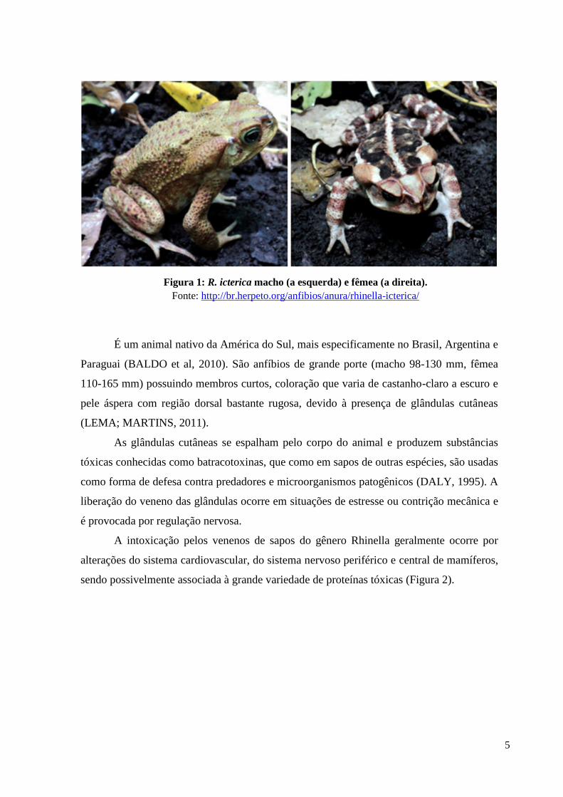
A espécie R. icterica foi descrita em 1824 por Spix inicialmente como Bufo icterica,
posteriormente, denominada de Chaunus icterica, e atualmente Rhinella icterica (Figura 1).
Classificação científica:
• Reino: Animalia
• Filo: Chordata
• Classe: Amphibia
• Ordem: Anura
• Família Bufonidae
• Gênero: Rhinella
• Espécie: Rhinella icterica
5
É um animal nativo da América do Sul, mais especificamente no Brasil, Argentina e
Paraguai (BALDO et al, 2010). São anfíbios de grande porte (macho 98-130 mm, fêmea
110-165 mm) possuindo membros curtos, coloração que varia de castanho-claro a escuro e
pele áspera com região dorsal bastante rugosa, devido à presença de glândulas cutâneas
(LEMA; MARTINS, 2011).
As glândulas cutâneas se espalham pelo corpo do animal e produzem substâncias
tóxicas conhecidas como batracotoxinas, que como em sapos de outras espécies, são usadas
como forma de defesa contra predadores e microorganismos patogênicos (DALY, 1995). A
liberação do veneno das glândulas ocorre em situações de estresse ou contrição mecânica e
é provocada por regulação nervosa.
A intoxicação pelos venenos de sapos do gênero Rhinella geralmente ocorre por
alterações do sistema cardiovascular, do sistema nervoso periférico e central de mamíferos,
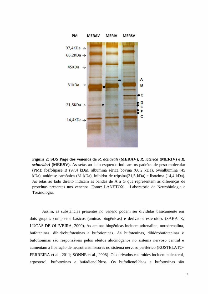
sendo possivelmente associada à grande variedade de proteínas tóxicas (Figura 2).
Figura 1: R. icterica macho (a esquerda) e fêmea (a direita).
Fonte: http://br.herpeto.org/anfibios/anura/rhinella-icterica/
6
Assim, as substâncias presentes no veneno podem ser divididas basicamente em
dois grupos: compostos básicos (aminas biogênicas) e derivados esteroides (SAKATE;
LUCAS DE OLIVEIRA, 2000). As aminas biogênicas incluem adrenalina, noradrenalina,
bufoteninas, dihidrobufoteninas e bufotioninas. As bufoteninas, dihidrobufoteninas e
bufotioninas são responsáveis pelos efeitos alucinógenos no sistema nervoso central e
aumentam a liberação de neurotransmissores no sistema nervoso periférico (ROSTELATO-
FERREIRA et al., 2011; SONNE et al., 2008). Os derivados esteroides incluem colesterol,
ergosterol, bufotoxinas e bufadienolídeos. Os bufodienolídeos e bufotoxinas são
Figura 2: SDS Page dos venenos de R. achavali (MERAV), R. icterica (MERIV) e R.
schneideri (MERSV). As setas ao lado esquerdo indicam os padrões de peso molecular
(PM): fosfolipase B (97,4 kDa), albumina sérica bovina (66,2 kDa), ovoalbumina (45
kDa), anidrase carbônica (31 kDa), inibidor de tripsina(21,5 kDa) e lisozima (14,4 kDa).
As setas ao lado direito indicam as bandas de A a G que representam as diferenças de
proteínas presentes nos venenos. Fonte: LANETOX – Laboratório de Neurobiologia e
Toxinologia.
7
responsáveis pela ação digitálica induzida pelos venenos de sapos (CUNHA-FILHO et al.,
2010). Dessa forma, o efeito digitálico é responsável pela inibição da Na+,K+-ATPase nas
células cardíacas, aumentando a concentração intracelular de sódio e, consequentemente,
inibindo a extrusão do cálcio (RANG et al., 2005). O aumento da concentração de cálcio
nos miócitos cardíacos, produz um aumento da força de contração cardíaca (inotropismo
positivo) e reduz a frequência dos batimentos cardíacos (cronotropismo negativo) por uma
ação vagal (CHEN et al., 2006). O bloqueio da bomba de Na+,K+-ATPase no sistema
nervoso induz um aumento da liberação de neurotransmissores, um efeito ainda pouco
estudado (ARNAIZ; BERSIER, 2014).
1.2. Neurotransmissão
A transmissão de mensagens entre duas células se dá através de sinapses, uma junção
especializada em que um terminal axonal faz contato com outro neurônio ou tipo de célula. O
sentido normal do fluxo de informação é do terminal axonal para o neurônio-alvo; desse
modo, o terminal axonal é o pré-sináptico, enquanto o neurônio-alvo é o pós-sináptico. A
transmissão sináptica envolve a conversão do impulso nervoso, de natureza elétrica, em uma
mensagem química carreada por substâncias neuromediadoras, e depois novamente em
impulsos elétricos já na célula pós-sináptica (BEAR; CONNORS; PARADISO, 2002; LENT,
2010; SILVERTHORN, 2010).
Há dois tipos básicos de sinapses: as químicas e as elétricas. As sinapses elétricas são
chamadas de junções comunicantes, possuem estrutura mais simples, transferem correntes
iônicas e até mesmo pequenas moléculas entre células acopladas. As sinapses químicas
podem modificar as mensagens que transmitem de acordo com inúmeras circunstâncias. Sua
estrutura é especializada no armazenamento de substâncias neurotransmissoras e
neuromoduladoras que, quando liberadas na fenda sináptica, provocam alterações de potencial
elétrico na membrana pós-sináptica, que poderão influenciar o disparo de potenciais de ação
do neurônio pós-sináptico. O resultado da interação dos efeitos excitatórios e inibitórios de
cada uma das sinapses sobre o potencial da membrana do neurônio pós-sináptico irá definir a
mensagem transportada pelo axônio do segundo neurônio, em direção a outras células
(BEAR; CONNORS; PARADISO, 2002; LENT, 2010).
8
1.2.1. Junção Neuromuscular (JNM)
A junção neuromuscular (JNM), também chamada de placa motora, é uma sinapse
química colinérgica com a função de transferir impulsos de uma terminação nervosa para uma
fibra muscular e, assim, desencadear a contração muscular, por meio da ação do
neurotransmissor acetilcolina (ACh) . A JNM é formada por três elementos estruturais: a
região pré-sináptica, onde se encontra a terminação nervosa; a região pós-sináptica, célula
muscular; e a fenda sináptica, fenda entre 20 e 30 nm que separa as membranas das células
pré e pós-sináptica (HUGHES; KUSNER; KAMINSKI, 2006) (Figura 3).
Os neurotransmissores são liberados para a fenda sináptica pelos botões sinápticos,
formado por expansões das extremidades do axônio que perde sua bainha de mielina quando
está próximo da placa motora. Cada botão sináptico sobrepõe depressões presentes na
superfície da fibra muscular, chamadas de fendas subneurais, sendo este um local de alta
densidade de receptores de ACh (AChR). Além de que, em cada botão sináptico está presente
a maquinaria necessária para liberar o neurotransmissor: vesículas sinápticas onde a ACh é
armazenada; complexo de acoplamento, complexo proteico de exocitose das vesículas; zona
ativa, sítio de liberação do neurotransmissor; e canais de Ca2+ voltagem-dependentes, os quais
permitem a entrada de Ca2+ na terminação nervosa a cada potencial de ação. O Ca2+ é
responsável por desencadear a fusão das vesículas sinápticas com a zona ativa, liberando seu
conteúdo para a fenda sináptica (BEAR; CONNORS; PARADISO, 2002; HUGHES;
KUSNER; KAMINSKI, 2006; KATZ, 1966; LENT, 2010). O resultado da reação entre o
neurotransmissor e o seu receptor, como acontece em todas as sinapses excitatórias, é a
abertura seletiva de canais de Na+ e K+, e a ocorrência de um potencial pós-sináptico
despolarizante (BEAR; CONNORS; PARADISO, 2002; LENT, 2010). Esta reação pode ser
impedida caso estejam presentes as toxinas d-Tubocurarina e/ou α-bungarotoxina pois estas
são capazes de inibir a transmissão neuromuscular por bloquearem os receptores colinérgicos
nicotínicos da placa motora (MELLA; AMBIEL; PRADO, 2000).
9
A ACh é sintetizada no terminal pré-sináptico pela enzima colina acetiltransferase
(ChAT) a partir da colina e do acetil-CoA. Após, a acetilcolina é armazenada no interior das
vesículas por seu transportador vesicular, VAChT. Durante a neurotransmissão, as vesículas
sinápticas, passam por um ciclo nos terminais nervosos, onde primeiramente os
neurotransmissores são transportados para o interior das vesículas sinápticas, que se agrupam
nas adjacências da zona ativa, após haverá ancoramento e estas estarão aptas para a fusão e
liberação de seu conteúdo na fenda sináptica (ARISI; NEDER; MOREIRA, 2001). Após a
exocitose e ativação dos receptores nicotínicos, a ACh é degradada pela enzima
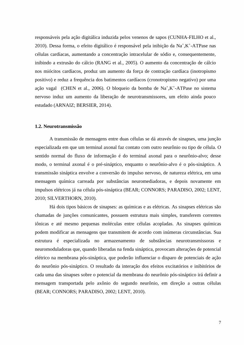
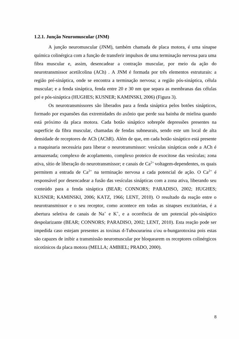
Figura 3: Eventos fisiológicos na junção neuromuscular. A chegada de potenciais de
ação no terminal do axônio abre canais de Ca2+ dependentes de voltagem. O Ca2+
difunde-se para dentro da célula a favor do gradiente eletroquímico, desencadeando a
liberação de ACh contida nas vesículas sinápticas. A ACh difunde-se através da fenda
sináptica e combina-se com receptores nicotínicos (nAChRs) na membrana do músculo
esquelético. Fonte: (SILVERTHORN, 2010).
10
acetilcolinesterase (AChE) presente na fenda sináptica, gerando colina e acetato, o que a torna
inativa para os AChRs. A colina é recaptada para o interior do terminal por meio de seu
transportador de membrana (CHT1) e será utilizada novamente para a síntese de acetilcolina.
A AChE é responsável por evitar a exposição ininterrupta de ACh na junção neuromuscular o
que pode causar dessensibilização dos receptores e bloqueio da transmissão neuromuscular.
Em pequenas quantidades, inibidores da AChE, como a Neostigmina, podem reforçar as
transmissões neuromusculares prolongando a ação da ACh liberada (BEAR; CONNORS;
PARADISO, 2002; BUAINAIN; MOURA; OLIVEIRA, 2000; SOUSA et al., 2006).
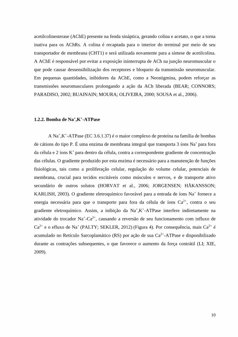
1.2.2. Bomba de Na+,K+-ATPase
A Na+,K+-ATPase (EC 3.6.1.37) é o maior complexo de proteína na família de bombas
de cátions do tipo P. É uma enzima de membrana integral que transporta 3 íons Na+ para fora
da célula e 2 íons K+ para dentro da célula, contra a correspondente gradiente de concentração
das células. O gradiente produzido por esta enzima é necessário para a manutenção de funções
fisiológicas, tais como a proliferação celular, regulação do volume celular, potenciais de
membrana, crucial para tecidos excitáveis como músculos e nervos, e de transporte ativo
secundário de outros solutos (HORVAT et al., 2006; JORGENSEN; HÅKANSSON;
KARLISH, 2003). O gradiente eletroquímico favorável para a entrada de íons Na+ fornece a
energia necessária para que o transporte para fora da célula de íons Ca2+, contra o seu
gradiente eletroquímico. Assim, a inibição da Na+,K+-ATPase interfere indiretamente na
atividade do trocador Na+-Ca2+, causando a reversão de seu funcionamento com influxo de
Ca2+ e o efluxo de Na+ (PALTY; SEKLER, 2012) (Figura 4). Por consequência, mais Ca2+ é
acumulado no Retículo Sarcoplamático (RS) por ação de sua Ca2+-ATPase e disponibilizado
durante as contrações subsequentes, o que favorece o aumento da força contrátil (LI; XIE,
2009).
11
Figura 4: Representação do mecanismo de ação das digitálicas. A Na+,K+-ATPase
controla a concentração citoplasmática de Na+ em repouso o que determina a concentração de
Ca2+ através do trocador Na+/Ca2+. A inibição da Na+,K+-ATPase causa a reversão do
funcionamento do trocador Na+-Ca2+, com influxo de Ca2+ e o efluxo de Na+. Estes eventos
influenciam a atividade da Ca2+-ATPase e a atividade do canal de Ca2+. Assim, mais Ca2+ é
acumulado no Retículo Sarcoplamático (RS) por ação de sua Ca2+-ATPase e disponibilizado
durante as contrações subsequentes. Em conjunto, todos esses eventos são responsáveis por
controlar o nível intracelular de Ca2+ e influenciam a contratilidade muscular, cardíaca e a
excitabilidade neuronal, o que favorece o aumento da força contrátil. (HORVAT et al., 2006;
LI; XIE, 2009). Fonte: Autor.
Esta enzima também é conhecida como ouabaina-sensível devido ao efeito
farmacológico de ser especificamente inibida por esteroides cardiotônicos, como um receptor
de ouabaína (HORVAT et al., 2006).
Os esteroides cardiotônicos, assim chamados por possuírem ação inotrópica positiva
sobre o coração, são inibidores da Na+,K+-ATPase e sua estrutura química básica está em
torno de um núcleo esteroidal. São encontrados tanto em plantas quanto em animais. A
digoxina e a ouabaína são extraídas das folhas secas da plantas Digitalis purpúrea L. e
Strophantus gratus (Wall. & Hook.) Baill, são esteroides cardiotônicos utilizados no
tratamento de insuficiência cardíaca congestiva, quando o coração não tem força suficiente
para a contração muscular (SHAMAGIAN et al., 2005). Os sapos contém esteroides nas
secreções de suas glândulas paratoides, sendo os bufadienolídeos presentes no veneno
responsáveis pelo envenenamento de pequenos animais, causando convulsões e parada
12
cardíaca (CUNHA-FILHO et al., 2010; TEMPONE et al., 2008). A ação cardiotóxica do
veneno é semelhante à intoxicação por digitálicas e apresenta o mesmo mecanismo que
promove inotropismo positivo decorrente da inibição da Na+,K+-ATPase (DE ROBERTIS;
HIB, 2006; GOWDA; COHEN; KHAN, 2003).
1.2.3. Viabilidade Celular
A viabilidade celular é caracterizada pela capacidade que uma célula possui de realizar
determinadas funções, como metabolismo, crescimento e reprodução. A divisão celular
garante a reparação tissular ou celular após uma lesão que tenha causado uma perda de células
(RANG et al., 2005). Porém, mesmo no estado adulto, as células morrem e é imprescindível
sua substituição (RANG et al., 2005). A morte celular pode ocorrer de duas formas distintas,
por necrose ou apoptose. A apoptose funciona como um sistema controlador da homeostase
através da eliminação não inflamatória de células irreversivelmente lesadas ou desnecessárias
(YIN et al., 2012). A mitocôndria pode desencadear a apoptose ao liberar para o citoplasma
proteínas pró-apoptóticas, como o citocromo c e fator indutor de apoptose (DENG et al.,
2014).
A membrana celular é a parte mais externa da célula o que a torna um sítio receptor
ideal para substâncias químicas, que podem ser toxinas, drogas e hormônios. Estas, por sua
vez, podem interagir com as proteínas presentes na membrana ou com seus componentes
lipídicos, alterar suas funções de transporte e a integridade da célula como um todo. Os
receptores apresentam sítios para ligação de moléculas, estas atuam como moduladores destes
receptores (RANG et al., 2005). Estudos sugerem que a enzima Na+,K+-ATPase possui além
de seu papel regulatório na homeostasia iônica, um papel importante na transdução de sinal e
na ativação da transcrição gênica, modulando na presença de ouabaína o crescimento e
migração celular e a apoptose (BAGROV; SHAPIRO; FEDOROVA, 2009; DANIEL et al.,
2010; DVELA et al., 2012; KOTOVA et al., 2006). Além disso, os estudos sugerem a
relevância fisiológica da ouabaína e glicosídeos cardiotônicos em regular a viabilidade celular
quando em concentrações da mesma amplitude de seus níveis circulantes (CHEN et al., 2006;
DVELA et al., 2012)
13
2. JUSTIFICATIVA
O Brasil é o país com maior biodiversidade de anfíbios anuros e o estado do Rio
Grande do Sul contém cerca de 10% dessa diversidade, com 98 espécies de anuros descritos
até o momento. Porém muitas dessas espécies ainda carecem de estudos farmacológicos e
toxicológicos. Apesar do óbvio interesse ecológico e clínico no estudo dos sapos, a presença
de substâncias biologicamente ativas no veneno, aumenta o interesse no estudo desses animais
como fonte de compostos com aplicabilidade biotecnológica (Monti e Cardello, 1994).
Considerando a grande biodiversidade de anfíbios ainda não catalogados e que
toxicologicamente ainda não foram explorados, e que as toxinas produzidas por eles podem
possuir propriedades terapêuticas para vertebrados, há interesse em investigar os seus
mecanismos de ação fisiológica, e assim, aumentar o conhecimento sobre anfíbios anuros e
posteriormente conduzir ao desenvolvimento de fármacos que possam ser adicionados a
coleção de compostos biologicamente ativos.
14
3. OBJETIVOS
3.1. Objetivo Geral
Caracterizar a atividade neurobiológica do veneno do sapo R. icterica em vertebrados.
3.2. Objetivos específicos
Caracterizar por meio de teste eletromiográfico o efeito do veneno de R. icterica in
vitro em preparação neuromuscular biventer cervicis de pintainhos (Gallus gallus
domesticus).
Comparar os efeitos do veneno de R. icterica com a ação de drogas inibidoras da
Na+,K+-ATPase em preparação neuromuscular de pintainho.
Verificar o efeito anticolinesterásico do veneno de R. icterica em preparação
neuromuscular de pintainho.
Avaliar a viabilidade celular do veneno de R. icterica em fatias de hipocampo de
camundongos (Mus musculus).
Comparar os efeitos do veneno de R. icterica com a ação de drogas inibidoras da
bomba de Na+/K+ em fatias hipocampais de camundongos.
Avaliar a atividade inibidora da Na+,K+-ATPase do veneno de R. icterica em tecido
cardíaco de ratos (Rattus norvegicus).

15
4. MANUSCRITO
Todos os Resultados, bem como os itens Materiais e Métodos, Discussão e Referências
Bibliográficas que fazem parte desta dissertação estão apresentados sob a forma de
manuscrito. Este manuscrito está disposto na forma na qual deverá ser submetido para o
periódico Chemico Biological Interactions (ISSN: 0009-2797) e será intitulado como: In
Vitro Neurobiology of Rhinella icterica (Spix, 1824) Toad Venom in Chicks and
Mammalian Preparations.
16
In vitro Neurobiology of Rhinella icterica (Spix, 1824) Toad Venom in Chicks and
Mammalian Preparations
Raquel Soares Oliveiraa, Allan Pinto Leala, Carlos Gabriel Moreira de Almeidaa, Douglas
Silva dos Santosb, Leandro Homrich Lorentza, Cleci Menezes Moreirac, Tiago Gomes dos
Santosa, Cháriston André Dal Beloa,b and Lúcia Vinadéa*
aLANETOX, Universidade Federal do Pampa (UNIPAMPA),Av. Antônio Trilha 1847, 97300-
000, São Gabriel, RS, Brazil bPrograma de Pós-Graduação em Ciências Biológicas: Bioquímica Toxicológica,
(PPGBTox), Universidade Federal de Santa Maria (UFSM), Av. Roraima 1000,97105-900,
Santa Maria, RS, Brazil
*Corresponding authors:
Dra. Lúcia Vinadé
CIPBiotec, campus SãoGabriel, Universidade Federal do Pampa
Av.Antônio Trilha,1847
97300000 São Gabriel, Brazil
Tel./fax: + 55 55 3237-0850.
Dr. Cháriston André Dal Belo
CIPBiotec, campus São Gabriel, Universidade Federal do Pampa
Av. Antônio Trilha,1847
97300000 São Gabriel, Brazil
Tel./fax: + 55 55 3237-0850.
17
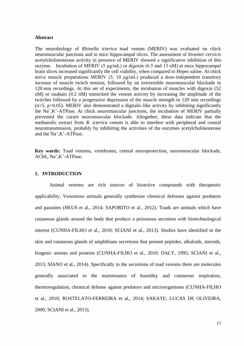
Abstract
The neurobiology of Rhinella icterica toad venom (MERIV) was evaluated on chick
neuromuscular junctions and in mice hippocampal slices. The assessment of biventer cervicis
acetylcholinesterase activity in presence of MERIV showed a significative inhibition of this
enzyme. Incubation of MERIV (5 μg/mL) or digoxin (6.5 and 13 nM) at mice hippocampal
brain slices increased significantly the cell viability, when compared to Hepes saline. At chick
nerve muscle preparations MERIV (5, 10 μg/mL) produced a dose-independent transitory
increase of muscle twitch tension, followed by an irreversible neuromuscular blockade in
120 min recordings. At this set of experiments, the incubation of muscles with digoxin (52
nM) or ouabain (0.2 nM) mimicked the venom activity by increasing the amplitude of the
twitches followed by a progressive depression of the muscle strength in 120 min recordings
(n=5, p<0.05). MERIV also demonstrated a digitalic-like activity by inhibiting significantly
the Na+,K+-ATPase. At chick neuromuscular junctions, the incubation of MERIV partially
prevented the curare neuromuscular blockade. Altogether, these data indicate that the
methanolic extract from R. icterica venom is able to interfere with peripheral and central
neurotransmission, probably by inhibiting the activities of the enzymes acetylcholinesterase
and the Na+,K+-ATPase.
Key words: Toad venoms, vertebrates, central neuroprotection, neuromuscular blockade,
AChE, Na+,K+-ATPase.
1. INTRODUCTION
Animal venoms are rich sources of bioactive compounds with therapeutic
applicability. Venomous animals generally synthesize chemical defenses against predators
and parasites (HEUS et al., 2014; SAPORITO et al., 2012). Toads are animals which have
cutaneous glands around the body that produce a poisonous secretion with biotechnological
interest (CUNHA-FILHO et al., 2010; SCIANI et al., 2013). Studies have identified in the
skin and cutaneous glands of amphibians secretions that present peptides, alkaloids, steroids,
biogenic amines and proteins (CUNHA-FILHO et al., 2010; DALY, 1995; SCIANI et al.,
2013; SIANO et al., 2014). Specifically in the secretions of toad venoms there are molecules
generally associated to the maintenance of humidity and cutaneous respiration,
thermoregulation, chemical defense against predators and microorganisms (CUNHA-FILHO
et al., 2010; ROSTELATO-FERREIRA et al., 2014; SAKATE; LUCAS DE OLIVEIRA,
2000; SCIANI et al., 2013).
18
Among the pharmacological activity induced by toad venoms there are a number of
works describing effects related to peripheral neurotoxicity. In this regard, Yoshida and Sakai
(SEIICHIRO YOSHIDA AND TAKESHI SAKAI, 1973, 1974) have published a couple of
articles showing the effects of a neuromuscular blocker compound named bufalin, that was
isolated from the venom of Bufo gargarizans (Cantor, 1842). Manika and Gomes (MANIKA
DAS; GOMES, 2001) have described that the lethal factor (TSE-LF) isolated and purified
from Bufo melanostictus (Schneider,1799) skin has induced neurotoxic action on chick
biventer cervicis. Recently, Rostelato and cols (ROSTELATO-FERREIRA et al., 2011, 2014)
have demonstrated an interesting presynaptic effect on Rhinella schneideri (Werner, 1894)
toad venom, using avian and mammalian neuromuscular preparations. The cardiotoxicity
induced by toad venoms in vertebrates are another well described biological activity and is
related to the presence of cardiac glycosides (GOWDA; COHEN; KHAN, 2003; KUO et al.,
2007; KWAN; PAIUSCO; KOHL, 1992; RADFORD et al., 1986; ROHRER et al., 1982).
R. icterica, the “cururu toad”, is a large anuran (up to 140 mm) of the Bufonidae
family, native to South America, with a broad geographic distribution, occurring in the
southeastern and southern Brazil, Paraguay, Uruguay and Argentine(COLOMBO et al.,
2008). However, as far as our knowledge, none preliminary pharmacological study has been
undertaken with R. icterica venom at vertebrate nervous system. In this work, we sought to
investigate the biological activity of R. icterica venom at mammalian central nervous system
and chicks’ neuromuscular junctions. The rationale of this work was determining the
mechanism of action of R. icterica venom by using biochemical and neurophysiological in
vitro preparations.
19
2. MATERIAL AND METHODS
2.1. Reagents and venom
All chemicals and reagents used were of the highest purity and were obtained from
Sigma-Aldrich, Merck or BioRad. The animals were collected by Prof. Tiago Gomes dos
Santos, with authorization of the System Authorization and Information on Biodiversity
(SISBIO) Collector License No: 24041-2. After extraction of the venom, animals were
tumbled in collection.Venom collection was made by milking toads obtained at Derrubadas
region located at the northwest of Rio Grande do Sul. R. icterica venom (MERIV) was
previously treated by methanol extraction followed by lyophilization according Rostelato
(2011), resulting in a methanolic extract used in all biological assays.
2.2. Animals
Adult Swiss mice and male Wistar rats were supplied by the animal facilities from the
Federal University of Santa Maria (UFSM, Brazil). Hyline chicks (1 - 10 days) were supplied
by local seller (Agropecuaria Sinuelo, São Gabriel, RS, Brazil). The animals were housed
with water and food ad libitum in controlled temperature and lighting ( ± 25°C and 12-hour
cycles light / dark). Adult cockroaches (Nauphoeta cinerea, 3-4 month after adult molt) were
used to assess the potential modulatory activity induced by the venom on acetylcholinesterase
activity. The animals were reared at laboratory conditions with controlled temperature (22-
25ºC) on a 12h:12h L:D cycle. All cockroaches were provided with water and dog chow ad
libitum. This work was approved by the Institutional Committee for Ethics in Animal Use
(CEUA/ UNIPAMPA, Protocol nº 037/2012).
2.3. Na+, K+-ATPase activity
20
The potential of MERIV to influence the Na+, K+-ATPase activity was investigated in
rat´s hearts as described elsewhere Stefanon and collaborators (STEFANON et al., 2009).
Briefly, ventricular tissue was homogenized in a solution containing 20 mM Tris–HCl and 1
mM EDTA, pH 7.5. The homogenized tissue was centrifuged at 8.800 rpm for 20 min and the
precipitate was discarded. The supernatant was then centrifuged at 10.000 rpm for 60 min.
The precipitate was resuspended in 20 mM Tris–HCl and 1 mM EDTA, pH 7.5 in a final
volume of 400 μL. Na+, K+-ATPase activity was assayed by measuring Pi liberation from 3
mM ATP in the presence of 125 mM NaCl, 3 mM MgCl2, 20 mM KCl, and 50 mM Tris–HCl
(pH 7.5). The enzyme was pre-incubated for 5 min at 37ºC, then the reaction was initiated by
adding ATP. The incubation times and protein concentration were chosen to ensure the
linearity of the reaction. The reaction was quenched by the addition of 200 μL of 10%
trichloroacetic acid. Control reactions, where the enzyme was added following addition of
trichloroacetic acid, were used to correct for any non-enzymatic hydrolysis of the substrate.
All samples were examined in duplicate. The specific activity was reported as nmol Pi
released per min per mg of protein, unless otherwise stated. The specific activity of the
enzyme was determined in the presence and absence of 5 μg/mL MERIV.
2.4. Assay for Acetylcholinesterase activity (AChE)
The in vitro inhibition of AChE was evaluated according to ELLMAN et al.(ELLMAN et
al., 1961) using both cockroach brain (STÜRMER et al., 2014) and in biventer cervicis
muscle homogenates. Briefly, after electromyographic protocols, the biventer cervicis
muscles were homogenized in phosphate-buffer saline (pH 7.0) and centrifuged at 1000xg for
5 min at 4ºC. The supernatant was used for determination of AChE activity in
spectrophotometer at 412 nm. The assays using cockroaches were performed after injection of
different doses of MERIV at the third abdominal region, by means of Hamilton syringe (final
21
volume 20µL). After six hours following the treatments the animals had their heads removed,
the brains (at least six animals) were homogenized in phosphate-saline buffer (pH 7.0) and
centrifuged at 1000xg for 3 min at 4ºC. The supernatant was used for determination of AChE
following the same protocol used for chick muscles.
2.5. Hippocampal Slices Preparation and MTT assay
The MTT colorimetric assay was performed on hippocampal slices in the presence or
absence of MERIV. Mice were decapitated, the brains removed immediately to hippocampus
dissection on ice, and the hippocampus dissected on ice and humidified in cold HEPES-saline
buffer gassed with O2 (124mM NaCl, 4mM KCl, 1.2mM MgSO4,12mM glucose, 1mM
CaCl2, and 25mM HEPES pH 7.4). Hippocampal slices were obtained according to Vinadé
and Rodnight (VINADÉ; RODNIGHT, 1996), modified by Dal Belo and collaborators (DAL
BELO et al., 2013): a Mcilwain tissue chopper was used to obtain the slices (0.4µm) that were
separated and preincubated at 37ºC for 30 min in micro well plates filled with HEPES saline
(200 µL/slice). Then, the medium was replaced (200 µL/slice) for control condition and
treatments with MERIV (5, 10, 20 and 40 µg/mL) and incubated for 60 min (37ºC).
Immediately after incubation, slices were assayed for a 3-(4, 5-dimethylthiazol-2-yl)-2, 5-
diphenyltetrazolium bromide (MTT) test (0.05% in HEPES-saline) for 30 min (37°C). The
MTT was converted into a purple formazan product after cleavage of the tetrazolium ring by
dehydrogenases. Formazan was dissolved by the addition of 100% DMSO, resulting in a
colored compound whose optical density (𝜆 = 490 nm) was measured in an ELISA reader
equipment.
2.6. Chick biventer cervicis preparation
22
The animals were sacrificed under anesthesia using halothane. The muscle biventer
cervicis was isolated and assembled according to the method described by Ginsborg and
Warriner (GINSBORG; WARRINER, 1960). The muscles were placed on an isolated organ
bath (AVS Projetos, mod IOB, São Paulo, Brazil) with capacity of 5 mL containing Krebs
solution (in mM: 136 NaCl, 5 KCl, 2.5 CaCl2 , 1.2 MgSO4, 1.2 KH2PO4, 11 Glucose, 23.8
NaHCO3 , pH 7.5). The solution was keept aerated with carbogen (95% O2 and 5% CO2) at
37°C. The preparation was subjected at 0.5 g/cm tension. Electrical stimulation was delivered
by bipolar electrodes positioned in the region between the tendon and muscle in order to
establish a field stimulation. Supraximal stimuli were applied to the muscle (0.1 Hz frequency
and 0.2 ms duration) from a stimulator (AVS Projetos, mod. 100-C4). Muscle contractions
resulting from maximal electrical stimulation and contractions in response to the addition of
KCl (13.4 mM) and ACh (120 mM) were recorded for 120 min on a physiograph via an 1g
isometric transducer (AVS Projetos, mod IOB, São Paulo, Brazil). To observe the effect of
MERIV or pharmacological treatments on muscle contractions in response and following the
application of KCl and ACh, treatments were added to the preparation after 20 min
preliminary stabilization period. After de addition of the treatments the recordings were done
during 120 min after that KCL and ACh were added again in order to compare the contraction
responses with control Krebs.
2.7. Statistical analysis
Each experimental protocol was repeated 3-8 times and the results were expressed as the
mean ± SEM. Differences were validated by ANOVA and Tukey test as post hoc with p<0.05
indicating significance. All data analyses were performed using SAS Enterprise Guide 4.3 and
GraphPad Prism.
23
3. RESULTS
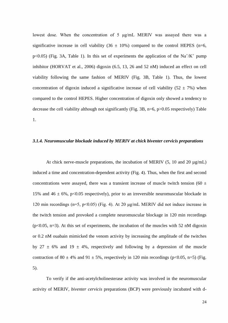
3.1.1. Na+,K+-ATPase activity
The analysis of the Na+,K+-ATPase in rat’s heart activity in presence of MERIV
(5µg/ml) showed that this compound inhibited significatively the electrogenic pump in
39±3% (n=4, p<0.05) (Fig. 2).
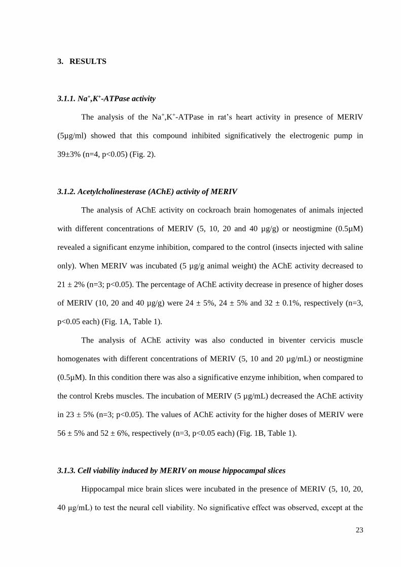
3.1.2. Acetylcholinesterase (AChE) activity of MERIV
The analysis of AChE activity on cockroach brain homogenates of animals injected
with different concentrations of MERIV (5, 10, 20 and 40 µg/g) or neostigmine (0.5µM)
revealed a significant enzyme inhibition, compared to the control (insects injected with saline
only). When MERIV was incubated (5 µg/g animal weight) the AChE activity decreased to
21 ± 2% (n=3; p<0.05). The percentage of AChE activity decrease in presence of higher doses
of MERIV (10, 20 and 40 µg/g) were 24 ± 5%, 24 ± 5% and 32 ± 0.1%, respectively (n=3,
p<0.05 each) (Fig. 1A, Table 1).
The analysis of AChE activity was also conducted in biventer cervicis muscle
homogenates with different concentrations of MERIV (5, 10 and 20 µg/mL) or neostigmine
(0.5µM). In this condition there was also a significative enzyme inhibition, when compared to
the control Krebs muscles. The incubation of MERIV (5 µg/mL) decreased the AChE activity
in 23 ± 5% (n=3; p<0.05). The values of AChE activity for the higher doses of MERIV were
56 ± 5% and 52 ± 6%, respectively (n=3, p<0.05 each) (Fig. 1B, Table 1).
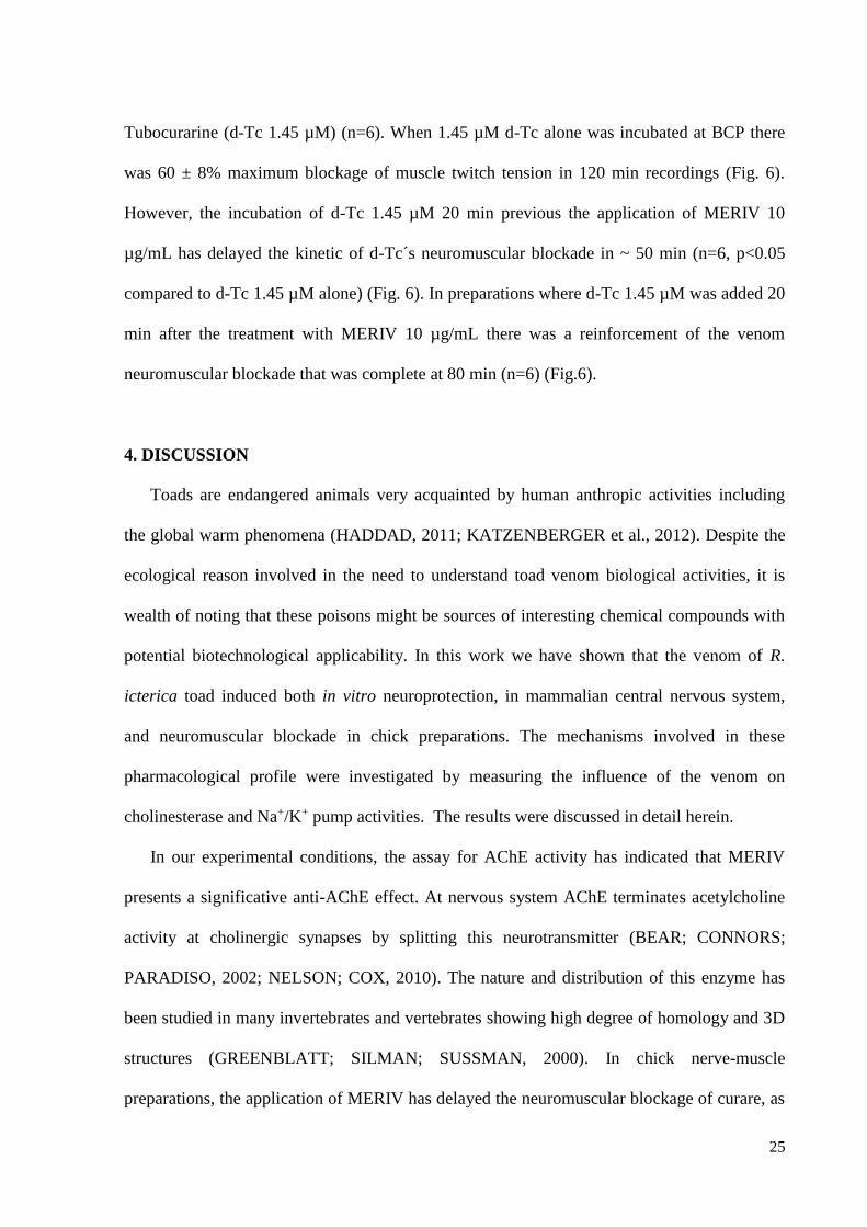
3.1.3. Cell viability induced by MERIV on mouse hippocampal slices
Hippocampal mice brain slices were incubated in the presence of MERIV (5, 10, 20,
40 μg/mL) to test the neural cell viability. No significative effect was observed, except at the
24
lowest dose. When the concentration of 5 μg/mL MERIV was assayed there was a
significative increase in cell viability (36 ± 10%) compared to the control HEPES (n=6,
p<0.05) (Fig. 3A, Table 1). In this set of experiments the application of the Na+/K+ pump
inhibitor (HORVAT et al., 2006) digoxin (6.5, 13, 26 and 52 nM) induced an effect on cell
viability following the same fashion of MERIV (Fig. 3B, Table 1). Thus, the lowest
concentration of digoxin induced a significative increase of cell viability (52 ± 7%) when
compared to the control HEPES. Higher concentration of digoxin only showed a tendency to
decrease the cell viability although not significantly (Fig. 3B, n=6, p>0.05 respectively) Table
1.
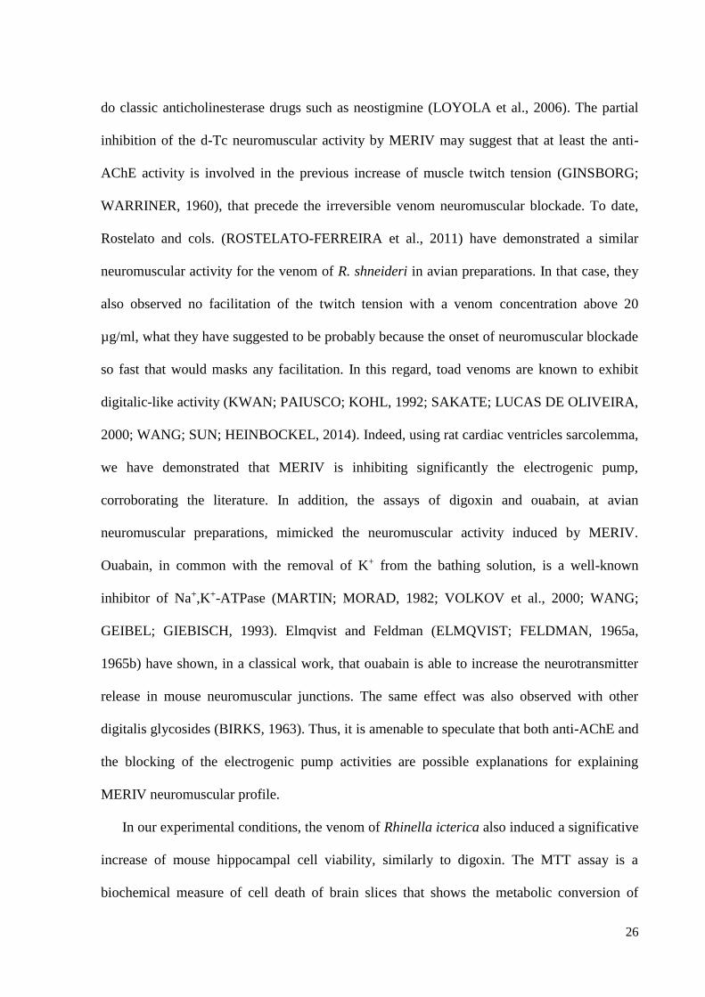
3.1.4. Neuromuscular blockade induced by MERIV at chick biventer cervicis preparations
At chick nerve-muscle preparations, the incubation of MERIV (5, 10 and 20 µg/mL)
induced a time and concentration-dependent activity (Fig. 4). Thus, when the first and second
concentrations were assayed, there was a transient increase of muscle twitch tension (60 ±
15% and 46 ± 6%, p<0.05 respectively), prior to an irreversible neuromuscular blockade in
120 min recordings (n=5, p<0.05) (Fig. 4). At 20 μg/mL MERIV did not induce increase in
the twitch tension and provoked a complete neuromuscular blockage in 120 min recordings
(p<0.05, n=3). At this set of experiments, the incubation of the muscles with 52 nM digoxin
or 0.2 nM ouabain mimicked the venom activity by increasing the amplitude of the twitches
by 27 ± 6% and 19 ± 4%, respectively and following by a depression of the muscle
contraction of 80 ± 4% and 91 ± 5%, respectively in 120 min recordings (p<0.05, n=5) (Fig.
5).
To verify if the anti-acetylcholinesterase activity was involved in the neuromuscular
activity of MERIV, biventer cervicis preparations (BCP) were previously incubated with d-
25
Tubocurarine (d-Tc 1.45 µM) (n=6). When 1.45 µM d-Tc alone was incubated at BCP there
was 60 ± 8% maximum blockage of muscle twitch tension in 120 min recordings (Fig. 6).
However, the incubation of d-Tc 1.45 µM 20 min previous the application of MERIV 10
µg/mL has delayed the kinetic of d-Tc´s neuromuscular blockade in ~ 50 min (n=6, p<0.05
compared to d-Tc 1.45 µM alone) (Fig. 6). In preparations where d-Tc 1.45 µM was added 20
min after the treatment with MERIV 10 µg/mL there was a reinforcement of the venom
neuromuscular blockade that was complete at 80 min (n=6) (Fig.6).
4. DISCUSSION
Toads are endangered animals very acquainted by human anthropic activities including
the global warm phenomena (HADDAD, 2011; KATZENBERGER et al., 2012). Despite the
ecological reason involved in the need to understand toad venom biological activities, it is
wealth of noting that these poisons might be sources of interesting chemical compounds with
potential biotechnological applicability. In this work we have shown that the venom of R.
icterica toad induced both in vitro neuroprotection, in mammalian central nervous system,
and neuromuscular blockade in chick preparations. The mechanisms involved in these
pharmacological profile were investigated by measuring the influence of the venom on
cholinesterase and Na+/K+ pump activities. The results were discussed in detail herein.
In our experimental conditions, the assay for AChE activity has indicated that MERIV
presents a significative anti-AChE effect. At nervous system AChE terminates acetylcholine
activity at cholinergic synapses by splitting this neurotransmitter (BEAR; CONNORS;
PARADISO, 2002; NELSON; COX, 2010). The nature and distribution of this enzyme has
been studied in many invertebrates and vertebrates showing high degree of homology and 3D
structures (GREENBLATT; SILMAN; SUSSMAN, 2000). In chick nerve-muscle
preparations, the application of MERIV has delayed the neuromuscular blockage of curare, as
26
do classic anticholinesterase drugs such as neostigmine (LOYOLA et al., 2006). The partial
inhibition of the d-Tc neuromuscular activity by MERIV may suggest that at least the anti-
AChE activity is involved in the previous increase of muscle twitch tension (GINSBORG;
WARRINER, 1960), that precede the irreversible venom neuromuscular blockade. To date,
Rostelato and cols. (ROSTELATO-FERREIRA et al., 2011) have demonstrated a similar
neuromuscular activity for the venom of R. shneideri in avian preparations. In that case, they
also observed no facilitation of the twitch tension with a venom concentration above 20
µg/ml, what they have suggested to be probably because the onset of neuromuscular blockade
so fast that would masks any facilitation. In this regard, toad venoms are known to exhibit
digitalic-like activity (KWAN; PAIUSCO; KOHL, 1992; SAKATE; LUCAS DE OLIVEIRA,
2000; WANG; SUN; HEINBOCKEL, 2014). Indeed, using rat cardiac ventricles sarcolemma,
we have demonstrated that MERIV is inhibiting significantly the electrogenic pump,
corroborating the literature. In addition, the assays of digoxin and ouabain, at avian
neuromuscular preparations, mimicked the neuromuscular activity induced by MERIV.
Ouabain, in common with the removal of K+ from the bathing solution, is a well-known
inhibitor of Na+,K+-ATPase (MARTIN; MORAD, 1982; VOLKOV et al., 2000; WANG;
GEIBEL; GIEBISCH, 1993). Elmqvist and Feldman (ELMQVIST; FELDMAN, 1965a,
1965b) have shown, in a classical work, that ouabain is able to increase the neurotransmitter
release in mouse neuromuscular junctions. The same effect was also observed with other
digitalis glycosides (BIRKS, 1963). Thus, it is amenable to speculate that both anti-AChE and
the blocking of the electrogenic pump activities are possible explanations for explaining
MERIV neuromuscular profile.
In our experimental conditions, the venom of Rhinella icterica also induced a significative
increase of mouse hippocampal cell viability, similarly to digoxin. The MTT assay is a
biochemical measure of cell death of brain slices that shows the metabolic conversion of
27
yellow MTT dye by active mitochondria to form insoluble purple precipitates in live cells.
Cell viability in mammalian central nervous system is mostly influenced by toxic conditions
which include level of reactive oxygen species, excitotoxicity due to excess of
neurotransmitters, necrosis and apoptosis (DAL BELO et al., 2013; DVELA et al., 2012;
FRANCO et al., 2009; TAKADA-TAKATORI et al., 2006). In this regard, drugs like
anticholinesterases, are good clinical choices for preventing both neuronal death and cognitive
impairment in neurodegenerative diseases (TAKADA-TAKATORI et al., 2006). Therefore,
despite the clinical relevance of the venom ability to inhibit the electrogenic pump, this
pharmacological activity may also reveal its potential as sources for novel drugs in cancer
therapy (DVELA et al., 2012).
5. CONCLUSION
Rhinella icterica toad venom induced both inhibitory activities over AChE and Na+/K+
pumps. These activities may be directly involved in the neurotoxicity observed in chick
neuromuscular junctions and in the increase of cell viability of mouse hippocampal slices.
The comprehension of the venom biology is important not only to understand the ecological
interactions of Rhinella icterica toad, but also reveal the potential biotechnological
applications of the poison components.
ACKNOWLEDGMENTS:
The authors thank Michele Corrêa Stach, Rafael Plá Matielo Lemos, Giulianna
Echeverria Macedo and Nathane Rosa Rodrigues for the technical assistance with
biochemical protocols. The authors also thank the PRONEM/FAPERGS/CNPq 003/2011,
Coordenação de Aperfeiçoamento de Pessoal de Nível Superior-CAPES 063/2010
Toxinologia and PROPESQ/PROPG UNIPAMPA, for financial support. R. S. Oliveira,
28
Santos, D.S and Almeida, C.G.M. was granted by CAPES. Leal, A.P. was granted by
Academic Program Development from UNIPAMPA.
CONFLICT OF INTERESTS: The authors declare that there is no conflict of interests
regarding this work.
REFERENCES
[1] F. Heus, R. a. Otvos, R.L.E.G. Aspers, R. van Elk, J.I. Halff, A.W. Ehlers, et al.,
Miniaturized Bioaffinity Assessment Coupled to Mass Spectrometry for Guided
Purification of Bioactives from Road and Cone Snail, Biology (Basel). 3 (2014) 139–
156. doi:10.3390/biology3010139.
[2] R. a. Saporito, M.A. Donnelly, T.F. Spande, H.M. Garraffo, A Review of Chemical
Ecology in Poison Frogs, Chemoecology. 22 (2012) 159–168. doi:10.1007/s00049-
011-0088-0.
[3] G. a. Cunha-Filho, I.S. Resck, B.C. Cavalcanti, C.Ó. Pessoa, M.O. Moraes, J.R.O.
Ferreira, et al., Cytotoxic Profile of Natural and Some Modified Bufadienolides from
Toad Rhinella schneideri Parotoid Gland Secretion, Toxicon. 56 (2010) 339–348.
doi:10.1016/j.toxicon.2010.03.021.
[4] J.M. Sciani, C.B. Angeli, M.M. Antoniazzi, C. Jared, D.C. Pimenta, Differences and
Similarities Among Parotoid Macrogland Secretions in South American Toads: A
Preliminary Biochemical Delineation, Sci. World J. 2013 (2013) 1–9.
doi:10.1155/2013/937407.
[5] A. Siano, P.I. Gatti, M.S. Imaz, E. Zerbini, C. Arturo, R. Lajmanovich, et al., A
Comparative Study of the Biological Activity of Skin and Granular Gland Secretions of
Leptodactylus latrans and Hypsiboas pulchellus from Argentina, Rec. Nat. Prod. 8
(2014) 128–135.
[6] J.W. Daly, The Chemistry of Poisons in Amphibian Skin., Proc. Natl. Acad. Sci. U. S.
A. 92 (1995) 9–13. doi:10.1073/pnas.92.1.9.
[7] S. Rostelato-ferreira, C. a D. Belo, G.B. Leite, S. Hyslop, L. Rodrigues-simioni,
Presynaptic Neuromuscular Action of a Methanolic Extract from the Venom of
Rhinella schneideri Toad, J. Venom. Anim. Toxins Incl. Trop. Dis. 20 (2014) 1–5.
doi:10.1186/1678-9199-20-30.
[8] M. Sakate, P.C. Lucas de Oliveira, Toad Envenoming in Dogs: Effects and Treatment,
J. Venom. Anim. Toxins. 6 (2000) 1–7. doi:10.1590/S0104-79302000000100003.
[9] Seiichiro Yoshida and Takeshi Sakai, Effects of Bufalin and Related Cardiotonic
Steroids in the Neuromuscular Junction, Japan. J. Pharmacol. 23 (1973) 859–869.
[10] Seiichiro Yoshida and Takeshi Sakai, Mechanism of Bufalin-Induced Blockade of
Neuromuscular Transmission in Isolated Rat Diaphragm, Japan. J. Pharmacol. 24
(1974) 97–108.
29
[11] S.C.D. Manika Das, A. Gomes, Isolation, Purification and Partial Chemical
Characterization of a Lethal Factor from Common Indian Toad (Bufo melanostictus,
Schneider) Skin Extract, Indian J. Exp. Biol. 39 (2001) 781–785.
[12] S. Rostelato-Ferreira, C. a Dal Belo, M.A. da Cruz-Höfling, S. Hyslop, L. Rodrigues-
Simioni, Presynaptic Effect of a Methanolic Extract of Toad (Rhinella schneideri)
Poison in Avian Neuromuscular Preparation, J. Venom Res. 2 (2011) 32–36.
[13] R.M. Gowda, R.A. Cohen, I.A. Khan, Toad Venom Poisoning: Resemblance to
Digoxin Toxicity and Therapeutic Implications, Heart. 89 (2003) 1–2.
doi:10.1136/heart.89.4.e14.
[14] H.-Y. Kuo, C.-W. Hsu, J.-H. Chen, Y.-L. Wu, Y.-S. Shen, Life-Threatening Episode
After Ingestion of Toad Eggs: a Case Report With Literature Review, Emerg. Med. J.
24 (2007) 215–216. doi:10.1136/emj.2006.044602.
[15] D. Radford, A. Gillies, J. Hinds, P. Duffy, Naturally Occurring Cardiac Glycosides,
Med. J. Aust. 144 (1986) 540–543. doi:10.1016/0378-8741(87)90017-1.
[16] D.C. Rohrer, D.S. Fullerton, E. Kitatsuji, T. Nambara, Y. Eiichi, Bufalin*, Acta Cryst.
B 38 (1982) 1865–1868.
[17] T. Kwan, A.D. Paiusco, L. Kohl, Digitalis Toxicity Caused by Toad Venom, Chest.
102 (1992) 949–950.
[18] P. Colombo, A. Kindel, G. Vinciprova, L. Krause, Composição e ameaças à
conservação dos anfíbios anuros do Parque Estadual de Itapeva, município de Torres,
Rio Grande do Sul, Brasil, Biota Neotrop. 8 (2008). doi:10.1590/S1676-
06032008000300020.
[19] I. Stefanon, J.R. Cade, A.A. Fernandes, R.F. Ribeiro Junior, G.P. Targueta, J.G. Mill,
et al., Ventricular Performance and Na+-K+ ATPase Activity are Reduced Early and
Late After Myocardial Infarction in Rats, Braz J Med Biol Res. 42 (2009) 902–911.
[20] M. Bradford, A Rapid and Sensitive Method for the Quantitation of Microgram
Quantities of Protein Utilizing the Principle of Protein-Dye Binding, Anal. Biochem.
254 (1976) 248–254.
[21] G.L. Ellman, K.D. Courtney, V. Andres, R.M. Featherstone, A New and Rapid
Colorimetric Determination of Acetylcholinesterase Activity, Biochem. Pharmacol. 7
(1961) 88–95. doi:10.1016/0006-2952(61)90145-9.
[22] G.D. Stürmer, T.C. de Freitas, M. de A. Heberle, D.R. de Assis, L. Vinadé, A.B.
Pereira, et al., Modulation of Dopaminergic Neurotransmission Induced by Sublethal
Doses of the Organophosphate Trichlorfon in Cockroaches., Ecotoxicol. Environ. Saf.
109 (2014) 56–62. doi:10.1016/j.ecoenv.2014.08.006.
[23] L. Vinadé, R. Rodnight, The Dephosphorylation of Glial Fibrillary Acidic Protein
(GFAP) in the Immature Rat Hippocampus is Catalyzed Mainly by a Type 1 Protein
Phosphatase, Brain Res. 732 (1996) 195–200.
[24] C.A. Dal Belo, A.P.D.B. Lucho, L. Vinadé, L. Rocha, H. Seibert França, S. Marangoni,
et al., In Vitro Antiophidian Mechanisms of Hypericum brasiliense Choisy
Standardized Extract: Quercetin-Dependent Neuroprotection, Biomed Res. Int. 2013
(2013) 1–6. doi:10.1155/2013/943520.
[25] B.L. Ginsborg, J. Warriner, The Isolated Chick Biventer Cervicis Nerve-Muscle
Preparation, Brit. J. Pharmacol. 15 (1960) 410–411.
30
[26] A. Horvat, T. Momic, A. Banjac, S. Petrovic, G. Nikezic, M. Demajo, Selective
Inhibition of Brain Na , K-ATPase by Drugs, Physiol. Res. 55 (2006) 325–338.
[27] D. Conrado, D.E.A. Munhoz, M.C. dos Santos, R.F.L. MEllo, V.B. Silva,
Vulnerabilidades às Mudanças Climáticas, IIEB. (2000) 1–10.
[28] M. Katzenberger, M. Tejedo, H. Duarte, F. Marangoni, J.F. Beltrán, Especial
Mudanças Ambientais, Rev. Da Biol. 8 (2012) 25–32. www.ib.usp.br/revista (accessed
February 4, 2016).
[29] C.F.B. Haddad, ANFÍBIOS - Uma Análise da Lista Brasileira de Anfíbios Ameaçados
de Extinção, ICMBIO. 2 (2011) 287–288.
http://www.icmbio.gov.br/portal/images/stories/biodiversidade/fauna-brasileira/livro-
vermelho/volumeII/Anfibios.pdf (accessed January 24, 2016).
[30] D.L. Nelson, M.M. Cox, Principles of Biochemistry, 5a ed., Sara Tenney, New York,
2010. doi:10.1016/0020-711X(94)90020-5.
[31] M.F. Bear, W.B. Connors, M.A. Paradiso, Neurociências: Desvendando o Sistema
Nervoso, Artmed Editora S.A., Porto Alegre, 2002.
[32] H.M. Greenblatt, I. Silman, J.L. Sussman, Structural Studies on Vertebrate and
Invertebrate Acetylcholinesterases and Their Complexes With Functional Ligands,
Drug Dev. Res. 50 (2000) 573–583.
[33] Y.C.S. Loyola, A. de F. de A. Braga, G.M.B. Potério, S.R. de Sousa, S.C.A.F.
Fernandes, F.S. da S. Braga, Influência da Lidocaína no Bloqueio Neuromuscular
Produzido pelo Rocurônio. Estudo em Preparação Nervo Frênico-Diafragma de Rato,
Rev Bras Anestesiol. 56 (2006) 147–156.
[34] Z.-J. Wang, L. Sun, T. Heinbockel, Resibufogenin and Cinobufagin Activate Central
Neurons through an Ouabain-Like Action, PLoS One. 9 (2014) e113272.
doi:10.1371/journal.pone.0113272.
[35] G. Martin, M. Morad, Activity-Induced Potassium Accumulation and its Uptake in
Frog Ventricular Muscle, J. Physiol. 328 (1982) 205–227.
[36] W.H. Wang, J. Geibel, G. Giebisch, Mechanism of Apical K+ Channel Modulation in
Principal Renal Tubule Cells. Effect of Inhibition of Basolateral Na+,K+-ATPase, J.
Gen. Physiol. 101 (1993) 673–694.
[37] E.M. Volkov, L.F. Nurullin, I. Svandová, E.E. Nikolscky, F. VysKocil, Participation of
Electrogenic Na+,K+,ATPase in the Membrane Potential of Earthworm Body Wall
Muscles, Physiol. Res. 49 (2000) 481–484.
[38] T. Clausen, Na+-K+ Pump Regulation and Skeletal Muscle Contractility., Physiol.
Rev. 83 (2003) 1269–1324. doi:10.1152/physrev.00011.2003.
[39] B.Y.D. Elmqvist, A.D.D.S. Feldman, Calcium Dependence of Spontaneous
Acetylcholine Release at Mammalian Motor Nerve Terminals, J. Physiol. 181 (1965)
487–497.
[40] B.Y.D. Elmqvist, D.S. Feldman, Effects of Sodium Pump Inhibitors on Spontaneous
Acetylcholine Release at the Neuromuscular Junction, J. Physiol. 181 (1965) 498–505.
[41] R.I. Birks, The Role of Sodium Ions in the Metabolism of Acetylcholine, Can. J.
Biochem. Physiol. 41 (1963) 2573–2597.
[42] M. Dvela, H. Rosen, H.C. Ben-Ami, D. Lichtstein, Endogenous Ouabain Regulates
31
Cell Viability, Am J Physiol Cell Physiol. 302 (2012) C442–C452.
doi:10.1152/ajpcell.00336.2011.
[43] J.L. Franco, T. Posser, P.R. Dunkley, P.W. Dickson, J.J. Mattos, R. Martins, et al.,
Methylmercury Neurotoxicity is Associated With Inhibition of the Antioxidant
Enzyme Glutathione Peroxidase, Free Radic. Biol. Med. 47 (2009) 449–457.
doi:10.1016/j.freeradbiomed.2009.05.013.
[44] Y. Takada-Takatori, T. Kume, M. Sugimoto, H. Katsuki, H. Sugimoto, A. Akaike,
Acetylcholinesterase Inhibitors Used in Treatment of Alzheimer’s Disease Prevent
Glutamate Neurotoxicity Via Nicotinic Acetylcholine Receptors and
Phosphatidylinositol 3-kinase Cascade, Neuropharmacology. 51 (2006) 474–486.
doi:10.1016/j.neuropharm.2006.04.007.
32
Figure Legends
Figure 1: Inhibition of acetylcholinesterase (AChE) activity induced by Rhinella icterica toad
venom (MERIV). Panel A: Representative graph of acetylcholinesterase inhibition in
cockroaches’ brain homogenates after 6 hours exposure to MERIV (5, 10, 20 and 40 µg/g)
and neostigmine (0.5 μM). Panel B: acetylcholinesterase activity in chick biventer cervicis
muscle after MERIV (5, 10 and 20μg/mL) treatments and neostigmine (0.5 μM), with indirect
stimulation, in comparison with those obtained only with Krebs control. Data were expressed
as mean ± S.E.M. Significance at *p<0.05 in comparison to Saline (control 100%).
Figure 2: Inhibition of rat cardiac Na+,K+-ATPase activity induced by Rhinella icterica toad
venom (MERIV). Na+,K+-ATPase activity was assayed by measuring Pi liberation by reacting
initiated by adding ATP. The specific activity was reported as nmol Pi released per min per
mg of protein. Results represent Mean ± S.E.M. (n=4). Significance *p<0.05 compared to
control.
Figure 3: Effects of Rhinella icterica toad venom (MERIV) and digoxin on the cell viability
of hippocampal slices. Panel A: Hippocampal slices were incubated with MERIV (5, 10, 20
and 40 µg/mL) during 60 min. Note that the concentration of 5 µg/mL induced a significant
increase in the cell viability (n=6, p<0.05). Panel B: Hippocampal slices were incubated with
digoxin (6.5, 13, 26 and 52 nM) during 60 min. Note that the concentration of 6.5 and 13 nM
induced a significative increase in cell viability (n=6, p<0.05). Data were expressed as mean ±
S.E.M. Significance at *p<0.05 compared to Hepes (control 100%).
Figure 4: Concentration-response of MERIV at chick muscle-nerve preparations. BCP
muscle-nerve preparation was indirectly stimulated (0.1 Hz, 0.2ms, 3-10V) during 120 min.
Panel A: In the graphic each point represents the mean ± S.E.M. (n = 3-5). Panel B:
Representative traces of the activity of MERIV 10 µg/mL on evoked responses of BCP
muscle-nerve preparation. Note that the venom (5 and 10 µg/mL) induced a transitory
increase of muscle twitch-tension followed by an irreversible neuromuscular blockade after
30min recordings. Significance at *p<0.05, compared to Krebs control.
Figure 5: Comparison of the Rhinella icterica toad venom (MERIV) neuromuscular activity
with different pharmacological treatments in biventer cervicis muscle nerve preparations
(BCP). Panel A: Shows the activity induced by 10 µg/mL MERIV, 52 nM digoxin and 0.2 nM
ouabain at BCP muscle-nerve preparations in 120min (0.1Hz, 0.2ms, 5-8V). In the graphic
each point represents the mean ± S.E.M. (n = 4-6). Note that ouabain and digoxin have a
similar activity compared to the MERIV. Panel B: representative traces of the effect of 0.2 nM
ouabain on evoked responses at BCP preparations. Significance at *p<0.05 compared to
MERIV.
Figure 6: Protective activity of Rhinella icterica toad venom (MERIV) against the
neuromuscular blockade induced by d-Tubocurarine (d-Tc) at chick muscle-nerve
preparations (BCP). BCP was indirectly stimulated (0.1 Hz, 0.2ms, 3-10V) during 120 min.
Panel A: Shows the activity induced by 1.45 µM d-Tc in BCP. Panel B: Demonstrates the
muscle response of d-Tc preincubated and post incubated with MERIV 10 µg/mL on evoked
responses at BCP. Note that the addition of d-Tc prior to MERIV partially prevented the
neuromuscular blockade induced by the curare. In the graphic each point represents the Mean
± S.E.M. (n = 3-5). Significance at *p<0.05, compared to d-Tc.
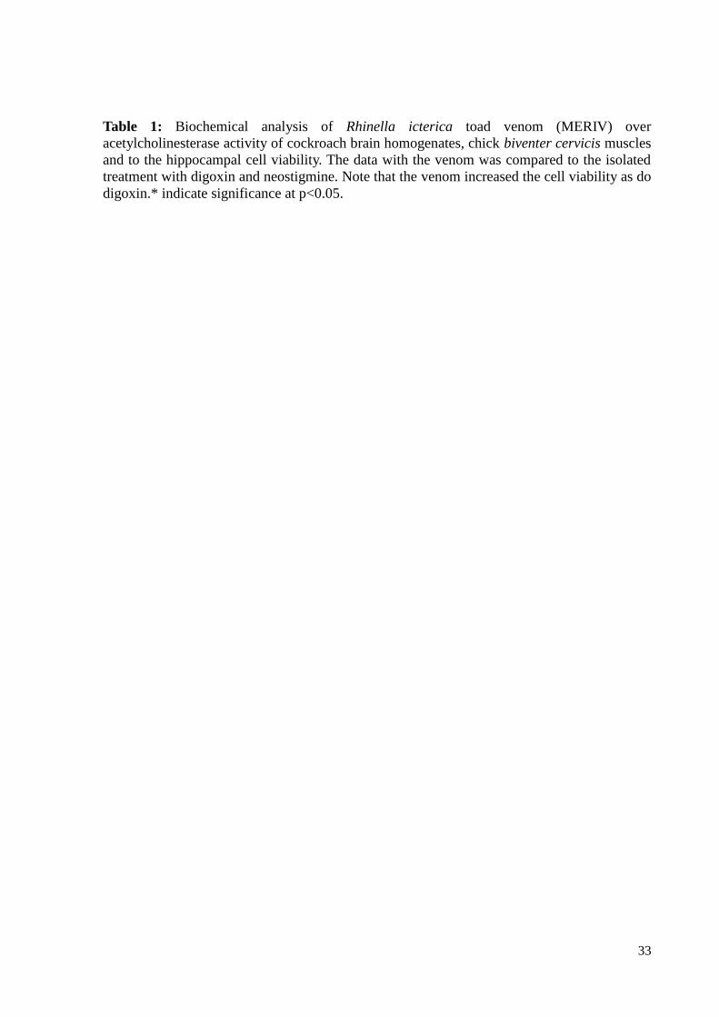
33
Table 1: Biochemical analysis of Rhinella icterica toad venom (MERIV) over
acetylcholinesterase activity of cockroach brain homogenates, chick biventer cervicis muscles
and to the hippocampal cell viability. The data with the venom was compared to the isolated
treatment with digoxin and neostigmine. Note that the venom increased the cell viability as do
digoxin.* indicate significance at p<0.05.
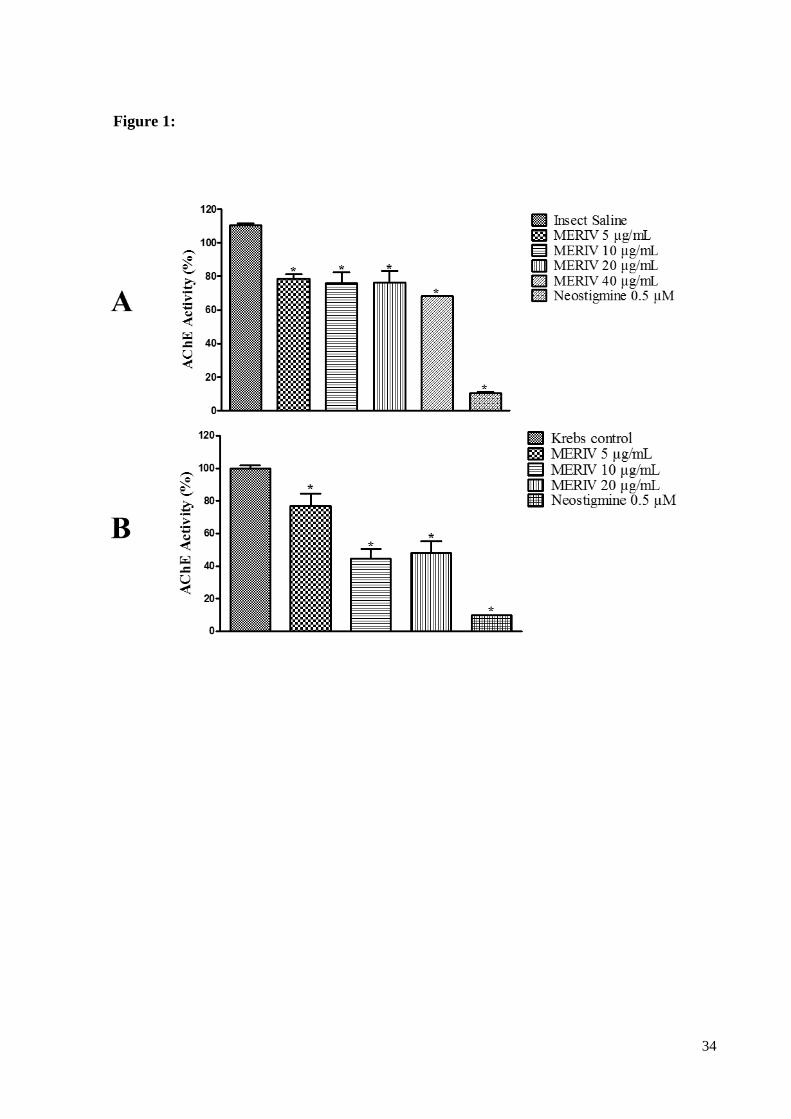
34
Figure 1:
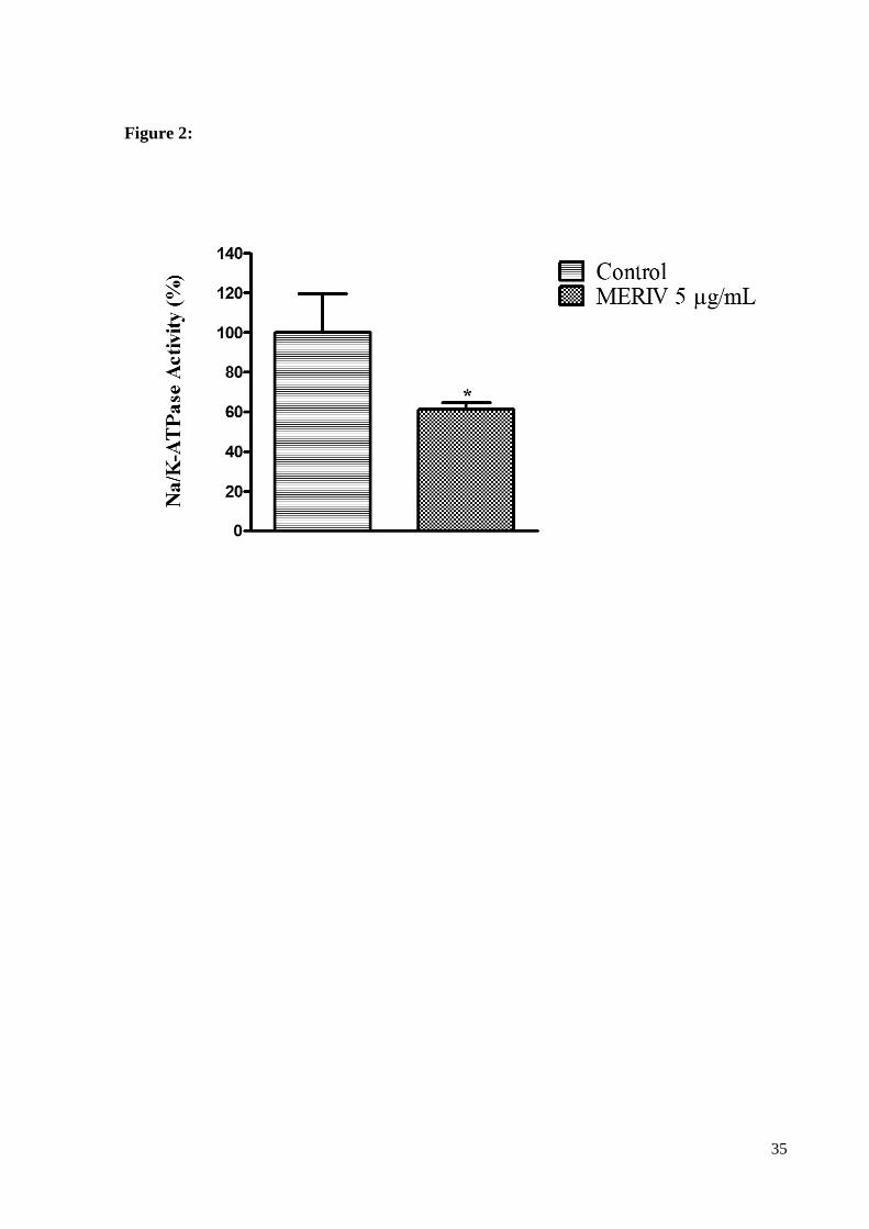
35
Figure 2:
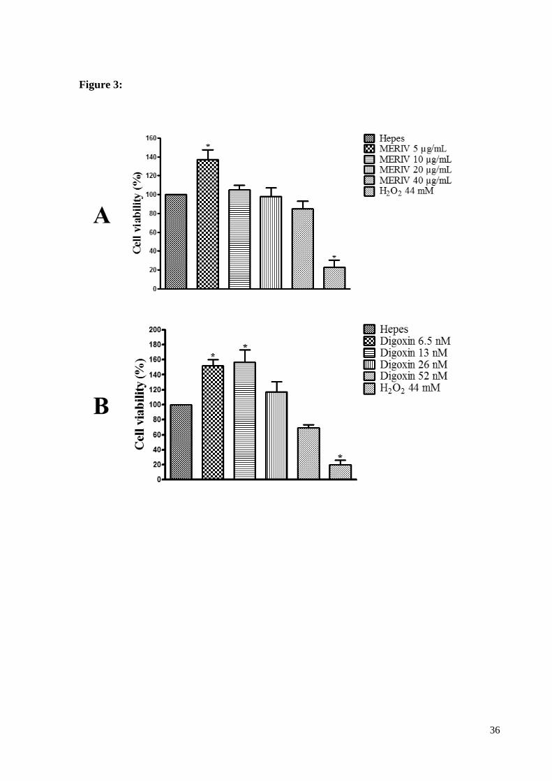
36
Figure 3:
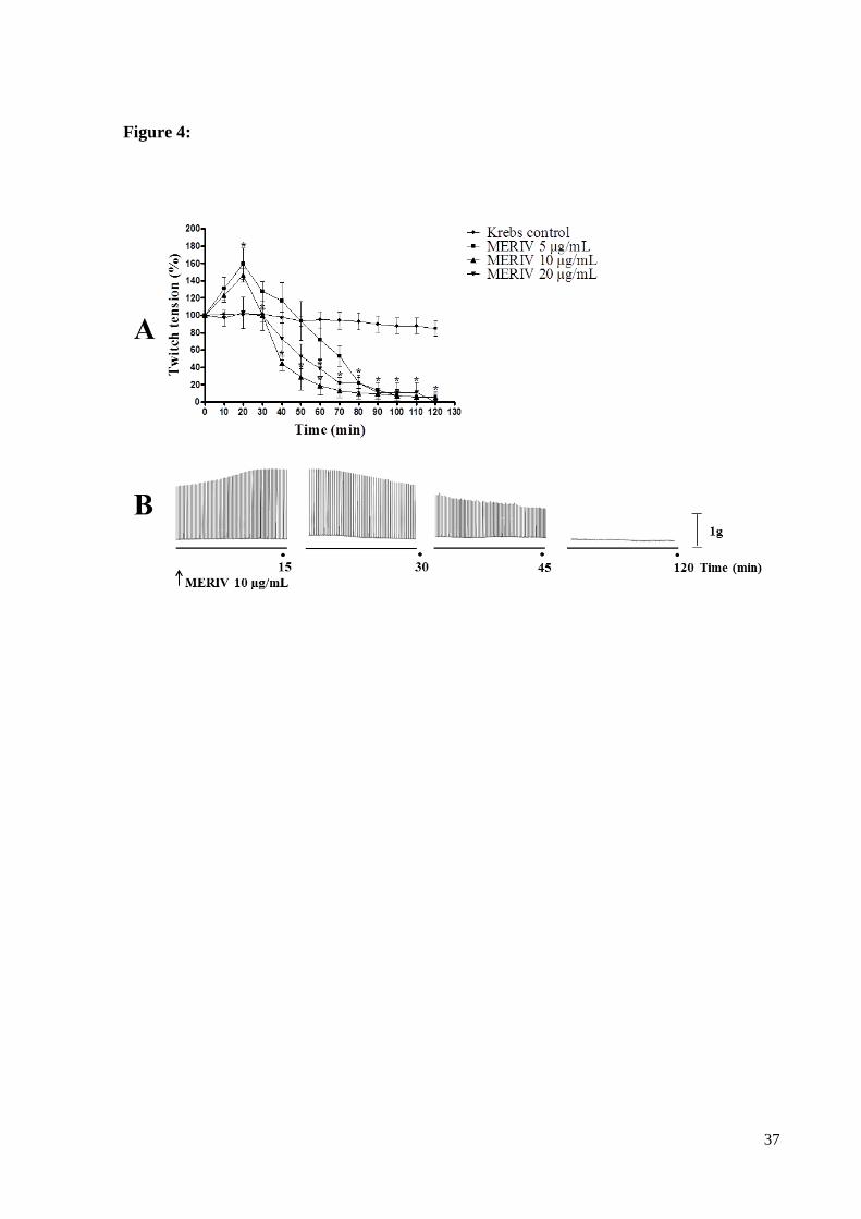
37
Figure 4:
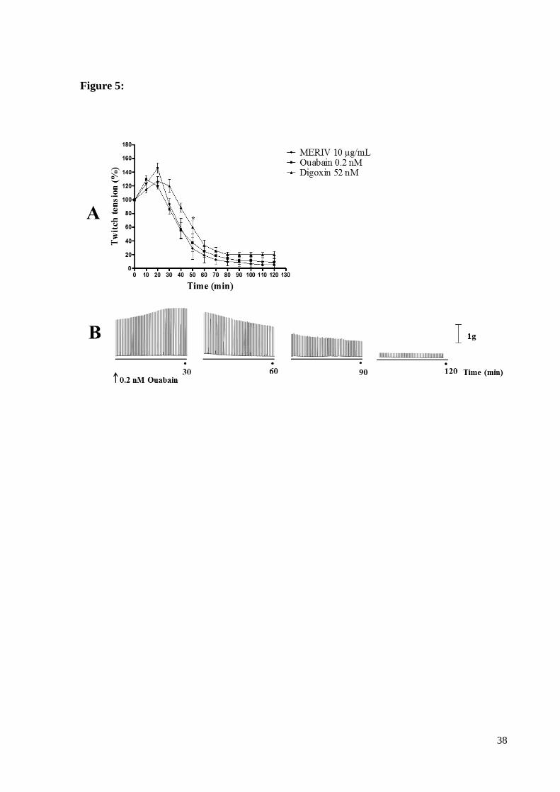
38
Figure 5:
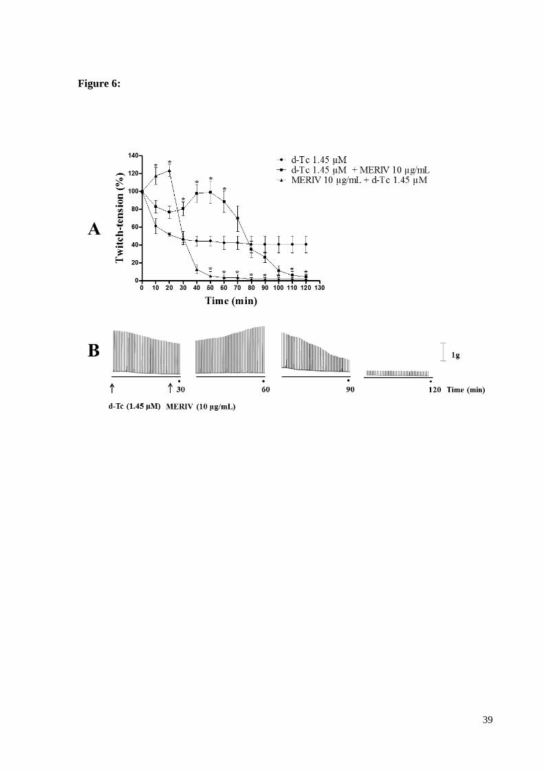
39
Figure 6:
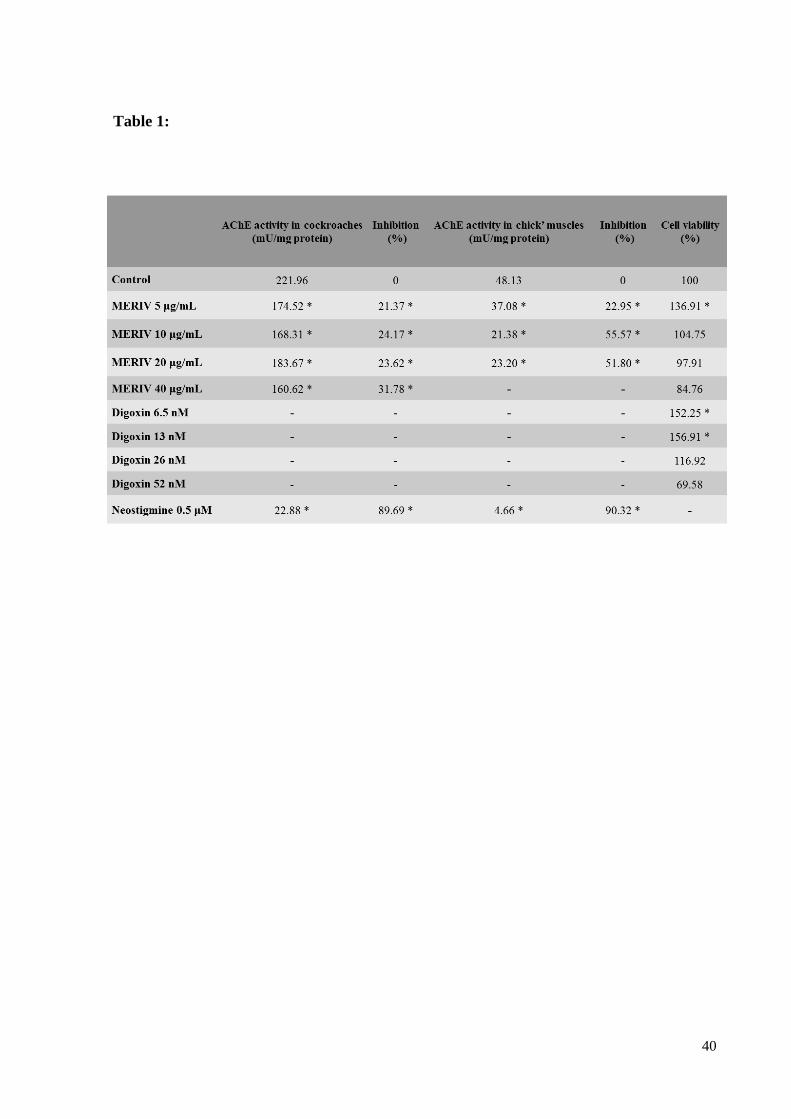
40
Table 1:
41
5. CONSIDERAÇÕES FINAIS
Com base nos dados obtidos no presente estudo, podemos considerar:
Nas menores concentrações, MERIV (5 μg/mL) e Digoxina (6,5 e 13 nM)
demonstraram aumento da viabilidade celular em fatias de hipocampo de
camundongos. Condizente com os resultados encontrados na literatura com
glicosídeos cardiotônicos.
MERIV inibiu significativamente a atividade da acetilcolinesterase em todas as doses
avaliadas, tanto em invertebrados (cérebro de baratas), quanto em músculos utilizados
na preparação biventer cervicis de pintainho.
Em preparação biventer cervicis de pintainho, MERIV 5 e 10 μg/mL induziram
bloqueio neuromuscular precedido de facilitação, possivelmente pela atividade
anticolinesterasica e por inibição da Na+,K+-ATPase.
Em preparação biventer cervicis de pintainho, a atividade de MERIV (10 μg/mL) foi
similar a dos inibidores da Na+,K+-ATPase, digoxina (52 nM) e Ouabaína (0,2 nM),
sugerindo que MERIV inibe a Na+,K+-ATPase.
MERIV prorrogou o bloqueio neuromuscular induzido por d-Tubocurarina,
demonstrando atividade similar a de anticolinesterásicos.
MERIV inibiu significativamente a atividade da Na+,K+-ATPase em tecido cardíaco
de ratos, comprovando que estão presentes no veneno toxinas que atuam na Na+,K+-
ATPase.
Este trabalho contribui para caracterizar a atividade biológica do veneno de sapos da
espécie R. icterica em sistema nervoso central e periférico de vertebrados. As
atividades anticolinesterasica e digitalic-like presentes no veneno demonstram a
importância das toxinas presentes no veneno desta espécie e sua aplicabilidade
terapêutica.
42
6. PERSPECTIVAS
Com base nos resultados obtidos neste estudo, pretendemos:
Contribuir para o conhecimento do mecanismo da neurotoxicidade por meio da
identificação bioquímica e farmacológica dos compostos ativos isolados do veneno de
R. icterica sobre o sistema nervoso de vertebrados.
Isolar e purificar as toxinas presentes neste veneno com atividade anticolinesterásica
e/ou que apresentem ação sobre a Na+,K+-ATPase.
Avaliar o mecanismo de ação dos compostos isolados do veneno junto à junção
neuromuscular de vertebrados, e investigar a sua farmacologia com ênfase na
identificação dos possíveis sítios ativos.
43
7. REFERÊNCIAS BIBLIOGRÁFICAS
ABDEL-RAHMAN, M. A.; AHMED, S. H.; NABIL, Z. I. In Vitro Cardiotoxicity and
Mechanism of Action of the Egyptian Green Toad Bufo viridis Skin Secretions. Toxicology
in vitro, v. 24, n. 2, p. 480–485, 2010.
ARISI, G. M.; NEDER, L.; MOREIRA, J. E. O Ciclo Da Vesícula Sináptica : Panorama
Molecular*. Medicina, Ribeirão Preto, v. 34, p. 154–169, 2001.
BAGROV, A. Y.; SHAPIRO, J. I.; FEDOROVA, O. V. Endogenous Cardiotonic Steroids:
Physiology, Pharmacology, and Novel Therapeutic Targets. Pharmacological reviews, v. 61,
n. 1, p. 9–38, 2009.
BARRAVIERA, B. Venenos Animais: Uma Visão Integrada. Rio de Janeiro: Editora de
Publicações Científicas, 1994.
BEAR, M. F.; CONNORS, W. B.; PARADISO, M. A. Neurociências: Desvendando o
Sistema Nervoso. Porto Alegre: Artmed Editora S.A., 2002. v. 2.
BUAINAIN, R. P.; MOURA, L. S.; OLIVEIRA, A. S. B. Fasciculação. Rev. Neurociências,
v. 8, n. 1, p. 31–34, 2000.
CHEN, J. Q. et al. Sodium/Potasium ATPase (Na+, K+-ATPase) and Ouabain/Related
Cardiac Glycosides: A New Paradigm for Development of Anti- Breast Cancer Drugs?
Breast Cancer Research and Treatment, v. 96, n. 1, p. 1–15, 2006.
CHEN, K. K.; KOVARIOVÁ, A. Pharmacology and Toxicology of Toad Venom. Journal of
Pharmaceutical Sciences, v. 56, n. 12, p. 1535–1541, 1967.
CHOI, Y. H.; HONG, S.-H. Bufalin Induces Apoptosis Through Activation of Both the
Intrinsic and Extrinsic athways in Human Bladder Cancer Cells. Oncology Reports, v. 27, n.
15, p. 114–120, 2012.
CUNHA-FILHO, G. A. et al. Cytotoxic Profile of Natural and Some Modified Bufadienolides
from Toad Rhinella schneideri Parotoid Gland Secretion. Toxicon, v. 56, n. 3, p. 339–348,
2010.
DALY, J. W. The Chemistry of Poisons in Amphibian Skin. Proceedings of the National
Academy of Sciences of the United States of America, v. 92, n. 1, p. 9–13, 1995.
DALY, J. W.; SPANDE, T. F.; GARRAFFO, H. M. Alkaloids from Amphibian Skin: A
Tabulation of Over Eight-Hundred Compounds. Journal of Natural Products, v. 68, n. 10,
p. 1556–1575, 2005.
DANIEL, L. et al. Regulation of the Sperm EGF Receptor by Ouabain Leads to Initiation of
the Acrosome Reaction. Developmental Biology, v. 344, n. 2, p. 650–657, 2010.
DE ROBERTIS, E. M. F.; HIB, J. Bases da Biologia Celular e Molecular. 4a. ed. Rio de
Janeiro: GUANABARA KOOGAN, 2006.
44
DENG, L.-J. et al. Hellebrigenin Induces Cell Cycle Arrest and Apoptosis in Human
Hepatocellular Carcinoma HepG2 Cells Through Inhibition of Akt. Chemico-biological
interactions, v. 219, p. 184–194, 2014. 41.
DVELA, M. et al. Endogenous Ouabain Regulates Cell Viability. Am J Physiol Cell Physiol,
v. 302, p. C442–C452, 2012.
FROST, D. R. 2015. Amphibian Species of the World: an Online Reference. Version 6.0
(20/01/2016). Electronic Database accessible at
http://research.amnh.org/herpetology/amphibia/index.html. American Museum of Natural
History, New York, USA.
GOWDA, R. M.; COHEN, R. A.; KHAN, I. A. Toad Venom Poisoning: Resemblance to
Digoxin Toxicity and Therapeutic Implications. Heart (British Cardiac Society), v. 89, n. 4, p.
1–2, 2003.
HORVAT, A. et al. Selective Inhibition of Brain Na , K-ATPase by Drugs. Physiol. Res., v.
55, p. 325–338, 2006.
HUGHES, B. W.; KUSNER, L. L.; KAMINSKI, H. J. Molecular Architecture of the
Neuromuscular Junction. Muscle & nerve, v. 33, n. 4, p. 445–461, 2006.
JORGENSEN, P. L.; HÅKANSSON, K. O.; KARLISH, S. J. D. Structure and Mechanism of
Na,K-ATPase: Functional Sites and Their Interactions. Annual Review of Physiology, v. 65,
n. 1, p. 817–849, 2003.
KAMBOJ, A.; RATHOUR, A.; KAUR, M. Bufadienolides and Their Medicinal Utility : a
Review. International Journal of Pharmacy and Pharmaceutical Sciences, v. 5, n. 4, p. 20–27,
2013.
KATZ, R. L. Neuromuscular Effects of Diethyl Ether and Its Interaction with Succinylcholine
and d-Tubocurarine. Anesthesiology, v. 27, n. 1, p. 52–63, 1966.
KOTOVA, O. et al. Metabolic and Signaling Events Mediated by Cardiotonic Steroid
Ouabain in Rat Skeletal Muscle. Cell and Molecular Biology, v. 52, n. 8, p. 48–57, 2006.
KUO, H.-Y. et al. Life-Threatening Episode After Ingestion of Toad Eggs: a Case Report
With Literature Review. Emerg. Med. J., v. 24, n. 3, p. 215–216, 2007.
KWAN, T.; PAIUSCO, A. D.; KOHL, L. Digitalis Toxicity Caused by Toad Venom. Chest,
v. 102, n. 3, p. 949–950, 1992.
LEMA, THALES DE; MARTINS, L. A. Anfíbios do Rio Grande do Sul: Catálogo,
Diagnoses, Distribuição, Iconografia. Porto Alegre: EDIPUCRS, 2011.
LENT, R. CEM BILHÕES DE NEURÔNIOS? CONCEITOS FUNDAMENTAIS DE
NEUROCIÊNCIA. 2a. ed. Rio de Janeiro: ATHENEU, 2010.
45
LI, Z.; XIE, Z. The Na/K-ATPase/Src Complex and Cardiotonic Steroid-Activated Protein
Kinase Cascades. Pflügers Archiv - European Journal of Physiology, v. 457, n. 3, p. 635–644,
2009.
MANIKA DAS, S. C. D.; GOMES, A. Isolation, Purification and Partial Chemical
Characterization of a Lethal Factor from Common Indian Toad (Bufo melanostictus,
Schneider) Skin Extract. Indian Journal of Experimental Biology, v. 39, n. 8, p. 781–785,
2001. 42.
MELLA, E. A. C.; AMBIEL, C. R.; PRADO, W. A. DO. A Potenciação Pós-Tetânica e a
Transmissao Neuromuscular. Arq. Ciênc. Saúde Unipar, v. 4, n. 3, p. 277–281, 2000.
MORENO Y BANULS, L. et al. Hellebrin and Its Aglycone Form Hellebrigenin Display
Similar In Vitro Growth Inhibitory Effects in Cancer Cells and Binding Profiles to the Alpha
Subunits of the Na+,K+-ATPase. Molecular cancer, v. 12, n. 33, p. 1–14, 2013.
PALTY, R.; SEKLER, I. The Mitochondrial Na+/Ca2+ Exchanger. Cell Calcium, v. 52, n. 1,
p. 9–15, 2012.
RADFORD, D. et al. Naturally Occurring Cardiac Glycosides. The Medical Journal of
Australia, v. 144, n. 3, p. 540–543, 1986.
RANG, H. P. et al. FARMACOLOGIA. 5a. ed. Rio de Janeiro: Elsevier, 2005.
ROHRER, D. C. et al. Bufalin*. Acta Cryst., v. B 38, p. 1865–1868, 1982.
ROMAN, I. et al. Síntese de Dihidropiridinas Via Reação Multicomponente de Hantzsch,
Aplicando os Conceitos da Química Verde. Perspectiva, v. 36, n. 135, p. 93–99, 2012.
ROSTELATO-FERREIRA, S. et al. Presynaptic Effect of a Methanolic Extract of Toad
(Rhinella schneideri) Poison in Avian Neuromuscular Preparation. J. Venom Res., v. 2, p. 32–
36, 2011.
ROSTELATO-FERREIRA, S. et al. Presynaptic Neuromuscular Action of a Methanolic
Extract from the Venom of Rhinella schneideri Toad. Journal of Venomous Animals and
Toxins including Tropical Diseases, v. 20, n. 30, p. 1–5, 2014.
SAKATE, M.; LUCAS DE OLIVEIRA, P. C. Toad Envenoming in Dogs: Effects and
Treatment. Journal of Venomous Animals & Toxins, v. 6, n. 1, p. 1–7, 2000.
SAPORITO, R. A et al. Formicine Ants: An Arthropod Source for the Pumiliotoxin Alkaloids
of Dendrobatid Poison Frogs. PNAS, v. 101, n. 21, p. 8045–8050, 2004.
SAPORITO, R. A. et al. A Review of Chemical Ecology in Poison Frogs. Chemoecology, v.
22, n. 3, p. 159–168, 2012.
SCIANI, J. M. et al. Differences and Similarities Among Parotoid Macrogland Secretions in
South American Toads: A Preliminary Biochemical Delineation. The Scientific World
Journal, v. 2013, p. 1–9, 2013.
46
SEGALLA, M. V et al. Brazilian Amphibians : List of Species. Herpetologia Brasileira, v. 3,
n. 2, p. 37–48, 2014.
SEIICHIRO YOSHIDA AND TAKESHI SAKAI. Effects of Bufalin and Related Cardiotonic
Steroids in the Neuromuscular Junction. Japan. J. Pharmacol., v. 23, p. 859–869, 1973.
SEIICHIRO YOSHIDA AND TAKESHI SAKAI. Mechanism of Bufalin-Induced Blockade
of Neuromuscular Transmission in Isolated Rat Diaphragm. Japan. J. Pharmacol., v. 24, p.
97–108, 1974.
SHAMAGIAN, L. G. et al. Evolución a Largo Plazo de la Prescripción de Fármacos en
Pacientes Hospitalizados por Insuficiencia Cardíaca Congestiva. Influencia del Patrón de
Disfunción. Rev Esp Cardiol., v. 58, n. 4, p. 381–388, 2005. 43.
SIANO, A. et al. A Comparative Study of the Biological Activity of Skin and Granular Gland
Secretions of Leptodactylus latrans and Hypsiboas pulchellus from Argentina. Rec. Nat.
Prod., v. 8, n. 2, p. 128–135, 2014.
SILVERTHORN, D. U. Fisiologia Humana - Uma Abordagem Integrada. 5a. ed. Porto
Alegre: Artmed, 2010.
SONNE, L. et al. Intoxicação por Veneno de Sapo em um Canino. Ciência Rural, v. 38, n. 6,
p. 1787–1789, 2008.
SOUSA, S. R. et al. Influência da Nifedipina no Bloqueio Neuromuscular Produzido pelo
Atracúrio e pelo Cisatracúrio. Estudo em Preparações Nervo Frênico-Diafragma de Rato. Rev
Bras de Anestesiol, v. 56, n. 2, p. 157–167, 2006.
TAKAI, N. et al. Bufalin, a Traditional Oriental Medicine, Induces Apoptosis in Human
Cancer Cells. Asian Pacific Journal of Cancer Prevention, 2012.
TEMPONE, A. G. et al. Antileishmanial and antitrypanosomal activity of bufadienolides
isolated from the toad Rhinella jimi parotoid macrogland secretion. Toxicon, v. 52, n. 1, p.
13–21, 2008.
YANG, Q. et al. Angel of Human Health : Current Research Updates in Toad Medicine. Am J
Transl Res, v. 7, n. 1, p. 1–14, 2015.
YIN, P. H. et al. Anti-Tumor Activity and Apoptosis-Regulation Mechanisms of Bufalin in
Various Cancers: New Hope for Cancer Patients. Asian Pacific Journal of Cancer Prevention,
v. 13, n. 11, p. 5339–5343, 2012.
47
8. ANEXOS
8.1. Aprovação do CEUA
48
8.2. Participação em Congresso Internacional
49
8.3. Artigo submetido para o periódico Journal of Neuroscience Research (ISSN: 1097-
4547)
Electrophysiological and Biochemical Investigation of the Stimulating Activity of
Canavalia ensiformis Urease in Mammalian Nervous System
Carlos Gabriel Moreira de Almeida1,2, Raquel Soares Oliveira1, Carolina Nunes da Silva4,
Rafael Stuani Floriano3, Maria Elena de Lima4, João Pereira Leite6, Edward G. Rowan5,
Alexandre Pinto Corrado6, Lucia Vinade1, Celia Regina Carlini2,8, Chariston André Dal
Belo1,2,7,8*
1LANETOX, Universidade Federal do Pampa, UNIPAMPA, São Gabriel, Brazil; 2LaNeurotox,BrainInstitute, Pontificia Universidade Católica do Rio Grande do Sul, Porto
Alegre, Brazil; 3Faculdade de Ciências Médicas, Departamento de
Farmacologia,Universidade Estadual de Campinas, UNICAMP, Campinas, Brazil; 4Laboratório de Venenos e Toxinas Animais (LVTA), Departamento de Bioquímica e
Imunologia, Instituto de Ciências Biológicas, Universidade Federal de Minas Gerais, UFMG,
Belo Horizonte, Brazil; 5SIPBS, University of Strathclyde, Glasgow, UK; 6FMRP, University
of São Paulo, Ribeirão Preto, SP, Brazil; 7PPGBTox, Universidade Federal de Santa Maria,
UFSM, Santa Maria, Brazil; 8Center of Biotechnology, Universidade Federal do Rio Grande
do Sul, UFRGS, Porto Alegre Brazil..
*Chariston André Dal Belo, PhD. Address Federal University of Pampa (UNIPAMPA) Av.
Antônio Trilha, 1847 – Centro, CEP 97300-000, São Gabriel-RS, Brazil.
Phone/Fax: +55 55 3232-6075;
e-mail:[email protected]
Abstract
The Canavalia ensiformis urease (Jack Bean Urease) has a 90,770 kDa polypeptide
containing 840 aminoacid residues. JBU is known to exhibit insecticidal and fungicidal
activities. When administered endovenously in mammalians, it induces tonic clonic
convulsions culminating in the death of the animals. This mechanisms involved in the
excitatory activity of JBU has not been elucidated so far. In this work, we sought to
investigate the central and peripheral electrophysiological patterns of Jack Bean Urease in
rodents, in vivo and in vitro, as well as the biochemical correlation of cell viability and
glutamate release. In the biochemical assays, JBU induced increase in L-Glutamate release in
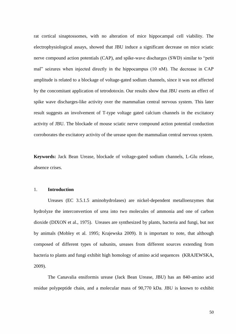
50
rat cortical sinaptossomes, with no alteration of mice hippocampal cell viability. The
electrophysiological assays, showed that JBU induce a significant decrease on mice sciatic
nerve compound action potentials (CAP), and spike-wave discharges (SWD) similar to “petit
mal” seizures when injected directly in the hippocampus (10 nM). The decrease in CAP
amplitude is related to a blockage of voltage-gated sodium channels, since it was not affected
by the concomitant application of tetrodotoxin. Our results show that JBU exerts an effect of
spike wave discharges-like activity over the mammalian central nervous system. This later
result suggests an involvement of T-type voltage gated calcium channels in the excitatory
activity of JBU. The blockade of mouse sciatic nerve compound action potential conduction
corroborates the excitatory activity of the urease upon the mammalian central nervous system.
Keywords: Jack Bean Urease, blockade of voltage-gated sodium channels, L-Glu release,
absence crises.
1. Introduction
Ureases (EC 3.5.1.5 aminohydrolases) are nickel-dependent metalloenzymes that
hydrolyze the interconvertion of urea into two molecules of ammonia and one of carbon
dioxide (DIXON et al., 1975). Ureases are synthesized by plants, bacteria and fungi, but not
by animals (Mobley et al. 1995; Krajewska 2009). It is important to note, that although
composed of different types of subunits, ureases from different sources extending from
bacteria to plants and fungi exhibit high homology of amino acid sequences (KRAJEWSKA,
2009).
The Canavalia ensiformis urease (Jack Bean Urease, JBU) has an 840-amino acid
residue polypeptide chain, and a molecular mass of 90,770 kDa. JBU is known to exhibit
51
insecticidal (Ferreira-DaSilva et al. 2000; Martinelli et al. 2014) and fungicidal (Becker-Ritt
et al. 2007; Carlini and Polacco 2008) activities.
In mammalians, the toxic effect of Canavalia ensiformis crude extract has been
assessed in rodents and showed to induce dyspnea, ataxia, hypothermia, coma, tonic
convulsions and death (Carlini et al., 1984). Its toxicity to murines is mainly devoid to the
presence of a toxic peptide named Canatoxin (CNTX) (Carlini et. al., 1984). The lethal and
convulsive effect of CNTX occurs in mice and rats and is observed after endogenous
administration (Carlini et al., 1984). The same effects are observed with JBU, although they
appear only by endovenous injection (FOLLMER et al., 2004). The observation of Straub
phenomena and the ability of reserpine, a drug which inhibits the vesicular monoamines
transporter (VMAT), to decrease the threshold of seizures, suggested the central nervous
system as the target for CNTX convulsive activity (Carlini et al., 1984). On the other hand, it
was also observed that the disruption of spinal column at T5 region, was able to inhibit the
seizures related to the hind limbs (lower limbs). In this regard, the development of Straub
phenomena requires an action of the sacro-coccygeus dorsalis muscle, and that it is also
necessary that the lumbo-sacral cord, with its peripheral nervous outflow, should be intact and
that these functioning units should have an adequate circulation (BILBEY; SALEM;
GROSSMAN, 1960).
Seizures are the clinical manifestation of an abnormal, excessive, hypersynchronous
discharge of a population of cortical neurons (Broomfield et al., 2006). Experimental studies
using animal epilepsy models have shown that NMDA, AMPA and kainate agonists induce
seizure activity, whereas their antagonists are suppressive modulators (Broomfield et al.,
2006). Thus, since glutamate is the main excitatory neurotransmitter at central nervous system
(CNS), it is the pivotal agent involved in seizures and other neurodegenerative disorders
(Meldrum, 1994; Platt, 2007).
52
In this work, we sought to investigate the central and peripheral electrophysiological
patterns of Jack Bean Urease in rodents, in vivo and in vitro, as well as the biochemical
correlation of cell viability and glutamate release. As far as our knowledge, this is the first
work to present the correlation between the EEG activity of Jack Bean Urease with cell
viability and the changes of glutamate release. With this on mind, we expected to contribute
to the understand of the ureases convulsive-like activities.
2. Materials and Methods
2.1 Animals
Adult male Swiss white mice and adult male Wistar rats were obtained from
PUCRS Animal House. The animals were housed in standard rodent cages in a colony
room maintained at 24 ºC under a 12 h light/12 h dark cycle with free access to food and
water.
The studies presented on this paper have been done in accordance with the
Brazilian Council for Animal Experimentation (CONCEA) guidelines and approved by
the local animal care committee (CEUA) with the protocol acceptance number 043/2015.
Experiments were designed to minimize the number of animals used and their suffering.
2.2 Reagents
All reagents used in these studies were of high purity, obtained from Sigma-
Aldrich Co. Brazil.
2.3 Biochemical assays
2.3.1 MTT Cell Viability Assay
53
2.3.1.1 Hippocampal Slices Preparation
Mice were decapitated, the brains removed immediately, and the hippocampus
dissected on ice and humidified in cold HEPES-saline buffer gassed with O2 (124mm NaCl,
4mM KCl, 1.2mM MgSO4,12mM glucose, 1mM CaCl2, and 25mM HEPES pH 7.4).
Hippocampal slices were obtained according to Vinadé and Rodnight (1996), briefly: a
Mcilwain tissue chopper was used to obtain the slices (400nm) that were separated and
preincubatedat 37 °C for 30min in microwell plates filled with HEPES saline (200 𝜇L/slice).
Subsequently, fresh medium was replaced (200 𝜇L/slice) for control condition and treatments
with JBU (1µM, 100nM, 10nM, 1nM, 100pM, 10pM and 1pM), and incubated for 1 hour and
30 minutes (37∘C).
2.3.1.2 Hippocampal Slices Viability
Immediately after incubation with different treatments, slices were assayed for a 3-
(4,5-dimethylthiazol-2-yl)-2,5-diphenyltetrazolium bromide (MTT) test (0.05% in HEPES-
saline) for 30 min (37°C). The MTT is converted into a purple formazan product after
cleavage of the tetrazolium ring by mitochondrial dehydrogenases. Formazan was dissolved
by the addition of DMSO, resulting in a colored compound whose optical density (𝜆 = 490
nm) was measured in an ELISA reader equipment. The MTT colorimetric assay was
performed as described by (DAL BELO et al., 2013).
2.3.2 Synaptosomes preparation
Male adult Wistar rats (200–250 g) were sacrificed by decapitation. The brain cortex
was removed and homogenized in 0.32 M sucrose solution containing dithiothreitol (0.25
mM) and EDTA (1 mM). Homogenates were then submitted to low-speed centrifugation
(1000 g for 10min) and the synaptossomes were purified from the supernatant by
54
discontinuous Percoll density gradient centrifugation (39,000 g for 15 min) (DUNKLEY;
JARVIE; ROBINSON, 2008). The isolated nerve terminals were resuspended in Krebs–
Ringer–HEPES (KRH) solution (124 mM NaCl, 4 mM KCl, 1.2 mM Mg2SO4, 10 mM
glucose, 25 mM HEPES, pH 7.4) to a final concentration of approximately 1 mg/ml. Aliquots
of 30 ml were prepared and kept on ice until use.
2.3.3 Measurement of continuous glutamate release
Glutamate release assessed by measuring the fluorescence increase due to the
production of NADPH in the presence of glutamate dehydrogenase and NADP+. This protocol
was performed essentially as described by (LOMEO et al., 2014). In short, the medium used
for the reaction contained a mixture of rat cortical synaptosomes (± 30 µg of protein/well) and
NADP+ (1 mM) in KRH (Krebs Ringer Hepes) was transferred to Elisa microplates (300
µl/well) to be read in a spectrofluorimeter (Synergy 2, Winooski, USA). After 1 min,
glutamate dehydrogenase (35 units per well) was added to the medium and the reading was
restarted until the fluorescence reached balance (about 10 min). Subsequently, treatments with
different concentrations of JBU were applied. Calibration curves were done in parallel by
adding known amounts of glutamate to the reaction medium. The experimental data were
expressed as nmol of glutamate released per mg of protein. The experiments were performed
at 37 ºC for 45 min continuous reading with 360 nm/450 nm excitation/emission wavelengths.
2.4 Electrophysiological recordings
2.4.1- In vitro recordings of compound action potentials from mouse sciatic
nerve preparation
Sciatic nerves were obtained from adult male Swiss mice (28 – 35g). The extraction
surgery was performed essentially as described by Bala et al. 2014. The mice sciatic nerves
55
were mounted on a recording chamber according to Dal Belo et al. 2005. Standard
extracellular recording techniques were used to record compound nerve action potentials.
Pellet-type silver electrodes were dipped into each of the three compartments of the recording
chambers, with stimulation occurring between the central and one of the external
compartments. Recordings were obtained from the central compartment. A Grass S48
stimulator was used to supply supramaximal electrical impulses (0.4 Hz, 0.04 ms duration)
via a model SIU 5A stimulus isolation unit (Grass Instrument Co.). The signals were
amplified with a CED1902 transducer (Cambridge Electronic Design, Cambridge, England),
digitized with a CED 1401 analogue-to-digital converter (Cambridge Electronic Design) and
analyzed with custom built software (Dempster, 1988). In each experiment, the amplitude,
rise time, latency and threshold of the action potentials recorded were measured. Prior to add
the test compounds, the sciatic nerve preparations were incubated in physiological solution for
15 min under constant supramaximal stimulation to demonstrate viability of the preparation
and consistency of the recordings.
2.4.2. In vivo rat electroencephalographic (EEG) Recordings
2.4.2.1. Implantation of animals for electroencephalographic recordings
Animals were anesthetized with ketamine 90 mg/kg and xylazine 13mg/kg. The
anesthesia was maintained by supplementary doses of ketamine/xylazine (30% of initial
dose), after checking tail pinch reflex and respiratory rate. Body temperature was maintained
at 37 ± 0.5 ºC using a heat pad. For the EEG recordings, A-M SYSTEMS .0045” tungsten
Teflon-insulated electrodes were employed. These electrodes were surgically implanted on
mPFC (3.0 mm anterior to bregma, 0.4 mm lateral to midline and 3.2 mm ventral to dura
mater); CA1 (5.7 mm anterior to bregma,4.6 mm lateral to midline and 2.5 mm ventral to dura
mater) and TMD (-1.9 mm anterior to bregma, -0.4 mm to midline and -4.8 mm ventral to
56
dura mater). The electrode implanting sites coordinates were followed according to Paxinos
and Watson (2007). At the hippocampal CA1 area, a chemitrode (electrode + cannula) was
implanted. The cannula was used for intrahippocampal injections of 10 nM JBU in a volume
of 2 µl. The skull was exposed, and holes were drilled so that the recording electrodes and the
chemitrode could be lowered at the mentioned stereotaxic coordinates. An additional hole was
drilled over the parietal to cortex to implant a microscrew that served as a recording reference
(ground). An additional electrode was used as miogram to detect the electrical activity of the
animal’s neck muscles in order to detect REM sleep, noise caused by body movements or
electrical oscilations. The apparatus employed in the EEG recordings is shown in Fig1.
2.4.2.2. Recording of SWDs
EEG was recorded by an electroencephalograph (NIHONCOHDEN, Japan, Tokyo)
attached to a data capture and analysis device (CED: Cambridge Electronic Design Ltd., UK,
Cambridge; POWER 1401 mkII). Sampling rate was 2000 Hz whereas the bandwidth of the
EEG recording was 0.3–150 Hz. Number of SWDs (frequency: 3–11 Hz; a train of
asymmetric spikes and slow waves starting and ending with sharp spikes; the average
amplitude at least twice as high as the basal EEG activity) (Kovács et al., 2006, 2015) and
time of SWDs (average time and total time of SWDs) were measured between 30 and 270
min of post-injection time (from 4.00 PM to 8.00 PM). Injections and handling induced stress
and behavioral alteration may change the SWD number Kovács et al., 2006, 2015). Thus, in
spite of that the changes in behavioral features after drug application disappeared within 30
min after injections and normal grooming was observed in all animals, data of the first 30 min
after the injections (from 3.30 PM to 4.00 PM) was excluded from the analysis (Kovács et al.,
2006). The recording periods were split into 60 min sections, which were evaluated
57
separately. SWDs were cut off from the raw data files and checked by FFT analysis (Kovács
et al., 2006).
EEG recordings were carried out in rats awaked without anesthesia during 6h in a recording
chamber containing food and water ad libitum. After 2h of basal recordings, the animals were
injected with 2µl of JBU 10nM by using a Hamilton syringe connected to a 30G dental needle
through a polypropylene flexible pipe. The needle was inserted into the cannula and stood
0,1mm above hippocampal CA1. Every microliter of JBU solution was injected in one minute.
2.5. Statistical analysis
For the MTT cell viability assay, the results were expressed as the mean ±SEM and
were compared statistically using ANOVA for repeated measures. Statistical analysis of the
data of glutamate release were made by the method of two-way ANOVA followed by
Bonferroni test. For compound action potentials recordings, the results were expressed as the
mean ±SEM and were compared statistically, using Student’s unpaired t-test.
3. Results
3.1 MTT Cell Viability Assay
On MTT Assay no meaningful cell loss could be verified according to our data (Fig.
2). Since MTT is based on mitochondrial dehydrogenases activity, our findings suggest no
mitochondrial dysfunctions.
3.2 L-Glutamate Liberation Assay
Rat brain cortex synaptosomes consist of isolated nerve terminals which still contain
the machinery related to neurotransmission and represent an excellent model for the study of
the effects of substances with potential activity in the central nervous system (CNS). The
58
analysis of L-Glu release in presence of different concentrations of JBU (1 nM, 10 nM, 20
nM, 50 nM and 100nM), showed a time-dependent significative increase in the release of L-
GLU (n=6) Fig.3. Thus, 33.34±8.56% maximum increase of JBU-induced L-Glu release was
observed with the concentration of 100nM in 45min readings (p<0.05) Table 1. Also, a
positive control in depolarizing conditions (KCl) was assayed for comparison.
3.3 Compound action potential (CAP)
The effect of JBU on compound action potentials was limited to significant changes in the
amplitude and rise time of the potentials. Fig. 4 displays JBU’s effect in different doses. This
result is summarized in Table 2.When 0.001 µM JBU was applied, the most significant
decrease (40) in CAP amplitude was observed (n = 6, p <0.05). The subsequent application of
Tetrodoxin (0.1µM) when applied after or before JBU in the preparation did not increase or
decrease the urease effect (Fig.5).
3.4 Electroencephalographic (EEG) Recordings
An intrahippocampal injection of JBU 10 nM, induced a 3 Hz generalized spike-wave
discharge as shown in Fig. 6. This suggests a non-convulsive seizure, a petit mal seizure,
since the animals did not respond to external sound stimulation or show any tonic clonic
convulsion.
3.4.1 Effects of Jack Bean Urease on mean number of SWDs
The intrusion criteria for SWDs in this study were that its duration should be more
than 3 seconds. At the end of recording we calculated numbers of SWDs that were more than
3 seconds. Numbers of SWDs in all groups are presented in Fig.7. There was a noticeable
number of SWDs in all tested areas, slightly higher but not statistically meaningful in CA1.
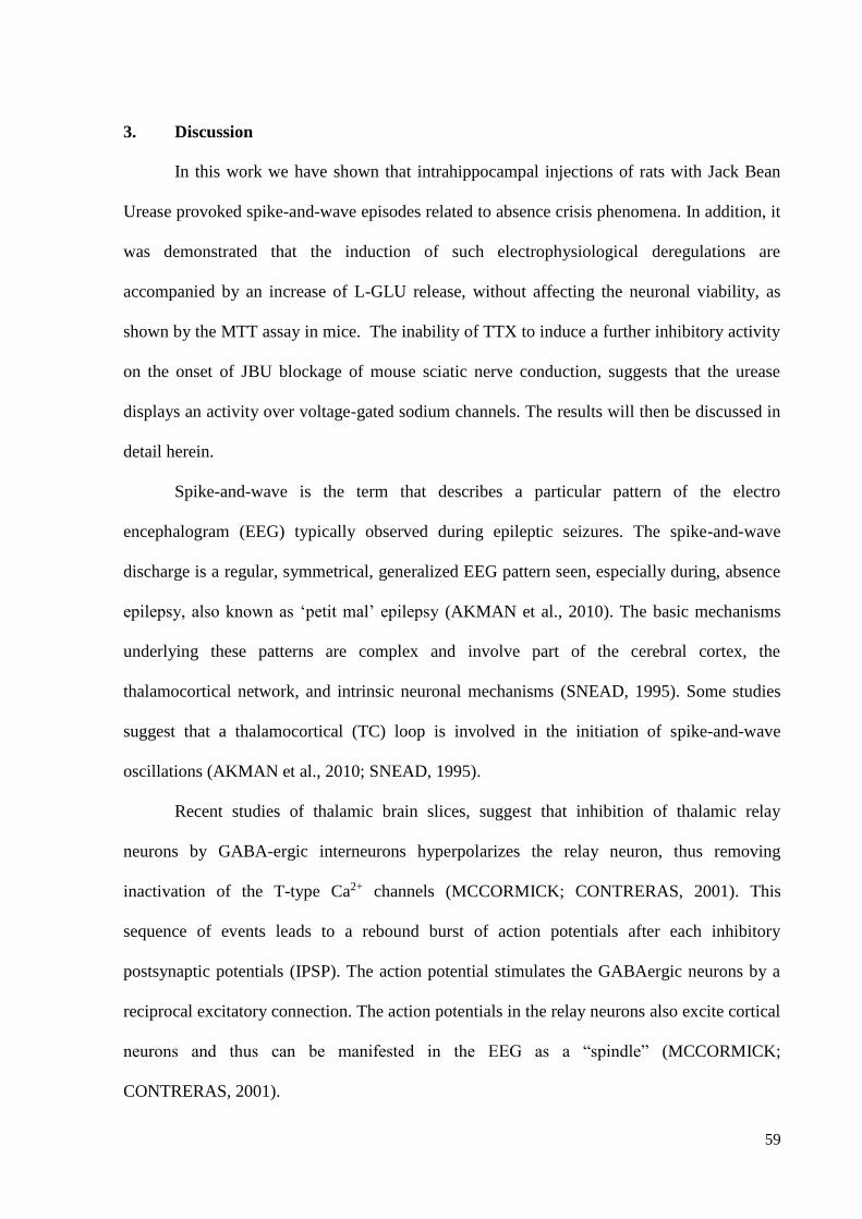
59
3. Discussion
In this work we have shown that intrahippocampal injections of rats with Jack Bean
Urease provoked spike-and-wave episodes related to absence crisis phenomena. In addition, it
was demonstrated that the induction of such electrophysiological deregulations are
accompanied by an increase of L-GLU release, without affecting the neuronal viability, as
shown by the MTT assay in mice. The inability of TTX to induce a further inhibitory activity
on the onset of JBU blockage of mouse sciatic nerve conduction, suggests that the urease
displays an activity over voltage-gated sodium channels. The results will then be discussed in
detail herein.
Spike-and-wave is the term that describes a particular pattern of the electro
encephalogram (EEG) typically observed during epileptic seizures. The spike-and-wave
discharge is a regular, symmetrical, generalized EEG pattern seen, especially during, absence
epilepsy, also known as ‘petit mal’ epilepsy (AKMAN et al., 2010). The basic mechanisms
underlying these patterns are complex and involve part of the cerebral cortex, the
thalamocortical network, and intrinsic neuronal mechanisms (SNEAD, 1995). Some studies
suggest that a thalamocortical (TC) loop is involved in the initiation of spike-and-wave
oscillations (AKMAN et al., 2010; SNEAD, 1995).
Recent studies of thalamic brain slices, suggest that inhibition of thalamic relay
neurons by GABA-ergic interneurons hyperpolarizes the relay neuron, thus removing
inactivation of the T-type Ca2+ channels (MCCORMICK; CONTRERAS, 2001). This
sequence of events leads to a rebound burst of action potentials after each inhibitory
postsynaptic potentials (IPSP). The action potential stimulates the GABAergic neurons by a
reciprocal excitatory connection. The action potentials in the relay neurons also excite cortical
neurons and thus can be manifested in the EEG as a “spindle” (MCCORMICK;
CONTRERAS, 2001).
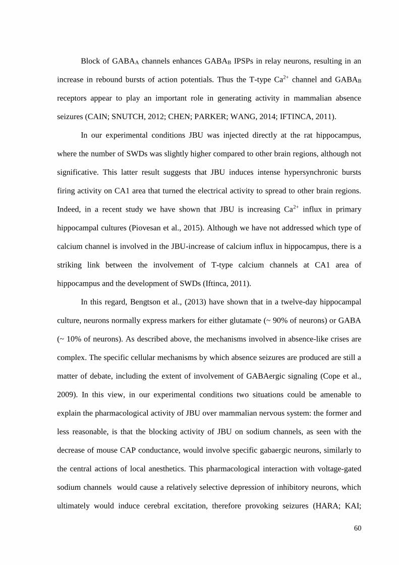
60
Block of GABAA channels enhances GABAB IPSPs in relay neurons, resulting in an
increase in rebound bursts of action potentials. Thus the T-type Ca2+ channel and GABAB
receptors appear to play an important role in generating activity in mammalian absence
seizures (CAIN; SNUTCH, 2012; CHEN; PARKER; WANG, 2014; IFTINCA, 2011).
In our experimental conditions JBU was injected directly at the rat hippocampus,
where the number of SWDs was slightly higher compared to other brain regions, although not
significative. This latter result suggests that JBU induces intense hypersynchronic bursts
firing activity on CA1 area that turned the electrical activity to spread to other brain regions.
Indeed, in a recent study we have shown that JBU is increasing Ca2+ influx in primary
hippocampal cultures (Piovesan et al., 2015). Although we have not addressed which type of
calcium channel is involved in the JBU-increase of calcium influx in hippocampus, there is a
striking link between the involvement of T-type calcium channels at CA1 area of
hippocampus and the development of SWDs (Iftinca, 2011).
In this regard, Bengtson et al., (2013) have shown that in a twelve-day hippocampal
culture, neurons normally express markers for either glutamate (~ 90% of neurons) or GABA
(~ 10% of neurons). As described above, the mechanisms involved in absence-like crises are
complex. The specific cellular mechanisms by which absence seizures are produced are still a
matter of debate, including the extent of involvement of GABAergic signaling (Cope et al.,
2009). In this view, in our experimental conditions two situations could be amenable to
explain the pharmacological activity of JBU over mammalian nervous system: the former and
less reasonable, is that the blocking activity of JBU on sodium channels, as seen with the
decrease of mouse CAP conductance, would involve specific gabaergic neurons, similarly to
the central actions of local anesthetics. This pharmacological interaction with voltage-gated
sodium channels would cause a relatively selective depression of inhibitory neurons, which
ultimately would induce cerebral excitation, therefore provoking seizures (HARA; KAI;
61
IKEMOTO, 1995). The second and more attractive possibility appears to be an increase of
gabaergic transmission by the blockage of voltage-gated sodium channels in glutamatergic
neurons, since these excitatory cells are more abundant in CNS. In this context, increased
GABAergic transmission may predominantly represent a compensatory response of the brain
in an attempt to decrease seizure propensity. However, enhanced GABA transmisson has been
reported paradoxically to promote seizures as well, at least in some specific types of epilepsy
models (Klaasen et al., 2006), such as absence epilepsy (Cope et al., 2009). However, how to
explain the JBU-increase of L-Glu release in brain synaptossomes? The explanation may
relay on the clinical observation that several antiepileptic drugs such as: phenobarbital,
benzodiazepines, phenytoin, carbamazepine, oxcarbazepine, valproate, ethosuximide among
others can exacerbate seizures, in which glutamate content is positively altered (OTOOM;
AL-HADIDI, 2000).
Finally, the inability of JBU to decrease the neuronal cell viability may suggests that
the amount of glutamate release during the onset of the toxin activity was not harmful to the
cells. Indeed a similar activity was observed by with pancreatic islets (Barja-Fidalgo et al.,
1991) and platelets (Carlini et al., 1985). This later result permit to infer that mitochondrial
dysfunctions are not associated with the JBU-induced seizure activity (Henshall 2007; Zsurka
and Kunz 2015).
Acknowledgments
The authors thank the Coodenação de Aperfeiçoamento de Pessoal de Nível Superior-
CAPES Edital Toxinologia 063/2010. Carlos Gabriel Moreira de Almeida was granted by
CAPES fellowship. The authors declare no conflicts of interest related to this work.
62
4. References
AKMAN, O. et al. Electroencephalographic differences between WAG/Rij and GAERS rat models of
absence epilepsy. Epilepsy Research, v. 89, n. 2-3, p. 185–193, 2010.
BALA, U. et al. Harvesting the maximum length of sciatic nerve from adult mice: a step-by-step
approach. BMC Research Notes, v. 7, n. 1, p. 714, 2014.
BECKER-RITT, A. B. et al. Antifungal activity of plant and bacterial ureases. Toxicon, v. 50, n. 7, p.
971–983, 2007.
BENGTSON, C. P. et al. Calcium responses to synaptically activated bursts of action potentials and
their synapse-independent replay in cultured networks of hippocampal neurons. Biochimica et
Biophysica Acta - Molecular Cell Research, v. 1833, n. 7, p. 1672–1679, 2013.
BILBEY, D. L. J.; SALEM, H.; GROSSMAN, M. H. The anatomical basis of the straub phenomenon.
Brit J Pharmacol, v. 15, p. 540–543, 1960.
CAIN, S. M.; SNUTCH, T. P. Voltage-gated calcium channels in epilepsy. Jasper’ s Basic
Mechanisms of the Epilepsies, 2012.
CARLINI, C. R.; POLACCO, J. C. Toxic Properties of Urease. Crop Science, v. 48, n. 5, p. 1665,
2008.
CHEN, Y.; PARKER, W. D.; WANG, K. The role of T-type calcium channel genes in absence
seizures. Frontiers in Neurology, v. 5 MAY, n. May, p. 1–8, 2014.
DAL BELO, C. A. et al. Pharmacological and structural characterization of a novel phospholipase A2
from Micrurus dumerilii carinicauda venom. Toxicon, v. 46, n. 7, p. 736–750, 2005.
DAL BELO, C. A. et al. In Vitro Antiophidian Mechanisms of Hypericum brasiliense Choisy
Standardized Extract: Quercetin-Dependent Neuroprotection. BioMed Research International, v.
2013, p. 1–6, 2013.
DIXON, N. E. et al. Letter: Jack bean urease (EC 3.5.1.5). A metalloenzyme. A simple biological role
for nickel? Journal of the American Chemical Society, v. 97, p. 4131–4133, 1975.
DUNKLEY, P. R.; JARVIE, P. E.; ROBINSON, P. J. A rapid Percoll gradient procedure for
preparation of synaptosomes. Nature protocols, v. 3, n. 11, p. 1718–1728, 2008.
FERREIRA-DASILVA, C. T. et al. Proteolytic activation of canatoxin, a plant toxic protein, by insect
cathepsin-like enzymes. Archives of insect biochemistry and physiology, v. 44, n. April, p. 162–
171, 2000.
FOLLMER, C. et al. Jackbean, soybean and Bacillus pasteurii ureases: Biological effects unrelated to
ureolytic activity. European Journal of Biochemistry, v. 271, n. 7, p. 1357–1363, 2004.
HARA, M.; KAI, Y.; IKEMOTO, Y. Local anesthetics reduce the inhibitory neurotransmitter-induced
current in dissociated hippocampal neurons of the rat. European Journal of Pharmacology, v. 283,
n. 1-3, p. 83–89, 1995.
HENSHALL, D. C. Apoptosis signalling pathways in seizure-induced neuronal death and epilepsy.
Biochemical Society transactions, v. 35, n. Pt 2, p. 421–423, 2007.
IFTINCA, M. C. Neuronal T-type calcium channels: what’s new? Iftinca: T-type channel regulation.
Journal of medicine and life, v. 4, n. 2, p. 126–138, 2011.
KRAJEWSKA, B. Ureases I. Functional, catalytic and kinetic properties: A review. Journal of
Molecular Catalysis B: Enzymatic, v. 59, n. 1-3, p. 9–21, 2009.
63
LOMEO, R. DA S. et al. Crotoxin from Crotalus durissus terrificus snake venom induces the release
of glutamate from cerebrocortical synaptosomes via N and P/Q calcium channels. Toxicon, v. 85, p.
5–16, 2014.
MARTINELLI, A. H. S. et al. Structure-function studies on jaburetox, a recombinant insecticidal
peptide derived from jack bean (Canavalia ensiformis) urease. Biochimica et Biophysica Acta -
General Subjects, v. 1840, n. 3, p. 935–944, 2014.
MCCORMICK, D. A.; CONTRERAS, D. O N T HE C ELLULAR AND N ETWORK B ASES. n. 1,
2001.
MOBLEY, H. L.; ISLAND, M. D.; HAUSINGER, R. P. Molecular biology of microbial ureases.
Microbiological reviews, v. 59, n. 3, p. 451–480, 1995.
OTOOM, S.; AL-HADIDI, H. Seizure induced by antiepileptic drugs. Annals of Saudi Medicine, v.
20, n. 3-4, p. 316–318, 2000.
PLATT, S. R. The role of glutamate in central nervous system health and disease - A review.
Veterinary Journal, v. 173, n. 2, p. 278–286, 2007.
SNEAD, O. C. Basic mechanisms of generalized absence seizures. Annals of Neurology, v. 37, n. 2,
p. 146–157, 1995.
VINADÉ, L.; RODNIGHT, R. The Dephosphorylation of Glial Fibrillary Acidic Protein (GFAP) in
the Immature Rat Hippocampus is Catalyzed Mainly by a Type 1 Protein Phosphatase. Brain
research, v. 732, p. 195–200, 1996.
WONG, M. Too much inhibition leads to excitation in absence epilepsy. Epilepsy currents /
American Epilepsy Society, v. 10, n. 5, p. 131–132, 2010.
ZSURKA, G.; KUNZ, W. S. Mitochondrial dysfunction and seizures: the neuronal energy crisis. The
Lancet Neurology, v. 14, n. 9, p. 956–966, 2015.
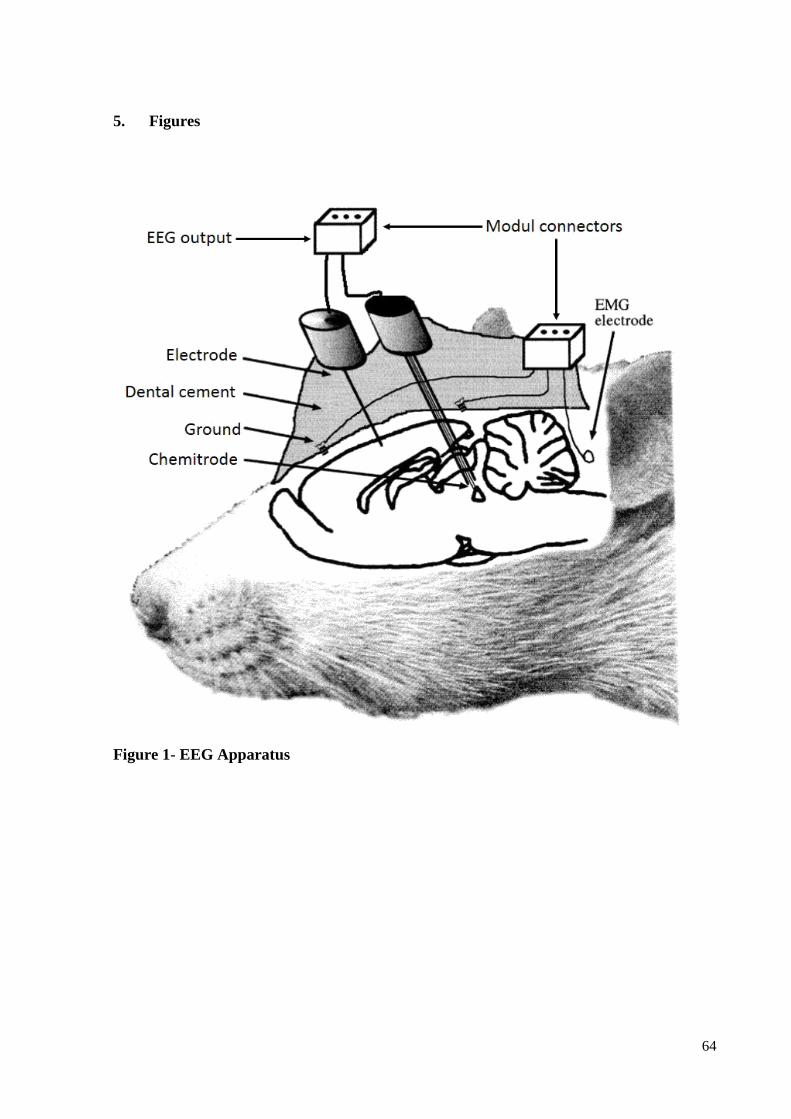
64
5. Figures
Figure 1- EEG Apparatus
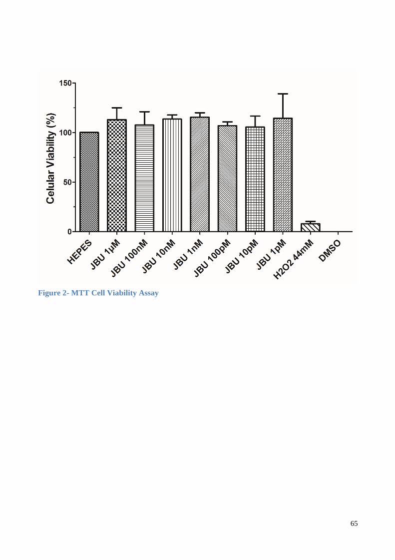
65
Figure 2- MTT Cell Viability Assay
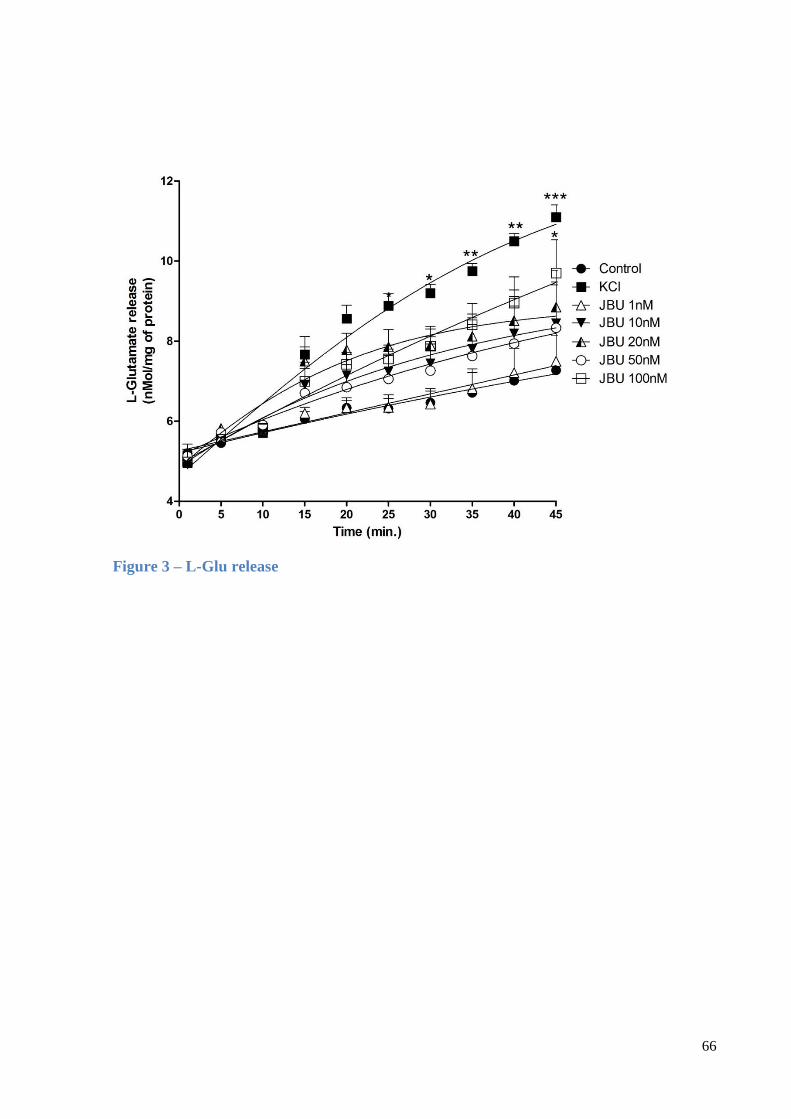
66
Figure 3 – L-Glu release
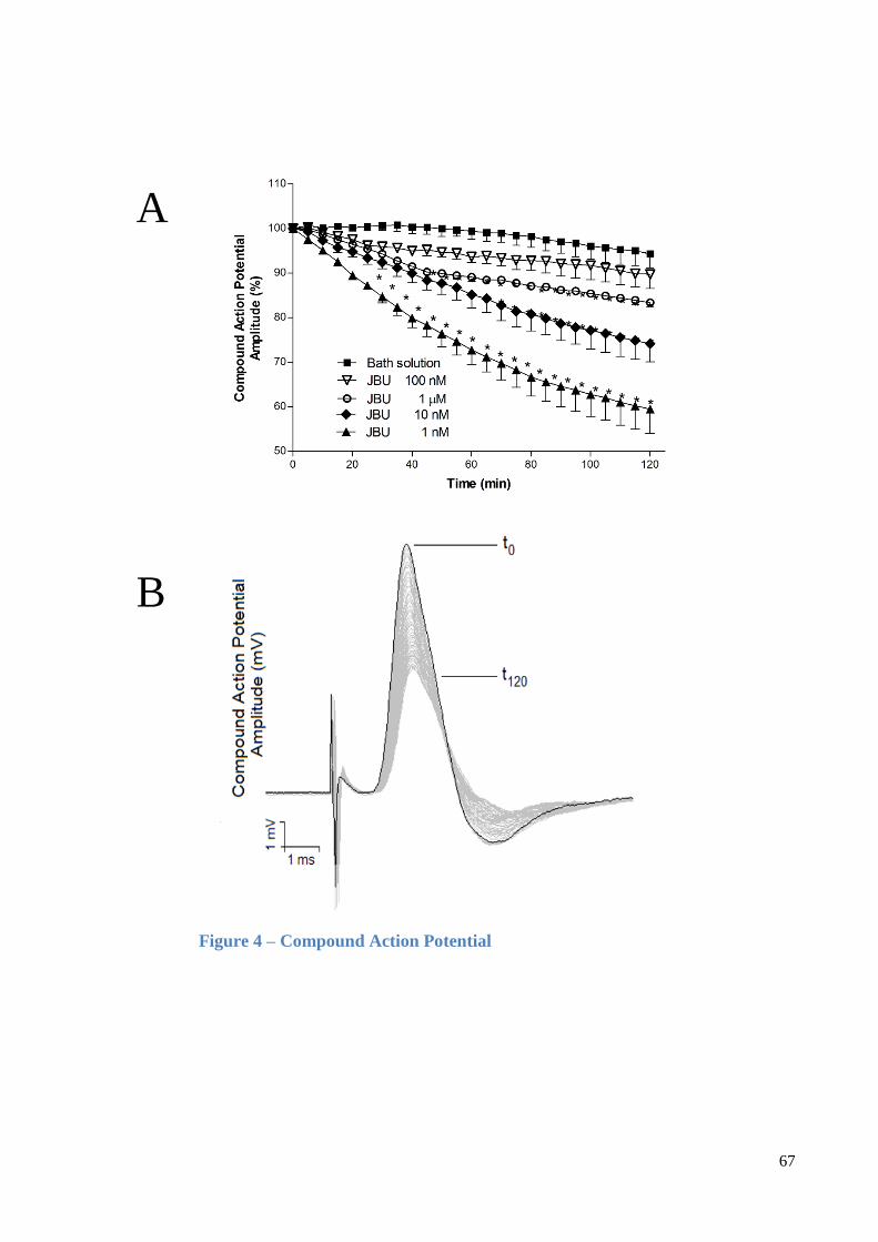
67
A
B
Figure 4 – Compound Action Potential
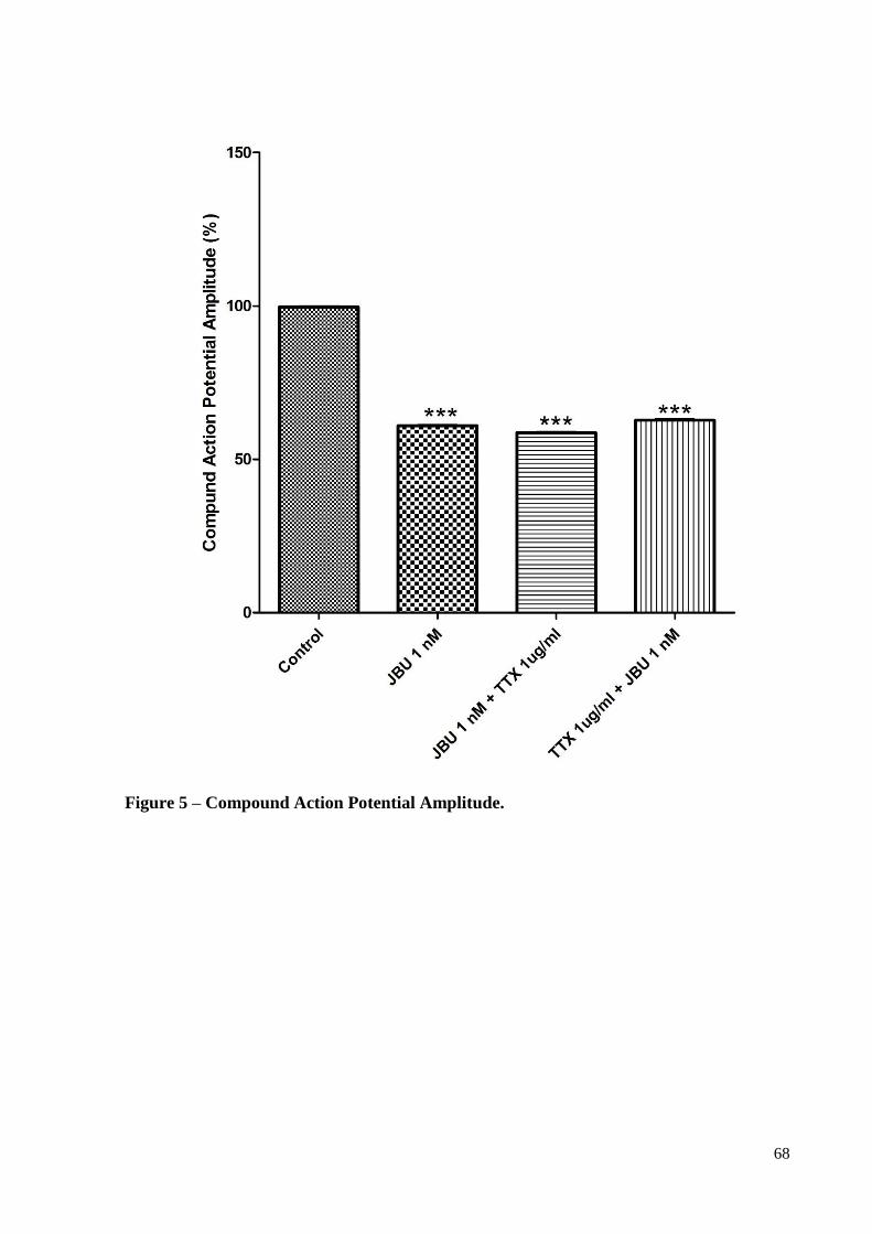
68
Figure 5 – Compound Action Potential Amplitude.
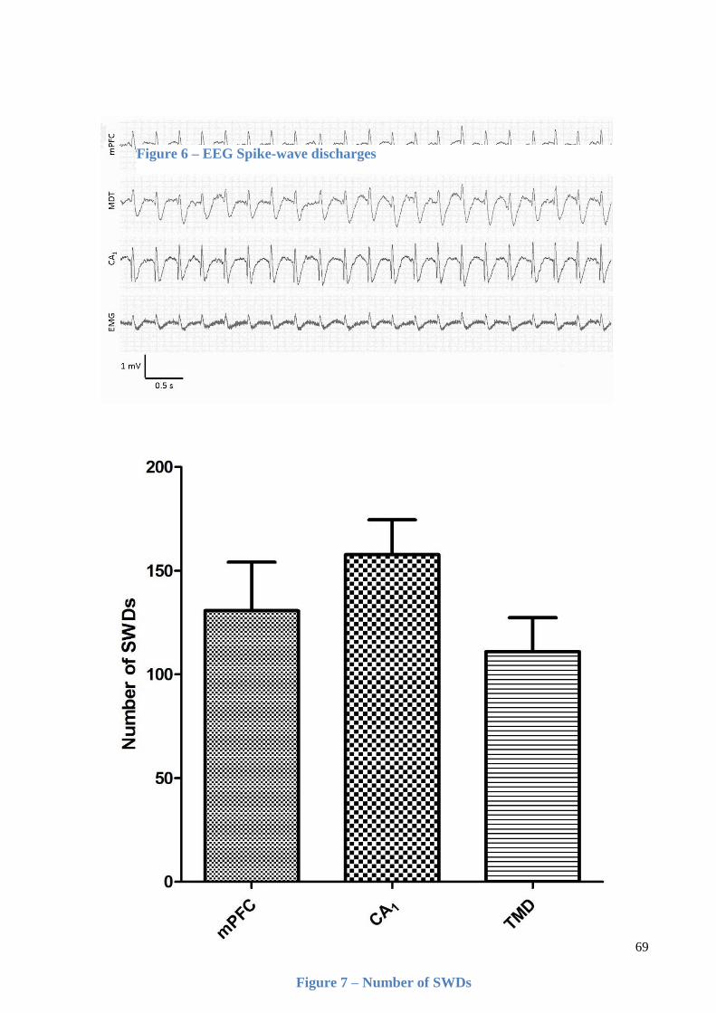
69
Figure 6 – EEG Spike-wave discharges
Figure 7 – Number of SWDs
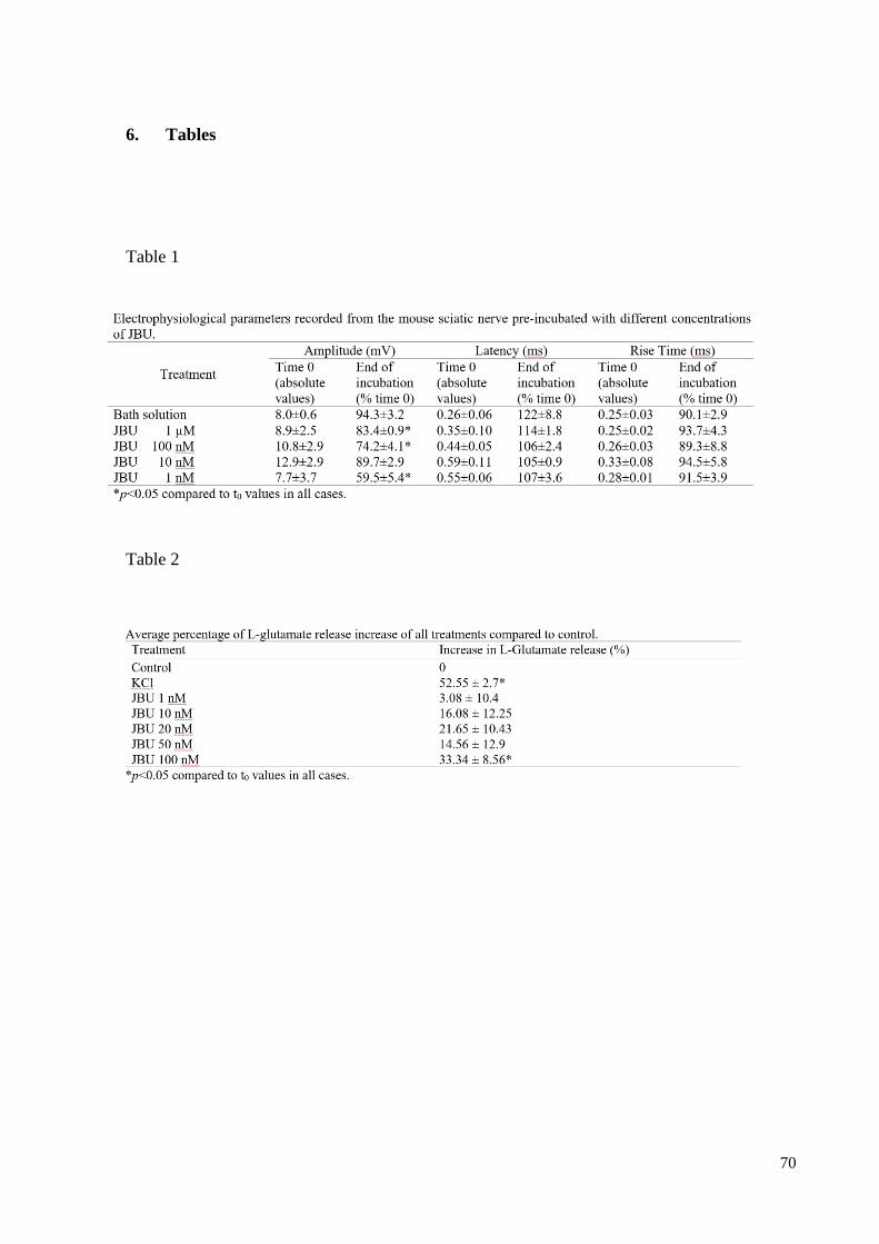
70
6. Tables
Table 1
Table 2


![Interação de Alquilfosfolipídios de atividade anti ... · A ação de ALPs é preferencialmente via a membrana celular [1]. Há um interesse crescente na sua atividade biológica,](https://img.pdfslide.tips/doc/110x75/5e9684e2f0a4db43a3642ecb/interao-de-alquilfosfolipdios-de-atividade-anti-a-ao-de-alps-preferencialmente.jpg)