Embed Size (px)
Citation preview

Università degli Studi di Ferrara
DOTTORATO DI RICERCA IN "Farmacologia e Oncologia Molecolare"
CICLO XXIII
COORDINATORE Prof. Antonio Cuneo
OSTEOPROTEGERIN: A PANCREATIC
ISLETS DYSFUNCTION AND
VASCULAR INJURY MODULATOR
Settore Scientifico Disciplinare BIO/16
Dottoranda Tutore
Dott.ssa Toffoli Barbara Prof.ssa Secchiero Paola
Anni 2008/2010

TABLE OF CONTENTS
1. INTRODUCTION
1.1 TNF-Superfamily page 1
1.2 OPG structure and expression page 3
1.3 OPG as a ‘decoy receptor’ page 5
1.3.1 OPG/RANKL and bone system page 5
1.3.2 OPG/TRAIL and tumorigenesis page 7
1.4 OPG and the vascular system page 9
1.5 OPG and diabetes page 11
1.6 Atherosclerosis and animal models page 13
1.7 Renin-angiotensin system and diabetes development page 18
1.7.1 Systemic versus local pancreatic RAS page 18
1.7.2 Physiological role of the RAS in the endocrine pancreas page 20
1.7.3 RAS and beta cell dysfunction page 20
1.7.4 RAS inhibition and prevention of type 2 diabetes page 23
2. RATIONALE AND OBJECTIVES page 25
MATERIALS AND METHODS
3. STUDY 1.A-B page 27
3.1 Animals and experimental protocol page 27
3.2 Evaluation of atherosclerotic plaques page 28
3.3 Masson’s trichrome staining page 28
3.4 Immunohistochemistry page 28
3.5 Real-time quantitative PCR page 29
3.6 Cell cultures page 29
3.7 Immunofluorescence page 30
3.8 Osteoprotegerin ELISA page 30
3.9 Statistical analysis page 31
4. STUDY 2. page 34
4.1 Animals and experimental protocol page 34
4.2 Measurement of physiological and biochemical parameters page 35
4.3 Oral glucose tolerance test page 35

4.4 Picrosirius red staining page 35
4.5 Immunohistochemistry page 35
4.6 In situ detection of apoptosis page 36
4.7 Real-time quantitative PCR page 37
4.8 Statistical analysis page 37
RESULTS
5. STUDY 1.A-B page 39
5.1 Increase of atherosclerotic plaque development in diabetic apoE-/-
mice after in vivo administration of full-length recombinant human
OPG page 39
5.2 Recombinant human OPG injected in vivo in apoE-/-
diabetic mice
induces changes in the histological composition of the atherosclerotic
plaques page 41
5.3 Increase of the mRNA expression of angiopoietin 2 in the aorta of
apoE-/-
diabetic animals after in vivo administration of recombinant
human OPG page 44
5.4 Induction of primary rodent VSMC proliferation by recombinant
human OPG page 44
5.5 The in vivo injection of recombinant human OPG modulates the
histological composition of aortic media in apoE-/-
mice page 46
5.6 TGFβ stimulates OPG production in mouse primary VSMC page 47
5.7 Recombinant full-length OPG increases the mRNA expression of NF-
kB, TGFβ and pro-fibrotic markers in mouse primary VSMC page 48
5.8 Recombinant full-length OPG increases both OPG protein and mRNA
expression in mouse primary VSMC page 50
6. STUDY 2. page 51
6.1 In vivo injection of low concentrations of full-length recombinant
human OPG induces a glycemic increase and an insulin levels
reduction in C57Bl/6J mice page 51
6.2 In vivo injection of recombinant human OPG promotes a reduction in
insulin staining density and in beta cell mass in C57Bl/6J mice page 53
6 weeks old apo E-null male mice ST
Z
R
(weeks
rOP
G
rOP
G
rOP
G
6 weeks old apo E-null male mice ST
Z
R
(weeks
rOP
G
rOP
G
rOP
G

6.3 In vivo administration of recombinant human OPG induces alterations
in the histological composition of the pancreatic islets in C57Bl/6J
mice page 53
6.4 Treatment with recombinant human OPG increases pancreatic mRNA
expression of MCP-1, VCAM-1, TGF and CTGF in C57Bl/6J mice page 56
6.5 In vivo injection of human recombinant OPG induces an activation of
the local pancreatic RAS in C57Bl/6J mice page 56
7. DISCUSSION page 58
7.1 OPG and diabetes associated atherosclerosis page 59
7.2 OPG and pancreatic beta cells dysfunction page 62
7.3 Perspective page 64
8. LIST OF PUBLICATIONS 2008-2010 page 66
9. REFERENCES page 67

1
INTRODUCTION
1.1 TNF-Superfamily
The Tumor Necrosis Factor (TNF) superfamily of cytokines and their receptors regulates
many areas of metazoan biology, and specifically plays foundamental roles in regulating
myriad aspects of immune development and functions (Chan, 2007). The biological functions
of this system encompass beneficial effects for the host in inflammation and protective
immune responses in infectious diseases as well as crucial roles in organogenesis of
secondary lymphoid organs and the maintenance of lymphoid structures throughout the body.
At the same time, some members of this superfamily are responsible for host damaging
effects in sepsis, fever syndromes, cachexia, and autoimmune diseases (e.g., psoriasis,
inflammatory bowel disease, rheumatoid arthritis) (Hehlgans and Pfeffer, 2005; Aggarwal,
2003; Ware, 2003; Kwon et al., 2003).
In 1984, two forms of TNF, TNFα and LTα (lymphotoxin, TNFβ), were isolated from
activated macrophages and T cells, respectively. Since their identification, these proteins have
become representative of a unique superfamily of ligands, which currently includes at least 18
different human homologues (Baker and Reddy, 1998; MacEwan, 2008). The TNF-related
ligands are type II transmembrane proteins (intracellular N-terminus) with a short cytoplasmic
tail (15 to 25 residues in length) and a larger extracellular region (approximately 150 amino
acids) containing the signature ‘TNF homology domain’ (THD) where the receptor-binding
sites are located (Figure 1). The THD, that shares approximately 20-25% of sequence identity
between family members, folds into an antiparallel β-sandwich that assembles into trimers,
the functional unit of the ligand (Paul, 2008).
Some of these ligands, e.g. TNF, are active both as a membrane integrated and as a soluble
form released after proteolytic cleavage, mainly by metalloproteinases induced by various
stimuli. Certain ligands are expressed only as soluble molecules, e.g. LTα; but may also be
recruited to the cell membrane to form heterotrimeric membrane anchored complexes and
thereby enhancing regulatory specificity and complexity (Hehlgans and Pfeffer, 2005).
Thus far, 29 TNF receptor family members have been identified in humans (Figure 1). These
are primarily type I transmembrane proteins characterized by cysteine-rich domains (CRD)
that are the hallmark of the TNF superfamily receptors (TNFR). These 40 amino acid
pseudorepeats are typically defined by 3 intrachain disulphides generated by 6 highly
conserved cysteine residues within the receptor chains (Locksley et al., 2001). Most receptors

2
also exist in a soluble form, and the solubility is achieved by proteolitic cleavage or by
alternative splicing of the exon encoding the transmembrane domain (Baker and Reddy,
1998).
Figure 1. The TNF/TNFR superfamily. The TNF-related ligands are shown in blue and arrows indicate
interactions with their receptors (Hehlgans and Pfeffer, 2005).
Although most members of the TNF superfamily of ligands interact with more than one
receptor of the corresponding superfamily of cognate receptors, genetic based approaches,
mainly conducted in gene targeted mouse strains, have clearly demonstrated that almost each
receptor-ligand system have a unique and non-redundant function.
Receptor activation by the TNF family ligands causes recruitment of several intracellular
adaptor proteins which activate multiple signal transduction pathways. Based on their
intracellular sequences the members of the TNFR superfamily can be classified into three
major groups. The first group, including molecules such as FAS and TNFRI, contains the so
called death domains (DD) in their cytoplasmic domains. Activation of these receptors leads
to recruitment of intracellular death domain containing adaptors such as FAS-associated death
domain (FADD) and TNFR-associated death domain (TRADD), which subsequently promote
the activation of the caspase cascade and induction of apoptosis. The second group of
3
receptors, that includes members like CD30, CD40 and receptor activator of NF-kB (RANK),
contains one or more TNF-receptor associated factor (TRAF)- interacting motif (TIMs) in
their cytoplasmic domain. Activation of TIM containing TNFR family members leads to the
recruitment of TRAF family members and the subsequent activation of signal transduction
pathways like nuclear factor kB (NF-kB), Jun N-terminal kinase (JNK), p38, extracellular
signal-related kinase (ERK) and phosphoinositide 3-kinase (PI3K), which regulate cellular
processes ranging from proliferation and differentiation to cell death. Finally, the third group
of receptors, including TRAIL-R3 (DcR1), DcR3 and Osteoprotegerin, doesn’t contain
functional intracellular signaling domains or motifs, but instead compete with the other two
groups of receptors for their corresponding ligands (Hehlgans and Pfeffer, 2005, Dempsey et
al., 2003).
1.2 OPG structure and expression
Osteoprotegerin (OPG) is a secreted glycoprotein belonging to the TNFR superfamily and
was initially identified by two separate groups in 1997. Both groups observed OPG to be
central in the regulation of bone turnover through the inhibition of osteoclastogenesis.
However, it was not until 1998 that the newly discovered proteins were found to be identical,
hence OPG was alternatively termed osteoclast inhibitory factor (OCIF) (Reid and Holen,
2009). Its international name according to the TNF nomenclature is TNFRSF11B (Baud’huin
et al., 2007).
The mouse and the human OPG genes have been cloned and characterized, and the human
OPG gene is a single-copy gene that consists of five exons and spans 29 kb of the human
genome located on chromosome 8 q23-24 (Morinaga et al, 1998; Mizuno et al.,a 1998).
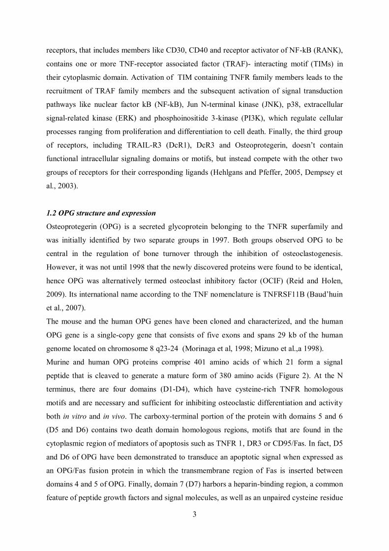
Murine and human OPG proteins comprise 401 amino acids of which 21 form a signal
peptide that is cleaved to generate a mature form of 380 amino acids (Figure 2). At the N
terminus, there are four domains (D1-D4), which have cysteine-rich TNFR homologous
motifs and are necessary and sufficient for inhibiting osteoclastic differentiation and activity
both in vitro and in vivo. The carboxy-terminal portion of the protein with domains 5 and 6
(D5 and D6) contains two death domain homologous regions, motifs that are found in the
cytoplasmic region of mediators of apoptosis such as TNFR 1, DR3 or CD95/Fas. In fact, D5
and D6 of OPG have been demonstrated to transduce an apoptotic signal when expressed as
an OPG/Fas fusion protein in which the transmembrane region of Fas is inserted between
domains 4 and 5 of OPG. Finally, domain 7 (D7) harbors a heparin-binding region, a common
feature of peptide growth factors and signal molecules, as well as an unpaired cysteine residue

4
at position 400 required for homodimerization of the molecule (Zauli et al., 2009; Schoppet et
al., 2002). OPG represents an atypical member of the TNFR family since it is a secreted
protein with no transmembrane domain. OPG is produced as a monomer (55-62 kDa),
undergoes homodimerization, and is secreted as a disulfide linked homodimeric glycoprotein
with four or five potential glycosylation sites, generating a mature form of OPG of 110-120
kDa. The monomeric and the homodimeric forms are indistinguishable in stability, but the
homodimeric exerts more potent biological activity and is stronger in heparin binding ability
(Zauli et al., 2009; Tomoyasu et al., 1998).
Figure 2. Schematic representation of the protein structure of OPG and OPG-Fc. Main domains and their biochemical and/or functional properties are indicated. Numbers in figure represent amino acids. NH2 indicates
N terminus, COOH, C terminus, Cys400, dimer formation site. Human OPG-Fc is a recombinant fusion protein,
in which the Fc fragment of human IgG1 is fused to the C terminus of the 22-194 fragment of native OPG to
maintain a dimeric molecule with a sustained circulating half-life (Zauli et al., 2009).
To enhance the pharmacological activity of native OPG, numerous constructs have been
created wherein the signal peptide, heparin binding domain, and death domains were removed
and the remaining peptide was fused to the Fc domain of human IgG1 (Figure 2). The Fc
fusion partner maintains the potent dimeric nature of OPG while significantly increasing its
circulating half-life (Kearns et al., 2008).
OPG is produced by a variety of tissues including the cardiovascular system (hearth, arteries,
veins), lung, kidney, intestine, and bone, as well as hematopoietic and immune cells. The
expression and production of the protein is modulated by various cytokines, peptides,
hormones, and drugs. Cytokines, including TNFα, interleukin (IL)-1α, IL-18, transforming
growth factor (TGFβ), bone morphogenetic proteins, and steroid hormones are known to up-
regulate OPG mRNA levels. In contrast, glucocorticoids (known to promote bone resorption)
and the immunosuppressant cyclosporine A (which has the propensity to cause osteoporosis

5
and vascular disease) or basic fibroblast growth factor, all suppress the expression of OPG
(Schoppet et al., 2002).
1.3 OPG as a ‘decoy receptor’
OPG acts as a decoy receptor for RANKL, preventing the stimulation and maturation of
osteoclast precursors instigated by normal binding of RANKL to its constitutive receptor and
as a soluble receptor for TRAIL (Emery et al, 1998).
1.3.1 OPG/RANKL and bone system
The important role of OPG in bone metabolism has been clearly demonstrated by the
development of transgenic and knock-out mice. OPG knock-out mice are viable and fertile,
but exhibit an osteoporotic phenotype due to enhance osteoclastogenesis. They are
characterized by marked bone loss accompanied by destruction of growth plate and lack of
trabecular bone in their femurs, and the strength of their bones dramatically decrease (Mizuno
et al.,b 1998). In contrast, systemic delivery of OPG via the expression of rat or murine opg
transgenes results in severe yet nonlethal osteopetrosis. The ostopetrotic phenotype caused by
OPG overexpression differs significantly from those observed in other ostopetrotic models.
opg transgenic mice are of normal size, have no apparent defects in tooth eruption, and have
normally shaped bones. Histologically, they have a marked reduction in trabecular osteoclast
but no deficiency of osteoclast precursors, suggesting a defect in the later stages of osteoclast
differentiation (Simonet et al., 1997). These evidences, supported by confirmations in the in
vitro experiments, demonstrate that the presence of OPG is absolutely required to maintain
bone mass in physiological situations. Subsequently, molecular binding experiments have
shown that OPG associates with the ligand of the receptor activator of NF-kB (RANKL), a
member of TNF superfamily ligand, functioning as a decoy receptor. RANKL genes gives
rise to splice variants that encode two forms of type II transmembrane proteins and one form
of a secreted protein. Although high RANKL expression can be found in lymph nodes,
thymus and lung, only low levels of RANKL can be detected in spleen, bone marrow,
peripheral blood, leukocytes, hearth, placenta, skeletal muscle, stomach or the thyroid. In
addition, RANKL expression is induced in mammary gland epithelial cells in pregnancy,
activated T cells, and malignant tumor cells (Wong et al., 1999; Wada et al., 2006).
The receptor that mediates all known activities for RANKL is called receptor activator of NF-
kB (RANK). The binding and activation of RANK, that is a homotrimeric TNFR family
member, involve direct interactions between the extracellular receptor binding domain of

6
trimeric RANKL and the extracellular cysteine-rich domains of trimeric RANK. This
interaction is thought to cause oligomerization of RANK and the subsequent activation of
several signal transduction cascades, as the NF-kB, MAPK, and phosphatidylinositol
pathways (Kearns et al., 2008; Wada et al., 2006).
One of the best characterized role of RANKL/RANK system is in the osteoclastogenesis. The
key regulators of bone turnover are osteoblasts, which are involved in bone formation, and
osteoclasts, which are responsible for bone resorption. RANKL expressed by osteoblasts as a
transmembrane protein binds to RANK on the surface of osteoclasts and osteoclasts
precursor. This leads to osteoclast formation, differentiation, activation and consequently
bone resorption (Figure 3). To regulate the balance between bone formation and bone
resorption, the RANKL-RANK interaction is inhibited by OPG. OPG produced by
osteoblastic cells binds as a homodimer to the homotrimeric RANKL, thus inhibiting the
terminal stage of osteoclastic differentiation (Reid and Holen, 2009; Baud’huin et al., 2007).
Although there are contradictory data, in general upregulation of RANKL is associated with
downregulation of OPG, or at least lower induction of OPG, such that the ratio of RANKL to
OPG changes in favour of osteoclastogenesis. Many reports have supported the assertion that
the RANKL/OPG ratio is a major determinant of bone mass. Moreover, consistent with the
osteoprotective role, mutation in the human OPG gene have been associated with idiopathic
hyperphosphatasia, also known as Juvenile Paget’s disease, an autosomal-recessive disorder
characterized by increased bone remodeling, osteopenia, and fractures (Boyce and Xing,
2007).
Figure 3. In (A) Schematic representation of RANKL in osteoclastogenesis (Zauli et al., 2009). In (B)
RANK signaling pathways (Theoleyre et al., b 2004).

7
1.3.2 OPG/TRAIL and tumorigenesis
In 1995 TNF-related apoptosis-inducing ligand (TRAIL) was identified and characterized as a
member of the TNF family of death-inducing ligands. TRAIL is a type II transmembrane
protein of about 33-35 kD, which can be cleaved from the cell surface to form a soluble
ligand that retains biological activity. The extra-cellular domain of TRAIL forms a bell
shaped homo-trimer, much like other ligands of the TNF family, but unlike the other
members, it carries a zinc ion at the trimer interface, coordinated by the single unpaired
cysteine residue of each monomer. This zinc ion is essential for structural integrity of TRAIL
and to maintain its capacity to induce apoptosis (Degli-Esposti, 1999; Corallini et al., 2008).
TRAIL is constitutively present in many tissues at the level of mRNA, but it is expressed
mainly by activated cells of the immune system, especially natural killer (NK) cells, B cells, T
cells, monocytes, and dendritic cells. TRAIL plays a crucial role in maintaining T cell
homeostasis, as well as in killing of tumor and virally transformed cells by NK cells. Over the
years, TRAIL has generated considerable interest among clinicians for its preferential toxicity
toward transformed cells and tumor xenografts, with generally little or no toxicity to normal
tissues, which makes it an ideal candidate for cancer therapy. As a result, recombinant
TRAIL, as well as agonistic antibodies against TRAIL receptors, are currently in Phase I/II
clinical trials for treatment of solid tumors and hematological malignancies (Griffith et al.,
2009; Guicciardi and Gores, 2009; Ashkenazi and Herbst, 2008; Finnberg and El-Deiry,
2008).
With respect to other members of the TNF ligand superfamily, TRAIL shows the most
complex ligand-receptor interaction, since it is able to bind to five different receptors found
on a variety of cell types: four membrane-bound (TRAIL-R1/death receptor 4, TRAIL-
R2/death receptor 5, TRAIL-R3/decoy receptor 1, TRAIL-R4/decoy receptor 2) and one
soluble receptor (OPG). OPG acting as a decoy receptor, binds TRAIL and prevents its
interaction with the functional death receptors, thus allowing cells to escape cell death (Figure
4). In this context, many different in vitro data demonstrated the ability of native OPG,
produced by tumor cells or by bone marrow stromal cells, to efficiently counteract the pro-
apoptotic activity of TRAIL in a variety of cell lines derived from prostate and breast cancers,
ameloblastomas and multiple myeloma (Zauli et al., 2009; Reid and Holen, 2009).

8
Of note, in the light of recent in vitro studies that demonstated that the affinity of OPG for
RANKL and TRAIL under physiological conditions is of a similar order of magnitude and
that TRAIL is able to inhibit OPG-mediated inhibition of osteoclastogenesis, OPG acquires a
key position in the regulation of the functions of these two important signaling pathways
(Figure 5) (Vitovski et al., 2007).
Recently, many studies have demonstrated that OPG can exert direct biological activities
independently of its neutralizing effects towards RANKL or TRAIL. In fact OPG has a highly
basic heparin-binding domain (D7) that besides being responsible for the homodimerization
of the molecule (Yamaguchi et al., 1998), makes interactions with heparin and heparan
sulfates possible. Heparan sulfate proteoglycans are important participants in cell-surface
Figure 4. Role of OPG in cell survival. TRAIL is produced by immune cells that
can infiltrate the tumor microenvironment.
TRAIL can then bind to the death
receptors 4 and 5 (DR4 and DR5) present
on the surface of tumor cells, resulting in
tumor cell apoptosis. OPG, secreted by
tumor cells, acts as a decoy receptor for TRAIL. As a result, tumor cells escape
from death (Reid and Holen, 2009).
Figure 5. Mechanism of action of OPG
on RANKL- and TRAIL- biological
activities. The pro-apoptotic activity of
TRAIL is mediated by two (TRAIL-R1 and
TRAIL-R2) of its four membrane
receptors. OPG by efficiently binding RANKL and TRAIL, counteracts both the
RANKL-mediated osteoclastogenesis as
well as the pro-apoptotic activity of TRAIL
(Zauli et al., 2009).

9
signaling and critical in controlling cell behavior. They are involved in actin cytoskeleton
regulation, cell adhesion and migration, and modulation of specific receptor interactions. At
the cell surface, proteoglycans of the syndecan family are the major source of heparan sulfate
(Wright et al., 2009). In this context, different reports indicate that the heparin binding
domain is involved in OPG-induced chemotaxis of human peripheral blood monocytes
(Mosheimer et al., 2005), in controlling OPG release by vascular cells (Nybo and Rasmussen,
2008) and in the OPG-mediated osteopontin-increasing in human periodontal ligament cells
(Yongchaitrakul et al., 2009). Of note, in multiple myeloma (Standal et al., 2002) and
osteosarcoma (Lamoureux et al., 2009) it has been shown that also the decreased of the
biological activity of the full-length OPG might be due to its bounding, internalization and
degradation, syndecan-1 mediated.
1.4 OPG and the vascular system
While OPG, RANK and RANKL are produced by numerous cell types and a variety of
tissues, their expression pattern targets three main biological systems where the molecular
triad could be more specifically involved: the osteoarticular, immune, and vascular systems
(Figure 6). Transgenic and knockout mice models as well as many in vitro experiments
clearly revealed the potential involvement of these effectors in the three biological systems
(Theoleyre et al., b 2004; Feige, 2001; Grĉević et al., 2001; Hofbauer et al., 2001; Josien et
al., 2000; Schoppet et al., 2002).
Figure 6. OPG/RANK/RANKL as common effectors of bone, immune and vascular system (Theoleyre et
al., b 2004).

10
In fact, the first evidence for a role of OPG in the vasculature was given by a study of OPG
knockout mice generated on a mixed genetic background. The selective deletion of OPG in
mice resulted in early-onset severe osteoporosis as well as significant medial calcification of
the aorta and renal arteries. The onset of arterial calcification could be completed prevented
by transgenic OPG delivery from mid gestation through adulthood; in contrast, post-natal
intravenous injection of recombinant OPG had no effect on the incidence of vascular
calcification, suggesting that OPG cannot reverse the calcification process once it had
occurred. Interestingly, both treatments effectively reversed osteoporotic bone loss phenotype.
A vascular-protective role for OPG was also indicated by rat studies, in which OPG
administration prevented calcification induced by warfarin (a vitamin K antagonist) or high
vitamin D doses (Van Campenhout and Golledge, 2009; Venuraju et al., 2010; Collin-
Osdoby, 2004). Furthermore, Morony et al. (2008), demonstrated that subcutaneous injection
of pharmacological concentrations of human recombinant OPG (OPG-Fc) decreased the
degree of atherosclerotic calcified lesions in atherogenic diet-fed ldlr knockout mice, without
affecting the total burden of atherosclerotic lesions. To determine whether OPG plays a role
in the calcification and chondrocyte metaplasia that has been reported in advanced
atherosclerotic lesions in mice, Bennet et al. (2006) generated mice deficient in both OPG and
apoE gene. They observed that the loss of OPG in this animal model led to larger
atherosclerotic lesions in the innominate arteries of 40 and 60 weeks of age mice, coupled
with more rapid and extensive calcification of both the media and the intima.
Although most animal studies supported a protective role for OPG in cardiovascular system,
many clinical investigations revealed a positive association between high serum OPG levels
and cardiovascular outcome. The first paper to report a relation between plasma OPG and
vascular disease was published in 2001 (Browner et al.). An association was discovered
between high plasma OPG levels and increased cardiovascular mortality in a cohort of elderly
women primarily gathered to study osteoporosis risk factors. Subsequently, the connection
between plasma OPG and cardiovascular disease was confirmed in populations of men with
coronary artery disease (Schoppet et al., 2003), patients with myocardial infarction and heart
insufficiency (Ueland et al., 2004), and in seemingly healthy individuals (Kiechl et al., 2004)
where a high level of serum OPG was an independent risk factor for incident CVD and
vascular mortality, but not for mortality due to non-vascular causes. Moreover, plasma OPG
has also been found to be associated with intima-media thickness of the carotid artery as
determined by ultrasound (Erdogan et al., 2004). Remarkably, two OPG genetic
polymorphisms have been associated with an increase risk of coronary artery disease in

11
Caucasian men, and serum OPG levels correlated with one of these polymorphisms (Soufi et
al., 2004). Thus, these studies strongly indicate that serum OPG levels frequently rise in
clinical conditions that favor vascular dysfunction or atherosclerosis. Morena et al. (2006)
demonstrated in hemodialysis patients as elevated plasma OPG predicted all-cause and CV
mortality when adjusted for age, gender, dialysis vintage, diabetes, hypertension, and
smoking. More recently OPG was described as an independent predictor factor both for early
vascular adverse changes in osteoporotic postmenopausal women (Shargorodsky et al., 2009)
and for cardiovascular mortality in patients with stable coronary artery disease (Jono et al.,
2010). Mesquita et al. (2009) showed that OPG predicted mortality in chronic kidney disease
patients and could be a valuable biomarker in early detection of coronary artery calcification
in these patients.
Moreover, many different in vitro studies have clearly demonstrated that inflammatory
cytokines promote the expression and release of OPG by endothelial cells (Secchiero et al., a
2006; Ben-Tal Cohen et al., 2007; Collin-Osdoby et al., 2001) and by vascular smooth muscle
cells (Zhang et al., 2002; Moran et al., 2005). Because of the enormous surface area of the
endothelium throughout the body as well as the relatively substantial levels of constitutive
and regulated OPG produced by endothelial cells and vascular smooth muscle cells, vascular
cells play a fundamental role in the contribution in circulating OPG in human serum. On the
other side, if we consider that recent studies have shown that recombinant full-length OPG is
able to promote leukocytes adhesion to endothelial cells (Zauli et al., 2007; Mangan et al.,
2007), OPG seems to have an active role in disease progression more than serving as a
compensatory/protective response to minimize disease progression. Anyway, considerable
controversy still exist regarding the role of OPG/RANKL/RANK/TRAIL pathways in
cardiovascular setting.
1.5 OPG and diabetes
Plasma osteoprotegerin concentration correlates to both diabetes and cardiovascular disease in
cross-sectional studies. This was first shown by Browner et al. (2001), who reported that
although no associations were seen between the bone parameters and plasma osteoprotegerin
levels, individual with diabetes as well as persons with cardiovascular disease had increased
values. In another investigation on 522 men that described a positive relationship between
coronary arteriosclerosis (determined by CAG) and plasma OPG, diabetic patients were also
observed to have elevated OPG plasma levels and the increase was more than could be
expected when the degree of coronary sclerosis was considered (Schoppet et al., 2003). Since

12
then, these findings have been confirmed in both type 1 (Xiang et al., 2007; Galluzzi et al.,
2005) and type 2 diabetes (Knudsen et al., 2003; Xiang et al., 2006) and it has been found that
the increased OPG production characterizes an early event in the natural history of diabetes
mellitus (Secchiero et al., a 2006).
In more recent studies performed in diabetic subjects a strong association between plasma
levels of OPG and micro- and macroangiopathy was observed (Avignon et al., 2005;
Grauslund et al., 2010; Knudsen et al., 2003; Xiang et al., 2009).
Osteoprotegerin has also been found to be accumulated in aorta from patients with type 1 and
type 2 diabetes (Olesen et al., 2005). This accumulation is seen in the tunica media from
areas of the tissue with and without atherosclerotic plaque. The accumulation of
osteoprotegerin throughout the deeper layers of the vessels wall may reflect generalized
changes in the arterial system in diabetes, as part of diffuse arterial changes such as alterations
in glycoproteins (Takemoto et al., 2000), collagens (Rasmussen and Ledet, 1993) and
glycosaminoglycans (Heickendorff et al., 1994). In addition, osteoprotegerin may be related
to the development of medial calcification, which is frequently present in patients with
diabetes (Niskanen et al., 1994; Lehto et al., 1996). Two recent experimental studies have
confirmed the finding of increased levels of OPG in animals with experimental diabetes
(Heinonen et al., 2007; Vaccarezza et al., 2007).
The mechanisms behind the increased circulating OPG levels in diabetes are unknown.
Diabetic vasculopathy has an underlying low-grade inflammatory component, manifesting
itself in the up-regulation of genes responsive to inflammatory processes (Secchiero et al.,
2005; Joussen et al., 2002; Bulotta et al., 2001; Fujiwara et al., 2004; Sjöholm and Nyström,
2005). In this respect, it should be emphasized that OPG is an NF-kB-inducible gene (Collin-
Osdoby et al., 2001), whose release in endothelial cell culture is significantly increased by
inflammatory cytokines.
Considering the elevated levels of OPG detected also in patients affected by type 1 diabetes,
hyperinsulinemia and insulin resistance are unlikely to play a key role in OPG induction.
Accordingly Jørgensen et al. (2009) have recently demonstrated as acute hyperinsulinemia
decreases plasma OPG in type 2 diabetes and obesity. In line with the hypothesis that insulin
is not involved in the induction of OPG expression and secretion, Secchiero et al. (a 2006)
have also demonstrated that OPG release is significantly up-regulated in the sera of diabetic
apoE-knockout mice early after the induction of diabetes mellitus by streptozotocin (STZ)
treatment. Of note, OPG serum levels in diabetic apoE-knockout mice positively correlated
with the glycemic levels whereas they were inversely correlated to the levels of free RANKL.

13
Elevated levels of OPG were also observed in C57Bl littermates concomitantly with the
induction of diabetes mellitus, suggesting that hypercholesterolemia, characterizing apoE-
knockout mice, did not play a major role in the upregulation of serum OPG associated to
diabetes mellitus.
Despite the in vivo data obtained in the mouse models of STZ-induced diabetes, in which it
has been demonstrated the existence of a positive correlation between OPG and glycemic
serum levels, high glucose levels per se were insufficient to modulate OPG release in
endothelial cells, PBMCs, and macrophages. On the other hand, the proinflammatory
cytokine TNF-, which is known to be elevated in the sera of patients with diabetes mellitus
(Joussen et al., 2002; Bulotta et al., 2001; Fujiwara et al., 2004), dose dependently up-
regulated OPG secretion by endothelial cells. Importantly, the concentrations of TNF- (10
pg/ml) required to induce a significant (approximately two-fold) increase in OPG, a situation
mimicking the OPG rise observed in the serum of diabetic patients, were in the range of
plasma concentrations reported in diabetic patients (Joussen et al. 2002; Bulotta et al., 2001;
Fujiwara et al., 2004). These in vitro findings, coupled to the data obtained in the diabetic
mouse models, clearly suggest that the inflammation-driven hyperglycemia, rather than the
high glucose levels per se, is involved in the increase of OPG observed in both diabetic
patients and diabetic mice. It is possible that the imbalance of OPG versus RANKL serum
levels in both diabetic patients and diabetic apoE-knockout mice might contribute to
endothelial cell dysfunction by blocking RANKL signaling, which is able to activate
protective intracellular pathways in endothelial cells, such as the eNOS pathway. In this
respect, it should be emphasized that diabetic vascular dysfunction is a major clinical problem
that predisposes patients to a variety of cardiovascular diseases. In fact, diabetic patients
frequently suffer from macroscopic and microscopic vasculopathy and accelerated
atherosclerosis. The early impairment of nitric oxide release is a key feature of endothelial
dysfunction, which invariably precedes permanent vascular alterations (Landmesser et al.,
2004).
At present it is unclear whether OPG plays a pathogenetic or compensatory role in the
vascular dysfunction and atherosclerosis associated to diabetes. Moreover the
physiopathological role of elevated levels of OPG in pancreatic islet function is not known.
1.6 Atherosclerosis and animal models
Atherosclerosis is a high-cost disease and its complications still represent the first cause of
death in most industrialized countries. Efficacious prevention includes treatment of the most

14
important cardiovascular risk factors, such as cigarette smoking, hypertension,
hypercholesterolemia, diabetes and obesity. However, the absence of such ‘traditional’ risk
factors does not completely protect from the disease and new ‘emerging factors’ have been
identified, including markers of inflammation (Corrado et al., 2010; Libby, 2002). Although
the clinical manifestations of cardiovascular disease, such as myocardial infarction, stroke,
and peripheral vascular disease, appear from middle age, the process of atherosclerosis can
begin early in childhood as an accumulation of fatty streaks-lipid engorged macrophages
(foam cells) and T lymphocytes in the intima of the arteries. Fatty streaks may or may not
progress, and may regress (Hong, 2010).
Under normal conditions, the endothelial cells of the arterial wall resist adhesion and
aggregation of leukocytes and promote fibrinolysis. When activated by stimuli, like
hypertension, an unhealthy diet, insulin resistance or inflammation, the endothelial cells
express a series of adhesion molecules that selectively recruit various classes of leukocytes.
Once the monocytes adhere to the activated endothelium, proinflammatory proteins provide a
chemotactic stimulus that induces them to enter the intima. Within the intima, the monocytes
mature into macrophages and start to express scavenger receptors, that allow them to engulf
modified lipoprotein particles (especially highly oxidized LDL). Subsequently, the cytoplasm
becomes engorged with lipid particles, giving the macrophages the typical microscopic frothy
appearance of the foam cells found in atherosclerotic lesions (Libby et al., 2010).
The transition from the relatively simple fatty streak to the more complex lesion is
characterized by the immigration of smooth muscle cells from the medial layer of the artery
wall trough the internal elastic lamina into the intimal, or subendothelial, space. Intimal
smooth muscle cells may proliferate and take up modified lipoproteins, contributing to foam
cells formation, and synthesize extracellular matrix proteins that lead to the development of
the fibrous cap. This phase of lesion development is influenced by interactions between
monocytes/macrophages and T cells that result in a broad range of cellular and humoral
responses and the acquisition of many features of a chronic inflammatory state.
Although advanced atherosclerotic lesions can lead to ischemic symptoms as a result of
progressive narrowing of the vessel lumen, acute cardiovascular events that result in
myocardial infarction and stroke are generally thought to result from plaque rupture and
thrombosis. Plaque ruptures generally occur at the shoulder regions of the plaque and are
more likely to occur in lesions with thin fibrous caps, a relatively high concentration of lipid-
filled macrophages within the shoulder region, and a large necrotic core (Glass and Witztum,
2001; Lusis, 2000) (Figure 7).

15
Numerous animal species, especially transgenic mouse models, have been used to study the
pathogenesis and the potential treatment of atherosclerotic lesions (Woollard and Geissmann,
2010). Until 1992, the majority of atherosclerotic research focused on mechanisms in rabbits,
with a lesser number of studies in pigs and nonhuman primates. Despite the fact that rabbits
do not develop spontaneous atherosclerosis, they have been useful for their high
responsiveness to cholesterol manipulation and the capacity of developing lesions in a fairly
short time.
Figure 7. Atherosclerosis progression.
Unfortunately, rabbit lesions are much more fatty and macrophage-rich than those in humans
and plasma cholesterol are extraordinary high. Pigs and monkeys are better suited to model
human atherosclerotic lesions, but, obviously, they present many problems related to costs
and maintaining/handling of the colonies. In recent years, there has been an explosion in the
number of in vivo studies that is largely attributable to the use of small mouse models to study
atherogenic mechanisms (Jawień et al., 2004). Among available models, the apolipoprotein E-
deficient (ApoE-knockout) mice is particularly popular, because of its propensity to
spontaneously develop a full range of atherosclerotic lesions from fatty streaks to fibrous
plaques, that are distributed throughout the arterial tree (Figure 8) and that present many
features characteristic in appearance and distribution of those observed in human lesions
(Nakashima et al., 1994).

16
ApoE, a glycoprotein synthesized mainly in the liver and brain in both human and mice, is a
constituent of all lipoproteins except low- density lipoproteins (LDL). It functions as a ligand
for receptors that clear chilomicrons and very low-density lipoprotein (VLDL) remnants.
ApoE is also synthesized by monocytes and macrophages in vessel, and is thought to have
local effects on cholesterol homeostasis and on inflammatory reactions in atherosclerotic
vessel. It may also function in dietary absorption and biliary excretion of cholesterol.
Figure 8. Line graphs showing the arteries from apoE-knockout mice. Left, Arteries that were observed
under the dissecting microscope and dissected for further microscopic analysis. Right, Sites of predilection for
lesions development are indicated in black: (1) aortic root, at the base of the valves; (2) lesser curvature of the
aortic arch; (3) principal branches of the aortic arch; (4) carotid artery; (5) principal branches of the abdominal
aorta; (6) aortic bifurcation; (7) iliac artery; and (8) pulmonary arteries (Nakashima et al., 1994).
The apoE-knockout mouse was created practically simultaneously in two separate
laboratories, through gene inactivation by targeting. On a standard chow diet (0.02%
cholesterol), the mice demonstrate a total cholesterol level >500mg/dl (5 times normal),
mostly in the VLDL and chylomicron remnant fractions. These levels are unaffected by the
age or sex of the animals. A Western- type diet (0.15% by weight cholesterol) quadruple these
fractions. Interestingly, mice homozygous or heterozygous for the disrupted ApoE gene
appear healthy and no difference in their body weights compared to normal mice is observed
(Jawień et al., 2004; Meir and Leitersdorf, 2004).
Unlike normal mice, which don’t develop atherosclerotic lesions (except for some strains on
high fat/high cholesterol diets) (Vischer, 1999), a chronological analysis of atherosclerosis in

17
the apoE-knockout mouse has shown that the sequential events involved in lesion formation
are strikingly similar to those in well-established larger animal models of atherosclerosis and
in humans. Lesions in the apoE-knockout mice, as in humans, tend to develop at vascular
branch points and progress from foam cell stage to the fibroproliferative stage with well-
defined fibrous caps and necrotic lipid cores, although plaque rupture has not been observed
in this or in any other mouse models. Progression of lesions appears to occur at a faster rate
than in humans atherosclerosis, but the rapidity of the progression can be advantageous in
many experimental situations. In particular, fatty streaks are first observed in the proximal
aortas of a chow-fed, 3 months old mouse. On this diet, as early as 10 weeks of age, foam
cells lesions are observed by light microscopy. Intermediate lesions containing foam cells and
smooth muscle cells emerge around 15 weeks, and fibrous plaques appear at 20 weeks of age
(Jawień et al., 2004; Meir and Leitersdorf, 2004).
These spontaneous lesions progress and cause severe occlusion of the coronary artery ostium
by 8 months (Zhang et al., 1992). A western diet accelerates all the process (Figure 9).
Figure 9. Diagram showing how lesion formation in chow-fed mice is delayed in comparison with mice fed
the Western-type diet (Jawień et al., 2004).
Of note, the genetic background has a major effect on atherosclerosis susceptibility in strains
of apoE-knockout mice. For example, lesions from 16-week chow diet C57BL/6 apoE-
knockout are relatively larger than from FVB apoE-knockout mice and, in contrast to FVB
mice, there is evidence of early development of fibrous caps (Jawień et al., 2004).

18
The development of murine models of atherosclerosis has revolutionized the approach to
evaluating potential roles of specific proteins in lesion development. The crossing of these
animals with mice that have been engineered to over-express or lack genes of interest,
generating double knockout mice (e.g. iNOS-/-
apoE-/-
; CCR2-/-
apoE-/-
), has led to a growing
list of proteins that accelerate or retard the rate at which lesions develop, and/or alter lesion
composition (Glass and Witztum, 2001).
1.7 Renin-angiotensin system and diabetes development
1.7.1 Systemic versus local pancreatic RAS
The renin-angiotensin system (RAS) plays a key role in the regulation of fluid, electrolyte
balance and arterial pressure (Reid et al., 1978). In the classical RAS, the glycoprotein
Angiotensinogen is secreted into the circulation by the liver, where it is cleaved by renin, an
aspartyl protease produced by the juxtaglomerular cells of the renal afferent arterioles, to
release the decapeptide Angiotensin I. This decapeptide is further hydrolized by angiotensin
converting enzyme (ACE), a metalloprotease produced by and anchored to the surface of
endothelial cells, into the eight-amino acid peptide Angiotensin II (Ang II), which is able to
bind to high affinity AT1 and AT2 cell-surface receptors (Lavoie and Sigmund; 2003). Ang
II can also be formed via non-ACE and non-renin enzymes, including chymase, cathepsin G,
cathepsin A, chymostatin-sensitive AII-generated enzyme (CAGE), tissue plasminogen
activator (t-PA), and tonin (Urata et al., 1995; Urata et al., 1996). On the other side, ACE also
breaks down bradykinin, a potent vasodilator and natriuretic hormone.
It has gradually become evident that in addition to the circulating RAS there is a local tissue
RAS in most organs and tissues (Johnston et al., 1992; Paul et al., 2006). This makes RAS
not only an endocrine, but also a paracrine and an intracrine system. Moreover the important
discoveries of the renin/pro-renin receptor (Nguyen et al., 2002), the ACE2 enzyme
(Donoghue et al., 2000; Tipnis et al., 2000), Angiotensin 1-7 as a biologic active metabolite of
RAS (Santos and Ferreira, 2007), the Ang IV receptor as an insulin regulated aminopeptidase
(IRAP) (Albiston et al., 2001), and the Mas as a receptor for Angiotensin 1-7 (Santos et al.,
2003), have contributed to extend our view of the RAS from classical linear cascade to a more
complex cascade with multiple mediators, receptors and functional enzymes (Figure 10).

19
Figure 10. Enzymatic cascade of the renin-angiotensin system: classic and alternative pathways.
There is growing evidence supporting the existence of all the components of a functioning
intrinsic RAS in the endocrine pancreas. (Pro)- renin is expressed in the islets of Langerhans,
chiefly in the connective tissue surrounding the blood vessels and in reticular fibers within the
islets (Leung and Chappell, 2003).
Both ACE and ACE2 mRNA has been found within islet with the former predominating in
the microvasculature and the latter identifying within the centre of the islets (Tikellis et al.,
2004).
Although initial studies, limited by low sensitivity of mRNA assays, found Angiotensinogen
undetectable in normal rat pancreas (Campbell and Habener, 1986), more recent studies
confirm that Angiotensinogen is expressed in glucagon-secreting -cells located at the
periphery of the islets of Langerhans (Regoli et al., 2003).
AT1 receptors have been detected in cells at the centre of the islet with its expression co-
localized with that of insulin secreting beta cells. In contrast AT2 receptor has been localized
to the outer region of islets and co-localizes with somatostatin-secreting cells (Tahmasebi et
al., 1999).
Ang II is immunohistochemically localized predominantly in the endothelial cells of
pancreatic blood vessels and the epithelial cells of pancreatic duct (Leung et al., 1998).
Moreover, more recent studies have demonstrated that local genesis of Ang II occurs within
pancreatic islets (Lau et al., 2004).

20
1.7.2 Physiological role of the RAS in the endocrine pancreas
There is converging evidence suggesting that in the endocrine pancreas, islet RAS has a role
in regulating pancreatic islet secretion. In fact, Ang II has been shown to induce a dose-
dependent reduction in both whole pancreatic and islet blood flow in the endocrine pancreas
(Carlsson et al., 1998), and this effect, reversed by RAS antagonists, has suggested that
locally released Ang II may consequently affect insulin release from pancreatic islets
(Carlsson et al., 1998).
However, the precise mechanism by which Ang II is involved in islet dysfunction has yet to
be elucidated. The regulation of insulin secretion by beta cells is different according to acute
or chronic exposure of the pancreas to Ang II. Acute studies have demonstrated that Ang II
inhibits insulin release in a dose-dependent manner from isolated mouse islets in response to a
high glucose concentration (Lau et al., 2004). Similar results have been found in humans,
where intravenous infusion of Ang II in pressor doses suppressed both basal and glucose-
stimulated insulin secretion and increased insulin sensitivity in healthy volunteers (Townsend
and DiPette, 1993). In chronic studies in mice, infusion of Ang II for 4 weeks cleared a
glucose bolus faster than in mice treated with saline despite similar basal serum
concentrations (Gletsu et al., 2005). Moreover, the increase in serum insulin was greater in
Ang II treated mice, suggesting that Ang II-induced hyperinsulinemia may play a role in the
development of insulin resistance in patients with hypertension (Gletsu et al., 2005).
Angiotensin II acting through AT2 receptor may stimulate somatostatin in a dose-dependent
manner (Wong et al., 2004).
Taked together, these data suggest that both the local and systemic RAS have a functional role
in regulating pancreatic islet insulin and somatostatin secretion although the relevance of
these actions for normal activity in the healthy state remains to be established.
1.7.3 RAS and beta cell dysfunction
At a local level, the activity of the RAS in the endocrine pancreas is significantly upregulated
in response to chronic hyperglycemia (Tikellis et al., 2004). Moreover it is now apparent that
blockade of the RAS has important and direct effects in the prevention of islet cell
dysfunction associated with type 2 diabetes (Tikellis et al., 2006; Lastra and Manrique, 2007).
Beta cell damage in type 2 diabetes is likely due to a combination of genetic and acquired
factors, still to be fully clarifed (Kahn et al., 2006). Among acquired factors, glucose toxicity,
free fatty acids, amylin, islet fibrosis and oxidative stress may contribute to beta cell

21
dysfunction. RAS may potentiate the action of each of these pathways thus contributing to
beta cell dysfunction.
Glucotoxicity: It is well established that chronic hyperglycemia leads to beta cell dysfunction
and impaired-induced insulin gene expression and secretion (Poitout and Robertson, 2002).
Moreover it has also been reported that hyperglycemia yields an increased apoptosis in
cultured human pancreatic islets (Federici et al., 2001). Mechanisms implicated in
glucotoxicity include: enhanced activity of protein kinase C (Oliveira et al., 2003),
overactivation of the hexosamine pathway (Andreozzi et al., 2004) and generation of
advanced glycation end products (AGEs) which in turn are associated with the reduced
transcription of genes involved in insulin production (Tajiri et al., 1997). Notably, all these
pathways lead to augmented production of reactive oxygen species (ROS) and the secondary
oxidative stress could explain most of the observed beta cells defects (Robertson, 2004).
Chronic hyperglycaemia per se can activate RAS in the endocrine pancreas (Lupi et al.,
2006), and, under high glucose concentrations, ACE inhibition exerted beneficial effects on
beta cells regarding insulin production and oxidative stress (Lupi et al., 2006). The RAS also
significantly interacts with the generation and accumulation of AGEs in diabetes. It has been
recently shown that blockade of the RAS attenuates the formation and tissue accumulation of
AGEs in experimental diabetes (Forbes et al., 2002; Davis et al., 2004).
Lipotoxicity: Chronically elevated levels of fatty acid (FA) in plasma and in pancreatic islets
have a negative impact on beta cell function resulting in decrease of glucose-stimulated
insulin secretion (Jacqueminet et al., 2000). Importantly in vitro and in vivo studies have
provided evidence that lipotoxicity only occurs in the presence of concomitantly elevated
glucose levels (Harmon et al., 2001). Among the possibly mechanisms involved, inhibition of
insulin gene expression, increased beta cell apoptosis and increased ceramide production are
likely to play the major roles (Poitout and Robertson, 2008; Lupi et al., 2002). Notably,
different FA have different effects on beta cells, with palmitate showing much more marked
deleterious action than oleate (Poitout and Robertson, 2008; Marchetti et al., 2008). An
increase of trigycerides accumulation in islets (Lee et al., 1994; Dubois et al., 2004) as well
as an alteration in cholesterol metabolism may also play a relevant role in beta cell
dysfunction. Experimental studies suggest that low density lipoprotein (LDL) and very low
density lipoprotein (VLDL) exert proapoptotic actions on beta cells, an action that appears to
be prevented by high density lipoprotein (HDL) (Roehrich et al., 2003).
22
Activation of the local RAS may interfere with some of the mechanisms of lipotoxicity.
Independent from its hypotensive actions, the AT1 receptor blocker, olmesartan, is able to
reduce the overproduction of trigycerides in fructose-fed (Okada et al., 2004) and Zucker fatty
rats (Ran et al., 2004).
Ang II also inhibits proliferation of adipocytes (Janke et al., 2002) whereas blockade of the
AT1 receptor stimulates adipogenesis (Schling and Löffler, 2001).
Fibrosis: The maintenance of the specialized architecture of the pancreatic islets is relevant
for normal function (Charollais et al., 2000). Type 2 diabetes is associated with fibrosis within
the islet interstitium causing disruption of islet architecture and loss of cell-to cell
comunication (Tikellis et al., 2004). The RAS has been linked to increased fibrosis in a
variety of tissues including the heart (Seccia et al., 2003), kidney (Satoh et al., 2001) and liver
(Yoshiji et al., 2001). Locally increased production of Ang II results in AT1 mediated up
regulation of the fibrogenic cytokines and growth factors including TGF and CTGF (Sun et
al., 2000). In experimental diabetes blockade of the RAS is associated with attenuation of
islet fibrosis and reduction of beta cell apoptosis (Tikellis et al., 2004).
Amyloid: Islet amyloid polipeptide (IAPP) or amylin is consistently found within the
pancreatic islet in >90% of the type 2 diabetic patients and may contribute to beta cell
dysfunction and death (Höppener et al., 2000). Amyloid-induced cytotoxicity appears to be
mediated, at least in part, by increased oxidative stress in addition to increased apoptosis
(Lastra and Manrique, 2007). IAPP gene is expressed almost exclusively in beta cells and the
protein is co-secreted with insulin (Marchetti et al., 2008).
Recently it has been shown that it is the process of amyloid fibril formation or the formation
of toxic IAPP oligomers, rather than the deposit of mature fibrils, to be cytotoxic (Meier et al.,
2006).
The interaction between amylin and RAS has long been established. Amylin has been shown
to increase plasma renin and aldosterone concentrations (Cooper et al., 1995), and, recently,
it has been reported that treatment with either ACE inhibitors or AT1 receptor blockers is
associated with a reduction in pancreatic amylin content (Satoh et al., 2001).
Oxidative stress: The beta cells are quite vulnerable to oxidative stress because of their low
endogenous antioxidant activity (Hayden and Sowers, 2007). In experimental models of type
2 diabetes, increased level of oxidative stress are observed in the pancreas (Bindokas et al.,
2003). Recent studies demonstrated that beta cells express p22 phox and gp91phox, the

23
membranous components of NADPH oxidase (Oliveira et al., 2003) and in beta cells glucose
promotes the production of ROS at least in part by protein kinase C-dependent activation of
NADPH oxidase (Oliveira et al., 2003). Ligand engagement of RAGE by ACE also results in
the production of cellular ROS by activation of NADPH oxidase (Thallas-Bonke et al., 2008).
It has been shown that the induction of oxidative stress results in decreased insulin expression
and increased rate of apoptosis of beta cells both in vivo and in vitro with reduced expression
and reduced nuclear translocation of PDX-1 (Kawamori et al., 2003).
Ang II increases NADPH oxidase activity in islets via AT1 receptors (Griendling et al.,
1994). It has been shown that ACE inhibitors and AT1 receptor blockers inhibit NADPH
oxidase in both in vitro (Griendling et al., 1994; Cai et al., 2002) and ex vivo (Onozato et al.,
2002; Shao et al., 2006) studies.
Furthermore blockade of the RAS with perindopril or irbesartan significantly decreases
oxidative stress within the endocrine pancreas as measured by percentage of nitrotyrosine in
ZDP rats (Tikellis et al., 2004).
1.7.4 RAS inhibition and prevention of type 2 diabetes
A large body of evidence suggests that RAS blockade with either an ACE inhibitor or an AT1
receptor blocker may protect against the development of type 2 diabetes in patients with or
without hypertension and at high risk for developing diabetes.
The first study was the Captopril Prevention Project (CAPPP), which evaluated the effects of
captopril in comparison with conventional antihypertensive therapy (beta blockers or thiazide
diuretic) in nearly 11000 hypertensive patients. Among the study participants, the incidence
of new onset diabetes was reduced by 11% in the captopril treated group compared with
conventional treatment after a mean follow up period of 6,1 years (Hansson et al., 1999).
However there has been some debate over whether this finding might be attributable to
adverse metabolic effects of the non-ACE inhibitor medications.
A post hoc analysis of the Heart Outcomes Prevention Evaluation (HOPE) trial showed a
reduced incidence of new-onset diabetes in patients treated with ramipril also as compared
with placebo, thus suggesting a true antidiabetic effect of ACE inhibitors (Yusuf et al., 2000).
In this context, relevant are the results of the Antihypertensive and Lipid-lowering Treatment
to prevent Heart Attack Trial (ALLHAT) study which demonstrated that new onset diabetes
was lowest in the lisinopril group when compared not only with the thiazide diuretic
chlortalidone, but also with amlodipine, a metabolically neutral calcium channel blocker
(ALLHAT Officers and Coordinators for the ALLHAT Collaborative Research Group ,
24
2002). Similar results have been reported in other 3 trials with ACE inhibitors. Like the ACE
inhibitors, also AT1 receptor blockers are known to exert positive metabolic effects. As with
ACE inhibitors, initial evidence of the effects of AT1 receptor blockers on new-onset of
diabetes come from studies like Losartan Intervention For Endpoint reduction (LIFE) trial
(Lindholm et al., 2002) and the Antihypertensive treatment and Lipid Profile In a North-
Sweden Evaluation (ALPINE) trial (Lindholm et al., 2003) in which they were compared with
conventional therapies. Nevertheless the favorable effect of AT1 receptor blockers on new
onset diabetes was observed also when they were compared with placebo (Yusuf et al., 2005)
or calcium channel blockers (Julius et al., 2004). Recently a meta-analysis of 13 major trials
was performed in an attempt to evaluate the effect of inhibiting the RAS system on the
incidence of diabetes mellitus. The meta-analysis found an overall decrease in the incidence
of diabetes mellitus from 9% to 7,1% when ACE inhibitors or AT1 receptor blockers were
used (Andraws and Brown, 2007).
Several mechanisms have been suggested to explain the reduction in diabetes with RAS
inhibition: enhanced insulin sensitivity, prevention of potassium depletion, effects on adipose
tissue and protective effects on pancreatic structure and function. These mechanisms are not
mutually exclusive and it is possible that several may combine to provide beneficial effects on
metabolic function.

25
RATIONALE AND OBJECTIVES
In the general population, OPG is an independent risk factor for the progression of
atherosclerosis and onset of cardiovascular disease (Kiechl et al., 2004; Ueland et al., 2005).
Moreover, in patients with coronary artery disease OPG is associated with the severity of
coronary atherosclerosis and mortality (Jono et al., 2002; Lieb et al., 2010; Schoppet et al.,
2003; Ueland et al., 2004). Interestingly, different groups of investigators have reported that
serum OPG levels are significantly increased in both type 1 and type 2 diabetic patients
(Browner et al., 2001; Galluzzi et al., 2005; Knudsen et al., 2003; Rasmussen et al., 2006;
Secchiero et al., a 2006) and in more recent studies performed in diabetic subjects a strong
association between plasma levels of OPG and micro- and macroangiopathy was observed
(Avignon et al., 2005; Grauslund et al., 2010; Knudsen et al., 2003; Xiang et al., 2009). A
possible pathogenetic link between elevated levels of OPG and inflammation has been
suggested by recent in vitro studies by our group (Zauli et al., 2007) and that of Mangan et al.
(2007) demonstrating that exposure to recombinant OPG promotes leukocyte adhesion to
endothelial cells. These findings are particularly noteworthy since atherosclerosis, which
constitutes the single most important contributor to the growing burden of cardiovascular
disease, is regarded as a form of chronic low-grade inflammatory process, which can
ultimately lead to the development of complex lesions, or plaques, that protrude into the
arterial lumen (Libby, 2002). Moreover, it has been recently demonstrated that OPG might be
involved in the pathogenesis of pulmonary hypertension by promoting the growth of human
vascular smooth muscle cells (VSMC), obtained from pulmonary artery (Lawrie et al., 2008).
OPG is produced by a wide range of tissues, but it is noteworthy that different studies in vitro
have demonstrated that OPG can be up-regulated in both endothelial cells (Secchiero et al., a
2006; Ben-Tal Cohen et al., 2007) and vascular smooth muscle cells (Zhang et al., 2002).
On these bases, my studies aimed to:
1. investigate whether OPG is involved in pathogenetic aspects of atherosclerosis, by
coupling in vitro studies, performed by using murine primary VSMC, with in vivo
studies, conducted in the apoE-knockout mice, that represent an optimal model for
studies on the pathogenesis/treatment of atherosclerosis, in particular after induction
of diabetes mellitus;

26
2. investigate whether OPG is involved in pancreatic beta cell dysfunction, using an in
vivo model represented by C57Bl/6J mice, that do not spontaneously develop
atherosclerotic lesions with a normal chow diet. Moreover, taking in consideration
some evidences for an interplay between OPG and the RAS pathways in human
aortas, where OPG has been shown to modulate angiotensin II type 1 receptor gene
expression (Moran et al., 2009) and, in turn, angiotensin II promotes OPG production
(Zhang et al., 2002), investigate whether the long term co-treatment with the ACE
inhibitor ramipril could eventually hinder the effect of exogenous OPG on beta cells
remodelling and function.

27
MATERIALS AND METHODS
STUDY 1. A-B
3.1 Animals and experimental protocol
Animal care and treatments were conducted in conformity with institutional guidelines in
compliance with national and international laws and policies (EEC Council Directive 86/609,
OJL 358, December 12th 1987). 80 apoE-knockout (apoE-/-
) male mice were further
randomized into 4 groups (n=20) and followed for 3 months. One group of non diabetic
animals received every 3 weeks an intraperitoneal (i.p.) injection of vehicle (HEPES-buffered
saline) and served as a control; another group of non-diabetic animals received every 3 weeks
an i.p. injection of human recombinant OPG (OPG) (R&D Systems) (1µg/mouse in a total of
200μl HEPES-buffered saline). The other two groups, rendered diabetic by 5 daily i.p.
injections of streptozotocin (STZ) (55mg/Kg body weight), received injections of OPG or an
equivalent volume of vehicle. After 3 months, the animals were anesthetized by an i.p.
injection of pentobarbital sodium (60 mg/Kg body weight) and sacrificed for blood tests and
histological examination. In half of the animals in each group, aortas were collected and
placed in 10% neutral buffered formalin for subsequent immunohistochemical analysis. In
the other half, aortas were snap frozen in liquid nitrogen and stored at –80oC for subsequent
RNA extraction. In each group of animals, serum glucose, glycosylated hemoglobin (HbA1c),
systolic blood pressure, triglycerides, total and HDL cholesterol were determined according to
standard procedure (Candido et al.; 2004, Secchiero et al., b 2006).

28
3.2 Evaluation of atherosclerotic plaques
To evaluate the atherosclerotic lesions, two complementary approaches were used: en face
whole and histological section analyses. The en face approach was used to obtain information
about distribution and extent of atherosclerosis in the aorta, whereas microscopic histological
analysis was used to evaluate the lesion composition and complexity. In brief the entire aorta,
stained with Sudan IV-Herxheimer’s solution (Sigma), was opened longitudinally and lesion
area measurements were performed by calculating the proportion of aortic intimal surface
area occupied by the red stain in the arch, descending thoracic and abdominal aorta, with the
use of a video-based image analysis program (MCID; Imaging Research). All aortic segments
were next paraffin-embedded, and 4-m thick cross-sectional serial sections were obtained.
Hematoxylin-eosin staining of the aorta was used for featuring plaque morphology.
3.3 Masson’s trichrome staining
Cross-sectional paraffin aortic serial sections four micron thick were prepared and stained
with Masson’s trichrome to evaluate the presence of collagen. Collagen was quantified by
calculation of the proportion of area occupied by the Masson’s trichrome staining within the
aortic media or within the plaque by use of an image analysis system (Image Pro Plus®
6.3
Software, Media- Cybernetics) associated with a videocamera and a computer (Candido et al.;
2004).
3.4 Immunohistochemistry
For immunohistochemical analysis, four micron paraffin serial sections of aorta were de-
waxed and hydrated, and the endogenous peroxidase was neutralized with 3% (v/v) hydrogen
peroxide in phosphate-buffered saline (PBS, pH 7.4) for 20 minutes. Subsequently, aortic
sections were incubated with the following primary antibodies: α-smooth muscle actin (α-
SMA, smooth muscle cell marker) and proliferating cell nuclear antigen PCNA (both from
DAKO; diluted 1:50). Biotinilated immunoglobulins (Vector Laboratories; diluted 1:200)
were then applied as a secondary antibody, followed by horseradish peroxidase-conjugated
streptavidin (DAKO; diluted 1:625). Macrophage detection was performed by using the
primary antibody for F4/80 (Serotec; diluted 1:200), followed by secondary anti-rat
immunoglobulins (Vector Laboratories; diluted 1:200) and the CSA mouse amplification kit
(DAKO), following manufacturer’s instructions. Syndecan-1 detection was performed using
monoclonal rat anti-mouse CD138 (BD Pharmingen; diluted 1:500), followed by secondary
biotinylated rabbit anti-rat antibody (Vector Laboratories; diluted 1:200) and the ABC kit

29
(Vector Laboratories), following the manufacturer’s instructions. OPG localization was
performed by using biotinylated primary antibody for OPG (R&D Systems; diluted 1:10) and
the CSA mouse amplification kit.
3,3’-diaminobenzidine tetrahydrochloride (Sigma Chemical) was used as a chromogen, with
subsequent nuclear counterstaining with hematoxylin. All the sections were then examined by
light microscopy and digitized using an high-resolution camera. Atherosclerotic lesions were
manually traced on the computer, taking care to exclude normal-appearing media and to
include only the intimal/subintimal atherosclerotic lesion. Quantifications of collagen staining
and of the specific immunostainings within the plaques or in the aortic media were assessed
using Image Pro Plus® 6.3 analysis system.
3.5 Real-time quantitative PCR
Total RNA was isolated from cultured vascular smooth muscle cells (VSMC) and tissue
samples by the TRIZOL method (Invitrogen, Milan, Italy) . A reverse transcriptase reaction
was performed on three micrograms of RNA using random hexamers, dNTPs and M-MLV
reverse transcriptase (Invitrogen). Angiopoietin 2, vascular cell adhesion molecule-1
(VCAM-1), osteoprotegerin (OPG), alpha-smooth muscle actin (αSMA), fibronectin (Fibr),
vimentin (Vim), connective tissue growth factor (CTGF), collagen I (col I), collagen III (col
III), collagen IV (col IV), nuclear factor kB (NF-kB), and transforming growth factor-beta
(TGFβ) gene expressions were analyzed by real-time quantitative RT-PCR using the TaqMan
system based on real-time detection of accumulated fluorescence (ABI Prism 7900HT,
Applied Biosystems). To control for variation in the amount of cDNA available for PCR in
the different samples, gene expression of the target sequence was normalised in relation to the
expression of an endogenous control, 18S ribosomal RNA (rRNA) (18S rRNA TaqMan
Control Reagent kit; Applied Biosystems). Specific primers and Taqman probes (Table 1)
were constructed with the help of Primer Express (PE Applied Biosystems). Results were
expressed relative to control which was arbitrarily assigned a value of 1. Values are shown as
means SEM unless otherwise specified.
3.6 Cell cultures
Isolated primary rat aortic VSMC were purified and used between passages 3 and 6 (Pandolfi
et al., 2003). Aortic mouse VSMC were kindly provided by Doctor Agrotis (Baker Institute,
Melbourne, Australia) and generated as previously described by his group (Agrotis et al.,
2004).

30
Subcultured mouse VSMC were maintained in high glucose (25mM) DMEM containing
10%FBS at 37°C, 5% CO2. Experimental treatments were in serum reduced conditions (2%
FBS) with recombinant human TGFβ1 (1-10ng/ml) or recombinant human OPG (1-40ng/ml)
(both from R&D Systems) for 48h. To examine the effect of the block of the TGFβ- type 1
receptor, the inhibitor SB431542 (R&D Systems) was added 1 hour before the addition of
TGFβ or OPG. The experiments were designed so that the cells, seeded in 6-well plates,
reached 90-95% confluence the day of the harvesting. At the end of the incubation time, the
media was collected, centrifuged to discard dead cells, and stored aliquoted at -80°C for the
subsequent soluble murine OPG quantification. The cells monolayer was then washed two
times with ice-cold PBS, and RNA was extracted for gene expression evaluation.
For cell cycle analysis, rat primary VSMC were seeded at subconfluence, made quiescent by
using serum-reduced (0.1% FBS) medium and either left untreated or exposed to OPG (10-
100pg/ml) for 36 hours before incubation with 5-bromodeoxyuridine (BrdU; Sigma
Chemical) at 37°C for 1 hour. Anti-BrdU antibody was bound to BrdU incorporated into
neosynthesized DNA, and the complex was detected by fluorescein isothiocyanate-conjugated
secondary antibody. Cells were stained with propidium iodide (PI) and analyzed by flow
cytometry. The percentage of cells in the S phase of the cell cycle was calculated from the
flow cytograms and expressed as the percentage of the total population. As positive control,
cells were treated with insulin. To avoid nonspecific fluorescence from dead cells, live cells
were gated tightly using forward and side scatter, according to standard method (Borgatti et
al., 1997).
3.7 Immunofluorescence
VSMC were grown on coverslips, washed twice with PBS, fixed in ice cold acetone for 10
minutes, permeabilized using 0.1% Triton X- 100, and incubated in a blocking buffer (10%
normal rabbit serum in PBS) for 30 minutes. Primary antibody against αSMA (DAKO) was
incubated with cells overnight, followed by a rabbit anti mouse 488. Coverslips were
mounted using Prolong Gold antifade reagent with DAPI (Invitrogen) and cells viewed using
an Olympus BX61 Fluorescence microscope.
3.8 Osteoprotegerin ELISA
Mouse OPG was measured in diluted growth media by using a sandwich-type enzyme-linked
immunosorbent assay (DuoSet ELISA Development System, R&D Systems) . Microtitre
wells were coated with 4.0µg/ml of a rat anti-mouse OPG capture antibody, and detection was

31
done with 200ng/ml of a biotinylated goat anti- mouse OPG antibody. Streptavidin
conjugated to horseradish-peroxidase was used for detection of bound biotynilated
immunoglobulin, and tetramethylbenzidine was added as substrate. The optical density was
read at 450nm on a microplate reader and compared to a seven point standard curve made
with recombinant mouse OPG. All standards and samples were loaded in duplicate.
3.9 Statistical analysis
The mean, median, minimum, and maximum values were calculated for each group of data.
Box plots were used to show the median, minimum and maximum values and 25th to 75th
percentiles. Data were analyzed by ANOVA and with the Mann-Whitney rank-sum test.
Comparison of group means was performed by Bonferroni method. Correlation coefficients
were calculated by the Spearman’s method. Statistical significance was defined as p<0.05.

32
Table 1. TaqMan Primers and Probe Sequences used for real-time RT-PCR in mouse VSMC
and aortas extracts
Name Sequence
Angiopoietin 2 probe FAM-5’- CAGCCAACCAGGAAG -MGB
Angiopoietin 2 primer F 5’- GTCCAACTACAGGATTCACCTTACAG -3’
Angiopoietin 2 primer R 5’- TTGTCCGAATCCTTTGTGCTAA -3’
Osteoprotegerin probe FAM-5’- CGAACCTCACCACAGAG -3’-MGB
Osteoprotegerin primer F 5’- GCGTGCAGCGGCATCT -3’
Osteoprotegerin primer R 5’- TCAATCTCTTCTGGGCTGATCTT -3’
CTGF probe FAM-5’-ACTGCCTGGTCCAGAC-MGB
CTGF primer F 5’-GCTGCCTACCGACTGGAAGA-3’
CTGF primer R 5’-CTTAGAACAGGCGCTCCACTCT-3’
VCAM-1 probe FAM-5’-CGGCATCCTGCAGCTGTGCCT-3’-TAMRA
VCAM-1 primer F 5’-AAGTCTGTGGATGGCTCGTACA-3’
VCAM-1 primer R 5’-TCAGTCTTAGATTCACACTCGTATATGC-3’
αSMA probe FAM-5’- TGCCAGATCTTTTCC -3’-MGB
αSMA primer F 5’- GACGCTGAAGTATCCGATAGAACA -3’
αSMA primer R 5’- GGCCACACGAAGCTCGTTAT -3’
Fibronectin probe FAM-5’- CCCCGTCAGGCTTA -3’-MGB
Fibronectin primer F 5’- ACATGGCTTTAGGCGGACAA -3’
Fibronectin primer R 5’- ACATTCGGCAGGTATGGTCTTG -3’
Vimentin probe FAM-5’- CCGCACCAACGAGA -3’-MGB
Vimentin primer F 5’- CGCCATCAACACTGAGTTCAA -3’
Vimentin primer R 5’- TGGCAAAGCGGTCATTCA -3’
collagen I probe FAM-5’- ATCGACCCTAACCAAG -3’-MGB
collagen I primer F 5’- GACTGGAAGAGCGGAGAGTACTG -3’
collagen I primer R 5’- CCTTGATGGCGTCCAGGTT -3’
collagen III probe FAM-5’- AATATCAAACACGCAAGGC -3’-MGB
collagen III primer F 5’- GGGAATGGAGCAAGACAGTCTT -3’
collagen III primer R 5’- TGCGATATCTATGATGGGTAGTCTCA -3’
collagen IV probe FAM-5’- CAGTGCCCTAACGGT -3’-MGB
collagen IV primer F 5’- GGCGGTACACAGTCAGACCAT -3’
collagen IV primer R 5’- GGAATAGCCGATCCACAGTGA -3’

33
NF-kB probe FAM-5’- AGCTCAAGATCTGCCG -3’-MGB
NF-kB primer F 5’- TCTCACATCCGATTTTTGATAACC -3’
NF-kB primer R 5’- CGAGGCAGCTCCCAGAGTT -3’
TGF probe FAM-5’-AAAGCCCTGTATTCCGT-MGB
TGF primer F 5’-GCAGTGGCTGAACCAAGGA-3’
TGF primer R 5’-GCAGTGAGCGCTGAATCGA-3’
FAM=6-carboxyfluorescein, TAMRA (quencher)=6-carboxy-tetramethylrhodamine, MGB=minor groove binder

34
STUDY 2
4.1 Animals and experimental protocol
Animal care and treatments were conducted in conformity with institutional guidelines in
compliance with national and international laws and policies for the care and use of laboratory
animals. 40 male mice C57B1/6J, aged 10 weeks, were randomized into 4 groups and studied
for 12 weeks. Group 1 received every 3 weeks an i.p. injection of vehicle and served as a
control. Group 2 received every 3 weeks an i.p. injection of human recombinant OPG (R&D
Systems), (1g/mouse in a total of 200 l HEPES-buffered saline). Group 3 received the ACE
inhibitor ramipril (Sigma Aldrich) at the dose of 10 mg/Kg/die in drinking water in co-
treatment with i.p. injections of vehicle. Group 4 received ramipril in co-treatment with i.p.
injections of OPG. The animals were maintained on regular mouse chow. Ramipril was added
to the drinking water, stored at 4°C and replaced twice a week. Previous studies had
demonstrated that ramipril remains stable in water for more than four days at room
temperature and that the dosage used in mice is well below the toxic range and equivalent to
the maximum therapeutic dose of 10 mg per day in humans (Gross et al., 2003). Adjustment
of drug concentration according to the fluid intake ensured that the daily dose was applied
during the whole experiment. In addition, our group has previously shown the effectiveness of
an oral chronic delivery of ACE inhibitor (Candido et al., 2002).

35
4.2 Measurement of physiological and biochemical parameters
At 4-week intervals, the following parameters were measured in all groups: body weight;
systolic blood pressure measured by tail-cuff plethysmography in conscious, prewarmed
mice; fasting glucose levels using a glucose oxidase method (Menarini); and fasting insulin
serum levels. After 3 months, animals were anesthetized by an i.p. injection of pentobarbital
sodium (60 mg/Kg body weight). Blood was collected from the left ventricle and centrifuged
and serum was stored at -20° C for subsequent analysis. The mouse pancreas was rapidly
dissected out and bisected longitudinally, with one half snap frozen in liquid nitrogen and
stored at -80° C for subsequent quantitative RT-PCR measurements, and the other half fixed
in 4% paraformaldehyde and embedded in paraffin for immunohistochemical studies.
4.3 Oral glucose tolerance test
An oral glucose tolerance test was performed at the end of the experimental study. For this
purpose, conscious mice, overnight fasted, received an oral glucose bolus (1g/Kg body
weight) by gavage and blood was collected from the tail vein at 0, 30, 90 and 120 minutes
(Chu et al., 2006). Serum insulin levels were determined by using the Mouse Insulin
ultrasensitive ELISA kit (DRG International).
4.4 Picrosirius red staining
Interstitial fibrosis was determined after picrosirius red staining. Four-micron paraffin
sections were prepared from 4% paraformaldeyde-fixed, paraffin-embedded mice pancreas.
Sections were stained with 0.1% Sirius red (Direct red 80; Fluka Chemika) in satured picric
acid for 90 minutes and mounted; subsequently all the sections were examined by bright-field
and polarization microscopy (Olympus BX50WI) and then digitized with a high resolution
camera (Q-Imaging Fast 1394). Collagen deposition appears red in sections analyzed in
bright-field microscopy, whereas exhibits strong birefringence in polarized light microscopy.
The quantification of fibrosis was performed calculating the proportional area of each islet or
of islet boundaries occupied by the positive red staining, using an image analysis system
(Image Pro-Plus 6.3 Software).
4.5 Immunohistochemistry
The expression of the beta cell marker insulin, ACE and AT1 receptor were examined by
immunohistochemistry on four-micron paraffin pancreas sections. The primary antibodies
used were a polyclonal guinea pig anti-swine insulin antibody (DAKO; diluted 1:100), a

36
monoclonal mouse anti-human ACE antibody (Chemicon, diluted 1:100), and a polyclonal
rabbit anti-human AT1 receptor antibody (Santa Cruz Biotechnology; diluted 1:200). After
neutralization of endogenous peroxidase, the sections were incubated with the primary
antibodies for 1 hour at room temperature for ACE and AT1 receptor or overnight at 4°C for
insulin. After washing, biotinylated secondary antibodies (all from Vector Laboratories) were
applied for 60 minutes for ACE and AT1 receptor or 30 minutes for insulin. Specific
immunohistochemical staining was detected using the standard avidin-biotin complex (Vector
Laboratories) method. After an extensive washing, the final detection step was carried out
using 3,3’-diaminobenzidine tetrahydrochloride as the chromogen.
Detection of cells of the monocytic/macrophagic lineage was performed by using the primary
antibody for F4/80 (AbD Serotec; diluted 1:200), followed by secondary anti-rat
immunoglobulins (Vector Laboratories; diluted 1:200) and the CSA mouse amplification kit,
following manufacturer’s instructions.
After counterstaining with hematoxylin, all the sections were examined by light microscopy
(Olympus BX50WI) and digitized with a high resolution camera (Q-Imaging Fast 1394).
Semiquantitative assessment of islet proteins was performed by determining the percentage
proportion of area or number of cells per islet section occupied by the brown (DAB) staining
within each islet (20X objective) using an image analysis system (Image Pro Plus 6.0
Software). A total of 40-50 islets per mouse pancreas (n=10 mice/group) were analyzed.
Pancreatic beta cell mass was estimated by multiplying the mean density of staining for
insulin in the islet sections by the mean islet area per area of pancreas. This was expressed in
arbitrary units adjusted for the pancreatic wet weight for individual animals (Tikellis et al.,
2004).
4.6 In situ detection of apoptosis
The localization of beta cell apoptosis was identified using dual immunofluorescence for
Terminal Deoxynucleotidyl Transferase-Mediated dUTP Nick End Labeling (TUNEL) and
insulin staining. After digestion with Proteinase K (20 g/ml; Sigma Chemical), four micron
paraffin sections were labeled with polyclonal guinea pig anti swine insulin (DAKO; diluted
1:100), followed by a Texas Red dye-conjugated secondary antibody (Vector Laboratories;
diluted 1:100). Slides were subsequently labeled for TUNEL-positive cells and visualized
using fluorescein dye according to the manufacturer’s instructions (Roche Diagnostic).
TUNEL-positive beta cells were identified by the presence of green nuclei and red cytoplasm.

37
4.7 Real-Time quantitative PCR
Total RNA was isolated from snap-frozen pancreatic tissue after homogenisation in Trizol
(Invitrogen), and processed as reported in paragraph 3.5 of the Material and Methods section.
Briefly, angiotensin converting enzyme (ACE), angiotensin II type 1 (AT1) receptor,
monocyte chemotactic protein (MCP-1), vascular adhesion molecule type 1 (VCAM-1),
transforming growth factor- (TGF) and connective tissue growth factor (CTGF) gene
expressions were analyzed by real-time quantitative RT-PCR using the TaqMan system (ABI
Prism 7900HT). To control for variation in the amount of cDNA available for PCR in the
different samples, gene expression of the target sequence was normalized in relation to the
expression of an endogenous control, 18S ribosomal RNA. Specific primers and Taqman
probes for ACE, AT1 receptor, MCP-1, VCAM-1, TGF and CTGF (Table 2) were
constructed with the help of Primer Express (PE Applied Biosystem). Each sample was tested
in triplicate. Results were expressed relative to control pancreas values, which were arbitrarily
assigned a value of 1.
4.8 Statistical analysis
Analysis of variance (ANOVA), calculated using Statview 512 software for Apple Macintosh
computer (Brainpower), was used to determine the presence of significant difference between
groups. Comparisons of group means were performed by Fisher’s least significant difference
(LSD) method unless otherwise specified. Results are expressed as mean SEM. A p value of
less than 0.05 was considered statistically significant.
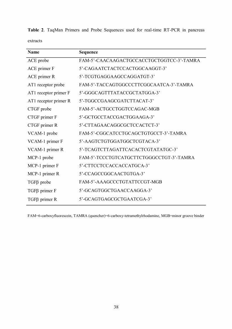
38
Table 2. TaqMan Primers and Probe Sequences used for real-time RT-PCR in pancreas
extracts
Name Sequence
ACE probe FAM-5’-CAACAAGACTGCCACCTGCTGGTCC-3’-TAMRA
ACE primer F 5’-CAGAATCTACTCCACTGGCAAGGT-3’
ACE primer R 5’-TCGTGAGGAAGCCAGGATGT-3’
AT1 receptor probe FAM-5’-TACCAGTGGCCCTTCGGCAATCA-3’-TAMRA
AT1 receptor primer F 5’-GGGCAGTTTATACCGCTATGGA-3’
AT1 receptor primer R 5’-TGGCCGAAGCGATCTTACAT-3’
CTGF probe FAM-5’-ACTGCCTGGTCCAGAC-MGB
CTGF primer F 5’-GCTGCCTACCGACTGGAAGA-3’
CTGF primer R 5’-CTTAGAACAGGCGCTCCACTCT-3’
VCAM-1 probe FAM-5’-CGGCATCCTGCAGCTGTGCCT-3’-TAMRA
VCAM-1 primer F 5’-AAGTCTGTGGATGGCTCGTACA-3’
VCAM-1 primer R 5’-TCAGTCTTAGATTCACACTCGTATATGC-3’
MCP-1 probe FAM-5’-TCCCTGTCATGCTTCTGGGCCTGT-3’-TAMRA
MCP-1 primer F 5’-CTTCCTCCACCACCATGCA-3’
MCP-1 primer R 5’-CCAGCCGGCAACTGTGA-3’
TGF probe FAM-5’-AAAGCCCTGTATTCCGT-MGB
TGF primer F 5’-GCAGTGGCTGAACCAAGGA-3’
TGF primer R 5’-GCAGTGAGCGCTGAATCGA-3’
FAM=6-carboxyfluorescein, TAMRA (quencher)=6-carboxy-tetramethylrhodamine, MGB=minor groove binder

39
RESULTS
STUDY 1.A
5.1 Increase of atherosclerotic plaque development in diabetic apoE-/-
mice after in vivo
administration of full-length recombinant human OPG
Since it has been previously demonstrated that the serum levels of OPG are elevated in
diabetic and nondiabetic patients affected by cardiovascular disease (Jono et al., 2002;
Schoppet et al., 2003; Kiechl et al., 2004; Moran et al., 2005; Secchiero et al., a 2006; Shin et
al., 2006; Abedin et al., 2007), in the first group of experiments we investigated the possible
role of OPG in vivo in the pathogenesis of atherosclerosis. For this purpose we utilized the
apoE-/-
mouse model, that spontaneously develop atherosclerotic lesions, which are increased
after induction of diabetes and resemble in appearance and distribution those observed in
humans (Calkin and Allen, 2006). Therefore, 3 weeks after STZ or vehicle (buffer citrate
alone) multiple injections, the animals were randomized to be i.p. injected either with
recombinant human OPG or with control vehicle (HEPES buffered saline). It should be
noticed that repeated i.p. injections of human recombinant OPG (every 3 weeks for 3 months)
were well tolerated by the apoE-/-
mice. After 3 months, diabetic and control animals were
analyzed for various parameters, as reported in Table 1, and then sacrificed to perform
pathological examination. Diabetic animals gained less weight than did control mice. Blood
glucose, HbA1c, total cholesterol, triglycerides and HDL cholesterol were increased in
diabetic apoE-/-
mice compared with nondiabetic control mice. All of these parameters were
not significantly different in diabetic animals injected or not with human recombinant OPG.
The OPG injections in apoE-/-
mice did not alter significantly the general parameters, however
a slight increase in serum glucose levels was seen in respect to control animals. Systolic blood
pressure levels did not differ significantly among all the groups studied. Moreover, no gross
abnormalities at necroscopic examination were observed in OPG-treated animals with respect
to vehicle-treated apoE-/-
mice.
En face dissection of aorta segments revealed that diabetes was associated with a significant
increase in percentage of plaque area in the entire aorta (means SEM: 1.64 0.15 vs 0.51
0.05, in diabetic vs control apoE-/-
mice respectively; p<0.0001) as well as in each segment
(means SEM in arch: 3.87 0.51 vs 1.24 0.34, p<0.001; means SEM in thoracic aorta:
1.0 0.14 vs 0.13 0.04, p<001; means SEM in abdominal aorta: 1.1 0.22 vs 0.19 0.13,
p<0.05).
Moreover, in non-diabetic control mice most plaques were fatty streaks, whereas in the aorta
6 weeks old apo E-null male mice
C
(n=10)
D
(n=20)
D+rOPG
(n=20)
Diabetic mice + human recombinant
OPG
Diabetic mice + vehicle
(HBS)
Control
ST
Z
R
-
3
+
3
+
6
+
9
0
(weeks
) +
1
rOP
G
rOP
G
rOP
G
6 weeks old apo E-null male mice
C
(n=10)
D
(n=20)
D+rOPG
(n=20)
Diabetic mice + human recombinant
OPG
Diabetic mice + vehicle
(HBS)
Control
ST
Z
R
-
3
+
3
+
6
+
9
0
(weeks
) +
1
rOP
G
rOP
G
rOP
G

40
of diabetic mice there were predominantly advanced lesions, characterized by an
asymmetrically thickened intima and an increase in the collagen content within the plaque,
clearly detected by Masson’s trichrome staining (Figure 1).
While non-diabetic apoE-/-
mice injected with OPG did not show a significant change in the
plaque area extent (means SEM: 0.74 0.23), interestingly, OPG treatment in diabetic apoE-
/- mice induced a significantly (means SEM: 2.25 0.24, p<0.05) increase in the total plaque
area with respect to diabetic mice injected with vehicle alone (Figure 2). Such increase could
not be ascribed to an aspecific, allergic or toxic effect consequent to the injection of a human
protein in the apoE-/-
animals since the administration of recombinant human TRAIL
promoted the opposite effect, decreasing the total plaque area extent (Secchiero et al., b
2006).
Parameters C
(n = 20)
OPG
(n = 20)
D
(n = 20)
D + OPG
(n = 20)
Body weight (g) 34 4 36 3 25 2* 24 3
*
Serum glucose (mg/dl) 157 38 187 73 549 42* 532 39
*
HbA1c (%) 3.9 0.9 4.0 0.7 14.0 1.2* 13.3 1.3
*
SBP (mmHg) 86 10 86 9 87 9 94 8
Total cholesterol (mg/dl) 448 100 538 155 915 105* 949 119
*
Triglycerides (mg/dl) 111 33 120 42 208 44* 200 16
*
HDL cholesterol (mg/dl) 66 14 68 14 116 27* 91 12
*
Table 1. Characteristics of the mice at the end of the study. Data are expressed as mean SD and statistically
(ANOVA) compared. SBP, systolic blood pressure; C, control mice; OPG, mice injected with recombinant human OPG; D, diabetic mice injected with vehicle; D + OPG, diabetic mice injected with OPG. *p<0.01 vs C
group
Figure 1. Increase in atherosclerotic
lesions in the aorta of apoE-/-
mice
following induction of diabetes.
Representative images of aorta of control
and diabetic apoE-/- mice stained with
hematoxylin and eosin (upper panel) and
with Masson’s trichrome for collagen
detection (lower panel, light blue staining). Original magnification X20.

41
Figure 2. Increase in the total extent of atherosclerotic plaques in diabetic mice following the OPG
injections. a. Representative images of en face dissection of aortic arch, thoracic and abdominal aorta, stained
with Sudan IV, showing atherosclerotic lesions (red, with arrows) in diabetic apoE-/- mice treated with either
vehicle (n=10) or recombinant human OPG (n=10). Atherosclerotic involvement for each mouse was
quantitatively determined in en face sections by measuring the proportion of aortic intimal surface area stained
red (Sudan IV-positive) in each of the 3 aortic segments. Horizontal bars are medians of the percentage of plaque
area in the total aortas, upper and lower edges of the boxes are 75th and 25th percentiles and vertical lines are 10th and 90th percentiles. b. Representative hematoxylin-eosin-stained histological cross-sections are shown. Original
magnification X20.
5.2 Recombinant human OPG injected in vivo in apoE-/-
diabetic mice induces changes in
the histological composition of the atherosclerotic plaques
Subsequently, we investigated whether, besides increasing the extension of atherosclerotic
lesions in diabetic mice, OPG treatment also affected the histological characteristics of the
plaques. For this purpose, cross-sectional serial paraffin sections of the aortic atherosclerotic
lesions of apoE-/-
diabetic mice were quantitatively analyzed for the presence of collagen and
the cell composition of the plaques (Figure 3,4). Of note, no significant differences in the

42
percentage of infiltrating macrophages or in the collagen content were observed between
OPG- and vehicle-injected mice (Figure 3a,b).
Figure 3. OPG treatment did not affect the macrophage infiltration or interstitial collagen content in the
atherosclerotic plaques of apoE-/-
diabetic mice. Serial sections from aortas of apoE-/- diabetic mice injected
intraperitoneally with either vehicle (n=10) or recombinant OPG (n=10) were analyzed to quantitatively evaluate
the presence/percentage of macrophages (using F4/80 marker), as well as the interstitial collagen content
(trichrome staining) in the aortic intima. Staining results, representatively shown in a (original magnification
X20), were quantitatively determined in each mouse with the Image Pro Plus® 6.3 analysis program and
expressed as the percentage of stained area. In b, horizontal bars are medians, upper and lower edges of the boxes are 75th and 25th percentiles and vertical lines extending from the boxes are 10th and 90th percentiles.
In the same way, the expression of Syndecan-1, which has been proposed to act as a ligand
for OPG in multiple myeloma (Standal et al., 2002), was not modified in the aortas of OPG-
rOP
G
rOP
G
rOP
G
+
1
(weeks
) 0 +
9
+
6
+
3
-
3
R ST
Z
6 weeks old apo E-null male mice
rOP
G
rOP
G
rOP
G
+
1
(weeks
) 0 +
9
+
6
+
3
-
3
R ST
Z
6 weeks old apo E-null male mice

43
injected mice compared to vehicle treated animals (means SEM (%): 6.6 1.8 vs 5.8 2.1,
in OPG treated diabetic mice vs diabetic apoE-/-
mice respectively).
Interestingly, on the other hand, the percentage of VSMC (αSMA-positive cells) was
significantly (p<0.05) increased in the aortic lesions of OPG-injected apoE-/-
diabetic animals
with respect to diabetic animals injected with vehicle alone (Figure 4a,b).
Figure 4. OPG treatment increased the content of VSMC and their proliferative activity in the
atherosclerotic plaques of apoE-/-
diabetic mice. Serial paraffin sections from aortas of apoE-/- diabetic mice
injected intraperitoneally with either vehicle (n=10) or recombinant OPG (n=10) were analyzed to quantitatively
evaluate the percentage of VSMC (using αSMA marker) and the content of PCNA-positive cells in the aortic
intima. Staining results, representatively shown in a (original magnification X20), were quantitatively
determined in each mouse with the Image Pro Plus® 6.3 analysis program and expressed as the percentage of
stained area or of positive cells. In b, horizontal bars are medians, upper and lower edges of the boxes are 75th
and 25th percentiles and vertical lines are 10th and 90th percentiles.
6 weeks old apo E-null male mice
ST
Z
R
(weeks
)
rOP
G
rOP
G
rOP
G
6 weeks old apo E-null male mice
ST
Z
R
(weeks
)
rOP
G
rOP
G
rOP
G
44
In order to evaluate whether the increased content of VSMC observed in the plaques of OPG-
injected animals was accompanied by an increase in cell proliferation, we analyzed the
percentage of PCNA-positive cells, which accurately reflects the degree of cell proliferation,
within the plaques. As shown in Figure 4a and b, OPG administration promoted a significant
(p<0.01) increase in PCNA-positive cells. Interestingly, the percentage of PCNA-positive
cells evaluated in all animals showed a significant positive correlation with the percentage of
the αSMA-positive area evaluated in the plaques of each mouse (R=0.76, p<0.05), suggesting
that VSMC were the major cell type involved in cell proliferation within the plaques.
5.3 Increase of the mRNA expression of angiopoietin 2 in the aorta of apoE-/-
diabetic
animals after in vivo administration of recombinant human OPG
Taking into account that a recent in vitro study demonstrated that angiopoietin 2 plays a key
role in promoting inflammation of the vessel wall (Fiedler et al., 2006), we next investigated
whether the in vivo injection of human full-length OPG was able to modulate angiopoietin 2
expression in the aortic wall of apoE-/-
diabetic mice. The constitutive steady-state mRNA
levels of angiopoietin 2 were quantitatively evaluated by real time RT-PCR in RNA extracted
from aortic samples of OPG- and vehicle-treated diabetic mice (Figure 5). The mRNA levels
of angiopoietin 2 were significantly (p<0.05) higher in the aortas of OPG-treated diabetic
animals than in the aortas obtained from vehicle-treated diabetic animals (Figure 5). On the
other hand, the inflammatory cell adhesion molecule VCAM-1 (Wara et al., 2008) did not
show significant modifications in OPG-treated versus vehicle-treated diabetic mice (Figure
5).
5.4 Induction of primary rodent VSMC proliferation by recombinant human OPG
In the light of the in vivo results, in which the plaques in OPG-treated diabetic mice showed a
significant increase in the number of VSMC, we have investigated in vitro whether the
exposure to recombinant full-length human OPG had any effect on cell cycle progression of
murine VSMC, analyzed by BrdU incorporation assay (Figure 6). As shown in Figure 6, full-
length recombinant human OPG at concentrations similar to those found in human serum
significantly (p<0.05) promoted the proliferation of VSMC (Secchiero et al., a 2006).

45
Figure 5. Effect of full-length OPG treatment on angiopoietin 2 and VCAM-1 mRNA levels in aortas from
diabetic apoE-/-
mice. mRNA levels of angiopoietin 2 and VCAM-1 in RNA extracted from aortic samples of OPG- and vehicle-treated apoE-/- diabetic mice were quantitatively analyzed by real-time RT-PCR. After
normalization to the level of 18S, gene specific RNA levels in control aortas (from vehicle-treated mice) were
set as 1. Bars indicate mean SEM.
Figure 6. Recombinant human OPG promoted the proliferation of primary aortic VSMC. Aortic murine
VSMC were seeded at subconfluence, serum-starved and either left untreated or exposed to the indicated concentrations of OPG for 36 hours. After BrdU/PI staining, percentage of cells in S phase of the cell cycle,
which have incorporated BrdU, was calculated from the flow cytograms and expressed as the percentage of the
total population. As positive control, cells were treated with Insulin and are shown for comparison. Values are
expressed as means SD of results from four independent experiments. *p<0.05 compared to untreated cells.

46
STUDY 1.B
5.5 The in vivo injection of recombinant human OPG modulates the histological
composition of aortic media in apoE-/-
mice
Taking in consideration our results on the effect of OPG treatment in inducing an increase in
the extension of atherosclerotic lesions and in promoting VSMC proliferation both in vitro
and in vivo in diabetic animals, and considering that an increase in OPG levels characterizes
the early onset of diabetes (Secchiero et al., a 2006), in the second part of our study, we
focalized our attention on the effect of OPG treatment in the aortic media of apoE-/-
mice
without diabetes in respect to control and diabetic mice.
As expected, aortas of diabetic mice injected with vehicle showed an increase in TGFβ and
fibronectin gene expression (by 7.5- and 11-fold, respectively; p<0.05), as well as an increase
in both osteoprotegerin mRNA expression (by 4-fold; p<0.05) and protein content compared
to nondiabetic control mice (Figure 7a,b). Furthermore, interstitial collagen content was
markedly increased in the aortic media of diabetic mice with respect to control animals
(p<0.0001) (Figure 8).
Figure 7. Changes in molecular and cellular parameters in OPG-treated and diabetic mice aortas. (a) Gene expression analysis was assessed by quantitative RT-PCR on whole aorta of control, apoE-/- mice injected
intraperitoneally with human recombinant OPG (OPG) and diabetic apoE-/- mice. Significant increases were
observed for osteoprotegerin, TGFβ and fibronectin in diabetic mice compared to controls (*p < 0.05). OPG
treatment promoted an increase in osteoprotegerin mRNA levels, whereas induced no significant modifications
on TGFβ and fibronectin mRNA levels. (b) Immunohistochemical analysis demonstrated increased staining for
osteoprotegerin in the aortic media of diabetic apoE-/- mice compared to controls (microscope magnification
X20). Positive staining is shown as brown in aortic medial layer. Sections are counterstained with hematoxylin.

47
The injections with recombinant human OPG promoted a significant increase in
osteoprotegerin gene expression, whereas TGFβ and fibronectin did not show significant
modifications in OPG-treated versus control mice (Figure 7a). Interestingly, the percentage of
collagen content was significantly (p<0.05) increased in the aortic media of OPG-injected
apoE-/-
animals compared to untreated animals (Figure 8).
Figure 8. Recombinant human OPG increased the interstitial collagen content in the aortic media of apoE-
/- mice. Representative histological sections (original magnification X20) of aortas from control, apoE-/- mice
injected intraperitoneally with human recombinant OPG (OPG) and diabetic apoE-/- mice. Serial sections were
analyzed to quantitatively evaluate the interstitial collagen content (trichrome staining) in the aortic media and
the results were expressed as the percentage of stained area. *p<0.05 or **p<0.0001 vs control group). All bars
represent mean SEM.
5.6 TGFβ stimulates OPG production in mouse primary VSMC
TGFβ is expressed by cells in the vessel wall and is capable of modulating vascular
development and remodeling by altering cell differentiation, proliferation, migration and
extracellular matrix production (Bobik, 2006). To assess the effect of TGFβ on VSMC, cells
were treated with 10ng/ml of TGFβ for 48h in high glucose media and in serum reduced
condition (2%). Cells incubated with TGFβ exhibited an elongated and broadened
morphology, associated with an increased staining for αSMA (Figure 9a). Significant
6 weeks old apo E-null male mice
C
(n=10)
Control
ST
Z
R
-
3
+
3
+
6
+
9
0
(weeks
) +
1
rOP
G
rOP
G
rOP
G
6 weeks old apo E-null male mice
C
(n=10)
Control
ST
Z
R
-
3
+
3
+
6
+
9
0
(weeks
) +
1
rOP
G
rOP
G
rOP
G

48
increases were observed in the mRNA levels of αSMA, fibronectin (Fibr), vimentin (Vim),
CTGF, collagen I (col I), collagen III (col III) and collagen IV (col IV) (Figure 9b).
Figure 9. TGFβ induced changes in VSMC morphology, protein and gene expression. (a) Mouse aortic
vascular smooth muscle cells (DMEM, 25mM glucose, 2% serum) were treated with TGFβ (10 ng/ml, 2 days).
Changes in morphology are evident by light microscopy (original magnification X10) with cells adopting a more
elongated appearance following TGFβ treatment. Immunostaining of VSMC revealed increased αSMA (green
fluorescence) after TGFβ treatment (original magnification X20). Nuclei were stained with DAPI (blue
fluorescence). (b) Gene expression analysis was assessed by quantitative RT-PCR and significant changes in
response to TGFβ are indicated (*p<0.05 or **p<0.0001 vs control group). Bars indicate mean SEM.
To determine whether OPG expression is regulated by TGFβ in VSMC, cells were stimulated
with increasing concentrations of TGFβ (1-10ng/ml) for 48h. The treatment increased the
OPG mRNA expression in a dose-dependent manner, whereas OPG protein expression
reached a plateau since from the lowest concentration used (Figure 10a). To further confirm
that the OPG release was mediated specifically by TGFβ, VSMC were pre-incubated with
SB431542, the selective TGFβ-type 1 receptor inhibitor, at the concentration of 2µM for 1
hour and then treated with TGFβ 1ng/ml for 48h. SB431542 completely blocked the OPG
expression both at mRNA and protein level (Figure 10b).
5.7 Recombinant full-length OPG increases the mRNA expression of NF-kB, TGFβ and
pro-fibrotic markers in mouse primary VSMC
According to the fact that OPG levels were increased in the aortic media of diabetic mice, the
effect of increasing concentrations of OPG on VSMC was investigated in vitro. Treatment
with 1-40ng/ml of OPG for 48h increased the mRNA expression of fibronectin, collagen I,
collagen III and collagen IV in a dose dependent manner, which peaks at 10ng/ml.

49
Interestingly, with the same pattern, an increased level of TGFβ and NF-kB was also observed
(Figure 11).
Figure 10. TGFβ induced OPG mRNA and protein expression in VSMC. (a) Gene expression analysis by
quantitative real-time PCR revealed a dose dependent induction of OPG mRNA expression in VSMC by TGFβ over 48h. Gene expression is expressed relative to not-treated group, which was arbitrarily designated as 1
(*p<0.05 compared to control group) (left panel). Evaluations of OPG production in VSMC supernates
following TGFβ (1-10ng/ml) treatment over 48h, determined by an ELISA kit (**p<0.0001 compared to control
group) (right panel). (b) VSMC were treated with TGFβ (1ng/ml) for 48h after pre-incubation with SB431542
(2µM) inhibitor for 1h. SB431542 completely blocked the OPG expression at mRNA and protein level. *p<0.05
or **p<0.0001 vs control group. Bars represent mean SEM.

50
Figure 11. Dose response effect of OPG treatment on VSMC gene expression. VSMC (DMEM, 25mM
glucose, 2% serum) were treated with increasing concentrations of OPG (1-40ng/ml) for 48h. The expression of
several genes was assessed by quantitative real-time PCR and the significant changes caused by OPG treatment
are indicated (*p<0.05 or †p<0.0001 vs control group). Bars indicate mean SEM.
5.8 Recombinant full-length OPG increases both OPG protein and mRNA expression in
mouse primary VSMC
In the light of the in vivo results, in which OPG-treated apoE-/-
mice showed an increase in
OPG mRNA expression in the whole aorta, we have investigated in vitro whether the
exposure to recombinant full-length human OPG had any effect on OPG release from aortic
VSMC. As shown in Figure 12, the 48 hours treatment with full-length OPG promoted an
increase in the OPG expression both at mRNA and protein level in murine aortic VSMC.
Figure 12. Full-length recombinant human OPG induced OPG mRNA and protein expression in murine VSMC. Gene expression analysis by quantitative real-time PCR revealed a dose dependent induction of OPG
mRNA expression in VSMC by full-length human OPG over 48h. Gene expression is expressed relative to not-
treated group, which was arbitrarily designated as 1 (*p<0.05 compared to control group) (left panel).
Evaluations of OPG production in VSMC supernates following full-length human OPG (1-40ng/ml) treatment
over 48h, determined by an ELISA kit (**p<0.0001 compared to control group) (right panel).

51
STUDY 2
6.1 In vivo injection of low concentrations of full-length recombinant human OPG induces
a glycemic increase and an insulin levels reduction in C57Bl/6J mice
Although serum osteoprotegerin is significantly increased in diabetic subjects (Browner et al.,
2001; Galluzzi et al., 2005; Knudsen et al., 2003; Rasmussen et al., 2006; Secchiero et al., a
2006), its potential role in beta cell dysfunction has not been investigated. For this purpose 40
male mice C57Bl/6J, aged 10 weeks, were further randomized to be i.p. injected either with
recombinant human OPG or with control vehicle. In addition, in order to explore the possible
interplay of OPG with the renin-angiotensin system (RAS), 20 mice received the ACE
inhibitor ramipril in co-treatment with i.p. injections of OPG or vehicle alone. It should be
noticed that repeated i.p. injections of human recombinant OPG (every 3 weeks for 3 months)
were well tolerated by the C57Bl/6J mice. After 3 months, animals were analyzed for various
parameters, as reported in Table 2, and then sacrificed to perform pathological examination.
No difference between body weight and baseline systolic blood pressure was determined in
animals prior to randomization. The body weight and systolic blood pressure were not
significantly different in animals injected or not with human recombinant OPG. Treatment
with ramipril did not influence body weight, but, however, induced a significant reduction in
systolic blood pressure. Moreover no gross abnormalities at necroscopic examination were
observed in OPG-treated animals with respect to vehicle- or ramipril-treated C57B1/6J mice.
Parameters CONT
(n=10)
OPG
(n=10)
CONT-R
(n=10)
OPG-R
(n=10)
Body weight (g) 29.5 1.5 31.2 1.3 30.1 1.6 28.5 2.0
SBP (mmHg) 95 2 98 3 80 2* 83 2
*
AUC (µg/L/min) 150 20 90 15**
145 18 143 17
Table 2. Characteristics of the mice at the end of the study. SBP, systolic blood pressure; AUC, area under
insulin curve; CONT, control mice; OPG, mice injected with recombinant human OPG; CONT-R, mice injected
with vehicle and treated with ramipril; OPG-R, mice injected with OPG and treated with ramipril. *p<0.05 vs
CONT and OPG groups; **p<0.01 vs other groups. Data are expressed as mean SEM.
Fasting blood glucose levels were measured every 4 weeks for 3 months using a glucose
oxidase method. Mice injected with recombinant human OPG had significantly higher blood
glucose levels after 2 and 3 months of treatment compared to control mice (p<0.05).
Treatment with ramipril completely prevented the glycemic increase induced by OPG
6 weeks old apo E-null male mice
C
(n=10)
D
(n=20)
D+rOPG
(n=20)
Diabetic mice + human recombinant
OPG
Diabetic mice + vehicle
(HBS)
Control
ST
Z
R
-
3
+
3
+
6
+
9
0
(weeks
) +
1
rOP
G
rOP
G
rOP
G
6 weeks old apo E-null male mice
C
(n=10)
D
(n=20)
D+rOPG
(n=20)
Diabetic mice + human recombinant
OPG
Diabetic mice + vehicle
(HBS)
Control
ST
Z
R
-
3
+
3
+
6
+
9
0
(weeks
) +
1
rOP
G
rOP
G
rOP
G

52
administration (p<0.05) (Figure 13a). Consistently with the situation of fasting
hyperglycemia, OPG-treated mice exhibited a reduction in serum insulin with respect to the
control animals, that was statistically significant after 3 months of treatment (p<0.05). In the
same way, co-treatment with the ACE inhibitor significantly improved insulin secretion in
OPG injected mice (p<0.05) (Figure 13b).
Figure 13. Effect of in vivo injections of recombinant OPG on fasting blood glucose and insulin serum levels. Kinetics of blood glucose levels, measured using a glucose oxidase method (a) and insulin serum levels,
determined by an ELISA kit (b). Results are reported as mean SEM. *p<0.05 vs other groups.
Similarly the area under the insulin curve (AUC), evaluated between 0 and 120 minutes after
the glucose challenge, was significantly reduced in OPG-treated mice compared to control
animals (p<0.01). The co-treatment with ramipril induced a statistically significant improve in

53
the AUC of the OPG injected mice (p<0.01). Treatment with ramipril alone didn’t alter the
AUC with respect to untreated mice (Table 2).
6.2 In vivo injection of recombinant human OPG promotes a reduction in insulin staining
density and in beta cell mass in C57Bl/6J mice
Immunohistochemical staining for the beta cell marker insulin was strong and intense in
control mice islets. In contrast, mice injected with recombinant human OPG presented islets
that showed an attenuated and variable pattern of staining suggestive of progressive beta cell
loss (Figure 14). Consistent with these evidences, the mean percentage of proportional area
staining positively for insulin in the OPG treated mice was significantly reduced with respect
to control mice (means SEM (%): CONT 47.1 1.3; OPG 39.6 1.4; p<0.0001). Treatment
with ramipril significantly increased the staining density for insulin (mean SEM (%): OPG-
R 50.7 1.5; vs OPG p<0.0001). No difference in insulin pattern was observed in mice
treated with ramipril alone compared to control mice islets (mean SEM (%): CONT-R 49.6
1.3).
Pancreatic beta cell mass was estimated by multiplying the mean islet density of staining for
insulin by the mean islet area per area of pancreas. This was expressed in arbitrary units
adjusted for the pancreatic wet weight for individual animals. OPG treated mice had a marked
reduction in beta cell mass with respect to the mice injected with vehicle (means SEM:
CONT 14.8 1.6; OPG 9.56 0.9; p<0.05). Blockade of the RAS further increased total
pancreas insulin content (mean SEM: OPG-R 14.2 1.6 vs OPG p<0.05). Treatment with
ramipril alone had no effect on the total beta cell mass (mean SEM: CONT-R 13.9 1.5).
6.3 In vivo administration of recombinant human OPG induces alterations in the
histological composition of the pancreatic islets in C57Bl/6J mice
Islets in OPG treated animals were significantly enlarged with disarray of islet architecture,
and irregular islet boundaries. Treatment with ramipril largely attenuated these changes
(Figure 14). The in vivo administration of human OPG increased significantly Picrosirius
staining both within and at the boundary of the islets (p<0.05) and this increase was
completely prevented by ramipril treatment. Ramipril alone didn’t alter the total collagen
content of the islets compared to the control animals (Table 3).
6 weeks old apo E-null male mice
C
(n=10)
D
(n=20)
D+rOPG
(n=20)
Diabetic mice + human recombinant
OPG
Diabetic mice + vehicle
(HBS)
Control
ST
Z
R
-
3
+
3
+
6
+
9
0
(weeks
) +
1
rOP
G
rOP
G
rOP
G
6 weeks old apo E-null male mice
C
(n=10)
D
(n=20)
D+rOPG
(n=20)
Diabetic mice + human recombinant
OPG
Diabetic mice + vehicle
(HBS)
Control
ST
Z
R
-
3
+
3
+
6
+
9
0
(weeks
) +
1
rOP
G
rOP
G
rOP
G

54
Figure 14. Effect of human recombinant OPG injections on pancreas islet morphology. Representative
histological sections of pancreas (microscope magnification X20); specimens from control (CONT), OPG
injected (OPG) and OPG injected treated with ramipril (OPG-R) mice. Dense immunostaining for insulin (upper
panel) in CONT islets (A) and diffuse staining in OPG islets (B). Increased staining density after blockade of the
RAS with ramipril (C). Staining for picrosirius red (middle panel) and for F4/80 (lower panel, brown staining) in
islets from CONT (D, G), OPG (E, H) and OPG-R (F, I) mice.
Picrosirius F4/80
Intra-islet Peri-islet Intra-islet Peri-islet
CONT 3.7 0.3 12.4 0.9 0.019 0.005 0.163 0.036
OPG 6.3 0.7* 16.6 1.2
* 0.098 0.01
* 0.406 0.079
*
CONT-R 3.0 0.4 11.3 0.3 0.013 0.009 0.197 0.086
OPG-R 3.9 0.1 13.5 0.8 0.017 0.007 0.132 0.038
Table 3. Intra- and peri-islet fibrosis and macrophages infiltration. Data for picrosirius are expressed as
proportional area (%) of each islet (intra-) or of islet boundaries (peri-) occupied by positive red staining, shown
as mean SEM. Data for F4/80 are expressed as average of the number of cells staining positive (brown
staining) in insular- or peri-insular level SEM. *p<0.05 vs all groups.

55
In order to evaluate whether the reduction in beta cell mass observed in OPG-treated animals
might involve the induction of apoptosis by OPG, a double immunofluorescence staining
performed by combining TUNEL plus a specific beta cell marker (insulin) was applied to
identify apoptotic cells within islet boundaries in C57Bl/6J mice treated or not with OPG. As
shown in Figure 15, intraislet cell death was significantly greater in OPG injected mice (7-
fold) compared with infrequent apoptotic cells seen in control islets (p<0.01). Blockade of the
RAS with ramipril reduced TUNEL staining within islet cells to control levels.
Figure 15. Effect of in vivo injections of recombinant full-length OPG on beta cell apoptosis. Representative
image of a pancreatic insula from OPG treated mice, double-stained for TUNEL (green) plus insulin antibody,
followed by a Texas Red-dye conjugated secondary antibody (red) to visualize the beta cells. Beta cell apoptosis
is expressed as the average number of TUNEL positive cells per insula in control (CONT), OPG injected (OPG), control treated with ramipril (CONT-R) and OPG injected treated with ramipril (OPG-R) C57B1/6J mice. Bars
indicate mean SEM (n=6 animals per group). *p<0.01 vs other groups.
To investigate whether OPG treatment affected the degree of infiltration of cells of the
monocytic/macrophagic lineage, serial sections of paraffin-embedded pancreas were stained
for F4/80 antigen (Figure 14). Peri-insular and intra-insular infiltrations were significantly
(p<0.05) increased in OPG-injected mice (2- and 3.7 fold, respectively) when compared with
control animals injected with vehicle. Co-treatment with lifelong ramipril significantly
reduced the expression of F4/80 protein in islets (Table 3).
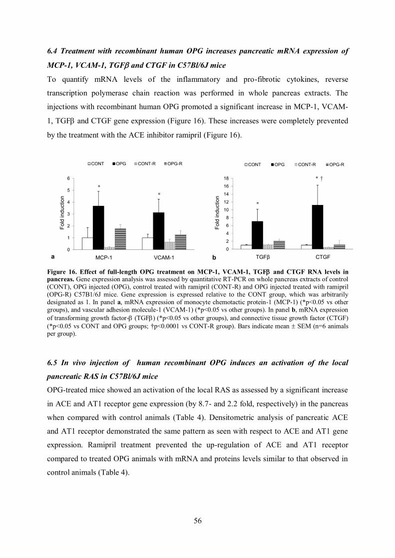
56
6.4 Treatment with recombinant human OPG increases pancreatic mRNA expression of
MCP-1, VCAM-1, TGF and CTGF in C57Bl/6J mice
To quantify mRNA levels of the inflammatory and pro-fibrotic cytokines, reverse
transcription polymerase chain reaction was performed in whole pancreas extracts. The
injections with recombinant human OPG promoted a significant increase in MCP-1, VCAM-
1, TGF and CTGF gene expression (Figure 16). These increases were completely prevented
by the treatment with the ACE inhibitor ramipril (Figure 16).
Figure 16. Effect of full-length OPG treatment on MCP-1, VCAM-1, TGF and CTGF RNA levels in pancreas. Gene expression analysis was assessed by quantitative RT-PCR on whole pancreas extracts of control
(CONT), OPG injected (OPG), control treated with ramipril (CONT-R) and OPG injected treated with ramipril
(OPG-R) C57B1/6J mice. Gene expression is expressed relative to the CONT group, which was arbitrarily
designated as 1. In panel a, mRNA expression of monocyte chemotactic protein-1 (MCP-1) (*p<0.05 vs other
groups), and vascular adhesion molecule-1 (VCAM-1) (*p<0.05 vs other groups). In panel b, mRNA expression
of transforming growth factor- (TGF) (*p<0.05 vs other groups), and connective tissue growth factor (CTGF)
(*p<0.05 vs CONT and OPG groups; †p<0.0001 vs CONT-R group). Bars indicate mean SEM (n=6 animals per group).
6.5 In vivo injection of human recombinant OPG induces an activation of the local
pancreatic RAS in C57Bl/6J mice
OPG-treated mice showed an activation of the local RAS as assessed by a significant increase
in ACE and AT1 receptor gene expression (by 8.7- and 2.2 fold, respectively) in the pancreas
when compared with control animals (Table 4). Densitometric analysis of pancreatic ACE
and AT1 receptor demonstrated the same pattern as seen with respect to ACE and AT1 gene
expression. Ramipril treatment prevented the up-regulation of ACE and AT1 receptor
compared to treated OPG animals with mRNA and proteins levels similar to that observed in
control animals (Table 4).

57
ACE AT1 receptor
Immunohistochemistry
CONT 8.0 1.7 2.7 1.0
OPG 16.1 3.2* 5.5 0.6
*
CONT-R 6.3 2.2 2.9 0.8
OPG-R 8.7 1.1 3.1 0.5
Real-time RT-PCR
CONT 1.0 0.6 1.0 0.2
OPG 8.7 2.8† 2.2 0.4*
CONT-R 0.6 0.1 1.0 0.2
OPG-R 0.7 0.3 1.1 0.3
Table 4. Expression of components of the RAS as quantified by immunohistochemistry and real-time RT-
PCR.
Data are proportional area (%) of each islet occupied by positive staining for specific islet proteins, shown as
mean SEM (for immunohistochemistry); and data are results from gene expression study presented as a ratio
compared with CONT, shown as mean SEM (for real-time RT-PCR). *p<0.05 vs other groups; †p<0.0001 vs other groups.

58
DISCUSSION
Many different studies have shown that increased amounts of serum OPG correlate with
increased cardiovascular risk in both diabetic and non-diabetic patients (Jono et al., 2002;
Schoppet et al., 2003; Kiechl et al., 2004; Moran et al., 2005; Secchiero et al., a 2006; Shin et
al., 2006; Abedin et al., 2007). Possible interpretations of this positive relationship include
OPG playing an active role in disease progression or OPG serving as a compensatory
response to reduce disease progression, or OPG representing a noncompensatory (neutral)
response to disease. The interpretation that OPG serum elevation represents a compensatory
mechanism mainly relies on studies conducted on OPG knockout mice showing a protective
role of OPG against vascular calcifications (Bucay et al., 1998; Price et al., a 2001; Bennet et
al., 2006). However, in each of these models the prevention of vascular calcification by OPG
was associated with strong suppression of bone turnover. Furthermore, both recombinant
OPG (Price et al., a 2001) and bisphosphonate (Price et al., b 2001) treatments of rats
suppressed vascular calcification in a warfarin model of vascular calcification, in association
with suppression of bone turnover. Taken these data together, seems that the ability of OPG to
inhibit vascular calcification is an indirect effect of its anti-osteoclastic activity. In contrast, in
vitro studies have clearly shown that the expression and release of OPG by vascular cells is
markedly induced in response to inflammatory cytokines, such as TNF-α and platelet-derived
growth factor (PDGF) (Olesen et al., 2005; Zhang et al., 2002), and that OPG promotes
leukocytes adhesion to endothelial cells (Zauli et al., 2007; Mangan et al., 2007), suggesting
an active role of OPG in disease progression. On these bases, the aims of our studies were
elucidate the potential roles of OPG in the pathogenesis of atherosclerosis and in pancreatic
beta cell dysfunction. To investigate whether OPG is involved in pathogenetic aspects of
atherosclerosis, apoE-/-
mice, with or without diabetes, were injected every 3 weeks with
recombinant full-length OPG and studied for 3 months. Subsequently, the animals were
analyzed for the total plaque area and for the plaque cellular content, as well as for the media
histology. Moreover, in parallel to in vivo studies, the effect of increasing concentrations of
OPG were analyzed in vitro, using murine primary VSMC [see 7.1-section].
On the other side, to investigate whether OPG is involved in pathogenetic aspects of diabetes
mellitus, C57Bl/6J mice, that do not spontaneously develop atherosclerotic lesions with a
normal chow diet, were injected every 3 weeks with recombinant full length OPG and, after 3
months, sacrificed to perform the analysis of the pancreatic beta cells structure and function.
59
In addition, starting from some evidences for an interplay between OPG and the RAS
pathways in human aortas, the long term co-treatment with the ACE inhibitor ramipril was
investigated in order to evaluate if it could hinder the effect of exogenous OPG on beta cells
remodelling and function [see 7.2-section].
7.1 OPG and diabetes associated atherosclerosis
The first part of our study showed that repeated injections of relatively low concentrations of
human OPG resulted in a significant increase in the total aortic plaque area in apoE-/-
diabetic
mice, a well characterized animal model of diabetes-accelerated atherosclerosis, which have
been shown to develop aortic lesions characteristic in appearance and distribution to those
observed in humans (Nakashima et al., 1994; Wu and Huan, 2007). Taking into account that a
series of in vitro and in vivo experiments, conducted previously by our group, have
demonstrated that soluble recombinant TRAIL shows anti-inflammatory (Secchiero et al.,
2003; Zauli et al., 2003; Secchiero et al., 2005) and anti-atherosclerotic (Secchiero et al., b
2006) activity, a mechanism by which OPG might accelerate the development of
atherosclerotic lesions is through the inhibition of soluble circulating TRAIL. Moreover, the
demonstration that OPG significantly increases the mRNA expression of angiopoietin 2 at the
aortic level is in line with a recent in vitro study (Mangan et al., 2007). Since angiopoietin 2
has been shown to play a key role in the early steps of atherosclerosis (Fiedler et al., 2006),
the ability of OPG to increase the expression of angiopoietin 2 might represent an important
additional pathogenetic mechanism to explain the ability of OPG to promote atherosclerosis.
On the other side, we also noticed a slight trend for an increase in systolic blood pressure in
the OPG-treated diabetic mice, which might account for the changes observed in
atherosclerotic plaques.
It is also noteworthy that the aortic plaques in OPG-treated diabetic apoE-/-
mice presented a
significant increase in the percentage of VSMC. Furthermore, in line with the in vivo results,
in vitro data demonstrated that recombinant full-length OPG promoted also the proliferation
of primary rodent cultured VSMC. In this context, is important to underline that the full-
length recombinant OPG, that we used for our experiments, possess an heparin binding
domain, that is essential to mediate important pro-inflammatory effects of OPG (Vitovski et
al., 2007; Zauli et al., 2007; Mangan et al., 2007), in an independent manner to its role of
decoy receptor for TRAIL and RANKL. According to this, we should consider that the

60
VSMC migration and proliferation, could be mediated by the interactions between the heparin
binding domain of OPG and cell surface proteoglycans (Rauch et al., 2005).
Besides contributing to vessel wall inflammation and lipoprotein retention, VSMC play a
foundamental role in the formation of the fibrous cap that provides stability to the plaques,
preserving them from rupture, and therefore the benefit/risk of therapeutic inhibition of
VSMC proliferation in atherosclerosis is unclear (Libby, 2002). In this context, the effect of
OPG on plaque formation seems to be twofold: on one side, it increases the extent of the total
plaque area, but on the other side it may contribute to plaque stabilization by promoting the
proliferation of VSMC.
Subsequently, in the second part of our study, we analyzed whether in vivo administration of
human recombinant OPG could induce some modifications in VSMC of the aortic media too,
in the absence of a diabetes associated atherosclerosis. Basically, the strategy adopted of
delivering human OPG intraperitoneally was chosen in order to try to mimic the effect of a
small elevation of OPG serum levels, by allowing a prolonged release of the molecule into the
general circulation.
The possible existence of a link between the high levels of OPG and TGFβ observed in
diabetes was suggested by our preliminary results, showing that OPG and TGFβ were
augmented in aortas of diabetic animals.
Interestingly, apoE-/-
mice injected with OPG presented a trend for an increase in TGFβ,
fibronectin and OPG gene expression and, more importantly, a significant increase in the
interstitial medial aortic collagen content, resembling an intermediate situation between
diabetic and control animals injected with vehicle.
TGFβ is generally accepted to be the main pro-fibrotic factor in diabetic complications, and
several evidences support its important role in development of glomerulosclerosis and
interstitial fibrosis (Ban and Twigg, 2008), and in regulating cardiac extracellular matrix
deposition (ECM) (Westermann et al., 2007). Several different investigations supported the
hypothesis that an increased expression of TGFβ, which is the main responsible for ECM
accumulation, is involved in many cardiovascular complications by participating in the
fibrotic process (Yokoyama and Deckert, 1996; Piao and Tokunaga, 2006; Pham et al., 2010;
Ruiz-Ortega et al., 2007).
On the other side, many studies demonstrated that OPG secretion is regulated in smooth
muscle cells by different stimuli, such as insulin (Olesen et al, 2005), angiotensin II (Moran et
al., 2009) or proinflammatory cytokines, like tumor necrosis α (Moran et al., 2005). Clearly,
many other factors could also play a role in high serum OPG levels that characterized both

61
type 1 and type 2 diabetes (Secchiero et al., a 2006; Galluzzi et al., 2005; Rasmussen et al.,
2006; Browner et al., 2001; Knudsen et al., 2003; Avignon et al., 2005; Anand et al., 2006).
To further explore this potential link between TGFβ and OPG, we carried out a series of in
vitro experiments using mouse primary aortic VSMC. Interestingly, VSMC treated with
TGFβ, as well as having an elongated and broadened morphology and an increased expression
of ECM components and profibrotic markers, showed an increased expression of OPG both at
protein and mRNA level since from the lowest concentration used. This finding suggests that
TGFβ could actively participate in the increased amounts of OPG seen in the aortic vessel of
the diabetic animals.
Subsequently, in order to analyze the possible phisiopathological role of high concentrations
of OPG observed in diabetes, we investigated the effect of this molecule in the same in vitro
model, represented by the mouse primary aortic VSMC. OF note, VSMC treated with OPG
presented an increased expression of fibronectin and profibrotic markers that resembled what
TGFβ, in the same experimental conditions, did. Furthermore, in this system, a significant
increase in TGFβ expression, as well as an auto-induction of OPG release were also observed,
suggesting that probably a vicious cycle exists between TGFβ and OPG.
As a major transcription factor in inflammatory responses, nuclear factor kB is involved in the
regulation of inflammatory and immune genes, apoptosis, and cell proliferation. Taking in
consideration that inflammatory events characterize all stages of the atherosclerotic process ,
NF-kB has been considered a crucial element in the initiation of atherogenesis (Monaco and
Paleolog, 2004; de Winther et al., 2005) . According to this, it is noteworthy that the NF-kB
increase, induced by OPG treatment, supports the potential proinflammatory role of OPG,
which is consistent to what we and other groups have previously shown (Zauli et al., 2007;
Mangan et al., 2007).
In summary, taking in consideration that collagen synthesis occurs very early in lesion
development (Rekhter, 1999) and an increase in OPG levels characterizes the early onset of
diabetes (Secchiero et al., a 2006), our data, from in vitro and in vivo models, suggest that
OPG is involved in diabetes-induced vascular ECM accumulation, probably via TGFβ
induction, and that the up-regulation of TGFβ actively participates in the increased amount of
OPG seen in diabetes, suggesting a positive feedback regulatory mechanism.
In conclusion, overall these data together, suggest that high OPG levels could definitely play
an important role in the development and progression of diabetic atherosclerosis.
62
7.2 OPG and pancreatic beta cells dysfunction
Results obtained from in vivo studies performed in C57Bl/6J mice have demonstrated that the
systemic administration of recombinant human OPG promotes significant histopathological
changes in pancreatic islets including selective loss of beta cells by apoptosis, increased
infiltration of monocytes/macrophages, increased expression of inflammatory molecules and
fibrosis. In addition, exogenous administration of OPG also induced a significant increase of
gene and protein expression of ACE and AT1 receptor and we have also observed the
attenuation of these changes after chronic blockade of the RAS, in keeping with previous
strong evidence that most of the components of functioning RAS are present locally in the
endocrine pancreas and that islet RAS has a role in regulating pancreatic islet function (Leung
and Carlsson, 2001; Tikellis et al., 2006). These findings are particularly relevant since it has
been shown that angiotensin II plays an important role in regulating insulin islet secretion
(Carlsson et al., 1998; Lau et al., 2004) and the process that leads to beta cell loss and
dysfunction in type 1 and type 2 diabetes are significantly influenced by local activation of
the RAS (Tikellis et al., 2006).
The molecular mechanism by which OPG controls ACE and AT1 gene expression is not
known. The expression of ACE and AT1 receptor have been shown to be associated with
activation of the mitogen-activated protein kinase (MAPK) and nuclear factor-kB
transduction pathways (Cowling et al., 2002; Koka et al., 2008; Martin et al., 2007). In this
respect, it should be emphasized that OPG is able to stimulate MAPK signaling (Theoleyre et
al., a 2004). Taking these data together with the present study it prompts us to hypothesize
that OPG might control transcription mechanism governing ACE and AT1 gene expression.
In this context, it should also be taken into account that pancreatic inflammation may also
result in the upregulation of local RAS as demonstrated in animal models of pancreatitis
(Kuno et al., 2003). In our experimental setting, an islet accumulation of cells of the
monocytic/macrophagic lineage was noted after OPG administration, and this was associated
with an increased expression of MCP-1, a proinflammatory chemoattractant whose expression
correlates with the progression of insulitis and beta cell destruction in different murine models
(Chen et al., 2001; Ehses et al., 2009). In addition, the endothelial adhesion molecule VCAM-
1, which is important for the adhesion of macrophages to the vascular wall, as demonstrated
in experimental diabetes (Candido et al., 2004), was also significantly increased in OPG-
treated animals. Previous studies have demonstrated that the pro-inflammatory effect of OPG
is mediated by a direct interactivity with cell surface heparan solfate (Mosheimer et al., 2005)

63
or alternatively by preventing the antiinflammatory activity of TRAIL (Zauli et al., 2009). In
this respect, it is particularly noteworthy that the TRAIL death pathway is present in islet beta
cells (Ou et al., 2002) and we have recently demonstrated that recombinant TRAIL
ameliorates the severity of streptozotocin-induced type 1 diabetes (Zauli et al., 2010).
Therefore, a possible mechanism by which OPG might contribute to beta cell dysfunction is
by inhibiting the antiinflammatory activity of circulating TRAIL. Notwithstanding these
multiple potential pathways mediating the inflammatory response to OPG, all the
inflammatory effects observed in our study after administration of OPG may be explained by
the local RAS upregulation and its downstream sequelae. In accordance with this hypothesis,
ACE inhibitor treatment prevented all the OPG-induced inflammatory changes.
In native tissues, the interaction between pancreatic beta cells and elements of their local
micro-environment including extracellular matrix is essential to activate intracellular
signaling pathways that regulate cell proliferation, survival and function. Loss of cell-to-cell
communications associated with increased intraislet fibrosis may reduce the secretory
efficiency of the islet and promote islet cell apoptosis (Tikellis et al., 2004). We report here
that chronic OPG treatment increases peri- and intrainsula fibrosis and promotes the
expression of TGF mRNA and its downstream effector CTGF in the pancreas. Both TGF
and CTGF have been associated with fibrosis in diabetic nephropathy (Murphy et al., 1999)
and pancreatitis (di Mola et al., 1999; Kuno et al., 2003). Also the RAS has been linked to
increased fibrosis in a variety of tissues including the heart (Seccia et al., 2003), kidney
(Satoh et al., 2001) and liver (Yoshiji et al., 2001). In fact, locally produced angiotensin II is
thought to upregulate, via AT1 receptor, the expression of fibrogenic cytokines and growth
factors including TGF and CTGF (Ruperez et al., 2003). Moreover, the use of an ACE
inhibitor or AT1 receptor antagonist have been shown to result in reduced fibrosis from
inhibtion of TGF and CTGF in animal models of diabetes mellitus (Tikellis et al., 2004) and
pancreatitis (Kuno et al., 2003). It is conceivable that the reduction in islet fibrosis observed
in this study after treatment with ramipril may be mediated through a similar pathway.
Pancreatic beta cell death by apoptosis is believed to be the primary mechanism for the
reduction of beta cell mass and volume in both type 1 and type 2 diabetes (Thomas et al.,
2009). Apoptosis in beta cell is a highly complex process and angiotensin II has been shown
to play a relevant role in the control of beta cell life and death (Yuan et al., 2010). In the
present study the OPG induction of islet apoptosis is closely related to upregulation of
pancreatic RAS and these changes were prevented after treatment with ramipril. The
64
maintenance of beta cell area/density is important for maintaining a correct insulin secretion.
Consistently with a protective role of ramipril the levels of insulinemia were significantly
higher in mice treated with OPG + ramipril, and OPG + ramipril treated animals showed
reduced level of hyperglycemia throughout the whole period of the study with respect to
OPG-injected animals. Moreover ramipril co-treatment significantly ameliorated insulin
response after the glucose challenge, suggesting an improvement in beta cell function.
Because disruption of contacts between beta cells and beta cell loss reduces the secretory
efficiency of islets, the results illustrated in our study suggest a potential mechanism to
explain why pathologically elevated serum OPG levels, frequently occurring in both type 1
and type 2 diabetes (Browner et al., 2001; Galluzzi et al., 2005; Knudsen et al., 2003;
Rasmussen et al., 2006; Secchiero et al., a 2006), are linked to the development and/or
maitenance of diabetes mellitus.
In conclusion the present study indicates in the overactivity of the local RAS one of the
possible mechanisms responsible for the OPG-induced islet beta cell dysfunction, suggesting
that elevated level of OPG may have an active role in pancreatic islet pathophysiology.
7.3 Perspective
In vitro and in vivo data indicate that OPG might have an important role in promoting pro-
atherogenic and pro-fibrotic effects in vascular physiopathology, as well as in inducing
pancreatic beta cell dysfunction. Furthermore, full-length OPG seems to have a clear
inflammatory activity, that is in line with many recent in vitro studies reported by our and
other groups (Zauli et al., 2007; Mangan et al., 2007).
Considerable controversy still exists regarding the role of OPG/RANKL/RANK/TRAIL in
cardiovascular disease and there is as yet no hypothesis unifying the apparent dichotomy in
the nature of this system noted in animal and human studies (Venuraju et al., 2010). As
already reported, serum OPG levels are elevated in both type 1 and type 2 diabetic patients
and progressively increase with the duration of diabetes. Unfortunately at the moment there
are no clear evidences of a relationship between an increase in OPG serum levels and any
prediabetic condition. Consequently, OPG serum levels that could be potentially responsible
for beta cell dysfunction are already unknown, also considering that once diabetes has been
established, serum OPG levels are influenced by several factors such as arterial accumulation
of the molecule and increased production from vascular smooth muscle cells induced by
many different cytokines like TNF- and TGFβ. Although it cannot be ruled out that high
plasma OPG levels are an epiphenomenon of inflammatory processes harbored in vascular

65
lesions, our data suggest rather that elevated level of OPG in diabetes may have an active role
in the development and progression of atherosclerosis as well as in pancreatic islet
pathophysiology. Moreover our results indicate in the overactivity of the local RAS one of the
possible mechanisms responsible for the OPG-induced islet beta cell dysfunction, and this
may explain why the use of an ACE inhibitor, currently and safely used in clinical setting,
may be suitable to target OPG pancreatic effect.
Of note, the triad RANKL/RANK/OPG plays a central role in coupling bone formation and
resorption during normal bone turnover and in a wide spectrum of diseases characterized by
disturbed bone remodeling, increased bone resorption and bone destruction (osteoporosis,
Paget’s disease of bone, rheumatoid arthritis, metastatic bone disease). While little doubts
exist on the ability of pharmacological concentrations of recombinant chimeric OPG-Fc, in
which the signal peptide, the heparin-binding domain and the death domain homologous
regions are removed, to efficiently counteract bone turnover associated to different
pathological conditions (Zauli et al., 2009), the overall effects of full-length OPG are much
less clear. Anyhow, an important implication of our data is that denosumab, a fully human
monoclonal antibody which blocks osteoclastogenesis by inhibiting RANKL (Kearns et al.,
2008; Geusens, 2009), might be preferable to recombinant OPG for the treatment of
pathologies associated with high bone turnover, such as osteoporosis.

66
LIST OF PUBLICATIONS 2008-2010
1) Cocchietto M, Zorzin L, Toffoli B, Candido R, Fabris B, Stebel M, Sava G. Orally
administered microencapsulated lysozyme downregulates serum AGE and reduces the
severity of early-stage diabetic nephropathy. Diabetes Metab 2008; 34:587-94.
2) Candido R, Toffoli B, Corallini F, Bernardi S, Zella D, Voltan R, Grill V, Celeghini C,
Fabris B. Human full-length osteoprotegerin induces the proliferation of rodent
vascular smooth muscle cells both in vitro and in vivo. J Vasc Res 2010; 47:252-61.
3) Zauli G, Toffoli B, di Iasio MG, Celeghini C, Fabris B, Secchiero P. Treatment with
recombinant tumor necrosis factor-related apoptosis-inducing ligand alleviates the
severity of streptozotocin-induced diabetes. Diabetes 2010; 59:1261-5.
4) Thomas MC, Pickering RJ, Tsorotes D, Koitka A, Sheehy K, Bernardi S, Toffoli B,
Nguyen-Huu TP, Head G, Fu Y, Chin-Dusting J, Cooper ME, Tikellis C. Genetic
Ace2 Deficiency Accentuates Vascular Inflammation and Atherosclerosis in the ApoE
Knockout Mouse. Circ Res 2010; 107:888-97.
5) Bernardi S, Candido R, Toffoli B, Carretta R, Fabris B. Prevention of accelerated
atherosclerosis by AT1 receptor blockade in experimental renal failure. Nephrol Dial
Transplant 2010. [Epub ahead of print].
6) Toffoli B, Bernardi S, Candido R, Sabato N, Carretta R, Corallini F, Secchiero P,
Zauli G, Fabris B. Osteoprotegerin induces morphological and functional alterations in
mouse pancreatic islets. Mol Cell Endocrinol 2011; 331:136-42.
7) Fabris B, Candido R, Bortoletto M, Toffoli B, Bernardi S, Stebel M, Bardelli M,
Zentilin L, Giacca M, Carretta R. Stimulation of cardiac apoptosis in ovariectomized
hypertensive rats: potential role of the renin-angiotensin system. J Hypertens 2011; 29:
273-81.
8) Toffoli B, Bernardi S, Candido R, Zacchigna S, Fabris B, Secchiero P. TRAIL shows
potential cardioprotective activity. Invest New Drugs 2011 Jan 4. [Epub ahead of print]
9) Fischetti F, Candido R, Toffoli B, Durigutto P, Bernardi S, Carretta R, Tedesco F,
Fabris B. Innate immunity, through late complement components activation,
contributes to the development of early vascular inflammation and morphologic
alterations in experimental diabetes. Atherosclerosis. IN PRESS

67
REFERENCES
Abedin M, Omland T, Ueland T, Khera A, Aukrust P, Murphy SA, Jain T, Gruntmanis U,
McGuire DK, de Lemos JA. Relation of osteoprotegerin to coronary calcium and aortic
plaque (from the Dallas Heart Study). Am J Cardiol 2007; 99:513-8.
Aggarwal BB. Signalling pathways of the TNF superfamily: a double-edged sword. Nat Rev
Immunol 2003; 3:745-56.
Agrotis A, Kanellakis P, Kostolias G, Di Vitto G, Wei C, Hannan R, Jennings G, Bobik A.
Proliferation of neointimal smooth muscle cells after arterial injury. Dependence on
interactions between fibroblast growth factor receptor-2 and fibroblast growth factor-9. J Biol
Chem 2004; 279:42221-9.
Albiston AL, McDowall SG, Matsacos D, Sim P, Clune E, Mustafa T, Lee J, Mendelsohn FA,
Simpson RJ, Connolly LM, Chai SY. Evidence that the angiotensin IV (AT(4)) receptor is the
enzyme insulin-regulated aminopeptidase. J Biol Chem 2001; 276:48623-6.
ALLHAT Officers and Coordinators for the ALLHAT Collaborative Research Group. The
Antihypertensive and Lipid-Lowering Treatment to Prevent Heart Attack Trial. Major
outcomes in high-risk hypertensive patients randomized to angiotensin-converting enzyme
inhibitor or calcium channel blocker vs diuretic: The Antihypertensive and Lipid-Lowering
Treatment to Prevent Heart Attack Trial (ALLHAT). JAMA 2002; 288:2981-97.
Anand DV, Lahiri A, Lim E, Hopkins D, Corder R. The relationship between plasma
osteoprotegerin levels and coronary artery calcification in uncomplicated type 2 diabetic
subjects. J Am Coll Cardiol 2006; 47:1850-7.
Andraws R, Brown DL. Effect of inhibition of the renin-angiotensin system on development
of type 2 diabetes mellitus (meta-analysis of randomized trials). Am J Cardiol 2007; 99:1006-
12.
Andreozzi F, D'Alessandris C, Federici M, Laratta E, Del Guerra S, Del Prato S, Marchetti P,
Lauro R, Perticone F, Sesti G. Activation of the hexosamine pathway leads to
phosphorylation of insulin receptor substrate-1 on Ser307 and Ser612 and impairs the
phosphatidylinositol 3-kinase/Akt/mammalian target of rapamycin insulin biosynthetic
pathway in RIN pancreatic beta-cells. Endocrinology 2004; 145:2845-57.
Ashkenazi A, Herbst RS. To kill a tumor cell: the potential of proapoptotic receptor agonists.
J Clin Invest 2008; 118:1979-90.
68
Avignon A, Sultan A, Piot C, Elaerts S, Cristol JP, Dupuy AM. Osteoprotegerin is associated
with silent coronary artery disease in high-risk but asymptomatic type 2 diabetic patients.
Diabetes Care 2005; 28:2176-80.
Baker SJ, Reddy EP. Modulation of life and death by the TNF receptor superfamily.
Oncogene 1998; 17:3261-70.
Ban CR, Twigg SM. Fibrosis in diabetes complications: pathogenic mechanisms and
circulating and urinary markers. Vasc Health Risk Manag 2008; 4:575-96.
Baud'huin M, Lamoureux F, Duplomb L, Rédini F, Heymann D. RANKL, RANK,
osteoprotegerin: key partners of osteoimmunology and vascular diseases. Cell Mol Life Sci
2007; 64:2334-50.
Bennett BJ, Scatena M, Kirk EA, Rattazzi M, Varon RM, Averill M, Schwartz SM, Giachelli
CM, Rosenfeld ME. Osteoprotegerin inactivation accelerates advanced atherosclerotic lesion
progression and calcification in older ApoE-/- mice. Arterioscler Thromb Vasc Biol 2006;
26:2117-24.
Ben-Tal Cohen E, Hohensinner PJ, Kaun C, Maurer G, Huber K, Wojta J. Statins decrease
TNF-alpha-induced osteoprotegerin production by endothelial cells and smooth muscle cells
in vitro. Biochem Pharmacol 2007; 73:77-83.
Bindokas VP, Kuznetsov A, Sreenan S, Polonsky KS, Roe MW, Philipson LH. Visualizing
superoxide production in normal and diabetic rat islets of Langerhans. J Biol Chem 2003;
278:9796-801.
Bobik A. Transforming growth factor-betas and vascular disorders. Arterioscler Thromb Vasc
Biol 2006; 26:1712-20.
Borgatti P, Zauli G, Colamussi ML, Gibellini D, Previati M, Cantley LL, Capitani S.
Extracellular HIV-1 Tat protein activates phosphatidylinositol 3- and Akt/PKB kinases in
CD4+ T lymphoblastoid Jurkat cells. Eur J Immunol 1997; 27:2805-11.
Boyce BF, Xing L. Biology of RANK, RANKL, and osteoprotegerin. Arthritis Res Ther
2007; 9 Suppl 1:S1.
Browner WS, Lui LY, Cummings SR. Associations of serum osteoprotegerin levels with
diabetes, stroke, bone density, fractures, and mortality in elderly women. J Clin Endocrinol
Metab 2001; 86:631-7.
Bucay N, Sarosi I, Dunstan CR, Morony S, Tarpley J, Capparelli C, Scully S, Tan HL, Xu W,
Lacey DL, Boyle WJ, Simonet WS. Osteoprotegerin-deficient mice develop early onset
osteoporosis and arterial calcification. Genes Dev 1998; 12:1260-8.

69
Bulotta S, Barsacchi R, Rotiroti D, Borgese N, Clementi E. Activation of the endothelial
nitric-oxide synthase by tumor necrosis factor-alpha. A novel feedback mechanism regulating
cell death. J Biol Chem 2001; 276:6529-36.
Cai H, Li Z, Dikalov S, Holland SM, Hwang J, Jo H, Dudley SC Jr, Harrison DG. NAD(P)H
oxidase-derived hydrogen peroxide mediates endothelial nitric oxide production in response
to angiotensin II. J Biol Chem 2002; 277:48311-7.
Calkin AC, Allen TJ. Diabetes mellitus-associated atherosclerosis: mechanisms involved and
potential for pharmacological invention. Am J Cardiovasc Drugs 2006; 6:15-40.
Campbell DJ, Habener JF. Angiotensinogen gene is expressed and differentially regulated in
multiple tissues of the rat. J Clin Invest 1986; 78:31-9.
Candido R, Jandeleit-Dahm KA, Cao Z, Nesteroff SP, Burns WC, Twigg SM, Dilley RJ,
Cooper ME, Allen TJ. Prevention of accelerated atherosclerosis by angiotensin-converting
enzyme inhibition in diabetic apolipoprotein E-deficient mice. Circulation 2002; 106:246-53.
Candido R, Allen TJ, Lassila M, Cao Z, Thallas V, Cooper ME, Jandeleit-Dahm KA.
Irbesartan but not amlodipine suppresses diabetes-associated atherosclerosis. Circulation
2004; 109:1536-42.
Carlsson PO, Berne C, Jansson L. Angiotensin II and the endocrine pancreas: effects on islet
blood flow and insulin secretion in rats. Diabetologia 1998; 41:127-33.
Chan FK. Three is better than one: pre-ligand receptor assembly in the regulation of TNF
receptor signaling. Cytokine 2007; 37:101-7.
Charollais A, Gjinovci A, Huarte J, Bauquis J, Nadal A, Martín F, Andreu E, Sánchez-Andrés
JV, Calabrese A, Bosco D, Soria B, Wollheim CB, Herrera PL, Meda P. Junctional
communication of pancreatic beta cells contributes to the control of insulin secretion and
glucose tolerance. J Clin Invest 2000; 106:235-43.
Chen MC, Proost P, Gysemans C, Mathieu C, Eizirik DL. Monocyte chemoattractant protein-
1 is expressed in pancreatic islets from prediabetic NOD mice and in interleukin-1 beta-
exposed human and rat islet cells. Diabetologia 2001; 44:325-32.
Chu KY, Lau T, Carlsson PO, Leung PS. Angiotensin II type 1 receptor blockade improves
beta-cell function and glucose tolerance in a mouse model of type 2 diabetes. Diabetes 2006;
55:367-74.
Collin-Osdoby P, Rothe L, Anderson F, Nelson M, Maloney W, Osdoby P. Receptor activator
of NF-kappa B and osteoprotegerin expression by human microvascular endothelial cells,

70
regulation by inflammatory cytokines, and role in human osteoclastogenesis. J Biol Chem
2001; 276:20659-72.
Collin-Osdoby P. Regulation of vascular calcification by osteoclast regulatory factors
RANKL and osteoprotegerin. Circ Res 2004; 95:1046-57.
Cooper ME, McNally PG, Phillips PA, Johnston CI. Amylin stimulates plasma renin
concentration in humans. Hypertension 1995; 26:460-4.
Corallini F, Rimondi E, Secchiero P. TRAIL and osteoprotegerin: a role in endothelial
physiopathology? Front Biosci 2008; 13:135-47.
Corrado E, Rizzo M, Coppola G, Fattouch K, Novo G, Marturana I, Ferrara F, Novo S. An
update on the role of markers of inflammation in atherosclerosis. J Atheroscler Thromb 2010;
17:1-11.
Cowling RT, Gurantz D, Peng J, Dillmann WH, Greenberg BH. Transcription factor NF-
kappa B is necessary for up-regulation of type 1 angiotensin II receptor mRNA in rat cardiac
fibroblasts treated with tumor necrosis factor-alpha or interleukin-1 beta. J Biol Chem 2002;
277:5719-24.
Davis BJ, Forbes JM, Thomas MC, Jerums G, Burns WC, Kawachi H, Allen TJ, Cooper ME.
Superior renoprotective effects of combination therapy with ACE and AGE inhibition in the
diabetic spontaneously hypertensive rat. Diabetologia 2004; 47:89-97.
de Winther MP, Kanters E, Kraal G, Hofker MH. Nuclear factor kappaB signaling in
atherogenesis. Arterioscler Thromb Vasc Biol 2005; 25:904-14.
Degli-Esposti M. To die or not to die--the quest of the TRAIL receptors. J Leukoc Biol 1999;
65:535-42.
Dempsey PW, Doyle SE, He JQ, Cheng G. The signaling adaptors and pathways activated by
TNF superfamily. Cytokine Growth Factor Rev 2003; 14:193-209.
di Mola FF, Friess H, Martignoni ME, Di Sebastiano P, Zimmermann A, Innocenti P, Graber
H, Gold LI, Korc M, Büchler MW. Connective tissue growth factor is a regulator for fibrosis
in human chronic pancreatitis. Ann Surg 1999; 230:63-71.
Donoghue M, Hsieh F, Baronas E, Godbout K, Gosselin M, Stagliano N, Donovan M, Woolf
B, Robison K, Jeyaseelan R, Breitbart RE, Acton S. A novel angiotensin-converting enzyme-
related carboxypeptidase (ACE2) converts angiotensin I to angiotensin 1-9. Circ Res 2000;
87:E1-9.

71
Dubois M, Kerr-Conte J, Gmyr V, Bouckenooghe T, Muharram G, D'Herbomez M, Martin-
Ponthieu A, Vantyghem MC, Vandewalle B, Pattou F. Non-esterified fatty acids are
deleterious for human pancreatic islet function at physiological glucose concentration.
Diabetologia 2004; 47:463-9.
Ehses JA, Lacraz G, Giroix MH, Schmidlin F, Coulaud J, Kassis N, Irminger JC, Kergoat M,
Portha B, Homo-Delarche F, Donath MY. IL-1 antagonism reduces hyperglycemia and tissue
inflammation in the type 2 diabetic GK rat. Proc Natl Acad Sci U S A 2009; 106:13998-4003.
Emery JG, McDonnell P, Burke MB, Deen KC, Lyn S, Silverman C, Dul E, Appelbaum ER,
Eichman C, DiPrinzio R, Dodds RA, James IE, Rosenberg M, Lee JC, Young PR.
Osteoprotegerin is a receptor for the cytotoxic ligand TRAIL. J Biol Chem 1998; 273:14363-
7.
Erdogan B, Aslan E, Bagis T, Gokcel A, Erkanli S, Bavbek M, Altinors N. Intima-media
thickness of the carotid arteries is related to serum osteoprotegerin levels in healthy
postmenopausal women. Neurol Res 2004; 26:658-61.
Federici M, Hribal M, Perego L, Ranalli M, Caradonna Z, Perego C, Usellini L, Nano R,
Bonini P, Bertuzzi F, Marlier LN, Davalli AM, Carandente O, Pontiroli AE, Melino G,
Marchetti P, Lauro R, Sesti G, Folli F. High glucose causes apoptosis in cultured human
pancreatic islets of Langerhans: a potential role for regulation of specific Bcl family genes
toward an apoptotic cell death program. Diabetes 2001; 50:1290-301.
Feige U. Osteoprotegerin. Ann Rheum Dis 2001; 60 Suppl 3:iii81-4.
Fiedler U, Reiss Y, Scharpfenecker M, Grunow V, Koidl S, Thurston G, Gale NW,
Witzenrath M, Rosseau S, Suttorp N, Sobke A, Herrmann M, Preissner KT, Vajkoczy P,
Augustin HG. Angiopoietin-2 sensitizes endothelial cells to TNF-alpha and has a crucial role
in the induction of inflammation. Nat Med 2006; 12:235-9.
Finnberg N, El-Deiry WS. TRAIL death receptors as tumor suppressors and drug targets. Cell
Cycle 2008; 7:1525-8.
Forbes JM, Cooper ME, Thallas V, Burns WC, Thomas MC, Brammar GC, Lee F, Grant SL,
Burrell LA, Jerums G, Osicka TM. Reduction of the accumulation of advanced glycation end
products by ACE inhibition in experimental diabetic nephropathy. Diabetes 2002; 51:3274-
82.
Fujiwara K, Nagasaka A, Nagata M, Yamamoto K, Imamura S, Oda N, Sawai Y, Hayakawa
N, Suzuki A, Itoh M. A stable prostacyclin analogue reduces high serum TNF-alpha levels in
diabetic patients. Exp Clin Endocrinol Diabetes 2004; 112:390-4.

72
Galluzzi F, Stagi S, Salti R, Toni S, Piscitelli E, Simonini G, Falcini F, Chiarelli F.
Osteoprotegerin serum levels in children with type 1 diabetes: a potential modulating role in
bone status. Eur J Endocrinol 2005; 153:879-85.
Geusens P. Emerging treatments for postmenopausal osteoporosis - focus on denosumab. Clin
Interv Aging 2009; 4:241-50.
Glass CK, Witztum JL. Atherosclerosis: the road ahead. Cell 2001; 104:503-16.
Gletsu N, Doan TN, Cole J, Sutliff RL, Bernstein KE. Angiotensin II-induced hypertension in
mice caused an increase in insulin secretion. Vascul Pharmacol 2005; 42:83-92.
Grauslund J, Rasmussen LM, Green A, Sjølie AK. Does osteoprotegerin relate to micro- and
macrovascular complications in long-term type 1 diabetes? Scand J Clin Lab Invest 2010;
70:188-93.
Grcević D, Katavić V, Lukić IK, Kovacić N, Lorenzo JA, Marusić A. Cellular and molecular
interactions between immune system and bone. Croat Med J 2001; 42:384-92.
Griendling KK, Minieri CA, Ollerenshaw JD, Alexander RW. Angiotensin II stimulates
NADH and NADPH oxidase activity in cultured vascular smooth muscle cells. Circ Res
1994; 74:1141-8.
Griffith TS, Stokes B, Kucaba TA, Earel JK Jr, VanOosten RL, Brincks EL, Norian LA.
TRAIL gene therapy: from preclinical development to clinical application. Curr Gene Ther
2009; 9:9-19.
Gross O, Beirowski B, Koepke ML, Kuck J, Reiner M, Addicks K, Smyth N, Schulze-Lohoff
E, Weber M. Preemptive ramipril therapy delays renal failure and reduces renal fibrosis in
COL4A3-knockout mice with Alport syndrome. Kidney Int 2003; 63:438-46.
Guicciardi ME, Gores GJ. Life and death by death receptors. FASEB J 2009; 23:1625-37.
Hansson L, Lindholm LH, Niskanen L, Lanke J, Hedner T, Niklason A, Luomanmäki K,
Dahlöf B, de Faire U, Mörlin C, Karlberg BE, Wester PO, Björck JE. Effect of angiotensin-
converting-enzyme inhibition compared with conventional therapy on cardiovascular
morbidity and mortality in hypertension: the Captopril Prevention Project (CAPPP)
randomised trial. Lancet 1999; 353:611-6.
Harmon JS, Gleason CE, Tanaka Y, Poitout V, Robertson RP. Antecedent hyperglycemia, not
hyperlipidemia, is associated with increased islet triacylglycerol content and decreased insulin
gene mRNA level in Zucker diabetic fatty rats. Diabetes 2001; 50:2481-6.

73
Hayden MR, Sowers JR. Isletopathy in type2 diabetes mellitus: implications of islet RAS,
islet fibrosis, islet amyloid, remodeling and oxidative stress. Antioxid Redox Signal 2007;
9:891-910.
Hehlgans T, Pfeffer K. The intriguing biology of the tumour necrosis factor/tumour necrosis
factor receptor superfamily: players, rules and the games. Immunology 2005; 115:1-20.
Heickendorff L, Ledet T, Rasmussen LM. Glycosaminoglycans in the human aorta in diabetes
mellitus: a study of tunica media from areas with and without atherosclerotic plaque.
Diabetologia 1994; 37:286-92.
Heinonen SE, Leppänen P, Kholová I, Lumivuori H, Häkkinen SK, Bosch F, Laakso M, Ylä-
Herttuala S. Increased atherosclerotic lesion calcification in a novel mouse model combining
insulin resistance, hyperglycemia, and hypercholesterolemia. Circ Res 2007; 101:1058-67.
Hofbauer LC, Heufelder AE, Erben RG. Osteoprotegerin, RANK, and RANK ligand: the
good, the bad, and the ugly in rheumatoid arthritis. J Rheumatol 2001; 28:685-7.
Hong YM. Atherosclerotic cardiovascular disease beginning in childhood. Korean Circ J
2010; 40:1-9.
Höppener JW, Ahrén B, Lips CJ. Islet amyloid and type 2 diabetes mellitus. N Engl J Med
2000; 343:411-9.
Jacqueminet S, Briaud I, Rouault C, Reach G, Poitout V. Inhibition of insulin gene expression
by long-term exposure of pancreatic beta cells to palmitate is dependent on the presence of a
stimulatory glucose concentration. Metabolism 2000; 49:532-6.
Janke J, Engeli S, Gorzelniak K, Luft FC, Sharma AM. Mature adipocytes inhibit in vitro
differentiation of human preadipocytes via angiotensin type 1 receptors. Diabetes 2002;
51:1699-707.
Jawień J, Nastałek P, Korbut R. Mouse models of experimental atherosclerosis. J Physiol
Pharmacol 2004; 55:503-17.
Johnston CI, Burrell LM, Perich R, Jandeleit K, Jackson B. The tissue renin-angiotensin
system and its functional role. Clin Exp Pharmacol Physiol Suppl 1992; 19:1-5.
Jono S, Ikari Y, Shioi A, Mori K, Miki T, Hara K, Nishizawa Y. Serum osteoprotegerin levels
are associated with the presence and severity of coronary artery disease. Circulation 2002;
106:1192-4.
74
Jono S, Otsuki S, Higashikuni Y, Shioi A, Mori K, Hara K, Hashimoto H, Ikari Y. Serum
osteoprotegerin levels and long-term prognosis in subjects with stable coronary artery disease.
J Thromb Haemost 2010; 8:1170-5.
Jørgensen GM, Vind B, Nybo M, Rasmussen LM, Højlund K. Acute hyperinsulinemia
decreases plasma osteoprotegerin with diminished effect in type 2 diabetes and obesity. Eur J
Endocrinol 2009; 161:95-101.
Josien R, Li HL, Ingulli E, Sarma S, Wong BR, Vologodskaia M, Steinman RM, Choi Y.
TRANCE, a tumor necrosis factor family member, enhances the longevity and adjuvant
properties of dendritic cells in vivo. J Exp Med 2000; 191:495-502.
Joussen AM, Poulaki V, Mitsiades N, Kirchhof B, Koizumi K, Döhmen S, Adamis AP.
Nonsteroidal anti-inflammatory drugs prevent early diabetic retinopathy via TNF-alpha
suppression. FASEB J 2002; 16:438-40.
Julius S, Kjeldsen SE, Weber M, Brunner HR, Ekman S, Hansson L, Hua T, Laragh J,
McInnes GT, Mitchell L, Plat F, Schork A, Smith B, Zanchetti A; VALUE trial group.
Outcomes in hypertensive patients at high cardiovascular risk treated with regimens based on
valsartan or amlodipine: the VALUE randomised trial. Lancet 2004; 363:2022-31.
Kahn SE, Hull RL, Utzschneider KM. Mechanisms linking obesity to insulin resistance and
type 2 diabetes. Nature 2006; 444:840-6.
Kawamori D, Kajimoto Y, Kaneto H, Umayahara Y, Fujitani Y, Miyatsuka T, Watada H,
Leibiger IB, Yamasaki Y, Hori M. Oxidative stress induces nucleo-cytoplasmic translocation
of pancreatic transcription factor PDX-1 through activation of c-Jun NH(2)-terminal kinase.
Diabetes 2003; 52:2896-904.
Kearns AE, Khosla S, Kostenuik PJ. Receptor activator of nuclear factor kappaB ligand and
osteoprotegerin regulation of bone remodeling in health and disease. Endocr Rev 2008;
29:155-92.
Kiechl S, Schett G, Wenning G, Redlich K, Oberhollenzer M, Mayr A, Santer P, Smolen J,
Poewe W, Willeit J. Osteoprotegerin is a risk factor for progressive atherosclerosis and
cardiovascular disease. Circulation 2004; 109:2175-80.
Knudsen ST, Foss CH, Poulsen PL, Andersen NH, Mogensen CE, Rasmussen LM. Increased
plasma concentrations of osteoprotegerin in type 2 diabetic patients with microvascular
complications. Eur J Endocrinol 2003; 149:39-42.
Koka V, Huang XR, Chung AC, Wang W, Truong LD, Lan HY. Angiotensin II up-regulates
angiotensin I-converting enzyme (ACE), but down-regulates ACE2 via the AT1-ERK/p38
MAP kinase pathway. Am J Pathol 2008; 172:1174-83.

75
Kuno A, Yamada T, Masuda K, Ogawa K, Sogawa M, Nakamura S, Nakazawa T, Ohara H,
Nomura T, Joh T, Shirai T, Itoh M. Angiotensin-converting enzyme inhibitor attenuates
pancreatic inflammation and fibrosis in male Wistar Bonn/Kobori rats. Gastroenterology
2003; 124:1010-9.
Kwon B, Kim BS, Cho HR, Park JE, Kwon BS. Involvement of tumor necrosis factor
receptor superfamily (TNFRSF) members in the pathogenesis of inflammatory diseases. Exp
Mol Med 2003; 35:8-16.
Lamoureux F, Picarda G, Garrigue-Antar L, Baud'huin M, Trichet V, Vidal A, Miot-Noirault
E, Pitard B, Heymann D, Rédini F. Glycosaminoglycans as potential regulators of
osteoprotegerin therapeutic activity in osteosarcoma. Cancer Res 2009; 69:526-36.
Landmesser U, Hornig B, Drexler H. Endothelial function: a critical determinant in
atherosclerosis? Circulation 2004; 109:II27-33.
Lastra G, Manrique C. The expanding role of oxidative stress, renin angiotensin system, and
beta-cell dysfunction in the cardiometabolic syndrome and Type 2 diabetes mellitus. Antioxid
Redox Signal 2007; 9:943-54.
Lau T, Carlsson PO, Leung PS. Evidence for a local angiotensin-generating system and dose-
dependent inhibition of glucose-stimulated insulin release by angiotensin II in isolated
pancreatic islets. Diabetologia 2004; 47:240-8.
Lavoie JL, Sigmund CD. Minireview: Overview of the Renin-Angiotensin System—An
Endocrine and Paracrine System. Endocrinology 2003; 144: 2179-2183.
Lawrie A, Waterman E, Southwood M, Evans D, Suntharalingam J, Francis S, Crossman D,
Croucher P, Morrell N, Newman C. Evidence of a role for osteoprotegerin in the pathogenesis
of pulmonary arterial hypertension. Am J Pathol 2008; 172:256-64.
Lee Y, Hirose H, Ohneda M, Johnson JH, McGarry JD, Unger RH. Beta-cell lipotoxicity in
the pathogenesis of non-insulin-dependent diabetes mellitus of obese rats: impairment in
adipocyte-beta-cell relationships. Proc Natl Acad Sci U S A 1994; 91:10878-82.
Lehto S, Niskanen L, Suhonen M, Rönnemaa T, Laakso M. Medial artery calcification. A
neglected harbinger of cardiovascular complications in non-insulin-dependent diabetes
mellitus. Arterioscler Thromb Vasc Biol 1996; 16:978-83.
Leung PS, Chan HC, Wong PY. Immunohistochemical localization of angiotensin II in the
mouse pancreas. Histochem J 1998; 30:21-5.

76
Leung PS, Carlsson PO. Tissue renin-angiotensin system: its expression, localization,
regulation and potential role in the pancreas. J Mol Endocrinol 2001; 26:155-64.
Leung PS, Chappell MC. A local pancreatic renin-angiotensin system: endocrine and exocrine
roles. Int J Biochem Cell Biol 2003; 35:838-46.
Libby P. Inflammation in atherosclerosis. Nature 2002; 420:868-74.
Libby P, Okamoto Y, Rocha VZ, Folco E. Inflammation in atherosclerosis: transition from
theory to practice. Circ J 2010; 74:213-20.
Lieb W, Gona P, Larson MG, Massaro JM, Lipinska I, Keaney JF Jr, Rong J, Corey D,
Hoffmann U, Fox CS, Vasan RS, Benjamin EJ, O'Donnell CJ, Kathiresan S. Biomarkers of
the osteoprotegerin pathway: clinical correlates, subclinical disease, incident cardiovascular
disease, and mortality. Arterioscler Thromb Vasc Biol 2010; 30:1849-54.
Lindholm LH, Ibsen H, Borch-Johnsen K, Olsen MH, Wachtell K, Dahlöf B, Devereux RB,
Beevers G, de Faire U, Fyhrquist F, Julius S, Kjeldsen SE, Kristianson K, Lederballe-
Pedersen O, Nieminen MS, Omvik P, Oparil S, Wedel H, Aurup P, Edelman JM, Snapinn S;
For the LIFE study group. Risk of new-onset diabetes in the Losartan Intervention For
Endpoint reduction in hypertension study. J Hypertens 2002; 20:1879-86.
Lindholm LH, Persson M, Alaupovic P, Carlberg B, Svensson A, Samuelsson O. Metabolic
outcome during 1 year in newly detected hypertensives: results of the Antihypertensive
Treatment and Lipid Profile in a North of Sweden Efficacy Evaluation (ALPINE study). J
Hypertens 2003; 21:1563-74.
Locksley RM, Killeen N, Lenardo MJ. The TNF and TNF receptor superfamilies: integrating
mammalian biology. Cell 2001; 104:487-501.
Lupi R, Dotta F, Marselli L, Del Guerra S, Masini M, Santangelo C, Patané G, Boggi U, Piro
S, Anello M, Bergamini E, Mosca F, Di Mario U, Del Prato S, Marchetti P. Prolonged
exposure to free fatty acids has cytostatic and pro-apoptotic effects on human pancreatic
islets: evidence that beta-cell death is caspase mediated, partially dependent on ceramide
pathway, and Bcl-2 regulated. Diabetes 2002; 51:1437-42.
Lupi R, Del Guerra S, Bugliani M, Boggi U, Mosca F, Torri S, Del Prato S, Marchetti P. The
direct effects of the angiotensin-converting enzyme inhibitors, zofenoprilat and enalaprilat, on
isolated human pancreatic islets. Eur J Endocrinol 2006; 154:355-61.
Lusis AJ. Atherosclerosis. Nature 2000; 407:233-41.
MacEwan DJ. Interactions between TNF and GnRH. Neurochem Res 2008; 33:678-82.

77
Mangan SH, Van Campenhout A, Rush C, Golledge J. Osteoprotegerin upregulates
endothelial cell adhesion molecule response to tumor necrosis factor-alpha associated with
induction of angiopoietin-2. Cardiovasc Res 2007; 76:494-505.
Marchetti P, Dotta F, Lauro D, Purrello F. An overview of pancreatic beta-cell defects in
human type 2 diabetes: implications for treatment. Regul Pept 2008; 146:4-11.
Martin MM, Buckenberger JA, Jiang J, Malana GE, Knoell DL, Feldman DS, Elton TS. TGF-
beta1 stimulates human AT1 receptor expression in lung fibroblasts by cross talk between the
Smad, p38 MAPK, JNK, and PI3K signaling pathways. Am J Physiol Lung Cell Mol Physiol
2007; 293:L790-9.
Meier JJ, Kayed R, Lin CY, Gurlo T, Haataja L, Jayasinghe S, Langen R, Glabe CG, Butler
PC. Inhibition of human IAPP fibril formation does not prevent beta-cell death: evidence for
distinct actions of oligomers and fibrils of human IAPP. Am J Physiol Endocrinol Metab
2006; 291:E1317-24.
Meir KS, Leitersdorf E. Atherosclerosis in the apolipoprotein-E-deficient mouse: a decade of
progress. Arterioscler Thromb Vasc Biol 2004; 24:1006-14.
Mesquita M, Demulder A, Damry N, Mélot C, Wittersheim E, Willems D, Dratwa M,
Bergmann P. Plasma osteoprotegerin is an independent risk factor for mortality and an early
biomarker of coronary vascular calcification in chronic kidney disease. Clin Chem Lab Med
2009; 47:339-46.
Mizuno A, Murakami A, Nakagawa N, Yasuda H, Tsuda E, Morinaga T, Higashio K.
Structure of the mouse osteoclastogenesis inhibitory factor (OCIF) gene and its expression in
embryogenesis. Gene 1998; 215:339-43. a
Mizuno A, Amizuka N, Irie K, Murakami A, Fujise N, Kanno T, Sato Y, Nakagawa N,
Yasuda H, Mochizuki S, Gomibuchi T, Yano K, Shima N, Washida N, Tsuda E, Morinaga T,
Higashio K, Ozawa H. Severe osteoporosis in mice lacking osteoclastogenesis inhibitory
factor/osteoprotegerin. Biochem Biophys Res Commun 1998; 247:610-5. b
Monaco C, Paleolog E. Nuclear factor kappaB: a potential therapeutic target in atherosclerosis
and thrombosis. Cardiovasc Res 2004; 61:671-82.
Moran CS, McCann M, Karan M, Norman P, Ketheesan N, Golledge J. Association of
osteoprotegerin with human abdominal aortic aneurysm progression. Circulation 2005;
111:3119-25.
Moran CS, Cullen B, Campbell JH, Golledge J. Interaction between angiotensin II,
osteoprotegerin, and peroxisome proliferator-activated receptor-gamma in abdominal aortic
aneurysm. J Vasc Res 2009; 46:209-17.
78
Morena M, Terrier N, Jaussent I, Leray-Moragues H, Chalabi L, Rivory JP, Maurice F,
Delcourt C, Cristol JP, Canaud B, Dupuy AM. Plasma osteoprotegerin is associated with
mortality in hemodialysis patients. J Am Soc Nephrol 2006; 17:262-70.
Morinaga T, Nakagawa N, Yasuda H, Tsuda E, Higashio K. Cloning and characterization of
the gene encoding human osteoprotegerin/osteoclastogenesis-inhibitory factor. Eur J Biochem
1998; 254:685-91.
Morony S, Tintut Y, Zhang Z, Cattley RC, Van G, Dwyer D, Stolina M, Kostenuik PJ, Demer
LL. Osteoprotegerin inhibits vascular calcification without affecting atherosclerosis in ldlr(-/-)
mice. Circulation 2008; 117:411-20.
Mosheimer BA, Kaneider NC, Feistritzer C, Djanani AM, Sturn DH, Patsch JR, Wiedermann
CJ. Syndecan-1 is involved in osteoprotegerin-induced chemotaxis in human peripheral blood
monocytes. J Clin Endocrinol Metab 2005; 90:2964-71.
Murphy M, Godson C, Cannon S, Kato S, Mackenzie HS, Martin F, Brady HR. Suppression
subtractive hybridization identifies high glucose levels as a stimulus for expression of
connective tissue growth factor and other genes in human mesangial cells. J Biol Chem 1999;
274:5830-4.
Nakashima Y, Plump AS, Raines EW, Breslow JL, Ross R. ApoE-deficient mice develop
lesions of all phases of atherosclerosis throughout the arterial tree. Arterioscler Thromb 1994;
14:133-40.
Nguyen G, Delarue F, Burcklé C, Bouzhir L, Giller T, Sraer JD. Pivotal role of the
renin/prorenin receptor in angiotensin II production and cellular responses to renin. J Clin
Invest 2002; 109:1417-27.
Niskanen L, Siitonen O, Suhonen M, Uusitupa MI. Medial artery calcification predicts
cardiovascular mortality in patients with NIDDM. Diabetes Care 1994; 17:1252-6.
Nybo M, Rasmussen LM. Osteoprotegerin released from the vascular wall by heparin mainly
derives from vascular smooth muscle cells. Atherosclerosis 2008; 201:33-5.
Okada K, Hirano T, Ran J, Adachi M. Olmesartan medoxomil, an angiotensin II receptor
blocker ameliorates insulin resistance and decreases triglyceride production in fructose-fed
rats. Hypertens Res 2004; 27:293-9.
Olesen P, Ledet T, Rasmussen LM. Arterial osteoprotegerin: increased amounts in diabetes
and modifiable synthesis from vascular smooth muscle cells by insulin and TNF-alpha.
Diabetologia 2005; 48:561-8.

79
Oliveira HR, Verlengia R, Carvalho CR, Britto LR, Curi R, Carpinelli AR. Pancreatic beta-
cells express phagocyte-like NAD(P)H oxidase. Diabetes 2003; 52:1457-63.
Onozato ML, Tojo A, Goto A, Fujita T, Wilcox CS. Oxidative stress and nitric oxide synthase
in rat diabetic nephropathy: effects of ACEI and ARB. Kidney Int 2002; 61:186-94.
Ou D, Metzger DL, Wang X, Huang J, Pozzilli P, Tingle AJ. TNF-related apoptosis-inducing
ligand death pathway-mediated human beta-cell destruction. Diabetologia 2002; 45:1678-88.
Ovchinnikova O, Gylfe A, Bailey L, Nordström A, Rudling M, Jung C, Bergström S,
Waldenström A, Hansson GK, Nordström P. Osteoprotegerin promotes fibrous cap formation
in atherosclerotic lesions of ApoE-deficient mice--brief report. Arterioscler Thromb Vasc Biol
2009; 29:1478-80.
Pandolfi A, Grilli A, Cilli C, Patruno A, Giaccari A, Di Silvestre, De Lutiis MA, Pellegrini G,
Capani F, Consoli A, Felaco M. Phenotype modulation in cultures of vascularsmooth muscle
cells from diabetic rats: association with increased nitric oxide synthase expression and
superoxide anion generation. J Cell Physiol 2003; 196:378–385.
Paul M, Poyan Mehr A, Kreutz R. Physiology of local renin-angiotensin systems. Physiol Rev
2006; 86:747-803.
Paul WE. Fundamental Immunology (Book). Lippincot Williams & Wilkins, 2008; chapter 25:
777
Pham Y, Tu Y, Wu T, Allen TJ, Calkin AC, Watson AM, Li J, Jandeleit-Dahm KA, Toh BH,
Cao Z, Cooper ME, Chai Z. Cell division autoantigen 1 plays a profibrotic role by modulating
downstream signalling of TGF-beta in a murine diabetic model of atherosclerosis.
Diabetologia 2010; 53:170-9.
Piao M, Tokunaga O. Significant expression of endoglin (CD105), TGFbeta-1 and TGFbeta
R-2 in the atherosclerotic aorta: an immunohistological study. J Atheroscler Thromb 2006;
13:82-9.
Poitout V, Robertson RP. Minireview: Secondary beta-cell failure in type 2 diabetes--a
convergence of glucotoxicity and lipotoxicity. Endocrinology 2002; 143:339-42.
Poitout V, Robertson RP. Glucolipotoxicity: fuel excess and beta-cell dysfunction. Endocr
Rev 2008; 29:351-66.
Price PA, June HH, Buckley JR, Williamson MK. Osteoprotegerin inhibits artery calcification
induced by warfarin and by vitamin D. Arterioscler Thromb Vasc Biol 2001; 21:1610-6. a

80
Price PA, Faus SA, Williamson MK. Bisphosphonates alendronate and ibandronate inhibit
artery calcification at doses comparable to those that inhibit bone resorption. Arterioscler
Thromb Vasc Biol 2001; 21:817-24. b
Ran J, Hirano T, Adachi M. Angiotensin II type 1 receptor blocker ameliorates
overproduction and accumulation of triglyceride in the liver of Zucker fatty rats. Am J Physiol
Endocrinol Metab 2004; 287:E227-32.
Rasmussen LM, Ledet T. Aortic collagen alterations in human diabetes mellitus. Changes in
basement membrane collagen content and in the susceptibility of total collagen to cyanogen
bromide solubilisation. Diabetologia 1993; 36:445-53.
Rasmussen LM, Tarnow L, Hansen TK, Parving HH, Flyvbjerg A. Plasma osteoprotegerin
levels are associated with glycaemic status, systolic blood pressure, kidney function and
cardiovascular morbidity in type 1 diabetic patients. Eur J Endocrinol 2006; 154:75-81.
Rauch BH, Millette E, Kenagy RD, Daum G, Fischer JW, Clowes AW. Syndecan-4 is
required for thrombin-induced migration and proliferation in human vascular smooth muscle
cells. J Biol Chem 2005; 280:17507-11.
Regoli M, Bendayan M, Fonzi L, Sernia C, Bertelli E. Angiotensinogen localization and
secretion in the rat pancreas. J Endocrinol 2003; 179:81-9.
Reid IA, Morris BJ, Ganong WF.The renin-angiotensin system. Annu. Rev. Physiol 1978;
40:377-410.
Reid P, Holen I. Pathophysiological roles of osteoprotegerin (OPG). Eur J Cell Biol 2009;
88:1-17.
Rekhter MD. Collagen synthesis in atherosclerosis: too much and not enough. Cardiovasc Res
1999; 41:376-84.
Robertson RP. Chronic oxidative stress as a central mechanism for glucose toxicity in
pancreatic islet beta cells in diabetes. J Biol Chem 2004; 279:42351-4.
Roehrich ME, Mooser V, Lenain V, Herz J, Nimpf J, Azhar S, Bideau M, Capponi A, Nicod
P, Haefliger JA, Waeber G. Insulin-secreting beta-cell dysfunction induced by human
lipoproteins. J Biol Chem 2003; 278:18368-75.
Ruiz-Ortega M, Rodríguez-Vita J, Sanchez-Lopez E, Carvajal G, Egido J. TGF-beta signaling
in vascular fibrosis. Cardiovasc Res 2007; 74:196-206.

81
Rupérez M, Ruiz-Ortega M, Esteban V, Lorenzo O, Mezzano S, Plaza JJ, Egido J.
Angiotensin II increases connective tissue growth factor in the kidney. Am J Pathol 2003;
163:1937-47.
Santos RA, Simoes e Silva AC, Maric C, Silva DM, Machado RP, de Buhr I, Heringer-
Walther S, Pinheiro SV, Lopes MT, Bader M, Mendes EP, Lemos VS, Campagnole-Santos
MJ, Schultheiss HP, Speth R, Walther T. Angiotensin-(1-7) is an endogenous ligand for the G
protein-coupled receptor Mas. Proc Natl Acad Sci U S A 2003; 100:8258-63.
Santos RA, Ferreira AJ. Angiotensin-(1-7) and the renin-angiotensin system. Curr Opin
Nephrol Hypertens 2007; 16:122-8.
Satoh M, Kashihara N, Yamasaki Y, Maruyama K, Okamoto K, Maeshima Y, Sugiyama H,
Sugaya T, Murakami K, Makino H. Renal interstitial fibrosis is reduced in angiotensin II type
1a receptor-deficient mice. J Am Soc Nephrol 2001; 12:317-25
Schling P, Löffler G. Effects of angiotensin II on adipose conversion and expression of genes
of the renin-angiotensin system in human preadipocytes. Horm Metab Res 2001; 33:189-95.
Schoppet M, Preissner KT, Hofbauer LC. RANK ligand and osteoprotegerin: paracrine
regulators of bone metabolism and vascular function. Arterioscler Thromb Vasc Biol 2002;
22:549-53.
Schoppet M, Sattler AM, Schaefer JR, Herzum M, Maisch B, Hofbauer LC. Increased
osteoprotegerin serum levels in men with coronary artery disease. J Clin Endocrinol Metab
2003; 88:1024-8.
Secchiero P, Gonelli A, Carnevale E, Milani D, Pandolfi A, Zella D, Zauli G. TRAIL
promotes the survival and proliferation of primary human vascular endothelial cells by
activating the Akt and ERK pathways. Circulation 2003; 107:2250-6.
Secchiero P, Corallini F, di Iasio MG, Gonelli A, Barbarotto E, Zauli G. TRAIL counteracts
the proadhesive activity of inflammatory cytokines in endothelial cells by down-modulating
CCL8 and CXCL10 chemokine expression and release. Blood 2005; 105:3413-9.
Secchiero P, Candido R, Corallini F, Zacchigna S, Toffoli B, Rimondi E, Fabris B, Giacca M,
Zauli G. Systemic tumor necrosis factor-related apoptosis-inducing ligand delivery shows
antiatherosclerotic activity in apolipoprotein E-null diabetic mice. Circulation 2006;
114:1522-30. b
Secchiero P, Corallini F, Pandolfi A, Consoli A, Candido R, Fabris B, Celeghini C, Capitani
S, Zauli G. An increased osteoprotegerin serum release characterizes the early onset of
diabetes mellitus and may contribute to endothelial cell dysfunction. Am J Pathol 2006;
169:2236-44. a

82
Seccia TM, Belloni AS, Kreutz R, Paul M, Nussdorfer GG, Pessina AC, Rossi GP. Cardiac
fibrosis occurs early and involves endothelin and AT-1 receptors in hypertension due to
endogenous angiotensin II. J Am Coll Cardiol 2003; 41:666-73.
Shao J, Iwashita N, Ikeda F, Ogihara T, Uchida T, Shimizu T, Uchino H, Hirose T, Kawamori
R, Watada H. Beneficial effects of candesartan, an angiotensin II type 1 receptor blocker, on
beta-cell function and morphology in db/db mice. Biochem Biophys Res Commun 2006;
344:1224-33.
Shargorodsky M, Boaz M, Luckish A, Matas Z, Gavish D, Mashavi M. Osteoprotegerin as an
independent marker of subclinical atherosclerosis in osteoporotic postmenopausal women.
Atherosclerosis 2009; 204:608-11.
Shin JY, Shin YG, Chung CH. Elevated serum osteoprotegerin levels are associated with
vascular endothelial dysfunction in type 2 diabetes. Diabetes Care 2006; 29:1664-6.
Simonet WS, Lacey DL, Dunstan CR, Kelley M, Chang MS, Lüthy R, Nguyen HQ, Wooden
S, Bennett L, Boone T, Shimamoto G, DeRose M, Elliott R, Colombero A, Tan HL, Trail G,
Sullivan J, Davy E, Bucay N, Renshaw-Gegg L, Hughes TM, Hill D, Pattison W, Campbell P,
Sander S, Van G, Tarpley J, Derby P, Lee R, Boyle WJ. Osteoprotegerin: a novel secreted
protein involved in the regulation of bone density. Cell 1997; 89:309-19.
Sjöholm A, Nyström T. Endothelial inflammation in insulin resistance. Lancet 2005; 365:610-
2.
Soufi M, Schoppet M, Sattler AM, Herzum M, Maisch B, Hofbauer LC, Schaefer JR.
Osteoprotegerin gene polymorphisms in men with coronary artery disease. J Clin Endocrinol
Metab 2004; 89:3764-8.
Standal T, Seidel C, Hjertner Ø, Plesner T, Sanderson RD, Waage A, Borset M, Sundan A.
Osteoprotegerin is bound, internalized, and degraded by multiple myeloma cells. Blood 2002;
100:3002-7.
Sun Y, Zhang J, Zhang JQ, Ramires FJ. Local angiotensin II and transforming growth factor-
beta1 in renal fibrosis of rats. Hypertension 2000; 35:1078-84.
Tahmasebi M, Puddefoot JR, Inwang ER, Vinson GP. The tissue renin-angiotensin system in
human pancreas. J Endocrinol 1999; 161:317-22.
Tajiri Y, Möller C, Grill V. Long-term effects of aminoguanidine on insulin release and
biosynthesis: evidence that the formation of advanced glycosylation end products inhibits B
cell function. Endocrinology 1997; 138:273-80.

83
Takemoto M, Yokote K, Nishimura M, Shigematsu T, Hasegawa T, Kon S, Uede T,
Matsumoto T, Saito Y, Mori S. Enhanced expression of osteopontin in human diabetic artery
and analysis of its functional role in accelerated atherogenesis. Arterioscler Thromb Vasc Biol
2000; 20:624-8.
Thallas-Bonke V, Thorpe SR, Coughlan MT, Fukami K, Yap FY, Sourris KC, Penfold SA,
Bach LA, Cooper ME, Forbes JM. Inhibition of NADPH oxidase prevents advanced glycation
end product-mediated damage in diabetic nephropathy through a protein kinase C-alpha-
dependent pathway. Diabetes 2008; 57:460-9.
Theoleyre S, Wittrant Y, Couillaud S, Vusio P, Berreur M, Dunstan C, Blanchard F, Rédini F,
Heymann D. Cellular activity and signaling induced by osteoprotegerin in osteoclasts:
involvement of receptor activator of nuclear factor kappaB ligand and MAPK. Biochim
Biophys Acta 2004; 1644:1-7. a
Theoleyre S, Wittrant Y, Tat SK, Fortun Y, Redini F, Heymann D. The molecular triad
OPG/RANK/RANKL: involvement in the orchestration of pathophysiological bone
remodeling. Cytokine Growth Factor Rev 2004; 15:457-75. b
Thomas HE, McKenzie MD, Angstetra E, Campbell PD, Kay TW. Beta cell apoptosis in
diabetes. Apoptosis 2009; 14:1389-404.
Tikellis C, Wookey PJ, Candido R, Andrikopoulos S, Thomas MC, Cooper ME. Improved
islet morphology after blockade of the renin- angiotensin system in the ZDF rat. Diabetes
2004; 53:989-97.
Tikellis C, Cooper ME, Thomas MC. Role of the renin-angiotensin system in the endocrine
pancreas: implications for the development of diabetes. Int J Biochem Cell Biol 2006; 38:737-
51.
Tipnis SR, Hooper NM, Hyde R, Karran E, Christie G, Turner AJ. A human homolog of
angiotensin-converting enzyme. Cloning and functional expression as a captopril-insensitive
carboxypeptidase. J Biol Chem 2000; 275:33238-43.
Tomoyasu A, Goto M, Fujise N, Mochizuki S, Yasuda H, Morinaga T, Tsuda E, Higashio K.
Characterization of monomeric and homodimeric forms of osteoclastogenesis inhibitory
factor. Biochem Biophys Res Commun 1998; 245:382-7.
Townsend RR, DiPette DJ. Pressor doses of angiotensin II increase insulin-mediated glucose
uptake in normotensive men. Am J Physiol 1993; 265:E362-6.
Ueland T, Jemtland R, Godang K, Kjekshus J, Hognestad A, Omland T, Squire IB, Gullestad
L, Bollerslev J, Dickstein K, Aukrust P. Prognostic value of osteoprotegerin in heart failure
after acute myocardial infarction. J Am Coll Cardiol 2004; 44:1970-6.

84
Ueland T, Yndestad A, Øie E, Florholmen G, Halvorsen B, Frøland SS, Simonsen S,
Christensen G, Gullestad L, Aukrust P. Dysregulated osteoprotegerin/RANK ligand/RANK
axis in clinical and experimental heart failure. Circulation 2005; 111:2461-8.
Urata H, Nishimura H, Ganten D. Mechanisms of angiotensin II formation in humans. Eur
Heart J 1995; 16 Suppl N:79-85.
Urata H, Nishimura H, Ganten D, Arakawa K. Angiotensin-converting enzyme-independent
pathways of angiotensin II formation in human tissues and cardiovascular diseases. Blood
Press Suppl 1996; 2:22-8.
Vaccarezza M, Bortul R, Fadda R, Zweyer M. Increased OPG expression and impaired
OPG/TRAIL ratio in the aorta of diabetic rats. Med Chem 2007; 3:387-91.
Van Campenhout A, Golledge J. Osteoprotegerin, vascular calcification and atherosclerosis.
Atherosclerosis 2009; 204:321-9.
Venuraju SM, Yerramasu A, Corder R, Lahiri A. Osteoprotegerin as a predictor of coronary
artery disease and cardiovascular mortality and morbidity. J Am Coll Cardiol 2010; 55:2049-
61.
Vischer UM. Hyperglycemia and the pathogenesis of atherosclerosis: lessons from murine
models. Eur J Endocrinol 1999; 140:1-3.
Vitovski S, Phillips JS, Sayers J, Croucher PI. Investigating the interaction between
osteoprotegerin and receptor activator of NF-kappaB or tumor necrosis factor-related
apoptosis-inducing ligand: evidence for a pivotal role for osteoprotegerin in regulating two
distinct pathways. J Biol Chem 2007; 282:31601-9.
Wada T, Nakashima T, Hiroshi N, Penninger JM. RANKL-RANK signaling in
osteoclastogenesis and bone disease. Trends Mol Med 2006; 12:17-25.
Wara AK, Mitsumata M, Yamane T, Kusumi Y, Yoshida Y. Gene expression in endothelial
cells and intimal smooth muscle cells in atherosclerosis-prone or atherosclerosis-resistant
regions of the human aorta. J Vasc Res 2008; 45:303-13.
Ware CF. The TNF superfamily. Cytokine Growth Factor Rev 2003; 14:181-4.
Westermann D, Rutschow S, Jäger S, Linderer A, Anker S, Riad A, Unger T, Schultheiss HP,
Pauschinger M, Tschöpe C. Contributions of inflammation and cardiac matrix
metalloproteinase activity to cardiac failure in diabetic cardiomyopathy: the role of
angiotensin type 1 receptor antagonism. Diabetes 2007; 56:641-6.

85
Wong BR, Josien R, Choi Y. TRANCE is a TNF family member that regulates dendritic cell
and osteoclast function. J Leukoc Biol 1999; 65:715-24.
Wong PF, Lee SS, Cheung WT. Immunohistochemical colocalization of type II angiotensin
receptors with somatostatin in rat pancreas. Regul Pept 2004; 117:195-205.
Woollard KJ, Geissmann F. Monocytes in atherosclerosis: subsets and functions. Nat Rev
Cardiol 2010; 7:77-86.
Wright HL, McCarthy HS, Middleton J, Marshall MJ. RANK, RANKL and osteoprotegerin
in bone biology and disease. Curr Rev Musculoskelet Med 2009; 2:56-64.
Wu KK, Huan Y. Diabetic atherosclerosis mouse models. Atherosclerosis 2007; 191:241-9.
Xiang GD, Xu L, Zhao LS, Yue L, Hou J. The relationship between plasma osteoprotegerin
and endothelium-dependent arterial dilation in type 2 diabetes. Diabetes 2006; 55:2126-31.
Xiang GD, Sun HL, Zhao LS. Changes of osteoprotegerin before and after insulin therapy in
type 1 diabetic patients. Diabetes Res Clin Pract 2007; 76:199-206.
Xiang GD, Pu JH, Zhao LS, Sun HL, Hou J, Yue L. Association between plasma
osteoprotegerin concentrations and urinary albumin excretion in Type 2 diabetes. Diabet Med
2009; 26:397-403.
Yamaguchi K, Kinosaki M, Goto M, Kobayashi F, Tsuda E, Morinaga T, Higashio K.
Characterization of structural domains of human osteoclastogenesis inhibitory factor. J Biol
Chem 1998; 273:5117-23.
Yokoyama H, Deckert T. Central role of TGF-beta in the pathogenesis of diabetic
nephropathy and macrovascular complications: a hypothesis. Diabet Med 1996; 13:313-20.
Yongchaitrakul T, Manokawinchoke J, Pavasant P. Osteoprotegerin induces osteopontin via
syndecan-1 and phosphoinositol 3-kinase/Akt in human periodontal ligament cells. J
Periodontal Res 2009; 44:776-83.
Yoshiji H, Kuriyama S, Yoshii J, Ikenaka Y, Noguchi R, Nakatani T, Tsujinoue H, Fukui H.
Angiotensin-II type 1 receptor interaction is a major regulator for liver fibrosis development
in rats. Hepatology 2001; 34:745-50.
Yuan L, Li X, Xu GL, Qi CJ. Effects of renin-angiotensin system blockade on islet function
in diabetic rats. J Endocrinol Invest 2010; 33:13-9.

86
Yusuf S, Sleight P, Pogue J, Bosch J, Davies R, Dagenais G. Effects of an angiotensin-
converting-enzyme inhibitor, ramipril, on cardiovascular events in high-risk patients. The
Heart Outcomes Prevention Evaluation Study Investigators. N Engl J Med 2000; 342:145-53.
Yusuf S, Ostergren JB, Gerstein HC, Pfeffer MA, Swedberg K, Granger CB, Olofsson B,
Probstfield J, McMurray JV; Candesartan in Heart Failure-Assessment of Reduction in
Mortality and Morbidity Program Investigators. Effects of candesartan on the development of
a new diagnosis of diabetes mellitus in patients with heart failure. Circulation 2005; 112:48-
53.
Zauli G, Pandolfi A, Gonelli A, Di Pietro R, Guarnieri S, Ciabattoni G, Rana R, Vitale M,
Secchiero P. Tumor necrosis factor-related apoptosis-inducing ligand (TRAIL) sequentially
upregulates nitric oxide and prostanoid production in primary human endothelial cells. Circ
Res 2003; 92:732-40.
Zauli G, Corallini F, Bossi F, Fischetti F, Durigutto P, Celeghini C, Tedesco F, Secchiero P.
Osteoprotegerin increases leukocyte adhesion to endothelial cells both in vitro and in vivo.
Blood 2007; 110:536-43.
Zauli G, Melloni E, Capitani S, Secchiero P. Role of full-length osteoprotegerin in tumor cell
biology. Cell Mol Life Sci 2009 ; 66:841-51.
Zauli G, Toffoli B, di Iasio MG, Celeghini C, Fabris B, Secchiero P. Treatment with
recombinant tumor necrosis factor-related apoptosis-inducing ligand alleviates the severity of
streptozotocin-induced diabetes. Diabetes 2010; 59:1261-5.
Zhang SH, Reddick RL, Piedrahita JA, Maeda N. Spontaneous hypercholesterolemia and
arterial lesions in mice lacking apolipoprotein E. Science 1992; 258:468-71.
Zhang J, Fu M, Myles D, Zhu X, Du J, Cao X, Chen YE. PDGF induces osteoprotegerin
expression in vascular smooth muscle cells by multiple signal pathways. FEBS Lett 2002;
521:180-4.