Embed Size (px)
Citation preview

RESEARCH Open Access
Upregulation of MIF as a defensemechanism and a biomarker of Alzheimer’sdiseaseSi Zhang1†, Jiehao Zhao2†, Yuhu Zhang2†, Yun Zhang1†, Fang Cai1, Lijuan Wang2* and Weihong Song1*
Abstract
Background: Macrophage migration inhibitory factor (MIF) is a pro-inflammatory cytokine. Chronic inflammationinduced by amyloid β proteins (Aβ) is one prominent neuropathological feature in Alzheimer’s disease (AD) brain.
Methods: Elisa, Western blot, and immunohistochemical staining analysis were performed to examine the level ofMIF protein in CSF and brain tissues. MTT and LDH assays were used to examine the neurotoxicity, and the MorrisWater Maze test was performed to examine the cognitive function in the MIF+/−/APP23 transgenic mice.
Results: MIF expression was upregulated in the brain of AD patients and AD model mice. Elevated MIF concentrationwas detected in the cerebrospinal fluid of AD patients but not in that of the patients suffering from mild cognitiveimpairment and vascular dementia. Reduced MIF expression impaired learning and memory in the AD model mice. MIFexpression largely associates with Aβ deposits and microglia. The binding assay revealed a direct association between MIFand Aβ oligomers. Neurons instead of glial cells were responsible for the secretion of MIF upon stimulation by Aβoligomers. In addition, overexpression of MIF significantly protected neuronal cells from Aβ-induced cytotoxicity.
Conclusion: Our study suggests that neuronal secretion of MIF may serve as a defense mechanism to compensate fordeclined cognitive function in AD, and increased MIF level could be a potential AD biomarker.
Keywords: Alzheimer’s disease, Amyloid, MIF, Microglia, Neuronal toxicity, Cognitive impairment
IntroductionAlzheimer’s disease (AD) is the most common cause of de-mentia. It is characterized by the intracellular neurofibril-lary tangles, extracellular neuritic plaques, and neuronalloss. Extracellular neuritic plaques are the unique featuredistinguishing AD from other forms of dementia andneurodegenerative diseases. Amyloid β protein (Aβ), thecentral component of neuritic plaques, is generated fromthe amyloid precursor protein by β– and γ–secretase cleav-ages [1, 2]. One of the common features of AD neuropath-ology is the activation of microglia and neuroinflammation.Aβ deposits are particularly potent in the activation of
microglia [3]. Despite clear evidence of the recruitment ofmicroglia to the vicinity of plaques, there are ongoingdebates on the function of the recruited microglia and howmicroglia participate in Aβ deposition and clearance duringAD pathogenesis. In addition, microglia are the primaryimmune cells responsible for cytokine production, includ-ing pro- and anti-inflammatory cytokines as well as growthfactors [4], thus potentially serving as a double-edgedsword in response to stimuli depending on the physiopath-ological status of the brain.Macrophage migration inhibitory factor (MIF) is a pleio-
tropic protein that participates in many cellular activitiesand plays an essential role in regulating the inflammatoryresponse, energy metabolism, and apoptosis. MIF hasbeen shown to be beneficial in promoting survival ofcardiomyocytes during cardiac ischemia/reperfusion (I/R)by inhibiting apoptosis, reducing ROS production andregulating glucose metabolism [5–7]. Previously, we haveshown upregulation of MIF by hypoxia in the acute phase
© The Author(s). 2019 Open Access This article is distributed under the terms of the Creative Commons Attribution 4.0International License (http://creativecommons.org/licenses/by/4.0/), which permits unrestricted use, distribution, andreproduction in any medium, provided you give appropriate credit to the original author(s) and the source, provide a link tothe Creative Commons license, and indicate if changes were made. The Creative Commons Public Domain Dedication waiver(http://creativecommons.org/publicdomain/zero/1.0/) applies to the data made available in this article, unless otherwise stated.
* Correspondence: [email protected]; [email protected]†Si Zhang, Jiehao Zhao, Yuhu Zhang and Yun Zhang contributed equally tothis work.2Department of Neurology, Guangdong Neuroscience Institute, GuangdongGeneral Hospital, Guangdong Academy of Medical Sciences, Guangzhou,Guangdong, China1Townsend Family Laboratories, Department of Psychiatry, The University ofBritish Columbia, 2255 Wesbrook Mall, Vancouver, BC V6T 1Z3, Canada
Zhang et al. Alzheimer's Research & Therapy (2019) 11:54 https://doi.org/10.1186/s13195-019-0508-x

of stroke [8, 9] and demonstrated the protective role ofMIF in suppressing oxidative stress-induced caspase-3 ac-tivation [10]. MIF has also been identified as a PARP-1(Poly (ADP-Ribose) Polymerase 1)-dependent AIF (apop-tosis-inducing factor)-associated nuclease (PAAN), anddisruption of MIF’s nuclease activity inhibited cell deathinduced by glutamate excitotoxicity and focal stroke [11].In AD pathogenesis, neuronal death through apoptoticpathways is observed, and there is evidence suggestingthat oxidative stress originating from dysregulation ofmitochondria functions serves as a cause of apoptosis [12].Therefore, MIF may be essential for neuronal survivalduring AD pathogenesis. In contrast, MIF could also play adeleterious role when overexpressed by immune cells,resulting in excessive inflammation in chronic inflamma-tory diseases in the systems outside of the CNS [13].Whether MIF plays a role in initiating and/or maintainingthe inflammatory status in the CNS remains unknown. InAD patients, Aβ deposits induce chronic neuroinflamma-tion which features in the activation of resident microgliaand infiltration of peripheral macrophages [14]. However,the current understanding of the expression regulation ofMIF and its role in AD is limited. This study aimed toinvestigate how MIF would participate in these seemlyparadoxical roles during AD pathogenesis.
Materials and methodsCerebrospinal fluid (CSF) and brain tissuesCSF samples were obtained from patients visiting theGuangzhou General Hospital. The control group in-cluded patients who had no history or evidence of cog-nitive decline. CSFs were taken by lumbar punctureunder anesthesia when the subjects had surgery fordiseases other than inflammatory diseases of the centralnervous system. CSFs from patients with AD, mild cog-nitive impairment (MCI), and vascular dementia (VD)were collected for neurological diagnosis. Table 1 listedthe sample information regarding the sample size, sex,and age in each group. Frozen control and AD humancortices were obtained from the Department of Path-ology, Columbia University. These samples were usedto examine the expression of MIF by immunoblottingexperiments. Table 2 listed the sample informationregarding the sex, age, and brain areas used in thestudy.
AnimalsAnimal experiment protocols were approved by The Uni-versity of British Columbia Animal Care and Use Commit-tee. APP23 transgenic mice carry human APP751 cDNAwith the Swedish double mutation at positions 670/671(KM→NL) under control of the murine Thy-1.2 expressioncassette [15, 16]. The PS45 transgenic mice carry humanpresenilin-1 cDNA with the G384A mutation [17]. Mif+/−
mice on the c57/BL6 background were generated by breed-ing Mif+/− mice on BALB/c background (The Jackson La-boratory), with in-house bred c57/BL6 mice. All mice wereallowed to access water and food ad libitum. APP23/PS45double transgenic mice were bred by cross APP23 withPS45 mice. APP23/MIF+/− mice were bred by cross APP23with Mif+/− mice on c57/BL6 background. Positive pupswere determined by genotyping [18].
The Morris water maze testThe Morris water maze test was performed as previouslydescribed [19]. Briefly, the test was performed in a 1.5-mdiameter pool with a 10-cm diameter platform placed inthe southeastern quadrant of the pool. The procedureconsisted of 1 day of visible platform tests and 4 days ofhidden platform tests, plus a probe trial 24 h after the lasthidden platform test. In the visible platform test, micewere tested for five continuous trials with an inter-trialinterval of 60 min. Mouse behavior including distancetraveled and escape latency was automatically video-recorded by automated video tracking (ANY-maze, Stoelt-ing). The tests were performed on APP23/MIF+/− mice,and APP23 mice, which were negative littler mates ofAPP23/MIF+/− mice. The tests were performed at the agesbetween 13 and 14 months.
ImmunohistologyHalf brains were fixed in 4% PFA in PBS. Fixed brainswere either dehydrated in 30% sucrose solution followedby cryosectioning at 30 μm thickness or prepared for
Table 1 Patient information for the CSF analysis
Subjects Sample size (female/male) Age (mean ± SD)
Control 30 (14/16) 61.2 ± 9.6
AD 28 (14/14) 69.4 ± 6.8
MCI 10 (4/6) 68.9 ± 9.6
VD 17 (8/9) 66.3 ± 10.2
Table 2 Patient information for the brain tissue analysis
Code Group Sex Age (years) Area
1751 AD M 76 Fc
1780 AD M 72 Fc
4556 AD F 70 Fc
4693 AD F 70 Fc
4854 AD M 54 Fc
794 Control F 51 Fc
1170 Control M 58 Fc
1226 Control M 23 Fc
1441 Control M 51 Fc
4263 Control M 61 Fc
AD Alzheimer’s disease, M male, F female, IB immunoblotting, Fc frontal cortex
Zhang et al. Alzheimer's Research & Therapy (2019) 11:54 Page 2 of 12
paraffin-embedded sectioning at 5 μm thickness. For im-munohistochemistry, the brain slices were incubatedwith 3% hydrogen peroxide in PBS for 10 min, perme-abilized in 0.3% Triton X-100 in PBS (PBS-Tx) for 30min, and blocked with 5% BSA in PBS for 1 h at 22 °C.Next, the slices were incubated with primary antibodiesat 4 °C overnight. After rinsing with PBS-Tx for threetimes, the slices were applied with secondary antibodiesfor 1 h at 22 °C, followed by 30-min incubation withavidin-biotin-peroxidase complex (ABC, Vector Labora-tories). Color development was achieved by the DAB(Vector Laboratories) method. After rinsing withddH2O, brain sections were subjected to additionalhematoxylin staining to visualize nuclei. The sectionswere observed under traditional microscopy. For Aβplaque detection, the procedure for secondary antibody in-cubation was omitted, and the slices were proceeded forABC incubation and color development. The number ofplaques was quantified manually following qualificationunder × 40 magnification. The area of the plaques wasquantified using ImageJ. Images were taken from 10matching areas (5 slices with 540-μm intervals for eachmouse) between APP23 and APP23/MIF+/− transgenicmice. All the images used for plaque quantification weretaken at the same time with the same exposure level. Forimmunofluorescent staining, the brain slices were permea-blized in PBS-Tx for 30 min followed by sequential incuba-tion with primary and fluorescent-labeled secondaryantibodies as above. After rinsing with PBS, brain sectionswere coverslipped using the VECTASHILD® mountingmedium with DAPI (Vector Laboratories) and observedunder fluorescent microscopy. The primary antibodies arerabbit anti-MIF antibody (Torrey Pines Biolabs), mouseanti-GFAP antibody, biotinylated 4G8 antibody, rabbitanti-Iba-1 (DAKO). Secondary antibodies are biotinylatedswine anti-rabbit IgG (DAKO) for immunohistochemistry,Alexa 488-labeled goat anti-rabbit IgG (Invitrogen), Alexa594-labeled goat anti-rabbit IgG (Invitrogen), Alexa 488-labeled goat anti-mouse IgG (Invitrogen), and Alexa 594-labeled goat anti-mouse IgG (Invitrogen) for immunofluor-escent staining. Primary and secondary antibodies werediluted in PBS with 1% BSA.
ImmunoblottingCortical tissues were homogenized by sonication with 5X(v/w) RIPA-DOC lysis buffer supplemented with acomplete mini protease inhibitor cocktail tablet (RocheMolecular Biochemicals). The samples were then centri-fuged at 16,000×g at 4 °C for 30 min. The supernatantswere removed and added to 2X Novex® tricine SDS samplebuffer (Invitrogen) followed by boiling at 100 °C for 2 min.The samples were resolved in 12% tris-tricine gels andtransferred to PVDF-FL membranes (Millipore). The mem-branes were blocked with 5% non-fat milk and incubated
with primary antibodies for MIF (Torrey Pines Biolabs)and β-actin (Sigma, AC-15). To detect the proteins,IDye680-labeled goat anti-rabbit and IDye800-labeled goatanti-mouse antibody were used. The blots were scannedusing the Odyssey Imager (Licor).
Cell culture, Aβ oligomer preparation, ELISA, LDH, andMTS assaysThe mouse microglia cell line BV-2, mouse macrophagecell line RAW264.7, human neuroblastoma cell lineSHSY-5Y, and a stable cell line overexpressing MIF(SYMS) were maintained in Dulbecco’s modified Eagle’smedium supplemented with 10% fetal bovine serum, 1mmol/L of sodium pyruvate, and 2 mmol/L of L-glutam-ine (Invitrogen). Cells were seeded onto 96-well platesand cultured at 37 °C in an incubator supplemented with5% CO2. Aβ oligomers were prepared as previously de-scribed with modification [20, 21]. Briefly, syntheticAβ1-42 was dissolved in 1,1,1,3,3,3-hexafluoro-2-pro-panol (HFIP, Fluka), vacuum dried, and dissolved inDMSO as a 5 mM stock. Aβ oligomer was prepared bydiluting the stock Aβ in sterile PBS to 100 μM and incu-bated at 4 °C for 12 h. The oligomers were further di-luted to 10 μM or 50 μM by culture medium to treat thecells. LPS was used as a positive control for MIF secre-tion and was used at the concentration of 100 ng/mL.Sixteen hours after Aβ treatment (10 μM), the culturemedium was collected and centrifuged at 3000×g for 2min at 4 °C prior to assays. MIF concentrations in cul-ture media were measured by a human (R&D systems)or mouse (Mybiosource) MIF ELISA kit following themanufacturer’s instruction. Culture media were dilutedfive and two times prior to the assays to measure humanand mouse MIF concentration, respectively. To assesscell membrane integrity, LDH assay (Promega) followingthe manufacturer’s instruction was performed using thesame batch of culture medium. SHSY-5Y and SYMSwere treated with Aβ oligomers (50 μM). To detect Aβ-induced cytotoxicity, MTS assay was performed follow-ing the manufacturer’s protocol (Promega).
Dot blot assayTo prepare the membrane for the dot blot assay, 2 μL ofoligomerized Aβ peptide (100 μM) or purified green fluor-escent protein (GFP) protein (approximately 50 μM) werespot on a nitrocellulose membrane and were let dry. Themembrane was then blocked in 0.3% BSA in PBS for 1 hat room temperature prior to incubation with mixed pro-teins of purified hMIF and GFP at the concentration ofapproximated 5 μM at 4 °C for overnight. The membranewas then washed, and immunoblotting was performed todetect MIF and GFP. The primary antibody to detect MIFwas a monoclonal anti-MIF antibody (D-2, Santa-Cruz).
Zhang et al. Alzheimer's Research & Therapy (2019) 11:54 Page 3 of 12

The primary antibody to detect GFP was a polyclonalanti-GFP antibody.
ResultsUpregulation of MIF expression in ADPostmortem cortical tissues collected from AD patientsand controls were assayed by immunoblotting to evaluatethe expression of MIF (Fig. 1a). The expression level ofMIF was significantly elevated for 1.58 ± 0.14 folds of thecontrols (p < 0.05) (Fig. 1b). To assess the MIF level in ADpatients, CSF was collected from control subjects andpatients diagnosed with MCI and AD. CSF from patientswith VD was also collected. The age and sex distribution ofthe patients are listed in Table 2. Our results showed thatthe MIF level in the CSF was significantly upregulated inthe patients diagnosed with AD (14.62 ± 1.15 ng/ml) com-pared to the control subjects (10.07 ± 0.60 ng/ml) (p <
0.05) (Fig. 1c). There were no significant differences in theconcentration of MIF between the MCI, VD patients (9.89± 1.48 and 9.86 ± 0.83 ng/ml, respectively) and the controlsubjects (p > 0.05). However, the MIF level in the MCI andVD patients was significantly lower than that in ADpatients (p < 0.05) (Fig. 1c). Notably, the CSF concentrationof MIF elevated approximately 1.4 folds in AD patientscompared to the that in control subjects, which was similaras the fold increase in the brain tissue, indicating the levelof MIF in CSF could be an indicator of the level of MIF inthe brain tissue. Our results demonstrated that MIF wasupregulated in AD, and the elevation of MIF in CSF couldbe a biomarker of AD.To determine the change of the MIF expression during
AD pathology, we measured the expression of MIF inthe brain tissue from APP23/PS45 double transgenicmice at different ages. APP23/PS45 mice have an accel-erated AD-like pathological progression [17, 22, 23], andby the age of 3 months, the mice have developed a sig-nificant amount of plaques and cognitive impairments inbehavioral tests. Brain slices obtained from APP23/PS45mice were stained with thioflavin S and 4G8 for plaquedetection. In 2-month old mice, a few plaques appearedin the cortical and hippocampal regions and were scat-tered in the cortical layers (Fig. 1d). As expected, thenumber of plaques significantly increased at 3 months ofage in all brain regions as indicated by both thioflavin Sand 4G8 staining (Fig. 1d). MIF expression levels incerebral parenchyma were also measured using the samegroup of mice. Immunoblotting results showed that MIFprotein level in the brain tissue was similar between
Fig. 1 Upregulation of MIF in AD patients and AD model mice. aHuman brain tissues obtained from Columbia University were lysedin RIPA-DOC buffer, and an equal amount of protein was resolvedon a 12% tris-tricine gel. MIF was detected by anti-MIF antibody, andβ-actin was detected by the β-actin antibody. b Quantification of(A). Values were expressed as mean ± SEM, n = 5. *p < 0.05 byStudent’s t test. c Concentrations of MIF in CSF collected frompatients with MCI, AD, and VD, and control subjects were measuredby ELISA. Values were expressed as mean ± SEM, n = 30 for control,28 for AD, 10 for MCI, and 17 for VD. *p < 0.05 by one-way ANOVAwith Newman-Keuls post hoc tests compared to control. #p < 0.05by one-way ANOVA with Newman-Keuls post hoc tests compared toAD. d APP23/PS45 double transgenic mice and the wildtypecontrols were euthanized at the ages of 2 and 3 months. Half of thebrain was fixed and sectioned for plaque assessment, and the otherhalf was used for MIF expression evaluation. Thioflavin S (a, b) and4G8 (c, d) were used to stained representative brain sections forplaque detection. Arrows point to neuritic plaques. e Brain tissueswere lysed in RIPA-DOC buffer, and an equal amount of protein wasresolved on a 12% tris-tricine gel. MIF was detected by anti-MIFantibody, and β-actin was detected by β-actin antibody serving asthe loading control. The ratio of MIF to β-actin was normalized towildtype mice. Values were expressed as mean ± SEM, n = 4~8 forwildtype mice and 4~10 for APP23/PS45 mice. p > 0.05 by Student’st tests. (I) *p < 0.05 by Student’s t tests
Zhang et al. Alzheimer's Research & Therapy (2019) 11:54 Page 4 of 12

wildtype and APP23/PS45 mice at the age of 2 months(1.00 ± 0.03 vs 1.02 ± 0.02 folds, p > 0.05) (Fig. 1e).However, at 3 months of age, MIF expression in theAPP23/PS45 mice increased to 1.37 ± 0.05 fold (p <0.05) of the WT mice at the same age (Fig. 1e). Our re-sults demonstrated that upregulation of MIF in theAPP23/PS45 mice occurred at the late stage of the dis-ease when a large amount of amyloid plaques had beenformed.
Increased MIF secretion protects neuronal cells from Aβ-induced neurotoxicityMIF is a secretory protein and exerts functions via auto-crine/paracrine mechanisms. To examine whether Aβcould trigger MIF secretion, we first tested whether MIFcould be secreted after Aβ treatment on mouse microgliaand mouse macrophage cell lines, BV-2 and RAW 264.7,respectively. LPS was used as a positive control as it trig-gers MIF secretion in RAW 264.7 [24, 25]. Baseline expres-sion of MIF was detected in the medium from RAW 264.7at the concentration of 11.73 ± 1.54 pg/mL (Fig. 2a), while
not detectable in that from BV-2 (Fig. 2b). After the LPStreatment, the concentration of MIF significantly increasedin the culture medium of both cell lines to 18.83 ± 0.88ng/mL (p < 0.05) (Fig. 2a) for RAW 264.7 and 14.35 ± 0.86ng/ml for BV-2 (p < 0.05) (Fig. 2b). However, Aβ treatmentdid not significantly alter the concentration of MIF in themedium from RAW 264.7 (9.43 ± 1.24 pg/mL) comparedto controls, but it was significantly lower than that fromLPS treatment (p < 0.01) (Fig. 2a). To our surprise, Aβtreatment did not elevate the concentration of MIF abovethe detection limit in the culture medium from BV-2(Fig. 2b). LDH assays were performed to examine the cellmembrane integrity that could affect the release of MIF.Our result demonstrated that Aβ treatment did not resultin cell membrane leakage in both cell lines.To examine neuronal secretion of MIF by Aβ, we treated
SH-SY5Y cells. The baseline release of MIF to the culturemedium from the SH-SY5Y was also observed at the con-centration of 26.98 ± 0.91 ng/mL (Fig. 2c). In contrast tothe increased MIF secretion in RAW264.7 and BV-2, LPStreatment slightly reduced MIF release in SH-SY5Y to the
Fig. 2 Increased MIF secretion protects neuronal cells from Aβ-induced neurotoxicity. Cells were seeded on seeded onto 96-well plates andcultured 24 h prior to treatment. Aβ treatment was achieved by adding medium diluted Aβ1-42 oligomer stock solution (100 μM in sterile PBS) atthe final concentration of 10 μM or 50 μM. LPS at the concentration of 100 ng/mL was used as a positive control for MIF secretion. Sixteen hoursafter treatment, the culture medium was collected and centrifuged prior to analysis. Media collected from RAW 264.7 (a), BV-2 (b), and SHSY-5Y(c) cell lines were measured for MIF concentrations by ELISA. Values represent mean ± SEM, n = 4. *p < 0.05 relative to controls by one-wayANOVA with Newman-Keuls post hoc tests. #p < 0.05 relative to LPS by one-way ANOVA with Newman-Keuls post hoc tests. d The same batchof media from SHSY-5Y cells were subjected to LDH assay to evaluate the cell membrane integrity. Values represent mean ± SEM, n = 4. *p <0.05 relative to controls by one-way ANOVA with Newman-Keuls post hoc tests. e SH-SY5Y and SYMS cell lines were subjected to Aβ oligomertreatment at the final concentration of 50 μM. After 24-h treatment, cell viability was assessed by MTS assays. Values represent mean ± SEM, n =3. *p < 0.05 relative to controls by two-way ANOVA with Bonferroni’s multiple comparison test
Zhang et al. Alzheimer's Research & Therapy (2019) 11:54 Page 5 of 12
concentration of 24.36 ± 1.12 ng/mL, compared to controls(p < 0.05) (Fig. 2c). The Aβ treatment on SH-SY5Y cellsinduced a significant increase of MIF concentrations in theculture medium to 32.97 ± 0.79 ng/mL (Fig. 2c), and theincrease was not due to the cell membrane leakage asshown by LDH assay (Fig. 2d). Taken together, our resultsuggested that Aβ-triggered MIF secretion by neurons.Next, we examined whether elevated MIF secretion
could protect neuronal cells from Aβ-induced toxicity. Wegenerated an SH-SY5Y cell line stably overexpressing MIF(SYMS). We treated the SH-SY5Y and SYMS cells with Aβoligomers (50 μM). After 24 h, the MTS assay was per-formed to detect cell viability. We found that Aβ treatmentreduced the cell survival rate to 73.64% ± 2.49% in SH-SY5Y cells. However, there was no significant difference incell viability between Aβ-treated SYMS cells and untreatedones, suggesting that elevated MIF secretion protectsneurons from Aβ-induced cytotoxicity (Fig. 2e).
MIF deficiency impairs cognitive functionsThe above results suggested that neurons were responsiblefor the MIF secretion triggered by the Aβ deposits. To testwhether MIF participates in cognitive performance duringAD pathogenesis, APP23/MIF+/− and APP23 mice weresubjected to memory function assessment by the Morriswater maze at the age between 12 and 13 months. In thevisible platform tests, APP23/MIF+/− and APP23 mice hadsimilar escape latencies (40.5 ± 3.0 s and 42.8 ± 4.6 s, p >0.05) (Fig. 3a) and path length (7.7 ± 0.7 m and 8.7 ± 0.7m, p > 0.05) (Fig. 3b), indicating that hemizygous knock-out of MIF did not affect mouse mobility or vision. In thehidden platform test, APP23/MIF+/− mice showed signifi-cant memory impairment on the fifth day of the test. Theescape latency on the last day of the hidden platform testwas significantly longer than in APP23 mice (28.1 ± 2.9 vs15.7 ± 1.4 s, p < 0.05) (Fig. 3c). The APP23/MIF+/− micealso swam significantly longer distances to reach the plat-form as compared to APP23 mice (4.3 ± 0.5 vs 2.6 ± 0.1m, p < 0.05) on the fifth day of hidden platform test(Fig. 3d). In the probe trial on the last day of testing, theplatform was removed. APP23/MIF+/− mice showed a sig-nificantly lower number of times passing through the pos-ition of the hidden platform than APP23 mice (P < 0.05)(2.2 ± 0.6 vs 0.8 ± 0.3, p < 0.05) (Fig. 3e). These results in-dicated that MIF deficiency affected spatial learning dur-ing the hidden platform training.
MIF expression is associated with amyloid plaquesSince our results suggested that sufficient MIF is neces-sary for normal cognitive performance, it seems contro-versial that AD patients with increased MIF levels stilldemonstrated memory loss. In order to address thisdiscrepancy, we thoroughly analyzed the expression pat-terns of MIF in the brain. Immunohistology was
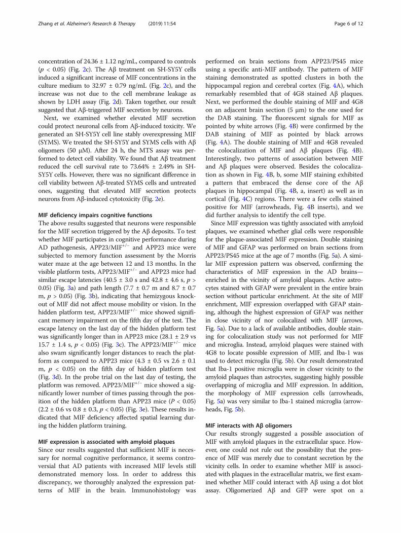
performed on brain sections from APP23/PS45 miceusing a specific anti-MIF antibody. The pattern of MIFstaining demonstrated as spotted clusters in both thehippocampal region and cerebral cortex (Fig. 4A), whichremarkably resembled that of 4G8 stained Aβ plaques.Next, we performed the double staining of MIF and 4G8on an adjacent brain section (5 μm) to the one used forthe DAB staining. The fluorescent signals for MIF aspointed by white arrows (Fig. 4B) were confirmed by theDAB staining of MIF as pointed by black arrows(Fig. 4A). The double staining of MIF and 4G8 revealedthe colocalization of MIF and Aβ plaques (Fig. 4B).Interestingly, two patterns of association between MIFand Aβ plaques were observed. Besides the colocaliza-tion as shown in Fig. 4B, b, some MIF staining exhibiteda pattern that embraced the dense core of the Aβplaques in hippocampal (Fig. 4B, a, insert) as well as incortical (Fig. 4C) regions. There were a few cells stainedpositive for MIF (arrowheads, Fig. 4B inserts), and wedid further analysis to identify the cell type.Since MIF expression was tightly associated with amyloid
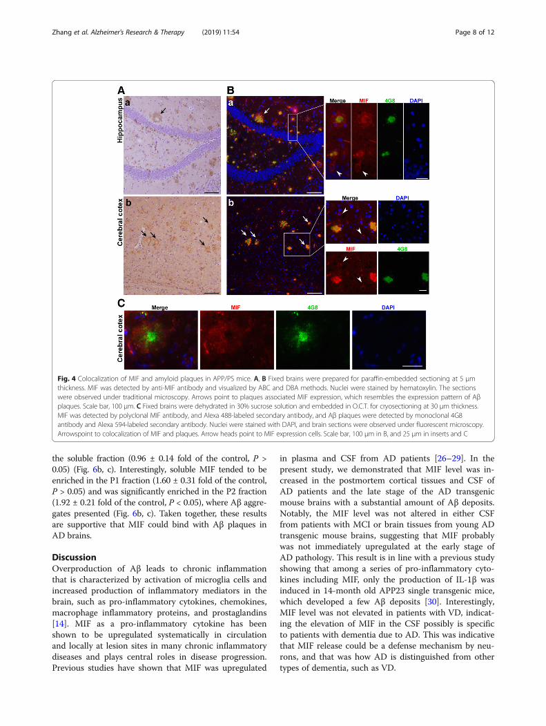
plaques, we examined whether glial cells were responsiblefor the plaque-associated MIF expression. Double stainingof MIF and GFAP was performed on brain sections fromAPP23/PS45 mice at the age of 7 months (Fig. 5a). A simi-lar MIF expression pattern was observed, confirming thecharacteristics of MIF expression in the AD brains—enriched in the vicinity of amyloid plaques. Active astro-cytes stained with GFAP were prevalent in the entire brainsection without particular enrichment. At the site of MIFenrichment, MIF expression overlapped with GFAP stain-ing, although the highest expression of GFAP was neitherin close vicinity of nor colocalized with MIF (arrows,Fig. 5a). Due to a lack of available antibodies, double stain-ing for colocalization study was not performed for MIFand microglia. Instead, amyloid plaques were stained with4G8 to locate possible expression of MIF, and Iba-1 wasused to detect microglia (Fig. 5b). Our result demonstratedthat Iba-1 positive microglia were in closer vicinity to theamyloid plaques than astrocytes, suggesting highly possibleoverlapping of microglia and MIF expression. In addition,the morphology of MIF expression cells (arrowheads,Fig. 5a) was very similar to Iba-1 stained microglia (arrow-heads, Fig. 5b).
MIF interacts with Aβ oligomersOur results strongly suggested a possible association ofMIF with amyloid plaques in the extracellular space. How-ever, one could not rule out the possibility that the pres-ence of MIF was merely due to constant secretion by thevicinity cells. In order to examine whether MIF is associ-ated with plaques in the extracellular matrix, we first exam-ined whether MIF could interact with Aβ using a dot blotassay. Oligomerized Aβ and GFP were spot on a
Zhang et al. Alzheimer's Research & Therapy (2019) 11:54 Page 6 of 12

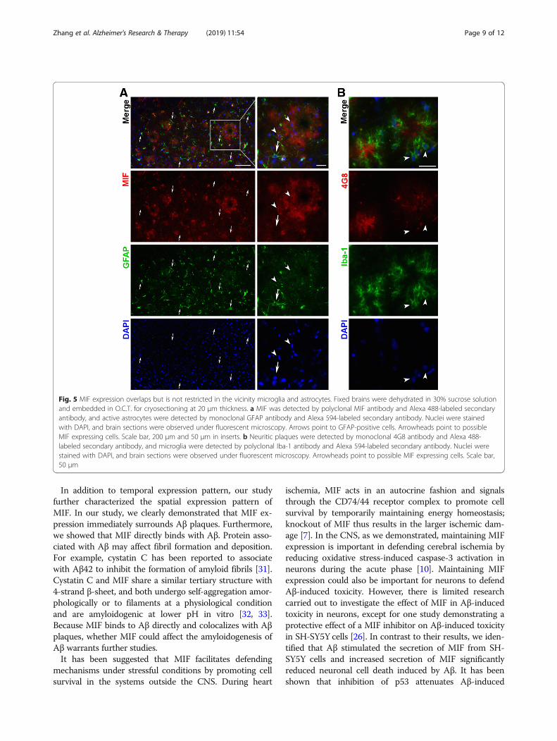
membrane and incubated with the protein mixture of MIFand GFP. GFP spotted on the membrane served as thecontrol for non-specific binding by MIF, and GFP in theprotein mixture served as the control for non-specificbinding by Aβ. The result demonstrated that MIF inter-acted with oligomerized Aβ, but neither did MIF nor Aβinteract with GFP, indicating a specific binding between oli-gomerized Aβ and MIF (Fig. 6a). We then studied whetherMIF and Aβ were associated in the AD brain by exploringwhether MIF and Aβ present at the same density fraction.Brain tissues were obtained from APP23/PS45 mice andhomogenized in PBS with 0.5% Triton X-100. The homog-enates were fractionated under 16,000×g for 15 min at 4°C. The homogenates were clearly separated into fourlayers, namely supernatant (S), pellet layer 1 (P1), pellet
layer 2 (P2), and pellet layer 3 (P3). The supernatants werecollected and subjected to ultracentrifugation at 100,000×gfor 1 h at 4 °C, and the second supernatants were collectedand labeled as S′. The layer P3 mainly consisted of non-uniform tissue debris was discarded. Subsequently, samplescollected from S′, P1, and P2 fractions were subjected toimmunoblots to locate the presence of Aβ enriched frac-tions. To study the potential association between plaquesand MIF, immunoblots were performed to detect the pres-ence of Aβ and the level of MIF in each collected fraction.Our results demonstrated that Aβ was not detectable inthe S′ fraction, but was detected in P1, P2, and P3 fractions(Fig. 6b). Concentrations of MIF were similar between con-trols and the APP23/PS45 mice in the supernatant, indicat-ing AD pathology did not increase the level of free MIF in
Fig. 3 MIF deficiency affects cognitive functions in the AD model mice. APP23/MIF+/− and APP23 mice (APP/MIF+/− and APP, respectively) at theage of 12 to 13 months were subjected to the Morris water maze test. a During the first day of visible platform tests, the APP23/MIF+/− andAPP23 mice exhibited a similar latency to escape onto the visible platform. P > 0.05 by Student’s t test. b APP23/MIF+/− and APP23 mice hadsimilar swimming distances before escaping onto the visible platform. P > 0.05 by Student’s t test. c In hidden platform tests, APP23/MIF+/− miceshowed a longer latency to escape on to the hidden platform on the 5th day. *p < 0.05 by two-way ANOVA with Bonferroni post hoc tests. dAPP23/MIF+/− mice had a shorter swimming length before escaping onto the hidden platform on the 5th day. *P < 0.05 by two-way ANOVAwith Bonferroni post hoc tests. e On the last day of the trial, APP23/MIF+/− showed a significantly lower number of passing times through thelocation of the platform than APP23 mice. P < 0.05 by Student’s t test
Zhang et al. Alzheimer's Research & Therapy (2019) 11:54 Page 7 of 12
the soluble fraction (0.96 ± 0.14 fold of the control, P >0.05) (Fig. 6b, c). Interestingly, soluble MIF tended to beenriched in the P1 fraction (1.60 ± 0.31 fold of the control,P > 0.05) and was significantly enriched in the P2 fraction(1.92 ± 0.21 fold of the control, P < 0.05), where Aβ aggre-gates presented (Fig. 6b, c). Taken together, these resultsare supportive that MIF could bind with Aβ plaques inAD brains.
DiscussionOverproduction of Aβ leads to chronic inflammationthat is characterized by activation of microglia cells andincreased production of inflammatory mediators in thebrain, such as pro-inflammatory cytokines, chemokines,macrophage inflammatory proteins, and prostaglandins[14]. MIF as a pro-inflammatory cytokine has beenshown to be upregulated systematically in circulationand locally at lesion sites in many chronic inflammatorydiseases and plays central roles in disease progression.Previous studies have shown that MIF was upregulated
in plasma and CSF from AD patients [26–29]. In thepresent study, we demonstrated that MIF level was in-creased in the postmortem cortical tissues and CSF ofAD patients and the late stage of the AD transgenicmouse brains with a substantial amount of Aβ deposits.Notably, the MIF level was not altered in either CSFfrom patients with MCI or brain tissues from young ADtransgenic mouse brains, suggesting that MIF probablywas not immediately upregulated at the early stage ofAD pathology. This result is in line with a previous studyshowing that among a series of pro-inflammatory cyto-kines including MIF, only the production of IL-1β wasinduced in 14-month old APP23 single transgenic mice,which developed a few Aβ deposits [30]. Interestingly,MIF level was not elevated in patients with VD, indicat-ing the elevation of MIF in the CSF possibly is specificto patients with dementia due to AD. This was indicativethat MIF release could be a defense mechanism by neu-rons, and that was how AD is distinguished from othertypes of dementia, such as VD.
Fig. 4 Colocalization of MIF and amyloid plaques in APP/PS mice. A, B Fixed brains were prepared for paraffin-embedded sectioning at 5 μmthickness. MIF was detected by anti-MIF antibody and visualized by ABC and DBA methods. Nuclei were stained by hematoxylin. The sectionswere observed under traditional microscopy. Arrows point to plaques associated MIF expression, which resembles the expression pattern of Aβplaques. Scale bar, 100 μm. C Fixed brains were dehydrated in 30% sucrose solution and embedded in O.C.T. for cryosectioning at 30 μm thickness.MIF was detected by polyclonal MIF antibody, and Alexa 488-labeled secondary antibody, and Aβ plaques were detected by monoclonal 4G8antibody and Alexa 594-labeled secondary antibody. Nuclei were stained with DAPI, and brain sections were observed under fluorescent microscopy.Arrowspoint to colocalization of MIF and plaques. Arrow heads point to MIF expression cells. Scale bar, 100 μm in B, and 25 μm in inserts and C
Zhang et al. Alzheimer's Research & Therapy (2019) 11:54 Page 8 of 12
In addition to temporal expression pattern, our studyfurther characterized the spatial expression pattern ofMIF. In our study, we clearly demonstrated that MIF ex-pression immediately surrounds Aβ plaques. Furthermore,we showed that MIF directly binds with Aβ. Protein asso-ciated with Aβ may affect fibril formation and deposition.For example, cystatin C has been reported to associatewith Aβ42 to inhibit the formation of amyloid fibrils [31].Cystatin C and MIF share a similar tertiary structure with4-strand β-sheet, and both undergo self-aggregation amor-phologically or to filaments at a physiological conditionand are amyloidogenic at lower pH in vitro [32, 33].Because MIF binds to Aβ directly and colocalizes with Aβplaques, whether MIF could affect the amyloidogenesis ofAβ warrants further studies.It has been suggested that MIF facilitates defending
mechanisms under stressful conditions by promoting cellsurvival in the systems outside the CNS. During heart
ischemia, MIF acts in an autocrine fashion and signalsthrough the CD74/44 receptor complex to promote cellsurvival by temporarily maintaining energy homeostasis;knockout of MIF thus results in the larger ischemic dam-age [7]. In the CNS, as we demonstrated, maintaining MIFexpression is important in defending cerebral ischemia byreducing oxidative stress-induced caspase-3 activation inneurons during the acute phase [10]. Maintaining MIFexpression could also be important for neurons to defendAβ-induced toxicity. However, there is limited researchcarried out to investigate the effect of MIF in Aβ-inducedtoxicity in neurons, except for one study demonstrating aprotective effect of a MIF inhibitor on Aβ-induced toxicityin SH-SY5Y cells [26]. In contrast to their results, we iden-tified that Aβ stimulated the secretion of MIF from SH-SY5Y cells and increased secretion of MIF significantlyreduced neuronal cell death induced by Aβ. It has beenshown that inhibition of p53 attenuates Aβ-induced
Fig. 5 MIF expression overlaps but is not restricted in the vicinity microglia and astrocytes. Fixed brains were dehydrated in 30% sucrose solutionand embedded in O.C.T. for cryosectioning at 20 μm thickness. a MIF was detected by polyclonal MIF antibody and Alexa 488-labeled secondaryantibody, and active astrocytes were detected by monoclonal GFAP antibody and Alexa 594-labeled secondary antibody. Nuclei were stainedwith DAPI, and brain sections were observed under fluorescent microscopy. Arrows point to GFAP-positive cells. Arrowheads point to possibleMIF expressing cells. Scale bar, 200 μm and 50 μm in inserts. b Neuritic plaques were detected by monoclonal 4G8 antibody and Alexa 488-labeled secondary antibody, and microglia were detected by polyclonal Iba-1 antibody and Alexa 594-labeled secondary antibody. Nuclei werestained with DAPI, and brain sections were observed under fluorescent microscopy. Arrowheads point to possible MIF expressing cells. Scale bar,50 μm
Zhang et al. Alzheimer's Research & Therapy (2019) 11:54 Page 9 of 12
neuronal apoptosis [34], and MIF can directly inhibit p53activation [35]. Thus, secretion of MIF precedes cell dam-age, and MIF is released under the autocrine fashion, inturn, activates cell survival signals.It should be noted that MIF acts in an autocrine fashion
and interacts with its cell surface receptors to transduce itssignals [36, 37]; therefore, secretion of MIF to the extracellu-lar space is necessary for MIF to exert its cellular functions.It has been suggested that MIF is constitutively expressedand is perhaps secreted constantly by neurons in the brain[38, 39]. However, at the late stage of AD, a large portion ofthe extracellular MIF is sequestered by Aβ plaques, as wedemonstrated, and perhaps has no functions anymore,which suggests that increased MIF secretion is essential formaintaining cognitive function during AD development.This also explains why MIF upregulation is observed at thelate stage of AD with significantly increased Aβ deposition.We found that neurons specifically secreted MIF fol-
lowing Aβ stimulation. However, we did not rule out thepossibility that upregulation of MIF is also contributedby microglia (local or infiltrated) at the late stage of AD,at which pro-inflammatory cytokines are predominantlyproduced [14], and they are known to trigger secretionof MIF [25]. Indeed, recent studies demonstrated a mix-ture of hyperactivated microglia at the late stage of ADpathology [40] and the upregulation of MIF in hyperacti-vated microglia [41]. We speculated that at the late stageof AD pathology, overproduced MIF could still be bene-ficial in promoting neuronal survival if it can be receivedby neurons; on the other hand, however, the pool ofMIF produced by immune cells may locally promotetheir own survival and proliferation, which in turn pro-duce and release more MIF, leading to a vicious circle asseen in chronic inflammatory diseases in the peripheral.Although it is debatable whether Aβ deposits serve as a
cause of cognitive decline during AD, inhibition of Aβ pro-duction and plaque formation have been shown to success-fully mitigate cognitive deficits in AD model mice [17, 18,42, 43]. Since MIF insufficiency had an impact on cognitiveperformance [44], we hypothesized that Aβ-triggered MIFsecretion could serve as a compensatory mechanism to im-prove cognitive performance during AD. In contrast, inhib-ition of MIF has been suggested for treating peripheralinflammatory diseases [13, 26], indicating the possibilitythat the potential pro-survival and pro-inflammatory func-tions of MIF counteract each other during AD, and inhib-ition of one would tip a balance to the other. It is possiblethat at the early stage with little Aβ plaques formation, MIFsecretion triggered by Aβ oligomer is pro-survival for neu-rons to maintain cognitive function; while at the late stagewith abundant Aβ plaques, the compensatory function mayfail to demonstrate effects due to the direct binding be-tween MIF and Aβ, leaving behind the pro-inflammatoryeffects. Therefore, it will be of importance to dissect the
Fig. 6 MIF interacts with Aβ oligomers. a 0.2 nmol of Aβ oligomersand 0.1 nmol of purified GFP protein were spot on a nitrocellulosemembrane, and the membrane was incubation with mixed proteinsof recombinant hMIF and purified GFP at the concentration ofapproximated 5 μM at 4 °C for overnight. The membrane was thensubjected to immunoblotting to detect MIF and GFP by a monoclonalanti-MIF antibody and a polyclonal anti-GFP antibody, respectively. Thered channel detects IR-dye-labeled goat anti-rabbit antibody, and thegreen channel detects IR-dye-labeled goat anti-mouse antibody. bBrains from 4-month-old APP23/PS45 and wildtype (as controls) micewere homogenized in 5x PBS with 0.5% Triton-100 (v/w) andcentrifuge at 16,000×g at 4 °C for 15 min. The supernatants werecollected and subjected to ultracentrifugation at 100,000×g for 1 h at 4°C, and the second supernatants were collected and labeled as S′.Homogenates from each layer were further dissolved in RIPA-DOCbuffer followed by brief sonication. The same amount of protein wasloaded on 16% and 12% Tris-tricine SDS PAGE gels for Aβ and MIFseparation, respectively. Aβ was detected by a 6E10 antibody, MIF wasdetected by anti-MIF antibody, CTFs were detected by a C20 antibodyto confirm the expression of the transgene, and β-actin was detectedby β-actin antibody serving as the loading control. S′, supernatant afterultracentrifugation; P1, pellet 1; P2 pellet 2. c Quantification of the levelof MIF protein from b. Values represent mean ± SEM, n = 3. *P < 0.05by Student’s t test
Zhang et al. Alzheimer's Research & Therapy (2019) 11:54 Page 10 of 12

MIF signal complex so that the deleterious effects could beinhibited without affecting the beneficial ones.
ConclusionsIn summary, we found that the upregulation of MIF expres-sion specifically occurs in patients with AD rather thanother types of dementia and identified that the level of MIFin CSF could serve as a biomarker of AD with global inflam-mation. In addition, hemizygous knockout of MIF exacer-bated memory task performance of APP23 AD model mice.More importantly, we for the first time revealed the directinteraction between MIF and Aβ. Taken together, our dataprovided first-hand evidence of MIF expression profile dur-ing AD and its effect on AD development. Our study sug-gested that proper regulation of MIF expression, secretion,and function is essential for the successful treatment of AD.
AbbreviationsAD: Alzheimer’s disease; APP: Amyloid β precursor proteins; Aβ: Amyloid βprotein; MIF: Macrophage migration inhibitory factor
Authors’ contributionsLW and WS conceived and designed the experiments. SZ, JZ, YuhuZ, andYunZ performed the experiments. SZ, JZ, YuhuZ, YunZ, FC, LW, and WSanalyzed and contributed reagents/materials/analysis tools. SZ, YunZ, and WSwrote the paper. All authors reviewed the manuscript. All authors read andapproved the final manuscript.
FundingThis work was supported by Canadian Institutes of Health Research (CIHR)Operating Grant MOP-142487 to WS. W.S. was the holder of the Tier 1Canada Research Chair in Alzheimer’s Disease. S.Z. is supported by UBC 4YFScholarship. YunZ. is the recipient of Michael Smith Foundation for HealthResearch Post-Doctoral Fellowship Award.
Availability of data and materialsThe authors agree the availability upon request.
Ethics approval and consent to participateThe study was approved by the Institutional Ethics Committee of theGuangdong General Hospital. Animal experiments were approved by andconducted in accordance with the University of British Columbia AnimalCare and Use Committee (Vancouver, BC, Canada) and the CanadianInstitutes of Health Research (CIHR) guidelines.
Consent for publicationN/A
Competing interestsThe authors declare that they have no competing interests.
Received: 21 January 2019 Accepted: 21 May 2019
References1. Zhang S, Wang Z, Cai F, Zhang M, Wu Y, Zhang J, Song W. BACE1 cleavage
site selection critical for amyloidogenesis and Alzheimer’s pathogenesis. JNeurosci. 2017;37(29):6915–25.
2. Zhang Y, Song W. Islet amyloid polypeptide: another key molecule inAlzheimer's pathogenesis? Prog Neurobiol. 2017;153:100–20.
3. Wyss-Coray T, Rogers J. Inflammation in Alzheimer disease-a brief review ofthe basic science and clinical literature. Cold Spring Harb Perspect Med.2012;2(1):a006346.
4. Colton CA. Heterogeneity of microglial activation in the innate immuneresponse in the brain. J Neuroimmune Pharmacol. 2009;4(4):399–418.
5. Koga K, Kenessey A, Powell SR, Sison CP, Miller EJ, Ojamaa K. Macrophagemigration inhibitory factor provides cardioprotection during ischemia/reperfusion by reducing oxidative stress. Antioxid Redox Signal. 2011;14(7):1191–202.
6. Luedike P, Hendgen-Cotta UB, Sobierajski J, Totzeck M, Reeh M, Dewor M, LueH, Krisp C, Wolters D, Kelm M, et al. Cardioprotection through S-nitros(yl)ationof macrophage migration inhibitory factor. Circulation. 2012;125(15):1880–9.
7. Miller EJ, Li J, Leng L, McDonald C, Atsumi T, Bucala R, Young LH.Macrophage migration inhibitory factor stimulates AMP-activated proteinkinase in the ischaemic heart. Nature. 2008;451(7178):578–82.
8. Wang L, Zis O, Ma G, Shan Z, Zhang X, Wang S, Dai C, Zhao J, Lin Q, Lin S,et al. Upregulation of macrophage migration inhibitory factor geneexpression in stroke. Stroke. 2009;40(3):973–6.
9. Zis O, Zhang S, Dorovini-Zis K, Wang L, Song W. Hypoxia signaling regulatesmacrophage migration inhibitory factor (MIF) expression in stroke. MolNeurobiol. 2015;51(1):155–67.
10. Zhang S, Zis O, Ly PT, Wu Y, Zhang M, Cai F, Bucala R, Shyu WC, Song W.Down-regulation of MIF by NFkappaB under hypoxia accelerated neuronalloss during stroke. FASEB J. 2014;28(10):4394–407.
11. Wang Y, An R, Umanah GK, Park H, Nambiar K, Eacker SM, Kim B, Bao L, HarrazMM, Chang C, et al. A nuclease that mediates cell death induced by DNAdamage and poly(ADP-ribose) polymerase-1. Science. 2016;354(6308):aad6872.
12. Eckert A, Keil U, Marques CA, Bonert A, Frey C, Schüssel K, Müller WE.Mitochondrial dysfunction, apoptotic cell death, and Alzheimer's disease.Biochem Pharmacol. 2003;66(8):1627–34.
13. Morand EF, Leech M, Bernhagen J. MIF: a new cytokine link between rheumatoidarthritis and atherosclerosis. Nat Rev Drug Discov. 2006;5(5):399–410.
14. Rubio-Perez JM, Morillas-Ruiz JM. A review: inflammatory process inAlzheimer's disease, role of cytokines. ScientificWorldJournal. 2012;2012:756357.
15. Sun X, He G, Qing H, Zhou W, Dobie F, Cai F, Staufenbiel M, Huang LE, SongW. Hypoxia facilitates Alzheimer's disease pathogenesis by up-regulatingBACE1 gene expression. Proc Natl Acad Sci U S A. 2006;103(49):18727–32.
16. Sturchler-Pierrat C, Abramowski D, Duke M, Wiederhold KH, Mistl C,Rothacher S, Ledermann B, Burki K, Frey P, Paganetti PA, et al. Two amyloidprecursor protein transgenic mouse models with Alzheimer disease-likepathology. Proc Natl Acad Sci U S A. 1997;94(24):13287–92.
17. Qing H, He G, Ly PT, Fox CJ, Staufenbiel M, Cai F, Zhang Z, Wei S, Sun X,Chen CH, et al. Valproic acid inhibits Abeta production, neuritic plaqueformation, and behavioral deficits in Alzheimer's disease mouse models. JExp Med. 2008;205(12):2781–9.
18. Ly PT, Wu Y, Zou H, Wang R, Zhou W, Kinoshita A, Zhang M, Yang Y, Cai F,Woodgett J, et al. Inhibition of GSK3β-mediated BACE1 expression reducesAlzheimer-associated phenotypes. J Clin Invest. 2013;123(1):224–35.
19. Zeng J, Chen L, Wang Z, Chen Q, Fan Z, Jiang H, Wu Y, Ren L, Chen J, Li T,et al. Marginal vitamin A deficiency facilitates Alzheimer's pathogenesis.Acta Neuropathol. 2017;133(6):967–82.
20. Stine WB Jr, Dahlgren KN, Krafft GA, LaDu MJ. In vitro characterization ofconditions for amyloid-beta peptide oligomerization and fibrillogenesis. JBiol Chem. 2003;278(13):11612–22.
21. Fa M, Orozco IJ, Francis YI, Saeed F, Gong Y, Arancio O. Preparation ofoligomeric beta-amyloid 1-42 and induction of synaptic plasticityimpairment on hippocampal slices. J Vis Exp. 2010;41:1884.
22. Dong Z, Han H, Li H, Bai Y, Wang W, Tu M, Peng Y, Zhou L, He W, Wu X, etal. Long-term potentiation decay and memory loss are mediated by AMPARendocytosis. J Clin Invest. 2015;125(1):234–47.
23. Huang D, Yu M, Yang S, Lou D, Zhou W, Zheng L, Wang Z, Cai F, Zhou W,Li T et al. Ethanol Alters APP Processing and Aggravates Alzheimer-Associated Phenotypes. Mol Neurobiol. 2018;55(6):5006-18.
24. Flieger O, Engling A, Bucala R, Lue H, Nickel W, Bernhagen J. Regulatedsecretion of macrophage migration inhibitory factor is mediated by a non-classical pathway involving an ABC transporter. FEBS Lett. 2003;551(1–3):78–86.
25. Calandra T, Bernhagen J, Mitchell RA, Bucala R. The macrophage is animportant and previously unrecognized source of macrophage migrationinhibitory factor. J Exp Med. 1994;179(6):1895–902.
26. Bacher M, Deuster O, Aljabari B, Egensperger R, Neff F, Jessen F, Popp J,Noelker C, Reese JP, Al-Abed Y, et al. The role of macrophage migrationinhibitory factor in Alzheimer's disease. Mol Med. 2010;16(3–4):116–21.
27. Craig-Schapiro R, Kuhn M, Xiong C, Pickering EH, Liu J, Misko TP, Perrin RJ,Bales KR, Soares H, Fagan AM, et al. Multiplexed immunoassay panelidentifies novel CSF biomarkers for Alzheimer's disease diagnosis andprognosis. PLoS One. 2011;6(4):e18850.
Zhang et al. Alzheimer's Research & Therapy (2019) 11:54 Page 11 of 12

28. Popp J, Bacher M, Kolsch H, Noelker C, Deuster O, Dodel R, Jessen F.Macrophage migration inhibitory factor in mild cognitive impairment andAlzheimer's disease. J Psychiatr Res. 2009;43(8):749–53.
29. Lee KS, Chung JH, Lee KH, Shin MJ, Oh BH, Hong CH. Bioplex analysis ofplasma cytokines in Alzheimer's disease and mild cognitive impairment.Immunol Lett. 2008;121(2):105–9.
30. Mehlhorn G, Hollborn M, Schliebs R. Induction of cytokines in glial cellssurrounding cortical beta-amyloid plaques in transgenic Tg2576 mice withAlzheimer pathology. Int J Dev Neurosci. 2000;18(4–5):423–31.
31. Sastre M, Calero M, Pawlik M, Mathews PM, Kumar A, Danilov V, Schmidt SD,Nixon RA, Frangione B, Levy E. Binding of cystatin C to Alzheimer's amyloid betainhibits in vitro amyloid fibril formation. Neurobiol Aging. 2004;25(8):1033–43.
32. Levy E, Jaskolski M, Grubb A. The role of cystatin C in cerebral amyloid angiopathyand stroke: cell biology and animal models. Brain Pathol. 2006;16(1):60–70.
33. Lashuel HA, Aljabari B, Sigurdsson EM, Metz CN, Leng L, Callaway DJ, BucalaR. Amyloid fibril formation by macrophage migration inhibitory factor.Biochem Biophys Res Commun. 2005;338(2):973–80.
34. Culmsee C, Zhu X, Yu QS, Chan SL, Camandola S, Guo Z, Greig NH, MattsonMP. A synthetic inhibitor of p53 protects neurons against death induced byischemic and excitotoxic insults, and amyloid beta-peptide. J Neurochem.2001;77(1):220–8.
35. Hudson JD, Shoaibi MA, Maestro R, Carnero A, Hannon GJ, Beach DH. Aproinflammatory cytokine inhibits p53 tumor suppressor activity. J Exp Med.1999;190(10):1375–82.
36. Bernhagen J, Krohn R, Lue H, Gregory JL, Zernecke A, Koenen RR, Dewor M,Georgiev I, Schober A, Leng L, et al. MIF is a noncognate ligand of CXCchemokine receptors in inflammatory and atherogenic cell recruitment. NatMed. 2007;13(5):587–96.
37. Leng L, Metz CN, Fang Y, Xu J, Donnelly S, Baugh J, Delohery T, Chen Y,Mitchell RA, Bucala R. MIF signal transduction initiated by binding to CD74.J Exp Med. 2003;197(11):1467–76.
38. Bacher M, Meinhardt A, Lan HY, Dhabhar FS, Mu W, Metz CN, Chesney JA,Gemsa D, Donnelly T, Atkins RC, et al. MIF expression in the rat brain:implications for neuronal function. Mol Med. 1998;4(4):217–30.
39. Ogata A, Nishihira J, Suzuki T, Nagashima K, Tashiro K. Identification ofmacrophage migration inhibitory factor mRNA expression in neural cells ofthe rat brain by in situ hybridization. Neurosci Lett. 1998;246(3):173–7.
40. Yin Z, Raj D, Saiepour N, Van Dam D, Brouwer N, Holtman IR, Eggen BJL, MollerT, Tamm JA, Abdourahman A, et al. Immune hyperreactivity of Abeta plaque-associated microglia in Alzheimer's disease. Neurobiol Aging. 2017;55:115–22.
41. Kamphuis W, Kooijman L, Schetters S, Orre M, Hol EM. Transcriptionalprofiling of CD11c-positive microglia accumulating around amyloid plaquesin a mouse model for Alzheimer’s disease. Biochim Biophys Acta. 2016;1862(10):1847–60.
42. Richard KL, Filali M, Préfontaine P, Rivest S. Toll-like receptor 2 acts as anatural innate immune receptor to clear amyloid beta 1-42 and delay thecognitive decline in a mouse model of Alzheimer's disease. J Neurosci.2008;28(22):5784–93.
43. Sevigny J, Chiao P, Bussiere T, Weinreb PH, Williams L, Maier M, Dunstan R,Salloway S, Chen T, Ling Y, et al. The antibody aducanumab reduces Abetaplaques in Alzheimer's disease. Nature. 2016;537(7618):50–6.
44. Conboy L, Varea E, Castro JE, Sakouhi-Ouertatani H, Calandra T, Lashuel HA,Sandi C. Macrophage migration inhibitory factor is critically involved inbasal and fluoxetine-stimulated adult hippocampal cell proliferation and inanxiety, depression, and memory-related behaviors. Mol Psychiatry. 2011;16(5):533–47.
Publisher’s NoteSpringer Nature remains neutral with regard to jurisdictional claims inpublished maps and institutional affiliations.
Zhang et al. Alzheimer's Research & Therapy (2019) 11:54 Page 12 of 12