Embed Size (px)
Citation preview
Verlag Dr. Friedrich PfeilISSN 0936-9902
Ichthyological Exploration of Freshwaters
Volume 23Number 3
An international journal for field-orientated ichthyology
Ichthyological Exploration of FreshwatersAn international journal for field-orientated ichthyology
Volume 23 • Number 3 • November 2012pages 193-288, 39 figs., 11 tabs.
Managing Editor Maurice Kottelat, Route de la Baroche 12, Case postale 57 CH-2952 Cornol, Switzerland Tel. + 41 32 4623175 · Fax + 41 32 4622259 · E-mail [email protected]
Editorial board Pier Giorgio Bianco, Dipartimento di Zoologia, Università, Napoli, Italy Ralf Britz, Department of Zoology, The Natural History Museum, London, United Kingdom Sven O. Kullander, Naturhistoriska Riksmuseet, Stockholm, Sweden Helen K. Larson, Museum and Art Gallery of the Northern Territory, Darwin, Australia Lukas Rüber, Department of Zoology, The Natural History Museum, London, United Kingdom Ivan Sazima, Museu de Zoologia, Unicamp, Campinas, Brazil Paul H. Skelton, South African Institute for Aquatic Biodiversity, Grahamstown, South Africa Tan Heok Hui, Raffles Museum of Biodiversity Research, National University of Singapore, Singapore
Ichthyological Exploration of Freshwaters is published quarterly
Subscriptions should be addressed to the Publisher:
Verlag Dr. Friedrich Pfeil, Wolfratshauser Str. 27, 81379 München, Germany PERSONAL SUBSCRIPTION : EURO 100 per Year/volume - 4 issues (includes surface mail shipping) INSTITUTIONAL SUBSCRIPTION : EURO 180 per Year/volume - 4 issues (includes surface mail shipping)
Manuscripts should be addressed to the Managing Editor:Maurice Kottelat, Route de la Baroche 12, Case postale 57, CH -2952 Cornol, Switzerland
CIP-Titelaufnahme der Deutschen Bibliothek
Ichthyological exploration of freshwaters : an internationaljournal for field-orientated ichthyology. – München : Pfeil.
Erscheint jährl. viermal. – Aufnahme nach Vol. 1, No. 1 (1990)ISSN 0936-9902
Vol. 1, No. 1 (1990) –
Copyright © 2012 by Verlag Dr. Friedrich Pfeil, München, Germany
All rights reserved.No part of this publication may be reproduced, stored in a retrieval system, or transmitted in any form or by anymeans, electronic, mechanical, photocopying or otherwise, without the prior permission of the copyright owner. Applications for such permission, with a statement of the purpose and extent of the reproduction, should be
addressed to the Publisher, Verlag Dr. Friedrich Pfeil, Wolfratshauser Str. 27, 81379 München, Germany.
Printed by Advantage Printpool, Gilching
ISSN 0936-9902Printed in the European Union
Verlag Dr. Friedrich Pfeil, Wolfratshauser Str. 27, 81379 München, GermanyPhone + 49 89 742827-0 · Fax + 49 89 7242772 · E-mail: [email protected] · www.pfeil-verlag.de
245
Ichthyol. Explor. Freshwaters, Vol. 23, No. 3
Description of Danio flagrans, and redescription of D. choprae, two closely related species
from the Ayeyarwaddy River drainage in northern Myanmar (Teleostei: Cyprinidae)
Sven O. Kullander*
Danio flagrans, new species, is described from headwaters of the Mali Hka River in the vicinity of Putao in north-ern Myanmar. It is distinguished from D. choprae by longer barbels, longer caudal peduncle, shorter anal-fin base, more caudal vertebrae, fewer anal-fin rays, short vs. usually absent lateral line, details of the colour pattern, and mitochondrial DNA sequences. The two species share a unique colour pattern combining dark vertical bars an-teriorly on the side with dark horizontal stripes postabdominally, and brilliant red or orange interstripes anteri-orly and posteriorly on the side. Pointed tubercles on the infraorbital bones are observed in both species, but were found to be mostly present and prominent in D. choprae and mostly absent in D. flagrans, and are considered as possibly being seasonal in expression. Danio choprae is known from three localities along the Mogaung Chaung southwest of Myitkyina.
* Department of Vertebrate Zoology, Swedish Museum of Natural History, PO Box 50007, SE-104 05 Stockholm, Sweden. E-mail: [email protected]
Introduction
The cyprinid fish genus Danio includes 16 valid species in South and South East Asia (Fang Kul-lander, 2001; Kullander et al., 2009; Kullander & Fang, 2009a,b). Ten valid species have been re-ported from Myanmar, including D. aesculapii Kullander & Fang, 2009, D. albolineatus (Blyth, 1860), D. choprae Hora, 1928, D. erythromicron (Annandale, 1918), D. feegradei Hora, 1937, D. ky-athit Fang, 1998, D. margaritatus (Roberts, 2007), D. nigrofasciatus (Day, 1870), D. quagga Kullander, Liao & Fang, 2009, and D. tinwini Kullander & Fang, 2009 (Kullander et al., 2009; Kullander & Fang, 2009a-b). Species of Danio have species specific colour
patterns, commonly in the form of horizontal stripes, more rarely light or dark spots, or vertical bars. Danio choprae, described from near Myitkyi-na on the Ayeyarwaddy River in northern My-anmar is remarkable for its distinctive colour pattern of dark vertical bars combined with strik-ing red horizontal interstripes giving it the name glowlight danio in the aquarium hobby (Cottle, 2010). Specimens identified as D. choprae or as a similar species have been reported from Putao, much further north in the Ayeyarwaddy River drainage (Kullander et al., 2009; Cottle, 2010). Glowlight danios from Putao were recently in-troduced in the aquarium hobby as Danio cf. choprae (Cottle, 2010). Morphological and DNA analyses of samples of glowlight danios from
Ichthyol. Explor. Freshwaters, Vol. 23, No. 3, pp. 245-262, 12 figs., 2 tabs., November 2012© 2012 by Verlag Dr. Friedrich Pfeil, München, Germany – ISSN 0936-9902
246
Kullander: Danio flagrans and D. choprae
Putao show that they represent a distinct species. The description of the new species and a rede-scription of D. choprae form the objectives of the present paper.
Material and methods
Specimens are kept in the fish collections of the Swedish Museum of Natural History, Stockholm (NRM), the Natural History Museum in London (BMNH), and the Zoological Survey of India in Kolkata (ZSI). Measurements were taken with digital callipers to a precision of 0.1 mm. Counts and measurements were made according to Fang (1997a). Colour pattern terminology follows Fang (1998). Horizontal dark stripes are identified by alphanumeric annotations: the P stripe is the dark stripe along the middle of the side, those above are numbered P+1, P+2, those below P-1, P-2, P-3; stripes on the anal fin are numbered with the middle one the A stripe, the proximal stripe A+1, and the distal stripe A-1. The term interstripe, used by Quigley et al. (2005) for xanthophore-rich areas between dark melanophore-rich stripes, is adopted here, but without numbering. Fin-ray counts from pectoral, pelvic, dorsal and anal fins were obtained directly from the specimens under a dissection microscope and with throughfalling light. Fin-ray counts from the caudal fin and vertebral counts were taken from X-radiographs made with a Philips MG-105 low voltage X-ray unit and Kodak X-Omat V plates. Abdominal vertebrae counts include the Weberian apparatus (assumed to contain four centra). Sexes were separated by the presence in males vs. absence in females of tubercles on the pectoral fin. When adults of both sexes are present in the same sam-ple this is a reliable criterion correlated with fuller abdomen in females; in samples of adults in which pectoral-fin tubercles are consistently absent, thickened interradial tissue may indicate males, but otherwise sex is recorded as indeter-minable if no other sex dimorphism is present. Statistics were calculated using IBM Statistics v. 20 (IBM, 2011). Photographs of morphological detail were taken with a Leica M165C stereo microscope with motor stand, Leica DFC450 camera, and composed with Leica Application Suite 4.0 multi-focus montage software. DNA sequences of D. choprae and D. flagrans were downloaded from GenBank. Only cytochrome b (D. choprae: Gen-Bank accession numbers EF452740, HM224264;
D. flagrans: EU241421), and rhodopsin (D. choprae: HM223904, JQ614128-614130; D. flagrans: EU 241356, JQ614112-614113) gene sequences were available for both species. Alignment using the ClustalW algorithm, and calculation of nucleotide divergence as uncorrected p distance was made in the Geneious computer software (Drummond et al., 2009).
Danio choprae Hora, 1928(Figs. 1a-b)
Material examined. Myanmar: Kachin State: Ayeyar-waddy River basin: Mogaung River drainage: BMNH 2011.3.25.5-22, 20 (4 males, 22.1-25.2 mm SL, 14 females, 23.2-29.7 mm SL); Mogaung area; U Tin Win, Feb 2007. – BMNH 2012.7.23.148-203, 56 (28 examined, 7 males 18.7-22.0 mm SL, 19 females, 16.7-23.0 mm SL); small stream and pond south of Mogaung, 25°15.583' N 96°57.374' E, 145 masl; R. Britz, O. Crimmen and local fishermen, 23 Feb 2011. – NRM 52001, 76 (41 males, 25.0-29.0 mm SL; 35 females, 24.3-30.7 mm SL); hill stream around Kamaing; Hla Ku & Mg Nyo, 17 May 2004. – NRM 51965, 2, not measured; hill stream around Kamaing; U Tin Win, 31 Oct 2004. – ZSI F10811/1, 1, 22.0 mm SL; holotype of D. choprae (photograph only); small rocky stream round about Kamaing; B. N. Chopra, 23-30 Dec 1926. Aquarium specimens: NRM 50141-50143, 3, cleared and stained; NRM 48378, 1, tissue sample; NRM 48377, 6, not measured; 2002. – NRM 51832, 1, not measured; 2004.
Diagnosis. Danio choprae is similar to D. flagrans in general colour pattern with several vertical bars on abdominal sides, followed by postab-dominal P and P+1 stripes, distinct P+2 stripe anterior to dorsal fin, and red interstripes between middorsal and P+2 stripes, and between P and P+1 stripes; and presence of well-developed tu-bercles on infraorbital ossicles. It is distinguished from D. flagrans by slightly deeper body (26.6-31.6 % SL vs. 22.5-26.6 % SL), shorter caudal peduncle (16.1-19.1 % SL vs. 20.5-24.7 % SL), longer anal-fin base (19.2-23.9 % SL vs. 14.2-18.3 % SL), shorter rostral barbel (5.9-10.1 % SL vs. 10.3-18.7 % SL), not reaching posterior margin of orbit in adults (vs. reaching caudally beyond preopercular margin); shorter maxillary barbels not reaching to pectoral-fin base in adults (vs. reaching to below pectoral-fin base), lateral line almost always absent, occasionally on up to three scales (vs. almost always present, on up to seven scales), fewer vertebrae contained in caudal pe-duncle (6-8 vs. 9-10), more anal-fin rays (12 1/2-
247
Ichthyol. Explor. Freshwaters, Vol. 23, No. 3
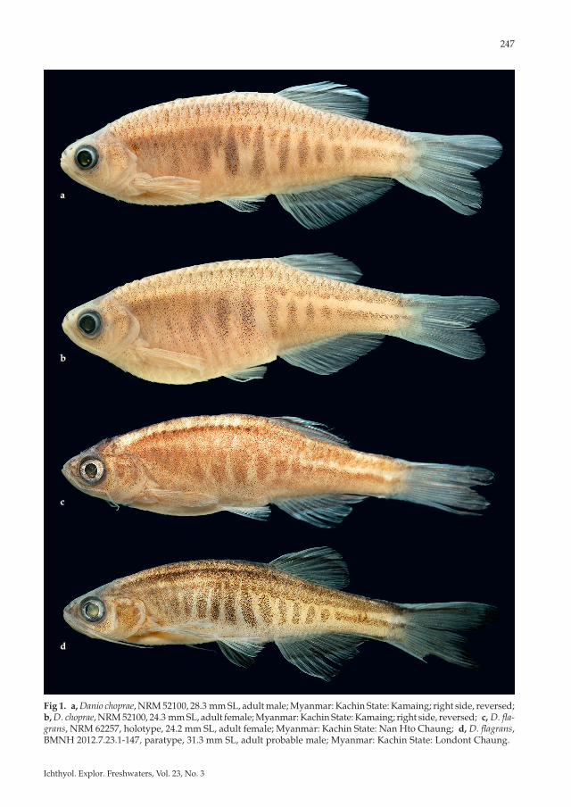
Fig 1. a, Danio choprae, NRM 52100, 28.3 mm SL, adult male; Myanmar: Kachin State: Kamaing; right side, reversed; b, D. choprae, NRM 52100, 24.3 mm SL, adult female; Myanmar: Kachin State: Kamaing; right side, reversed; c, D. fla-grans, NRM 62257, holotype, 24.2 mm SL, adult female; Myanmar: Kachin State: Nan Hto Chaung; d, D. flagrans, BMNH 2012.7.23.1-147, paratype, 31.3 mm SL, adult probable male; Myanmar: Kachin State: Londont Chaung.
aa
bb
cc
dd
248
13 1/2, rarely 11 1/2 branched rays vs. 9 1/2-11 1/2), anal-fin base dark (vs. light); and black streak usually present on lower lobe of caudal fin (vs. absent). It is distinguished from all other species of Danio by characters in combination: rostral barbel present (absent in D. erythromicron, D. mar-garitatus, D. nigrofasciatus, D. tinwini, variable in D. rerio); mandibular barbel present (absent in D. erythromicron and D. margaritatus); lateral line abbreviated or absent (complete in D. dangila, D. feegradei, D. meghalayensis; absent in D. erythro-micron, D. margaritatus, D. nigrofasciatus, D. rerio, D. tinwini), colour pattern consisting of vertical bars or spots anteriorly on side, horizontal stripes posteriorly on side (only vertical bars in D. eryth-romicron; light spots on dark ground in D dangila and D. margaritatus; dark spots on light ground in D. kyathit and D. tinwini; horizontal stripes only in D. rerio, D. jaintianensis, D. quagga, D. meghalay-ensis, D. nigrofasciatus, D. kerri, D. albolineatus, D. roseus; bars anteriorly, two horizontal rows of spots posteriorly in D. aesculapii); branched dorsal-fin rays 7 1/2 (6 1/2 in D. aesculapii and D. tinwini); circumpeduncular scale rows 10 (12 in D. aescu-
lapii, D. albolineatus, D. erythromicron, D. kerri; 14 in D. dangila, D. feegradei, D. meghalayensis).
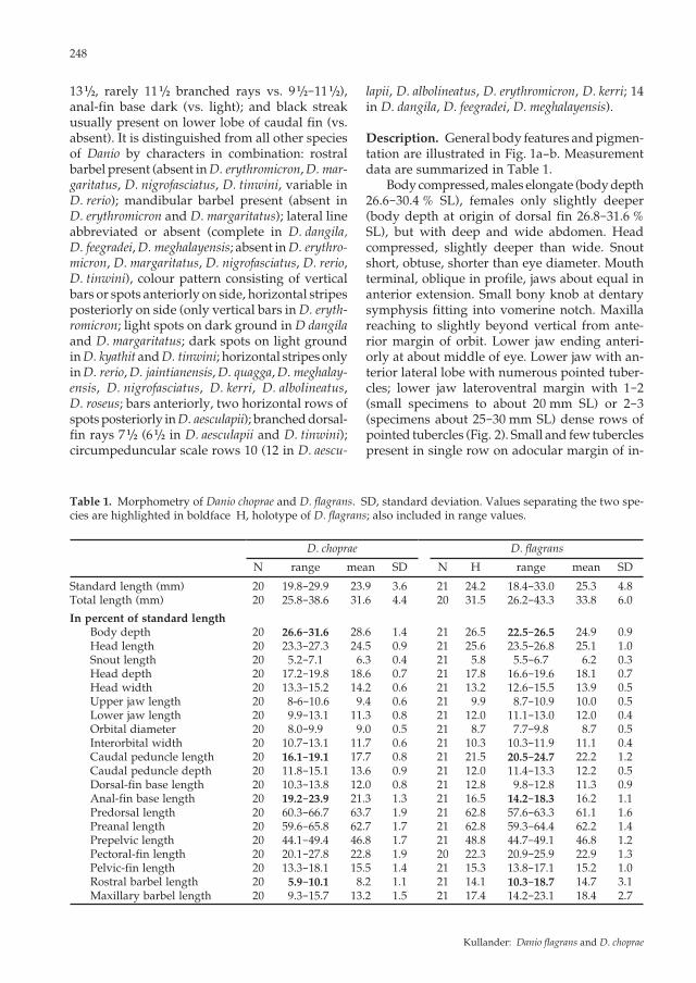
Description. General body features and pigmen-tation are illustrated in Fig. 1a-b. Measurement data are summarized in Table 1. Body compressed, males elongate (body depth 26.6-30.4 % SL), females only slightly deeper (body depth at origin of dorsal fin 26.8-31.6 % SL), but with deep and wide abdomen. Head compressed, slightly deeper than wide. Snout short, obtuse, shorter than eye diameter. Mouth terminal, oblique in profile, jaws about equal in anterior extension. Small bony knob at dentary symphysis fitting into vomerine notch. Maxilla reaching to slightly beyond vertical from ante-rior margin of orbit. Lower jaw ending anteri-orly at about middle of eye. Lower jaw with an-terior lateral lobe with numerous pointed tuber-cles; lower jaw lateroventral margin with 1-2 (small specimens to about 20 mm SL) or 2-3 (specimens about 25-30 mm SL) dense rows of pointed tubercles (Fig. 2). Small and few tubercles present in single row on adocular margin of in-
Table 1. Morphometry of Danio choprae and D. flagrans. SD, standard deviation. Values separating the two spe-cies are highlighted in boldface H, holotype of D. flagrans; also included in range values.
D. choprae D. flagrans
N range mean SD N H range mean SD
Standard length (mm) 20 19.8-29.9 23.9 3.6 21 24.2 18.4-33.0 25.3 4.8Total length (mm) 20 25.8-38.6 31.6 4.4 20 31.5 26.2-43.3 33.8 6.0
In percent of standard lengthBody depth 20 26.6-31.6 28.6 1.4 21 26.5 22.5-26.5 24.9 0.9Head length 20 23.3-27.3 24.5 0.9 21 25.6 23.5-26.8 25.1 1.0Snout length 20 5.2-7.1 6.3 0.4 21 5.8 5.5-6.7 6.2 0.3Head depth 20 17.2-19.8 18.6 0.7 21 17.8 16.6-19.6 18.1 0.7Head width 20 13.3-15.2 14.2 0.6 21 13.2 12.6-15.5 13.9 0.5Upper jaw length 20 8-6-10.6 9.4 0.6 21 9.9 8.7-10.9 10.0 0.5Lower jaw length 20 9.9-13.1 11.3 0.8 21 12.0 11.1-13.0 12.0 0.4Orbital diameter 20 8.0-9.9 9.0 0.5 21 8.7 7.7-9.8 8.7 0.5Interorbital width 20 10.7-13.1 11.7 0.6 21 10.3 10.3-11.9 11.1 0.4Caudal peduncle length 20 16.1-19.1 17.7 0.8 21 21.5 20.5-24.7 22.2 1.2Caudal peduncle depth 20 11.8-15.1 13.6 0.9 21 12.0 11.4-13.3 12.2 0.5Dorsal-fin base length 20 10.3-13.8 12.0 0.8 21 12.8 9.8-12.8 11.3 0.9Anal-fin base length 20 19.2-23.9 21.3 1.3 21 16.5 14.2-18.3 16.2 1.1Predorsal length 20 60.3-66.7 63.7 1.9 21 62.8 57.6-63.3 61.1 1.6Preanal length 20 59.6-65.8 62.7 1.7 21 62.8 59.3-64.4 62.2 1.4Prepelvic length 20 44.1-49.4 46.8 1.7 21 48.8 44.7-49.1 46.8 1.2Pectoral-fin length 20 20.1-27.8 22.8 1.9 20 22.3 20.9-25.9 22.9 1.3Pelvic-fin length 20 13.3-18.1 15.5 1.4 21 15.3 13.8-17.1 15.2 1.0Rostral barbel length 20 5.9-10.1 8.2 1.1 21 14.1 10.3-18.7 14.7 3.1Maxillary barbel length 20 9.3-15.7 13.2 1.5 21 17.4 14.2-23.1 18.4 2.7
Kullander: Danio flagrans and D. choprae
249
Ichthyol. Explor. Freshwaters, Vol. 23, No. 3
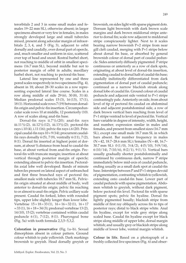
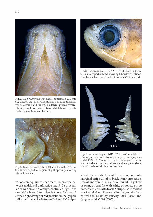
terorbitals 2 and 3 in some small males and fe-males 19-22 mm SL), otherwise absent; in larger specimens absent or very few in females, in males strongly developed large and small tubercles mixed, present along adocular margin of infraor-bitals 2, 3, 4, and 5 (Fig. 3), adjacent to orbit dorsally and caudally, over dorsal part of opercle, and, much smaller and uniform in size, scattered over top of head and snout. Rostral barbel short, not reaching to middle of orbit in smallest speci-mens (16.7 mm SL), beyond middle but not to posterior margin of orbit in adults; maxillary barbel short, not reaching to pectoral-fin base. Lateral line represented by one and three pored scales respectively in two specimens (Fig. 4), absent in 18; about 28-30 scales in a row repre-senting expected lateral line course. Scales in a row along middle of side 29 (6), 30 (8), 31 (4). Median predorsal scales 15 (3), 16 (13), 17 (1), 18 (1). Horizontal scale rows 7 (19) between dorsal-fin origin and pelvic-fin insertion. Circumpedun-cular scale rows 10 at middle, 12 posteriorly (10). A row of scales along anal-fin base. Dorsal-fin rays ii.7 1/2 (20); anal-fin rays iii.11 1/2 (2), iii.12 1/2 (12), iii.13 1/2 (2); pectoral-fin rays i.10 (4), i.11 (16); pelvic-fin rays i.6 (20). Prin-cipal caudal-fin rays 10 + 9 (14); procurrent caudal-fin rays dorsally 6 (5), 7 (9), ventrally 6 (11), 7 (2), 8 (1). Dorsal fin inserted at highest point of dor-sum, at about 3/5 distance from head to caudal-fin base, at about vertical from anal-fin origin. Pec-toral-fin with truncate margin; insertion at about vertical through posterior margin of opercle; extending almost to pelvic-fin insertion. Pectoral-fin axial lobe well developed. Bands of minute tubercles present on lateral aspect of unbranched and first three branched rays of pectoral fin; smallest male with tubercles 18.7 mm SL. Pelvic-fin origin situated at about middle of body, well anterior to dorsal-fin origin; pelvic fin reaching to or almost to anal-fin origin. Pelvic axillary scale present. Caudal fin forked, lobes with rounded tips, upper lobe slightly longer than lower lobe. Vertebrae 15 + 18 = 33 (1), 16 + 16 = 32 (1), 16 + 17 = 33 (1), 16 + 18 = 34 (3); predorsal vertebrae 13 (2), 14 (10), 15 (2); vertebrae contained within caudal peduncle 6 (1), 7 (12), 8 (1). Pharyngeal bone (Fig. 5a) with tooth formula 2,3,5-5,3,2.
Coloration in preservative (Fig. 1a-b). Sexual dimorphism absent in colour pattern. Ground colour whitish to pale yellowish. Dark markings brownish to greyish. Head dorsally greyish or
brownish, on sides light with sparse pigment dots. Dorsum light brownish with dark brown scale margins and dark brown middorsal stripe ante-rior to dorsal fin; scale row adjacent to middorsal stripe conspicuously lighter. Next to it scales bearing narrow brownish P+2 stripe from near gill cleft caudad, merging with P+1 stripe below about dorsal fin base, or absorbed by general brownish colour of dorsal part of caudal pedun-cle. Sides anteriorly diffusely pigmented. P stripe continuous or anteriorly as a row of dark spots, originating at about level of dorsal-fin insertion, extending caudad to dorsal half of caudal-fin base; caudally indistinctly differentiated from dark pigmentation of dorsal part of caudal peduncle; continued as a narrow blackish streak along dorsal lobe of caudal fin. Ground colour of caudal peduncle and adjacent side ventral to P+1 stripe contrastingly pale. Anteriorly on side, from about level of tip of pectoral fin caudad on abdominal side and adjacent postabdominal side, a row of dark brown vertical bars reaching from level of P+1 stripe ventrad to level of pectoral fin. Vertical bars variable in degree of intensity, width, height, and number; expression similar in males and females, and present from smallest sizes (16.7 mm SL), except one small male 18.7 mm SL in which bars absent. Bar number frequency in males (N = 42, 18.7-28.6 mm SL)/females (N = 48, 16.7-30.7 mm SL): 0 (1/0), 3 (4/2), 4 (5/10), 5 (9/14), 6 (10/14), 7 (10/6), 8 (2/1), 9 (1/1). Vertical bars usually gradually shorter postabdominally and continued by continuous dark, narrow P stripe immediately below mid-axis of caudal peduncle, ending usually as a small dark spot at caudal-fin base. Interstripe between P and P+1 stripes devoid of pigmentation, contrasting whitish to yellowish, extending onto caudal-fin base. Lower part of caudal peduncle with sparse pigmentation. Abdo-men whitish to greyish, without dark pigment, below pectoral-fin level. Pectoral fin with sparse pigment spots; pelvic fin hyaline. Dorsal fin lightly pigmented basally; blackish stripe from middle of first ray obliquely across fin to tips of posterior rays; distal to black stripe white. Anal fin hyaline, except for wide grey stripe along scaled base. Caudal fin hyaline except for black stripe along middle of upper lobe, dorsal margin whitish; and usually grey or blackish stripe along middle of lower lobe, ventral margin whitish.
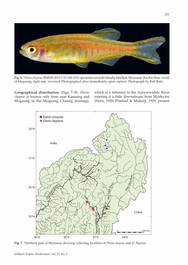
Colour in life. Based on a photograph of a freshly collected live specimen (Fig. 6) and obser-
250
vations on aquarium specimens: Interstripe be-tween middorsal dark stripe and P+2 stripe an-terior to dorsal fin orange, continued lighter to caudal-fin base. Interstripe between P+1 and P stripe bright orange or red postabdominally; pale yellowish interstripe between P+1 and P+2 stripes
anteriorly on side. Dorsal fin with orange sub-marginal stripe distal to black transverse stripe. Dorsal and ventral margins of caudal fin yellow or orange. Anal fin with white or yellow stripe immediately distal to black A stripe. Danio choprae was included and illustrated in analyses of colour patterns in Danio by Parichy (2006, 2007) and Quigley et al. (2004, 2005).
IO2
lachrymal
IO3
IO4
12 3
Fig. 4. Danio choprae, NRM 52001, adult female, 29.9 mm SL; lateral aspect of region of gill opening, showing lateral line scales.
Fig. 2. Danio choprae, NRM 52001, adult male, 27.0 mm SL; ventral aspect of head showing pointed tubercles ventrolaterally and tuberculate lateral process ventro-laterally on lower jaw. Infraorbital tubercles partly visible lateral to rostral barbels.
Fig. 3. Danio choprae, NRM 52001, adult male, 27.0 mm SL; lateral aspect of head, showing tubercles on infraor-bital bones. Lachrymal and infraorbitals 2-4 labelled.
a
b
a
b
500 µm500 µm
Fig. 5. a, Danio choprae, NRM 52001, 26.5 mm SL; left pharyngeal bone in ventromedial aspect; b, D. flagrans, NRM 41270, 21.9 mm SL; right pharyngeal bone in ventromedial aspect, lateral margin damaged and one medial tooth lost during preparation.
Kullander: Danio flagrans and D. choprae
251
Ichthyol. Explor. Freshwaters, Vol. 23, No. 3
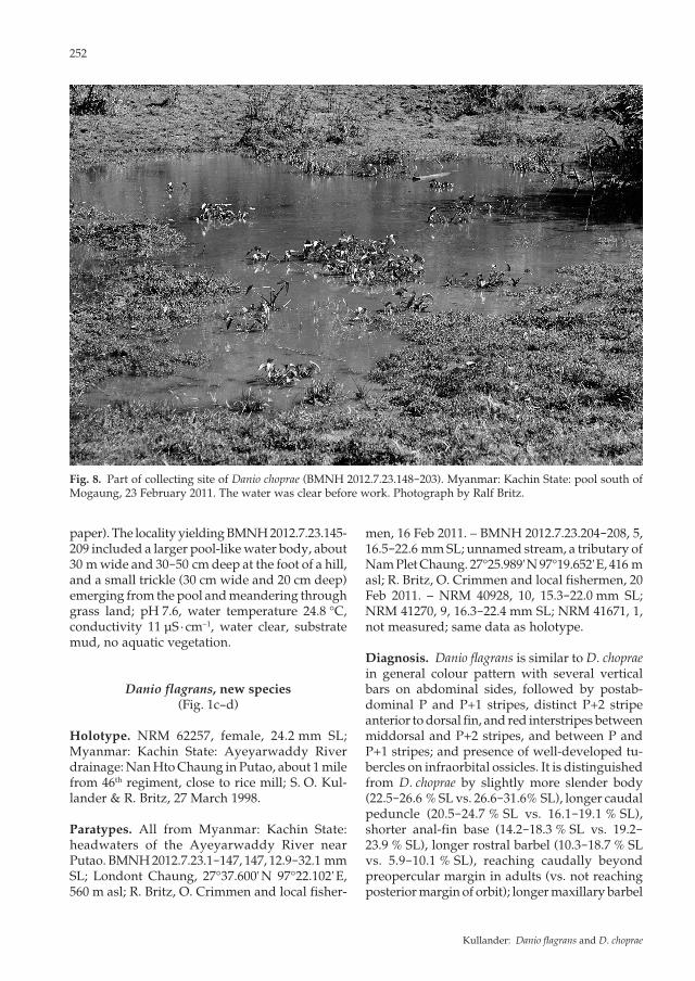
Geographical distribution (Figs. 7-8). Danio choprae is known only from near Kamaing and Mogaung in the Mogaung Chaung drainage,
Fig. 6. Danio choprae, BMNH 2012.7.23.148-203, specimen not individually labelled; Myanmar: Kachin State: south of Mogaung; right side, reversed. Photographed alive immediately upon capture. Photograph by Ralf Britz.
Fig. 7. Northern part of Myanmar showing collecting localities of Danio choprae and D. flagrans.
which is a tributary to the Ayeyarwaddy River entering it a little downstream from Myitkyina (Hora, 1928; Prashad & Mukerji, 1929; present
0 100 km
India
China
Danio chopraeDanio flagrans
98°E97°E96°E95°E
28°N
27°N
26°N
25°N
Chi
ndwin
Rive
r
Aye
yarw
addy
Riv
er
Mal
i Hka
Rive
r
May
Hka
Riv
er
Chi
ndwin
Rive
r
Aye
yarw
addy
Riv
er
Mal
i Hka
Rive
r
May
Hka
Riv
er
252
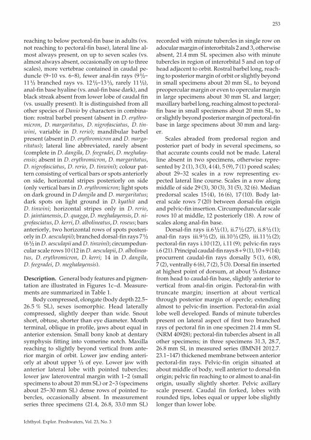
paper). The locality yielding BMNH 2012.7.23.145-209 included a larger pool-like water body, about 30 m wide and 30-50 cm deep at the foot of a hill, and a small trickle (30 cm wide and 20 cm deep) emerging from the pool and meandering through grass land; pH 7.6, water temperature 24.8 °C, conductivity 11 μS · cm−1, water clear, substrate mud, no aquatic vegetation.
Danio flagrans, new species (Fig. 1c-d)
Holotype. NRM 62257, female, 24.2 mm SL; Myanmar: Kachin State: Ayeyarwaddy River drainage: Nan Hto Chaung in Putao, about 1 mile from 46th regiment, close to rice mill; S. O. Kul-lander & R. Britz, 27 March 1998.
Paratypes. All from Myanmar: Kachin State: headwaters of the Ayeyarwaddy River near Putao. BMNH 2012.7.23.1-147, 147, 12.9-32.1 mm SL; Londont Chaung, 27°37.600' N 97°22.102' E, 560 m asl; R. Britz, O. Crimmen and local fisher-
men, 16 Feb 2011. – BMNH 2012.7.23.204-208, 5, 16.5-22.6 mm SL; unnamed stream, a tributary of Nam Plet Chaung. 27°25.989' N 97°19.652' E, 416 m asl; R. Britz, O. Crimmen and local fishermen, 20 Feb 2011. – NRM 40928, 10, 15.3-22.0 mm SL; NRM 41270, 9, 16.3-22.4 mm SL; NRM 41671, 1, not measured; same data as holotype.
Diagnosis. Danio flagrans is similar to D. choprae in general colour pattern with several vertical bars on abdominal sides, followed by postab-dominal P and P+1 stripes, distinct P+2 stripe anterior to dorsal fin, and red interstripes between middorsal and P+2 stripes, and between P and P+1 stripes; and presence of well-developed tu-bercles on infraorbital ossicles. It is distinguished from D. choprae by slightly more slender body (22.5-26.6 % SL vs. 26.6-31.6% SL), longer caudal peduncle (20.5-24.7 % SL vs. 16.1-19.1 % SL), shorter anal-fin base (14.2-18.3 % SL vs. 19.2-23.9 % SL), longer rostral barbel (10.3-18.7 % SL vs. 5.9-10.1 % SL), reaching caudally beyond preopercular margin in adults (vs. not reaching posterior margin of orbit); longer maxillary barbel
Fig. 8. Part of collecting site of Danio choprae (BMNH 2012.7.23.148-203). Myanmar: Kachin State: pool south of Mogaung, 23 February 2011. The water was clear before work. Photograph by Ralf Britz.
Kullander: Danio flagrans and D. choprae
253
Ichthyol. Explor. Freshwaters, Vol. 23, No. 3
reaching to below pectoral-fin base in adults (vs. not reaching to pectoral-fin base), lateral line al-most always present, on up to seven scales (vs. almost always absent, occasionally on up to three scales), more vertebrae contained in caudal pe-duncle (9-10 vs. 6-8), fewer anal-fin rays (9 1/2-11 1/2 branched rays vs. 12 1/2-13 1/2, rarely 11 1/2), anal-fin base hyaline (vs. anal-fin base dark), and black streak absent from lower lobe of caudal fin (vs. usually present). It is distinguished from all other species of Danio by characters in combina-tion: rostral barbel present (absent in D. erythro-micron, D. margaritatus, D. nigrofasciatus, D. tin-wini, variable in D. rerio); mandibular barbel present (absent in D. erythromicron and D. marga-ritatus); lateral line abbreviated, rarely absent (complete in D. dangila, D. feegradei, D. meghalay-ensis; absent in D. erythromicron, D. margaritatus, D. nigrofasciatus, D. rerio, D. tinwini); colour pat-tern consisting of vertical bars or spots anteriorly on side, horizontal stripes posteriorly on side (only vertical bars in D. erythromicron; light spots on dark ground in D dangila and D. margaritatus; dark spots on light ground in D. kyathit and D. tinwini; horizontal stripes only in D. rerio, D. jaintianensis, D. quagga, D. meghalayensis, D. ni-grofasciatus, D. kerri, D. albolineatus, D. roseus; bars anteriorly, two horizontal rows of spots posteri-orly in D. aesculapii); branched dorsal-fin rays 7 1/2 (6 1/2 in D. aesculapii and D. tinwini); circumpedun-cular scale rows 10 (12 in D. aesculapii, D. albolinea-tus, D. erythromicron, D. kerri; 14 in D. dangila, D. feegradei, D. meghalayensis).
Description. General body features and pigmen-tation are illustrated in Figures 1c-d. Measure-ments are summarized in Table 1. Body compressed, elongate (body depth 22.5-26.5 % SL), sexes isomorphic. Head laterally compressed, slightly deeper than wide. Snout short, obtuse, shorter than eye diameter. Mouth terminal, oblique in profile, jaws about equal in anterior extension. Small bony knob at dentary symphysis fitting into vomerine notch. Maxilla reaching to slightly beyond vertical from ante-rior margin of orbit. Lower jaw ending anteri-orly at about upper 1/3 of eye. Lower jaw with anterior lateral lobe with pointed tubercles; lower jaw lateroventral margin with 1-2 (small specimens to about 20 mm SL) or 2-3 (specimens about 25-30 mm SL) dense rows of pointed tu-bercles, occasionally absent. In measurement series three specimens (21.4, 26.8, 33.0 mm SL)
recorded with minute tubercles in single row on adocular margin of interorbitals 2 and 3, otherwise absent, 21.4 mm SL specimen also with minute tubercles in region of interorbital 5 and on top of head adjacent to orbit. Rostral barbel long, reach-ing to posterior margin of orbit or slightly beyond in small specimens about 20 mm SL, to beyond preopercular margin or even to opercular margin in large specimens about 30 mm SL and larger; maxillary barbel long, reaching almost to pectoral-fin base in small specimens about 20 mm SL, to or slightly beyond posterior margin of pectoral-fin base in large specimens about 30 mm and larg-er. Scales abraded from predorsal region and posterior part of body in several specimens, so that accurate counts could not be made. Lateral line absent in two specimens, otherwise repre-sented by 2 (1), 3 (3), 4 (4), 5 (9), 7 (1) pored scales; about 29-32 scales in a row representing ex-pected lateral line course. Scales in a row along middle of side 29 (3), 30 (3), 31 (5), 32 (6). Median predorsal scales 15 (4), 16 (6), 17 (10). Body lat-eral scale rows 7 (20) between dorsal-fin origin and pelvic-fin insertion. Circumpeduncular scale rows 10 at middle, 12 posteriorly (18). A row of scales along anal-fin base. Dorsal-fin rays ii.6 1/2 (1), ii.7 1/2 (27), ii.8 1/2 (1); anal-fin rays iii.9 1/2 (2), iii.10 1/2 (25), iii.11 1/2 (2); pectoral-fin rays i.10 (12), i.11 (9); pelvic-fin rays i.6 (21). Principal caudal-fin rays 8 + 9 (1), 10 + 9 (14); procurrent caudal-fin rays dorsally 5 (1), 6 (8), 7 (2), ventrally 6 (6), 7 (2), 5 (3). Dorsal fin inserted at highest point of dorsum, at about 3/5 distance from head to caudal-fin base, slightly anterior to vertical from anal-fin origin. Pectoral-fin with truncate margin; insertion at about vertical through posterior margin of opercle; extending almost to pelvic-fin insertion. Pectoral-fin axial lobe well developed. Bands of minute tubercles present on lateral aspect of first two branched rays of pectoral fin in one specimen 21.4 mm SL (NRM 40928); pectoral-fin tubercles absent in all other specimens; in three specimens 31.3, 28.7, 26.8 mm SL in measured series (BMNH 2012.7. 23.1-147) thickened membrane between anterior pectoral-fin rays. Pelvic-fin origin situated at about middle of body, well anterior to dorsal-fin origin; pelvic fin reaching to or almost to anal-fin origin, usually slightly shorter. Pelvic axillary scale present. Caudal fin forked, lobes with rounded tips, lobes equal or upper lobe slightly longer than lower lobe.
254
Vertebrae 15 + 19 = 34 (1), 16 + 18 = 34 (13), 16 + 19 = 35 (1), 17 + 17 = 34 (1), 17 + 18 = 35 (1), 17 + 19 = 36 (1), 18 + 17 = 35 (1), last abdominal vertebra as counted here, with short or long haemal apophysis and articulating with long second anal-fin pterygiophore, succeeding verte-bra with short haemal apophysis and inserted between two anal-fin pterygiophores; predorsal vertebrae 13 (2), 14 (14), 15 (2), 16 (1); contained within caudal peduncle 8 (1), 9 (11), 10 (7). Pha-ryngeal bone (Fig. 5b) with tooth formula 2,4,5-5,4,2.
Colouration in preservative. Sexual dimorphism absent in colour pattern. Ground colour whitish to pale yellowish. Dark markings brownish to greyish. Head dorsally pale brownish grey. Sides of head sparsely pigmented. Dorsum light brown-ish with dark brown middorsal stripe anterior to dorsal fin; scale row adjacent to middorsal stripe conspicuously lighter. Next to it, scales bearing narrow brownish P+2 stripe from near gill cleft caudad, merging with P+1 stripe below about dorsal fin base. Sides anteriorly diffusely pig-mented, from about distal part of pectoral fin followed by a number of dark vertical bars or blotches, which continuous dorsally; extending from level of P+1 stripe level to about level of pectoral-fin base. Number of bars (excluding spots) in subsample of 38 specimens 19.3-26.2 mm SL, 2 (1), 3 (4), 4 (23), 5 (7), 6 (3), in subsample of 10 specimen 26.8-33.0 mm SL, 5 (6), 6 (3), 10 (1). Bars gradually shorter caudally and integrating with continuous P stripe above anal-fin base. Vertical bars developing from small spots ante-
riorly in stripes P+1 and P, spots coalescing vertically, but often disarranged in smaller spots mixed with bars. Specimens 20 mm SL and longer all possess vertical bars or blotches; specimens smaller show incipient spots or bars, but generally up to about 18 mm SL blotches absent and P and P+1 stripes extending uniform and distinctly separated by lighter stripe to close to head. P stripe narrow, extending onto caudal-fin base where usually ending in a small spot. Usually two dark spots above anal-fin base, fre-quently indistinct, representing P-2 stripe. P+1 stripe obsolete anteriorly on side, initiated as a few indistinct spots in transition abdominal to postabdominal region, and then continuous to caudal-fin base, but barely distinct from dark colour of dorsal part of caudal peduncle. Narrow interstripe between P and P+1 stripes contrast-ingly light. Pectoral and pelvic fins hyaline. Dorsal fin hyaline with dark brown or blackish stripe from middle of anterior margin obliquely across rays to tip of posterior rays; beyond that stripe hyaline. Anal fin basally greyish; blackish stripe from middle of anterior margin caudad across rays to tip of posterior rays; distal to that stripe hyaline. Caudal fin hyaline or slightly pigmented; brown or black stripe along upper rays, dorsal margin white; ventral margin white. In juveniles smaller than 18 mm dark stripe on caudal fin absent, dark stripe in dorsal fin present but faint, A stripe distinct, occasionally absent.
Colour in life. A specimen from BMNH 2012.7. 23.1-147 (Fig. 9) has interstripe between mid-
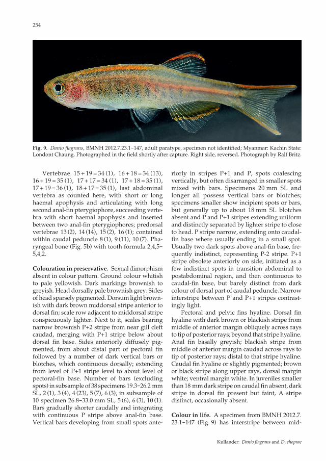
Fig. 9. Danio flagrans, BMNH 2012.7.23.1-147, adult paratype, specimen not identified; Myanmar: Kachin State: Londont Chaung. Photographed in the field shortly after capture. Right side, reversed. Photograph by Ralf Britz.
Kullander: Danio flagrans and D. choprae
255
Ichthyol. Explor. Freshwaters, Vol. 23, No. 3
dorsal dark stripe and P+2 stripe anterior to dorsal fin bright orange. Interstripe between P+1 and P stripes bright orange postabdominally; weaker orange interstripe between P+1 and P+2 stripes anteriorly on side. Bright orange spots on iris. Dorsal fin with bright red submarginal stripe distal to black transverse stripe. Caudal fin dorsal margin red; ventral margin orange. Anal fin with orange stripe immediately distal to black A stripe. A much smaller specimen sampled in 1998 (NRM 40928 or 41270) did not show body markings, but orange or yellowish orange stripes present in dorsal, anal, and caudal fins.
Etymology. The specific name flagrans is a Latin participial adjective meaning flaming, blazing, burning, glowing, and is given with reference to the red to orange colour in living specimens, and with inspiration from the name glowlight danio applied on this species and D. choprae.
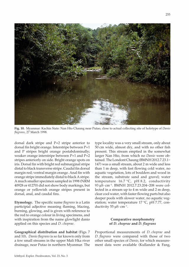
Geographical distribution and habitat (Figs. 7 and 10). Danio flagrans is so far known only from a few small streams in the upper Mali Hka river drainage, near Putao in northern Myanmar. The
type locality was a very small stream, only about 50 cm wide, almost dry, and with no other fish present. This stream emptied in the somewhat larger Nan Hto, from which no Danio were ob-tained. The Londont Chaung (BMNH 2012.7.23.1-147) was a small stream, about 2 m wide and less than 1 m deep, with fast flowing cold water, no aquatic vegetation, lots of boulders and wood in the stream, substrate sand and gravel; water temperature 16.7 °C, pH 8.2, conductivity 93 μS · cm−1. BMNH 2012.7.23.204-208 were col-lected in a stream up to 4 m wide and 2 m deep, clear cool water, with faster flowing parts but also deeper pools with slower water, no aquatic veg-etation; water temperature 17 °C, pH 7.77, con-ductivity 55 μS · cm−1.
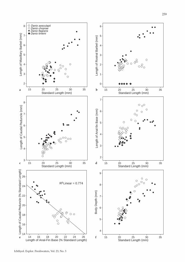
Comparative morphometry of D. choprae and D. flagrans
Proportional measurements of D. choprae and D. flagrans were compared with those of two other small species of Danio, for which measure-ment data were available (Kullander & Fang,
Fig. 10. Myanmar: Kachin State: Nan Hto Chaung near Putao, close to actual collecting site of holotype of Danio flagrans, 27 March 1998.
256
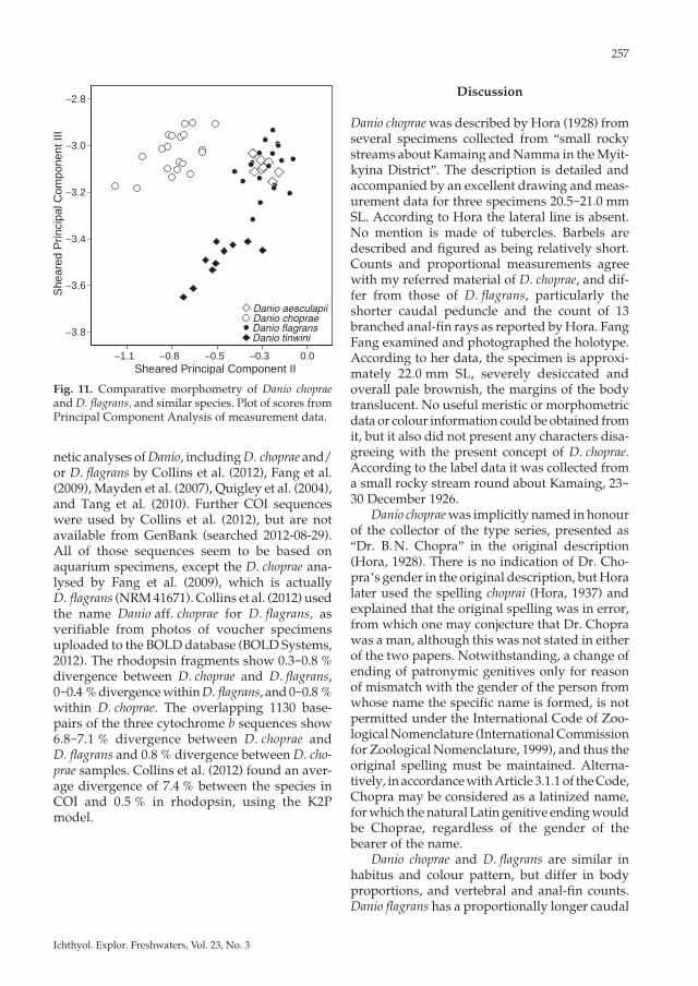
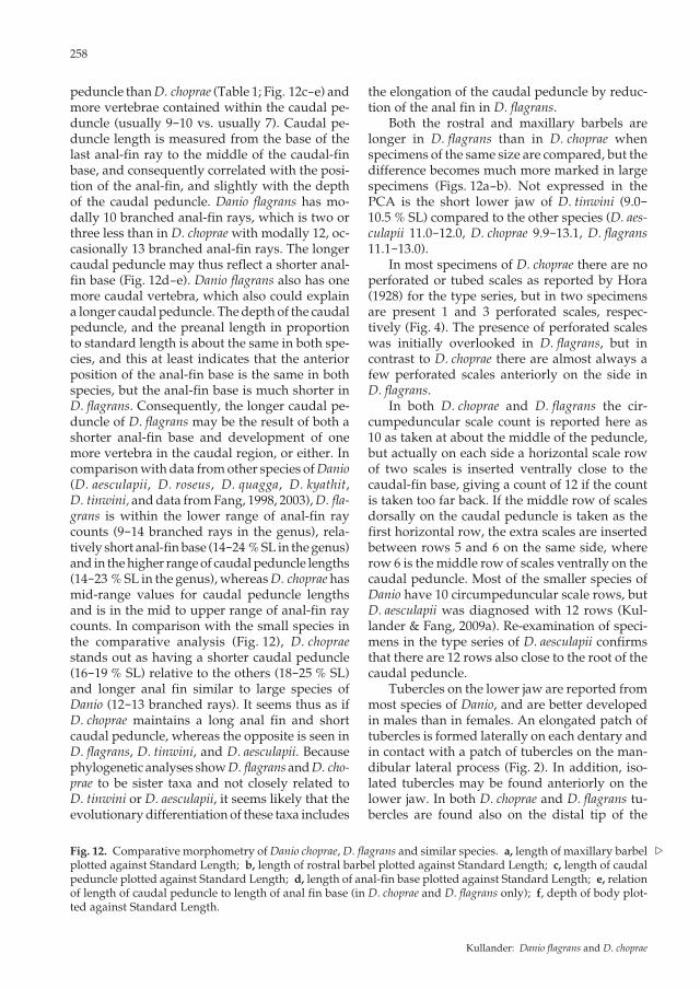
2009a,b), viz. D. tinwini and D. aesculapii, expect-ing that Danio species of the same small size would be informative about the direction of character transformation in the caudal peduncle and anal fin proportions. Danio aesculapii also has a colour pattern similar to D. choprae and D. flagrans, and D. tinwini was collected in the Mogaung River upstream of D. choprae localities. The principal component analysis shows D. choprae separated from D. flagrans + D. aesculapii on Component II and D. tinwini from the rest on Component III (Table 2; Fig. 11). Most of the variation is ex-pressed in the length of the maxillary barbel, which is very short in D. choprae (9.3-15.7 % SL), and very long in D. aesculapii (16.8-19.0 %) and D. flagrans (14.2-23.1 %) (Fig. 12a). The rostral barbel was excluded from the PCA because it is absent in D. tinwini. It is relatively long in D. aes-culapii and much longer in D. flagrans than in D. choprae (10.3-18.7 % SL vs. 5.9-10.1 in D. choprae (Fig. 12b). Remaining variation separating species the PCA and biplots, are above all caudal pedun-cle length and anal-fin base length, separating D. flagrans and D. tinwini, both with short anal-fin
Table 2. Component loadings from Principal Component Analysis of morphometric data from Danio choprae (N = 20), D. flagrans (N = 20), D. aesculapii (N = 10; data from Kullander & Fang, 2009a), and D. tinwini (N = 10; data from Kullander & Fang, 2009b). Rostral barbel length was excluded because the rostral barbel is absent in D. tin-wini. The four highest loadings for each component are highlighted in boldface.
PC I PC II Sheared PC II
PC III Sheared PC III
PC IV Sheared PC IV
Standard length 0.203 -0.016 -0.012 -0.189 -0.171 -0.042 -0.029Body depth 0.210 -0.259 -0.256 -0.197 -0.179 -0.172 -0.158Head length 0.185 0.037 0.040 -0.162 -0.147 -0.019 -0.007Snout length 0.230 -0.008 -0.004 0.079 -0.097 0.000 0.015Head depth 0.235 -0.110 -0.106 -0.020 -0.001 0.276 0.292Head width 0.205 -0.053 -0.049 -0.061 -0.044 0.035 0.049Upper jaw length 0.272 0.095 0.099 0.283 0.303 0.189 0.207Lower jaw length 0.222 0.120 0.123 0.110 0.127 0.113 0.127Orbital diameter 0.152 -0.014 -0.012 -0.087 -0.075 -0.105 -0.094Interorbital width 0.215 -0.089 -0.085 0.038 0.055 0.079 0.093Caudal peduncle length 0.173 0.305 0.308 -0.424 -0.408 0.415 0.427Caudal peduncle depth 0.245 -0.218 -0.214 0.009 0.029 0.026 0.042Dorsal-fin base length 0.306 -0.198 -0.193 0.333 0.356 0.436 0.456Anal-fin base length 0.235 -0.405 -0.401 0.345 0.362 -0.438 -0.422Predorsal length 0.215 -0.071 -0.067 -0.198 -0.179 -0.158 -0.143Preanal length 0.201 -0.016 -0.012 -0.260 -0.243 -0.155 -0.142Prepelvic length 0.191 -0.011 -0.007 -0.310 -0.293 -0.153 -0.140Pectoral-fin length 0.153 0.086 0.088 -0.240 -0.226 -0.273 -0.262Pelvic-fin length 0.220 -0.052 -0.049 -0.072 -0.054 0.136 0.151Maxillary barbel length 0.322 0.725 0.730 0.334 0.358 -0.324 -0.302
Eigenvalue 0.5473 0.0642 N/A 0.0304 N/A 0.0125 N/A% Variance 80.2 % 89.6 % N/A 94.1 % N/A 95.9 % N/A
base, as well as D. choprae with short caudal pe-duncle and long anal-fin base, and D. flagrans with long caudal peduncle (Fig. 12c-e). Body depth is important in Component II, probably because of the deeper D. choprae and slender D. flagrans at larger sizes (Fig. 12f; 26.6-31.6 % SL in D. choprae, 23.1-27.2 in D. aesculapii, 23.0-30.3 in D. tinwini, and 22.5-26.5 in D. flagrans). Com-ponents III-IV also reflect dorsal-fin base length, which is short in D. aesculapii (7.7-10.4 % SL) and D. tinwini (8.9-9.2) compared with D. choprae (10.3-13.8) and D. flagrans (9.8-12.8).
Genetic comparison of Danio choprae and D. flagrans
Complete or partial sequences of the mitochon-drial cytochrome b, cytochrome c oxidase subunit I (COI), NADH dehydrogenase subunit 4 (ND4), 16S ribosomal RNA, and 12S ribosomal RNA genes, and parts of the nuclear recombination activating protein 1 (RAG1) and rhodopsin genes were used in various combinations in phyloge-
Kullander: Danio flagrans and D. choprae
257
Ichthyol. Explor. Freshwaters, Vol. 23, No. 3
Discussion
Danio choprae was described by Hora (1928) from several specimens collected from “small rocky streams about Kamaing and Namma in the Myit-kyina District”. The description is detailed and accompanied by an excellent drawing and meas-urement data for three specimens 20.5-21.0 mm SL. According to Hora the lateral line is absent. No mention is made of tubercles. Barbels are described and figured as being relatively short. Counts and proportional measurements agree with my referred material of D. choprae, and dif-fer from those of D. flagrans, particularly the shorter caudal peduncle and the count of 13 branched anal-fin rays as reported by Hora. Fang Fang examined and photographed the holotype. According to her data, the specimen is approxi-mately 22.0 mm SL, severely desiccated and overall pale brownish, the margins of the body translucent. No useful meristic or morphometric data or colour information could be obtained from it, but it also did not present any characters disa-greeing with the present concept of D. choprae. According to the label data it was collected from a small rocky stream round about Kamaing, 23-30 December 1926. Danio choprae was implicitly named in honour of the collector of the type series, presented as “Dr. B. N. Chopra” in the original description (Hora, 1928). There is no indication of Dr. Cho-pra’s gender in the original description, but Hora later used the spelling choprai (Hora, 1937) and explained that the original spelling was in error, from which one may conjecture that Dr. Chopra was a man, although this was not stated in either of the two papers. Notwithstanding, a change of ending of patronymic genitives only for reason of mismatch with the gender of the person from whose name the specific name is formed, is not permitted under the International Code of Zoo-logical Nomenclature (International Commission for Zoological Nomenclature, 1999), and thus the original spelling must be maintained. Alterna-tively, in accordance with Article 3.1.1 of the Code, Chopra may be considered as a latinized name, for which the natural Latin genitive ending would be Choprae, regardless of the gender of the bearer of the name. Danio choprae and D. flagrans are similar in habitus and colour pattern, but differ in body proportions, and vertebral and anal-fin counts. Danio flagrans has a proportionally longer caudal
She
ared
Prin
cipa
l Com
pone
nt II
I
Sheared Principal Component II
Danio aesculapiiDanio chopraeDanio flagransDanio tinwini
2.8 –
3.0 –
3.2 –
3.4 –
3.6 –
3.8 –
ı ı ı ı ı 1.1 0.8 0.5 0.3 0.0
Fig. 11. Comparative morphometry of Danio choprae and D. flagrans, and similar species. Plot of scores from Principal Component Analysis of measurement data.
netic analyses of Danio, including D. choprae and/or D. flagrans by Collins et al. (2012), Fang et al. (2009), Mayden et al. (2007), Quigley et al. (2004), and Tang et al. (2010). Further COI sequences were used by Collins et al. (2012), but are not available from GenBank (searched 2012-08-29). All of those sequences seem to be based on aquarium specimens, except the D. choprae ana-lysed by Fang et al. (2009), which is actually D. flagrans (NRM 41671). Collins et al. (2012) used the name Danio aff. choprae for D. flagrans, as verifiable from photos of voucher specimens uploaded to the BOLD database (BOLD Systems, 2012). The rhodopsin fragments show 0.3-0.8 % divergence between D. choprae and D. flagrans, 0-0.4 % divergence within D. flagrans, and 0-0.8 % within D. choprae. The overlapping 1130 base-pairs of the three cytochrome b sequences show 6.8-7.1 % divergence between D. choprae and D. flagrans and 0.8 % divergence between D. cho-prae samples. Collins et al. (2012) found an aver-age divergence of 7.4 % between the species in COI and 0.5 % in rhodopsin, using the K2P model.
258
peduncle than D. choprae (Table 1; Fig. 12c-e) and more vertebrae contained within the caudal pe-duncle (usually 9-10 vs. usually 7). Caudal pe-duncle length is measured from the base of the last anal-fin ray to the middle of the caudal-fin base, and consequently correlated with the posi-tion of the anal-fin, and slightly with the depth of the caudal peduncle. Danio flagrans has mo-dally 10 branched anal-fin rays, which is two or three less than in D. choprae with modally 12, oc-casionally 13 branched anal-fin rays. The longer caudal peduncle may thus reflect a shorter anal-fin base (Fig. 12d-e). Danio flagrans also has one more caudal vertebra, which also could explain a longer caudal peduncle. The depth of the caudal peduncle, and the preanal length in proportion to standard length is about the same in both spe-cies, and this at least indicates that the anterior position of the anal-fin base is the same in both species, but the anal-fin base is much shorter in D. flagrans. Consequently, the longer caudal pe-duncle of D. flagrans may be the result of both a shorter anal-fin base and development of one more vertebra in the caudal region, or either. In comparison with data from other species of Danio (D. aesculapii, D. roseus, D. quagga, D. kyathit, D. tinwini, and data from Fang, 1998, 2003), D. fla-grans is within the lower range of anal-fin ray counts (9-14 branched rays in the genus), rela-tively short anal-fin base (14-24 % SL in the genus) and in the higher range of caudal peduncle lengths (14-23 % SL in the genus), whereas D. choprae has mid-range values for caudal peduncle lengths and is in the mid to upper range of anal-fin ray counts. In comparison with the small species in the comparative analysis (Fig. 12), D. choprae stands out as having a shorter caudal peduncle (16-19 % SL) relative to the others (18-25 % SL) and longer anal fin similar to large species of Danio (12-13 branched rays). It seems thus as if D. choprae maintains a long anal fin and short caudal peduncle, whereas the opposite is seen in D. flagrans, D. tinwini, and D. aesculapii. Because phylogenetic analyses show D. flagrans and D. cho-prae to be sister taxa and not closely related to D. tinwini or D. aesculapii, it seems likely that the evolutionary differentiation of these taxa includes
the elongation of the caudal peduncle by reduc-tion of the anal fin in D. flagrans. Both the rostral and maxillary barbels are longer in D. flagrans than in D. choprae when specimens of the same size are compared, but the difference becomes much more marked in large specimens (Figs. 12a-b). Not expressed in the PCA is the short lower jaw of D. tinwini (9.0-10.5 % SL) compared to the other species (D. aes-culapii 11.0-12.0, D. choprae 9.9-13.1, D. flagrans 11.1-13.0). In most specimens of D. choprae there are no perforated or tubed scales as reported by Hora (1928) for the type series, but in two specimens are present 1 and 3 perforated scales, respec-tively (Fig. 4). The presence of perforated scales was initially overlooked in D. flagrans, but in contrast to D. choprae there are almost always a few perforated scales anteriorly on the side in D. flagrans. In both D. choprae and D. flagrans the cir-cumpeduncular scale count is reported here as 10 as taken at about the middle of the peduncle, but actually on each side a horizontal scale row of two scales is inserted ventrally close to the caudal-fin base, giving a count of 12 if the count is taken too far back. If the middle row of scales dorsally on the caudal peduncle is taken as the first horizontal row, the extra scales are inserted between rows 5 and 6 on the same side, where row 6 is the middle row of scales ventrally on the caudal peduncle. Most of the smaller species of Danio have 10 circumpeduncular scale rows, but D. aesculapii was diagnosed with 12 rows (Kul-lander & Fang, 2009a). Re-examination of speci-mens in the type series of D. aesculapii confirms that there are 12 rows also close to the root of the caudal peduncle. Tubercles on the lower jaw are reported from most species of Danio, and are better developed in males than in females. An elongated patch of tubercles is formed laterally on each dentary and in contact with a patch of tubercles on the man-dibular lateral process (Fig. 2). In addition, iso-lated tubercles may be found anteriorly on the lower jaw. In both D. choprae and D. flagrans tu-bercles are found also on the distal tip of the
Fig. 12. Comparative morphometry of Danio choprae, D. flagrans and similar species. a, length of maxillary barbel plotted against Standard Length; b, length of rostral barbel plotted against Standard Length; c, length of caudal peduncle plotted against Standard Length; d, length of anal-fin base plotted against Standard Length; e, relation of length of caudal peduncle to length of anal fin base (in D. choprae and D. flagrans only); f, depth of body plot-ted against Standard Length.
/
Kullander: Danio flagrans and D. choprae
259
Ichthyol. Explor. Freshwaters, Vol. 23, No. 3
Leng
th o
f Cau
dal P
edun
cle
( % S
tand
ard
Leng
th)
Length of Anal-Fin Base (% Standard Length)e
a
Leng
th o
f Ros
tral
Bar
bel (
mm
)Standard Length (mm)
Leng
th o
f Cau
dal P
edun
cle
( mm
)
Standard Length (mm)c
Leng
th o
f Max
illar
y B
arbe
l (m
m)
Standard Length (mm)b
Leng
th o
f Ana
l-fin
Bas
e ( m
m)
Standard Length (mm)d
Standard Length (mm)
Bod
y D
epth
(m
m)
f
8 –
7 –
6 –
5 –
4 –
3 –
2 –
26 –
24 –
22 –
20 –
18 –
16 –
14 –
7 –
6 –
5 –
4 –
3 –
2 –
9 –
8 –
7 –
6 –
5 –
4 –
8 –
7 –
6 –
5 –
4 –
3 –
6 –
5 –
4 –
3 –
2 –
1 –
0 –
Danio aesculapiiDanio chopraeDanio flagransDanio tinwini
R2 Linear = 0.774
ı ı ı ı ı 15 20 25 30 35
ı ı ı ı ı 15 20 25 30 35
ı ı ı ı ı ı ı 14 16 18 20 22 24 26
ı ı ı ı ı 15 20 25 30 35
ı ı ı ı ı 15 20 25 30 35
ı ı ı ı ı 15 20 25 30 35
260
lachrymal, and on infraorbitals 2, 3, and 4 close to the orbit, and to some extent on infraorbital 5, frontals, and opercle. In large males of D. choprae those sharp projections along the lower margin of the orbit are very impressive. The tubercles appear to be smaller and less numerous in females and both sexes of D. flagrans. In many specimens only shallow pits remain in place of all or sev-eral of the tubercles, but it is not clear if this reflects natural shedding or abrasion from handling of museum samples. In the NRM paratypes of D. flagrans infraorbital tubercles are present only in the largest male, and in the BMNH paratypes they are absent except in two specimens, one of which is a possible male with wide mandibular tubercle band, and thickened interradial pectoral-fin skin. In D. flagrans, however, the infraorbital tubercles are relatively small. Because the large BMNH series of D. flagrans does not contain any specimens with pectoral-fin tubercles, it seems possible that both infraorbital and pectoral fin tubercles are expressed only seasonally. In a BMNH few specimens, thickened interradial skin is present between pectoral-fin rays, probably representing a state in the development of the pectoral-fin tuberculation, and those specimens may be regarded as males. Three such specimens are included in the measurement series. Infraorbital tuberculation appears not to have been observed in danionine cyprinids before. Only D. choprae and D. flagrans are known to develop the very conspicuous tubercles illustrated in Figure 3. Infraorbital tubercles (restricted to in-fraorbitals 2, 3, occasionally 4) were observed also in males of D. albolineatus, D. kerri, D. feegradei, D. margaritatus, D. meghalayensis, D. nigrofasciatus, D. quagga, D. roseus, and D. tinwini. In the small-er species, however, they are very small and ar-ranged in a single series, restricted to infraorbitals 2, 3, occasionally 4. In the larger species, they are minute and components of groups of scattered tubercles over much of the head. Infraorbital tubercles were not observed in males of D. aescu-lapii, D. dangila, D. erythromicron, D. kyathit, or D. rerio. As the infraorbital tubercles were absent in a large number of D. flagrans, it is obvious, however, that larger series of specimens of the latter five species may be needed to confirm ab-sence. In alcohol specimens of both D. choprae and D. flagrans infraorbital 2 is very short and flexible, giving the impression that it may not be ossified, but it is ossified in a cleared and stained specimen of D. choprae.
The colour patterns of D. flagrans and D. cho-prae are strikingly similar, and also similar to that of D. aesculapii from the western slope of the Rakhine Yoma. In D. flagrans the P+1 and P stripes are present in small juveniles from 13 mm SL and a gradual transition to a pattern of blotches or bars anteriorly on the side can be observed at a little shorter than 20 mm SL, whereby the P+1 and P stripes break up into spots eventually meet-ing to form a vertical bars. Already the smallest D. choprae available, 19 mm SL, possess vertical bars, but it seems likely that the same bar onto-geny is present in that species. In D. choprae the bars tend to be better defined and more sepa-rated, whereas in D. flagrans they may be wider and often appear in a pattern of irregular blotch-es. The largest D. flagrans have up to about 10 bars, but otherwise 4-5 bars seem to be prevalent in medium sized specimens of both species. The P+2 stripe tends to be better developed in D. fla-grans, and the light stripe between it and the middorsal stripe tends to contrast more. The caudal-fin pigmentation differs in that the lower black stripe present in most D. choprae is absent in D. flagrans, and where D. choprae has a dark band covering the inner half of the anal fin, D. fla-grans has a typical A stripe across the middle of the fin. In both species the precise pattern of dark markings varies considerably, each individual having its proper pattern, and thus there are extremes of each species approaching the modal of the other species. In Danio aesculapii horizontal stripes are absent, but the pattern of vertical bars on the abdominal sides is apparently homologous to that in D. flagrans and D. choprae. In D. aesculapii dark spots continuing the bars postabdominally represent fragmented P+1 and P stripes (Kul-lander & Fang, 2009a), distinguishing it from D. choprae and D. flagrans in which the P+1 and P stripes are continuous at least posteriorly. Danio aesculapii does not show any red markings on the body in life. Danio flagrans and D. choprae share a red or orange interstripe between the P+1 and P stripes, and between the predorsal stripe and anterior part of the P+2 stripe. In other striped species of Danio the light interstripe between the P+1 and P+1 stripes is simply pale or yellowish, and it is only in D. roseus and D. albolineatus that it is red, from pink to bright red. The combination of a red median and a red dorsal interstripe is unique to D. choprae and D. flagrans. The shared elaborate colour pattern of D. fla-grans and D. choprae suggests a close phyloge-
Kullander: Danio flagrans and D. choprae
261
Ichthyol. Explor. Freshwaters, Vol. 23, No. 3
netic relationship, further supported by the presence of well-developed tubercles on the in-fraorbital ossicles. Danio flagrans was recovered in a clade containing D. margaritatus and D. eryth-romicron by Fang et al. (2009) and Pramod et al. (2010) based on cytochrome b and rhodopsin. Tang et al. (2010) included the cytochrome b and rhodopsin data from Fang et al. (2009), but also reported the same two genes, RAG1, and cyto-chrome c oxidase I (COI), from a second specimen, probably representing the true D. choprae. Their two D. choprae come out as sister taxa, and form the sister clade to D. erythromicron and D. marga-ritatus. The latter two were found to be sister species also by morphological characters by Con-way et al. (2008). Danio erythromicron shares the barred colour pattern with D. flagrans and D. cho-prae, but in this species there is sexual dimor-phism, and horizontal markings are absent. Males have relatively broad dark bars, similar to bars in D. flagrans and D. choprae, but extending postab-dominally; females possess approximately the double number of narrow vertical bars. The close phylogenetic relationship of D. choprae and D. fla-grans with D. erythromicron suggests a transi-tional state of the colour pattern in the former two, from the usually striped colour pattern in danionines, to the exclusively barred colour pat-tern in D. erythromicron. Danio margaritatus has a unique body colour pattern, dark with numerous small white spots in irregular horizontal rows on the side (Conway et al., 2008), which then stands out as autapomorphic in an otherwise barred clade. The colour pattern of D. choprae and D. fla-grans also provides a very striking parallel to that of several species of Devario in which there is a series of dark vertical bars on the anterior side, followed by a postabdominal dark P stripe with light dorsal border, most marked in, i. e., D. mae-taengensis (Fang, 1997), and D. shanensis (Hora, 1928), but also expressed in D. apopyris (Fang & Kottelat, 1999), D. interruptus (Day, 1869) and D. apogon (Chu, 1981) (images in Fang, 1997b, 2000; Fang & Kottelat, 1999). All these species of Devario occur only east of the localities of D. cho-prae and D. flagrans. Phylogenetic analyses includ-ing both Danio and Devario (Fang et al., 2009; Pramod et al. 2010; Tang et al. 2010) do not indi-cate that this similarity in colour pattern is ex-plained by shared recent ancestry, but must have evolved in parallel within each genus. Despite a similar colour pattern, D. aesculapii did not cluster with D. choprae and D. flagrans in
molecular analyses, but instead with D. nigrofas-catus, D. kyathit and undescribed species (Fang et al., 2009, as D. “snakeskin”, cytochrome b; Pramod et al., 2010), or positioned basal to other Danio except D. dangila (Kullander et al., 2009, rho-dopsin), or a clade including D. rerio, D. kyathit, D. nigrofasciatus, and undescribed species (Tang et al., 2010, as D. “Panther” and D. aesculapii).
Comparative material. Danio albolineatus, NRM 37308; D. dangila, NRM 51441; D. erythromicron, NRM 51629; D. feegradei, NRM 55111; D. jaintianensis, NRM 60762; D. kerri, NRM 36414; D. kyathit, NRM 50496; D. marga-ritatus, NRM 55113; D. meghalayensis, UMMZ 243666; D. nigrofasciatus, NRM 51630; D. rerio, NRM 40446; D. roseus, NRM 44799; and material of D. aesculapii, D. kyathit, D. quagga, and D. tinwini already listed in Fang (1998), Kullander & Fang (2009a,b), and Kul-lander et al. (2009).
Acknowledgements
Ralf Britz was energetic company during field work in Myanmar in 1998, when D. flagrans was first discovered, and generously made available specimens and photo-graphs from his later field work in Myanmar. U Tin Win presented specimens of D. choprae, and Maurice Kottelat commented on an early draft of this manuscript. Work on this paper benefitted from notes and photo-graphs taken by Fang Fang in 1998.
Literature cited
BOLD Systems. 2012. BOLD database http://www.barcodinglife.org/. Accessed 2012-08-17.
Collins, R. A., K. F. Armstrong, R. Meier, Y. Yi, S. D. J. Brown, R. H. Cruickshank, S. Keeling & C. Johnston. 2012. Barcoding and border biosecurity: identifying cyprinid fishes in the aquarium trade. PLoS ONE, 7 (1): e28381.
Conway, K. W., W.-J. Chen & R. L. Mayden. 2008. The “Celestial Pearl danio” is a miniature Danio (s. s) (Ostariophysi: Cyprinidae): evidence from morphol-ogy and molecules. Zootaxa, 1686: 1-28.
Cottle, P. W. 2010. Danios and devarios. Cottle, Roches-ter, 150 pp.
Drummond, A. J., B. Ashton, S. Buxton, M. Cheung, A. Cooper, C. Duran, M. Field, J. Heled, J., Kearse, S. Markowitz, R. Moir, S. Stones-Havas, S. Sturrock, T. Thierer & A. Wilson. 2012. Geneious v5.6. Avail-able from http://www.geneious.com.
Fang, F. 1997a. Redescription of Danio kakhienensis, a poorly known cyprinid fish from the Irrawaddy basin. Ichthyological Exploration of Freshwaters, 7: 289-298.
262
— 1997b. Danio maetaengensis, a new species of cyprinid fish from northern Thailand. Ichthyological Explo-ration of Freshwaters, 8: 41-48.
— 1998. Danio kyathit, a new species of cyprinid fish from Myitkyina, northern Myanmar. Ichthyological Exploration of Freshwaters, 8: 273-280.
— 2000. Barred Danio species from the Irrawaddy River drainage (Teleostei, Cyprinidae). Ichthyo-logical Research, 47: 13-26.
— 2003. Phylogenetic analysis of the Asian cyprinid genus Danio (Teleostei: Cyprinidae). Copeia, 2003: 714-728.
Fang F. & M. Kottelat. 1999. Danio species from north-ern Laos, with descriptions of three new species (Teleostei: Cyprinidae). Ichthyological Exploration of Freshwaters, 10: 281-295.
Fang, F., M. Norén, T. Y. Liao, M. Källersjö & S. O. Kul-lander. 2009. Molecular phylogenetic interrelation-ships of the South Asian cyprinid genera Danio, Devario and Microrasbora (Teleostei, Cyprinidae, Danioninae). Zoologica Scripta, 38: 237-256.
Fang Kullander, F. 2001. Phylogeny and species diver-sity of the South and Southeast Asian cyprinid genus Danio Hamilton (Teleostei, Cyprinidae). PhD dissertation, Stockholm University, Stockholm, 26 pp.
Hora, S. L. 1928. Notes on fishes in the Indian Museum No. XV. Notes on Burmese fishes. Records of the Indian Museum, 30: 37-40.
— 1937. Notes on fishes in the Indian Museum XXXI. On a small collection of fish from Sandoway, Lower Burma. Records of the Indian Museum, 39: 323-331.
International Commission on Zoological Nomenclature. 1999. International Code of Zoological Nomencla-ture. Fourth edition. International Trust for Zoo-logical Nomenclature, London, xxix + 306 pp.
Kullander, S. O. & F. Fang. 2009a. Danio aesculapii, a new species of danio from south-western Myanmar (Teleostei: Cyprinidae). Zootaxa, 2164: 41-48.
Kullander, S. O. & F. Fang. 2009b. Danio tinwini, a new species of spotted danio from northern Myanmar (Teleostei: Cyprinidae). Ichthyological Exploration of Freshwaters, 20: 223-228.
Kullander, S. O., T. Y. Liao & F. Fang. 2009. Danio quagga, a new species of striped danio from western Myanmar (Teleostei: Cyprinidae). Ichthyological Exploration of Freshwaters, 20: 193-199.
Parichy, D. M. 2006. Evolution of danio pigment pattern development. Heredity, 97: 200-210.
— 2007. Homology and the evolution of novelty dur-ing danio adult pigment pattern development. Journal of Experimental Zoology Part B: Molecular and Developmental Evolution, 306B: 578-590.
Pramod, P. K., F. Fang, K. Rema Devi, T. Y. Liao, T. J. Indra, K. S. Jameela Beevi, & S. O. Kullander. 2010. Betadevario ramachandrani, a new danionine genus and species from the Western Ghats of India (Tele-ostei: Cyprinidae: Danioninae). Zootaxa, 2519, 31-47.
Quigley, I. K., J. L. Manuel, R. A. Roberts, R. J. Nuckels, E. R. Herrington, E. L. MacDonald, & D. M. Parichy. 2005. Evolutionary diversification of pigment pat-tern in Danio fishes: differential fms dependence and stripe loss in D. albolineatus. Development, 132: 89-104.
Quigley, I. K., J. M. Turner, R. J. Nuckels, J. L. Manuel, E. H. Budi, E. L. MacDonald & D. M. Parichy. 2004. Pigment pattern evolution by differential deploy-ment of neural crest and post-embryonic melano-phore lineages in Danio fishes. Development, 131: 6053-6069.
Tang, K. L., M. K. Agnew, M. V. Hirt, T. Sado, L. M. Schneider, J. Freyhof, Z. Sulaiman, E. Swartz, C. Vidthayanon, M. Miya, K. Saitoh, A. M. Simons, R. M. Wood & R. L. Mayden. 2010. Systematics of the subfamily Danioninae (Teleostei: Cypriniformes: Cyprinidae). Molecular Phylogenetics and Evolu-tion, 57: 189-214.
Received 29 September 2012Revised 3 October 2012
Accepted 4 October 2012
Kullander: Danio flagrans and D. choprae
WarningProspective authors should read carefully the following instructions and follow them when submitting a manuscript. Doing so significantly hastens publication and saves money and efforts. Manuscripts which do not satisfy the instructions below may be rejected at the Editor’s discretion and will not be returned.
Submission of manuscriptsThe original manuscript should be sent to the Editor, Maurice Kottelat, by e-mail ([email protected]). Additional information is requested:1) the name, postal and e-mail addresses, telephone and fax numbers of the corresponding author;2) the names, postal and e-mail addresses of up to four persons outside the authors’ institutions who are qualified to review the paper; and3) a statement that the material has not been published and is not consid-ered for publication elsewhere and that it will not be submitted elsewhere unless it is rejected or withdrawn. In submitting a manuscript, the author(s) accept(s) transfer of the copyright to the Publisher.
Co-authors, corresponding authorAuthors are those who have played a significant role in designing and conducting the research and in writing the manuscript. Individuals who have only collected data, provided material or financial support, or re-viewed the manuscript should be listed in acknowledgments. Honorary authorship is not accepted. Co-authors should designate a single corresponding author to whom correspondence and proofs will be sent. All correspondence regarding the paper should go through the corresponding author. Correspondence will not be sent to other co-authors and correspondence from other co-authors re-garding the manuscript will neither be answered nor taken into consideration.
FormatFiles. The manuscript should be submitted in DOC or RTF format only. The text, captions, tables etc. must all be included in the same file. It the manuscript includes only a few illustrations, include them in low resolution in the word file. If the manuscript includes numerous illustrations they must be submitted in a separate PDF file; send all figures in low resolution and with caption in a single file. The files should be less than 8 MB.
Text. All manuscripts are subject to editorial revision before final acceptance for publication. Nothing in the manuscript should be underlined. Titles with numerical series designations are not permitted. Titles should be brief, fewer than 20 words and should indicate clearly the field of study and the group of fishes investigated. All abbreviations should be explained in the Method section (or figure caption when appropriate) or a reference to published explanations should be provided; exceptions are very common abbreviations, such as mm, km, kg, sec, min, yr, vs., SL. Footnotes are not permitted. All measurements must be in metric units. The first page should include: title of the paper, author(s), addresses and abstract, all left justified. The text should be followed by Material Examined (if ap-propriate), Acknowledgments (if any), Appendix (if any) and Literature Cited, in that order. Keys are desirable in taxonomic papers. They should be dichotomous and not serially indented.
Nomenclature. Names of living organisms should follow the appropriate and current International Codes of Nomenclature. Only formal names of genera and species should be written in italics. Names of authors and pub-lication dates of scientific names should be mentioned once, in introduction or discussion, depending where most convenient, exceptionally as a table; bibliographical references must be included in the Literature cited section. Very old and classical works can be omitted if not absolutely justified.
Language. Manuscripts should be written in English. All papers must have a concise but informative abstract in English. In taxonomic papers, the abstract must include at least clear diagnosis of the new taxa. This maybe omitted for papers including the descriptions of many new taxa; consult the editor first. A second abstract, provided by the author(s), in the language of the country or area concerned by the text is acceptable. A maximum of two abstracts is permitted.
Acknowledgments. Identify individuals by first name(s) and surname. Do not list titles, position or institution. Acknowledge individuals, not positions. Idiosyncrasy and private jokes are not permitted.
Literature cited. Format for Literature Cited is that of the most recent issue. Do not abbreviate the names of journals. For books, give full name of publishing company or institution, and city. Manuscripts in preparation, abstracts, in-house reports and other literature not obtainable through
normal library channels cannot be cited. In-press manuscripts can be cited only if they have been formally accepted.
Tables. Tables should be included in the text file, at the end. Use Word format and do not anchor them. Tables must be numbered sequentially with Arabic numerals; they should have concise but self-explanatory headings. Do not insert frames, vertical rules, dotted lines or footnotes. The location of first citation of each table should be clearly indicated in the text.
Figures. Detailed instructions for the preparation of digital images are here: http://pfeil-verlag.de/div/eimag.php
For the submission of new manuscript only low resolution copies are needed. Do not send large files at this stage. Case by case, if needed, we may ask you to send the original files at the time of submission.
All maps, graphs, charts, drawings and photographs are regarded as figures and are to be numbered consecutively and in the sequence of their first citation in the text. When several charts or photographs are grouped as one figure, they must be trimmed and spaced as intended for final reproduction. Each part of such a group figure should be lettered with a lower case block letter in the lower left corner. Where needed, scale should be indicated on the figure by a scale bar.
All illustrations should be designed to fit a width of 68 or 140 mm and a depth no greater than 200 mm. Lettering should be large enough to be easily seen when reduced onto a journal column (68 mm).
If a vector-graphics program is used, the original files saved by this program and all linked files must be submitted. Do not export or save the figure in a different format (for more details see the informations on http://pfeil-verlag.de/div/eimag.phpIf line drawings are scanned, the resolution must be 1200 dpi or more and the format must be bitmap (1 pixel = 1 bit).If halftones are scanned, the resolution should never be lower than 400 dpi, applied to a width of 14 cm, even for photographs designed for column width.
Photographic prints and slides and original drawings must be scanned for submission. We will ask to send the original after acceptance of the manuscript.
Colour illustrations should preferably be submitted as slides (photographic slides, not slides prepared by a printer). Digital images should be only unmodified (raw) data files as originally saved by the camera or the scanner. If the data files are modified, a copy of the original, unmodified file should be submitted too. The decision to print in colour or in black and white any figure originally submitted in colour remains with the editor and publisher. This decision will be based on scientific justification, quality of the original, layout and other editorial, financial and production constraints. By submitting colour originals, the authors know and accept that they may be published in black and white.
ReviewEach manuscript will be sent to two reviewers for confidential evaluation. When justified, the reviewer’s comments will be forwarded to the corre-sponding author. When submitting a revised manuscript, authors should briefly indicate the reasons for disregarding any suggestion they consider unacceptable. Remember that if a reviewer had questions or did not under-stand you, other readers may make the same experience and the answers should be in the manuscript and not in a letter to the editor. Changes in style, format and layout requested by the Editor are non-negotiable and non-observance will result in rejection of the manuscript. Revised manuscripts received more than 6 months after the reviewers’ comments had been sent will not be considered or will be treated as new submissions.
Proofs, Reprints and Page ChargesA PDF proof file will be sent to the corresponding author; it should be checked and returned to the Editor within one week. If corrections are not received within this delay, they may be done by the Editor, at the author’s risks. Authors may be charged for any changes other than printer’s error. Reprint orders must be forwarded with the corrections. The correspond-ing author is responsible for contacting the co-authors and forwarding their reprint orders. The authors will receive a PDF file for personal use free of charge; high-resolution PDF files for unlimited use may be ordered. There will be no page charges and no charges for justified colour illustrations.
INSTRUCTIONS TO CONTRIBUTORS
Ichthyological Exploration of FreshwatersAn international journal for field-orientated ichthyology
Articles appearing in this journal are indexed in:
AQUATIC SCIENCES and FISHERIES ABSTRACTSBIOLIS - BIOLOGISCHE LITERATUR INFORMATION SENCKENBERG
CAMBRIDGE SCIENTIFIC ABSTRACTSCURRENT CONTENTS/AGRICULTURE, BIOLOGY & ENVIRONMENTAL SCIENCES and SCIE
FISHLITZOOLOGICAL RECORD
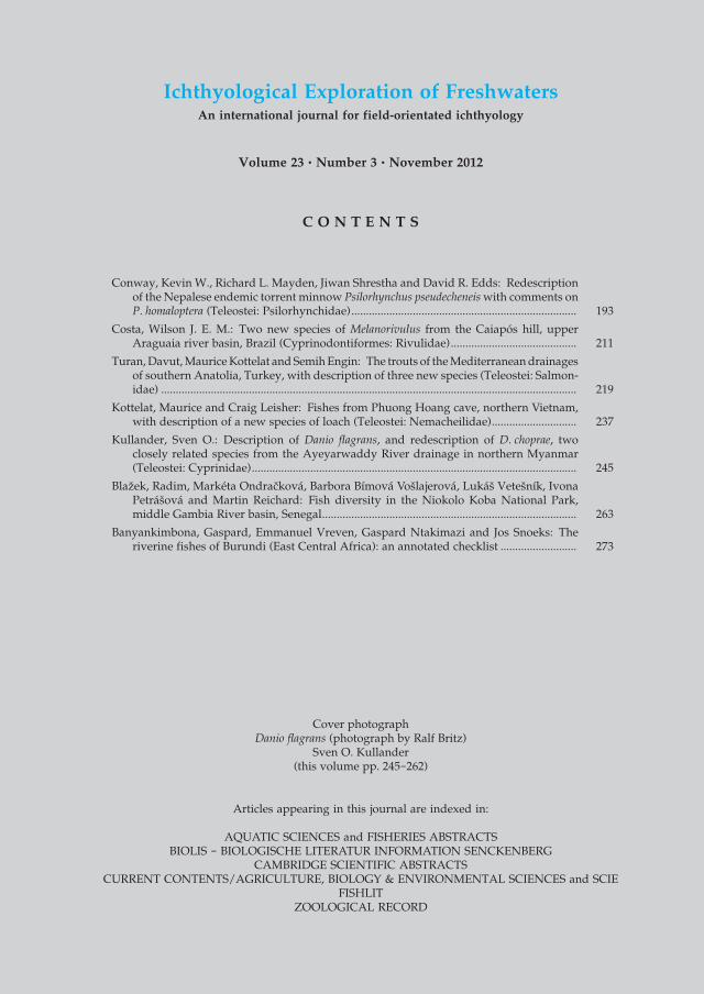
C O N T E N T S
Conway, Kevin W., Richard L. Mayden, Jiwan Shrestha and David R. Edds: Redescription of the Nepalese endemic torrent minnow Psilorhynchus pseudecheneis with comments on P. homaloptera (Teleostei: Psilorhynchidae) ............................................................................. 193
Costa, Wilson J. E. M.: Two new species of Melanorivulus from the Caiapós hill, upper Araguaia river basin, Brazil (Cyprinodontiformes: Rivulidae) ........................................... 211
Turan, Davut, Maurice Kottelat and Semih Engin: The trouts of the Mediterranean drainages of southern Anatolia, Turkey, with description of three new species (Teleostei: Salmon-idae) .............................................................................................................................................. 219
Kottelat, Maurice and Craig Leisher: Fishes from Phuong Hoang cave, northern Vietnam, with description of a new species of loach (Teleostei: Nemacheilidae) ............................. 237
Kullander, Sven O.: Description of Danio flagrans, and redescription of D. choprae, two closely related species from the Ayeyarwaddy River drainage in northern Myanmar (Teleostei: Cyprinidae) ............................................................................................................... 245
Blažek, Radim, Markéta Ondracková, Barbora Bímová Vošlajerová, Lukáš Vetešník, Ivona Petrášová and Martin Reichard: Fish diversity in the Niokolo Koba National Park, middle Gambia River basin, Senegal ....................................................................................... 263
Banyankimbona, Gaspard, Emmanuel Vreven, Gaspard Ntakimazi and Jos Snoeks: The riverine fishes of Burundi (East Central Africa): an annotated checklist .......................... 273
Ichthyological Exploration of FreshwatersAn international journal for field-orientated ichthyology
Volume 23 • Number 3 • November 2012
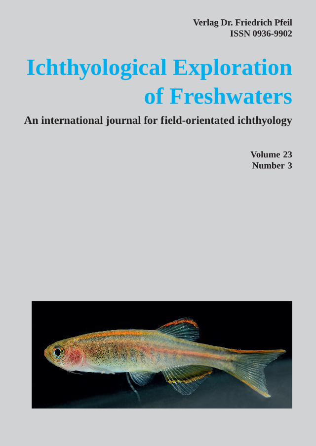
Cover photographDanio flagrans (photograph by Ralf Britz)
Sven O. Kullander(this volume pp. 245-262)