Embed Size (px)
Citation preview
Hindawi Publishing CorporationEvidence-Based Complementary and Alternative MedicineVolume 2012, Article ID 827278, 12 pagesdoi:10.1155/2012/827278
Research Article
Vinegar-Baked Radix Bupleuri Regulates Lipid Disordersvia a Pathway Dependent on Peroxisome-Proliferator-ActivatedReceptor-α in High-Fat-Diet-Induced Obese Rats
Thing-Fong Tzeng,1 Hung-Jen Lu,2 Shorong-Shii Liou,3 Chia Ju Chang,4 and I-Min Liu3
1 Department of Internal Medicine, Pao Chien Hospital, Pingtung City, Pingtung County 90065, Taiwan2 Traditional Medicinal Center, Kaohsiung Veterans General Hospital, Kaohsiung City 81362, Taiwan3 Department of Pharmacy & Graduate Institute of Pharmaceutical Technology, Tajen University, Yanpu Shiang,Pingtung Shien 90701, Taiwan
4 School of Chinese Pharmaceutical Sciences and Chinese Medicine Resources, China Medical University, Taichung 40402, Taiwan
Correspondence should be addressed to I-Min Liu, [email protected]
Received 19 June 2011; Accepted 23 September 2011
Academic Editor: Vincenzo De Feo
Copyright © 2012 Thing-Fong Tzeng et al. This is an open access article distributed under the Creative Commons AttributionLicense, which permits unrestricted use, distribution, and reproduction in any medium, provided the original work is properlycited.
The aim of this study was to investigate the antiobesity and antihyperlipidemic effects of vinegar-baked Radix Bupleuri (VBRB)on high-fat diet- (HFD-) induced obese rats. After being fed HFD for two weeks, rats were dosed orally with VBRB or fenofibrate,once daily for further twelve weeks. VBRB (1.0 g kg−1 per day) produced effects similar to fenofibrate (100 mg kg−1) in reducingbody weight (BW) gain, visceral fat-pad weights, plasma lipid levels, as well as hepatic TG and cholesterol content of HFD-fed rats. VBRB also lowered hepatic lipid droplet accumulation and the size of epididymal adipocytes in HFD-fed rats. VBRBand fenofibrate reversed the HFD-induced downregulation of hepatic peroxisome proliferator-activated receptor (PPAR)α. HFD-induced reductions in the hepatic levels of acyl-CoA oxidase (ACO) and cytochrome P450 isoform 4A1 (CYP4A1) proteins werereversed by VBRB and fenofibrate. The elevated expression of hepatic sterol regulatory element binding proteins (SREBPs) inHFD-fed rats was lowered by VBRB and fenofibrate. The results of this study show that VBRB suppresses BW gain and body fataccumulation by increasing fatty acid oxidation, an effect which is likely mediated via upregulation of PPARα and downregulationof SREBP expression in the liver of HFD-fed rats.
1. Introduction
Obesity is a common chronic disorder of carbohydrate andfat metabolism, characterized by an excessive fat depositionin adipose tissue and other internal organs, such as liver,heart, skeletal muscle, and pancreatic islets [1]. Obesityremains a major global public health issue because of itsincreasing prevalence, which cuts across issues of sex, agegroup, ethnicity, or race [1]. Obesity alone can induce all thesymptoms of metabolic syndrome, which is associated withmany additional health problems, including increased riskof insulin resistance, nonalcoholic fatty liver, atherosclerosis,degenerative disorders such as dementia, some immune-mediated disorders such as asthma, and certain cancers[2, 3]. Pharmacological approaches to weight control have
become an overriding priority [4]. Current trends for obesitymanagement involve multiple pharmacological strategies,including blocking nutrient absorption, modulating fat me-tabolism, regulating adipose signals, and modulating thesatiety center. However, these approaches have been asso-ciated with several serious adverse effects in the clinic, in-cluding adverse gastrointestinal effects and significant unfa-vorable cardiovascular effects [4]. As a result, a much safertherapeutic approach is necessary.
Radix Bupleuri, with a Chinese name Chaihu, is recordedas the roots of Bupleurum chinense and B. scorzonerifolium(family Umbelliferae) in the Chinese Pharmacopoeia, whichhas been widely practiced to treat influenza, fever, malaria,hepatitis, jaundice, nephritis, dizziness, bitter taste in the
2 Evidence-Based Complementary and Alternative Medicine
mouth, lung diseases, cancer, and menstrual disorders inChina, Japan, and other Asian countries [5]. In previouschemical studies on Bupleurum plants, saponins, flavonoids,coumarins, fatty acids, steroids, polysaccharides, and poly-acetylenes were identified [5]. Among them saponins wereknown to be the major bioactive compounds, which werecommonly used as chemical standards for quality evaluationof Radix Bupleuri in the current Chinese Pharmacopoeiaand recent publications [6]. When Radix Bupleuri was mixedthoroughly with vinegar and then baked to dry, it waschanged to vinegar-baked Radix Bupleuri (VBRB). The phar-macological effect and components in the drug changed alittle bit due to the vinegar-baked procedure [7]. It wasdemonstrated that VBRB has a much stronger effect onacesodyne and bile secretion than that of the Radix Bupleuri[8]. Therefore, VBRB was extensively used in traditional Chi-nese medicines for liver diseases treatment [9]. At present,although there are some reports on the pharmacologicaleffect of VBRB, little is known about the effect of VBRB onlipid regulation besides its clinical usage.
Diet-induced obesity in rodents has been used as a modelto investigate the interactions between the environment andgenetics. Rats fed a high-fat diet (HFD) become obese andshow distinctive visceral adiposity, dyslipidemia, hyperinsu-linemia, and hepatic steatosis, which are typical of humanobesity [10]. Therefore, this study investigated the effects ofVBRB on body fat and lipid profiles in rats with diet-inducedobesity and sought possible mechanisms of action.
2. Materials
2.1. Plant Preparations. The crude Radix Bupleuri waspurchased from Jinbaoan Trade Co., Ltd. (Zhunan Township,Miaoli County 350, Taiwan) in December 2010. The identityof the plants was confirmed by Hong T.Y. (Departmentof Biotechnology, College of Pharmacy and Health Care,Tajen University) using macroscopic and microscopic exam-inations as well as thin-layered chromatography and high-performance liquid chromatography (HPLC) methods. Ran-dom amplified polymorphic DNA analysis was also appliedto identify DNA polymorphisms in crude Radix Bupleuri. Avoucher specimen (Lot No. 20101205) has been deposited atour laboratory. The crude Radix Bupleuri was chopped intosmall pieces (10 kg) and incubated with vinegar (2 kg); untilvinegar was totally absorbed into Radix Bupleuri. Then, thematerial was dried by stir-firing to obtain VBRB.
2.2. Analytical Method. VBRB was ground to a powderand sieved through a 40-mesh screen. The powder wasdried at 60◦C until constant weight and was well-blendedbefore use. An ASE 100 System (Dionex, Sunnyvale, CA,USA) with 34 mL stainless steel ASE vessels was used forthe pressurized liquid extraction. Extraction conditionswere optimized by single-factor experiments (sequentiallyvarying the experimental parameters, one at a time, whileall the other parameters remained fixed). The extract wasevaporated to dryness using a rotary evaporator under 45◦C.The residue was then dissolved in 5.0 mL of methanol and
filtered through a 0.45 μm nylon filter membrane prior toinjection into the HPLC system.
An Agilent 1100 HPLC system was used, equipped with aquaternary pump, an autosampler, a degasser, an automaticthermostatic column compartment, and a DAD detector.Chromatography was performed on a TSKgel ODS-100 VC18 column (3 μm, 15064.6 mm id, Tosoh, Tokyo, Japan) ata column temperature of 30◦C and a flow rate of 1 mL min−1
using CH3OH (solvent A) and water (solvent B) as a mobilephase with linear gradients: 0–60 min (30–40% A), 60–80 min (40–50% A), and 80–90 min (50–100% A). DAD wasset to monitor at 210 nm, and the online UV spectra wererecorded in the range 190–400 nm.
For LC-ESI-MS/MS analysis, an Agilent 1100 HPLCsystem coupled with a LC/MSD Trap XCT mass spectrometer(Agilent Technologies, MA, USA) was used. The acquisitionparameters were as follows: collision gas, ultrahighpurityhelium; nebulizer gas, high purity nitrogen; nebulizer gas(N2), 35 psi; dry gas (N2), 12 L min−1; dry temperature,350◦C; HV voltage, 3500 V; mass range recorded mass-to-charge ratio (m/z) 200–1600, target mass m/z 800;compound stability, 100%; trap drive level, 100%; collisionenergy (Ampl), 0.3–2 V. Data-dependent MS/MS scanningwas used in negative-ion mode so that the two mostabundant ions in each MS scan were selected in turn andsubjected to IT-MS (MS/MS, with MS/MS ranging from 2–4)analyses.
A 1000 μg mL−1 standard stock solution of saikosaponina (SSa, Sigma-Aldrich, Inc., Saint Louis, MO, USA) orsaikosaponin d (SSd, Sigma-Aldrich, Inc.) was prepared anddiluted in 10 μg mL−1 to provide the working solution. Todetermine SSa and SSd simultaneously, SSa and SSd wereused to prepare a series of standards having concentrationsof 12.5, 25, 50, 100, 250, and 400 ng mL−1 in pure 70%methanol solution and also in the extract from VBRB toallow calibration of the samples. A calibration curve wasobtained by plotting the ratio of the peak areas of theanalyte and the IS as a function of the analyte concentration.A weighted (1/x2) linear regression line was fitted overthe concentration range from 12.5 to 400 ng mL−1. Theconcentrations of SSa and SSc in VBRB samples werecalculated from the ratios; they are expressed in units ofmicrograms per gram of preparation powder.
2.3. Extract of VBRB. The extract of VBRB was carried outaccording to the method reported previously [9]. Briefly,200 g of VBRB was socked in 2000 mL of water for 0.5 h, andthen heated to boil, kept boiling for 45 min, thereafter, fil-trated the extract. Gruffs were extracted again with 1600 mLof water for a further 0.5 h, pooled the filtrate, and condensedto 200 mL and then stored the extract at −20◦C until use.
2.4. Cell Culture. 3T3-L1 preadipocytes, obtained fromBioresource Collection and Research Center (BCRC 60159)of the Food Industry Research and Development Institute(Hsinchu, Taiwan), were cultured in Dulbecco’s ModifiedEagle’s Medium (DMEM) (GIBCO BRL Life Technologies,Invitrogen Corporation, CA, USA) with 10% fetal bovineserum (FBS) (GIBCO BRL) and antibiotics (100 units mL−1
Evidence-Based Complementary and Alternative Medicine 3
penicillin and 100 μg mL−1 streptomycin). When cellswere confluent, differentiation was induced by adding0.5 mmol L−1 isobutylmethylxanthine (Sigma-Aldrich Co.)and 1 μmol L−1 dexamethasone (Sigma-Aldrich Co.) to thecultures. After 2 days, cells were allowed to differentiatefurther by adding 10% FBS and 10 μg mL−1 insulin (Sigma-Aldrich Co.) and the medium was changed every 2 days.At day 10, about 80% of cultures were induced to containtriglyceride (TG). Treatments including serum starvation(DMEM only), SSa, SSd, or fenofibrate (Sigma-Aldrich Co.)were given to differentiated cultures for 8 hours. All otherreagents were of analytical grade.
2.5. Measurement of the Triglyceride Content. Oil Red O(Sigma-Aldrich Co.) at 0.2% in isopropanol (Sigma-AldrichCo.) was mixed with water (3 : 2, v v−1) and filtered. Exper-imental cultured cells were washed with PBS, fixed by 4%paraformaldehyde (Sigma-Aldrich Co.) in PBS for 5 minutes,incubated with filtered Oil Red O for 30 minutes, and washedtwice with PBS. The stained TG was extracted by isopropanoland its quantity was measured at 490 nm absorbance [11].A total of 5 independent experiments, with triplicates each,were performed.
2.6. Animal Models and Treatment Protocols. Male Wistarrats, 8 weeks of age, were obtained from the NationalLaboratory Animal Center (Taipei, Taiwan). They weremaintained in a temperature-controlled room (25 ± 1◦C)on a 12 h : 12 h light-dark cycle (lights on at 06:00 h) in theanimal center (Tajen University, Ping Tung Shien, Taiwan).Food and water were available ad libitum. Regular rat chowdiet (RCD, #D12450B, Research Diets, New Brunswick, NJ)with 20 kcal% protein, 70 kcal% carbohydrate, and 10 kcal%fat from lard was used as the maintenance and controldiet. A purified ingredient HFD with 20 kcal% protein,35 kcal% carbohydrate, and 45 kcal% fat primarily fromlard (#D12451, Research Diets) was used to induce a rapidincrease in body weight (BW) and obesity [12]. The caloricdensity of the control diet was 3.85 kcal g−1; that of the HFDwas 4.73 kcal g−1. All animal procedures were performedaccording to the Guide for the Care and Use of LaboratoryAnimals of the National Institutes of Health as well as theguidelines of the Animal Welfare Act. These studies wereconducted with the approval of the Institutional Animal Careand Use Committee (IACUC) at Tajen University (approvalnumber: IACUC 98-13; approval date: October 19, 2009).
After being fed a HFD for two weeks, rats were dosed byoral gavage once per day for 12 weeks with VBRB doses of0.3, 0.5, and 1.0 g kg−1 in a volume of 1.5 mL kg−1 distilledwater. Another group of HFD-fed rats was treated orallyfor 12 weeks with 100 mg kg−1 per day fenofibrate. In folkmedicine, the recommended daily oral dose of VBRB is 3–10 g per adult per day. The dose of VBRB was calculated bymultiplying the recommended dose of intake per kilogramby the human metabolism coefficient to get the dose ofintake per day of rats according to the equation: the intakedose per kilogram of rat body weight = recommendedintake dose for humans ÷ body weight 60 kg × 6.25 [13].The approximate daily oral dose of VBRB for a rat is
ranged from 0.3–1.0 g kg−1. The dose of fenofibrate wasbased on studies with long-term fenofibrate treatment inrats [14]. A vehicle-control group of HFD-fed and RCD-fed rats was treated with 1.5 mL kg−1 distilled water onlyover the same treatment period. Each experimental groupcontained 8 rats in the study. Twelve weeks after treatmentwith VBRB or fenofibrate, HFD-fed rats were weighedand anesthetized with sodium pentobarbital (30 mg kg−1)administered intraperitoneally (i.p.), and blood samples werecollected from the lateral tail vein. Feed and water weresupplied ad libitum throughout the 12-week experimentalperiod. Samples were centrifuged at 2,000×g for 10 minutesat 4◦C. The plasma was then removed and placed intoaliquots for the respective analytical determinations. Afterblood was collected, the liver and visceral and subcutaneouswhite adipose tissues (WAT) were removed, rinsed withphysiological saline, weighed, and immediately stored at−70◦C.
2.7. Biochemical Parameter Analysis. Diagnostic kits for themeasurement of plasma glucose (Cat. No. 10009582), totalcholesterol (TC; Cat. No. 10007640), and triglycerides (TG;Cat. No. 10010303) were purchased from Cayman ChemicalCompany (Michigan, USA). The diagnostic kit to determineplasma and hepatic levels of high density lipoprotein choles-terol (HDL-C) was purchased from Bio-Quant Diagnostics(Cat. No. BQ 019CR, CA, USA), and low density lipoproteincholesterol (LDL-C) levels were calculated using Friedewald’sequation [15]. Plasma-free fatty acid (FFA) levels weredetermined using an FFA-quantification kit obtained fromAbcam plc (Cat. No. ab65341, MA, USA). All samples wereanalyzed in triplicate. Atherogenic index (AI) and coronaryrisk index (CRI) were calculated as LDL-C/HDL-C andTC/HDL-C, respectively [16, 17].
2.8. Extraction of Hepatic Lipid. A section of each liver wascollected for lipid content analysis. The liver (1.25 g) washomogenized with chloroform/methanol (1 : 2, 3.75 mL),and then chloroform (1.25 mL) and distilled water (1.25 mL)were added to the homogenate and mixed well. Aftercentrifugation (1,500×g for 10 min), the lower clear organicphase solution was transferred into a new glass tube andthen lyophilized. The lyophilized powders were dissolved inchloroform/methanol (1 : 2) as the hepatic lipid extracts andstored at −20◦C for fewer than three days [18].
2.9. Hepatic Pathological Evaluation. Small pieces of hepatictissues taken from experimental animals were fixed in10% neutral formalin, dehydrated with alcohol, embeddedin paraffin, and sectioned to a mean thickness of 4 μm.Hematoxylin- and- eosin- (H&E-) stained tissues wereexamined histologically to evaluate the index of diabetic-induced necrosis. Liver biopsy was scored according tothe following criteria: grade 0: no steatosis, normal liver;grade 1: <25% of hepatocytes affected; grade 2: 26–50%of hepatocytes affected; grade 3: 51–75% of hepatocytesaffected; and grade 4: >76% of hepatocytes affected [19].A total of 5 independent experiments were carried out forstatistical analysis.
4 Evidence-Based Complementary and Alternative Medicine
2.10. Adipocyte Pathological Evaluation. Histological pho-tomicrographs of adipose tissue were analyzed with a lightmicroscope using the paraffin method. Fresh tissues werefixed immediately in Bouin’s solution for 6 to 12 hours, andthen fixed tissues were washed under running water. Afterbeing dehydrated through different grades of alcohol, thetissues were embedded in paraffin block at 60◦C. Sections(8 μm) were cut and mounted on glass slides coated withan egg albumin, and the paraffin was removed using xyleneand alcohol. The tissues were stained with H&E. After beingdehydrated and cleared of alcohol and xylene, the glassslides were mounted in Canada Balsam. Photomicrographswere taken with a Zeiss Axiolab light microscope equippedwith a Nikon Microflex HFX microscope camera. The sizesof epididymal adipocytes were calculated using Image-ProPlus 7.0 (Media Cybernetics, MD, USA). A total of 5independent experiments were carried out for statisticalanalysis.
2.11. Preparation of Hepatic Fractions. Hepatic fractions wereprepared as described previously [20]. To prepare nuclearfractions, hepatic tissue was homogenized with ice-coldlysis buffer containing 5 mmol L−1 Tris-HCl (pH 7.5),2 mmol L−1 MgCl2, 15 mmol L−1 L CaCl2, 1.5 mol L−1 su-crose, 0.1 mol L−1 dithiothreitol (DTT), and protease in-hibitor cocktail. After centrifugation (10,500×g for 20 mi-nutes at 4◦C), the pellet was suspended in extrac-tion buffer containing 20 mmol L−1 2-[4-(2-hydroxyethyl)-1-piperazinyl]ethanesulfonic acid (pH 7.9), 1.5 mmol L−1
MgCl2, 0.42 mol L−1 NaCl, 0.2 mmol L−1 L EDTA, 25%(v v−1) glycerol, 0.1 mol L−1 DTT, and protease inhibitorcocktail. The mixture was placed on ice for 30 minutes. Thenuclear fraction was prepared by centrifugation at 20,500×gfor 5 minutes at 4◦C. The postnuclear fraction was extractedfrom the liver of each rat as described below. In brief, hepatictissue was homogenized with ice-cold lysis buffer (pH 7.4)containing 137 mmol L−1 NaCl, 20 mmol L−1 Tris-HCl, 1%Tween 20, 10% glycerol, 1 mmol L−1 phenylmethylsulfonylfluoride, and protease inhibitor cocktail solution in DMSO.The homogenate was then centrifuged at 2,000×g for10 minutes at 4◦C. The protein concentration of eachfraction was determined using a commercial kit (Bio-RadLaboratories, Hercules, CA, USA).
2.12. Western Blot Analyses. For the determination of per-oxisome-proliferators-activated-receptor (PPAR), sterol-reg-ulatory-element-binding protein (SREBP)-1 and SREBP-2, 30 mg protein of each nuclear fraction was resolvedusing 8% sodium dodecylsulfate polyacrylamide gel elec-trophoresis (SDS-PAGE). Separated proteins were trans-ferred electrophoretically to a nitrocellulose membrane,blocked with 5% (w v−1) skim milk solution for 1 hour,and then incubated with primary antibodies to PPARα(Santa Cruz Biotechnology, Inc., CA, USA; Cat. No. sc-1985), SREBP-1 (Santa Cruz Biotechnology, Inc.; Cat. No. sc-367), SREBP-2 (Santa Cruz Biotechnology, Inc.; Cat. No. sc-5603), or β-actin (Santa Cruz Biotechnology, Inc.; Cat. No.sc-130656) overnight at 4◦C. After the blots were washed,
they were incubated with goat anti-rabbit and/or goat anti-mouse IgG HRP-conjugated secondary antibody for 1.5hours at room temperature. The blots were stripped withRestore Western Blot Stripping Buffer (CANDOR BioscienceGmbH, Wangen, Germany) for 15 minutes and incubatedwith the antibodies. In addition, 30 mg protein from eachpostnuclear fraction for acyl-CoA oxidase (ACO; Santa CruzBiotechnology, Inc.; Cat. No. sc-98499) and cytochromeP450 isoform 4A1 (CYP4A1; Santa Cruz Biotechnology,Inc.; Cat. No. sc-53248) was subjected to 10% SDS-PAGE. Each antigen-antibody complex was visualized usingECL Western Blotting Detection Reagents and detected bychemiluminescence with LAS-1000 plus (Fujifilm, Tokyo,Japan). Band densities were determined using ATTO Den-sitograph Software (ATTO Corporation, Tokyo, Japan) andquantified as the ratio to β-actin. The mean value forsamples from the vehicle-treated RCD-fed group on eachimmunoblot, expressed in densitometry units, was adjustedto a value of 1.0. All experimental sample values were thenexpressed relative to this adjusted mean value. A total of5 independent experiments were carried out for statisticalanalysis.
2.13. Statistical Analysis. Data are expressed as the mean± standard deviation (SD) for each group of animals atthe number (n) indicated in tables. Statistical analysis wasperformed with one-way analysis of variance (ANOVA). TheDunnett range posthoc comparisons were used to determinethe source of significant differences where appropriate. A Pvalue <.05 was considered statistically significant.
3. Results
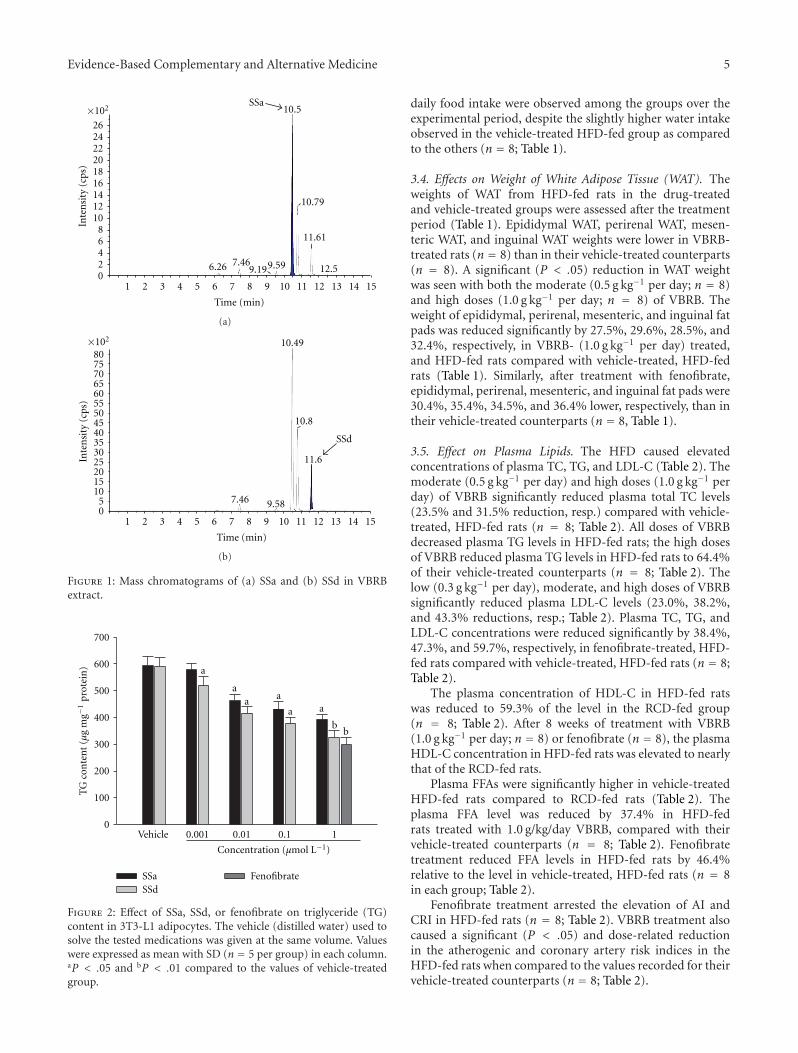
3.1. Quantitative Analysis. The regression equations of SSaand SSd are y = 190.65x − 1219.72 (R2 = 0.9992) andy = 42.87x−111.25 (R2 = 0.9983), respectively. The contentof SSa (724.63 ± 0.06 μg/g) was higher than that of SSd (9.62± 0.04 μg/g) in VBRB. The chromatogram of the samplesolution has been shown in Figure 1.
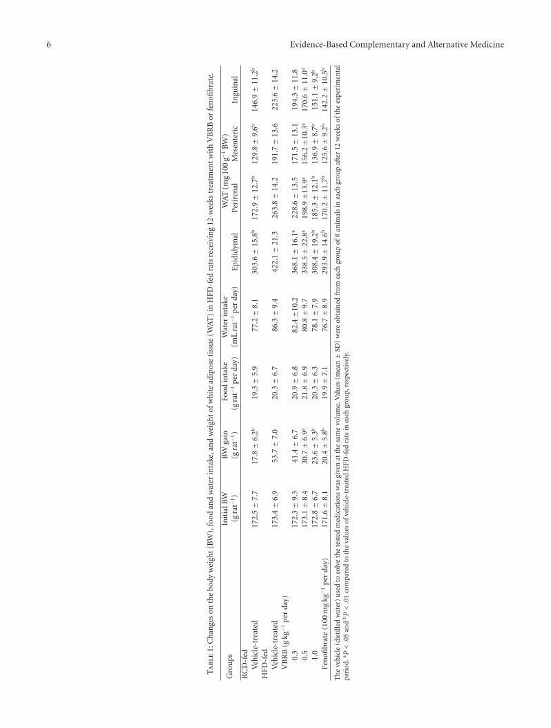
3.2. Effects on Lipogenic Differentiated 3T3-L1 Adipocyte. SSaand SSd exhibited a significant concentration-dependentdecrease in the intracellular accumulation of TG in 3T3-L1 adipocytes; the most significant effect (over 30% TGreduction) was observed in treatment at 1 μmol L−1 (n = 5;Figure 2). Fenofibrate (1 μmol L−1) caused a decrease in theTG content of differentiated 3T3-L1 adipocytes by 41% (n =5; Figure 2).
3.3. Effects on Body Weight (BW) and Food Intake. As shownin Table 1, the BWs of HFD-fed rats in the drug-treatedand vehicle-treated groups were monitored over the 12-weektreatment period. At the end of treatment, the BW of VBRB-treated rats (n = 8) was significantly lower than that of ratsin the vehicle-treated group (n = 8). VBRB significantlysuppressed BW gain at both the moderate (0.5 g kg−1 perday; n = 8) and high doses (1.0 g kg−1 per day; n = 8).Similar results were seen in rats treated with fenofibrate(100 mg kg−1 per day; n = 8). No significant differences in
Evidence-Based Complementary and Alternative Medicine 5
SSa×102
262422201816141210
86420
1 2 3 4 5 6 7 8 9 10 11 12 13 14 15
6.26 7.469.199.59
10.5
10.79
11.61
12.5
Time (min)
Inte
nsi
ty(c
ps)
(a)
1 2 3 4 5 6 7 8 9 10 11 12 13 14 15
Time (min)
SSd
×102
807570656055504540353025201510
50
7.46 9.58
10.49
10.8
11.6Inte
nsi
ty(c
ps)
(b)
Figure 1: Mass chromatograms of (a) SSa and (b) SSd in VBRBextract.
0
100
200
300
400
500
600
700
SSaSSd
Fenofibrate
Vehicle 0.001 0.01 0.1 1
a
aa
b
a
a a
b
TG
con
ten
t(μ
gm
g−1
prot
ein
)
Concentration (μmol L−1)
Figure 2: Effect of SSa, SSd, or fenofibrate on triglyceride (TG)content in 3T3-L1 adipocytes. The vehicle (distilled water) used tosolve the tested medications was given at the same volume. Valueswere expressed as mean with SD (n = 5 per group) in each column.aP < .05 and bP < .01 compared to the values of vehicle-treatedgroup.
daily food intake were observed among the groups over theexperimental period, despite the slightly higher water intakeobserved in the vehicle-treated HFD-fed group as comparedto the others (n = 8; Table 1).
3.4. Effects on Weight of White Adipose Tissue (WAT). Theweights of WAT from HFD-fed rats in the drug-treatedand vehicle-treated groups were assessed after the treatmentperiod (Table 1). Epididymal WAT, perirenal WAT, mesen-teric WAT, and inguinal WAT weights were lower in VBRB-treated rats (n = 8) than in their vehicle-treated counterparts(n = 8). A significant (P < .05) reduction in WAT weightwas seen with both the moderate (0.5 g kg−1 per day; n = 8)and high doses (1.0 g kg−1 per day; n = 8) of VBRB. Theweight of epididymal, perirenal, mesenteric, and inguinal fatpads was reduced significantly by 27.5%, 29.6%, 28.5%, and32.4%, respectively, in VBRB- (1.0 g kg−1 per day) treated,and HFD-fed rats compared with vehicle-treated, HFD-fedrats (Table 1). Similarly, after treatment with fenofibrate,epididymal, perirenal, mesenteric, and inguinal fat pads were30.4%, 35.4%, 34.5%, and 36.4% lower, respectively, than intheir vehicle-treated counterparts (n = 8, Table 1).
3.5. Effect on Plasma Lipids. The HFD caused elevatedconcentrations of plasma TC, TG, and LDL-C (Table 2). Themoderate (0.5 g kg−1 per day) and high doses (1.0 g kg−1 perday) of VBRB significantly reduced plasma total TC levels(23.5% and 31.5% reduction, resp.) compared with vehicle-treated, HFD-fed rats (n = 8; Table 2). All doses of VBRBdecreased plasma TG levels in HFD-fed rats; the high dosesof VBRB reduced plasma TG levels in HFD-fed rats to 64.4%of their vehicle-treated counterparts (n = 8; Table 2). Thelow (0.3 g kg−1 per day), moderate, and high doses of VBRBsignificantly reduced plasma LDL-C levels (23.0%, 38.2%,and 43.3% reductions, resp.; Table 2). Plasma TC, TG, andLDL-C concentrations were reduced significantly by 38.4%,47.3%, and 59.7%, respectively, in fenofibrate-treated, HFD-fed rats compared with vehicle-treated, HFD-fed rats (n = 8;Table 2).
The plasma concentration of HDL-C in HFD-fed ratswas reduced to 59.3% of the level in the RCD-fed group(n = 8; Table 2). After 8 weeks of treatment with VBRB(1.0 g kg−1 per day; n = 8) or fenofibrate (n = 8), the plasmaHDL-C concentration in HFD-fed rats was elevated to nearlythat of the RCD-fed rats.
Plasma FFAs were significantly higher in vehicle-treatedHFD-fed rats compared to RCD-fed rats (Table 2). Theplasma FFA level was reduced by 37.4% in HFD-fedrats treated with 1.0 g/kg/day VBRB, compared with theirvehicle-treated counterparts (n = 8; Table 2). Fenofibratetreatment reduced FFA levels in HFD-fed rats by 46.4%relative to the level in vehicle-treated, HFD-fed rats (n = 8in each group; Table 2).
Fenofibrate treatment arrested the elevation of AI andCRI in HFD-fed rats (n = 8; Table 2). VBRB treatment alsocaused a significant (P < .05) and dose-related reductionin the atherogenic and coronary artery risk indices in theHFD-fed rats when compared to the values recorded for theirvehicle-treated counterparts (n = 8; Table 2).
6 Evidence-Based Complementary and Alternative Medicine
Ta
ble
1:C
han
ges
onth
ebo
dyw
eigh
t(B
W),
food
and
wat
erin
take
,an
dw
eigh
tof
wh
ite
adip
ose
tiss
ue
(WA
T)
inH
FD-f
edra
tsre
ceiv
ing
12-w
eeks
trea
tmen
tw
ith
VB
RB
orfe
nofi
brat
e.
Gro
ups
Init
ialB
W(g
rat−
1)
BW
gain
(gra
t−1)
Food
inta
ke(g
rat−
1p
erda
y)W
ater
inta
ke(m
Lra
t−1
per
day)
WA
T(m
g10
0g−
1B
W)
Epi
didy
mal
Peri
ren
alM
esen
teri
cIn
guin
alR
CD
-fed
Veh
icle
-tre
ated
172.
5±
7.7
17.8±
6.2b
19.3±
5.9
77.2±
8.1
303.
6±
15.8
b17
2.9±
12.7
b12
9.8±
9.6b
146.
9±
11.2
b
HFD
-fed
Veh
icle
-tre
ated
173.
4±
6.9
53.7±
7.0
20.3±
6.7
86.3±
9.4
422.
1±
21.3
263.
8±
14.2
191.
7±
13.6
223.
6±
14.2
VB
RB
(gkg−1
per
day)
0.3
172.
3±
9.3
41.4±
6.7
20.9±
6.8
82.4±1
0.2
368.
1±
16.1
a22
8.6±
13.5
171.
5±
13.1
194.
3±
11.8
0.5
173.
1±
8.4
30.7±
6.9a
21.8±
6.9
80.8±
9.7
338.
5±
22.8
a19
8.9±1
3.9a
156.
2±1
0.3a
170.
6±
11.0
a
1.0
172.
8±
6.7
23.6±
5.3b
20.3±
6.3
78.1±
7.9
308.
4±
19.2
b18
5.3±
12.1
b13
6.9±
8.7b
151.
1±
9.2b
Fen
ofibr
ate
(100
mg
kg−1
per
day)
171.
6±
8.1
20.4±
5.8b
19.9±
7.1
76.7±
8.9
293.
9±
14.6
b17
0.2±
11.7
b12
5.6±
9.2b
142.
2±
10.5
b
Th
eve
hic
le(d
isti
lled
wat
er)
use
dto
solv
eth
ete
sted
med
icat
ion
sw
asgi
ven
atth
esa
me
volu
me.
Val
ues
(mea
n±
SD)
wer
eob
tain
edfr
omea
chgr
oup
of8
anim
als
inea
chgr
oup
afte
r12
wee
ksof
the
expe
rim
enta
lp
erio
d.a P
<.0
5an
dbP<.0
1co
mpa
red
toth
eva
lues
ofve
hic
le-t
reat
edH
FD-f
edra
tsin
each
grou
p,re
spec
tive
ly.
Evidence-Based Complementary and Alternative Medicine 7
Ta
ble
2:C
han
ges
inth
epl
asm
alip
ids,
hep
atic
lipid
s,at
her
ogen
icin
dex
(AI)
,an
dco
ron
ary
arte
ryin
dex
(CR
I)in
HFD
-fed
rats
rece
ivin
g12
-wee
kstr
eatm
ent
wit
hV
BR
Bor
fen
ofibr
ate.
Gro
ups
Pla
sma
lipid
s(m
gdL
−1)
AI
CR
IH
epat
iclip
ids
(μm
olg−
1liv
er)
TC
TG
LDL-
CH
DL-
CFF
As
TC
TG
RC
D-f
edV
ehic
le-t
reat
ed72
.9±
5.8b
56.8±
6.6b
31.8±
3.2b
46.9±
3.4b
28.2±
2.8b
0.7±
0.3b
1.6±
0.4b
19.3±
2.7b
24.8±
6.2b
HFD
-fed
Veh
icle
-tre
ated
130.
2±
12.3
124.
5±
6.3
120.
2±
4.2
27.8±
3.8
61.9±
5.3
4.3±
0.2
4.7±
0.3
40.2±
3.1
54.4±
8.4
VB
RB
(gkg−1
per
day)
0.3
116.
7±
10.3
110.
9±
6.8a
92.5±
5.7a
32.1±
4.2a
55.3±
4.6
2.9±
0.2a
3.6±
0.3a
33.9±
3.5
48.6±
7.9
0.5
99.6±
8.3a
95.0±
5.2a
74.3±
5.1b
39.1±
2.9a
47.1±
3.8a
1.9±
0.1b
2.5±
0.3b
31.7±
4.1a
40.8±
7.1
1.0
89.1±
7.9a
80.3±
4.8b
68.1±
4.5b
43.9±
3.2b
38.7±
3.2b
1.5±
0.1b
2.0±
0.3b
26.9±
3.8a
35.2±
6.5a
Fen
ofibr
ate
(100
mg
kg−1
per
day)
80.2±
8.0b
65.6±
4.7b
48.4±
4.3b
45.9±
4.4b
33.2±
4.1b
1.0±
0.2b
1.7±
0.4b
22.4±
4.5b
30.6±
7.8b
Th
eve
hic
le(d
isti
lled
wat
er)
use
dto
solv
eth
ete
sted
med
icat
ion
sw
asgi
ven
atth
esa
me
volu
me.
Val
ues
(mea
n±
SD)
wer
eob
tain
edfr
omea
chgr
oup
of8
anim
als
inea
chgr
oup
afte
r12
wee
ksof
the
expe
rim
enta
lp
erio
d.a P
<.0
5an
dbP<.0
1co
mpa
red
toth
eva
lues
ofve
hic
le-t
reat
edH
FD-f
edra
tsin
each
grou
p,re
spec
tive
ly.
8 Evidence-Based Complementary and Alternative Medicine
3.6. Effect on Hepatic Lipids. The hepatic TC level wassignificantly higher in HFD-fed rats than in rats from theRCD-fed group (n = 8; Table 2). The hepatic TC levels werereduced by 33.4% in HFD-fed rats treated with 1.0 g kg−1
per day VBRB (n = 8; Table 2). Similarly, VBRB treatment(1.0 g kg−1 per day) also produced a significant reductionin hepatic TG concentration, to 64.5% of that in vehicle-treated, HFD-fed rats (n = 8; Table 2). Hepatic TC and TGlevels were significantly reduced (by 44.2% and 43.7%, resp.)in fenofibrate-treated rats compared with vehicle-treated,HFD-fed rats (n = 8; Table 2).
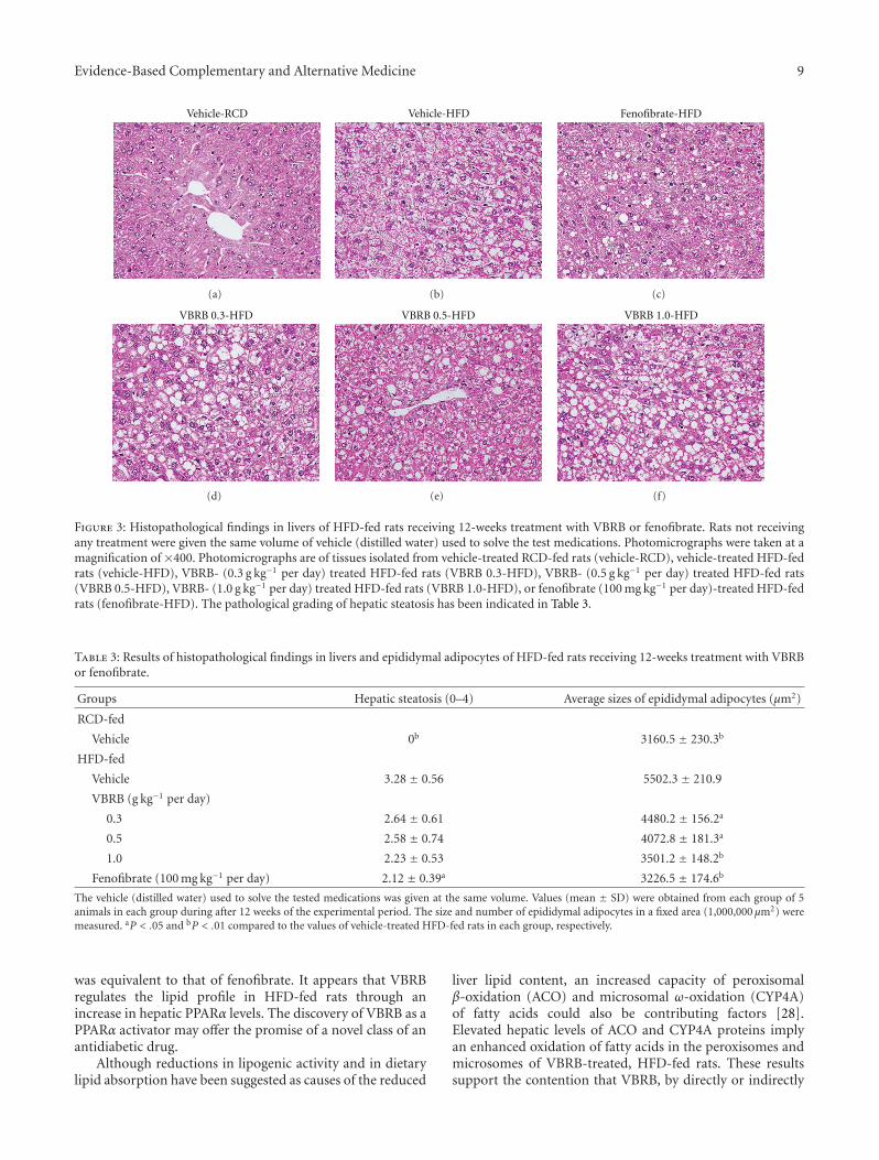
3.7. Morphological Changes in Hepatocytes. HFD-fed ratsshowed significantly greater hepatic lipid accumulation thanRCD-fed animals (Figure 3). The extent of hepatic lipid ac-cumulation after 12 weeks in fenofibrate-treated, HFD-fed rats was similar to that in RCD-fed rats (Figure 3).HFD-fed rats treated with 1.0 g kg−1 per day VBRB showedconsiderably lower hepatic lipid accumulation than theirvehicle-treated counterparts (Figure 3). The pathologicalgrading of hepatic steatosis has been indicated in Table 3(n = 5).
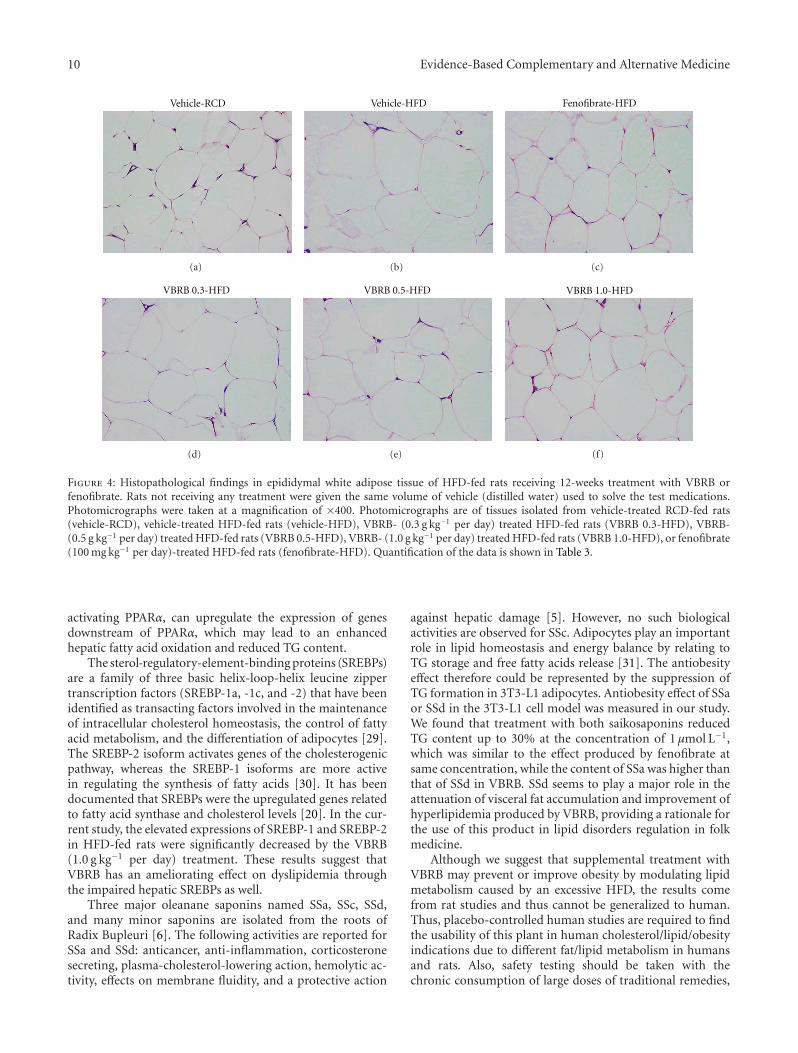
3.8. Morphological Changes in Epididymal Adipocytes. Thehistological appearance of epididymal adipocytes was irreg-ular in HFD-fed rats compared with animals in the RCD-fed group (n = 5; Table 2). This morphological changewas not evident in HFD-fed rats after fenofibrate treat-ment (Figure 4). The histological appearance of epididymaladipocytes in HFD-fed rats treated with 1.0 g kg−1 per dayVBRB was not as normal as in fenofibrate-treated rats,but was better than in vehicle-treated controls (Figure 4).Epididymal adipocytes were also significantly larger in HFD-fed compared to RCD-fed rats (Figure 4). The average sizeof epididymal adipocytes was reduced by approximately36.4% and 4.3%, respectively, in HFD-fed rats treated with1.0 g kg−1 per day VBRB or fenofibrate compared with theirvehicle-treated counterparts (n = 5; Table 3).
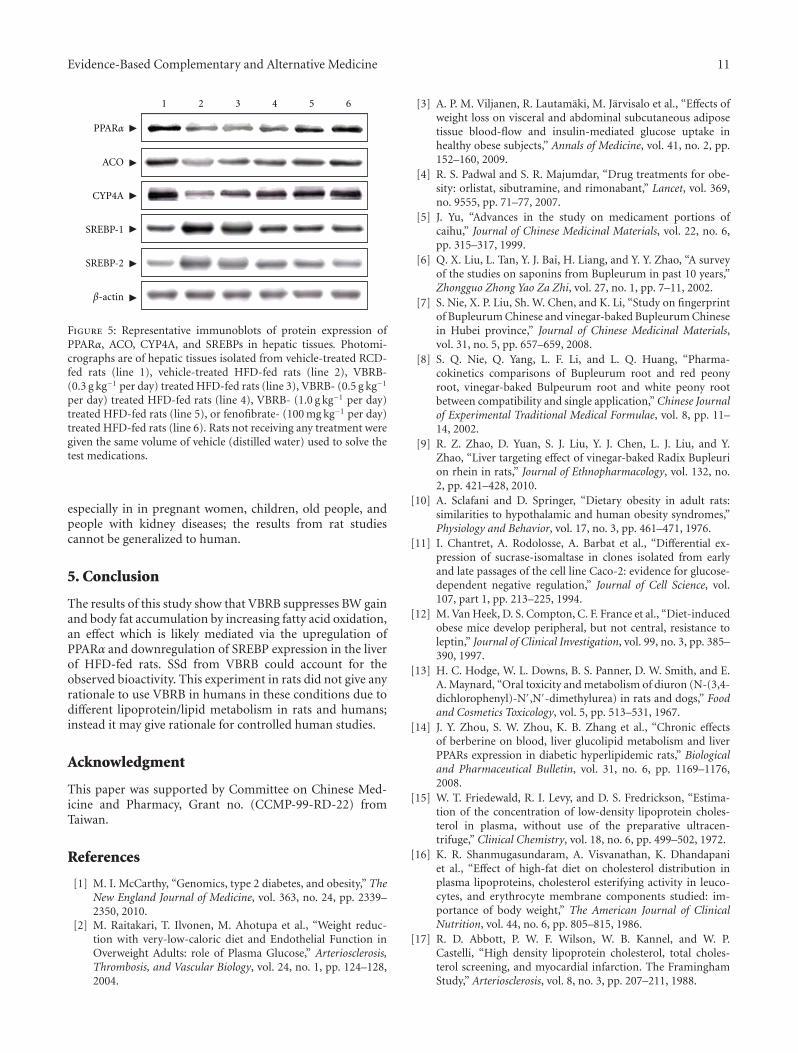
3.9. Protein Expressions of PPARα, ACO, CYP4A, and SREBPsin Hepatic Tissues. Hepatic PPARα protein expression inHFD-fed rats was lower than that in RCD-fed animals, butwas elevated significantly by treatment with 1.0 g kg−1 perday VBRB (Figure 5). In addition, the hepatic expressionlevels of ACO and CYP4A proteins in HFD-fed rats weremarkedly lower than in RCD-fed rats, but were significantlyelevated in VBRB- (1.0 g kg−1 per day) treated HFD-fed rats(Figure 5). Similar results were seen in rats treated withfenofibrate.
The expression levels of hepatic SREBP-1 and SREBP-2 proteins in HFD-fed rats were significantly higher inRCD-fed rats (Figure 5). The proteins levels of hepaticSREBP-1 and SREBP-2 were decreased by 49.3% and 53.7%in HFD-fed rats treated with VBRB (1.0 g kg−1 per day),relative to those in vehicle-treated, HFD-fed rats, respectively(Figure 5). Hepatic SREBP-1 and SREBP-2 protein expres-sion levels in HFD-fed rats treated with fenofibrate were48.1% and 43.1%, respectively, lower than those in theirvehicle-treated counterparts (Figure 5).
4. Discussion
In the this study, BW loss in HFD-fed rats was accompaniedby a depletion of body fat stores, since treatment with VBRBalso significantly reduced the weight of the visceral andsubcutaneous WAT compared with that of vehicle-treatedHFD-fed rats. Excessive growth of adipose tissue results inobesity, which involves two growth mechanisms: hyperplasia(cell number increase) and hypertrophy (cell size increase)[21]. The histological appearance of white adipocytes inHFD-fed animals supplemented with fenofibrate or VBRB(1.0 g kg−1 per day) was more regular, and adipocytes weresimilar in size to those in RCD-fed rats. This suggests thatVBRB suppresses the HFD-induced increase in adipose tissuemass and BW gain and that it may inhibit lipid accumulationin adipose tissue in particular.
Obesity, especially abdominal obesity, is associated withdyslipidemia, characterized by elevated TG and reducedHDL-C concentrations [22]. TG is involved in the ectopicaccumulation of lipid stores in the liver and is associatedwith a number of diseases such as metabolic syndrome andtype 2 diabetes [23]. High TC levels increase the risk ofdeveloping coronary heart disease, and high levels of LDL-C are a risk factor for coronary heart disease, while highHDL-C is helpful in transporting excess cholesterol to theliver for excretion in the bile [24]. As a result, HDL-C levelsare inversely related to coronary heart disease risk [25].Similar to fenofibrate treatment, the oral administrationof VBRB significantly lowered plasma TC, TG, and LDL-C levels in rats with HFD-induced obesity. Thus, VBRBmay be of benefit to patients with hypercholesterolemia andhypertriglyceridemia.
The effect of VBRB on the atherogenic and coronaryartery risk indices is also notable. The ratio of total choles-terol to HDL-C (i.e., the atherogenic index) and the ratioof LDL-C to HDL-C (i.e., the coronary artery index) arestrong and reliable indicators of whether or not cholesterolis deposited into tissues or metabolized and excreted [26].The results of this study show that treatment with VBRB orfenofibrate causes profound reductions in the atherogenicand coronary indices in experimental hyperlipidemic rats.This strongly suggests that VBRB has therapeutic potentialfor the management of obesity and hyperlipidemia and forthe prevention of atherogenic cardiovascular diseases.
Due to the ability of VBRB to reduce serum levels ofTGs and total cholesterol, as well as adipose tissue mass andBW gain functions similar to those of PPARα activation;we hypothesized that the actions of VBRB are related tothe regulation of hepatic expression of PPARα-target genesinvolved in lipid metabolism. In hepatocytes and othertissues (e.g., heart), ligand- (natural long-chain fatty acids)activated PPARα binds to peroxisome proliferator responseelements of DNA and increases the transcription of genesencoding enzymes involved in fatty acid oxidation andlipoprotein metabolism [27]. The outcome is an increasein hepatic fatty acid oxidation and ketogenesis, decreasedlipid levels in tissues, and protection against lipotoxicity.We found that VBRB-treated HFD-fed rats had significantlyhigher hepatic PPARα protein, and that the effect of VBRB
Evidence-Based Complementary and Alternative Medicine 9
Vehicle-RCD
(a)
Vehicle-HFD
(b)
Fenofibrate-HFD
(c)
VBRB 0.3-HFD
(d)
VBRB 0.5-HFD
(e)
VBRB 1.0-HFD
(f)
Figure 3: Histopathological findings in livers of HFD-fed rats receiving 12-weeks treatment with VBRB or fenofibrate. Rats not receivingany treatment were given the same volume of vehicle (distilled water) used to solve the test medications. Photomicrographs were taken at amagnification of ×400. Photomicrographs are of tissues isolated from vehicle-treated RCD-fed rats (vehicle-RCD), vehicle-treated HFD-fedrats (vehicle-HFD), VBRB- (0.3 g kg−1 per day) treated HFD-fed rats (VBRB 0.3-HFD), VBRB- (0.5 g kg−1 per day) treated HFD-fed rats(VBRB 0.5-HFD), VBRB- (1.0 g kg−1 per day) treated HFD-fed rats (VBRB 1.0-HFD), or fenofibrate (100 mg kg−1 per day)-treated HFD-fedrats (fenofibrate-HFD). The pathological grading of hepatic steatosis has been indicated in Table 3.
Table 3: Results of histopathological findings in livers and epididymal adipocytes of HFD-fed rats receiving 12-weeks treatment with VBRBor fenofibrate.
Groups Hepatic steatosis (0–4) Average sizes of epididymal adipocytes (μm2)
RCD-fed
Vehicle 0b 3160.5 ± 230.3b
HFD-fed
Vehicle 3.28 ± 0.56 5502.3 ± 210.9
VBRB (g kg−1 per day)
0.3 2.64 ± 0.61 4480.2 ± 156.2a
0.5 2.58 ± 0.74 4072.8 ± 181.3a
1.0 2.23 ± 0.53 3501.2 ± 148.2b
Fenofibrate (100 mg kg−1 per day) 2.12 ± 0.39a 3226.5 ± 174.6b
The vehicle (distilled water) used to solve the tested medications was given at the same volume. Values (mean ± SD) were obtained from each group of 5animals in each group during after 12 weeks of the experimental period. The size and number of epididymal adipocytes in a fixed area (1,000,000 μm2) weremeasured. aP < .05 and bP < .01 compared to the values of vehicle-treated HFD-fed rats in each group, respectively.
was equivalent to that of fenofibrate. It appears that VBRBregulates the lipid profile in HFD-fed rats through anincrease in hepatic PPARα levels. The discovery of VBRB as aPPARα activator may offer the promise of a novel class of anantidiabetic drug.
Although reductions in lipogenic activity and in dietarylipid absorption have been suggested as causes of the reduced
liver lipid content, an increased capacity of peroxisomalβ-oxidation (ACO) and microsomal ω-oxidation (CYP4A)of fatty acids could also be contributing factors [28].Elevated hepatic levels of ACO and CYP4A proteins implyan enhanced oxidation of fatty acids in the peroxisomes andmicrosomes of VBRB-treated, HFD-fed rats. These resultssupport the contention that VBRB, by directly or indirectly
10 Evidence-Based Complementary and Alternative Medicine
Vehicle-RCD
(a)
Vehicle-HFD
(b)
Fenofibrate-HFD
(c)
VBRB 0.3-HFD
(d)
VBRB 0.5-HFD
(e)
VBRB 1.0-HFD
(f)
Figure 4: Histopathological findings in epididymal white adipose tissue of HFD-fed rats receiving 12-weeks treatment with VBRB orfenofibrate. Rats not receiving any treatment were given the same volume of vehicle (distilled water) used to solve the test medications.Photomicrographs were taken at a magnification of ×400. Photomicrographs are of tissues isolated from vehicle-treated RCD-fed rats(vehicle-RCD), vehicle-treated HFD-fed rats (vehicle-HFD), VBRB- (0.3 g kg−1 per day) treated HFD-fed rats (VBRB 0.3-HFD), VBRB-(0.5 g kg−1 per day) treated HFD-fed rats (VBRB 0.5-HFD), VBRB- (1.0 g kg−1 per day) treated HFD-fed rats (VBRB 1.0-HFD), or fenofibrate(100 mg kg−1 per day)-treated HFD-fed rats (fenofibrate-HFD). Quantification of the data is shown in Table 3.
activating PPARα, can upregulate the expression of genesdownstream of PPARα, which may lead to an enhancedhepatic fatty acid oxidation and reduced TG content.
The sterol-regulatory-element-binding proteins (SREBPs)are a family of three basic helix-loop-helix leucine zippertranscription factors (SREBP-1a, -1c, and -2) that have beenidentified as transacting factors involved in the maintenanceof intracellular cholesterol homeostasis, the control of fattyacid metabolism, and the differentiation of adipocytes [29].The SREBP-2 isoform activates genes of the cholesterogenicpathway, whereas the SREBP-1 isoforms are more activein regulating the synthesis of fatty acids [30]. It has beendocumented that SREBPs were the upregulated genes relatedto fatty acid synthase and cholesterol levels [20]. In the cur-rent study, the elevated expressions of SREBP-1 and SREBP-2in HFD-fed rats were significantly decreased by the VBRB(1.0 g kg−1 per day) treatment. These results suggest thatVBRB has an ameliorating effect on dyslipidemia throughthe impaired hepatic SREBPs as well.
Three major oleanane saponins named SSa, SSc, SSd,and many minor saponins are isolated from the roots ofRadix Bupleuri [6]. The following activities are reported forSSa and SSd: anticancer, anti-inflammation, corticosteronesecreting, plasma-cholesterol-lowering action, hemolytic ac-tivity, effects on membrane fluidity, and a protective action
against hepatic damage [5]. However, no such biologicalactivities are observed for SSc. Adipocytes play an importantrole in lipid homeostasis and energy balance by relating toTG storage and free fatty acids release [31]. The antiobesityeffect therefore could be represented by the suppression ofTG formation in 3T3-L1 adipocytes. Antiobesity effect of SSaor SSd in the 3T3-L1 cell model was measured in our study.We found that treatment with both saikosaponins reducedTG content up to 30% at the concentration of 1 μmol L−1,which was similar to the effect produced by fenofibrate atsame concentration, while the content of SSa was higher thanthat of SSd in VBRB. SSd seems to play a major role in theattenuation of visceral fat accumulation and improvement ofhyperlipidemia produced by VBRB, providing a rationale forthe use of this product in lipid disorders regulation in folkmedicine.
Although we suggest that supplemental treatment withVBRB may prevent or improve obesity by modulating lipidmetabolism caused by an excessive HFD, the results comefrom rat studies and thus cannot be generalized to human.Thus, placebo-controlled human studies are required to findthe usability of this plant in human cholesterol/lipid/obesityindications due to different fat/lipid metabolism in humansand rats. Also, safety testing should be taken with thechronic consumption of large doses of traditional remedies,
Evidence-Based Complementary and Alternative Medicine 11
PPARα
ACO
CYP4A
SREBP-1
SREBP-2
β-actin
1 2 3 4 5 6
Figure 5: Representative immunoblots of protein expression ofPPARα, ACO, CYP4A, and SREBPs in hepatic tissues. Photomi-crographs are of hepatic tissues isolated from vehicle-treated RCD-fed rats (line 1), vehicle-treated HFD-fed rats (line 2), VBRB-(0.3 g kg−1 per day) treated HFD-fed rats (line 3), VBRB- (0.5 g kg−1
per day) treated HFD-fed rats (line 4), VBRB- (1.0 g kg−1 per day)treated HFD-fed rats (line 5), or fenofibrate- (100 mg kg−1 per day)treated HFD-fed rats (line 6). Rats not receiving any treatment weregiven the same volume of vehicle (distilled water) used to solve thetest medications.
especially in in pregnant women, children, old people, andpeople with kidney diseases; the results from rat studiescannot be generalized to human.
5. Conclusion
The results of this study show that VBRB suppresses BW gainand body fat accumulation by increasing fatty acid oxidation,an effect which is likely mediated via the upregulation ofPPARα and downregulation of SREBP expression in the liverof HFD-fed rats. SSd from VBRB could account for theobserved bioactivity. This experiment in rats did not give anyrationale to use VBRB in humans in these conditions due todifferent lipoprotein/lipid metabolism in rats and humans;instead it may give rationale for controlled human studies.
Acknowledgment
This paper was supported by Committee on Chinese Med-icine and Pharmacy, Grant no. (CCMP-99-RD-22) fromTaiwan.
References
[1] M. I. McCarthy, “Genomics, type 2 diabetes, and obesity,” TheNew England Journal of Medicine, vol. 363, no. 24, pp. 2339–2350, 2010.
[2] M. Raitakari, T. Ilvonen, M. Ahotupa et al., “Weight reduc-tion with very-low-caloric diet and Endothelial Function inOverweight Adults: role of Plasma Glucose,” Arteriosclerosis,Thrombosis, and Vascular Biology, vol. 24, no. 1, pp. 124–128,2004.
[3] A. P. M. Viljanen, R. Lautamaki, M. Jarvisalo et al., “Effects ofweight loss on visceral and abdominal subcutaneous adiposetissue blood-flow and insulin-mediated glucose uptake inhealthy obese subjects,” Annals of Medicine, vol. 41, no. 2, pp.152–160, 2009.
[4] R. S. Padwal and S. R. Majumdar, “Drug treatments for obe-sity: orlistat, sibutramine, and rimonabant,” Lancet, vol. 369,no. 9555, pp. 71–77, 2007.
[5] J. Yu, “Advances in the study on medicament portions ofcaihu,” Journal of Chinese Medicinal Materials, vol. 22, no. 6,pp. 315–317, 1999.
[6] Q. X. Liu, L. Tan, Y. J. Bai, H. Liang, and Y. Y. Zhao, “A surveyof the studies on saponins from Bupleurum in past 10 years,”Zhongguo Zhong Yao Za Zhi, vol. 27, no. 1, pp. 7–11, 2002.
[7] S. Nie, X. P. Liu, Sh. W. Chen, and K. Li, “Study on fingerprintof Bupleurum Chinese and vinegar-baked Bupleurum Chinesein Hubei province,” Journal of Chinese Medicinal Materials,vol. 31, no. 5, pp. 657–659, 2008.
[8] S. Q. Nie, Q. Yang, L. F. Li, and L. Q. Huang, “Pharma-cokinetics comparisons of Bupleurum root and red peonyroot, vinegar-baked Bulpeurum root and white peony rootbetween compatibility and single application,” Chinese Journalof Experimental Traditional Medical Formulae, vol. 8, pp. 11–14, 2002.
[9] R. Z. Zhao, D. Yuan, S. J. Liu, Y. J. Chen, L. J. Liu, and Y.Zhao, “Liver targeting effect of vinegar-baked Radix Bupleurion rhein in rats,” Journal of Ethnopharmacology, vol. 132, no.2, pp. 421–428, 2010.
[10] A. Sclafani and D. Springer, “Dietary obesity in adult rats:similarities to hypothalamic and human obesity syndromes,”Physiology and Behavior, vol. 17, no. 3, pp. 461–471, 1976.
[11] I. Chantret, A. Rodolosse, A. Barbat et al., “Differential ex-pression of sucrase-isomaltase in clones isolated from earlyand late passages of the cell line Caco-2: evidence for glucose-dependent negative regulation,” Journal of Cell Science, vol.107, part 1, pp. 213–225, 1994.
[12] M. Van Heek, D. S. Compton, C. F. France et al., “Diet-inducedobese mice develop peripheral, but not central, resistance toleptin,” Journal of Clinical Investigation, vol. 99, no. 3, pp. 385–390, 1997.
[13] H. C. Hodge, W. L. Downs, B. S. Panner, D. W. Smith, and E.A. Maynard, “Oral toxicity and metabolism of diuron (N-(3,4-dichlorophenyl)-N′,N′-dimethylurea) in rats and dogs,” Foodand Cosmetics Toxicology, vol. 5, pp. 513–531, 1967.
[14] J. Y. Zhou, S. W. Zhou, K. B. Zhang et al., “Chronic effectsof berberine on blood, liver glucolipid metabolism and liverPPARs expression in diabetic hyperlipidemic rats,” Biologicaland Pharmaceutical Bulletin, vol. 31, no. 6, pp. 1169–1176,2008.
[15] W. T. Friedewald, R. I. Levy, and D. S. Fredrickson, “Estima-tion of the concentration of low-density lipoprotein choles-terol in plasma, without use of the preparative ultracen-trifuge,” Clinical Chemistry, vol. 18, no. 6, pp. 499–502, 1972.
[16] K. R. Shanmugasundaram, A. Visvanathan, K. Dhandapaniet al., “Effect of high-fat diet on cholesterol distribution inplasma lipoproteins, cholesterol esterifying activity in leuco-cytes, and erythrocyte membrane components studied: im-portance of body weight,” The American Journal of ClinicalNutrition, vol. 44, no. 6, pp. 805–815, 1986.
[17] R. D. Abbott, P. W. F. Wilson, W. B. Kannel, and W. P.Castelli, “High density lipoprotein cholesterol, total choles-terol screening, and myocardial infarction. The FraminghamStudy,” Arteriosclerosis, vol. 8, no. 3, pp. 207–211, 1988.
12 Evidence-Based Complementary and Alternative Medicine
[18] J. Folch, M. Lees, and G. H. Sloane-Stanley, “A simple methodfor the isolation and purification of total lipides from animaltissues,” The Journal of Biological Chemistry, vol. 226, no. 1, pp.497–509, 1957.
[19] E. M. Brunt, C. G. Janney, A. M. Di Bisceglie, B. A. Neusch-wander-Tetri, and B. R. Bacon, “Nonalcoholic steatohepatitis:a proposal for grading and staging the histological lesions,”American Journal of Gastroenterology, vol. 94, no. 9, pp. 2467–2474, 1999.
[20] C. H. Park, N. Yamabe, J. S. Noh, K. S. Kang, T. Tanaka, andT. Yokozawa, “The beneficial effects of morroniside on theinflammatory response and lipid metabolism in the liver ofdb/db mice,” Biological and Pharmaceutical Bulletin, vol. 32,no. 10, pp. 1734–1740, 2009.
[21] F. P. Mancini, A. Lanni, L. Sabatino et al., “Fenofibrate pre-vents and reduces body weight gain and adiposity in diet-induced obese rats,” FEBS Letters, vol. 491, no. 1-2, pp. 154–158, 2001.
[22] P. Bjorntorp, “Number and size of adipose tissue fat cells inrelation to metabolism in human obesity,” Metabolism, vol. 20,no. 7, pp. 703–713, 1971.
[23] F. Paccaud, V. Schluter-Fasmeyer, V. Wietlisbach, and P. Bovet,“Dyslipidemia and abdominal obesityan assessment in threegeneral populations,” Journal of Clinical Epidemiology, vol. 53,no. 4, pp. 393–400, 2000.
[24] M. J. Malloy and J. P. Kane, “A risk factor for atherosclerosis:triglyceride-rich Lipoproteins,” Advances in Internal Medicine,vol. 47, pp. 111–136, 2001.
[25] P. Libby, “Current concepts of the pathogenesis of the acutecoronary syndromes,” Circulation, vol. 104, no. 3, pp. 365–372,2001.
[26] D. F. Van Wijk, E. S. G. Stroes, and J. J. P. Kastelein, “Lipid mea-sures and cardiovascular disease prediction,” Disease Mark-ers, vol. 26, no. 5-6, pp. 209–216, 2009.
[27] S. Mandard, M. Muller, and S. Kersten, “Peroxisomeproliferator-activated receptor α target genes,” Cellular andMolecular Life Sciences, vol. 61, no. 4, pp. 393–416, 2004.
[28] Z. Fatehi-Hassanabad and C. B. Chan, “Transcriptional regu-lation of lipid metabolism by fatty acids: a key determinant ofpancreatic β-cell function,” Nutrition and Metabolism, vol. 2,no. 1, pp. 1–12, 2005.
[29] T. Nieminen, J. Matinheikki, A. Nenonen et al., “The relation-ship of sterol regulatory element-binding protein cleavage-activation protein and apolipoprotein E gene polymorphismswith metabolic changes during weight reduction,” Metabolism:Clinical and Experimental, vol. 56, no. 7, pp. 876–880, 2007.
[30] R. Laaksonen, K. M. Thelen, H. Paiva et al., “Genetic variantof the SREBF-1 gene is significantly related to cholesterolsynthesis in man,” Atherosclerosis, vol. 185, no. 1, pp. 206–209,2006.
[31] T. Jeon, S. G. Hwang, S. Hirai et al., “Red yeast rice ex-tracts suppress adipogenesis by down-regulating adipogenictranscription factors and gene expression in 3T3-L1 cells,” LifeSciences, vol. 75, no. 26, pp. 3195–3203, 2004.

Submit your manuscripts athttp://www.hindawi.com
Stem CellsInternational
Hindawi Publishing Corporationhttp://www.hindawi.com Volume 2014
Hindawi Publishing Corporationhttp://www.hindawi.com Volume 2014
MEDIATORSINFLAMMATION
of
Hindawi Publishing Corporationhttp://www.hindawi.com Volume 2014
Behavioural Neurology
EndocrinologyInternational Journal of
Hindawi Publishing Corporationhttp://www.hindawi.com Volume 2014
Hindawi Publishing Corporationhttp://www.hindawi.com Volume 2014
Disease Markers
Hindawi Publishing Corporationhttp://www.hindawi.com Volume 2014
BioMed Research International
OncologyJournal of
Hindawi Publishing Corporationhttp://www.hindawi.com Volume 2014
Hindawi Publishing Corporationhttp://www.hindawi.com Volume 2014
Oxidative Medicine and Cellular Longevity
Hindawi Publishing Corporationhttp://www.hindawi.com Volume 2014
PPAR Research
The Scientific World JournalHindawi Publishing Corporation http://www.hindawi.com Volume 2014
Immunology ResearchHindawi Publishing Corporationhttp://www.hindawi.com Volume 2014
Journal of
ObesityJournal of
Hindawi Publishing Corporationhttp://www.hindawi.com Volume 2014
Hindawi Publishing Corporationhttp://www.hindawi.com Volume 2014
Computational and Mathematical Methods in Medicine
OphthalmologyJournal of
Hindawi Publishing Corporationhttp://www.hindawi.com Volume 2014
Diabetes ResearchJournal of
Hindawi Publishing Corporationhttp://www.hindawi.com Volume 2014
Hindawi Publishing Corporationhttp://www.hindawi.com Volume 2014
Research and TreatmentAIDS
Hindawi Publishing Corporationhttp://www.hindawi.com Volume 2014
Gastroenterology Research and Practice
Hindawi Publishing Corporationhttp://www.hindawi.com Volume 2014
Parkinson’s Disease
Evidence-Based Complementary and Alternative Medicine
Volume 2014Hindawi Publishing Corporationhttp://www.hindawi.com