Embed Size (px)
Citation preview
World Journal of HepatologyWorld J Hepatol 2015 April 8; 7(4): 638-724
Published by Baishideng Publishing Group Inc
ISSN 1948-5182 (online)
EDITORS-IN-CHIEFClara Balsano, RomeWan-Long Chuang, Kaohsiung
GUEST EDITORIAL BOARD MEMBERSKing-Wah Chiu, KaohsiungTai-An Chiang, TainanChi-Tan Hu, HualienSen-Yung Hsieh, TaoyuanWenya Huang, TainanLiang-Yi Hung, TainanJih RU Hwu, HsinchuJing-Yi Lee, TaipeiMei-Hsuan Lee, TaipeiChih-Wen Lin, KaohsiungChun-Che Lin, TaichungWan-Yu Lin, TaichungTai-Long Pan, Tao-YuanSuh-Ching Yang, TaipeiChun-Yan Yeung, Taipei
MEMBERS OF THE EDITORIAL BOARD
Algeria
Samir Rouabhia, Batna
Argentina
Fernando O Bessone, RosarioMaria C Carrillo, RosarioMelisa M Dirchwolf, Buenos AiresBernardo Frider, Buenos Aires
Jorge Quarleri, Buenos AiresAdriana M Torres, Rosario
Armenia
Narina Sargsyants, Yerevan
Australia
Mark D Gorrell, Sydney
Austria
Harald Hofer, ViennaGustav Paumgartner, ViennaMatthias Pinter, ViennaThomas Reiberger, Vienna
Bangladesh
Shahinul Alam, DhakaMamun Al Mahtab, Dhaka
Belgium
Nicolas Lanthier, BrusselsPhilip Meuleman, GhentLuisa Vonghia, Antwerp
Botswana
Francesca Cainelli, Gaborone
Sandro Vento, Gaborone
Brazil
Edson Abdala, Sao PauloIlka FSF Boin, CampinasNiels OS Camara, Sao PauloAna Carolina FN Cardoso, Rio de JaneiroRoberto J Carvalho-Filho, Sao PauloJulio CU Coelho, CuritibaFlavio Henrique Ferreira Galvao, São PauloJanaina L Narciso-Schiavon, FlorianopolisSílvia HC Sales-Peres, BauruLeonardo L Schiavon, FlorianópolisLuciana D Silva, Belo HorizonteVanessa Souza-Mello, Rio de JaneiroJaques Waisberg, Santo André
Bulgaria
Mariana P Penkova-Radicheva, Stara ZagoraMarieta Simonova, Sofia
Canada
Runjan Chetty, TorontoMichele Molinari, HalifaxGiada Sebastiani, Montreal
Chile
Luis A Videla, Santiago
I
Editorial Board2014-2017
The World Journal of Hepatology Editorial Board consists of 469 members, representing a team of worldwide experts in hepatology. They are from 53 countries, including Algeria (1), Argentina (6), Armenia (1), Australia (1), Austria (4), Bangladesh (2), Belgium (3), Botswana (2), Brazil (13), Bulgaria (2), Canada (3), Chile (1), China (98), Czech Repoublic (1), Denmark (2), Egypt (12), France (6), Germany (19), Greece (11), Hungary (5), India (15), Indonesia (2), Iran (4), Israel (1), Italy (52), Japan (35), Jordan (1), Malaysia (2), Mexico (3), Moldova (1), Netherlands (3), Nigeria (1), Pakistan (1), Philippines (2), Poland (1), Portugal (2), Qatar (1), Romania (6), Russia (2), Saudi Arabia (4), Singapore (1), South Korea (11), Spain (20), Sri Lanka (1), Sudan (1), Sweden (1), Switzerland (1), Thailand (4), Turkey (21), Ukraine (3), United Kingdom (17), and United States (56).
January 27, 2014WJH|www.wjgnet.com
World Journal of HepatologyW J H
ChinaGuang-Wen Cao, ShanghaiEn-Qiang Chen, ChengduGong-Ying Chen, HangzhouJin-lian Chen, ShanghaiJun Chen, ChangshaAlfred Cheng, Hong KongChun-Ping Cui, BeijingShuang-Suo Dang, Xi’an Ming-Xing Ding, JinhuaZhi-Jun Duang, DalianHe-Bin Fan, WuhanXiao-Ming Fan, ShanghaiJames Yan Yue Fung, Hong Kong Yi Gao, GuangzhouZuo-Jiong Gong, WuhanZhi-Yong Guo, GuangzhouShao-Liang Han, WenzhouTao Han, TianjinJin-Yang He, GuangzhouMing-Liang He, Hong KongCan-Hua Huang, ChengduBo Jin, BeijingShan Jin, Hohhot Hui-Qing Jiang, ShijiazhuangWan-Yee Joseph Lau, Hong KongGuo-Lin Li, ChangshaJin-Jun Li, ShanghaiQiang Li, JinanSheng Li, JinanZong-Fang Li, Xi'anXu Li, Guangzhou Xue-Song Liang, Shanghai En-Qi Liu, Xi‘anPei Liu, ShenyangZhong-Hui Liu, ChangchunGuang-Hua Luo, ChangzhouYi Lv, Xi'anGuang-Dong Pan, LiuzhouWen-Sheng Pan, HangzhouJian-Min Qin, Shanghai Wai-Kay Seto, Hong KongHong Shen, ChangshaXiao Su, ShanghaiLi-Ping Sun, BeijingWei-Hao Sun, NanjingXue-Ying Sun, HarbinHua Tang, TianjinLing Tian, ShanghaiEric Tse, Hong KongGuo-Ying Wang, ChangzhouYue Wang, BeijingShu-Qiang Wang, ChengduMary MY Waye, Hong KongHong-Shan Wei, BeijingDanny Ka-Ho Wong, Hong KongGrace Lai-Hung Wong, Hong KongBang-Fu Wu, DongguanFeng Wu, ChongqingXiong-Zhi Wu, Tianjin Chun-Fang Xu, SuzhouRui-An Xu, QuanzhouRui-Yun Xu, GuangzhouWei-Li Xu, ShijiazhuangShi-Ying Xuan, Qingdao Ming-Xian Yan, JinanLv-Nan Yan, ChengduJin Yang, HangzhouJi-Hong Yao, DalianWinnie Yeo, Hong Kong
Zheng Zeng, BeijingQi Zhang, HangzhouShi-Jun Zhang, GuangzhouXiao-Lan Zhang, ShijiazhuangXiao-Yong Zhang, GuangzhouXin-Chen Zhang, HarbinYong Zhang, Xi'anHong-Chuan Zhao, HefeiMing-Hua Zheng, WenzhouYu-Bao Zheng, GuangzhouRen-Qian Zhong, ShanghaiFan Zhu, WuhanXiao Zhu, Dongguan
Czech Repoublic
Kamil Vyslouzil, Olomouc
Denmark
Henning Gronbaek, AarhusChristian Mortensen, Hvidovre
Egypt
Ihab T Abdel-Raheem, DamanhourNGB G Bader EL Din, CairoHatem Elalfy, MansouraMahmoud M El-Bendary, MansouraMona El SH El-Raziky, CairoMohammad El-Sayed, CairoYasser M Fouad, MiniaMohamed AA Metwally, BenhaHany Shehab, CairoMostafa M Sira, Shebin El-koomAshraf Taye, MiniaMA Ali Wahab, Mansoura
France
Laurent Alric, ToulouseSophie Conchon, NantesDaniel J Felmlee, StrasbourgHerve Lerat, CreteilDominique Salmon, ParisJean-Pierre Vartanian, Paris
Germany
Laura E Buitrago-Molina, HannoverEnrico N De Toni, MunichOliver Ebert, MuenchenRolf Gebhardt, LeipzigJanine V Hartl, RegensburgSebastian Hinz, KielBenjamin Juntermanns, EssenRoland Kaufmann, JenaViola Knop, FrankfurtVeronika Lukacs-Kornek, HomburgBenjamin Maasoumy, HannoverJochen Mattner, ErlangenNadja M Meindl-Beinker, MannheimUlf P Neumann, AachenMargarete Odenthal, CologneYoshiaki Sunami, Munich
Christoph Roderburg, AachenFrank Tacke, AachenYuchen Xia, Munich
Greece
Alex P Betrosian, AthensGeorge N Dalekos, LarissaIoanna K Delladetsima, AthensNikolaos K Gatselis, LarissaStavros Gourgiotis, AthensChristos G Savopoulos, ThessalonikiTania Siahanidou, AthensEmmanouil Sinakos, ThessalonikiNikolaos G Symeonidi, ThessalonikiKonstantinos C Thomopoulos, LarissaKonstantinos Tziomalos, Thessaloniki
Hungary
Gabor Banhegyi, BudapestPeter L Lakatos, BudapestMaria Papp, DebrecenFerenc Sipos, BudapestZsolt J Tulassay, Budapest
India
Deepak N Amarapurkar, Mumbai Girish M Bhopale, PuneSibnarayan Datta, TezpurNutan D Desai, MumbaiSorabh Kapoor, MumbaiJaswinder S Maras, New DelhiNabeen C Nayak, New DelhiC Ganesh Pai, ManipalAmit Pal, ChandigarhK Rajeshwari, New DelhiAnup Ramachandran, VelloreD Nageshwar Reddy, HyderabadShivaram P Singh, CuttackAjith TA, ThrissurBalasubramaniyan Vairappan, Pondicherry
Indonesia
Cosmas RA Lesmana, JakartaNeneng Ratnasari, Yogyakarta
Iran
Seyed M Jazayeri, TehranSedigheh Kafi-Abad, TehranIradj Maleki, SariFakhraddin Naghibalhossaini, Shiraz
Israel
Stephen DH Malnick, Rehovot
Italy
Francesco Angelico, Rome
II January 27, 2014WJH|www.wjgnet.com
III January 27, 2014WJH|www.wjgnet.com
Alfonso W Avolio, RomeFrancesco Bellanti, FoggiaMarcello Bianchini, ModenaGuglielmo Borgia, NaplesMauro Borzio, MilanoEnrico Brunetti, PaviaValeria Cento, RomaBeatrice Conti, RomeFrancesco D'Amico, PadovaSamuele De Minicis, FermoFabrizio De Ponti, BolognaGiovan Giuseppe Di Costanzo, NapoliLuca Fabris, PadovaGiovanna Ferraioli, PaviaAndrea Galli, FlorenceeMatteo Garcovich, RomeEdoardo G Giannini, GenovaRossano Girometti, UdineAlessandro Granito, BolognaAlberto Grassi, RiminiAlessandro Grasso, SavonaSalvatore Gruttadauria, PalermoFrancesca Guerrieri, RomeQuirino Lai, AquilaAndrea Lisotti, BolognaMarcello F Maida, PalermoLucia Malaguarnera, CataniaAndrea Mancuso, PalermoLuca Maroni, AnconaFrancesco Marotta, MilanoPierluigi Marzuillo, NaplesSara Montagnese, PadovaGiuseppe Nigri, RomeClaudia Piccoli, FoggiaCamillo Porta, PaviaChiara Raggi, Rozzano (MI)Maria Rendina, BariMaria Ripoli, San Giovanni RotondoKryssia I Rodriguez-Castro, PaduaRaffaella Romeo, MilanAmedeo Sciarra, MilanoAntonio Solinas, SassariAurelio Sonzogni, BergamoGiovanni Squadrito, MessinaSalvatore Sutti, NovaraValentina Svicher, RomeLuca Toti, RomeElvira Verduci, MilanUmberto Vespasiani-Gentilucci, RomeMaria A Zocco, Rome
Japan
Yasuhiro Asahina, TokyoNabil AS Eid, TakatsukiKenichi Ikejima, TokyoShoji Ikuo, KobeYoshihiro Ikura, TakatsukiShinichi Ikuta, NishinomiyaKazuaki Inoue, YokohamaToshiya Kamiyama, SapporoTakanobu Kato, TokyoSaiho Ko, NaraHaruki Komatsu, SakuraMasanori Matsuda, Chuo-city Yasunobu Matsuda, NiigataYoshifumi Nakayama, KitakyushuTaichiro Nishikawa, Kyoto
Satoshi Oeda, SagaKenji Okumura, UrayasuMichitaka Ozaki, SapporoTakahiro Sato, SapporoJunichi Shindoh, TokyoRyo Sudo, YokohamaAtsushi Suetsugu, GifuHaruhiko Sugimura, HamamatsuReiji Sugita, SendaiKoichi Takaguchi, TakamatsuShinji Takai, TakatsukiAkinobu Takaki, OkayamaYasuhito Tanaka, NagoyaTakuji Tanaka, Gifu CityAtsunori Tsuchiya, NiigataKoichi Watashi, TokyoHiroshi Yagi, TokyoTaro Yamashita, KanazawaShuhei Yoshida, ChibaHitoshi Yoshiji, Kashihara
Jordan
Kamal E Bani-Hani, Zarqa
Malaysia
Peng Soon Koh, Kuala LumpurYeong Yeh Lee, Kota Bahru
Mexico
Francisco J Bosques-Padilla, MonterreyMaría de F Higuera-de la Tijera, Mexico CityJosé A Morales-Gonzalez, México City
Moldova
Angela Peltec, Chishinev
Netherlands
Wybrich R Cnossen, NijmegenFrank G Schaap, MaastrichtFareeba Sheedfar, Groningen
Nigeria
CA Asabamaka Onyekwere, Lagos
Pakistan
Bikha Ram Devrajani, Jamshoro
Philippines
Janus P Ong, PasigJD Decena Sollano, Manila
Poland
Jacek Zielinski, Gdansk
Portugal
Rui T Marinho, LisboaJoao B Soares, Braga
Qatar
Reem Al Olaby, Doha
Romania
Bogdan Dorobantu, BucharestLiana Gheorghe, BucharestGeorge S Gherlan, BucharestRomeo G Mihaila, SibiuBogdan Procopet, Cluj-NapocaStreba T Streba, Craiova
Russia
Anisa Gumerova, KazanPavel G Tarazov, St.Petersburg
Saudi Arabia
Abdulrahman A Aljumah, RiyadhIhab MH Mahmoud, RiyadhIbrahim Masoodi, RiyadhMhoammad K Parvez, Riyadh
Singapore
Ser Yee Lee, Singapore
South Korea
Young-Hwa Chung, SeoulDae-Won Jun, SeoulBum-Joon Kim, SeoulDo Young Kim, SeoulJi Won Kim, SeoulMoon Young Kim, WonuMi-Kyung Lee, SuncheonKwan-Kyu Park, DaeguYoung Nyun Park, SeoulJae-Hong Ryoo, SeoulJong Won Yun, Kyungsan
Spain
Ivan G Marina, MadridJuan G Acevedo, BarcelonaJavier Ampuero, SevillaJaime Arias, MadridAndres Cardenas, BarcelonaAgustin Castiella, MendaroIsrael Fernandez-Pineda, SevillaRocio Gallego-Duran, SevillaRita Garcia-Martinez, Barcelona
IV January 27, 2014WJH|www.wjgnet.com
José M González-Navajas, AlicanteJuan C Laguna, BarcelonaElba Llop, MadridLaura Ochoa-Callejero, La Rioja Albert Pares, BarcelonaSonia Ramos, MadridFrancisco Rodriguez-Frias, CórdobaManuel L Rodriguez-Peralvarez, CórdobaMarta R Romero, Salamanca Carlos J Romero, Madrid Maria Trapero-Marugan, Madrid
Sri Lanka
Niranga M Devanarayana, Ragama
Sudan
Hatim MY Mudawi, Khartoum
Sweden
Evangelos Kalaitzakis, Lund
Switzerland
Christoph A Maurer, Liestal
Thailand
Taned Chitapanarux, Chiang maiTemduang Limpaiboon, Khon KaenSith Phongkitkarun, BangkokYong Poovorawan, Bangkok
Turkey
Osman Abbasoglu, AnkaraMesut Akarsu, IzmirUmit Akyuz, IstanbulHakan Alagozlu, SivasYasemin H Balaban, IstanbulBulent Baran, VanMehmet Celikbilek, Yozgat
Levent Doganay, IstanbulFatih Eren, IstanbulAbdurrahman Kadayifci, GaziantepAhmet Karaman, KayseriMuhsin Kaya, DiyarbakirOzgur Kemik, VanSerdar Moralioglu, UskudarA Melih Ozel, Gebze - KocaeliSeren Ozenirler, AnkaraAli Sazci, KocaeliGoktug Sirin, KocaeliMustafa Sunbul, SamsunNazan Tuna, SakaryaOzlem Yonem, Sivas
Ukraine
Rostyslav V Bubnov, KyivNazarii K Kobyliak, KyivIgor N Skrypnyk, Poltava
United Kingdom
Safa Al-Shamma, BournemouthJayantha Arnold, SouthallMarco Carbone, CambridgeRajeev Desai, BirminghamAshwin Dhanda, BristolMatthew Hoare, CambridgeStefan G Hubscher, BirminghamNikolaos Karidis, LondonLemonica J Koumbi, LondonPatricia Lalor, BirminghamJi-Liang Li, OxfordEvaggelia Liaskou, BirminghamRodrigo Liberal, LondonWei-Yu Lu, EdinburghRichie G Madden, TruroChristian P Selinger, LeedsEsther Una Cidon, Bournemouth
United States
Naim Alkhouri, Cleveland Robert A Anders, BaltimoreMohammed Sawkat Anwer, North GraftonKalyan Ram Bhamidimarri, Miami
Brian B Borg, JacksonRonald W Busuttil, Los AngelesAndres F Carrion, MiamiSaurabh Chatterjee, ColumbiaDisaya Chavalitdhamrong, GainesvilleMark J Czaja, BronxJonathan M Fenkel, PhiladelphiaCatherine Frenette, La JollaLorenzo Gallon, ChicagoKalpana Ghoshal, ColumbusGrigoriy E Gurvits, New YorkHie-Won L Hann, PhiladelphiaShuang-Teng He, Kansas CityWendong Huang, DuarteRachel Hudacko, SuffernLu-Yu Hwang, HoustonIjaz S Jamall, SacramentoNeil L Julie, BethesdaHetal Karsan, AtlantaAhmed O Kaseb, HoustonZeid Kayali, PasadenaKusum K Kharbanda, OmahaTimothy R Koch, WashingtonGursimran S Kochhar, ClevelandSteven J Kovacs, East HanoverMary C Kuhns, Abbott ParkJiang Liu, Silver SpringLi Ma, StanfordFrancisco Igor Macedo, SouthfieldSandeep Mukherjee, OmahaNatalia A Osna, OmahaJen-Jung Pan, HoustonChristine Pocha, MinneapolisYury Popov, BostonDavide Povero, La JollaPhillip Ruiz, MiamiTakao Sakai, ClevelandNicola Santoro, New HavenEva Schmelzer, PittsburghZhongjie Shi, PhiladelphiaNathan J Shores, New OrleansSiddharth Singh, RochesterVeysel Tahan, Iowa CityMehlika Toy, BostonHani M Wadei, JacksonvilleGulam Waris, North ChicagoRuliang Xu, New YorkJun Xu, Los AngelesMatthew M Yeh, SeattleXuchen Zhang, West HavenLixin Zhu, BuffaloSasa Zivkovic, Pittsburgh
Contents Three issues per month Volume 7 Number 4 April 8, 2015
April 8, 2015|Volume 7|Issue 4|WJH|www.wjgnet.com I
REVIEW638 Non-invasivemethodsforthediagnosisofnonalcoholicfattyliverdisease
Papagianni M, Sofogianni A, Tziomalos K
649 Strategiestoimproveoutcomeofpatientswithhepatocellularcarcinomareceivingalivertransplantation
Guerrero-Misas M, Rodríguez-Perálvarez M, De la Mata M
662 Arrhythmiariskinlivercirrhosis
Mozos I
673 Recentadvancesinmultidisciplinarymanagementofhepatocellularcarcinoma
Gomaa AI, Waked I
MINIREVIEWS688 Cirrhosisandportalhypertension:Theimportanceofriskstratification,theroleofhepaticvenous
pressuregradientmeasurement
La Mura V, Nicolini A, Tosetti G, Primignani M
696 HostcellularmicroRNAinvolvementinthecontrolofhepatitisBvirusgeneexpressionandreplication
Mizuguchi Y, Takizawa T, Uchida E
ORIGINAL ARTICLE
Retrospective Study703 Pre-treatmentpredictionofresponsetopeginterferonplusribavirininchronichepatitisCgenotype3
Marciano S, Borzi SM, Dirchwolf M, Ridruejo E, Mendizabal M, Bessone F, Sirotinsky ME, Giunta DH, Trinks J, Olivera PA,
Galdame OA, Silva MO, Fainboim HA, Gadano AC
SYSTEMATIC REVIEWS710 Endotipsitis:Acasereportwithaliteraturereviewonanemergingprostheticrelatedinfection
Navaratnam AMD, Grant M, Banach DB
CASE REPORT717 Skincancerinimmunosuppressedtransplantpatients:Vigilancematters
Unlu O, Roach EC, Okoh A, Olayan M, Yilmaz B, Uzunaslan D, Shatnawei A
721 Firstjejunalartery,analternativegraftforrighthepaticarteryreconstruction
Aryal B, Komokata T, Kadono J, Motodaka H, Ueno T, Furoi A, Imoto Y
ContentsWorld Journal of Hepatology
Volume 7 Number 4 April 8, 2015
FLYLEAF
EDITORS FOR THIS ISSUE
Responsible Assistant Editor: Xiang Li Responsible Science Editor: Fang-Fang JiResponsible Electronic Editor: Su-Qing Liu Proofing Editorial Office Director: Xiu-Xia SongProofing Editor-in-Chief: Lian-Sheng Ma
NAMEOFJOURNALWorld Journal of Hepatology
ISSNISSN 1948-5182 (online)
LAUNCHDATEOctober 31, 2009
FREQUENCY36 Issues/Year (8th, 18th, and 28th of each month)
EDITORS-IN-CHIEFClara Balsano, PhD, Professor, Departement of Biomedicine, Institute of Molecular Biology and Pathology, Rome 00161, Italy
Wan-Long Chuang, MD, PhD, Doctor, Professor, Hepatobiliary Division, Department of Internal Medicine, Kaohsiung Medical University Hospital, Kaohsiung Medical University, Kaohsiung 807, Taiwan
EDITORIALOFFICEJin-Lei Wang, Director
Xiu-Xia Song, Vice DirectorWorld Journal of HepatologyRoom 903, Building D, Ocean International Center, No. 62 Dongsihuan Zhonglu, Chaoyang District, Beijing 100025, ChinaTelephone: +86-10-59080039Fax: +86-10-85381893E-mail: [email protected] Desk: http://www.wjgnet.com/esps/helpdesk.aspxhttp://www.wjgnet.com
PUBLISHERBaishideng Publishing Group Inc8226 Regency Drive, Pleasanton, CA 94588, USATelephone: +1-925-223-8242Fax: +1-925-223-8243E-mail: [email protected] Desk: http://www.wjgnet.com/esps/helpdesk.aspxhttp://www.wjgnet.com
PUBLICATIONDATEApril 8, 2015
COPYRIGHT© 2015 Baishideng Publishing Group Inc. Articles pub-lished by this Open Access journal are distributed under the terms of the Creative Commons Attribution Non-commercial License, which permits use, distribution, and reproduction in any medium, provided the original work is properly cited, the use is non commercial and is otherwise in compliance with the license.
SPECIALSTATEMENTAll articles published in journals owned by the Baishideng Publishing Group (BPG) represent the views and opinions of their authors, and not the views, opinions or policies of the BPG, except where other-wise explicitly indicated.
INSTRUCTIONSTOAUTHORSFull instructions are available online at http://www.wjgnet.com/1948-5182/g_info_20100316080002.htm
ONLINESUBMISSIONhttp://www.wjgnet.com/esps/
April 8, 2015|Volume 7|Issue 4|WJH|www.wjgnet.com II
ABOUT COVER
AIM AND SCOPE
EditorialBoardMemberofWorldJournalofHepatology ,AngelaPeltec,PhD,AssistantProfessor,DepartmentofInternalMedicine,ClinicofGastroenterol-ogyandHepatology,UniversityofMedicineandPharmacy"NicolaeTestemi-tanu",Chishinev2019,Moldova
World Journal of Hepatology (World J Hepatol, WJH, online ISSN 1948-5182, DOI: 10.4254), is a peer-reviewed open access academic journal that aims to guide clinical practice and improve diagnostic and therapeutic skills of clinicians.
WJH covers topics concerning liver biology/pathology, cirrhosis and its complications, liver fibrosis, liver failure, portal hypertension, hepatitis B and C and inflammatory disorders, steatohepatitis and metabolic liver disease, hepatocellular carcinoma, biliary tract disease, autoimmune disease, cholestatic and biliary disease, transplantation, genetics, epidemiology, microbiology, molecular and cell biology, nutrition, geriatric and pediatric hepatology, diagnosis and screening, endoscopy, imaging, and advanced technology. Priority publication will be given to articles concerning diagnosis and treatment of hepatology diseases. The following aspects are covered: Clinical diagnosis, laboratory diagnosis, differential diagnosis, imaging tests, pathological diagnosis, molecular biological diagnosis, immunological diagnosis, genetic diagnosis, functional diagnostics, and physical diagnosis; and comprehensive therapy, drug therapy, surgical therapy, interventional treatment, minimally invasive therapy, and robot-assisted therapy.
We encourage authors to submit their manuscripts to WJH. We will give priority to manuscripts that are supported by major national and international foundations and those that are of great basic and clinical significance.
World Journal of Hepatology is now indexed in PubMed Central, PubMed, Digital Object Identifier, Directory of Open Access Journals, and Scopus.
I-IV EditorialBoard
INDEXING/ABSTRACTING
Marianthi Papagianni, Areti Sofogianni, Konstantinos Tziomalos
Marianthi Papagianni, Areti Sofogianni, Konstantinos Tziomalos, First Propedeutic Department of Internal Medicine, Medical School, Aristotle University of Thessaloniki, AHEPA Hospital, 54636 Thessaloniki, GreeceAuthor contributions: Papagianni M and Sofogianni A drafted the paper; Tziomalos K revised the draft critically for important intellectual content.Conflict-of-interest: We have no conflict of interest to declare.Open-Access: This article is an open-access article which was selected by an in-house editor and fully peer-reviewed by external reviewers. It is distributed in accordance with the Creative Commons Attribution Non Commercial (CC BY-NC 4.0) license, which permits others to distribute, remix, adapt, build upon this work non-commercially, and license their derivative works on different terms, provided the original work is properly cited and the use is non-commercial. See: http://creativecommons.org/licenses/by-nc/4.0/Correspondence to: Konstantinos Tziomalos, MD, PhD, First Propedeutic Department of Internal Medicine, Medical School, Aristotle University of Thessaloniki, AHEPA Hospital, Kiriakidi 1, 54636 Thessaloniki, Greece. [email protected] Telephone: +30-231-994621Fax: +30-231-994773Received: November 3, 2014Peer-review started: November 3, 2014First decision: December 4, 2014Revised: December 13, 2014Accepted: January 15, 2015Article in press: January 19, 2015Published online: April 8, 2015
AbstractNonalcoholic fatty liver disease (NAFLD) is the commonest chronic liver disease and includes simple steatosis and nonalcoholic steatohepatitis (NASH). Since NASH progresses to cirrhosis more frequently and increases liver-related and cardiovascular disease risk substantially more than simple steatosis, there is a great need to differentiate the two entities. Liver biopsy is the gold standard for the diagnosis of NAFLD but its disadvantages, including the risk of complications
and sampling bias, stress the need for developing alternative diagnostic methods. Accordingly, several non-invasive markers have been evaluated for the diagnosis of simple steatosis and NASH, including both serological indices and imaging methods. The present review summarizes the current knowledge on the role of these markers in the diagnosis of NAFLD. Current data suggest that ultrasound and the fibrosis-4 score are probably the most appealing methods for detecting steatosis and for distinguishing NASH from simple steatosis, respectively, because of their low cost and relatively high accuracy. However, currently available methods, both serologic and imaging, cannot obviate the need for liver biopsy for diagnosing NASH due to their substantial false positive and false negative rates. Therefore, the current role of these methods is probably limited in patients who are unwilling or have contraindications for undergoing biopsy.
Key words: Nonalcoholic steatohepatitis; Steatosis; Fibrosis; Imaging; Nonalcoholic fatty liver disease
© The Author(s) 2015. Published by Baishideng Publishing Group Inc. All rights reserved.
Core tip: Current data suggest that ultrasound and the fibrosis-4 score are probably the most appealing methods for detecting steatosis and for distinguishing nonalcoholic steatohepatitis from simple steatosis, respectively, because of their low cost and relatively high accuracy. However, currently available methods, both serologic and imaging, cannot obviate the need for liver biopsy for diagnosing nonalcoholic steatohepatitis due to their substantial false positive and false negative rates. Therefore, the current role of these methods is probably limited in patients who are unwilling or have contraindications for undergoing biopsy.
Papagianni M, Sofogianni A, Tziomalos K. Non-invasive methods for the diagnosis of nonalcoholic fatty liver disease.
REVIEW
Submit a Manuscript: http://www.wjgnet.com/esps/Help Desk: http://www.wjgnet.com/esps/helpdesk.aspxDOI: 10.4254/wjh.v7.i4.638
638 April 8, 2015|Volume 7|Issue 4|WJH|www.wjgnet.com
World J Hepatol 2015 April 8; 7(4): 638-648ISSN 1948-5182 (online)
© 2015 Baishideng Publishing Group Inc. All rights reserved.
Non-invasive methods for the diagnosis of nonalcoholic fatty liver disease
World J Hepatol 2015; 7(4): 638-648 Available from: URL: http://www.wjgnet.com/1948-5182/full/v7/i4/638.htm DOI: http://dx.doi.org/10.4254/wjh.v7.i4.638
INTRODUCTIONNonalcoholic fatty liver disease (NAFLD) is defined as the presence of hepatic steatosis in the absence of other causes of hepatic fat accumulation[1-3]. NAFLD includes simple steatosis, steatosis accompanied by varying degrees of inflammation and fibrosis [non-alcoholic steatohepatitis, (NASH)] and cirrhosis[3]. NAFLD is the commonest chronic liver disease; the prevalence of simple steatosis and NASH in the general population is approximately 20%-30% and 5%-12%, respectively[4-7]. However, in patients with obesity and type 2 diabetes mellitus (T2DM), NAFLD is substantially more common, affecting up to 70% of patients[8,9].
Simple steatosis is associated with a relatively low risk for progression to cirrhosis[10-12]. Moreover, it is unclear whether patients with simple steatosis have increased mortality compared with the general population[13-15]. On the other hand, approximately 7% of patients with NASH will progress to cirrhosis within 3 years[10-12]. In addition, several prospective studies showed that NASH is independently associated with increased mortality, from both liver disease-related and cardiovascular causes[15,16]. Therefore, there is a clear need for differentiating patients with simple steatosis from those with NASH.
Liver biopsy remains the golden standard for the diagnosis of NAFLD and for distinguishing simple steatosis from NASH. However, biopsy is an invasive method carrying a small but not negligible risk of complications[17,18]. Sampling bias has also been reported in patients with NAFLD and might affect both diagnosis and staging of the disease[19]. Given these limitations of liver biopsy, several non-invasive markers have been evaluated for the diagnosis of simple steatosis and NASH, including both serological indices and imaging methods. The present review summarizes the current knowledge on the role of these markers in the diagnosis of NAFLD.
SEROLOGIC MARKERSSerologic markers for detecting hepatic steatosisCytokeratin-18 (CK18) is the major intermediate filament protein in the liver and plasma levels of caspase-generated CK18 fragments reflects hepatocellular apoptosis, which is implicated in the pathogenesis of NAFLD[20-22]. In an early study (n = 157 patients from Hong-Kong with biopsy-proven NAFLD), CK18 levels had an area under the receiving-operating characteristics curve (AUROC) 0.90 for detecting steatosis[20]. However, a very recent large study (n = 318) performed in the United States reported a considerably lower AUROC
(0.77)[21]. Similar results were observed in a smaller cohort from Germany[22]. Different CK18 fragments reflecting total hepatocellular death do not appear to be more accurate[20,22] (Table 1).
Fibroblast growth factor 21 (FGF21) is involved in the regulation of glucose and lipid metabolism[23-26]. Patients with steatosis have elevated FGF21 levels, which also correlate with the degree of steatosis[23-26]. Moreover, in a recent prospective study, elevated FGF21 levels independently predicted the development of steatosis[23]. However, in a comparative study, measurement of FGF21 levels was less accurate in diagnosing steatosis than CK18 fragments[25].
In addition to these isolated markers, several algorithms incorporating multiple clinical and biochemicalparameters have been evaluated for the diagnosis of simple steatosis. Perhaps the most promising is the fatty liver index (FLI), which incorporates readily available parameters [body mass index (BMI), waist circumference and serum levels of triglycerides and γ-glutamyl-transpeptidase (GGT)] to detect hepatic steatosis. In a study in the general population, this algorithm had an AUROC of 0.84 for detecting steatosis[27]. The Lipid Accumulation Product (LAP) is an even simpler algorithm that takes into account gender, waist circum-ference and fasting triglyceride levels. However, in the same population where the FLI was developed, LAP had a smaller AUROC for identifying steatosis (0.79)[28]. In addition, the diagnostic accuracy of the FLI was reported to be similar to that of a model including BMI and FGF21 levels[23].
The Hepatic Steatosis Index is another panel of simple biomarkers [gender, history of T2DM, BMI, alanine transaminase (ALT) and aspartate transaminase (AST)] and had an AUROC of 0.81 for diagnosing NAFLD [defined as presence of fatty liver in ultrasound (US) in the absence of other causes of chronic liver disease] in the derivation study (n = 5362 Korean patients)[29]. However, this algorithm had poor agreement with magnetic resonance spectroscopy (MRS) in the assessment of steatosis[30]. Finally, the SteatoTest includes levels of α2-macroglobulin, apolipoprotein Α-Ι, haptoglobin, total bilirubin, GGT, fasting glucose, triglycerides, cholesterol and ALT adjusted for age, gender and BMI[31]. In addition to the cost of measuring the parameters included in the SteatoTest, this index has limited sensitivity and specificity for detecting steatosis (69% and 74%, respectively)[31]. Moreover, the SteatoTest showed poor agreement with MRS in the assessment of steatosis[30].
In summary, among the serologic markers that have been evaluated for the detection of steatosis, measurement of CK18 levels and the FLI appear to be the most accurate. Since the FLI is inexpensive and readily available in clinical practice, it appears to be more appealing than measuring CK18 levels. However, available data for this algorithm are rather limited and it should be validated in large studies in different populations.
639 April 8, 2015|Volume 7|Issue 4|WJH|www.wjgnet.com
Papagianni M et al . Non-invasive methods for the diagnosis of NAFLD
Serologic markers for differentiating simple steatosis from NASHIsolated markers have limited accuracy for the diagnosis of NASH. Thus, normal ALT levels do not exclude the presence of advanced fibrosis or cirrhosis and ALT levels do not correlate with the severity of fibrosis[32,33]. Other simple markers, such as the AST/platelet ratio index (APRI), defined as (AST/upper limit of normal AST levels)*100/platelet count, also have very low accuracy (AUROC < 0.60)[34,35]. In contrast with its satisfactory performance in detecting steatosis, CK18 fragments also have moderate accuracy (AUROC = 0.70-0.83) in the diagnosis of NASH[20-22,36-38]. Moreover, measurement of CK18 has limited accuracy in distinguishing fibrosis stages[21,22,37]. Different CK18 fragments reflecting total hepatocellular death do not appear to be more accurate[20,22]. In a recent meta-analysis of 10 studies in patients with NASH (n = 838), uncleaved and caspase-cleaved CK18 fragments had an AUROC of 0.82 and 0.84 for diagnosing NASH, respectively[39]. A recent study suggested that measuring serum levels of Fas, a regulator of apoptotic death, might improve the ability of CK18 to diagnose NASH but additional studies
are needed to validate these findings[40]. Another marker used to detect steatosis, FGF21, is elevated in patients with NASH compared with controls[25]. However, in a comparative study, measurement of FGF21 levels was less accurate in diagnosing NASH than CK18 fragments[25]. Nevertheless, combining the measurement of FGF21 and CK18 improved the accuracy of CK18 alone[25].
Given the suboptimal diagnostic performance of isolated markers for distinguishing NASH from simple steatosis, several algorithms that combine different parameters have been developed. Some of these panels include readily available variables. The BMI, AST/ALT ratio, diabetes (BARD) score takes into account BMI, AST/ALT ratio and the presence of T2DM and had a high negative predictive value for excluding advanced fibrosis (stages 3-4) in a population of obese patients from the United States[41]. This score was validated in a Polish population where it showed similarly high negative predictive value (97%)[42]. The NAFLD fibrosis score incorporates age, BMI, hyperglycemia (fasting glucose levels ≥ 110 mg/dL or previously diagnosed T2DM), platelet count, albumin and AST/ALT ratio and had an AUROC of 0.82 for detecting advanced fibrosis in the study in United States where it was developed (n = 733)[43]. In a validation study in 162 Chinese patients with NAFLD, this score had 91% negative predicted value and obviated the need for 79% of liver biopsies[44]. The Nippon score includes gender, age and history of T2DM or hypertension and had an AUROC of 0.78 for detecting severe fibrosis (stages 3-4) in the derivation study (n = 182 Japanese patients with biopsy-proven NAFLD)[45].
Fibrosis-4 (FIB-4) appears to be the most promising simple scoring system for distinguishing NASH from steatosis and incorporates age, AST, ALT and platelet count. Indeed, in a comparative study from the United Kingdom (n = 145), FIB-4 had greater AUROC for detecting advanced fibrosis compared with the AST/ALT ratio, NAFLD fibrosis score, BARD score and APRI (0.86, 0.83, 0.81, 0.77 and 0.67, respectively)[46]. In another study in 165 Caucasian patients with NAFLD, the FIB-4 score had similar accuracy with the NAFLD fibrosis score and greater than the BARD score (AUROC 0.96, 0.94 and 0.84, respectively)[47]. In a larger study performed in the United States (n = 541), FIB-4 again was more predictive of advanced fibrosis than the latter scores, even though reported AUROCs were smaller (0.70-0.80)[48]. Moreover, accuracies for detecting significant fibrosis (stage 2-4) were even lower (AUROC 0.68-0.75)[48]. In a more recent large comparative study in 576 Japanese patients with NAFLD, FIB-4 again had better accuracy than the NAFLD fibrosis score, APRI, age/platelet index, AST/ALT ratio, BARD score and Nippon score for the diagnosis of advanced fibrosis (0.87, 0.86, 0.82, 0.81, 0.79, 0.76 and 0.71, respectively)[49]. In the above-mentioned studies, the sensitivity and
640 April 8, 2015|Volume 7|Issue 4|WJH|www.wjgnet.com
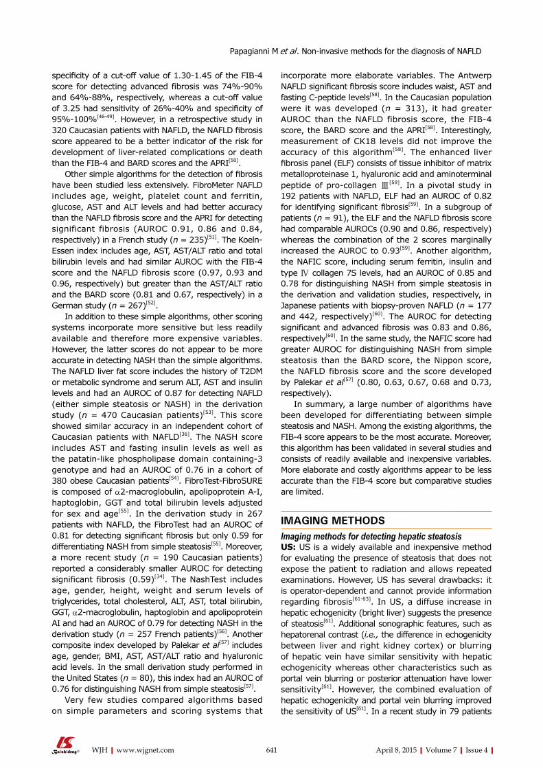
Marker AUROC n Ref.
Serologic markers for detecting steatosis CK18 0.90 157 [20]
0.77 318 [21] FLI 0.84 496 [27] LAP 0.79 588 [28] Hepatic steatosis index 0.81 5362 [29] SteatoTest 0.79 69 [31]Serologic markers for differentiating simple steatosis from nonalcoholic steatohepatitis APRI 0.60 190 [34] CK18 0.82 838 [39] NAFLD fibrosis score 0.82 733 [43]Comparative studies of serologic markers for differentiating simple steatosis from nonalcoholic steatohepatitis FIB-4 0.86 145 [46] NAFLD fibrosis score 0.81 BARD score 0.77 APRI 0.67 FIB-4 0.96 165 [47] NAFLD fibrosis score 0.94 BARD score 0.84 FIB-4 0.80 541 [48] NAFLD fibrosis score 0.77 BARD score 0.70 APRI 0.73 FIB-4 0.87 576 [49] NAFLD fibrosis score 0.86 APRI 0.79 BARD score 0.76
Table 1 Accuracy of the most-well studied serologic markers for detecting steatosis and for differentiating simple steatosis from nonalcoholic steatohepatitis
AUROC: Area under the receiving-operating characteristics curve; AST: Aspartate transaminase; CK18: Cytokeratin-18; FLI: Fatty liver index; LAP: Lipid accumulation product; APRI: AST/platelet ratio index; NAFLD: Non-alcoholic fatty liver disease; FIB-4: Fibrosis-4; BARD: BMI, AST/ALT Ratio, Diabetes; BMI: Body mass index; ALT: Alanine transaminase.
Papagianni M et al . Non-invasive methods for the diagnosis of NAFLD
641 April 8, 2015|Volume 7|Issue 4|WJH|www.wjgnet.com
incorporate more elaborate variables. The Antwerp NAFLD significant fibrosis score includes waist, AST and fasting C-peptide levels[58]. In the Caucasian population were it was developed (n = 313), it had greater AUROC than the NAFLD fibrosis score, the FIB-4 score, the BARD score and the APRI[58]. Interestingly, measurement of CK18 levels did not improve the accuracy of this algorithm[58]. The enhanced liver fibrosis panel (ELF) consists of tissue inhibitor of matrix metalloproteinase 1, hyaluronic acid and aminoterminal peptide of pro-collagen Ⅲ[59]. In a pivotal study in 192 patients with NAFLD, ELF had an AUROC of 0.82 for identifying significant fibrosis[59]. In a subgroup of patients (n = 91), the ELF and the NAFLD fibrosis score had comparable AUROCs (0.90 and 0.86, respectively) whereas the combination of the 2 scores marginally increased the AUROC to 0.93[59]. Another algorithm, the NAFIC score, including serum ferritin, insulin and type Ⅳ collagen 7S levels, had an AUROC of 0.85 and 0.78 for distinguishing NASH from simple steatosis in the derivation and validation studies, respectively, in Japanese patients with biopsy-proven NAFLD (n = 177 and 442, respectively)[60]. The AUROC for detecting significant and advanced fibrosis was 0.83 and 0.86, respectively[60]. In the same study, the NAFIC score had greater AUROC for distinguishing NASH from simple steatosis than the BARD score, the Nippon score, the NAFLD fibrosis score and the score developed by Palekar et al[57] (0.80, 0.63, 0.67, 0.68 and 0.73, respectively).
In summary, a large number of algorithms have been developed for differentiating between simple steatosis and NASH. Among the existing algorithms, the FIB-4 score appears to be the most accurate. Moreover, this algorithm has been validated in several studies and consists of readily available and inexpensive variables. More elaborate and costly algorithms appear to be less accurate than the FIB-4 score but comparative studies are limited.
IMAGING METHODSImaging methods for detecting hepatic steatosis US: US is a widely available and inexpensive method for evaluating the presence of steatosis that does not expose the patient to radiation and allows repeated examinations. However, US has several drawbacks: it is operator-dependent and cannot provide information regarding fibrosis[61-63]. In US, a diffuse increase in hepatic echogenicity (bright liver) suggests the presence of steatosis[61]. Additional sonographic features, such as hepatorenal contrast (i.e., the difference in echogenicity between liver and right kidney cortex) or blurring of hepatic vein have similar sensitivity with hepatic echogenicity whereas other characteristics such as portal vein blurring or posterior attenuation have lower sensitivity[61]. However, the combined evaluation of hepatic echogenicity and portal vein blurring improved the sensitivity of US[61]. In a recent study in 79 patients
specificity of a cut-off value of 1.30-1.45 of the FIB-4 score for detecting advanced fibrosis was 74%-90% and 64%-88%, respectively, whereas a cut-off value of 3.25 had sensitivity of 26%-40% and specificity of 95%-100%[46-49]. However, in a retrospective study in 320 Caucasian patients with NAFLD, the NAFLD fibrosis score appeared to be a better indicator of the risk for development of liver-related complications or death than the FIB-4 and BARD scores and the APRI[50].
Other simple algorithms for the detection of fibrosis have been studied less extensively. FibroMeter NAFLD includes age, weight, platelet count and ferritin, glucose, AST and ALT levels and had better accuracy than the NAFLD fibrosis score and the APRI for detecting significant fibrosis (AUROC 0.91, 0.86 and 0.84, respectively) in a French study (n = 235)[51]. The Koeln-Essen index includes age, AST, AST/ALT ratio and total bilirubin levels and had similar AUROC with the FIB-4 score and the NAFLD fibrosis score (0.97, 0.93 and 0.96, respectively) but greater than the AST/ALT ratio and the BARD score (0.81 and 0.67, respectively) in a German study (n = 267)[52].
In addition to these simple algorithms, other scoring systems incorporate more sensitive but less readily available and therefore more expensive variables. However, the latter scores do not appear to be more accurate in detecting NASH than the simple algorithms. The NAFLD liver fat score includes the history of T2DM or metabolic syndrome and serum ALT, AST and insulin levels and had an AUROC of 0.87 for detecting NAFLD (either simple steatosis or NASH) in the derivation study (n = 470 Caucasian patients)[53]. This score showed similar accuracy in an independent cohort of Caucasian patients with NAFLD[36]. The NASH score includes AST and fasting insulin levels as well as the patatin-like phospholipase domain containing-3 genotype and had an AUROC of 0.76 in a cohort of 380 obese Caucasian patients[54]. FibroTest-FibroSURE is composed of α2-macroglobulin, apolipoprotein A-I, haptoglobin, GGT and total bilirubin levels adjusted for sex and age[55]. In the derivation study in 267 patients with NAFLD, the FibroTest had an AUROC of 0.81 for detecting significant fibrosis but only 0.59 for differentiating NASH from simple steatosis[55]. Moreover, a more recent study (n = 190 Caucasian patients) reported a considerably smaller AUROC for detecting significant fibrosis (0.59)[34]. The NashTest includes age, gender, height, weight and serum levels of triglycerides, total cholesterol, ALT, AST, total bilirubin, GGT, α2-macroglobulin, haptoglobin and apolipoprotein AI and had an AUROC of 0.79 for detecting NASH in the derivation study (n = 257 French patients)[56]. Another composite index developed by Palekar et al[57] includes age, gender, BMI, AST, AST/ALT ratio and hyaluronic acid levels. In the small derivation study performed in the United States (n = 80), this index had an AUROC of 0.76 for distinguishing NASH from simple steatosis[57].
Very few studies compared algorithms based on simple parameters and scoring systems that
Papagianni M et al . Non-invasive methods for the diagnosis of NAFLD
642 April 8, 2015|Volume 7|Issue 4|WJH|www.wjgnet.com
(21 with NAFLD) who underwent both US and liver biopsy, the sensitivity and specificity of the US for detecting macrovesicular steatosis ≥ 5% of total hepatocyte area were 82% and 100%, respectively, but the sensitivity and specificity for detecting microvesicular steatosis were only 59% and 74%, respectively[61]. In patients with steatosis ≥ 20% of total hepatocyte area, sensitivity increased to 96% for macrovesicular steatosis but only to 67% for microvesicular steatosis; specificity decreased to 98% and 66%, respectively[61]. In contrast, a larger study in 94 patients with NAFLD reported an AUROC of 0.98 of US for detecting steatosis; the sensitivity and specificity was 92% and 100%, respectively[64]. In a meta-analysis of 49 studies (n = 4720), US had an AUROC of 0.93 for detecting steatosis; the sensitivity and specificity was 85% and 94%, respectively[65]. Moreover, in 5 small comparative studies (n = 215), US was as accurate as computed tomography (CT), magnetic resonance imaging (MRI) and MRS for detecting steatosis and had a sensitivity and specificity of 94% and 80%, respectively[65].
CT: CT provides an objective evaluation of the presence of steatosis but is more expensive than US and exposes the patient to radiation. Similar to US, CT cannot distinguish NASH from simple steatosis[63]. In a comparative study, CT was less accurate than dual gradient-echo MRI and MRS for identifying steatosis ≥ 5% whereas the latter 2 methods had similar accuracy (AUROC 0.65, 0.88 and 0.85, respectively)[66]. Notably, at higher degrees of steatosis (≥ 30%), the accuracy of the 3 methods was similar (AUROC 0.92, 0.99 and 0.91, respectively)[66]. In the same study, CT was also less accurate than US[66]. However, other studies reported similar accuracy of MRI, CT and US in assessing steatosis[63,65].
MRI: In patients with NAFLD, MRI has shown excellent accuracy for detecting steatosis[67-70], which is similar with the accuracy of MRS[66,71-73] and superior or similar compared with US and CT[63,65,74]. However, in patients with advanced fibrosis or cirrhosis, MRI appears to be less reliable for grading steatosis[67,73]. Compared with CT, MRI has the advantage that it does not expose the patient to radiation and can therefore be used for follow-up. On the other hand, MRI is more expensive than CT, it cannot be performed in patients with claustrophobia and the measurements are affected by hepatic iron deposition, which is frequently present in patients with NAFLD[75,76]. MRI also does not provide information regarding the presence of fibrosis. Indeed, in a small study in 10 patients with NAFLD, chemical-shift MRI was very accurate in identifying steatosis but could not differentiate between NASH and isolated steatosis[77]. A larger study in 25 patients with NAFLD also showed that MRI is not useful in distinguishing NASH from simple steatosis[63].
1H-magnetic resonance spectroscopy: 1H-magnetic
resonance spectroscopy is an accurate method for evaluating hepatic steatosis[66,72,73,78,79]. Furthermore, MRS is operator-independent and fast[66,78,79]. However, MRS has some important disadvantages, including limited availability and high cost[66,78,79]. The results might also be affected by respiratory movements, since MRS is a free-breathing method[66]. Some studies also suggested that advanced fibrosis is also associated with less accurate evaluation of steatosis using MRS[73]. Claustrophobia and the presence of implanted devices are additional limitations in the use of MRS[66,78,79].
In summary, the different imaging methods for detecting steatosis appear to have comparable accuracy. Since US is the least expensive, readily available, does not expose the patient to radiation and can be used for repeat evaluations, it appears to represent the most useful imaging method for detecting steatosis.
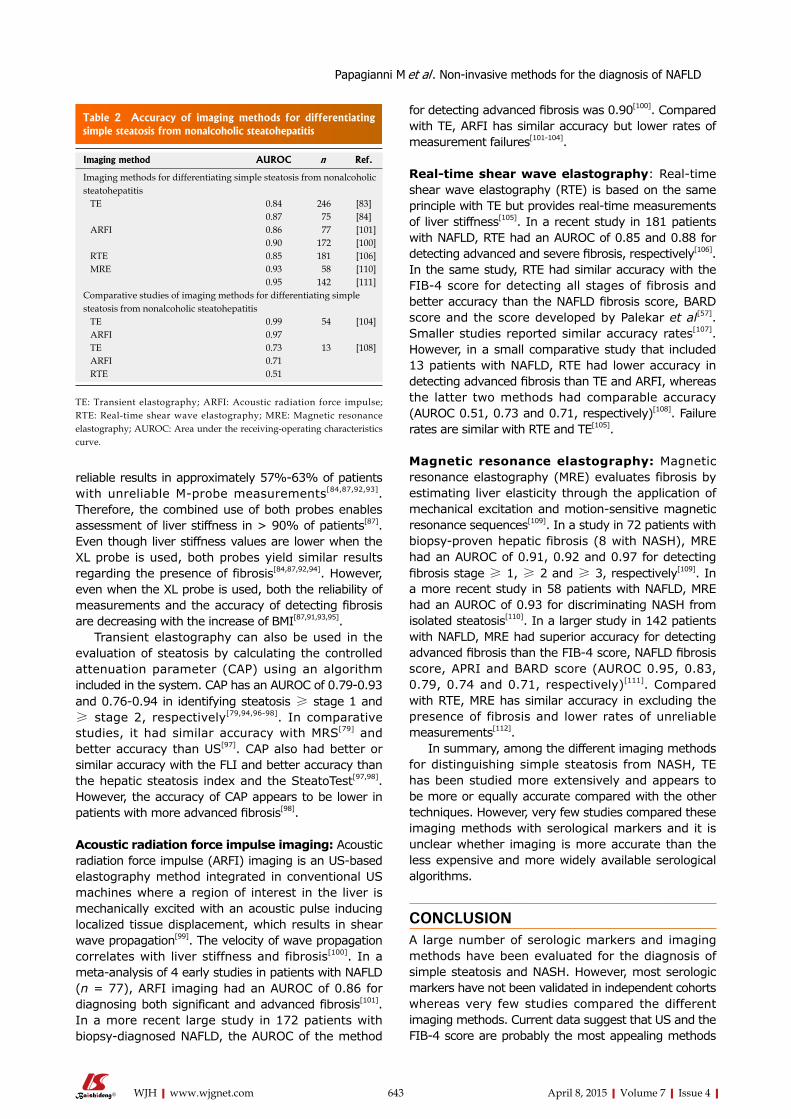
Imaging methods for differentiating simple steatosis from NASHTransient elastography: In transient elastography (TE), an M-probe that includes an ultrasonic transducer is used. The transducer is placed above the right lobe of the liver through an intercostal space and produces a vibration that generates a wave, which is transmitted through the skin into the liver. The velocity of the wave correlates directly with liver stiffness. In turn, liver stiffness correlates inversely with the degree of fibrosis[80-82]. In a large study (n = 246 patients with NAFLD), TE had an AUROC of 0.84 and 0.93 for detecting significant and severe fibrosis, respectively[83]. In the same study, TE was more accurate in identifying fibrosis than APRI, FIB-4 score, NAFLD fibrosis score and BARD score[83]. Other smaller studies in Caucasian and Japanese patients with NAFLD also reported similarly high accuracies of TE in detecting significant and severe fibrosis[79,82,84,85] (Table 2).
Liver stiffness evaluation with TE is considered reliable when the interquartile range/median ratio of measurements is ≤ 0.30[86]. Unreliable measurements are more frequent in older subjects and in overweight or obese patients[83,87,88]. In a large study that analyzed 13369 examinations of liver stiffness using TE (13.7% with NAFLD), 15.8% of the examinations yielded unreliable measurements[89]. In overweight and obese patients, 24% and 35% of measurements, respectively, were considered unreliable[89]. Obesity not only hampers the measurement of liver stiffness but also increases liver stiffness independently of the presence of fibrosis[90]. The presence of steatosis also appears to affect liver stiffness evaluation, particularly in non-cirrhotic patients[80,81,88]. To overcome these limitations, another probe has been developed, the XL-probe, which provides more reliable measurements in obese patients[84,87,91,92]. The XL-probe generates a lower frequency (1.75 MHz vs 3.5 MHz with the M-probe) and higher amplitude (3 mm and 2 mm, respectively) vibration resulting in greater measurement depth (3.5-7.5 cm vs 2.5-6.5 cm, respectively) and yields
Papagianni M et al . Non-invasive methods for the diagnosis of NAFLD
643 April 8, 2015|Volume 7|Issue 4|WJH|www.wjgnet.com
reliable results in approximately 57%-63% of patients with unreliable M-probe measurements[84,87,92,93]. Therefore, the combined use of both probes enables assessment of liver stiffness in > 90% of patients[87]. Even though liver stiffness values are lower when the XL probe is used, both probes yield similar results regarding the presence of fibrosis[84,87,92,94]. However, even when the XL probe is used, both the reliability of measurements and the accuracy of detecting fibrosis are decreasing with the increase of BMI[87,91,93,95].
Transient elastography can also be used in the evaluation of steatosis by calculating the controlled attenuation parameter (CAP) using an algorithm included in the system. CAP has an AUROC of 0.79-0.93 and 0.76-0.94 in identifying steatosis ≥ stage 1 and ≥ stage 2, respectively[79,94,96-98]. In comparative studies, it had similar accuracy with MRS[79] and better accuracy than US[97]. CAP also had better or similar accuracy with the FLI and better accuracy than the hepatic steatosis index and the SteatoTest[97,98]. However, the accuracy of CAP appears to be lower in patients with more advanced fibrosis[98].
Acoustic radiation force impulse imaging: Acoustic radiation force impulse (ARFI) imaging is an US-based elastography method integrated in conventional US machines where a region of interest in the liver is mechanically excited with an acoustic pulse inducing localized tissue displacement, which results in shear wave propagation[99]. The velocity of wave propagation correlates with liver stiffness and fibrosis[100]. In a meta-analysis of 4 early studies in patients with NAFLD (n = 77), ARFI imaging had an AUROC of 0.86 for diagnosing both significant and advanced fibrosis[101]. In a more recent large study in 172 patients with biopsy-diagnosed NAFLD, the AUROC of the method
for detecting advanced fibrosis was 0.90[100]. Compared with TE, ARFI has similar accuracy but lower rates of measurement failures[101-104].
Real-time shear wave elastography: Real-time shear wave elastography (RTE) is based on the same principle with TE but provides real-time measurements of liver stiffness[105]. In a recent study in 181 patients with NAFLD, RTE had an AUROC of 0.85 and 0.88 for detecting advanced and severe fibrosis, respectively[106]. In the same study, RTE had similar accuracy with the FIB-4 score for detecting all stages of fibrosis and better accuracy than the NAFLD fibrosis score, BARD score and the score developed by Palekar et al[57]. Smaller studies reported similar accuracy rates[107]. However, in a small comparative study that included 13 patients with NAFLD, RTE had lower accuracy in detecting advanced fibrosis than TE and ARFI, whereas the latter two methods had comparable accuracy (AUROC 0.51, 0.73 and 0.71, respectively)[108]. Failure rates are similar with RTE and TE[105].
Magnetic resonance elastography: Magnetic resonance elastography (MRE) evaluates fibrosis by estimating liver elasticity through the application of mechanical excitation and motion-sensitive magnetic resonance sequences[109]. In a study in 72 patients with biopsy-proven hepatic fibrosis (8 with NASH), MRE had an AUROC of 0.91, 0.92 and 0.97 for detecting fibrosis stage ≥ 1, ≥ 2 and ≥ 3, respectively[109]. In a more recent study in 58 patients with NAFLD, MRE had an AUROC of 0.93 for discriminating NASH from isolated steatosis[110]. In a larger study in 142 patients with NAFLD, MRE had superior accuracy for detecting advanced fibrosis than the FIB-4 score, NAFLD fibrosis score, APRI and BARD score (AUROC 0.95, 0.83, 0.79, 0.74 and 0.71, respectively)[111]. Compared with RTE, MRE has similar accuracy in excluding the presence of fibrosis and lower rates of unreliable measurements[112].
In summary, among the different imaging methods for distinguishing simple steatosis from NASH, TE has been studied more extensively and appears to be more or equally accurate compared with the other techniques. However, very few studies compared these imaging methods with serological markers and it is unclear whether imaging is more accurate than the less expensive and more widely available serological algorithms.
CONCLUSIONA large number of serologic markers and imaging methods have been evaluated for the diagnosis of simple steatosis and NASH. However, most serologic markers have not been validated in independent cohorts whereas very few studies compared the different imaging methods. Current data suggest that US and the FIB-4 score are probably the most appealing methods
TE: Transient elastography; ARFI: Acoustic radiation force impulse; RTE: Real-time shear wave elastography; MRE: Magnetic resonance elastography; AUROC: Area under the receiving-operating characteristics curve.
Table 2 Accuracy of imaging methods for differentiating simple steatosis from nonalcoholic steatohepatitis
Imaging method AUROC n Ref.
Imaging methods for differentiating simple steatosis from nonalcoholic steatohepatitis TE 0.84 246 [83]
0.87 75 [84] ARFI 0.86 77 [101]
0.90 172 [100] RTE 0.85 181 [106] MRE 0.93 58 [110]
0.95 142 [111]Comparative studies of imaging methods for differentiating simple steatosis from nonalcoholic steatohepatitis TE 0.99 54 [104] ARFI 0.97 TE 0.73 13 [108] ARFI 0.71 RTE 0.51
Papagianni M et al . Non-invasive methods for the diagnosis of NAFLD
644 April 8, 2015|Volume 7|Issue 4|WJH|www.wjgnet.com
for detecting steatosis and for distinguishing NASH from simple steatosis, respectively, because of their low cost and relatively high accuracy. However, currently available methods, both serologic and imaging, cannot obviate the need for liver biopsy for diagnosing NASH due to their substantial false positive and false negative rates. The current role of these methods is probably limited in patients who are unwilling or have contraindications for undergoing biopsy.
REFERENCES1 Farrell GC, Larter CZ. Nonalcoholic fatty liver disease: from
steatosis to cirrhosis. Hepatology 2006; 43: S99-S112 [PMID: 16447287 DOI: 10.1002/hep.20973]
2 Dowman JK, Tomlinson JW, Newsome PN. Systematic review: the diagnosis and staging of non-alcoholic fatty liver disease and non-alcoholic steatohepatitis. Aliment Pharmacol Ther 2011; 33: 525-540 [PMID: 21198708 DOI: 10.1111/j.1365-2036.2010.04556.x]
3 Chalasani N, Younossi Z, Lavine JE, Diehl AM, Brunt EM, Cusi K, Charlton M, Sanyal AJ. The diagnosis and management of non-alcoholic fatty liver disease: practice guideline by the American Gastroenterological Association, American Association for the Study of Liver Diseases, and American College of Gastroenterology. Gastroenterology 2012; 142: 1592-1609 [PMID: 22656328 DOI: 10.1053/j.gastro.2012.04.001]
4 Vernon G, Baranova A, Younossi ZM. Systematic review: the epidemiology and natural history of non-alcoholic fatty liver disease and non-alcoholic steatohepatitis in adults. Aliment Pharmacol Ther 2011; 34: 274-285 [PMID: 21623852 DOI: 10.1111/j.1365-2036.2011.04724.x]
5 Younossi ZM, Stepanova M, Afendy M, Fang Y, Younossi Y, Mir H, Srishord M. Changes in the prevalence of the most common causes of chronic liver diseases in the United States from 1988 to 2008. Clin Gastroenterol Hepatol 2011; 9: 524-530.e1; quiz e60 [PMID: 21440669 DOI: 10.1016/j.cgh.2011.03.020]
6 Williams CD, Stengel J, Asike MI, Torres DM, Shaw J, Contreras M, Landt CL, Harrison SA. Prevalence of nonalcoholic fatty liver disease and nonalcoholic steatohepatitis among a largely middle-aged population utilizing ultrasound and liver biopsy: a prospective study. Gastroenterology 2011; 140: 124-131 [PMID: 20858492 DOI: 10.1053/j.gastro.2010.09.038]
7 Browning JD, Szczepaniak LS, Dobbins R, Nuremberg P, Horton JD, Cohen JC, Grundy SM, Hobbs HH. Prevalence of hepatic steatosis in an urban population in the United States: impact of ethnicity. Hepatology 2004; 40: 1387-1395 [PMID: 15565570 DOI: 10.1002/hep.20466]
8 Leite NC, Salles GF, Araujo AL, Villela-Nogueira CA, Cardoso CR. Prevalence and associated factors of non-alcoholic fatty liver disease in patients with type-2 diabetes mellitus. Liver Int 2009; 29: 113-119 [PMID: 18384521 DOI: 10.1111/j.1478-3231.2008.01718.x]
9 Targher G, Bertolini L, Padovani R, Rodella S, Tessari R, Zenari L, Day C, Arcaro G. Prevalence of nonalcoholic fatty liver disease and its association with cardiovascular disease among type 2 diabetic patients. Diabetes Care 2007; 30: 1212-1218 [PMID: 17277038 DOI: 10.2337/dc06-2247]
10 Wong VW, Wong GL, Choi PC, Chan AW, Li MK, Chan HY, Chim AM, Yu J, Sung JJ, Chan HL. Disease progression of non-alcoholic fatty liver disease: a prospective study with paired liver biopsies at 3 years. Gut 2010; 59: 969-974 [PMID: 20581244 DOI: 10.1136/gut.2009.205088]
11 Dam-Larsen S, Franzmann M, Andersen IB, Christoffersen P, Jensen LB, Sørensen TI, Becker U, Bendtsen F. Long term prognosis of fatty liver: risk of chronic liver disease and death. Gut 2004; 53: 750-755 [PMID: 15082596 DOI: 10.1136/gut.2003.019984]
12 Adams LA, Sanderson S, Lindor KD, Angulo P. The histological
course of nonalcoholic fatty liver disease: a longitudinal study of 103 patients with sequential liver biopsies. J Hepatol 2005; 42: 132-138 [PMID: 15629518 DOI: 10.1016/j.jhep.2004.09.012]
13 Haring R, Wallaschofski H, Nauck M, Dörr M, Baumeister SE, Völzke H. Ultrasonographic hepatic steatosis increases prediction of mortality risk from elevated serum gamma-glutamyl transpeptidase levels. Hepatology 2009; 50: 1403-1411 [PMID: 19670414 DOI: 10.1002/hep.23135]
14 Targher G, Bertolini L, Rodella S, Tessari R, Zenari L, Lippi G, Arcaro G. Nonalcoholic fatty liver disease is independently associated with an increased incidence of cardiovascular events in type 2 diabetic patients. Diabetes Care 2007; 30: 2119-2121 [PMID: 17519430 DOI: 10.2337/dc07-0349]
15 Ekstedt M, Franzén LE, Mathiesen UL, Thorelius L, Holmqvist M, Bodemar G, Kechagias S. Long-term follow-up of patients with NAFLD and elevated liver enzymes. Hepatology 2006; 44: 865-873 [PMID: 17006923 DOI: 10.1002/hep.21327]
16 Adams LA, Lymp JF, St Sauver J, Sanderson SO, Lindor KD, Feldstein A, Angulo P. The natural history of nonalcoholic fatty liver disease: a population-based cohort study. Gastroenterology 2005; 129: 113-121 [PMID: 16012941 DOI: 10.1053/j.gastro.2005.04.014]
17 Myers RP, Fong A, Shaheen AA. Utilization rates, complications and costs of percutaneous liver biopsy: a population-based study including 4275 biopsies. Liver Int 2008; 28: 705-712 [PMID: 18433397 DOI: 10.1111/j.1478-3231.2008.01691.x]
18 Cadranel JF, Rufat P, Degos F. Practices of liver biopsy in France: results of a prospective nationwide survey. For the Group of Epidemiology of the French Association for the Study of the Liver (AFEF). Hepatology 2000; 32: 477-481 [PMID: 10960438 DOI: 10.1053/jhep.2000.16602]
19 Ratziu V, Charlotte F, Heurtier A, Gombert S, Giral P, Bruckert E, Grimaldi A, Capron F, Poynard T; LIDO Study Group. Sampling variability of liver biopsy in nonalcoholic fatty liver disease. Gastroenterology 2005; 128: 1898-1906 [PMID: 15940625 DOI: 10.1053/j.gastro.2005.03.084]
20 Shen J, Chan HL, Wong GL, Chan AW, Choi PC, Chan HY, Chim AM, Yeung DK, Yu J, Chu WC, Wong VW. Assessment of non-alcoholic fatty liver disease using serum total cell death and apoptosis markers. Aliment Pharmacol Ther 2012; 36: 1057-1066 [PMID: 23066946 DOI: 10.1111/apt.12091]
21 Cusi K, Chang Z, Harrison S, Lomonaco R, Bril F, Orsak B, Ortiz-Lopez C, Hecht J, Feldstein AE, Webb A, Louden C, Goros M, Tio F. Limited value of plasma cytokeratin-18 as a biomarker for NASH and fibrosis in patients with non-alcoholic fatty liver disease. J Hepatol 2014; 60: 167-174 [PMID: 23973932 DOI: 10.1016/j.jhep.2013.07.042]
22 Joka D, Wahl K, Moeller S, Schlue J, Vaske B, Bahr MJ, Manns MP, Schulze-Osthoff K, Bantel H. Prospective biopsy-controlled evaluation of cell death biomarkers for prediction of liver fibrosis and nonalcoholic steatohepatitis. Hepatology 2012; 55: 455-464 [PMID: 21993925 DOI: 10.1002/hep.24734]
23 Li H, Dong K, Fang Q, Hou X, Zhou M, Bao Y, Xiang K, Xu A, Jia W. High serum level of fibroblast growth factor 21 is an independent predictor of non-alcoholic fatty liver disease: a 3-year prospective study in China. J Hepatol 2013; 58: 557-563 [PMID: 23142063 DOI: 10.1016/j.jhep.2012.10.029]
24 Li H, Fang Q, Gao F, Fan J, Zhou J, Wang X, Zhang H, Pan X, Bao Y, Xiang K, Xu A, Jia W. Fibroblast growth factor 21 levels are increased in nonalcoholic fatty liver disease patients and are correlated with hepatic triglyceride. J Hepatol 2010; 53: 934-940 [PMID: 20675007 DOI: 10.1016/j.jhep.2010.05.018]
25 Shen J, Chan HL, Wong GL, Choi PC, Chan AW, Chan HY, Chim AM, Yeung DK, Chan FK, Woo J, Yu J, Chu WC, Wong VW. Non-invasive diagnosis of non-alcoholic steatohepatitis by combined serum biomarkers. J Hepatol 2012; 56: 1363-1370 [PMID: 22314419 DOI: 10.1016/j.jhep.2011.12.025]
26 Dushay J, Chui PC, Gopalakrishnan GS, Varela-Rey M, Crawley M, Fisher FM, Badman MK, Martinez-Chantar ML, Maratos-Flier E. Increased fibroblast growth factor 21 in obesity and nonalcoholic fatty liver disease. Gastroenterology 2010; 139: 456-463 [PMID: 20451522 DOI: 10.1053/j.gastro.2010.04.054]
Papagianni M et al . Non-invasive methods for the diagnosis of NAFLD
645 April 8, 2015|Volume 7|Issue 4|WJH|www.wjgnet.com
27 Bedogni G, Bellentani S, Miglioli L, Masutti F, Passalacqua M, Castiglione A, Tiribelli C. The Fatty Liver Index: a simple and accurate predictor of hepatic steatosis in the general population. BMC Gastroenterol 2006; 6: 33 [PMID: 17081293 DOI: 10.1186/1471-230X-6-33]
28 Bedogni G, Kahn HS, Bellentani S, Tiribelli C. A simple index of lipid overaccumulation is a good marker of liver steatosis. BMC Gastroenterol 2010; 10: 98 [PMID: 20738844 DOI: 10.1186/1471-230X-10-98]
29 Lee JH, Kim D, Kim HJ, Lee CH, Yang JI, Kim W, Kim YJ, Yoon JH, Cho SH, Sung MW, Lee HS. Hepatic steatosis index: a simple screening tool reflecting nonalcoholic fatty liver disease. Dig Liver Dis 2010; 42: 503-508 [PMID: 19766548 DOI: 10.1016/j.dld.2009.08.002]
30 Guiu B, Crevisy-Girod E, Binquet C, Duvillard L, Masson D, Lepage C, Hamza S, Krausé D, Verges B, Minello A, Cercueil JP, Hillon P, Petit JM. Prediction for steatosis in type-2 diabetes: clinico-biological markers versus 1H-MR spectroscopy. Eur Radiol 2012; 22: 855-863 [PMID: 22101800 DOI: 10.1007/s00330-011-2326-9]
31 Poynard T, Ratziu V, Naveau S, Thabut D, Charlotte F, Messous D, Capron D, Abella A, Massard J, Ngo Y, Munteanu M, Mercadier A, Manns M, Albrecht J. The diagnostic value of biomarkers (SteatoTest) for the prediction of liver steatosis. Comp Hepatol 2005; 4: 10 [PMID: 16375767 DOI: 10.1186/1476-5926-4-10]
32 Fracanzani AL, Valenti L, Bugianesi E, Andreoletti M, Colli A, Vanni E, Bertelli C, Fatta E, Bignamini D, Marchesini G, Fargion S. Risk of severe liver disease in nonalcoholic fatty liver disease with normal aminotransferase levels: a role for insulin resistance and diabetes. Hepatology 2008; 48: 792-798 [PMID: 18752331 DOI: 10.1002/hep.22429]
33 Mofrad P, Contos MJ, Haque M, Sargeant C, Fisher RA, Luketic VA, Sterling RK, Shiffman ML, Stravitz RT, Sanyal AJ. Clinical and histologic spectrum of nonalcoholic fatty liver disease associated with normal ALT values. Hepatology 2003; 37: 1286-1292 [PMID: 12774006 DOI: 10.1053/jhep.2003.50229]
34 Sebastiani G, Castera L, Halfon P, Pol S, Mangia A, Di Marco V, Pirisi M, Voiculescu M, Bourliere M, Alberti A. The impact of liver disease aetiology and the stages of hepatic fibrosis on the performance of non-invasive fibrosis biomarkers: an international study of 2411 cases. Aliment Pharmacol Ther 2011; 34: 1202-1216 [PMID: 21981787 DOI: 10.1111/j.1365-2036.2011.04861.x]
35 Loaeza-del-Castillo A, Paz-Pineda F, Oviedo-Cárdenas E, Sánchez-Avila F, Vargas-Vorácková F. AST to platelet ratio index (APRI) for the noninvasive evaluation of liver fibrosis. Ann Hepatol 2008; 7: 350-357 [PMID: 19034235]
36 Musso G, Gambino R, Durazzo M, Cassader M. Noninvasive assessment of liver disease severity with liver fat score and CK-18 in NAFLD: Prognostic value of liver fat equation goes beyond hepatic fat estimation. Hepatology 2010; 51: 715-717 [PMID: 19821531 DOI: 10.1002/hep.23255]
37 Feldstein AE, Wieckowska A, Lopez AR, Liu YC, Zein NN, McCullough AJ. Cytokeratin-18 fragment levels as noninvasive biomarkers for nonalcoholic steatohepatitis: a multicenter validation study. Hepatology 2009; 50: 1072-1078 [PMID: 19585618 DOI: 10.1002/hep.23050]
38 Malik R, Chang M, Bhaskar K, Nasser I, Curry M, Schuppan D, Byrnes V, Afdhal N. The clinical utility of biomarkers and the nonalcoholic steatohepatitis CRN liver biopsy scoring system in patients with nonalcoholic fatty liver disease. J Gastroenterol Hepatol 2009; 24: 564-568 [PMID: 19378390 DOI: 10.1111/j.1440-1746.2008.05731.x]
39 Chen J, Zhu Y, Zheng Q, Jiang J. Serum cytokeratin-18 in the diagnosis of non-alcoholic steatohepatitis: A meta-analysis. Hepatol Res 2014; 44: 854-862 [PMID: 23834322 DOI: 10.1111/hepr.12197]
40 Tamimi TI, Elgouhari HM, Alkhouri N, Yerian LM, Berk MP, Lopez R, Schauer PR, Zein NN, Feldstein AE. An apoptosis panel for nonalcoholic steatohepatitis diagnosis. J Hepatol 2011; 54: 1224-1229 [PMID: 21145805 DOI: 10.1016/j.jhep.2010.08.023]
41 Harrison SA, Oliver D, Arnold HL, Gogia S, Neuschwander-Tetri BA. Development and validation of a simple NAFLD clinical scoring system for identifying patients without advanced disease. Gut 2008; 57: 1441-1447 [PMID: 18390575 DOI: 10.1136/gut.2007.146019]
42 Raszeja-Wyszomirska J, Szymanik B, Ławniczak M, Kajor M, Chwist A, Milkiewicz P, Hartleb M. Validation of the BARD scoring system in Polish patients with nonalcoholic fatty liver disease (NAFLD). BMC Gastroenterol 2010; 10: 67 [PMID: 20584330 DOI: 10.1186/1471-230X-10-67]
43 Angulo P, Hui JM, Marchesini G, Bugianesi E, George J, Farrell GC, Enders F, Saksena S, Burt AD, Bida JP, Lindor K, Sanderson SO, Lenzi M, Adams LA, Kench J, Therneau TM, Day CP. The NAFLD fibrosis score: a noninvasive system that identifies liver fibrosis in patients with NAFLD. Hepatology 2007; 45: 846-854 [PMID: 17393509 DOI: 10.1002/hep.21496]
44 Wong VW, Wong GL, Chim AM, Tse AM, Tsang SW, Hui AY, Choi PC, Chan AW, So WY, Chan FK, Sung JJ, Chan HL. Validation of the NAFLD fibrosis score in a Chinese population with low prevalence of advanced fibrosis. Am J Gastroenterol 2008; 103: 1682-1688 [PMID: 18616651 DOI: 10.1111/j.1572-0241.2008.01933.x]
45 Miyaaki H, Ichikawa T, Nakao K, Yatsuhashi H, Furukawa R, Ohba K, Omagari K, Kusumoto Y, Yanagi K, Inoue O, Kinoshita N, Ishibashi H, Yano M, Eguchi K. Clinicopathological study of nonalcoholic fatty liver disease in Japan: the risk factors for fibrosis. Liver Int 2008; 28: 519-524 [PMID: 17976158 DOI: 10.1111/j.1478-3231.2007.01614.x]
46 McPherson S, Stewart SF, Henderson E, Burt AD, Day CP. Simple non-invasive fibrosis scoring systems can reliably exclude advanced fibrosis in patients with non-alcoholic fatty liver disease. Gut 2010; 59: 1265-1269 [PMID: 20801772 DOI: 10.1136/gut.2010.216077]
47 Demir M, Lang S, Nierhoff D, Drebber U, Hardt A, Wedemeyer I, Schulte S, Quasdorff M, Goeser T, Töx U, Steffen HM. Stepwise combination of simple noninvasive fibrosis scoring systems increases diagnostic accuracy in nonalcoholic fatty liver disease. J Clin Gastroenterol 2013; 47: 719-726 [PMID: 23442837 DOI: 10.1097/MCG.0b013e3182819a89]
48 Shah AG, Lydecker A, Murray K, Tetri BN, Contos MJ, Sanyal AJ; Nash Clinical Research Network. Comparison of noninvasive markers of fibrosis in patients with nonalcoholic fatty liver disease. Clin Gastroenterol Hepatol 2009; 7: 1104-1112 [PMID: 19523535 DOI: 10.1016/j.cgh.2009.05.033]
49 Sumida Y, Yoneda M, Hyogo H, Itoh Y, Ono M, Fujii H, Eguchi Y, Suzuki Y, Aoki N, Kanemasa K, Fujita K, Chayama K, Saibara T, Kawada N, Fujimoto K, Kohgo Y, Yoshikawa T, Okanoue T. Validation of the FIB4 index in a Japanese nonalcoholic fatty liver disease population. BMC Gastroenterol 2012; 12: 2 [PMID: 22221544 DOI: 10.1186/1471-230X-12-2]
50 Angulo P, Bugianesi E, Bjornsson ES, Charatcharoenwitthaya P, Mills PR, Barrera F, Haflidadottir S, Day CP, George J. Simple noninvasive systems predict long-term outcomes of patients with nonalcoholic fatty liver disease. Gastroenterology 2013; 145: 782-789.e4 [PMID: 23860502 DOI: 10.1053/j.gastro.2013.06.057]
51 Calès P, Lainé F, Boursier J, Deugnier Y, Moal V, Oberti F, Hunault G, Rousselet MC, Hubert I, Laafi J, Ducluzeaux PH, Lunel F. Comparison of blood tests for liver fibrosis specific or not to NAFLD. J Hepatol 2009; 50: 165-173 [PMID: 18977552 DOI: 10.1016/j.jhep.2008.07.035]
52 Demir M, Lang S, Schlattjan M, Drebber U, Wedemeyer I, Nierhoff D, Kaul I, Sowa J, Canbay A, Töx U, Steffen HM. NIKEI: a new inexpensive and non-invasive scoring system to exclude advanced fibrosis in patients with NAFLD. PLoS One 2013; 8: e58360 [PMID: 23555578 DOI: 10.1371/journal.pone.0058360]
53 Kotronen A, Peltonen M, Hakkarainen A, Sevastianova K, Bergholm R, Johansson LM, Lundbom N, Rissanen A, Ridderstråle M, Groop L, Orho-Melander M, Yki-Järvinen H. Prediction of non-alcoholic fatty liver disease and liver fat using metabolic and genetic factors. Gastroenterology 2009; 137: 865-872 [PMID: 19524579 DOI: 10.1053/j.gastro.2009.06.005]
54 Hyysalo J, Männistö VT, Zhou Y, Arola J, Kärjä V, Leivonen M,
Papagianni M et al . Non-invasive methods for the diagnosis of NAFLD
646 April 8, 2015|Volume 7|Issue 4|WJH|www.wjgnet.com
Juuti A, Jaser N, Lallukka S, Käkelä P, Venesmaa S, Simonen M, Saltevo J, Moilanen L, Korpi-Hyövalti E, Keinänen-Kiukaanniemi S, Oksa H, Orho-Melander M, Valenti L, Fargion S, Pihlajamäki J, Peltonen M, Yki-Järvinen H. A population-based study on the prevalence of NASH using scores validated against liver histology. J Hepatol 2014; 60: 839-846 [PMID: 24333862 DOI: 10.1016/j.jhep.2013.12.009]
55 Poynard T, Lebray P, Ingiliz P, Varaut A, Varsat B, Ngo Y, Norha P, Munteanu M, Drane F, Messous D, Bismut FI, Carrau JP, Massard J, Ratziu V, Giordanella JP. Prevalence of liver fibrosis and risk factors in a general population using non-invasive biomarkers (FibroTest). BMC Gastroenterol 2010; 10: 40 [PMID: 20412588 DOI: 10.1186/1471-230X-10-40]
56 Poynard T, Ratziu V, Charlotte F, Messous D, Munteanu M, Imbert-Bismut F, Massard J, Bonyhay L, Tahiri M, Thabut D, Cadranel JF, Le Bail B, de Ledinghen V. Diagnostic value of biochemical markers (NashTest) for the prediction of non alcoholo steato hepatitis in patients with non-alcoholic fatty liver disease. BMC Gastroenterol 2006; 6: 34 [PMID: 17096854 DOI: 10.1186/1471-230X-6-34]
57 Palekar NA, Naus R, Larson SP, Ward J, Harrison SA. Clinical model for distinguishing nonalcoholic steatohepatitis from simple steatosis in patients with nonalcoholic fatty liver disease. Liver Int 2006; 26: 151-156 [PMID: 16448452 DOI: 10.1111/j.1478-3231.2005.01209.x]
58 Francque SM, Verrijken A, Mertens I, Hubens G, Van Marck E, Pelckmans P, Michielsen P, Van Gaal L. Noninvasive assessment of nonalcoholic fatty liver disease in obese or overweight patients. Clin Gastroenterol Hepatol 2012; 10: 1162-1168; quiz e87 [PMID: 22796457 DOI: 10.1016/j.cgh.2012.06.019]
59 Guha IN, Parkes J, Roderick P, Chattopadhyay D, Cross R, Harris S, Kaye P, Burt AD, Ryder SD, Aithal GP, Day CP, Rosenberg WM. Noninvasive markers of fibrosis in nonalcoholic fatty liver disease: Validating the European Liver Fibrosis Panel and exploring simple markers. Hepatology 2008; 47: 455-460 [PMID: 18038452 DOI: 10.1002/hep.21984]
60 Sumida Y, Yoneda M, Hyogo H, Yamaguchi K, Ono M, Fujii H, Eguchi Y, Suzuki Y, Imai S, Kanemasa K, Fujita K, Chayama K, Yasui K, Saibara T, Kawada N, Fujimoto K, Kohgo Y, Okanoue T. A simple clinical scoring system using ferritin, fasting insulin, and type IV collagen 7S for predicting steatohepatitis in nonalcoholic fatty liver disease. J Gastroenterol 2011; 46: 257-268 [PMID: 20842510 DOI: 10.1007/s00535-010-0305-6]
61 Dasarathy S, Dasarathy J, Khiyami A, Joseph R, Lopez R, McCullough AJ. Validity of real time ultrasound in the diagnosis of hepatic steatosis: a prospective study. J Hepatol 2009; 51: 1061-1067 [PMID: 19846234 DOI: 10.1016/j.jhep.2009.09.001]
62 Mathiesen UL, Franzén LE, Aselius H, Resjö M, Jacobsson L, Foberg U, Frydén A, Bodemar G. Increased liver echogenicity at ultrasound examination reflects degree of steatosis but not of fibrosis in asymptomatic patients with mild/moderate abnormalities of liver transaminases. Dig Liver Dis 2002; 34: 516-522 [PMID: 12236486 DOI: 10.1016/S1590-8658(02)80111-6]
63 Saadeh S, Younossi ZM, Remer EM, Gramlich T, Ong JP, Hurley M, Mullen KD, Cooper JN, Sheridan MJ. The utility of radiological imaging in nonalcoholic fatty liver disease. Gastroenterology 2002; 123: 745-750 [PMID: 12198701 DOI: 10.1053/gast.2002.35354]
64 Hamaguchi M, Kojima T, Itoh Y, Harano Y, Fujii K, Nakajima T, Kato T, Takeda N, Okuda J, Ida K, Kawahito Y, Yoshikawa T, Okanoue T. The severity of ultrasonographic findings in nonalcoholic fatty liver disease reflects the metabolic syndrome and visceral fat accumulation. Am J Gastroenterol 2007; 102: 2708-2715 [PMID: 17894848 DOI: 10.1111/j.1572-0241.2007.01526.x]
65 Hernaez R, Lazo M, Bonekamp S, Kamel I, Brancati FL, Guallar E, Clark JM. Diagnostic accuracy and reliability of ultrasonography for the detection of fatty liver: a meta-analysis. Hepatology 2011; 54: 1082-1090 [PMID: 21618575 DOI: 10.1002/hep.24452]
66 Lee SS, Park SH, Kim HJ, Kim SY, Kim MY, Kim DY, Suh DJ, Kim KM, Bae MH, Lee JY, Lee SG, Yu ES. Non-invasive
assessment of hepatic steatosis: prospective comparison of the accuracy of imaging examinations. J Hepatol 2010; 52: 579-585 [PMID: 20185194 DOI: 10.1016/j.jhep.2010.01.008]
67 Permutt Z, Le TA, Peterson MR, Seki E, Brenner DA, Sirlin C, Loomba R. Correlation between liver histology and novel magnetic resonance imaging in adult patients with non-alcoholic fatty liver disease - MRI accurately quantifies hepatic steatosis in NAFLD. Aliment Pharmacol Ther 2012; 36: 22-29 [PMID: 22554256 DOI: 10.1111/j.1365-2036.2012.05121.x]
68 Tang A, Tan J, Sun M, Hamilton G, Bydder M, Wolfson T, Gamst AC, Middleton M, Brunt EM, Loomba R, Lavine JE, Schwimmer JB, Sirlin CB. Nonalcoholic fatty liver disease: MR imaging of liver proton density fat fraction to assess hepatic steatosis. Radiology 2013; 267: 422-431 [PMID: 23382291 DOI: 10.1148/radiol.12120896]
69 Hatta T, Fujinaga Y, Kadoya M, Ueda H, Murayama H, Kurozumi M, Ueda K, Komatsu M, Nagaya T, Joshita S, Kodama R, Tanaka E, Uehara T, Sano K, Tanaka N. Accurate and simple method for quantification of hepatic fat content using magnetic resonance imaging: a prospective study in biopsy-proven nonalcoholic fatty liver disease. J Gastroenterol 2010; 45: 1263-1271 [PMID: 20625773 DOI: 10.1007/s00535-010-0277-6]
70 Mennesson N, Dumortier J, Hervieu V, Milot L, Guillaud O, Scoazec JY, Pilleul F. Liver steatosis quantification using magnetic resonance imaging: a prospective comparative study with liver biopsy. J Comput Assist Tomogr 2009; 33: 672-677 [PMID: 19820490 DOI: 10.1097/RCT.0b013e318199d883]
71 Yokoo T, Bydder M, Hamilton G, Middleton MS, Gamst AC, Wolfson T, Hassanein T, Patton HM, Lavine JE, Schwimmer JB, Sirlin CB. Nonalcoholic fatty liver disease: diagnostic and fat-grading accuracy of low-flip-angle multiecho gradient-recalled-echo MR imaging at 1.5 T. Radiology 2009; 251: 67-76 [PMID: 19221054 DOI: 10.1148/radiol.2511080666]
72 Noworolski SM, Lam MM, Merriman RB, Ferrell L, Qayyum A. Liver steatosis: concordance of MR imaging and MR spectroscopic data with histologic grade. Radiology 2012; 264: 88-96 [PMID: 22723561 DOI: 10.1148/radiol.12110673]
73 McPherson S, Jonsson JR, Cowin GJ, O’Rourke P, Clouston AD, Volp A, Horsfall L, Jothimani D, Fawcett J, Galloway GJ, Benson M, Powell EE. Magnetic resonance imaging and spectroscopy accurately estimate the severity of steatosis provided the stage of fibrosis is considered. J Hepatol 2009; 51: 389-397 [PMID: 19505740 DOI: 10.1016/j.jhep.2009.04.012]
74 Fishbein M, Castro F, Cheruku S, Jain S, Webb B, Gleason T, Stevens WR. Hepatic MRI for fat quantitation: its relationship to fat morphology, diagnosis, and ultrasound. J Clin Gastroenterol 2005; 39: 619-625 [PMID: 16000931 DOI: 10.1097/00004836-200508000-00012]
75 Bugianesi E, Manzini P, D’Antico S, Vanni E, Longo F, Leone N, Massarenti P, Piga A, Marchesini G, Rizzetto M. Relative contribution of iron burden, HFE mutations, and insulin resistance to fibrosis in nonalcoholic fatty liver. Hepatology 2004; 39: 179-187 [PMID: 14752836 DOI: 10.1002/hep.20023]
76 Chitturi S, Weltman M, Farrell GC, McDonald D, Kench J, Liddle C, Samarasinghe D, Lin R, Abeygunasekera S, George J. HFE mutations, hepatic iron, and fibrosis: ethnic-specific association of NASH with C282Y but not with fibrotic severity. Hepatology 2002; 36: 142-149 [PMID: 12085358 DOI: 10.1053/jhep.2002.33892]
77 Kalra N, Duseja A, Das A, Dhiman RK, Virmani V, Chawla Y, Singh P, Khandelwal N. Chemical shift magnetic resonance imaging is helpful in detecting hepatic steatosis but not fibrosis in patients with nonalcoholic fatty liver disease (NAFLD). Ann Hepatol 2009; 8: 21-25 [PMID: 19221529]
78 Roldan-Valadez E, Favila R, Martínez-López M, Uribe M, Ríos C, Méndez-Sánchez N. In vivo 3T spectroscopic quantification of liver fat content in nonalcoholic fatty liver disease: Correlation with biochemical method and morphometry. J Hepatol 2010; 53: 732-737 [PMID: 20594607 DOI: 10.1016/j.jhep.2010.04.018]
79 Karlas T, Petroff D, Garnov N, Böhm S, Tenckhoff H, Wittekind C, Wiese M, Schiefke I, Linder N, Schaudinn A, Busse H, Kahn T,
Papagianni M et al . Non-invasive methods for the diagnosis of NAFLD
647 April 8, 2015|Volume 7|Issue 4|WJH|www.wjgnet.com
Mössner J, Berg T, Tröltzsch M, Keim V, Wiegand J. Non-invasive assessment of hepatic steatosis in patients with NAFLD using controlled attenuation parameter and 1H-MR spectroscopy. PLoS One 2014; 9: e91987 [PMID: 24637477 DOI: 10.1371/journal.pone.0091987]
80 Ziol M, Kettaneh A, Ganne-Carrié N, Barget N, Tengher-Barna I, Beaugrand M. Relationships between fibrosis amounts assessed by morphometry and liver stiffness measurements in chronic hepatitis or steatohepatitis. Eur J Gastroenterol Hepatol 2009; 21: 1261-1268 [PMID: 19478678 DOI: 10.1097/MEG.0b013e32832a20f5]
81 Gaia S, Carenzi S, Barilli AL, Bugianesi E, Smedile A, Brunello F, Marzano A, Rizzetto M. Reliability of transient elastography for the detection of fibrosis in non-alcoholic fatty liver disease and chronic viral hepatitis. J Hepatol 2011; 54: 64-71 [PMID: 20932598 DOI: 10.1016/j.jhep.2010.06.022]
82 Lupsor M, Badea R, Stefanescu H, Grigorescu M, Serban A, Radu C, Crişan D, Sparchez Z, Iancu S, Maniu A. Performance of unidimensional transient elastography in staging non-alcoholic steatohepatitis. J Gastrointestin Liver Dis 2010; 19: 53-60 [PMID: 20361076]
83 Wong VW, Vergniol J, Wong GL, Foucher J, Chan HL, Le Bail B, Choi PC, Kowo M, Chan AW, Merrouche W, Sung JJ, de Lédinghen V. Diagnosis of fibrosis and cirrhosis using liver stiffness measurement in nonalcoholic fatty liver disease. Hepatology 2010; 51: 454-462 [PMID: 20101745 DOI: 10.1002/hep.23312]
84 Myers RP, Pomier-Layrargues G, Kirsch R, Pollett A, Duarte-Rojo A, Wong D, Beaton M, Levstik M, Crotty P, Elkashab M. Feasibility and diagnostic performance of the FibroScan XL probe for liver stiffness measurement in overweight and obese patients. Hepatology 2012; 55: 199-208 [PMID: 21898479 DOI: 10.1002/hep.24624]
85 Yoneda M, Yoneda M, Mawatari H, Fujita K, Endo H, Iida H, Nozaki Y, Yonemitsu K, Higurashi T, Takahashi H, Kobayashi N, Kirikoshi H, Abe Y, Inamori M, Kubota K, Saito S, Tamano M, Hiraishi H, Maeyama S, Yamaguchi N, Togo S, Nakajima A. Noninvasive assessment of liver fibrosis by measurement of stiffness in patients with nonalcoholic fatty liver disease (NAFLD). Dig Liver Dis 2008; 40: 371-378 [PMID: 18083083 DOI: 10.1016/j.dld.2007.10.019]
86 Boursier J, Zarski JP, de Ledinghen V, Rousselet MC, Sturm N, Lebail B, Fouchard-Hubert I, Gallois Y, Oberti F, Bertrais S, Calès P; Multicentric Group from ANRS/HC/EP23 FIBROSTAR Studies. Determination of reliability criteria for liver stiffness evaluation by transient elastography. Hepatology 2013; 57: 1182-1191 [PMID: 22899556 DOI: 10.1002/hep.25993]
87 de Lédinghen V, Wong VW, Vergniol J, Wong GL, Foucher J, Chu SH, Le Bail B, Choi PC, Chermak F, Yiu KK, Merrouche W, Chan HL. Diagnosis of liver fibrosis and cirrhosis using liver stiffness measurement: comparison between M and XL probe of FibroScan®. J Hepatol 2012; 56: 833-839 [PMID: 22173167 DOI: 10.1016/j.jhep.2011.10.017]
88 Fraquelli M, Rigamonti C, Casazza G, Conte D, Donato MF, Ronchi G, Colombo M. Reproducibility of transient elastography in the evaluation of liver fibrosis in patients with chronic liver disease. Gut 2007; 56: 968-973 [PMID: 17255218 DOI: 10.1136/gut.2006.111302]
89 Castéra L, Foucher J, Bernard PH, Carvalho F, Allaix D, Merrouche W, Couzigou P, de Lédinghen V. Pitfalls of liver stiffness measurement: a 5-year prospective study of 13,369 examinations. Hepatology 2010; 51: 828-835 [PMID: 20063276 DOI: 10.1002/hep.23425]
90 Baba M, Furuya K, Bandou H, Kasai K, Sadaoka K. Discrimi-nation of individuals in a general population at high-risk for alcoholic and non-alcoholic fatty liver disease based on liver stiffness: a cross section study. BMC Gastroenterol 2011; 11: 70 [PMID: 21669003 DOI: 10.1186/1471-230X-11-70]
91 Wong VW, Vergniol J, Wong GL, Foucher J, Chan AW, Chermak F, Choi PC, Merrouche W, Chu SH, Pesque S, Chan HL, de Lédinghen V. Liver stiffness measurement using XL probe in patients with
nonalcoholic fatty liver disease. Am J Gastroenterol 2012; 107: 1862-1871 [PMID: 23032979 DOI: 10.1038/ajg.2012.331]
92 Friedrich-Rust M, Hadji-Hosseini H, Kriener S, Herrmann E, Sircar I, Kau A, Zeuzem S, Bojunga J. Transient elastography with a new probe for obese patients for non-invasive staging of non-alcoholic steatohepatitis. Eur Radiol 2010; 20: 2390-2396 [PMID: 20526777 DOI: 10.1007/s00330-010-1820-9]
93 Şirli R, Sporea I, Deleanu A, Culcea L, Szilaski M, Popescu A, Dănilă M. Comparison between the M and XL probes for liver fibrosis assessment by transient elastography. Med Ultrason 2014; 16: 119-122 [PMID: 24791843 DOI: 10.11152/mu.201.3.2066.162.rs1is2]
94 Friedrich-Rust M, Romen D, Vermehren J, Kriener S, Sadet D, Herrmann E, Zeuzem S, Bojunga J. Acoustic radiation force impulse-imaging and transient elastography for non-invasive assessment of liver fibrosis and steatosis in NAFLD. Eur J Radiol 2012; 81: e325-e331 [PMID: 22119555 DOI: 10.1016/j.ejrad.2011.10.029]
95 Myers RP, Pomier-Layrargues G, Kirsch R, Pollett A, Beaton M, Levstik M, Duarte-Rojo A, Wong D, Crotty P, Elkashab M. Discordance in fibrosis staging between liver biopsy and transient elastography using the FibroScan XL probe. J Hepatol 2012; 56: 564-570 [PMID: 22027584 DOI: 10.1016/j.jhep.2011.10.007]
96 Shen F, Zheng RD, Mi YQ, Wang XY, Pan Q, Chen GY, Cao HX, Chen ML, Xu L, Chen JN, Cao Y, Zhang RN, Xu LM, Fan JG. Controlled attenuation parameter for non-invasive assessment of hepatic steatosis in Chinese patients. World J Gastroenterol 2014; 20: 4702-4711 [PMID: 24782622 DOI: 10.3748/wjg.v20.i16.4702]
97 de Lédinghen V, Vergniol J, Foucher J, Merrouche W, le Bail B. Non-invasive diagnosis of liver steatosis using controlled attenuation parameter (CAP) and transient elastography. Liver Int 2012; 32: 911-918 [PMID: 22672642 DOI: 10.1111/j.1478-3231.2012.02820.x]
98 Myers RP, Pollett A, Kirsch R, Pomier-Layrargues G, Beaton M, Levstik M, Duarte-Rojo A, Wong D, Crotty P, Elkashab M. Controlled Attenuation Parameter (CAP): a noninvasive method for the detection of hepatic steatosis based on transient elastography. Liver Int 2012; 32: 902-910 [PMID: 22435761 DOI: 10.1111/j.1478-3231.2012.02781.x]
99 Palmeri ML, Wang MH, Dahl JJ, Frinkley KD, Nightingale KR. Quantifying hepatic shear modulus in vivo using acoustic radiation force. Ultrasound Med Biol 2008; 34: 546-558 [PMID: 18222031 DOI: 10.1016/j.ultrasmedbio.2007.10.009]
100 Palmeri ML, Wang MH, Rouze NC, Abdelmalek MF, Guy CD, Moser B, Diehl AM, Nightingale KR. Noninvasive evaluation of hepatic fibrosis using acoustic radiation force-based shear stiffness in patients with nonalcoholic fatty liver disease. J Hepatol 2011; 55: 666-672 [PMID: 21256907 DOI: 10.1016/j.jhep.2010.12.019]
101 Friedrich-Rust M, Nierhoff J, Lupsor M, Sporea I, Fierbinteanu-Braticevici C, Strobel D, Takahashi H, Yoneda M, Suda T, Zeuzem S, Herrmann E. Performance of Acoustic Radiation Force Impulse imaging for the staging of liver fibrosis: a pooled meta-analysis. J Viral Hepat 2012; 19: e212-e219 [PMID: 22239521 DOI: 10.1111/j.1365-2893.2011.01537.x]
102 Ebinuma H, Saito H, Komuta M, Ojiro K, Wakabayashi K, Usui S, Chu PS, Umeda R, Ishibashi Y, Takayama T, Kikuchi M, Nakamoto N, Yamagishi Y, Kanai T, Ohkuma K, Sakamoto M, Hibi T. Evaluation of liver fibrosis by transient elastography using acoustic radiation force impulse: comparison with Fibroscan(®). J Gastroenterol 2011; 46: 1238-1248 [PMID: 21779759 DOI: 10.1007/s00535-011-0437-3]
103 Boursier J, Isselin G, Fouchard-Hubert I, Oberti F, Dib N, Lebigot J, Bertrais S, Gallois Y, Calès P, Aubé C. Acoustic radiation force impulse: a new ultrasonographic technology for the widespread noninvasive diagnosis of liver fibrosis. Eur J Gastroenterol Hepatol 2010; 22: 1074-1084 [PMID: 20440210 DOI: 10.1097/MEG.0b013e328339e0a1]
104 Yoneda M, Suzuki K, Kato S, Fujita K, Nozaki Y, Hosono K, Saito S, Nakajima A. Nonalcoholic fatty liver disease: US-based acoustic radiation force impulse elastography. Radiology 2010;
Papagianni M et al . Non-invasive methods for the diagnosis of NAFLD
648 April 8, 2015|Volume 7|Issue 4|WJH|www.wjgnet.com
256: 640-647 [PMID: 20529989 DOI: 10.1148/radiol.10091662]105 Poynard T, Munteanu M, Luckina E, Perazzo H, Ngo Y, Royer
L, Fedchuk L, Sattonnet F, Pais R, Lebray P, Rudler M, Thabut D, Ratziu V. Liver fibrosis evaluation using real-time shear wave elastography: applicability and diagnostic performance using methods without a gold standard. J Hepatol 2013; 58: 928-935 [PMID: 23321316 DOI: 10.1016/j.jhep.2012.12.021]
106 Ochi H, Hirooka M, Koizumi Y, Miyake T, Tokumoto Y, Soga Y, Tada F, Abe M, Hiasa Y, Onji M. Real-time tissue elastography for evaluation of hepatic fibrosis and portal hypertension in nonalcoholic fatty liver diseases. Hepatology 2012; 56: 1271-1278 [PMID: 22488593 DOI: 10.1002/hep.25756]
107 Orlacchio A, Bolacchi F, Antonicoli M, Coco I, Costanzo E, Tosti D, Francioso S, Angelico M, Simonetti G. Liver elasticity in NASH patients evaluated with real-time elastography (RTE). Ultrasound Med Biol 2012; 38: 537-544 [PMID: 22341049 DOI: 10.1016/j.ultrasmedbio.2011.12.023]
108 Chung JH, Ahn HS, Kim SG, Lee YN, Kim YS, Jeong SW, Jang JY, Lee SH, Kim HS, Kim BS. The usefulness of transient elastography, acoustic-radiation-force impulse elastography, and real-time elastography for the evaluation of liver fibrosis. Clin
Mol Hepatol 2013; 19: 156-164 [PMID: 23837140 DOI: 10.3350/cmh.2013.19.2.156]
109 Asbach P, Klatt D, Schlosser B, Biermer M, Muche M, Rieger A, Loddenkemper C, Somasundaram R, Berg T, Hamm B, Braun J, Sack I. Viscoelasticity-based staging of hepatic fibrosis with multifrequency MR elastography. Radiology 2010; 257: 80-86 [PMID: 20679447 DOI: 10.1148/radiol.10092489]
110 Chen J, Talwalkar JA, Yin M, Glaser KJ, Sanderson SO, Ehman RL. Early detection of nonalcoholic steatohepatitis in patients with nonalcoholic fatty liver disease by using MR elastography. Radiology 2011; 259: 749-756 [PMID: 21460032 DOI: 10.1148/radiol.11101942]
111 Kim D, Kim WR, Talwalkar JA, Kim HJ, Ehman RL. Advanced fibrosis in nonalcoholic fatty liver disease: noninvasive assessment with MR elastography. Radiology 2013; 268: 411-419 [PMID: 23564711 DOI: 10.1148/radiol.13121193]
112 Yoon JH, Lee JM, Woo HS, Yu MH, Joo I, Lee ES, Sohn JY, Lee KB, Han JK, Choi BI. Staging of hepatic fibrosis: comparison of magnetic resonance elastography and shear wave elastography in the same individuals. Korean J Radiol 2013; 14: 202-212 [PMID: 23483022 DOI: 10.3348/kjr.2013.14.2.202]
P- Reviewer: Pan JJ, Pan TL, Quarleri J S- Editor: Tian YL L- Editor: A E- Editor: Liu SQ
Papagianni M et al . Non-invasive methods for the diagnosis of NAFLD
Marta Guerrero-Misas, Manuel Rodríguez-Perálvarez, Manuel De la Mata
Marta Guerrero-Misas, Manuel Rodríguez-Perálvarez, Manuel De la Mata, Department of Hepatology and Liver Transplantation, Reina Sofía University Hospital, Maimónides Institute of Biomedical Research of Córdoba, CIBERehd, 14004 Córdoba, SpainAuthor contributions: All authors contributed equally to the manuscript; GuerreroMisas M and RodríguezPerálvarez M performed research and wrote the paper; De la Mata M designed research and contributed new reagents. Conflict-of-interest: The authors of the present manuscript do not have any conflict of interest to disclose.Open-Access: This article is an openaccess article which was selected by an inhouse editor and fully peerreviewed by external reviewers. It is distributed in accordance with the Creative Commons Attribution Non Commercial (CC BYNC 4.0) license, which permits others to distribute, remix, adapt, build upon this work noncommercially, and license their derivative works on different terms, provided the original work is properly cited and the use is noncommercial. See: http://creativecommons.org/licenses/bync/4.0/Correspondence to: Manuel De la Mata, MD, PhD, Head, Department of Hepatology and Liver Transplantation, Reina Sofía University Hospital, Maimónides Institute of Biomedical Research of Córdoba, CIBERehd, Avda/ Menéndez Pidal s/n, 14004 Córdoba, Spain. [email protected]: +34957010450 Fax: +34957736014Received: August 25, 2014Peer-review started: August 25, 2014First decision: November 27, 2014Revised: December 15, 2014Accepted: January 15, 2015Article in press: January 19, 2015Published online: April 8, 2015
AbstractLiver transplantation is the only therapeutic option which allows to treat both, the hepatocellular carcinoma and the underlying liver disease. Indeed, liver transplantation is considered the standard of care for a subset of patients with cirrhosis and hepatocellular carcinoma.
However, tumour recurrence rates are as high as 20%, and once the recurrence is established the therapeutic options are scarce and with little impact on prognosis. Strategies to minimize tumour recurrence and thus to improve outcome may be classified into 3 groups: (1) An adequate selection of candidates for liver transplantation by using the Milan criteria; (2) An optimized management within waiting list including prioritization of patients at high risk of tumour progression, and the implementation of bridging therapies, particularly when the expected length within the waiting list is longer than 6 mo; and (3) Tailored immunosuppression comprising reduced exposure to calcineurin inhibitors, particularly early after liver transplantation, and the addition of mammalian target of rapamycin inhibitors. In the present manuscript the available scientific evidence supporting these strategies is comprehensively reviewed, and future directions are provided for novel research approaches, which may contribute to the final target: to cure more patients with hepatocellular carcinoma and with an improved long term outcome.
Key words: Hepatocellular carcinoma; Recurrence; Bridging therapy; Milan criteria; Immunosuppression; Liver transplantation
© The Author(s) 2015. Published by Baishideng Publishing Group Inc. All rights reserved.
Core tip: Liver transplantation is the only therapeutic option which allows to treat both, the hepatocellular carcinoma and the underlying liver disease. However, tumour recurrence rates are 15%-20% with a very poor prognosis. Strategies to minimize tumour recurrence and thus to improve outcome are focused in a careful selection of candidates for liver transplantation, an optimized management within waiting list and a tailored immunosuppression. The available scientific evidence supporting these strategies is reviewed, and future directions are provided for novel research approaches, which may contribute to the final target: to cure more
REVIEW
Submit a Manuscript: http://www.wjgnet.com/esps/Help Desk: http://www.wjgnet.com/esps/helpdesk.aspxDOI: 10.4254/wjh.v7.i4.649
649 April 8, 2015|Volume 7|Issue 4|WJH|www.wjgnet.com
World J Hepatol 2015 April 8; 7(4): 649-661ISSN 1948-5182 (online)
© 2015 Baishideng Publishing Group Inc. All rights reserved.
Strategies to improve outcome of patients with hepatocellular carcinoma receiving a liver transplantation
patients with hepatocellular carcinoma with an improved long term outcome.
GuerreroMisas M, RodríguezPerálvarez M, De la Mata M. Strategies to improve outcome of patients with hepatocellular carcinoma receiving a liver transplantation. World J Hepatol 2015; 7(4): 649661 Available from: URL: http://www.wjgnet.com/19485182/full/v7/i4/649.htm DOI: http://dx.doi.org/10.4254/wjh.v7.i4.649
INTRODUCTIONHepatocellular carcinoma (HCC) is the most frequent primary liver cancer, which accounts for 70%-80% of the hepatic malignancies[1]. It generally appears on a cirrhotic liver, and thus the common causes of cirrhosis are also involved in the oncogenesis of HCC, being particularly relevant hepatitis B virus (HBV) and HCV chronic viral infections[2-4]. It has been estimated that HBV is responsible for 50%-80% of HCC cases, whereas 10%-20% of cases are related to HCV[1].
HCC represents the fifth most common malignancy (554000 cases worldwide) and the second leading cause of cancer-related mortality, (746000 deaths annually)[5]. HCC rates are 2-4 fold increased in male population. The incidence is higher in developing countries, although its incidence in developed countries is increasing, mainly due to HCV infection[6]. Among developed countries the highest incidence rates are found in North America (9.3 cases/100000) and southern Europe (9.5 cases/100000)[5]. However this picture may change in the next decades given the exponential raising of infant obesity in United States and Europe which may trigger the number of HCCs attributable to non-alcoholic fatty liver disease[7-9].
Liver Transplantation (LT) is the only therapeutic option able to treat both the HCC and the underlying liver disease, and it is currently considered the standard of care for patients with small unresectable HCC. The proportion of patients diagnosed at early stage, who potentially would benefit from LT, is expected to increase due to the screening programs with ultrasound implemented for patients with chronic liver disease[10]. Compared to liver resection and locoregional ablative therapies, LT offers an improved long term survival (70% at 5 years). However the tumour recurrence rates after LT are 15%-20%, and the therapeutic options are very limited in this situation[11].
Current strategies to minimize HCC recurrence after LT are grouped in: (1) Adequate selection of candidates for LT; (2) Waiting list management and bridging locoregional therapies; and (3) Tailored immunosuppression. In the present manuscript the scientific evidence supporting these strategies is comprehensively reviewed, and future directions are drawn in order to improve long term outcome of LT patients with HCC.
SELECTION OF HCC PATIENTS FOR LT The initial experiences of LT to treat HCC were dis-appointing since the tumour recurrence was frequent, and survival was shorter when compared with other LT aetiologies (5-year survival rates 18%-40%)[12]. Many institutions included the HCC as a formal contraindication for LT until the early 90’s, when some studies showed that patients with small HCC under-going LT had reduced tumour recurrence rates when compared to liver resection. Clinicians soon became aware of the improved outcomes when LT was restricted to patients with a limited tumour burden. Indeed in a cohort of 221 consecutive LT patients [38 patients with small HCC (< 5 cm) and 136 with cirrhosis without HCC], the survival rates were similar irrespective of whether HCC was present or not (63% vs 68% at 5 years respectively; P = 0.84). In 1996 Mazzaferro et al[13] prospectively analyzed a cohort of 48 patients with small unresectable HCC undergoing LT. The authors considered LT when there was a single nodule ≤ 5 cm or up to 3 nodules, ≤ 3 cm each, in the absence of macrovascular invasion or extrahepatic metastases. The 4-year survival rate was 75%, with a recurrence-free survival rate of 83%. These premises, currently known as Milan criteria, found a wide acceptance and most LT institutions implemented them as the standard of care to select HCC patients for LT[10]. However according to the Milan criteria, only 6% of patients with HCC would be eligible for LT[14], and therefore these criteria may be considered too restrictive. A significant proportion of patients above Milan criteria could still benefit from LT without increasing HCC recurrence rates[15].
There have been many attempts to expand Milan criteria, being the most frequently used summarized in Table 1. The expansion of the Milan criteria may be performed by increasing the diameter and/or the number of nodules allowed, and in some cases with additional criteria such as histological tumour differentiation, serum PIVKA-Ⅱ or α-fetoprotein.
One of the most popular criteria is the so called “up-to-seven”, according to which patients without macrovascular invasion could be candidates for LT as long as the sum of the number of nodules and the diameter of the largest nodule is ≤ 7. These criteria were derived from a retrospective multicenter study with 1556 patients with HCC, from which 1070 had a HCC beyond Milan criteria. Patients outside Milan criteria but within the up-to-seven criteria had an overall 5 year-survival rate of 71.2%, similar to those found in patients within Milan criteria (73.3%). HCC recurrence rates were also similar provided that microvascular invasion was absent[16]. However this study had important limitations. The tumour was evaluated in the explanted liver rather than by using radiological techniques, and a certain grade of disagreement between both approaches is expected in clinical practice. Most importantly, the favorable outcome showed by patients within the up-to-seven criteria was only present after excluding patients
650 April 8, 2015|Volume 7|Issue 4|WJH|www.wjgnet.com
Guerrero-Misas M et al . Liver transplantation for hepatocellular carcinoma
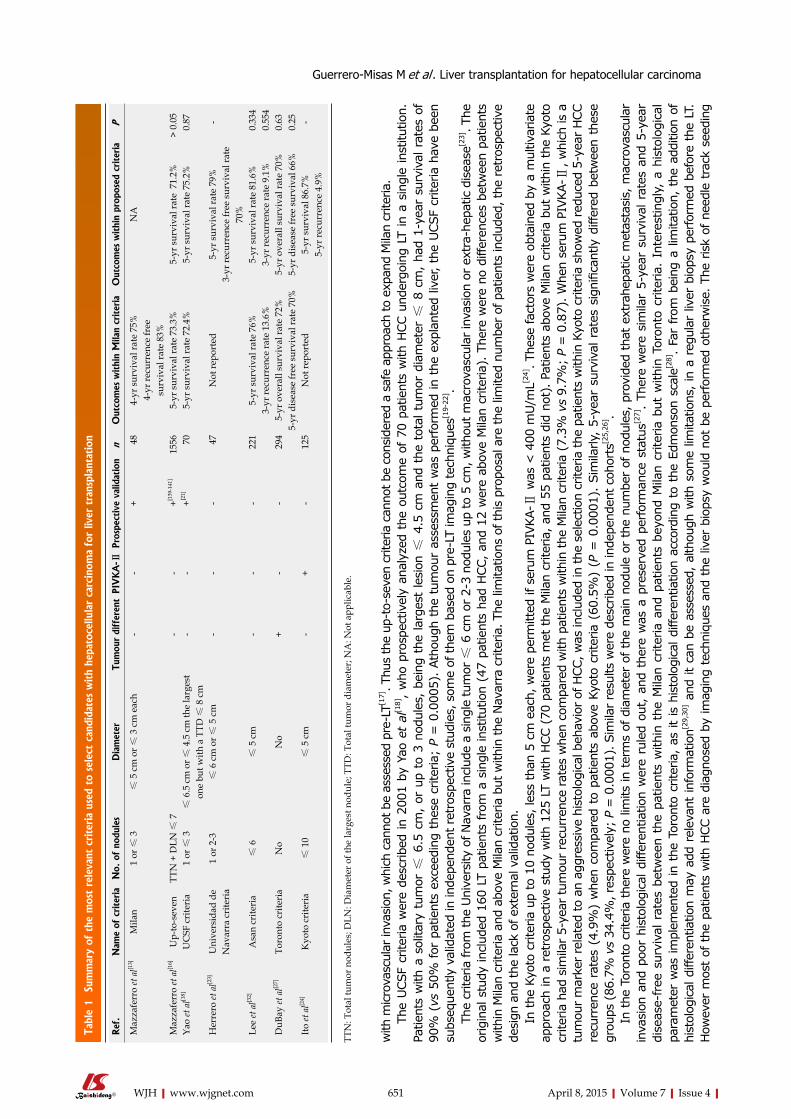
with
micro
vasc
ular
inva
sion
, whi
ch c
anno
t be
asse
ssed
pre
-LT[1
7]. T
hus
the
up-t
o-se
ven
crite
ria c
anno
t be
cons
ider
ed a
saf
e ap
proa
ch to
exp
and
Mila
n cr
iteria
. Th
e UCS
F cr
iteria
wer
e de
scrib
ed in
200
1 by
Yao
et
al[1
8],
who
pro
spec
tivel
y an
alyz
ed t
he o
utco
me
of 7
0 pa
tient
s w
ith H
CC u
nder
goin
g LT
in a
sin
gle
inst
itutio
n.
Patie
nts
with
a s
olita
ry t
umor
≤ 6
.5 c
m,
or u
p to
3 n
odul
es,
bein
g th
e la
rges
t le
sion
≤ 4
.5 c
m a
nd t
he t
otal
tum
or d
iam
eter
≤ 8
cm
, ha
d 1-
year
sur
viva
l rat
es o
f 90
% (
vs 5
0% for
pat
ient
s ex
ceed
ing
thes
e cr
iteria
; P
= 0
.000
5).
Atho
ugh
the
tum
our
asse
ssm
ent
was
per
form
ed in
the
exp
lant
ed li
ver,
the
UCS
F cr
iteria
hav
e be
en
subs
eque
ntly
val
idat
ed in
inde
pend
ent r
etro
spec
tive
stud
ies,
som
e of
them
bas
ed o
n pr
e-LT
imag
ing
tech
niqu
es[1
9-22
] .Th
e cr
iteria
from
the
Uni
vers
ity o
f Nav
arra
incl
ude
a si
ngle
tum
or ≤
6 c
m o
r 2-
3 no
dule
s up
to 5
cm
, with
out m
acro
vasc
ular
inva
sion
or ex
tra-
hepa
tic d
isea
se[2
3]. T
he
orig
inal
stu
dy in
clud
ed 1
60 L
T pa
tient
s from
a s
ingl
e in
stitu
tion
(47
patie
nts
had
HCC
, an
d 12
wer
e ab
ove
Mila
n cr
iteria
). T
here
wer
e no
diff
eren
ces
betw
een
patie
nts
with
in M
ilan
crite
ria a
nd a
bove
Mila
n cr
iteria
but
with
in th
e N
avar
ra c
riter
ia. T
he li
mita
tions
of t
his
prop
osal
are
the
limite
d nu
mbe
r of
pat
ient
s in
clud
ed, t
he ret
rosp
ectiv
e de
sign
and
the
lack
of e
xter
nal v
alid
atio
n.
In the
Kyo
to c
riter
ia u
p to
10
nodu
les,
less
tha
n 5
cm e
ach,
wer
e pe
rmitt
ed if
ser
um P
IVKA
- Ⅱ w
as <
400
mU/m
L[24]. T
hese
fact
ors
wer
e ob
tain
ed b
y a
mul
tivar
iate
ap
proa
ch in
a r
etro
spec
tive
stud
y w
ith 1
25 L
T w
ith H
CC (
70 p
atie
nts
met
the
Mila
n cr
iteria
, an
d 55
pat
ient
s di
d no
t). Pa
tient
s ab
ove
Mila
n cr
iteria
but
with
in t
he K
yoto
cr
iteria
had
sim
ilar
5-ye
ar tum
our
recu
rren
ce rat
es w
hen
com
pare
d w
ith p
atie
nts
with
in the
Mila
n cr
iteria
(7.
3% v
s 9.
7%;
P =
0.8
7). W
hen
seru
m P
IVKA
- Ⅱ, w
hich
is a
tu
mou
r m
arke
r re
late
d to
an
aggr
essi
ve h
isto
logi
cal b
ehav
ior of
HCC
, was
incl
uded
in th
e se
lect
ion
crite
ria th
e pa
tient
s w
ithin
Kyo
to c
riter
ia s
how
ed red
uced
5-y
ear H
CC
recu
rren
ce r
ates
(4.
9%)
whe
n co
mpa
red
to p
atie
nts
abov
e Ky
oto
crite
ria (
60.5
%)
(P =
0.0
001)
. Si
mila
rly,
5-ye
ar s
urvi
val r
ates
sig
nific
antly
diff
ered
bet
wee
n th
ese
grou
ps (
86.7
% v
s 34
.4%
, res
pect
ivel
y; P
= 0
.000
1). S
imila
r re
sults
wer
e de
scrib
ed in
inde
pend
ent c
ohor
ts[2
5,26
] . In
the
Tor
onto
crit
eria
the
re w
ere
no li
mits
in t
erm
s of
dia
met
er o
f the
mai
n no
dule
or
the
num
ber
of n
odul
es, p
rovi
ded
that
ext
rahe
patic
met
asta
sis,
mac
rova
scul
ar
inva
sion
and
poo
r hi
stol
ogic
al d
iffer
entia
tion
wer
e ru
led
out, a
nd t
here
was
a p
rese
rved
per
form
ance
sta
tus[2
7].
Ther
e w
ere
sim
ilar
5-ye
ar s
urvi
val r
ates
and
5-y
ear
dise
ase-
free
sur
viva
l rat
es b
etw
een
the
patie
nts
with
in t
he M
ilan
crite
ria a
nd p
atie
nts
beyo
nd M
ilan
crite
ria b
ut w
ithin
Tor
onto
crit
eria
. In
tere
stin
gly,
a h
isto
logi
cal
para
met
er w
as im
plem
ente
d in
the
Tor
onto
crit
eria
, as
it is
his
tolo
gica
l diff
eren
tiatio
n ac
cord
ing
to t
he E
dmon
son
scal
e[28].
Far
from
bei
ng a
lim
itatio
n, t
he a
dditi
on o
f hi
stol
ogic
al d
iffer
entia
tion
may
add
rel
evan
t in
form
atio
n[29,
30] a
nd it
can
be
asse
ssed
, al
thou
gh w
ith s
ome
limita
tions
, in
a r
egul
ar li
ver
biop
sy p
erfo
rmed
bef
ore
the
LT.
How
ever
mos
t of
the
pat
ient
s w
ith H
CC a
re d
iagn
osed
by
imag
ing
tech
niqu
es a
nd t
he li
ver
biop
sy w
ould
not
be
perfor
med
oth
erw
ise.
The
ris
k of
nee
dle
trac
k se
edin
g
651 April 8, 2015|Volume 7|Issue 4|WJH|www.wjgnet.com
Tabl
e 1 Su
mm
ary
of t
he m
ost
rele
vant
crite
ria
used
to
sele
ct c
andi
date
s w
ith
hepa
toce
llula
r ca
rcin
oma
for
liver
tra
nspl
anta
tion
Ref
.N
ame
of c
rite
ria
No.
of
nodu
les
Dia
met
erTu
mou
r di
ffer
ent
PIV
KA
-ⅡPr
ospe
ctiv
e va
lidat
ion
nO
utco
mes
withi
n M
ilan
criter
iaO
utco
mes
withi
n pr
opos
ed c
rite
ria
P
Maz
zafe
rro
et a
l[13]
Mila
n 1
or ≤
3≤
5 c
m o
r ≤ 3
cm
eac
h-
-+
4
84-
yr s
urvi
val r
ate
75%
4-yr
recu
rren
ce fr
ee
surv
ival
rate
83%
NA
Maz
zafe
rro
et a
l[16]
Up-
to-s
even
TTN
+ D
LN ≤
7
--
+
[139
-141
]15
565-
yr s
urvi
val r
ate
73.3
%5-
yr s
urvi
val r
ate
71.
2% >
0.0
5Ya
o et
al[1
8]U
CSF
cri
teri
a1
or ≤
3
≤ 6
.5 c
m o
r ≤ 4
.5 c
m th
e la
rges
t on
e bu
t with
a T
TD ≤
8 c
m-
-
+[2
1]
70
5-yr
sur
viva
l rat
e 72
.4%
5-yr
sur
viva
l rat
e 75
.2%
0.87
Her
rero
et a
l[23]
Uni
vers
idad
de
Nav
arra
cri
teri
a1
or 2
-3
≤ 6
cm
or ≤
5 c
m-
--
4
7N
ot re
port
ed5-
yr s
urvi
val r
ate
79%
3-yr
recu
rren
ce fr
ee s
urvi
val r
ate
70%
-
Lee
et a
l[32]
Asa
n cr
iteri
a≤
6≤
5 c
m-
--
221
5-yr
sur
viva
l rat
e 76
%3-
yr re
curr
ence
rate
13.
6%5-
yr s
urvi
val r
ate
81.6
%3-
yr re
curr
ence
rate
9.1
% 0
.334
0.5
54D
uBay
et a
l[27]
Toro
nto
crite
ria
No
No
+-
- 2
945-
yr o
vera
ll su
rviv
al ra
te 7
2%5-
yr d
isea
se fr
ee s
urvi
val r
ate
70%
5-yr
ove
rall
surv
ival
rate
70%
5-
yr d
isea
se fr
ee s
urvi
val 6
6%0.
630.
25Ito
et a
l[24]
Kyo
to c
rite
ria
≤ 1
0 ≤
5 c
m-
+-
125
Not
repo
rted
5-yr
sur
viva
l 86.
7%5-
yr re
curr
ence
4.9
%
-
TTN
: Tot
al tu
mor
nod
ules
; DLN
: Dia
met
er o
f the
larg
est n
odul
e; T
TD: T
otal
tum
or d
iam
eter
; NA
: Not
app
licab
le.
Guerrero-Misas M et al . Liver transplantation for hepatocellular carcinoma
652 April 8, 2015|Volume 7|Issue 4|WJH|www.wjgnet.com
(multinodular disease), or in other organs (extrahepatic spreading). Thus mVI is a critical hallmark in HCC progression, and the strongest prognostic factor as demonstrated in a metaanalysis of observational studies (RR = 3.41 for tumour recurrence and RR = 2.41 for mortality at 3 years)[17]. However the diagnosis of mVI has proven to be difficult even for experienced pathologists with the whole HCC specimen[33]. The mVI assessment in a regular liver biopsy has not been validated, but implementing this information to the selection of HCC patients for LT would allow to expand safely the Milan criteria[10,34]. Hitherto there have been many attempts to identify surrogate biomarkers of mVI, including serum markers [i.e., alpha-fetoprotein (AFP), PIVKA-Ⅱ, neutrophil to lymphocyte ratio][35-38], histological markers (i.e., poor differentiation and extranodular growth)[39-41], and imaging techniques (i.e., presence of capsule in an magnetic resonance imaging, smooth margin in computed tomography scan or positive positron emission tomography)[42-44], but none of them are reliable enough to impact on the decision-making process. Further studies are needed either to identify new not invasive biomarkers of mVI, or to validate its diagnosis in a regular liver biopsy. There are other histological features related to poor prognosis in HCC patients such as capsular invasion, lymphatic permeation, presence of satellite nodules and tumour differentiation, being the later the only one able to be detected in a liver biopsy before LT. Many studies have shown that patients with poorly differentiated tumours have increased risk of recurrence and reduced survival rates[39-41].
Among serum markers, AFP is the most widely used. Monitoring AFP levels was used in the past as an screening to detect early HCC in patients with chronic liver disease, but in the most recent guidelines the only recognized screening technique was liver ultrasound[10]. AFP was abandoned because of its suboptimal sen-sitivity. In patients with HCC candidates to LT there is controversy about what is the best threshold to exclude a patient from the waiting list[45,46]. In addition AFP serum levels may be modified within waiting list by the use of locoregional therapies such as transarterial chemoembolization (TACE)[47]. Serum PIVKA-Ⅱ, also known as Des-gamma-carboxyprothrombin, is preferred to AFP in some LT institutions, particularly in eastern countries, because of an increased accuracy reported in some studies[48]. However AFP appears more sensitive than PIVKA-Ⅱ for early HCC[49]. Increased serum PIVKA-Ⅱ concentrations are found in patients with more advanced HCC, and in patients with mVI[50-52]. The combination of PIVKA-Ⅱ and AFP provided increased accuracy than any of them alone[53]. Other biomarkers related to systemic inflammation such as neutrophil-to-lymphocyte ratio, platelet-to-lymphocyte ratio and inflammation-based index have been associated with poor survival in HCC[54,55], but their role in predicting HCC recurrence after LT is controversial[29,56,57], probably because these parameters are highly influenced by
after the liver biopsy[31] casts doubts on recommending this procedure to all patients with HCC before LT. Unfor-tunately the Toronto criteria have not been validated in independent cohorts.
In the Asan criteria ≤ 6 tumors with a maximum tumor diameter of ≤ 5 cm, and without macro-vascular invasion or extra-hepatic involvement were considered[32]. The original study analyzed the outcome of 221 patients with HCC undergoing LT in a single institution. The 5-year survival rates within Milan criteria were 76%, similar to those found in patients above Milan criteria and within Asan criteria (76.3%) (P = 0.334).
Hitherto these attempts to expand Milan criteria had a little impact in clinical practice because of inherent methodological limitations. Further studies are needed to identify the best approach to expand Milan criteria safely. Although there is a general agreement to exclude HCC patients with macrovascular invasion or extrahepatic spreading, the intrahepatic tumour burden allowed is a matter of debate, as they are the additional parameters to be included. The ideal criteria to select candidates with HCC for LT should be based on solid data, and future studies addressing this issue should fulfill the following premises: (1) Enough sample size and statistical power; (2) Criteria based on objective parameters with prognostic capability, easily measured before LT; (3) Cut-off points derived from robust statistical methods; (4) Similar overall and disease free survival rates as Milan criteria; and (5) External validation in a prospective multicentric cohort.
On the other hand the expansion of the Milan criteria should be tempered. A liberal policy would not only impair outcomes, but would also limit the access to LT of patients with other liver diseases, particularly in areas with increased incidence rates of HCC. Thus any attempt to expand Milan criteria need to provide similar long term outcome when compared to other aetiologies for LT, and specific studies will be needed in each area considering the HCC prevalence, the number of donors available, and the impact of this strategy on the waiting list.
The idea of expanding Milan criteria by using only the size of the tumour and the number of nodules is too simplistic. Each series may show a different threshold for the maximum tumour diameter or for the number of nodules permitted, but the results may not be reproducible in other countries, or even in a different institution within the same country. The reason may be that the biological tumour behavior is widely variable between patients with similar HCC burden. There are many surrogate markers related to an aggressive tumour behavior in HCC, which can be categorized into histological and serum markers.
Microvascular invasion (mVI) occurs when the tumour phenotype is sufficiently evolved to degrade extracellular matrix which surround vascular structures, and invades the vascular lumen. HCC cells are then free to metastasize either in a different location of the liver
Guerrero-Misas M et al . Liver transplantation for hepatocellular carcinoma
653 April 8, 2015|Volume 7|Issue 4|WJH|www.wjgnet.com
many environmental factors different from tumour progression.
The combination of demographic features, the underlying liver disease, tumour burden, histological characteristics and serum biomarkers by using novel multivariate approaches allowing to manage an in-creased amount of information such as machine learning classifiers or artificial neural networks, which have already proven their utility in other LT scenarios[58], might be the key for a safe expansion of the Milan criteria.
WAITING LIST MANAGEMENT AND BRIDGING THERAPYThe shortage of donors is universal and causes an imbalance between candidates for LT and number of organs available. The patients included in the waiting list should face a risk of drop-out, either because of death or due to a significant worsening of the underlying liver disease. In patients with HCC the drop-out is usually related to tumour progression. The waiting list management should be programmed carefully, and adapted to each clinical scenario in order to guarantee an equal access to LT for patients with different aetiologies of liver disease.
From 2002 the Model for end stage liver disease (MELD) was widely accepted as the standard of care to predict short term survival within waiting list, and has been adopted to prioritize the sickest patients for LT. However the MELD score does not reflect the risk of HCC progression. The proposal to palliate this problem consisted in adding extra-MELD points on an empirical basis according to the time within the waiting list. First experiences resulted in an increased benefit to patients with HCC, with more patients transplanted, decreased waiting list mortality and drop-out rates[59,60]. Indeed this was a very positive picture for HCC patients but it was linked to an imbalance in the access to LT between HCC patients and patients with other liver diseases[59-63].
A moderate delay within the waiting list would allow for a better selection of HCC candidates for LT according to some authors. The patients with the most aggressive tumours would experience an early tumour progression and they would not be transplanted. Indeed a recent analysis of a nationwide American database showed that a longer waiting time for LT resulted in improved survival rates after LT for HCC patients, while the disparities in the access to LT among different aetiologies were reduced[64]. The optimal balance between the length within waiting list and the outcome after LT for HCC patients has not been established yet. The current allocation policy for patients with HCC is to add extra-MELD points only when there is a significant risk of drop-out (i.e., T2 HCC stage). This may palliate the problem but it is far from solving it. A recent study used Monte-Carlo simulations and multiple logistic regression to calculate a corrected MELD score for HCC patients. The so called HCC-MELD formula included also
AFP and provided the same priority for HCC patients as the equivalent of conventional MELD score did for other LT aetiologies[65]. The main limitations were the lack of consideration of tumour volume at listing and tumour progression within the waiting list. In addition changes in AFP after bridging therapies may decrease the HCC-MELD score in patients with positive response to therapy, and these patients have shown particularly reduced tumour recurrence rates.
Neoadjuvant locoregional therapies are recom-mended when the expected time to LT is longer than 6 mo in order to prevent drop-out and increase long term survival, while minimizing the risk of tumour recurrence after LT[23,66-72]. However many LT institutions treat most of the patients within the waiting list, since the actual time to LT may be unpredictable, and this strategy has demonstrated a favourable cost-effect balance[73]. The radiological response to bridging therapy may also help to assess the HCC biological behavior[74-76], and to prioritize HCC patients for LT[77-81]. Patients with tumour growth beyond Milan criteria after locoregional therapies should not undergo LT.
There are different modalities of locoregional therapies to be used as bridging for LT. The most frequently applied are liver resection (LR), TACE and radiofrequency ablation (RFA). None of these therapies have shown to be superior to the others and the selection should be tailored according to the BCLC schema[72]. LR can be used as a first line-bridging therapy procedure to LT in experienced units, with a morbidity of 39%, and an early mortality rate of 3%[82]. The main advantage of LR is that the whole HCC specimen will be available for histological examination. The information coming from the histology is very valuable as noted above, and may serve to identify predictors of poor outcome. In the presence of these factors the tumour recurrence is almost universal and many LT teams include high-risk patients within waiting list for LT immediately after LR. Other authors would consider LT only in patients with tumour recurrence after LR (salvage LT), but this strategy may result in worse survival rates and increased recurrence rates, unless a careful selection of cases is carried out[83-87].
TACE is the most frequently used locoregional bridging therapy for LT. It has been hypothesized an increased risk of arterial and biliary complications after LT in patients with a previous TACE due to an endothelial damage, but this was not confirmed in a recent study with 456 HCC transplanted patients[88]. The use of TACE with drug eluting beads has improved the performance of the technique with complete tumour necrosis rates as high as 76.2%, and with a better safety profile[67,89]. In spite of this, RFA is preferred for single tumours less than 5 cm[80]. The available studies comparing RFA vs TACE suggested that complete response is more frequent with RFA, while drop-out rates are diminished[80,90]. In addition, RFA is a safer procedure with reduced rates of adverse events (4.6%)[91,92]. However the new protocols of TACE with drug eluting
Guerrero-Misas M et al . Liver transplantation for hepatocellular carcinoma
654 April 8, 2015|Volume 7|Issue 4|WJH|www.wjgnet.com
beads have not been tested against RFA in a randomized fashion. The heterogeneity in reporting outcome and in the inclusion criteria among the available studies make impossible to perform pooled data analysis, and no recommendation can be made of which is the optimal bridging protocol in HCC patients candidates for LT. Other locoregional therapies have been evaluated with promising results (i.e., percutaneous ethanol injection, percutaneous laser ablation, microwave ablation, and radioembolization)[72,91,93], but further studies are needed to confirm their utility, and to describe which patients may benefit the most of these novel approaches.
Sorafenib is an oral multi-tyrosine kinase inhibitor with antiangiogenic properties which has shown to prolong survival in patients with advanced HCC[94]. The role of sorafenib in the LT setting has been nicely reviewed by Castelli et al[95]. Theoretically, sorafenib would be used as an adjuvant therapy to locoregional ablation to reduce tumour recurrence after LT, and this approach is thought to be cost-effective for T2 HCC patients[96]. However the antiangiogenic effects of sorafenib could be deleterious in the perioperative period, and important safety concerns were arisen in the available series including biliary complications and hepatic artery thrombosis[97]. The combination of radioembolization and sorafenib as bridging for LT was poorly tolerated in a pilot prospective study with 23 patients, and the risk of biliary complications after LT was enhanced[98,99]. The combination of sorafenib with locoregional therapies as bridging for LT should not be recommended.
STRATEGIES TO IMPROVE OUTCOME AFTER LIVER TRANSPLANTATIONDespite a careful selection of candidates for LT by the Milan criteria, and an optimization of bridging locoregional therapies within the waiting list, HCC recurrence rates are 15%-20%[100]. In addition pre-LT imaging techniques may lead to misdiagnosis either by not detecting HCC nodules (incidental HCC), or by inducing a wrong staging, which usually means patients transplanted above Milan criteria and increased tumour recurrence rates[16]. Even in some patients fulfilling Milan criteria the histological evaluation shows features of poor prognosis such as mVI, poor differentiation or capsular invasion. In these situations the implementation of post-LT strategies to minimize HCC recurrence may be the only option to improve outcome[101].
The whole concept of tumour recurrence requires that a remnant of circulating HCC cells should be left behind after the LT, and remained unnoticed by the immune system. The use of immunosuppressive drugs after LT is needed to prevent the consequences of acute cellular rejection, including chronic rejection and graft loss[102]. In normal conditions the immune system is able
to detect tumour cells and to destroy them[103]. However the use of high doses of immunosuppressants may abolish the immune surveillance in the early post-LT period[104-108], as happens in other immunosuppressive conditions such as HIV chronic infection[109].
In LT patients with HCC the relationship between immunosuppression and tumour recurrence is poorly understood, but it is attracting more attention in the recent years. However the variability in the immu-nosuppression protocols among different institutions make it difficult to design studies addressing this issue with reduced risk of bias[106]. The current evidence is mainly based in observational studies, most of them retrospective and with a limited sample size, and thus their results should be taken with caution. Among immunosuppressive drugs used in LT patients, only calcineurin inhibitors and mammalian target of rapamycin (mTOR) inhibitors have shown to influence HCC recurrence, increasing and decreasing the risk respectively. Azathioprine is preferred in some centres for patients transplanted with HCV claiming for ame-lioration of viral recurrence and prevention of graft loss[110]. Long term use of azathioprine increases the risk of non-melanoma skin cancer[111] and lymphoma, the later when high doses are used especially in elderly patients[112], but there is no proved role on HCC. On the other hand mycophenolate seems to be protective against malignancy in the transplant population[113,114] but again there is no evidence supporting any significant effect on HCC. With regard to induction agents, the anti-interleukin-2 receptor basiliximab does not increase the risk of cancer, but anti-thymocyte globulins have been associated with an increased risk of lymphoma[115].
The use of calcineurin inhibitors, which are the mainstay of immunosuppression protocols after LT, is able to activate proto-oncogenes and pathways of cancer in a dose-dependent fashion such as transforming growth factor beta, thus promoting tumour proliferation, resistance to apoptosis and metastasis[116,117]. In a retrospective study with 70 LT patients receiving cyclosporine, the drug exposure calculated with the trapezoidal rule was increased in patients with HCC recurrence (trough concentrations 278.3 ng/mL vs 169.9 ng/mL; P < 0.001)[118]. However there were only 7 patients with HCC recurrence in this cohort, and it was not possible to control for confounding factors. In another study from the same group, 139 LT patients with HCC were analyzed, being 60 patients under tacrolimus and 79 patients under cyclosporine[119]. The rates of HCC recurrence were increased in patients with higher exposure to calcineurin inhibitors defined as tacrolimus > 10 ng/mL or cyclosporine > 220 ng/mL (RR = 4.01; P = 0.014). However the wide interval of recruitment with patients transplanted before and after the implementation of the Milan criteria, the heterogeneous length of drug exposure considered for each patient, and the lack of control for concomitant immunosuppression weakened the
Guerrero-Misas M et al . Liver transplantation for hepatocellular carcinoma
655 April 8, 2015|Volume 7|Issue 4|WJH|www.wjgnet.com
conclusions. Another study with 219 LT patients from two European institutions evaluated the exposure to calcineurin inhibitors within the first month after LT with respect to HCC recurrence[120]. After controlling for possible confounding factors such as tumour features and concomitant immunosuppression, the increased exposure to calcineurin inhibitors within the first month after LT (tacrolimus > 10 ng/mL or cyclosporine > 300 ng/mL) was an independent predictor of HCC recurrence (RR = 2.82; P = 0.005), either if the patient was within or above Milan criteria. The exposure to calcineurin inhibitors after the first month post-LT was similar in patients with and without tumour recurrence, highlighting the early post-LT period as one in which the minimization of calcineurin inhibitors should be encouraged.
The mTOR is a serine/threonine kinase involved in cellular growth, proliferation, metabolism and angiogenesis. The mTOR pathway is up-regulated in approximately half of patients with HCC[121]. The mTOR inhibitors sirolimus and everolimus have shown anti-cancer properties in animal models including HCC[121-123]. The mTOR inhibitors are able to prevent acute cellular rejection after LT, and allow for reducing the exposure to calcineurin inhibitors, and thus acting as renal sparing agents[124]. Regarding prevention of HCC recurrence there are five retrospective studies with sirolimus[125-129], whose results have been recently summarized in two meta-analyses[130,131] with the same conclusion: sirolimus may protect against HCC recurrence after LT, and patients treated with sirolimus showed improved overall survival rates. However the level of evidence is poor. These studies are heterogeneous, retrospective, and with an increased risk of reporting bias. There are no randomized controlled trials evaluating the role of mTOR inhibitors in preventing HCC recurrence. The SILVER study is a multicentre randomized controlled trial which preliminary results are expected to be available in 2016, and may shed some light in the actual role of sirolimus in LT patients with HCC[132]. The major concern with sirolimus relies in its safety profile. A large phase Ⅱ randomized trial (n = 222) evaluated de novo sirolimus and reduced tacrolimus after liver transplantation compared with a control arm composed by conventional tacrolimus[133]. The study had to be prematurely stopped due to an imbalance of adverse outcomes between groups. Patients under sirolimus experienced increased rates of graft failure (26.4% vs 12.5%) and mortality (20% vs 8% at 24 mo; P = 0.010), and a trend towards more hepatic artery thrombosis (8.3% vs 2.7%; P = 0.065). In addition the analysis of the Scientific Registry of Transplant Recipients (SRTR) database (n = 26414) showed an increased risk of all-cause mortality in patients with hepatitis C treated with sirolimus (HR = 1.29; 95%CI: 1.08-1.55)[134]. Everolimus has a selective effect on mTOR complex 1, and it has been proposed to be more potent than sirolimus[135], and with an improved metabolic profile[136]. Unfortunately
the evidence linking everolimus and HCC recurrence after LT is lacking. In the current scenario the syste-matic use of mTOR inhibitors after LT to prevent HCC recurrence may not be justified, but selected patients either with features of poor prognosis in the explanted liver (i.e., above Milan criteria, poor tumour differentiation and/or microvascular invasion), or with up-regulated mTOR pathway may benefit of combining early tacrolimus minimization and de novo everolimus. Future randomized controlled trials should evaluate the convenience, efficacy and safety of this approach.
Sorafenib have shown to delay HCC recurrence and metastasis after LT in a rat model[137]. In a prospective not randomized pilot study, 7 patients with HCC above Milan criteria were treated with sorafenib after LT, and compared with 12 matched historical controls in whom sorafenib had not been used[138]. Sorafenib was well tolerated with no severe adverse effects and there was a trend to less HCC recurrence in the group treated with sorafenib (29% vs 75%; P = 0.07). The combination of sorafenib and mTOR inhibitors should be avoided because of increased risk of severe adverse events[95]. At any rate these are very early experiences and no further recommendations should be derived until larger randomized controlled trials are performed.
CONCLUSIONThe efforts to improve outcome of patients with HCC undergoing LT should be driven to prevent tumour recurrence by combining the following approaches: (1) Adequate selection of candidates for LT by using Milan criteria. A moderate expansion of the Milan criteria may be possible without a significant increase in HCC recurrence rates, but this expansion should be based in objective criteria strongly associated with the biological tumour behavior; (2) Optimization in waiting-list management. Bridging locoregional therapies should be used whenever possible to prevent drop-out and to minimize HCC recurrence after LT, particularly when the expected time to LT is longer than 6 mo. The best protocol to be used remains as a matter of debate; and (3) Tailored immunosuppression protocols: Currently, early minimization of calcineurin inhibitors combined with an mTOR inhibitor may be the most rationale schema, but specific randomized controlled trials are needed for a general recommendation.
Taken as a whole the scientific evidence regarding strategies to prevent HCC recurrence after LT needs to be strengthened. Research projects addressing this issue face important caveats such as the increased sample size needed, prolonged length of recruitment and follow up of patients, and increased costs. Further studies are needed to identify non-invasive biomarkers of HCC with prognostic capability, to establish the optimal management within waiting list, and to develop new immunosuppressive drugs with antiproliferative properties, able to prevent tumour recurrence in high-
Guerrero-Misas M et al . Liver transplantation for hepatocellular carcinoma
656 April 8, 2015|Volume 7|Issue 4|WJH|www.wjgnet.com
risk patients.
ACKNOWLEDGMENTSWe would like to thank Encarna Díaz Sillero and Maribel Gómez Nuñez for their continuous effort in improving medical care, and to make possible the clinical research within our department.
REFERENCES1 Rosenthal GE, Mettler G, Pare S, Riegger M, Ward M, Landefeld
CS. A new diagnostic index for predicting cervical infection with either Chlamydia trachomatis or Neisseria gonorrhoeae. J Gen Intern Med 2010; 5: 319326 [PMID: 2115576 DOI: 10.1634/theoncologist.2010S405]
2 Raimondi S, Bruno S, Mondelli MU, Maisonneuve P. Hepatitis C virus genotype 1b as a risk factor for hepatocellular carcinoma development: a metaanalysis. J Hepatol 2009; 50: 11421154 [PMID: 19395111 DOI: 10.1016/j.jhep.2009.01.019]
3 Fattovich G, Bortolotti F, Donato F. Natural history of chronic hepatitis B: special emphasis on disease progression and prognostic factors. J Hepatol 2008; 48: 335352 [PMID: 18096267 DOI: 10.1016/j.jhep.2007.11.011]
4 El-Serag HB. Hepatocellular carcinoma: recent trends in the United States. Gastroenterology 2004; 127: S27S34 [PMID: 15508094 DOI: 10.1053/j.gastro.2004.09.013]
5 World Health Organizations. Globocan 2012: Estimated Cancer Incidence, Mortality and Prevalence Worldwide in 2012. [Cited 2014 July 24]. Available from: URL: http: //globocan.iarc.fr/Pages/fact_sheets_population.aspx
6 Forner A, Llovet JM, Bruix J. Hepatocellular carcinoma. Lancet 2012; 379: 12451255 [PMID: 22353262 DOI: 10.1016/S01406736(11)613470S01406736(11)613470]
7 Haslam DW, James WP. Obesity. Lancet 2005; 366: 11971209 [PMID: 16198769 DOI: 10.1016/S01406736(05)674831]
8 Sun B, Karin M. Obesity, inflammation, and liver cancer. J Hepatol 2012; 56: 704713 [PMID: 22120206 DOI: 10.1016/j.jhep.2011.09.020]
9 Rocchini AP. Childhood obesity and a diabetes epidemic. N Engl J Med 2002; 346: 854855 [PMID: 11893799 DOI: 10.1056/NEJM200203143461112]
10 European Association For The Study Of The Liver; European Organisation For Research And Treatment Of Cancer. EASLEORTC clinical practice guidelines: management of hepatocellular carcinoma. J Hepatol 2012; 56: 908943 [PMID: 22424438]
11 Galuppo R, McCall A, Gedaly R. The role of bridging therapy in hepatocellular carcinoma. Int J Hepatol 2013; 2013: 419302 [PMID: 24455285 DOI: 10.1155/2013/419302]
12 Mazzaferro V, Chun YS, Poon RT, Schwartz ME, Yao FY, Marsh JW, Bhoori S, Lee SG. Liver transplantation for hepatocellular carcinoma. Ann Surg Oncol 2008; 15: 10011007 [PMID: 18236119 DOI: 10.1245/s1043400795595]
13 Mazzaferro V, Regalia E, Doci R, Andreola S, Pulvirenti A, Bozzetti F, Montalto F, Ammatuna M, Morabito A, Gennari L. Liver transplantation for the treatment of small hepatocellular carcinomas in patients with cirrhosis. N Engl J Med 1996; 334: 693699 [PMID: 8594428 DOI: 10.1056/NEJM199603143341104]
14 Ulahannan SV, Duffy AG, McNeel TS, Kish JK, Dickie LA, Rahma OE, McGlynn KA, Greten TF, Altekruse SF. Earlier presentation and application of curative treatments in hepatocellular carcinoma. Hepatology 2014; 60: 16371644 [PMID: 24996116 DOI: 10.1002/hep.27288]
15 Silva MF, Sherman M. Criteria for liver transplantation for HCC: what should the limits be? J Hepatol 2011; 55: 11371147 [PMID: 21718672 DOI: 10.1016/j.jhep.2011.05.012]
16 Mazzaferro V, Llovet JM, Miceli R, Bhoori S, Schiavo M, Mariani L, Camerini T, Roayaie S, Schwartz ME, Grazi GL, Adam
R, Neuhaus P, Salizzoni M, Bruix J, Forner A, De Carlis L, Cillo U, Burroughs AK, Troisi R, Rossi M, Gerunda GE, Lerut J, Belghiti J, Boin I, Gugenheim J, Rochling F, Van Hoek B, Majno P. Predicting survival after liver transplantation in patients with hepatocellular carcinoma beyond the Milan criteria: a retrospective, exploratory analysis. Lancet Oncol 2009; 10: 3543 [PMID: 19058754 DOI: 10.1016/S14702045(08)702845]
17 Rodríguez-Perálvarez M, Luong TV, Andreana L, Meyer T, Dhillon AP, Burroughs AK. A systematic review of microvascular invasion in hepatocellular carcinoma: diagnostic and prognostic variability. Ann Surg Oncol 2013; 20: 325339 [PMID: 23149850 DOI: 10.1245/s1043401225131]
18 Yao FY, Ferrell L, Bass NM, Watson JJ, Bacchetti P, Venook A, Ascher NL, Roberts JP. Liver transplantation for hepatocellular carcinoma: expansion of the tumor size limits does not adversely impact survival. Hepatology 2001; 33: 13941403 [PMID: 11391528 DOI: 10.1053/jhep.2001.24563]
19 Yao FY, Bass NM, Nikolai B, Davern TJ, Kerlan R, Wu V, Ascher NL, Roberts JP. Liver transplantation for hepatocellular carcinoma: analysis of survival according to the intentiontotreat principle and dropout from the waiting list. Liver Transpl 2002; 8: 873883 [PMID: 12360427 DOI: 10.1053/jlts.2002.34923]
20 Yao FY, Xiao L, Bass NM, Kerlan R, Ascher NL, Roberts JP. Liver transplantation for hepatocellular carcinoma: validation of the UCSFexpanded criteria based on preoperative imaging. Am J Transplant 2007; 7: 25872596 [PMID: 17868066 DOI: 10.1111/j.16006143.2007.01965.x]
21 Duffy JP, Vardanian A, Benjamin E, Watson M, Farmer DG, Ghobrial RM, Lipshutz G, Yersiz H, Lu DS, Lassman C, Tong MJ, Hiatt JR, Busuttil RW. Liver transplantation criteria for hepatocellular carcinoma should be expanded: a 22year experience with 467 patients at UCLA. Ann Surg 2007; 246: 502509; discussion 509511 [PMID: 17717454]
22 Chen JW, Kow L, Verran DJ, McCall JL, Munn S, Balderson GA, Fawcett JW, Gow PJ, Jones RM, Jeffrey GP, House AK, Strasser SI. Poorer survival in patients whose explanted hepatocellular carcinoma (HCC) exceeds Milan or UCSF Criteria. An analysis of liver transplantation in HCC in Australia and New Zealand. HPB (Oxford) 2009; 11: 8189 [PMID: 19590628 DOI: 10.1111/j.14772574.2009.00022.x]
23 Herrero JI, Sangro B, Quiroga J, Pardo F, Herraiz M, Cienfuegos JA, Prieto J. Influence of tumor characteristics on the outcome of liver transplantation among patients with liver cirrhosis and hepatocellular carcinoma. Liver Transpl 2001; 7: 631636 [PMID: 11460231 DOI: 10.1053/jlts.2001.25458]
24 Ito T, Takada Y, Ueda M, Haga H, Maetani Y, Oike F, Ogawa K, Sakamoto S, Ogura Y, Egawa H, Tanaka K, Uemoto S. Expansion of selection criteria for patients with hepatocellular carcinoma in living donor liver transplantation. Liver Transpl 2007; 13: 16371644 [PMID: 18044766 DOI: 10.1002/lt.21281]
25 Kaido T, Ogawa K, Mori A, Fujimoto Y, Ito T, Tomiyama K, Takada Y, Uemoto S. Usefulness of the Kyoto criteria as expanded selection criteria for liver transplantation for hepatocellular carcinoma. Surgery 2013; 154: 10531060 [PMID: 24074704 DOI: 10.1016/j.surg.2013.04.056]
26 Fujiki M, Takada Y, Ogura Y, Oike F, Kaido T, Teramukai S, Uemoto S. Significance of desgammacarboxy prothrombin in selection criteria for living donor liver transplantation for hepatocellular carcinoma. Am J Transplant 2009; 9: 23622371 [PMID: 19656125 DOI: 10.1111/j.16006143.2009.02783.x]
27 DuBay D, Sandroussi C, Sandhu L, Cleary S, Guba M, Cattral MS, McGilvray I, Ghanekar A, Selzner M, Greig PD, Grant DR. Liver transplantation for advanced hepatocellular carcinoma using poor tumor differentiation on biopsy as an exclusion criterion. Ann Surg 2011; 253: 166172 [PMID: 21294289 DOI: 10.1097/SLA.0b013e31820508f1]
28 Edmondson HA, Steiner PE. Primary carcinoma of the liver: a study of 100 cases among 48,900 necropsies. Cancer 1954; 7: 462503 [PMID: 13160935]
29 Bertuzzo VR, Cescon M, Ravaioli M, Grazi GL, Ercolani G, Del Gaudio M, Cucchetti A, D’ErricoGrigioni A, Golfieri R, Pinna
Guerrero-Misas M et al . Liver transplantation for hepatocellular carcinoma
657 April 8, 2015|Volume 7|Issue 4|WJH|www.wjgnet.com
AD. Analysis of factors affecting recurrence of hepatocellular carcinoma after liver transplantation with a special focus on inflammation markers. Transplantation 2011; 91: 12791285 [PMID: 21617590 DOI: 10.1097/TP.0b013e3182187cf0]
30 Bhangui P, Vibert E, Majno P, Salloum C, Andreani P, Zocrato J, Ichai P, Saliba F, Adam R, Castaing D, Azoulay D. Intentiontotreat analysis of liver transplantation for hepatocellular carcinoma: living versus deceased donor transplantation. Hepatology 2011; 53: 15701579 [PMID: 21520172 DOI: 10.1002/hep.24231]
31 Silva MA, Hegab B, Hyde C, Guo B, Buckels JA, Mirza DF. Needle track seeding following biopsy of liver lesions in the diagnosis of hepatocellular cancer: a systematic review and metaanalysis. Gut 2008; 57: 15921596 [PMID: 18669577 DOI: 10.1136/gut.2008.149062]
32 Lee SG, Hwang S, Moon DB, Ahn CS, Kim KH, Sung KB, Ko GY, Park KM, Ha TY, Song GW. Expanded indication criteria of living donor liver transplantation for hepatocellular carcinoma at one largevolume center. Liver Transpl 2008; 14: 935945 [PMID: 18581465 DOI: 10.1002/lt.21445]
33 Fan L, Mac MT, Frishberg DP, Fan X, Dhall D, Balzer BL, Geller SA, Wang HL. Interobserver and intraobserver variability in evaluating vascular invasion in hepatocellular carcinoma. J Gastroenterol Hepatol 2010; 25: 15561561 [PMID: 20796155 DOI: 10.1111/j.14401746.2010.06304.x]
34 Clavien PA, Lesurtel M, Bossuyt PM, Gores GJ, Langer B, Perrier A. Recommendations for liver transplantation for hepatocellular carcinoma: an international consensus conference report. Lancet Oncol 2012; 13: e11e22 [PMID: 22047762]
35 McHugh PP, Gilbert J, Vera S, Koch A, Ranjan D, Gedaly R. Alphafetoprotein and tumour size are associated with microvascular invasion in explanted livers of patients undergoing transplantation with hepatocellular carcinoma. HPB (Oxford) 2010; 12: 5661 [PMID: 20495646 DOI: 10.1111/j.14772574.2009.00128.x]
36 Eguchi S, Takatsuki M, Hidaka M, Soyama A, Tomonaga T, Muraoka I, Kanematsu T. Predictor for histological microvascular invasion of hepatocellular carcinoma: a lesson from 229 consecutive cases of curative liver resection. World J Surg 2010; 34: 10341038 [PMID: 20127241 DOI: 10.1007/s0026801004245]
37 Pawlik TM, Delman KA, Vauthey JN, Nagorney DM, Ng IO, Ikai I, Yamaoka Y, Belghiti J, Lauwers GY, Poon RT, Abdalla EK. Tumor size predicts vascular invasion and histologic grade: Implications for selection of surgical treatment for hepatocellular carcinoma. Liver Transpl 2005; 11: 10861092 [PMID: 16123959 DOI: 10.1002/lt.20472]
38 Kaibori M, Ishizaki M, Matsui K, Kwon AH. Predictors of microvascular invasion before hepatectomy for hepatocellular carcinoma. J Surg Oncol 2010; 102: 462468 [PMID: 20872949 DOI: 10.1002/jso.21631]
39 Sumie S, Kuromatsu R, Okuda K, Ando E, Takata A, Fukushima N, Watanabe Y, Kojiro M, Sata M. Microvascular invasion in patients with hepatocellular carcinoma and its predictable clinicopathological factors. Ann Surg Oncol 2008; 15: 13751382 [PMID: 18324443 DOI: 10.1245/s1043400898469]
40 Kim BK, Han KH, Park YN, Park MS, Kim KS, Choi JS, Moon BS, Chon CY, Moon YM, Ahn SH. Prediction of microvascular invasion before curative resection of hepatocellular carcinoma. J Surg Oncol 2008; 97: 246252 [PMID: 18095300 DOI: 10.1002/jso.20953]
41 Esnaola NF, Lauwers GY, Mirza NQ, Nagorney DM, Doherty D, Ikai I, Yamaoka Y, Regimbeau JM, Belghiti J, Curley SA, Ellis LM, Vauthey JN. Predictors of microvascular invasion in patients with hepatocellular carcinoma who are candidates for orthotopic liver transplantation. J Gastrointest Surg 2002; 6: 224232; discussion 232 [PMID: 11992808]
42 Witjes CD, Willemssen FE, Verheij J, van der Veer SJ, Hansen BE, Verhoef C, de Man RA, Ijzermans JN. Histological differentiation grade and microvascular invasion of hepatocellular carcinoma predicted by dynamic contrastenhanced MRI. J Magn Reson Imaging 2012; 36: 641647 [PMID: 22532493 DOI: 10.1002/jmri.23681]
43 Kornberg A, Küpper B, Thrum K, Katenkamp K, Steenbeck J, Sappler A, Habrecht O, Gottschild D. Increased 18FFDG uptake of hepatocellular carcinoma on positron emission tomography independently predicts tumor recurrence in liver transplant patients. Transplant Proc 2009; 41: 25612563 [PMID: 19715974]
44 Chou CT, Chen RC, Lee CW, Ko CJ, Wu HK, Chen YL. Prediction of microvascular invasion of hepatocellular carcinoma by preoperative CT imaging. Br J Radiol 2012; 85: 778783 [PMID: 21828149]
45 Hameed B, Mehta N, Sapisochin G, Roberts JP, Yao FY. Alphafetoprotein level & gt; 1000 ng/mL as an exclusion criterion for liver transplantation in patients with hepatocellular carcinoma meeting the Milan criteria. Liver Transpl 2014; 20: 945951 [PMID: 24797281 DOI: 10.1002/lt.23904]
46 Duvoux C, RoudotThoraval F, Decaens T, Pessione F, Badran H, Piardi T, Francoz C, Compagnon P, Vanlemmens C, Dumortier J, Dharancy S, Gugenheim J, Bernard PH, Adam R, Radenne S, Muscari F, Conti F, Hardwigsen J, Pageaux GP, Chazouillères O, Salame E, Hilleret MN, Lebray P, Abergel A, DebetteGratien M, Kluger MD, Mallat A, Azoulay D, Cherqui D. Liver transplantation for hepatocellular carcinoma: a model including α-fetoprotein improves the performance of Milan criteria. Gastroenterology 2012; 143: 986994.e3; quiz e1415 [PMID: 22750200]
47 Vibert E, Azoulay D, Hoti E, Iacopinelli S, Samuel D, Salloum C, Lemoine A, Bismuth H, Castaing D, Adam R. Progression of alphafetoprotein before liver transplantation for hepatocellular carcinoma in cirrhotic patients: a critical factor. Am J Transplant 2010; 10: 129137 [PMID: 20070666]
48 Hamamura K, Shiratori Y, Shiina S, Imamura M, Obi S, Sato S, Yoshida H, Omata M. Unique clinical characteristics of patients with hepatocellular carcinoma who present with high plasma desgammacarboxy prothrombin and low serum alphafetoprotein. Cancer 2000; 88: 15571564 [PMID: 10738213]
49 Marrero JA, Feng Z, Wang Y, Nguyen MH, Befeler AS, Roberts LR, Reddy KR, Harnois D, Llovet JM, Normolle D, Dalhgren J, Chia D, Lok AS, Wagner PD, Srivastava S, Schwartz M. Alphafetoprotein, desgamma carboxyprothrombin, and lectinbound alphafetoprotein in early hepatocellular carcinoma. Gastroenterology 2009; 137: 110118 [PMID: 19362088]
50 Shirabe K, Itoh S, Yoshizumi T, Soejima Y, Taketomi A, Aishima S, Maehara Y. The predictors of microvascular invasion in candidates for liver transplantation with hepatocellular carcinomawith special reference to the serum levels of desgammacarboxy prothrombin. J Surg Oncol 2007; 95: 235240 [PMID: 17323337 DOI: 10.1002/jso.20655]
51 Shirabe K, Toshima T, Kimura K, Yamashita Y, Ikeda T, Ikegami T, Yoshizumi T, Abe K, Aishima S, Maehara Y. New scoring system for prediction of microvascular invasion in patients with hepatocellular carcinoma. Liver Int 2014; 34: 937941 [PMID: 24393295 DOI: 10.1111/liv.12459]
52 Koike Y, Shiratori Y, Sato S, Obi S, Teratani T, Imamura M, Yoshida H, Shiina S, Omata M. Desgammacarboxy prothrombin as a useful predisposing factor for the development of portal venous invasion in patients with hepatocellular carcinoma: a prospective analysis of 227 patients. Cancer 2001; 91: 561569 [PMID: 11169939 DOI: 10.1002/10970142(20010201)91:3<561::AIDCNCR1035>3.0.CO;2N]
53 Inagaki Y, Tang W, Makuuchi M, Hasegawa K, Sugawara Y, Kokudo N. Clinical and molecular insights into the hepatocellular carcinoma tumour marker des-γ-carboxyprothrombin. Liver Int 2011; 31: 2235 [PMID: 20874725 DOI: 10.1111/j.14783231.2010.02348.x]
54 Pinato DJ, Stebbing J, Ishizuka M, Khan SA, Wasan HS, North BV, Kubota K, Sharma R. A novel and validated prognostic index in hepatocellular carcinoma: the inflammation based index (IBI). J Hepatol 2012; 57: 10131020 [PMID: 22732513 DOI: 10.1016/j.jhep.2012.06.022]
55 Oh BS, Jang JW, Kwon JH, You CR, Chung KW, Kay CS, Jung HS, Lee S. Prognostic value of Creactive protein and neutrophiltolymphocyte ratio in patients with hepatocellular carcinoma. BMC Cancer 2013; 13:
Guerrero-Misas M et al . Liver transplantation for hepatocellular carcinoma
658 April 8, 2015|Volume 7|Issue 4|WJH|www.wjgnet.com
78 [PMID: 23409924 DOI: 10.1186/147124071378]56 Parisi I, Tsochatzis E, Wijewantha H, RodríguezPerálvarez M, De
Luca L, Manousou P, Fatourou E, Pieri G, Papastergiou V, Davies N, Yu D, Luong T, Dhillon AP, Thorburn D, Patch D, O’Beirne J, Meyer T, Burroughs AK. Inflammationbased scores do not predict posttransplant recurrence of hepatocellular carcinoma in patients within Milan criteria. Liver Transpl 2014; 20: 13271335 [PMID: 25088400 DOI: 10.1002/lt.23969]
57 Lai Q, Castro Santa E, Rico Juri JM, Pinheiro RS, Lerut J. Neutrophil and platelettolymphocyte ratio as new predictors of dropout and recurrence after liver transplantation for hepatocellular cancer. Transpl Int 2014; 27: 3241 [PMID: 24118272 DOI: 10.1111/tri.12191]
58 Briceño J, CruzRamírez M, Prieto M, Navasa M, Ortiz de Urbina J, Orti R, GómezBravo MÁ, Otero A, Varo E, Tomé S, Clemente G, Bañares R, Bárcena R, CuervasMons V, Solórzano G, Vinaixa C, Rubín A, Colmenero J, Valdivieso A, Ciria R, HervásMartínez C, de la Mata M. Use of artificial intelligence as an innovative donorrecipient matching model for liver transplantation: results from a multicenter Spanish study. J Hepatol 2014; 61: 10201028 [PMID: 24905493 DOI: 10.1016/j.jhep.2014.05.039]
59 Wiesner RH. Patient selection in an era of donor liver shortage: current US policy. Nat Clin Pract Gastroenterol Hepatol 2005; 2: 2430 [PMID: 16265097 DOI: 10.1038/ncpgasthep0070]
60 Wiesner RH, Freeman RB, Mulligan DC. Liver transplantation for hepatocellular cancer: the impact of the MELD allocation policy. Gastroenterology 2004; 127: S261S267 [PMID: 15508092 DOI: 10.1053/j.gastro.2004.09.040]
61 Sharma P, Balan V, Hernandez JL, Harper AM, Edwards EB, RodriguezLuna H, Byrne T, Vargas HE, Mulligan D, Rakela J, Wiesner RH. Liver transplantation for hepatocellular carcinoma: the MELD impact. Liver Transpl 2004; 10: 3641 [PMID: 14755775]
62 Park SJ, Freise CE, Hirose R, Kerlan RK, Yao FY, Roberts JP, Vagefi PA. Risk factors for liver transplant waitlist dropout in patients with hepatocellular carcinoma. Clin Transplant 2012; 26: E359E364 [PMID: 22693962 DOI: 10.1111/j.13990012.2012.01668.x]
63 Piscaglia F, Camaggi V, Ravaioli M, Grazi GL, Zanello M, Leoni S, Ballardini G, Cavrini G, Pinna AD, Bolondi L. A new priority policy for patients with hepatocellular carcinoma awaiting liver transplantation within the model for endstage liver disease system. Liver Transpl 2007; 13: 857866 [PMID: 17539006 DOI: 10.1002/lt.21155]
64 Schlansky B, Chen Y, Scott DL, Austin D, Naugler WE. Waiting time predicts survival after liver transplantation for hepatocellular carcinoma: a cohort study using the United Network for Organ Sharing registry. Liver Transpl 2014; 20: 10451056 [PMID: 24838471 DOI: 10.1002/lt.23917]
65 Vitale A, Volk ML, De Feo TM, Burra P, Frigo AC, Ramirez Morales R, De Carlis L, Belli L, Colledan M, Fagiuoli S, Rossi G, Andorno E, Baccarani U, Regalia E, Vivarelli M, Donataccio M, Cillo U. A method for establishing allocation equity among patients with and without hepatocellular carcinoma on a common liver transplant waiting list. J Hepatol 2014; 60: 290297 [PMID: 24161408 DOI: 10.1016/j.jhep.2013.10.010]
66 Yamashiki N, Tateishi R, Yoshida H, Shiina S, Teratani T, Sato S, Mine N, Kondo Y, Kawabe T, Omata M. Ablation therapy in containing extension of hepatocellular carcinoma: a simulative analysis of dropout from the waiting list for liver transplantation. Liver Transpl 2005; 11: 508514 [PMID: 15838878 DOI: 10.1002/lt.20392]
67 Nicolini D, SvegliatiBaroni G, Candelari R, Mincarelli C, Mandolesi A, Bearzi I, Mocchegiani F, Vecchi A, Montalti R, Benedetti A, Risaliti A, Vivarelli M. Doxorubicineluting bead vs conventional transcatheter arterial chemoembolization for hepatocellular carcinoma before liver transplantation. World J Gastroenterol 2013; 19: 56225632 [PMID: 24039354 DOI: 10.3748/wjg.v19.i34.5622]
68 De Luna W, Sze DY, Ahmed A, Ha BY, Ayoub W, Keeffe EB, Cooper A, Esquivel C, Nguyen MH. Transarterial chemoinfusion
for hepatocellular carcinoma as downstaging therapy and a bridge toward liver transplantation. Am J Transplant 2009; 9: 11581168 [PMID: 19344435 DOI: 10.1111/j.16006143.2009.02576.x]
69 Graziadei IW, Sandmueller H, Waldenberger P, Koenigsrainer A, Nachbaur K, Jaschke W, Margreiter R, Vogel W. Chemoembolization followed by liver transplantation for hepatocellular carcinoma impedes tumor progression while on the waiting list and leads to excellent outcome. Liver Transpl 2003; 9: 557563 [PMID: 12783395 DOI: 10.1053/jlts.2003.50106]
70 Hayashi PH, Ludkowski M, Forman LM, Osgood M, Johnson S, Kugelmas M, Trotter JF, Bak T, Wachs M, Kam I, Durham J, Everson GT. Hepatic artery chemoembolization for hepatocellular carcinoma in patients listed for liver transplantation. Am J Transplant 2004; 4: 782787 [PMID: 15084175 DOI: 10.1111/j.16006143.2004.00413.x]
71 Dharancy S, Boitard J, Decaens T, Sergent G, Boleslawski E, Duvoux C, Vanlemmens C, Meyer C, Gugenheim J, Durand F, Boillot O, Declerck N, Louvet A, Canva V, Romano O, Ernst O, Mathurin P, Pruvot FR. Comparison of two techniques of transarterial chemoembolization before liver transplantation for hepatocellular carcinoma: a casecontrol study. Liver Transpl 2007; 13: 665671 [PMID: 17427172 DOI: 10.1002/lt.21109]
72 Cescon M, Cucchetti A, Ravaioli M, Pinna AD. Hepatocellular carcinoma locoregional therapies for patients in the waiting list. Impact on transplantability and recurrence rate. J Hepatol 2013; 58: 609618 [PMID: 23041304 DOI: 10.1016/j.jhep.2012.09.021S01688278(12)007520]
73 Llovet JM, Mas X, Aponte JJ, Fuster J, Navasa M, Christensen E, Rodés J, Bruix J. Cost effectiveness of adjuvant therapy for hepatocellular carcinoma during the waiting list for liver transplantation. Gut 2002; 50: 123128 [PMID: 11772979 DOI: 10.1136/gut.50.1.123]
74 Kim DJ, Clark PJ, Heimbach J, Rosen C, Sanchez W, Watt K, Charlton MR. Recurrence of hepatocellular carcinoma: importance of mRECIST response to chemoembolization and tumor size. Am J Transplant 2014; 14: 13831390 [PMID: 24801862 DOI: 10.1111/ajt.12684]
75 Millonig G, Graziadei IW, Freund MC, Jaschke W, Stadlmann S, Ladurner R, Margreiter R, Vogel W. Response to preoperative chemoembolization correlates with outcome after liver transplantation in patients with hepatocellular carcinoma. Liver Transpl 2007; 13: 272279 [PMID: 17256758 DOI: 10.1002/lt.21033]
76 Decaens T, RoudotThoraval F, BressonHadni S, Meyer C, Gugenheim J, Durand F, Bernard PH, Boillot O, Boudjema K, Calmus Y, Hardwigsen J, Ducerf C, Pageaux GP, Dharancy S, Chazouilleres O, Dhumeaux D, Cherqui D, Duvoux C. Impact of pretransplantation transarterial chemoembolization on survival and recurrence after liver transplantation for hepatocellular carcinoma. Liver Transpl 2005; 11: 767775 [PMID: 15973710 DOI: 10.1002/lt.20418]
77 Baccarani U, Adani GL, Serraino D, Lorenzin D, Gambato M, Buda A, Zanus G, Vitale A, Piselli P, De Paoli A, Bresadola V, Risaliti A, Toniutto P, Cillo U, Bresadola F, Burra P. De novo tumors are a major cause of late mortality after orthotopic liver transplantation. Transplant Proc 2009; 41: 13031305 [PMID: 19460546 DOI: 10.1016/j.transproceed.2009.03.079]
78 Yao FY, Bass NM, Nikolai B, Merriman R, Davern TJ, Kerlan R, Ascher NL, Roberts JP. A followup analysis of the pattern and predictors of dropout from the waiting list for liver transplantation in patients with hepatocellular carcinoma: implications for the current organ allocation policy. Liver Transpl 2003; 9: 684692 [PMID: 12827553 DOI: 10.1053/jlts.2003.50147]
79 De Giorgio M, Vezzoli S, Cohen E, Armellini E, Lucà MG, Verga G, Pinelli D, Nani R, Valsecchi MG, Antolini L, Colledan M, Fagiuoli S, Strazzabosco M. Prediction of progressionfree survival in patients presenting with hepatocellular carcinoma within the Milan criteria. Liver Transpl 2010; 16: 503512 [PMID: 20373461 DOI: 10.1002/lt.22039]
80 Cucchetti A, Cescon M, Bigonzi E, Piscaglia F, Golfieri R, Ercolani G, Cristina Morelli M, Ravaioli M, Daniele Pinna A. Priority of candidates with hepatocellular carcinoma awaiting liver
Guerrero-Misas M et al . Liver transplantation for hepatocellular carcinoma
659 April 8, 2015|Volume 7|Issue 4|WJH|www.wjgnet.com
transplantation can be reduced after successful bridge therapy. Liver Transpl 2011; 17: 13441354 [PMID: 21837731 DOI: 10.1002/lt.22397]
81 Mehta N, Dodge JL, Goel A, Roberts JP, Hirose R, Yao FY. Identification of liver transplant candidates with hepatocellular carcinoma and a very low dropout risk: implications for the current organ allocation policy. Liver Transpl 2013; 19: 13431353 [PMID: 24285611 DOI: 10.1002/lt.23753]
82 Xu XS, Liu C, Qu K, Song YZ, Zhang P, Zhang YL. Liver transplantation versus liver resection for hepatocellular carcinoma: a metaanalysis. Hepatobiliary Pancreat Dis Int 2014; 13: 234241 [PMID: 24919605 DOI: 10.1016/S14993872(14)600370]
83 Hu Z, Zhou J, Xu X, Li Z, Zhou L, Wu J, Zhang M, Zheng S. Salvage liver transplantation is a reasonable option for selected patients who have recurrent hepatocellular carcinoma after liver resection. PLoS One 2012; 7: e36587 [PMID: 22574187 DOI: 10.1371/journal.pone.0036587]
84 Fuks D, Dokmak S, Paradis V, Diouf M, Durand F, Belghiti J. Benefit of initial resection of hepatocellular carcinoma followed by transplantation in case of recurrence: an intentiontotreat analysis. Hepatology 2012; 55: 132140 [PMID: 21932387 DOI: 10.1002/hep.24680]
85 Lei JY, Yan LN, Wang WT. Transplantation vs resection for hepatocellular carcinoma with compensated liver function after downstaging therapy. World J Gastroenterol 2013; 19: 44004408 [PMID: 23885153 DOI: 10.3748/wjg.v19.i27.4400]
86 Wang HQ, Yang J, Yan LN, Zhang XW, Yang JY. Liver resection in hepatitis B relatedhepatocellular carcinoma: clinical outcomes and safety in elderly patients. World J Gastroenterol 2014; 20: 66206625 [PMID: 24914386 DOI: 10.3748/wjg.v20.i21.6620]
87 Adam R, Azoulay D, Castaing D, Eshkenazy R, Pascal G, Hashizume K, Samuel D, Bismuth H. Liver resection as a bridge to transplantation for hepatocellular carcinoma on cirrhosis: a reasonable strategy? Ann Surg 2003; 238: 508518; discussion 518519 [PMID: 14530722]
88 Goel A, Mehta N, Guy J, Fidelman N, Yao F, Roberts J, Terrault N. Hepatic artery and biliary complications in liver transplant recipients undergoing pretransplant transarterial chemoembolization. Liver Transpl 2014; 20: 12211228 [PMID: 25045002 DOI: 10.1002/lt.23945]
89 Pompili M, Francica G, Ponziani FR, Iezzi R, Avolio AW. Bridging and downstaging treatments for hepatocellular carcinoma in patients on the waiting list for liver transplantation. World J Gastroenterol 2013; 19: 75157530 [PMID: 24282343 DOI: 10.3748/wjg.v19.i43.7515]
90 Huo TI, Huang YH, Su CW, Lin HC, Chiang JH, Chiou YY, Huo SC, Lee PC, Lee SD. Validation of the HCCMELD for dropout probability in patients with small hepatocellular carcinoma undergoing locoregional therapy. Clin Transplant 2008; 22: 469475 [PMID: 18318736 DOI: 10.1111/j.13990012.2008.00811.xCTR811]
91 Atabani SF, Smith C, Atkinson C, Aldridge RW, RodriguezPerálvarez M, Rolando N, Harber M, Jones G, O’Riordan A, Burroughs AK, Thorburn D, O’Beirne J, Milne RS, Emery VC, Griffiths PD. Cytomegalovirus replication kinetics in solid organ transplant recipients managed by preemptive therapy. Am J Transplant 2012; 12: 24572464 [PMID: 22594993 DOI: 10.1111/j.16006143.2012.04087.x]
92 Feng K, Ma KS. Value of radiofrequency ablation in the treatment of hepatocellular carcinoma. World J Gastroenterol 2014; 20: 59875998 [PMID: 24876721 DOI: 10.3748/wjg.v20.i20.5987]
93 Mearini L. High intensity focused ultrasound, liver disease and bridging therapy. World J Gastroenterol 2013; 19: 74947499 [PMID: 24282341 DOI: 10.3748/wjg.v19.i43.7494]
94 Llovet JM, Ricci S, Mazzaferro V, Hilgard P, Gane E, Blanc JF, de Oliveira AC, Santoro A, Raoul JL, Forner A, Schwartz M, Porta C, Zeuzem S, Bolondi L, Greten TF, Galle PR, Seitz JF, Borbath I, Häussinger D, Giannaris T, Shan M, Moscovici M, Voliotis D, Bruix J. Sorafenib in advanced hepatocellular carcinoma. N Engl J Med 2008; 359: 378390 [PMID: 18650514]
95 Castelli G, Burra P, Giacomin A, Vitale A, Senzolo M, Cillo U, Farinati F. Sorafenib use in the transplant setting. Liver Transpl 2014; 20: 10211028 [PMID: 24809799 DOI: 10.1002/lt.23911]
96 Vitale A, Volk ML, Pastorelli D, Lonardi S, Farinati F, Burra P, Angeli P, Cillo U. Use of sorafenib in patients with hepatocellular carcinoma before liver transplantation: a costbenefit analysis while awaiting data on sorafenib safety. Hepatology 2010; 51: 165173 [PMID: 19877181 DOI: 10.1002/hep.23260]
97 Saidi RF, Shah SA, Rawson AP, Grossman S, Piperdi B, Bozorgzadeh A. Treating hepatocellular carcinoma with sorafenib in liver transplant patients: an initial experience. Transplant Proc 2010; 42: 45824584 [PMID: 21168742 DOI: 10.1016/j.transproceed.2010.09.147]
98 Hoffmann K, Glimm H, Radeleff B, Richter G, Heining C, Schenkel I, ZahltenHinguranage A, Schirrmacher P, Schmidt J, Büchler MW, Jaeger D, von Kalle C, Schemmer P. Prospective, randomized, doubleblind, multicenter, Phase III clinical study on transarterial chemoembolization (TACE) combined with Sorafenib versus TACE plus placebo in patients with hepatocellular cancer before liver transplantation HeiLivCa [ISRCTN24081794]. BMC Cancer 2008; 8: 349 [PMID: 19036146 DOI: 10.1186/147124078349147124078349]
99 Kulik L, Vouche M, Koppe S, Lewandowski RJ, Mulcahy MF, Ganger D, Habib A, Karp J, AlSaden P, Lacouture M, Cotliar J, Abecassis M, Baker T, Salem R. Prospective randomized pilot study of Y90+/sorafenib as bridge to transplantation in hepatocellular carcinoma. J Hepatol 2014; 61: 309317 [PMID: 24681342 DOI: 10.1016/j.jhep.2014.03.023]
100 Bruix J, Sherman M. Management of hepatocellular carcinoma. Hepatology 2005; 42: 12081236 [PMID: 16250051 DOI: 10.1002/hep.20933]
101 Chen K, Man K, Metselaar HJ, Janssen HL, Peppelenbosch MP, Pan Q. Rationale of personalized immunosuppressive medication for hepatocellular carcinoma patients after liver transplantation. Liver Transpl 2014; 20: 261269 [PMID: 24376158 DOI: 10.1002/lt.23806]
102 Wiesner RH, Fung JJ. Present state of immunosuppressive therapy in liver transplant recipients. Liver Transpl 2011; 17 Suppl 3: S1S9 [PMID: 21850697 DOI: 10.1002/lt.22410]
103 Hanahan D, Weinberg RA. Hallmarks of cancer: the next generation. Cell 2011; 144: 646674 [PMID: 21376230 DOI: 10.1016/j.cell.2011.02.013]
104 Kim R, Emi M, Tanabe K. Cancer immunoediting from immune surveillance to immune escape. Immunology 2007; 121: 114 [PMID: 17386080 DOI: 10.1111/j.13652567.2007.02587.x]
105 Teng MW, Swann JB, Koebel CM, Schreiber RD, Smyth MJ. Immunemediated dormancy: an equilibrium with cancer. J Leukoc Biol 2008; 84: 988993 [PMID: 18515327 DOI: 10.1189/jlb.1107774]
106 Rodríguez-Perálvarez M, De la Mata M, Burroughs AK. Liver transplantation: immunosuppression and oncology. Curr Opin Organ Transplant 2014; 19: 253 [DOI: 10.1097/MOT.0000000000000069]
107 Schulz TF. Cancer and viral infections in immunocompromised individuals. Int J Cancer 2009; 125: 17551763 [PMID: 19588503 DOI: 10.1002/ijc.24741]
108 Mbulaiteye SM, Clarke CA, Morton LM, Gibson TM, Pawlish K, Weisenburger DD, Lynch CF, Goodman MT, Engels EA. Burkitt lymphoma risk in US solid organ transplant recipients. Am J Hematol 2013; 88: 245250 [PMID: 23386365 DOI: 10.1002/ajh.23385]
109 Grulich AE, van Leeuwen MT, Falster MO, Vajdic CM. Incidence of cancers in people with HIV/AIDS compared with immunosuppressed transplant recipients: a metaanalysis. Lancet 2007; 370: 5967 [PMID: 17617273 DOI: 10.1016/S01406736(07)610502]
110 Manousou P, Cholongitas E, Samonakis D, Tsochatzis E, Corbani A, Dhillon AP, Davidson J, RodríguezPerálvarez M, Patch D, O’Beirne J, Thorburn D, Luong T, Rolles K, Davidson B, McCormick PA, Hayes P, Burroughs AK. Reduced fibrosis in
Guerrero-Misas M et al . Liver transplantation for hepatocellular carcinoma
660 April 8, 2015|Volume 7|Issue 4|WJH|www.wjgnet.com
recurrent HCV with tacrolimus, azathioprine and steroids versus tacrolimus: randomised trial long term outcomes. Gut 2014; 63: 10051013 [PMID: 24131637 DOI: 10.1136/gutjnl2013305606]
111 Karran P, Attard N. Thiopurines in current medical practice: molecular mechanisms and contributions to therapyrelated cancer. Nat Rev Cancer 2008; 8: 2436 [PMID: 18097462 DOI: 10.1038/nrc2292]
112 Smith MA, Irving PM, Marinaki AM, Sanderson JD. Review article: malignancy on thiopurine treatment with special reference to inflammatory bowel disease. Aliment Pharmacol Ther 2010; 32: 119130 [PMID: 20412066 DOI: 10.1111/j.13652036.2010.04330.x]
113 Robson R, Cecka JM, Opelz G, Budde M, Sacks S. Prospective registrybased observational cohort study of the longterm risk of malignancies in renal transplant patients treated with mycophenolate mofetil. Am J Transplant 2005; 5: 29542960 [PMID: 16303010 DOI: 10.1111/j.16006143.2005.01125.x]
114 O’Neill JO, Edwards LB, Taylor DO. Mycophenolate mofetil and risk of developing malignancy after orthotopic heart transplantation: analysis of the transplant registry of the International Society for Heart and Lung Transplantation. J Heart Lung Transplant 2006; 25: 11861191 [PMID: 17045930 DOI: 10.1016/j.healun.2006.06.010]
115 Turner AP, Knechtle SJ. Induction immunosuppression in liver transplantation: a review. Transpl Int 2013; 26: 673683 [PMID: 23651083 DOI: 10.1111/tri.12100]
116 Maluccio M, Sharma V, Lagman M, Vyas S, Yang H, Li B, Suthanthiran M. Tacrolimus enhances transforming growth factorbeta1 expression and promotes tumor progression. Transplantation 2003; 76: 597602 [PMID: 12923450 DOI: 10.1097/01.TP.0000081399.75231.3B]
117 Datta D, Contreras AG, Basu A, Dormond O, Flynn E, Briscoe DM, Pal S. Calcineurin inhibitors activate the protooncogene Ras and promote protumorigenic signals in renal cancer cells. Cancer Res 2009; 69: 89028909 [PMID: 19903851 DOI: 10.1158/00085472.CAN091404]
118 Vivarelli M, Cucchetti A, Piscaglia F, La Barba G, Bolondi L, Cavallari A, Pinna AD. Analysis of risk factors for tumor recurrence after liver transplantation for hepatocellular carcinoma: key role of immunosuppression. Liver Transpl 2005; 11: 497503 [PMID: 15838913 DOI: 10.1002/lt.20391]
119 Vivarelli M, Cucchetti A, La Barba G, Ravaioli M, Del Gaudio M, Lauro A, Grazi GL, Pinna AD. Liver transplantation for hepatocellular carcinoma under calcineurin inhibitors: reassessment of risk factors for tumor recurrence. Ann Surg 2008; 248: 857862 [PMID: 18948815 DOI: 10.1097/SLA.0b013e3181896278]
120 Rodríguez-Perálvarez M, Tsochatzis E, Naveas MC, Pieri G, GarcíaCaparrós C, O’Beirne J, PoyatoGonzález A, FerrínSánchez G, MonteroÁlvarez JL, Patch D, Thorburn D, Briceño J, De la Mata M, Burroughs AK. Reduced exposure to calcineurin inhibitors early after liver transplantation prevents recurrence of hepatocellular carcinoma. J Hepatol 2013; 59: 11931199 [PMID: 23867318 DOI: 10.1016/j.jhep.2013.07.012]
121 Villanueva A, Chiang DY, Newell P, Peix J, Thung S, Alsinet C, Tovar V, Roayaie S, Minguez B, Sole M, Battiston C, Van Laarhoven S, Fiel MI, Di Feo A, Hoshida Y, Yea S, Toffanin S, Ramos A, Martignetti JA, Mazzaferro V, Bruix J, Waxman S, Schwartz M, Meyerson M, Friedman SL, Llovet JM. Pivotal role of mTOR signaling in hepatocellular carcinoma. Gastroenterology 2008; 135: 19721983, 1983.e111 [PMID: 18929564 DOI: 10.1053/j.gastro.2008.08.008]
122 Gaumann A, Schlitt HJ, Geissler EK. Immunosuppression and tumor development in organ transplant recipients: the emerging dualistic role of rapamycin. Transpl Int 2008; 21: 207217 [PMID: 18069922]
123 Schumacher G, Oidtmann M, Rueggeberg A, Jacob D, Jonas S, Langrehr JM, Neuhaus R, Bahra M, Neuhaus P. Sirolimus inhibits growth of human hepatoma cells alone or combined with tacrolimus, while tacrolimus promotes cell growth. World J Gastroenterol 2005; 11: 14201425 [PMID: 15770715 DOI: 10.3748/wjg.v11.i10.1420]
124 De Simone P, Nevens F, De Carlis L, Metselaar HJ, Beckebaum S, Saliba F, Jonas S, Sudan D, Fung J, Fischer L, Duvoux C, Chavin KD, Koneru B, Huang MA, Chapman WC, Foltys D, Witte S, Jiang H, Hexham JM, Junge G. Everolimus with reduced tacrolimus improves renal function in de novo liver transplant recipients: a randomized controlled trial. Am J Transplant 2012; 12: 30083020 [PMID: 22882750 DOI: 10.1111/j.16006143.2012.04212.x]
125 Zhou J, Wang Z, Wu ZQ, Qiu SJ, Yu Y, Huang XW, Tang ZY, Fan J. Sirolimusbased immunosuppression therapy in liver transplantation for patients with hepatocellular carcinoma exceeding the Milan criteria. Transplant Proc 2008; 40: 35483553 [PMID: 19100435]
126 Vivarelli M, Dazzi A, Zanello M, Cucchetti A, Cescon M, Ravaioli M, Del Gaudio M, Lauro A, Grazi GL, Pinna AD. Effect of different immunosuppressive schedules on recurrencefree survival after liver transplantation for hepatocellular carcinoma. Transplantation 2010; 89: 227231 [PMID: 20098287]
127 Campsen J, Zimmerman MA, Trotter JF, Wachs M, Bak T, Steinberg T, Kam I. Clinically recurrent primary sclerosing cholangitis following liver transplantation: a time course. Liver Transpl 2008; 14: 181185 [PMID: 18236392 DOI: 10.1002/lt.21420]
128 Chinnakotla S, Davis GL, Vasani S, Kim P, Tomiyama K, Sanchez E, Onaca N, Goldstein R, Levy M, Klintmalm GB. Impact of sirolimus on the recurrence of hepatocellular carcinoma after liver transplantation. Liver Transpl 2009; 15: 18341842 [PMID: 19938137 DOI: 10.1002/lt.21953]
129 Toso C, Merani S, Bigam DL, Shapiro AM, Kneteman NM. Sirolimusbased immunosuppression is associated with increased survival after liver transplantation for hepatocellular carcinoma. Hepatology 2010; 51: 12371243 [PMID: 20187107 DOI: 10.1002/hep.23437]
130 Menon KV, Hakeem AR, Heaton ND. Metaanalysis: recurrence and survival following the use of sirolimus in liver transplantation for hepatocellular carcinoma. Aliment Pharmacol Ther 2013; 37: 411419 [PMID: 23278125 DOI: 10.1111/apt.12185]
131 Liang W, Wang D, Ling X, Kao AA, Kong Y, Shang Y, Guo Z, He X. Sirolimusbased immunosuppression in liver transplantation for hepatocellular carcinoma: a metaanalysis. Liver Transpl 2012; 18: 6269 [PMID: 21964956 DOI: 10.1002/lt.22441]
132 Schnitzbauer AA, Zuelke C, Graeb C, Rochon J, Bilbao I, Burra P, de Jong KP, Duvoux C, Kneteman NM, Adam R, Bechstein WO, Becker T, Beckebaum S, Chazouillères O, Cillo U, Colledan M, Fändrich F, Gugenheim J, Hauss JP, Heise M, Hidalgo E, Jamieson N, Königsrainer A, Lamby PE, Lerut JP, Mäkisalo H, Margreiter R, Mazzaferro V, Mutzbauer I, Otto G, Pageaux GP, Pinna AD, Pirenne J, Rizell M, Rossi G, Rostaing L, Roy A, Turrion VS, Schmidt J, Troisi RI, van Hoek B, Valente U, Wolf P, Wolters H, Mirza DF, Scholz T, Steininger R, Soderdahl G, Strasser SI, Jauch KW, Neuhaus P, Schlitt HJ, Geissler EK. A prospective randomised, openlabeled, trial comparing sirolimuscontaining versus mTORinhibitorfree immunosuppression in patients undergoing liver transplantation for hepatocellular carcinoma. BMC Cancer 2010; 10: 190 [PMID: 20459775]
133 Asrani SK, Wiesner RH, Trotter JF, Klintmalm G, Katz E, Maller E, Roberts J, Kneteman N, Teperman L, Fung JJ, Millis JM. De novo sirolimus and reduceddose tacrolimus versus standarddose tacrolimus after liver transplantation: the 20002003 phase II prospective randomized trial. Am J Transplant 2014; 14: 356366 [PMID: 24456026 DOI: 10.1111/ajt.12543]
134 Watt KD, Dierkhising R, Heimbach JK, Charlton MR. Impact of sirolimus and tacrolimus on mortality and graft loss in liver transplant recipients with or without hepatitis C virus: an analysis of the Scientific Registry of Transplant Recipients Database. Liver Transpl 2012; 18: 10291036 [PMID: 22641474 DOI: 10.1002/lt.23479]
135 Jin YP, Valenzuela NM, Ziegler ME, Rozengurt E, Reed EF. Everolimus inhibits antiHLA I antibodymediated endothelial cell signaling, migration and proliferation more potently than sirolimus. Am J Transplant 2014; 14: 806819 [PMID: 24580843 DOI: 10.1111/ajt.12669]
Guerrero-Misas M et al . Liver transplantation for hepatocellular carcinoma
661 April 8, 2015|Volume 7|Issue 4|WJH|www.wjgnet.com
136 Laplante M, Sabatini DM. mTOR signaling in growth control and disease. Cell 2012; 149: 274293 [PMID: 22500797]
137 Yan J, Tan C, Gu F, Jiang J, Xu M, Huang X, Dai Z, Wang Z, Fan J, Zhou J. Sorafenib delays recurrence and metastasis after liver transplantation in a rat model of hepatocellular carcinoma with high expression of phosphorylated extracellular signalregulated kinase. Liver Transpl 2013; 19: 507520 [PMID: 23408515 DOI: 10.1002/lt.23619]
138 Shetty K, Dash C, Laurin J. Use of adjuvant sorafenib in liver transplant recipients with highrisk hepatocellular carcinoma. J Transplant 2014; 2014: 913634 [PMID: 24818010 DOI: 10.1155/2014/913634]
139 Lei JY, Wang WT, Yan LN. Uptoseven criteria for hepato
cellular carcinoma liver transplantation: a single center analysis. World J Gastroenterol 2013; 19: 60776083 [PMID: 24106409 DOI: 10.3748/wjg.v19.i36.6077]
140 D’Amico F, Schwartz M, Vitale A, Tabrizian P, Roayaie S, Thung S, Guido M, del Rio Martin J, Schiano T, Cillo U. Predicting recurrence after liver transplantation in patients with hepatocellular carcinoma exceeding the uptoseven criteria. Liver Transpl 2009; 15: 12781287 [PMID: 19790142 DOI: 10.1002/lt.21842]
141 de Ataide EC, Garcia M, Mattosinho TJ, Almeida JR, Escanhoela CA, Boin IF. Predicting survival after liver transplantation using uptoseven criteria in patients with hepatocellular carcinoma. Transplant Proc 2012; 44: 24382440 [PMID: 23026614]
P- Reviewer: Maroni L, NarcisoSchiavon JL, Peltec A, PenkovaRadicheva MP, ShimadaY
S- Editor: Tian YL L- Editor: A E- Editor: Liu SQ
Guerrero-Misas M et al . Liver transplantation for hepatocellular carcinoma
impaired autonomic function, hepatorenal syndrome, metabolic abnormalities, advanced age, inflammatory syndrome, stressful events, impaired drug metabolism and comorbidities. Close monitoring of cirrhotic patientsis needed for arrhythmias, particularly when QT interval-prolonging drugs are given, or if electrolyte imbalances or hepatorenal syndrome appear. Arrhythmia risk may persist after liver transplantation due to possible QT interval prolongation, persistence of the parasympathetic impairment, post-transplant reperfusion and chronic immunosuppression, as well as consideration of the fact that the transplant itself is a stressful event for the cardiovascular system. The aims of the present article were to provide a review of the most important data regarding the epidemiology, pathophysiology, and biomarkers of arrhythmia risk in patients with liver cirrhosis, to elucidate the association with long-term outcome, and to propose future research directions.
Key words: Arrhythmia; Atrial fibrillation; Cirrhotic cardiomyopathy; Electrocardiography; Liver cirrhosis; Liver transplantation; Sudden cardiac death; Tpeak-Tend interval; Ventricular tachycardia; Long-QT syndrome
© The Author(s) 2015. Published by Baishideng Publishing Group Inc. All rights reserved.
Core tip: Arrhythmias and electrocardiographic changes occur in several non-cardiac diseases, including liver cirrhosis. Supraventricular and ventricular arrhythmias, including atrial fibrillation and flutter, and premature atrial and ventricular contractions, have been reported in cirrhotic patients. It is questionable whether the prevalence of atrial fibrillation and flutter is high in patients with liver cirrhosis, or if liver cirrhosis protects against supraventricular arrhythmias.
Mozos I. Arrhythmia risk in liver cirrhosis. World J Hepatol 2015; 7(4): 662-672 Available from: URL: http://www.wjgnet.com/1948-5182/full/v7/i4/662.htm DOI: http://dx.doi.org/10.4254/wjh.v7.i4.662
Ioana Mozos
Ioana Mozos, Department of Functional Sciences, “Victor Babes” University of Medicine and Pharmacy, 300173 Timisoara, RomaniaAuthor contributions: Mozos I reviewed the literature and wrote the manuscript.Conflict-of-interest: No potential conflicts of interest relevant to this article were reported.Open-Access: This article is an open-access article which was selected by an in-house editor and fully peer-reviewed by external reviewers. It is distributed in accordance with the Creative Commons Attribution Non Commercial (CC BY-NC 4.0) license, which permits others to distribute, remix, adapt, build upon this work non-commercially, and license their derivative works on different terms, provided the original work is properly cited and the use is non-commercial. See: http://creativecommons.org/licenses/by-nc/4.0/Correspondence to: Ioana Mozos, MD, PhD, Associate Professor, Department of Functional Sciences, “Victor Babes” University of Medicine and Pharmacy, T. Vladimirescu Street 14, 300173 Timisoara, Romania. [email protected]: +40-745-610004Fax: +40-256-490626Received: August 23, 2014Peer-review started: August 24, 2014First decision: October 14, 2014Revised: December 4, 2014Accepted: January 15, 2015 Article in press: January 19, 2015 Published online: April 8, 2015
AbstractInteractions between the functioning of the heart and the liver have been described, with heart diseases affecting the liver, liver diseases affecting the heart, and conditions that simultaneously affect both. The heart is one of the most adversely affected organs in patients with liver cirrhosis. For example, arrhythmias and electrocardiographic changes are observed in patients with liver cirrhosis. The risk for arrhythmia is influenced by factors such as cirrhotic cardiomyopathy, cardiac ion channel remodeling, electrolyte imbalances,
REVIEW
Submit a Manuscript: http://www.wjgnet.com/esps/Help Desk: http://www.wjgnet.com/esps/helpdesk.aspxDOI: 10.4254/wjh.v7.i4.662
662 April 8, 2015|Volume 7|Issue 4|WJH|www.wjgnet.com
World J Hepatol 2015 April 8; 7(4): 662-672ISSN 1948-5182 (online)
© 2015 Baishideng Publishing Group Inc. All rights reserved.
Arrhythmia risk in liver cirrhosis
INTRODUCTIONInteractions between the functioning of the heart and the liver have been described, with heart diseases affecting the liver, liver diseases affecting the heart, and conditions that simultaneously affect both[1,2]. Thus, it is important for both hepatologists and cardiologists to understand the relationship between the liver and the heart. Indeed, involvement of the cardiovascular system in end-stage liver disease is well recognized, and there are reports of cardiovascular symptoms in patients with liver cirrhosis, including chronotropic incompetence, cardiomyopathy, prolonged QT intervals, hyperdynamic circulation with an increased cardiac output and decreased peripheral vascular resistance, and impaired ventricular contractility in response to physiologic and pharmacologic stimuli[3,4].
Liver cirrhosis is a fatal condition, and is most often caused by harmful alcohol consumption, metabolic syndrome related to being overweight or obese, or hepatitis B or C virus infection[5,6]. Arrhythmias and electrocardiographic changes can occur with liver cirrhosis, for which cases of atrial fibrillation and flutter, premature atrial and ventricular contractions, and ventricular arrhythmias have been reported[7]. The most important risk factors for arrhythmias in patients with cirrhosis include cirrhotic cardiomyopathy, cardiac ion channel remodeling, electrolyte imbalances, impaired autonomic function, hepatorenal syndrome, metabolic abnormalities, advanced age, inflammatory syndrome, and comorbidities. The aims of the present article were to provide a review of the most important data regarding the epidemiology, pathophysiology and biomarkers of arrhythmia risk in patients with liver cirrhosis, to elucidate the association with long-term outcome, and to propose future research directions.
CIRRHOTIC CARDIOMYOPATHYThe heart is one of the most adversely affected organs in patients with liver cirrhosis[8]. Cirrhotic cardiomyopathy can appear in all forms of cirrhosis due to physical or pharmacologic stress, and includes increased cardiac output, decreased response to physiologic and phar-macologic stimuli, systolic and diastolic dysfunction, and electrophysiologic abnormalities in the absence of any known cardiac disease[1,9-11]. Cirrhotic cardiomyopathy involves changes affecting the cardiomyocyte plasma membrane, attenuated stimulatory pathways, and increased activities of inhibitory systems[3]. In order to differentiate between cardiomyopathy resulting from cirrhosis with cardiomyopathy due to the underlying cause of cirrhosis, Zaky et al[8] prefer the term “cirrhosis-associated cardiomyopathy”.
Diastolic dysfunction at rest is present in most cirrhotic patients, is more prevalent in those with ascites[12], and precedes the development of systolic dysfunction[9]. Although severe heart failure due to cirrhotic cardio-myopathy is rare, its prevalence is unknown, considering
that the disease is latent, and becomes apparent when the patient is subjected to a stressful event, including exercise, drugs, hemorrhage, infections, and surgery[9,13]. At least one feature of cardiomyopathy is present in the majority of patients with severe or moderate liver failure, though the association between liver disease severity and cardiac dysfunction is controversial[9,12]. Cirrhotic cardiomyopathy is reversible after liver transplantation[14] and may contribute to the pathogenesis of hepatorenal syndrome[9].
Structural and histologic changes in cardiac chambers and subsequent structural myocardial heterogeneity may contribute to electrical instability. Increased left ventricular wall thickness was described as a supportive criterion in patients with cirrhotic cardiomyopathy[15], and it is known to impair myocardial oxygen demand. Myocardial hypertrophy (left ventricular hypertrophy and increased interventricular septum) and fibrosis cause diastolic dysfunction and contribute to structural heterogeneity and arrhythmia risk[2]. Autopsy studies have described subendocardial and myocyte edema and patchy fibrosis, in addition to myocardial hypertrophy[16]. However, further studies are needed to confirm the relationship between cardiac structural heterogeneity and arrhythmic events in cirrhotic patients.
VENTRICULAR ABNORMALITIES IN LIVER CIRRHOSISMultiple electrophysiologic abnormalities have been described in liver cirrhosis, including prolonged QT intervals, increased QT dispersion, chronotropic incom-petence, and electromechanical uncoupling. These signs occur in the absence of known cardiovascular disease, and are related to autonomic dysfunction, severe portal hypertension, liver dysfunction, cytokines and endotoxins, and are independent of the cause of cirrhosis[1,7,17-19].
The QT interval varies from daytime to nighttime due to the diurnal variations in autonomic tone, circulatory status and oxygen demands[18,20]; the minimum value of the corrected QT (QTc), rather than the maximum value, shows a significant diurnal variation[20]. The Bazett formula incompletely corrects the QT interval for heart rate, and the Fridericia method is therefore suggested to be the most reliable and valid[7]. Chronotropic incompetence refers to lack of heart rate response to physiologic and pharmacologic demands, including exercise, head tilt, inotropes, and increased norepinephrine concentrations[8], which limits exercise capacity. Electromechanical uncoupling leads to the dyssynchrony between electrical and mechanical systole[8].
Long QT intervalsA prolonged QT interval, found incidentally by Kowalski et al[21], is the electrophysiologic hallmark of cirrhotic cardiomyopathy. It represents the most common
663 April 8, 2015|Volume 7|Issue 4|WJH|www.wjgnet.com
Mozos I. Arrhythmias in liver cirrhosis
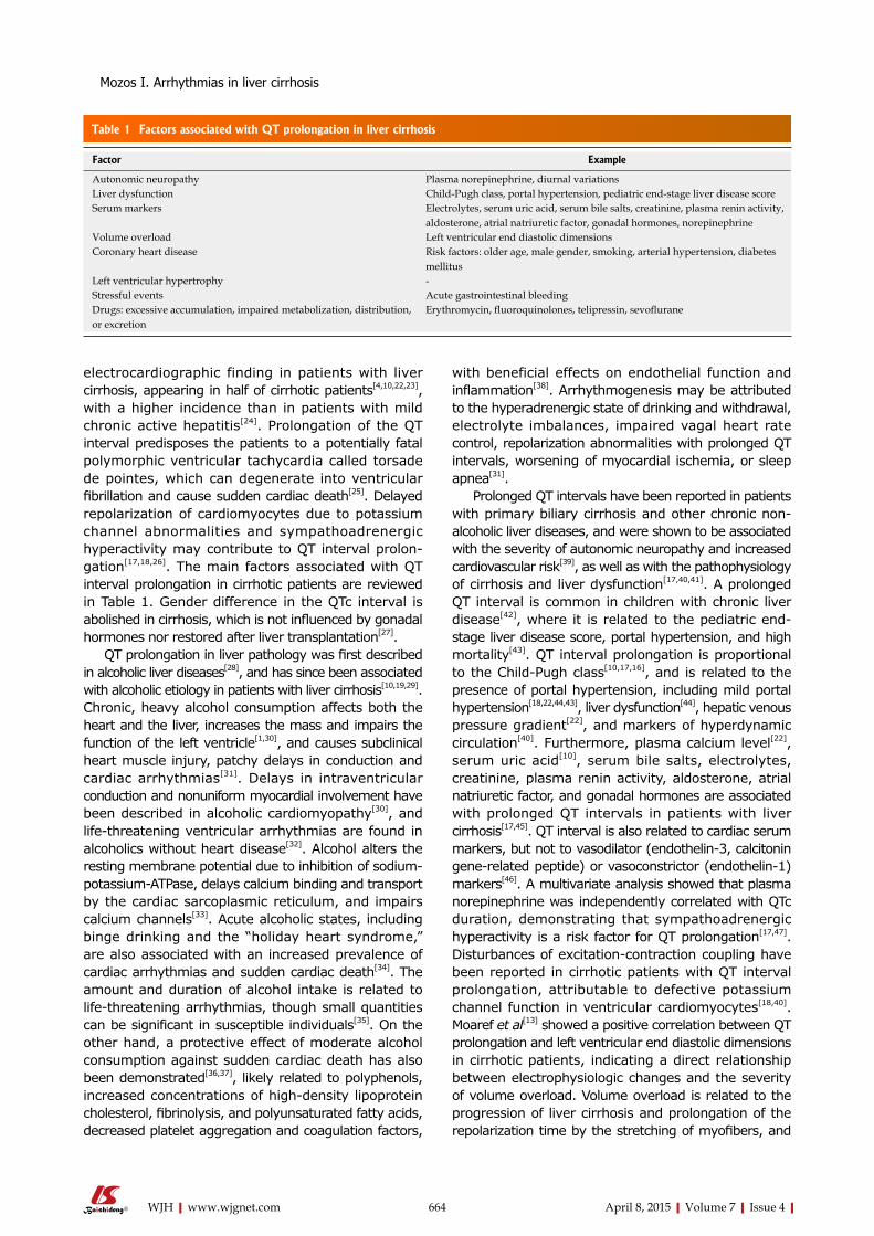
electrocardiographic finding in patients with liver cirrhosis, appearing in half of cirrhotic patients[4,10,22,23], with a higher incidence than in patients with mild chronic active hepatitis[24]. Prolongation of the QT interval predisposes the patients to a potentially fatal polymorphic ventricular tachycardia called torsade de pointes, which can degenerate into ventricular fibrillation and cause sudden cardiac death[25]. Delayed repolarization of cardiomyocytes due to potassium channel abnormalities and sympathoadrenergic hyperactivity may contribute to QT interval prolon-gation[17,18,26]. The main factors associated with QT interval prolongation in cirrhotic patients are reviewed in Table 1. Gender difference in the QTc interval is abolished in cirrhosis, which is not influenced by gonadal hormones nor restored after liver transplantation[27].
QT prolongation in liver pathology was first described in alcoholic liver diseases[28], and has since been associated with alcoholic etiology in patients with liver cirrhosis[10,19,29]. Chronic, heavy alcohol consumption affects both the heart and the liver, increases the mass and impairs the function of the left ventricle[1,30], and causes subclinical heart muscle injury, patchy delays in conduction and cardiac arrhythmias[31]. Delays in intraventricular conduction and nonuniform myocardial involvement have been described in alcoholic cardiomyopathy[30], and life-threatening ventricular arrhythmias are found in alcoholics without heart disease[32]. Alcohol alters the resting membrane potential due to inhibition of sodium-potassium-ATPase, delays calcium binding and transport by the cardiac sarcoplasmic reticulum, and impairs calcium channels[33]. Acute alcoholic states, including binge drinking and the “holiday heart syndrome,” are also associated with an increased prevalence of cardiac arrhythmias and sudden cardiac death[34]. The amount and duration of alcohol intake is related to life-threatening arrhythmias, though small quantities can be significant in susceptible individuals[35]. On the other hand, a protective effect of moderate alcohol consumption against sudden cardiac death has also been demonstrated[36,37], likely related to polyphenols, increased concentrations of high-density lipoprotein cholesterol, fibrinolysis, and polyunsaturated fatty acids, decreased platelet aggregation and coagulation factors,
with beneficial effects on endothelial function and inflammation[38]. Arrhythmogenesis may be attributed to the hyperadrenergic state of drinking and withdrawal, electrolyte imbalances, impaired vagal heart rate control, repolarization abnormalities with prolonged QT intervals, worsening of myocardial ischemia, or sleep apnea[31].
Prolonged QT intervals have been reported in patients with primary biliary cirrhosis and other chronic non-alcoholic liver diseases, and were shown to be associated with the severity of autonomic neuropathy and increased cardiovascular risk[39], as well as with the pathophysiology of cirrhosis and liver dysfunction[17,40,41]. A prolonged QT interval is common in children with chronic liver disease[42], where it is related to the pediatric end-stage liver disease score, portal hypertension, and high mortality[43]. QT interval prolongation is proportional to the Child-Pugh class[10,17,16], and is related to the presence of portal hypertension, including mild portal hypertension[18,22,44,43], liver dysfunction[44], hepatic venous pressure gradient[22], and markers of hyperdynamic circulation[40]. Furthermore, plasma calcium level[22], serum uric acid[10], serum bile salts, electrolytes, creatinine, plasma renin activity, aldosterone, atrial natriuretic factor, and gonadal hormones are associated with prolonged QT intervals in patients with liver cirrhosis[17,45]. QT interval is also related to cardiac serum markers, but not to vasodilator (endothelin-3, calcitonin gene-related peptide) or vasoconstrictor (endothelin-1) markers[46]. A multivariate analysis showed that plasma norepinephrine was independently correlated with QTc duration, demonstrating that sympathoadrenergic hyperactivity is a risk factor for QT prolongation[17,47]. Disturbances of excitation-contraction coupling have been reported in cirrhotic patients with QT interval prolongation, attributable to defective potassium channel function in ventricular cardiomyocytes[18,40]. Moaref et al[13] showed a positive correlation between QT prolongation and left ventricular end diastolic dimensions in cirrhotic patients, indicating a direct relationship between electrophysiologic changes and the severity of volume overload. Volume overload is related to the progression of liver cirrhosis and prolongation of the repolarization time by the stretching of myofibers, and
664 April 8, 2015|Volume 7|Issue 4|WJH|www.wjgnet.com
Table 1 Factors associated with QT prolongation in liver cirrhosis
Factor Example
Autonomic neuropathy Plasma norepinephrine, diurnal variationsLiver dysfunction Child-Pugh class, portal hypertension, pediatric end-stage liver disease scoreSerum markers Electrolytes, serum uric acid, serum bile salts, creatinine, plasma renin activity,
aldosterone, atrial natriuretic factor, gonadal hormones, norepinephrineVolume overload Left ventricular end diastolic dimensionsCoronary heart disease Risk factors: older age, male gender, smoking, arterial hypertension, diabetes
mellitusLeft ventricular hypertrophy -Stressful events Acute gastrointestinal bleedingDrugs: excessive accumulation, impaired metabolization, distribution, or excretion
Erythromycin, fluoroquinolones, telipressin, sevoflurane
Mozos I. Arrhythmias in liver cirrhosis
665 April 8, 2015|Volume 7|Issue 4|WJH|www.wjgnet.com
an analogue of vasopressin.
Ventricular repolarizationThe Tpe corresponds to the transmural dispersion of repolarization, and is a predictor of ventricular arrhythmias and sudden cardiac death[60-62]. The Tpe/QT ratio is also used as an index of ventricular arrhy-thmogenesis[63]. A prolonged Tpe interval and Tpe/QT interval ratio have been reported in patients with chronic hepatitis B infection, indicating an increased ventricular repolarization heterogeneity[64]. Liver cirrhosis affects ventricular repolarization via electrolyte imbalances, impaired autonomic function, subclinical cardiomyopathy, reduced β-adrenergic receptor function, post-receptor pathway defects, altered physical properties of myocyte plasma membranes, elevated levels of cardiotoxins, ion channel remodeling, portosystemic shunting, and systemic circulatory disturbances[10,16,22,44,65,66].
Late ventricular potentialsChronic alcoholics exhibit late ventricular potentials, low-amplitude and high-frequency waveforms appearing in the terminal part of the ECG QRS complex, which are predictors of re-entry ventricular tachycardia and sudden cardiac death[35,67]. Late ventricular potentials are associated with histologically significant fatty liver caused by chronic alcohol intake, revealing preclinical myocardial lesions and identifying alcoholic patients at risk of lethal arrhythmias[68].
Ion channel remodelingCardiac ion channel remodeling, particularly of pota-ssium channels, occurs in patients with liver cirrhosis[26]. Moreover, reduced transient outward and delayed rectifier potassium currents have been detected in ventricular myocytes from cirrhotic animals[26], which prolong the action potential and the QT interval[7]. Ionic channels, as well β-adrenergic receptors and G proteins, are altered by endotoxins and increased biliary acids in patients with cholestasis[16].
AUTONOMIC FUNCTIONPatients with liver cirrhosis show impaired autonomic cardiovascular reflexes, with the parasympathetic system more commonly affected than the sympathetic system[7]. The escape of systemic and intestinal vasodilators from degraded, diseased liver and the formation of new blood vessels in the gut explain arteriolar vasodilation of the systemic and splanchnic circulations[8]. The reduction in circulating blood volume and hyperdynamic circulation enhances the activities of the sympathetic nervous and renin-angiotensin-aldosterone systems. The resulting increased cardiac output and reduced systemic vascular resistance may induce myocardial remodeling and left ventricular hypertrophy, causing systolic and diastolic dysfunction and cardiomyopathy[7,8]. Sympathetic overactivity is
volume control is recommended in cirrhotic patients to prevent decompensation[13].
Prolonged QTc is related to an increased mortality rate in patients with chronic liver diseases[48]. Among these, patients with a QTc longer than 440 ms have a significantly lower survival rate than those with normal QTc[17]. The clinical significance of QT prolongation in liver cirrhosis is unclear, considering that sudden cardiac death and torsade de pointes are rare[9]. However, acute gastrointestinal bleeding further prolongs QTc in patients with liver cirrhosis, which predicts bleeding-induced mortality[49]. QT prolongation and electromechanical dyssynchrony have not been observed in septic cardiodepression, the inflammatory phenotype of cardiac dysfunction that is mediated through cytokines[15].
Drug-induced QT prolongationChild-Pugh and model for end-stage liver disease scores correlate with drug clearance[50]. As a result, patients with liver disease often require dosage adjustments in order to prevent adverse effects caused by excessive drug or metabolite accumulation[51]. Accumulation results from altered activity of drug-metabolizing enzymes and drug distribution, as well as from impaired renal excretion. For example, the activity of cytochrome P450 3A, the most abundant hepatic drug-metabolizing enzyme, is reduced in liver cirrhosis[51,52]. The activity of this enzyme varies according to the etiology and severity of liver disease[51,53]. Patients with transjugular intrahepatic portosystemic shunts are at increased risk for abnormal QT prolongation when exposed to oral cytochrome P450 substrates with QT-prolonging effects[54].
Drugs affecting the QT interval should be avoided in patients with liver cirrhosis, or used with caution under close ECG monitoring[2]. For example, the use of fluoroquinolones as secondary prophylaxis for spontaneous bacterial peritonitis in cirrhotic patients can predict QT prolongation[19]. Drug administration should be critically reviewed, with consideration of indications, interactions and adverse reactions, to prevent drug-induced torsade de pointes[55] and QT prolongation, particularly in patients with hepatic failure[56,57]. Werner et al[55] described a case of secondary torsade de pointes tachycardia in a 50-year-old patient with alcoholic liver cirrhosis who was admitted for hematemesis and melena after administration of QT-active drugs. Chung et al[58] reported a case of torsade de pointes after induction of anesthesia for liver transplantation with QT prolonging drugs: sevoflurane (to maintain anesthesia) and palonosetron (for postoperative nausea and vomiting). Faigel et al[45] also reported prolonged QT intervals and torsade de pointes in three cirrhotic patients with bleeding esophageal varices who received endoscopic sclerotherapy, vasopressin and neuroleptics. Lehmann et al[59] presented a patient with newly diagnosed cirrhosis and kidney failure who underwent cardiopulmonary resuscitation twice after terlipressin,
Mozos I. Arrhythmias in liver cirrhosis
666 April 8, 2015|Volume 7|Issue 4|WJH|www.wjgnet.com
associated with an increase in inflammatory cytokines, such as interleukin-1b, -6 and -8, tumor necrosis factor (TNF)-α, and transforming growth factor-β[8], which is a profibrogenic and proapoptotic stimulant[18]. Cardiovascular autonomic dysfunction has also been described in chronic alcoholic liver disease and chronic hepatitis B and C virus infections[64].
CARDIAC MANIFESTATIONS WITH HEPATITISPalpitations, dyspnea, angina chest discomfort, electro-cardiographic changes, tachycardia and bradycardia have all been described in patients with viral hepatitis[69], myocarditis, acute pericarditis and cardiomyopathy[70-72]. Sinus tachycardia occurs in most patients and is related to the febrile response[72]. Myocarditis may be a serious extrahepatic complication, and hepatitis B virus antigens have been detected in small intramyocardial vessels[71]. The cardiac abnormalities may be caused by viral infection, hyperbilirubinemia, hemorrhage in the myocardium and pericardium, or by immune me-chanisms[69,71]. Chronic hepatitis B infection triggers autoimmune disorders and several extrahepatic disorders may appear, including of the ganglia and the heart[73]. Endothelial progenitor cells may serve as a virus carrier, enabling transinfection in injured endothelial cells to cause hepatitis B virus-associated myocarditis[73]. Hayashi et al[69] reported a case of fulminant hepatitis complicated with myocarditis, with myocardial infarction-like electrocardiographic changes. Hepatitis C virus infection has been detected often in patients with dilated and hypertrophic cardiomyopathy, and may be an important causal agent in the pathogenesis of the disease and cause arrhythmias[72,74]. Interferon, successfully used to treat patients with chronic hepatitis C infections, may induce several cardiovascular com-plications, such as tachycardia, myocardial infarction and congestive heart failure[75].
MARKERS OF CARDIAC DYSFUNCTIONCell death is a central mechanism involved in liver damage, for which several promising noninvasive biomarkers have been associated with QT prolongation, including soluble cytokeratin 18, TNF and TNF-related apoptosis-inducing ligand receptors and their ligands, various isoforms of high mobility group box-1, small non-coding RNAs (microRNAs) and microparticles (extracellular vesicles)[76]. These biomarkers could be utilized in future studies to assess arrhythmia risk in liver cirrhosis. Fibrosis serum markers, such as hyaluronic acid and laminin[77], may also be indicators of electrophysiologic abnormalities in cirrhotic patients.
Natriuretic peptides are produced by the cardiac atrial and ventricular myocytes[78], and are higher in myocardial ischemia, heart failure and left ventricular tachycardia, as well as in liver cirrhosis and renal failure[79]. Plasma
levels of N-terminal pro-brain natriuretic peptide (BNP) are useful markers of increased cardiovascular risk, cardiac subclinical dysfunction, atrial volume, and early decompensation of cirrhosis, and are increased proportionate to the stage of chronic liver disease[78]. Elevated levels of BNP are related to interventricular septal thickness and the impairment of diastolic function in asymptomatic patients with cirrhosis, and may be a marker of the presence of cirrhotic cardiomyopathy[80]. Henriksen et al[81] also reported that circulating pro-BNP and BNP are related to severity of liver disease (Child-Pugh score, serum albumin, coagulation factors and hepatic venous pressure gradient) and markers of cardiac dysfunction (QT interval, heart rate and plasma volume), but not to indicators of hyperdynamic circulation.
RELATED COMPLICATIONS AND CONDITIONSCirrhotic patients also have an increased risk and pre-valence of coronary heart disease, which is also a cause of QT prolongation[1,82,83]. Risk factors for coronary heart disease, such as older age, male gender, smoking and arterial hypertension[82,84], are independent predictors of several electrocardiographic abnormalities in cirrhotic patients[19]. Moreover, liver disease severity is associated with many electrocardiographic features of coronary heart disease[19]. Considering low serum cholesterol, low blood pressure values and higher levels of circulating estrogens, cirrhosis should protect against coronary atherosclerosis[82]. However, recent reports have demonstrated an increased prevalence of major risk factors for atherosclerosis and cardiovascular disease in liver cirrhosis, especially in nonalcoholic steatohepatitis-cirrhosis[19,85]. Hypercholesterolemia in patients with primary biliary cirrhosis should be considered a cardiovascular risk factor, and further studies are needed to confirm if arrhythmias are related to it.
Arrhythmias are also associated with hypoxia and orthodeoxia due to hepatopulmonary syndrome. Hepatorenal syndrome may be another important contributor, influenced by systolic dysfunction and insu-fficient ventricular contractile reserve[2,86]. Ventricular arrhythmia risk and sudden cardiac death are increased in patients with renal failure, and even mild reductions in kidney function can alter the electrophysiologic properties of the myocardium[87]. Arrhythmia risk is related not only to renal function, but also to electrolyte imbalances, sympathetic activity, and levels of parathyroid hormone, hemoglobin, hematocrit and inflammatory markers[87].
Accumulation of bile acids in the liver due to obstructed ducts results in high circulating concentrations[88], with immunosuppressive effects[89]. In addition to the concentration, the composition of bile acids is important for arrhythmogenesis[90]. Taurocholic acid, a conjugated primary bile acid, has a negative inotropic effect and reduces the duration of the action potentials in the ventricular myocytes by reducing inward sodium and
Mozos I. Arrhythmias in liver cirrhosis
667 April 8, 2015|Volume 7|Issue 4|WJH|www.wjgnet.com
calcium and increasing outward potassium currents[88]. The increased level of non-ursodeoxycholic acids in patients with arrhythmias suggests that ursodeoxycholic acids provide cardioprotective and hepatoprotective effects[90]. Although the exact intracellular effects of bile salts are not clear, they may act on muscarinic or cell-surface bile acid receptors involved in the regulation of macrophage functions[89] or directly damage cardiac calcium channels due to the detergent-like properties[91].
SUPRAVENTRICULAR ARRHYTHMIAS AND CONDUCTION DISORDERS IN LIVER CIRRHOSISAtrial fibrillation and flutter are arrhythmias that are more frequently diagnosed in cirrhotic patients, and are significantly associated with arteriosclerosis, hypercholesterolemia and diabetes mellitus[92]. Atrial fibrillation after septic shock and sinus bradycardia with cardiac arrest were reported after living-donor liver transplantation in a 58-year-old man diagnosed with hepatocellular carcinoma and liver cirrhosis, which required resuscitation and temporary pacing[93]. Josefsson et al[19] reported several supraventricular arrhythmias in cirrhotic patients, such as atrial and junctional premature beats, atrial flutter or fibrillation, sinus tachycardia or bradycardia. Pre-transplant evaluation of cirrhotic patients also revealed atrioventricular-conduction defects, such as complete or incomplete right or left bundle branch block and intraventricular blocks.
Inflammation may promote cardiac and arrhyth-mogenic complications in non-alcoholic fatty liver disease[94]. Patients with liver fibrosis have elevated plasma levels of inflammatory markers, and several studies have indicated that inflammation plays a significant role in the generation, maintenance, and perpetuation of atrial fibrillation[95]. However, Zamirian et al[96] suggested that liver cirrhosis has a protective effect against atrial fibrillation, despite significant metabolic abnormalities, inflammatory syndrome and enlarged left atria. The low prevalence of atrial fibrillation observed in their study may be the result of the accumulation of anti-arrhythmic or anti-inflammatory substances that are normally metabolized by an intact functioning liver; this would explain the development of atrial fibrillation after liver transplantation[96]. However, no data concerning the influence of inflammation in the relationship of arrhythmias and liver cirrhosis have been reported, which should be the aim of future studies.
The low prevalence atrial fibrillation in cirrhotic patients reported by Zamirian et al[96] may also have been related to the low prevalence of systemic hypertension in their patients or the administration of medications (spironolactone and beta-blockers) that reduce atrial excitability. Spironolactone reduces myocardial fibrosis of dilated atria and P-wave duration, producing an antifibrotic effect in the ventricles and
reducing QT interval duration[97,98]. Beta-blockers are given as prophylaxis for variceal bleeding, such as for large esophageal varices, resulting in vasoconstriction in the splanchnic compartment, which increases preload and improves diastolic function[78]. Beta-blocker therapy may also prevent bleeding from portal hypertensive gastropathy and the development of spontaneous bacterial peritonitis. However, recent studies have warned about their use in decompensated cirrhosis, as they are associated with poor survival[99,100]. Beta-blockers decrease chronotropy and depress atrioventricular conduction, resulting in bradycardia or high-grade heart block[100].
Myocardial fibrosis is dysrhythmogenic[98], and atrial interstitial fibrosis is associated with changes in the electrical properties of the atria, including depressed excitability, increased refractoriness and conduction slowing or block[96,101]. Angiotensin-converting enzyme (ACE) inhibitors protect against myocardial fibrosis and prevent cardiac remodeling[102] and atrial fibrillation[103]. Drugs that interfere with the renin-angiotensin system, such as angiotensin Ⅱ-receptor blockers, also prevent atrial remodeling[103]. However, ACE inhibitors and other afterload-reducing drugs should be used with caution considering the risk for aggravating the vasodilatory state[2].
Statins are known for their pleiotropic and anti-hypertrophic effects, suppressing arrhythmogenesis and improving endothelial function[104]. Desensitization of cardiac myocytes to catecholamines due to down-regulation of beta-adrenergic receptors in the myocardium of cirrhotic patients could also be a protective mechanism against occurrence of tachyarrhythmia and atrial fibrillation[96].
The main mechanisms explaining the influence of cirrhosis on the higher prevalence or the protection against atrial fibrillation are reviewed in Table 2.
THERAPYNo specific therapy can be recommended for cirrhotic patients with heart conditions, but it should be supportiveand directed against heart failure and pulmonary stasis[1,15]. Surgical stress, including transjugular intra-hepatic portosystemic shunt insertion, surgical porto-systemic shunting and liver transplantation can facilitate heart failure[15,103]. However, severe heart failure can be prevented by vasodilated peripheral circulation, which unloads the heart, and a compensatory decrease of some negatively inotropic regulatory mechanisms[15]. Aldosterone antagonists may reduce left ventricular dilatation and wall thickness, and improve diastolic function[15]. QT interval prolongation may be improved by beta-blockers, which also lower portal pressure and reduce the hyperdynamic load, but their effect on contractile dysfunction and mortality should be the focus of further studies[15,105].
Liver transplantation is currently the only proven treatment for patients with cirrhotic cardiomyopathy[11]
Mozos I. Arrhythmias in liver cirrhosis
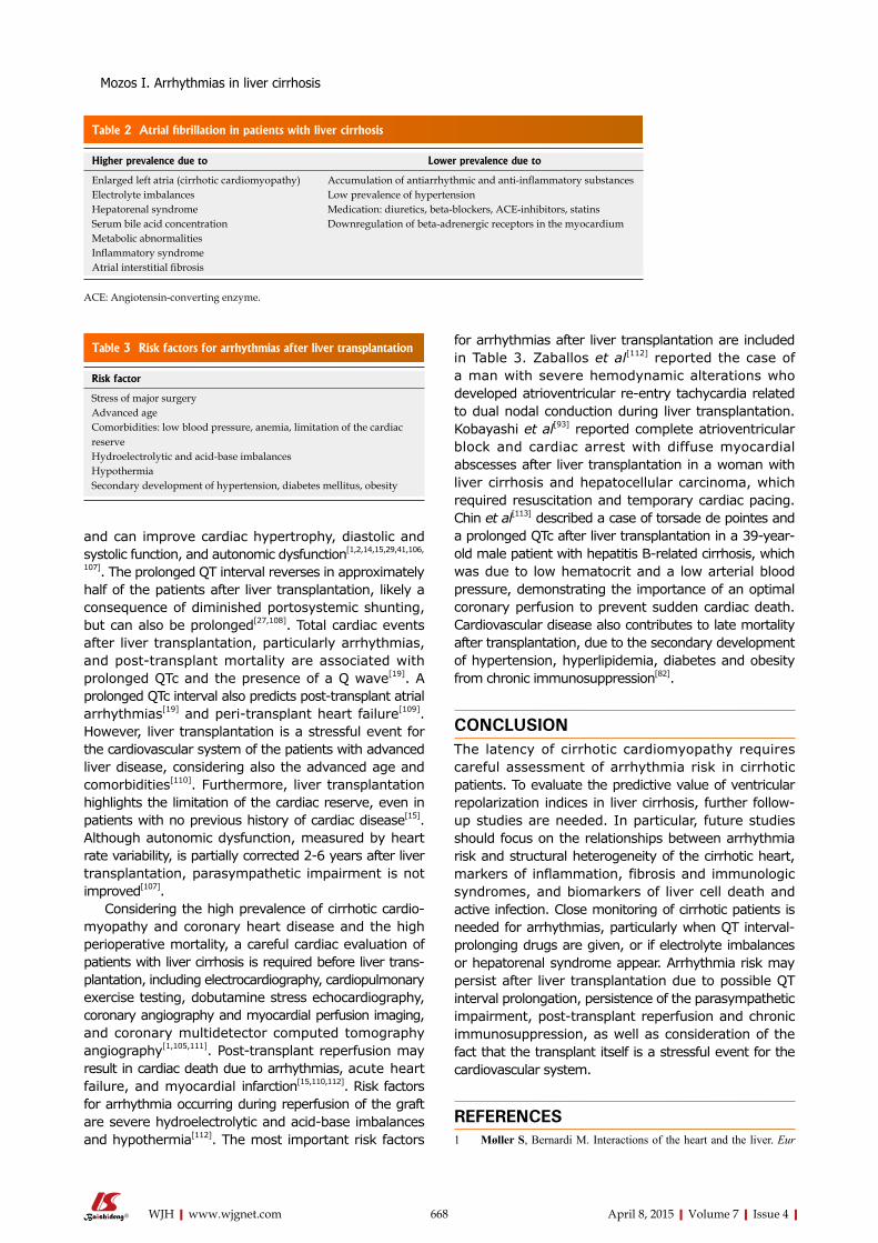
668 April 8, 2015|Volume 7|Issue 4|WJH|www.wjgnet.com
and can improve cardiac hypertrophy, diastolic and systolic function, and autonomic dysfunction[1,2,14,15,29,41,106,
107]. The prolonged QT interval reverses in approximatelyhalf of the patients after liver transplantation, likely a consequence of diminished portosystemic shunting, but can also be prolonged[27,108]. Total cardiac events after liver transplantation, particularly arrhythmias, and post-transplant mortality are associated with prolonged QTc and the presence of a Q wave[19]. A prolonged QTc interval also predicts post-transplant atrial arrhythmias[19] and peri-transplant heart failure[109]. However, liver transplantation is a stressful event for the cardiovascular system of the patients with advanced liver disease, considering also the advanced age and comorbidities[110]. Furthermore, liver transplantation highlights the limitation of the cardiac reserve, even in patients with no previous history of cardiac disease[15]. Although autonomic dysfunction, measured by heart rate variability, is partially corrected 2-6 years after liver transplantation, parasympathetic impairment is not improved[107].
Considering the high prevalence of cirrhotic cardio-myopathy and coronary heart disease and the high perioperative mortality, a careful cardiac evaluation of patients with liver cirrhosis is required before liver trans-plantation, including electrocardiography, cardiopulmonary exercise testing, dobutamine stress echocardiography, coronary angiography and myocardial perfusion imaging, and coronary multidetector computed tomography angiography[1,105,111]. Post-transplant reperfusion may result in cardiac death due to arrhythmias, acute heart failure, and myocardial infarction[15,110,112]. Risk factors for arrhythmia occurring during reperfusion of the graft are severe hydroelectrolytic and acid-base imbalances and hypothermia[112]. The most important risk factors
for arrhythmias after liver transplantation are included in Table 3. Zaballos et al[112] reported the case of a man with severe hemodynamic alterations who developed atrioventricular re-entry tachycardia related to dual nodal conduction during liver transplantation. Kobayashi et al[93] reported complete atrioventricular block and cardiac arrest with diffuse myocardial abscesses after liver transplantation in a woman with liver cirrhosis and hepatocellular carcinoma, which required resuscitation and temporary cardiac pacing. Chin et al[113] described a case of torsade de pointes and a prolonged QTc after liver transplantation in a 39-year-old male patient with hepatitis B-related cirrhosis, which was due to low hematocrit and a low arterial blood pressure, demonstrating the importance of an optimal coronary perfusion to prevent sudden cardiac death. Cardiovascular disease also contributes to late mortality after transplantation, due to the secondary development of hypertension, hyperlipidemia, diabetes and obesity from chronic immunosuppression[82].
CONCLUSIONThe latency of cirrhotic cardiomyopathy requires careful assessment of arrhythmia risk in cirrhotic patients. To evaluate the predictive value of ventricular repolarization indices in liver cirrhosis, further follow-up studies are needed. In particular, future studies should focus on the relationships between arrhythmia risk and structural heterogeneity of the cirrhotic heart, markers of inflammation, fibrosis and immunologic syndromes, and biomarkers of liver cell death and active infection. Close monitoring of cirrhotic patients is needed for arrhythmias, particularly when QT interval-prolonging drugs are given, or if electrolyte imbalances or hepatorenal syndrome appear. Arrhythmia risk may persist after liver transplantation due to possible QT interval prolongation, persistence of the parasympathetic impairment, post-transplant reperfusion and chronic immunosuppression, as well as consideration of the fact that the transplant itself is a stressful event for the cardiovascular system.
REFERENCES1 Møller S, Bernardi M. Interactions of the heart and the liver. Eur
Table 2 Atrial fibrillation in patients with liver cirrhosis
Higher prevalence due to Lower prevalence due to
Enlarged left atria (cirrhotic cardiomyopathy) Accumulation of antiarrhythmic and anti-inflammatory substancesElectrolyte imbalances Low prevalence of hypertensionHepatorenal syndrome Medication: diuretics, beta-blockers, ACE-inhibitors, statinsSerum bile acid concentration Downregulation of beta-adrenergic receptors in the myocardiumMetabolic abnormalitiesInflammatory syndromeAtrial interstitial fibrosis
ACE: Angiotensin-converting enzyme.
Table 3 Risk factors for arrhythmias after liver transplantation
Risk factor
Stress of major surgeryAdvanced ageComorbidities: low blood pressure, anemia, limitation of the cardiac reserveHydroelectrolytic and acid-base imbalancesHypothermiaSecondary development of hypertension, diabetes mellitus, obesity
Mozos I. Arrhythmias in liver cirrhosis
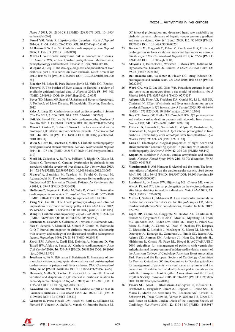
669 April 8, 2015|Volume 7|Issue 4|WJH|www.wjgnet.com
Heart J 2013; 34: 2804-2811 [PMID: 23853073 DOI: 10.1093/eurheartj/eht246]
2 Fouad YM, Yehia R. Hepato-cardiac disorders. World J Hepatol 2014; 6: 41-54 [PMID: 24653793 DOI: 10.4254/wjh.v6.i1.41]
3 Al Hamoudi W, Lee SS. Cirrhotic cardiomyopathy. Ann Hepatol 2006; 5: 132-139 [PMID: 17060868]
4 Mozos I. Ventricular arrhythmia risk in noncardiac diseases. In: Aronow WS, editor. Cardiac arrhythmias. Mechanisms, pathophysiology and treatment. Croatia: In Tech, 2014: 89-109
5 Wiegand J, Berg T. The etiology, diagnosis and prevention of liver cirrhosis: part 1 of a series on liver cirrhosis. Dtsch Arztebl Int 2013; 110: 85-91 [PMID: 23451000 DOI: 10.3238/arztebl.2013.0085]
6 Blachier M, Leleu H, Peck-Radosavljevic M, Valla DC, Roudot-Thoraval F. The burden of liver disease in Europe: a review of available epidemiological data. J Hepatol 2013; 58: 593-608 [PMID: 23419824 DOI: 10.1016/j.jhep.2012.12.005]
7 Boyer TD, Manns MP, Sanyal AJ. Zakim and Boyerʼs Hepatology. A Textbook of Liver Disease. Philadelphia: Elsevier, Saunders, 2012
8 Zaky A, Lang JD. Cirrhosis-associated cardiomyopathy. J Anesth Clin Res 2012; 3: 266 [DOI: 10.4172/2155-6148.1000266]
9 Baik SK, Fouad TR, Lee SS. Cirrhotic cardiomyopathy. Orphanet J Rare Dis 2007; 2: 15 [PMID: 17389039 DOI: 10.1186/1750-1172-2-15]
10 Mozos I, Costea C, Serban C, Susan L. Factors associated with a prolonged QT interval in liver cirrhosis patients. J Electrocardiol 2011; 44: 105-108 [PMID: 21146831 DOI: 10.1016/j.jelectrocard.2010.10.034]
11 Wiese S, Hove JD, Bendtsen F, Møller S. Cirrhotic cardiomyopathy: pathogenesis and clinical relevance. Nat Rev Gastroenterol Hepatol 2014; 11: 177-186 [PMID: 24217347 DOI: 10.1038/nrgastro.2013.210]
12 Merli M, Calicchia A, Ruffa A, Pellicori P, Riggio O, Giusto M, Gaudio C, Torromeo C. Cardiac dysfunction in cirrhosis is not associated with the severity of liver disease. Eur J Intern Med 2013; 24: 172-176 [PMID: 22958907 DOI: 10.1016/j.ejim.2012.08.007]
13 Moaref A, Zamirian M, Yazdani M, Salehi O, Sayadi M, Aghasadeghi K. The Correlation between Echocardiographic Findings and QT Interval in Cirrhotic Patients. Int Cardiovasc Res J 2014; 8: 39-43 [PMID: 24936479]
14 Huffman C, Wagman G, Fudim M, Zolty R, Vittorio T. Reversible cardiomyopathies--a review. Transplant Proc 2010; 42: 3673-3678 [PMID: 21094837 DOI: 10.1016/j.transproceed.2010.08.034]
15 Yang YY, Lin HC. The heart: pathophysiology and clinical implications of cirrhotic cardiomyopathy. J Chin Med Assoc 2012; 75: 619-623 [PMID: 23245476 DOI: 10.1016/j.jcma.2012.08.015]
16 Wong F. Cirrhotic cardiomyopathy. Hepatol Int 2009; 3: 294-304 [PMID: 19669380 DOI: 10.1007/s12072-008-9109-7]
17 Bernardi M, Calandra S, Colantoni A, Trevisani F, Raimondo ML, Sica G, Schepis F, Mandini M, Simoni P, Contin M, Raimondo G. Q-T interval prolongation in cirrhosis: prevalence, relationship with severity, and etiology of the disease and possible pathogenetic factors. Hepatology 1998; 27: 28-34 [PMID: 9425913]
18 Zardi EM, Abbate A, Zardi DM, Dobrina A, Margiotta D, Van Tassell BW, Afeltra A, Sanyal AJ. Cirrhotic cardiomyopathy. J Am Coll Cardiol 2010; 56: 539-549 [PMID: 20688208 DOI: 10.1016/j.jacc.2009.12.075]
19 Josefsson A, Fu M, Björnsson E, Kalaitzakis E. Prevalence of pre-transplant electrocardiographic abnormalities and post-transplant cardiac events in patients with liver cirrhosis. BMC Gastroenterol 2014; 14: 65 [PMID: 24708568 DOI: 10.1186/1471-230X-14-65]
20 Hansen S, Møller S, Bendtsen F, Jensen G, Henriksen JH. Diurnal variation and dispersion in QT interval in cirrhosis: relation to haemodynamic changes. J Hepatol 2007; 47: 373-380 [PMID: 17459513 DOI: 10.1016/j.jhep.2007.03.013]
21 Kowalski HJ, Abelmann WH. The cardiac output at rest in Laennec’s cirrhosis. J Clin Invest 1953; 32: 1025-1033 [PMID: 13096569 DOI: 10.1172/JCI102813]
22 Genovesi S, Prata Pizzala DM, Pozzi M, Ratti L, Milanese M, Pieruzzi F, Vincenti A, Stella A, Mancia G, Stramba-Badiale M.
QT interval prolongation and decreased heart rate variability in cirrhotic patients: relevance of hepatic venous pressure gradient and serum calcium. Clin Sci (Lond) 2009; 116: 851-859 [PMID: 19076059 DOI: 10.1042/CS20080325]
23 Bernardi M, Maggioli C, Dibra V, Zaccherini G. QT interval prolongation in liver cirrhosis: innocent bystander or serious threat? Expert Rev Gastroenterol Hepatol 2012; 6: 57-66 [PMID: 22149582 DOI: 10.1586/egh.11.86]
24 Akiyama T, Batchelder J, Worsman J, Moses HW, Jedlinski M. Hypocalcemic Torsades de Pointes. J Electrocardiol 1989; 22: 89-92 [PMID: 2921582]
25 Del Rosario ME, Weachter R, Flaker GC. Drug-induced QT prolongation and sudden death. Mo Med 2010; 107: 53-58 [PMID: 20222297]
26 Ward CA, Ma Z, Lee SS, Giles WR. Potassium currents in atrial and ventricular myocytes from a rat model of cirrhosis. Am J Physiol 1997; 273: G537-G544 [PMID: 9277435]
27 Adigun AQ, Pinto AG, Flockhart DA, Gorski JC, Li L, Hall SD, Chalasani N. Effect of cirrhosis and liver transplantation on the gender difference in QT interval. Am J Cardiol 2005; 95: 691-694 [PMID: 15721125 DOI: 10.1016/j.amjcard.2004.10.054]
28 Day CP, James OF, Butler TJ, Campbell RW. QT prolongation and sudden cardiac death in patients with alcoholic liver disease. Lancet 1993; 341: 1423-1428 [PMID: 8099138]
29 Finucci G, Lunardi F, Sacerdoti D, Volpin R, Bortoluzzi A, Bombonato G, Angeli P, Gatta A. Q-T interval prolongation in liver cirrhosis. Reversibility after orthotopic liver transplantation. Jpn Heart J 1998; 39: 321-329 [PMID: 9711183]
30 Luca C. Electrophysiological properties of right heart and atrioventricular conducting system in patients with alcoholic cardiomyopathy. Br Heart J 1979; 42: 274-281 [PMID: 508449]
31 Kupari M, Koskinen P. Alcohol, cardiac arrhythmias and sudden death. Novartis Found Symp 1998; 216: 68-79; discussion 79-85 [PMID: 9949788]
32 Moushmoush B, Abi-Mansour P. Alcohol and the heart. The long-term effects of alcohol on the cardiovascular system. Arch Intern Med 1991; 151: 36-42 [PMID: 1985607 DOI: 10.1001/archinte.1991.00400010060007]
33 Lorsheyd A, de Lange DW, Hijmering ML, Cramer MJ, van de Wiel A. PR and OTc interval prolongation on the electrocardiogram after binge drinking in healthy individuals. Neth J Med 2005; 63: 59-63 [PMID: 15766009]
34 Mozos I, Serban C, Mihaescu R. Late ventricular potentials in cardiac and extracardiac diseases. In: Breijo-Marquez FR, editor. Cardiac arrhythmias-New considerations, Croatia: In Tech, 2012: 227-256
35 Zipes DP, Camm AJ, Borggrefe M, Buxton AE, Chaitman B, Fromer M, Gregoratos G, Klein G, Moss AJ, Myerburg RJ, Priori SG, Quinones MA, Roden DM, Silka MJ, Tracy C, Priori SG, Blanc JJ, Budaj A, Camm AJ, Dean V, Deckers JW, Despres C, Dickstein K, Lekakis J, McGregor K, Metra M, Morais J, Osterspey A, Tamargo JL, Zamorano JL, Smith SC, Jacobs AK, Adams CD, Antman EM, Anderson JL, Hunt SA, Halperin JL, Nishimura R, Ornato JP, Page RL, Riegel B. ACC/AHA/ESC 2006 guidelines for management of patients with ventricular arrhythmias and the prevention of sudden cardiac death: a report of the American College of Cardiology/American Heart Association Task Force and the European Society of Cardiology Committee for Practice Guidelines (Writing Committee to Develop guidelines for management of patients with ventricular arrhythmias and the prevention of sudden cardiac death) developed in collaboration with the European Heart Rhythm Association and the Heart Rhythm Society. Europace 2006; 8: 746-837 [PMID: 16935866 DOI: 10.1093/europace/eul108]
36 Priori SG, Aliot E, Blomstrom-Lundqvist C, Bossaert L, Breithardt G, Brugada P, Camm AJ, Cappato R, Cobbe SM, Di Mario C, Maron BJ, McKenna WJ, Pedersen AK, Ravens U, Schwartz PJ, Trusz-Gluza M, Vardas P, Wellens HJ, Zipes DP. Task Force on Sudden Cardiac Death of the European Society of Cardiology. Eur Heart J 2001; 22: 1374-1450 [PMID: 11482917
Mozos I. Arrhythmias in liver cirrhosis
670 April 8, 2015|Volume 7|Issue 4|WJH|www.wjgnet.com
DOI: 10.1053/euhj.2001.2824]37 de Vreede-Swagemakers JJ, Gorgels AP, Weijenberg MP, Dubois-
Arbouw WI, Golombeck B, van Ree JW, Knottnerus A, Wellens HJ. Risk indicators for out-of-hospital cardiac arrest in patients with coronary artery disease. J Clin Epidemiol 1999; 52: 601-607 [PMID: 10391652]
38 di Giuseppe R, de Lorgeril M, Salen P, Laporte F, Di Castelnuovo A, Krogh V, Siani A, Arnout J, Cappuccio FP, van Dongen M, Donati MB, de Gaetano G, Iacoviello L. Alcohol consumption and n-3 polyunsaturated fatty acids in healthy men and women from 3 European populations. Am J Clin Nutr 2009; 89: 354-362 [PMID: 19056552 DOI: 10.3945/ajcn.2008.26661]
39 Kempler P, Szalay F, Váradi A, Keresztes K, Kádár E, Tánczos E, Petrik J. Prolongation of the QTc-interval reflects the severity of autonomic neuropathy in primary biliary cirrhosis and in other non-alcoholic liver diseases. Z Gastroenterol 1993; 31 Suppl 2: 96-98 [PMID: 7483730]
40 Henriksen JH, Fuglsang S, Bendtsen F, Christensen E, Møller S. Dyssynchronous electrical and mechanical systole in patients with cirrhosis. J Hepatol 2002; 36: 513-520 [PMID: 11943423 DOI: 10.1016/S0168-8278(02)00010-7]
41 Zamirian M, Tavassoli M, Aghasadeghi K. Corrected QT interval and QT dispersion in cirrhotic patients before and after liver transplantation. Arch Iran Med 2012; 15: 375-377 [PMID: 22642249]
42 Fishberger SB, Pittman NS, Rossi AF. Prolongation of the QT interval in children with liver failure. Clin Cardiol 1999; 22: 658-660 [PMID: 10526691]
43 Arikan C, Kilic M, Tumgor G, Levent E, Yuksekkaya HA, Yagci RV, Aydogdu S. Impact of liver transplantation on rate-corrected QT interval and myocardial function in children with chronic liver disease*. Pediatr Transplant 2009; 13: 300-306 [PMID: 18537904 DOI: 10.1111/j.1399-3046.2008.00909.x]
44 Ytting H, Henriksen JH, Fuglsang S, Bendtsen F, Møller S. Prolonged Q-T(c) interval in mild portal hypertensive cirrhosis. J Hepatol 2005; 43: 637-644 [PMID: 16083986 DOI: 10.1016/j.jhep.2005.04.015]
45 Faigel DO, Metz DC, Kochman ML. Torsade de pointes complicating the treatment of bleeding esophageal varices: association with neuroleptics, vasopressin, and electrolyte imbalance. Am J Gastroenterol 1995; 90: 822-824 [PMID: 7733096]
46 Henriksen JH, Gülberg V, Fuglsang S, Schifter S, Bendtsen F, Gerbes AL, Møller S. Q-T interval (QT(C)) in patients with cirrhosis: relation to vasoactive peptides and heart rate. Scand J Clin Lab Invest 2007; 67: 643-653 [PMID: 17852825 DOI: 10.1080/00365510601182634]
47 Camm AJ, Yap YG, Malik M. Acquired long QT syndrome. Wiley Online Library, 2007 [DOI: 10.1002/9780470994771.ch11]
48 Kosar F, Ates F, Sahin I, Karincaoglu M, Yildirim B. QT interval analysis in patients with chronic liver disease: a prospective study. Angiology 2007; 58: 218-224 [PMID: 17495272 DOI: 10.1177/0003319707300368]
49 Trevisani F, Di Micoli A, Zambruni A, Biselli M, Santi V, Erroi V, Lenzi B, Caraceni P, Domenicali M, Cavazza M, Bernardi M. QT interval prolongation by acute gastrointestinal bleeding in patients with cirrhosis. Liver Int 2012; 32: 1510-1515 [PMID: 22776742 DOI: 10.1111/j.1478-3231.2012.02847.x]
50 Albarmawi A, Czock D, Gauss A, Ehehalt R, Lorenzo Bermejo J, Burhenne J, Ganten TM, Sauer P, Haefeli WE. CYP3A activity in severe liver cirrhosis correlates with Child-Pugh and model for end-stage liver disease (MELD) scores. Br J Clin Pharmacol 2014; 77: 160-169 [PMID: 23772874 DOI: 10.1111/bcp.12182]
51 Vuppalanchi R, Liang T, Goswami CP, Nalamasu R, Li L, Jones D, Wei R, Liu W, Sarasani V, Janga SC, Chalasani N. Relationship between differential hepatic microRNA expression and decreased hepatic cytochrome P450 3A activity in cirrhosis. PLoS One 2013; 8: e74471 [PMID: 24058572 DOI: 10.1371/journal.pone.0074471]
52 Chalasani N, Gorski JC, Patel NH, Hall SD, Galinsky RE. Hepatic and intestinal cytochrome P450 3A activity in cirrhosis: effects of transjugular intrahepatic portosystemic shunts. Hepatology 2001;
34: 1103-1108 [PMID: 11731998]53 Frye RF, Zgheib NK, Matzke GR, Chaves-Gnecco D, Rabinovitz
M, Shaikh OS, Branch RA. Liver disease selectively modulates cytochrome P450--mediated metabolism. Clin Pharmacol Ther 2006; 80: 235-245 [PMID: 16952490 DOI: 10.1016/j.clpt.2006.05.006]
54 Vuppalanchi R, Juluri R, Ghabril M, Kim S, Thong N, Gorski JC, Chalasani N, Hall SD. Drug-induced QT prolongation in cirrhotic patients with transjugular intrahepatic portosystemic shunt. J Clin Gastroenterol 2011; 45: 638-642 [PMID: 20962670 DOI: 10.1097/MCG.0b013e3181f8c522]
55 Werner CR, Riessen R, Gregor M, Bitzer M. [Unexpected complication following esophageal variceal hemorrhage - Case 2/2011]. Dtsch Med Wochenschr 2011; 136: 217 [PMID: 21271486 DOI: 10.1055/s-0030-1247621]
56 Stanek EJ, Simko RJ, DeNofrio D, Pavri BB. Prolonged quinidine half-life with associated toxicity in a patient with hepatic failure. Pharmacotherapy 1997; 17: 622-625 [PMID: 9165569 DOI: 10.1002/j.1875-9114.1997.tb03075.x]
57 Barre J, Mallat A, Rosenbaum J, Deforges L, Houin G, Dhumeaux D, Tillement JP. Pharmacokinetics of erythromycin in patients with severe cirrhosis. Respective influence of decreased serum binding and impaired liver metabolic capacity. Br J Clin Pharmacol 1987; 23: 753-757 [PMID: 3606934]
58 Chung EJ, Jeon YS, Kim HJ, Lee KH, Lee JW, Han KA, Jung SH. Torsade de pointes in liver transplantation recipient after induction of general anesthesia: a case report. Korean J Anesthesiol 2014; 66: 80-84 [PMID: 24567820 DOI: 10.4097/kjae.2014.66.1.80]
59 Lehmann M, Bruns T, Herrmann A, Fritzenwanger M, Stallmach A. [54-year-old male with hepatic cirrhosis and therapy-associated torsade de pointes tachycardia]. Internist (Berl) 2011; 52: 445-48, 450 [PMID: 20938628 DOI: 10.1007/s00108-010-2667-5]
60 Castro Hevia J, Antzelevitch C, Tornés Bárzaga F, Dorantes Sánchez M, Dorticós Balea F, Zayas Molina R, Quiñones Pérez MA, Fayad Rodríguez Y. Tpeak-Tend and Tpeak-Tend dispersion as risk factors for ventricular tachycardia/ventricular fibrillation in patients with the Brugada syndrome. J Am Coll Cardiol 2006; 47: 1828-1834 [PMID: 16682308 DOI: 10.1016/j.jacc.2005.12.049]
61 Antzelevitch C, Sicouri S, Di Diego JM, Burashnikov A, Viskin S, Shimizu W, Yan GX, Kowey P, Zhang L. Does Tpeak-Tend provide an index of transmural dispersion of repolarization? Heart Rhythm 2007; 4: 1114-1116; author reply 1116-1119 [PMID: 17675094 DOI: 10.1016/j.hrthm.2007.05.028]
62 Arteyeva NV, Goshka SL, Sedova KA, Bernikova OG, Azarov JE. What does the T(peak)-T(end) interval reflect? An experimental and model study. J Electrocardiol 2013; 46: 296.e1-296.e8 [PMID: 23473669 DOI: 10.1016/j.jelectrocard.2013.02.001]
63 Kilicaslan F, Tokatli A, Ozdag F, Uzun M, Uz O, Isilak Z, Yiginer O, Yalcin M, Guney MS, Cebeci BS. Tp-e interval, Tp-e/QT ratio, and Tp-e/QTc ratio are prolonged in patients with moderate and severe obstructive sleep apnea. Pacing Clin Electrophysiol 2012; 35: 966-972 [PMID: 22671991 DOI: 10.1111/j.1540-8159.2012.03439.x]
64 Demir C, Demir M. Evaluation of Tp-e interval and Tp-e/QT ratio in patients with chronic hepatitis B. Prague Med Rep 2013; 114: 239-245 [PMID: 24485341]
65 Zambruni A, Trevisani F, Caraceni P, Bernardi M. Cardiac electrophysiological abnormalities in patients with cirrhosis. J Hepatol 2006; 44: 994-1002 [PMID: 16510203 DOI: 10.1016/j.jhep.2005.10.034]
66 Møller S, Hove JD, Dixen U, Bendtsen F. New insights into cirrhotic cardiomyopathy. Int J Cardiol 2013; 167: 1101-1108 [PMID: 23041091 DOI: 10.1016/j.ijcard.2012.09.089]
67 Benchimol Barbosa PR, Sousa MO, Barbosa EC, Bomfim Ade S, Ginefra P, Nadal J. Analysis of the prevalence of ventricular late potentials in the late phase of myocardial infarction based on the site of infarction. Arq Bras Cardiol 2002; 78: 352-363 [PMID: 12011951 DOI: 10.1590/S0066-782X2002000400002]
68 Pochmalicki G, Genest M, Jibril H. Late ventricular potentials and heavy drinking. Heart 1997; 78: 163-165 [PMID: 9326991]
69 Hayashi J, Kashiwagi S, Okeda T, Okamura H, Ishibashi H,
Mozos I. Arrhythmias in liver cirrhosis
671 April 8, 2015|Volume 7|Issue 4|WJH|www.wjgnet.com
Hiramatsu Y, Fujino T. Electrocardiographic changes related to hypersecretion of catecholamine in a patient with fulminant hepatitis. Jpn J Med 1988; 27: 187-190 [PMID: 3418984 DOI: 10.2169/internalmedicine1962.27.187]
70 Adler R, Takahashi M, Wright HT. Acute pericarditis associated with hepatitis B infection. Pediatrics 1978; 61: 716-719 [PMID: 149291]
71 Ursell PC, Habib A, Sharma P, Mesa-Tejada R, Lefkowitch JH, Fenoglio JJ. Hepatitis B virus and myocarditis. Hum Pathol 1984; 15: 481-484 [PMID: 6373562]
72 Matsumori A, Sasayama S. Newer aspects of pathogenesis of heart failure: hepatitis C virus infection in myocarditis and cardiomyopathy. J Card Fail 1996; 2: S187-S194 [PMID: 8951578]
73 Rong Q, Huang J, Su E, Li J, Li J, Zhang L, Cao K. Infection of hepatitis B virus in extrahepatic endothelial tissues mediated by endothelial progenitor cells. Virol J 2007; 4: 36 [PMID: 17407553 DOI: 10.1186/1743-422X-4-36]
74 Matsumori A, Ohashi N, Hasegawa K, Sasayama S, Eto T, Imaizumi T, Izumi T, Kawamura K, Kawana M, Kimura A, Kitabatake A, Matsuzaki M, Nagai R, Tanaka H, Hiroe M, Hori M, Inoko H, Seko Y, Sekiguchi M, Shimotohno K, Sugishita Y, Takeda N, Takihara K, Tanaka M, Yokoyama M. Hepatitis C virus infection and heart diseases: a multicenter study in Japan. Jpn Circ J 1998; 62: 389-391 [PMID: 9626910 DOI: 10.1253/jcj.62.389]
75 Matsumori A, Matoba Y, Sasayama S. Dilated cardiomyopathy associated with hepatitis C virus infection. Circulation 1995; 92: 2519-2525 [PMID: 7586353 DOI: 10.1161/01.CIR.92.9.2519]
76 Eguchi A, Wree A, Feldstein AE. Biomarkers of liver cell death. J Hepatol 2014; 60: 1063-1074 [PMID: 24412608 DOI: 10.1016/j.jhep.2013.12.026]
77 Li F, Zhu CL, Zhang H, Huang H, Wei Q, Zhu X, Cheng XY. Role of hyaluronic acid and laminin as serum markers for predicting significant fibrosis in patients with chronic hepatitis B. Braz J Infect Dis 2012; 16: 9-14 [PMID: 22358349]
78 Licata A, Corrao S, Petta S, Genco C, Cardillo M, Calvaruso V, Cabibbo G, Massenti F, Cammà C, Licata G, Craxì A. NT pro BNP plasma level and atrial volume are linked to the severity of liver cirrhosis. PLoS One 2013; 8: e68364 [PMID: 23940514 DOI: 10.1371/journal.pone.0068364]
79 Panagopoulou V, Deftereos S, Kossyvakis C, Raisakis K, Giannopoulos G, Bouras G, Pyrgakis V, Cleman MW. NTproBNP: an important biomarker in cardiac diseases. Curr Top Med Chem 2013; 13: 82-94 [PMID: 23470072 DOI: 10.2174/1568026611313020002]
80 Wong F, Siu S, Liu P, Blendis LM. Brain natriuretic peptide: is it a predictor of cardiomyopathy in cirrhosis? Clin Sci (Lond) 2001; 101: 621-628 [PMID: 11724649]
81 Henriksen JH, Gøtze JP, Fuglsang S, Christensen E, Bendtsen F, Møller S. Increased circulating pro-brain natriuretic peptide (proBNP) and brain natriuretic peptide (BNP) in patients with cirrhosis: relation to cardiovascular dysfunction and severity of disease. Gut 2003; 52: 1511-1517 [PMID: 12970147]
82 Garg A, Armstrong WF. Echocardiography in liver transplant candidates. JACC Cardiovasc Imaging 2013; 6: 105-119 [PMID: 23328568 DOI: 10.1016/j.jcmg.2012.11.002]
83 Keeffe BG, Valantine H, Keeffe EB. Detection and treatment of coronary artery disease in liver transplant candidates. Liver Transpl 2001; 7: 755-761 [PMID: 11552207 DOI: 10.1053/jlts.2001.26063]
84 Kalaitzakis E, Rosengren A, Skommevik T, Björnsson E. Coronary artery disease in patients with liver cirrhosis. Dig Dis Sci 2010; 55: 467-475 [PMID: 19242795 DOI: 10.1007/s10620-009-0738-z]
85 Kadayifci A, Tan V, Ursell PC, Merriman RB, Bass NM. Clinical and pathologic risk factors for atherosclerosis in cirrhosis: a comparison between NASH-related cirrhosis and cirrhosis due to other aetiologies. J Hepatol 2008; 49: 595-599 [PMID: 18662837 DOI: 10.1016/j.jhep.2008.05.024]
86 Ruiz-del-Arbol L, Monescillo A, Arocena C, Valer P, Ginès P, Moreira V, Milicua JM, Jiménez W, Arroyo V. Circulatory function
and hepatorenal syndrome in cirrhosis. Hepatology 2005; 42: 439-447 [PMID: 15977202 DOI: 10.1002/hep.20766]
87 Mozos I. Laboratory markers of ventricular arrhythmia risk in renal failure. Biomed Res Int 2014; 2014: 509204 [PMID: 24982887]
88 Binah O, Rubinstein I, Bomzon A, Better OS. Effects of bile acids on ventricular muscle contraction and electrophysiological properties: studies in rat papillary muscle and isolated ventricular myocytes. Naunyn Schmiedebergs Arch Pharmacol 1987; 335: 160-165 [PMID: 3561530]
89 Kawamata Y, Fujii R, Hosoya M, Harada M, Yoshida H, Miwa M, Fukusumi S, Habata Y, Itoh T, Shintani Y, Hinuma S, Fujisawa Y, Fujino M. A G protein-coupled receptor responsive to bile acids. J Biol Chem 2003; 278: 9435-9440 [PMID: 12524422]
90 Desai MS, Penny DJ. Bile acids induce arrhythmias: old metabolite, new tricks. Heart 2013; 99: 1629-1630 [PMID: 23969477 DOI: 10.1136/heartjnl-2013-304546]
91 Rainer PP, Primessnig U, Harenkamp S, Doleschal B, Wallner M, Fauler G, Stojakovic T, Wachter R, Yates A, Groschner K, Trauner M, Pieske BM, von Lewinski D. Bile acids induce arrhythmias in human atrial myocardium--implications for altered serum bile acid composition in patients with atrial fibrillation. Heart 2013; 99: 1685-1692 [PMID: 23894089 DOI: 10.1136/heartjnl-2013-304163]
92 Gundling F, Schmidtler F, Zelihic E, Seidl H, Haller B, Ronel J, Löffler N, Schepp W. [Frequency of cardiac arrhythmia in patients with liver cirrhoses and evaluation of associated factors]. Z Gastroenterol 2012; 50: 1149-1155 [PMID: 23150106 DOI: 10.1055/s-0032-1313182]
93 Kobayashi T, Sato Y, Yamamoto S, Oya H, Takeishi T, Kokai H, Hatakeyama K. Temporary cardiac pacing for fatal arrhythmia in living-donor liver transplantation: three case reports. Transplant Proc 2008; 40: 2818-2820 [PMID: 18929869 DOI: 10.1016/j.transproceed.2008.07.018]
94 Ballestri S, Lonardo A, Bonapace S, Byrne CD, Loria P, Targher G. Risk of cardiovascular, cardiac and arrhythmic complications in patients with non-alcoholic fatty liver disease. World J Gastroenterol 2014; 20: 1724-1745 [PMID: 24587651 DOI: 10.3748/wjg.v20.i7.1724]
95 Yap YG. Inflammation and atrial fibrillation: cause or para-phenomenon? Europace 2009; 11: 980-981 [PMID: 19635815 DOI: 10.1093/europace/eup191]
96 Zamirian M, Sarmadi T, Aghasadeghi K, Kazemi MB. Liver cirrhosis prevents atrial fibrillation: A reality or just an illusion? J Cardiovasc Dis Res 2012; 3: 109-112 [PMID: 22629027 DOI: 10.4103/0975-3583.95363]
97 Milliez P, Deangelis N, Rucker-Martin C, Leenhardt A, Vicaut E, Robidel E, Beaufils P, Delcayre C, Hatem SN. Spironolactone reduces fibrosis of dilated atria during heart failure in rats with myocardial infarction. Eur Heart J 2005; 26: 2193-2199 [PMID: 16141258 DOI: 10.1093/eurheartj/ehi478]
98 Wong KY, Wong SY, McSwiggan S, Ogston SA, Sze KY, MacWalter RS, Struthers AD. Myocardial fibrosis and QTc are reduced following treatment with spironolactone or amiloride in stroke survivors: a randomised placebo-controlled cross-over trial. Int J Cardiol 2013; 168: 5229-5233 [PMID: 23993727 DOI: 10.1016/j.ijcard.2013.08.027]
99 Sersté T, Melot C, Francoz C, Durand F, Rautou PE, Valla D, Moreau R, Lebrec D. Deleterious effects of beta-blockers on survival in patients with cirrhosis and refractory ascites. Hepatology 2010; 52: 1017-1022 [PMID: 20583214 DOI: 10.1002/hep.23775]
100 Ge PS, Runyon BA. The changing role of beta-blocker therapy in patients with cirrhosis. J Hepatol 2014; 60: 643-653 [PMID: 24076364 DOI: 10.1016/j.jhep.2013.09.016]
101 Sanders P, Morton JB, Davidson NC, Spence SJ, Vohra JK, Sparks PB, Kalman JM. Electrical remodeling of the atria in congestive heart failure: electrophysiological and electroanatomic mapping in humans. Circulation 2003; 108: 1461-1468 [PMID: 12952837 DOI: 10.1161/01.CIR.0000090688.49283.67]
102 Yu M, Zheng Y, Sun HX, Yu DJ. Inhibitory effects of enalaprilat on rat cardiac fibroblast proliferation via ROS/P38MAPK/TGF-β1
Mozos I. Arrhythmias in liver cirrhosis
672 April 8, 2015|Volume 7|Issue 4|WJH|www.wjgnet.com
signaling pathway. Molecules 2012; 17: 2738-2751 [PMID: 22395404 DOI: 10.3390/molecules17032738]
103 Schaer BA, Schneider C, Jick SS, Conen D, Osswald S, Meier CR. Risk for incident atrial fibrillation in patients who receive antihypertensive drugs: a nested case-control study. Ann Intern Med 2010; 152: 78-84 [PMID: 20083826 DOI: 10.7326/0003-4819-152-2-201001190-00005]
104 Tousoulis D, Oikonomou E, Siasos G, Stefanadis C. Statins in heart failure--With preserved and reduced ejection fraction. An update. Pharmacol Ther 2014; 141: 79-91 [PMID: 24022031 DOI: 10.1016/j.pharmthera.2013.09.001]
105 Møller S, Dümcke CW, Krag A. The heart and the liver. Expert Rev Gastroenterol Hepatol 2009; 3: 51-64 [PMID: 19210113 DOI: 10.1586/17474124.3.1.51]
106 Bal JS, Thuluvath PJ. Prolongation of QTc interval: relationship with etiology and severity of liver disease, mortality and liver transplantation. Liver Int 2003; 23: 243-248 [PMID: 12895263 DOI: 10.1034/j.1600-0676.2003.00833.x]
107 Baratta L, Tubani L, Merli M, Labbadia F, Facchini D, De Marco R, Rossi M, Attili AF, Berloco P, Ginanni Corradini S. Long-term effect of liver transplantation on cirrhotic autonomic cardiac dysfunction. Dig Liver Dis 2010; 42: 131-136 [PMID: 19540819 DOI: 10.1016/j.dld.2009.05.009]
108 Carey EJ, Gautam M, Ingall T, Douglas DD. The effect of liver transplantation on autonomic dysfunction in patients with end-stage liver disease. Liver Transpl 2008; 14: 235-239 [PMID:
18236403 DOI: 10.1002/lt.21350]109 Josefsson A, Fu M, Allayhari P, Björnsson E, Castedal M, Olausson
M, Kalaitzakis E. Impact of peri-transplant heart failure & amp; left-ventricular diastolic dysfunction on outcomes following liver transplantation. Liver Int 2012; 32: 1262-1269 [PMID: 22621679 DOI: 10.1111/j.1478-3231.2012.02818.x]
110 Rugină M, Predescu L, Sălăgean M, Gheorghe L, Gheorghe C, Tulbure D, Popescu I, Bubenek-Turconi S. Pre-liver transplantation, cardiac assessment. Chirurgia (Bucur) 2012; 107: 283-290 [PMID: 22844825]
111 Keeling AN, Flaherty JD, Davarpanah AH, Ambrosy A, Farrelly CT, Harinstein ME, Flamm SL, Abecassis MI, Skaro AI, Carr JC, Gheorghiade M. Coronary multidetector computed tomographic angiography to evaluate coronary artery disease in liver transplant candidates: methods, feasibility and initial experience. J Cardiovasc Med (Hagerstown) 2011; 12: 460-468 [PMID: 21610507 DOI: 10.2459/JCM.0b013e3283483916]
112 Zaballos M, Jimeno C, Jiménez C, Fraile JR, Almendral E. [Dual atrioventricular nodal conduction and arrhythmia with severe hemodynamic alterations during liver retransplantation]. Rev Esp Anestesiol Reanim 2005; 52: 355-358 [PMID: 16038175]
113 Chin JH, Park JY, Kim YK, Kim SH, Kong YG, Park PH, Hwang GS. Torsades de pointes triggered by severe diastolic hypotension with low hematocrit in the neohepatic stage of liver transplantation: a case report. Transplant Proc 2010; 42: 1959-1962 [PMID: 20620555 DOI: 10.1016/j.transproceed.2010.02.093]
P- Reviewer: Sirin G, Sunami Y, Xiong XJ, Wang CX S- Editor: Qi Y L- Editor: A E- Editor: Liu SQ
Mozos I. Arrhythmias in liver cirrhosis
team should decide clinical management of HCC, according to tumor characteristics and stage of liver disease. Potentially curative treatments are suitable for very-early- and early-stage HCC. However, the vast majority of HCC patients are diagnosed in later stages, where the tumor characteristics or progress of liver disease prevent curative interventions. For patients with intermediate-stage HCC, TACE and radioembolization improve survival and are being evaluated in addition to potentially curative therapies or with systemic targeted therapy. There is currently no effective systemic chemotherapy, immunologic, or hormonal therapy for HCC, and sorafenib is the only approved molecular-targeted treatment for advanced HCC. Other targeted agents are under investigation; trials comparing new agents in combination with sorafenib are ongoing. Combinations of systemic targeted therapies with local treatments are being evaluated for further improvements in HCC patient outcomes. This article provides an updated and comprehensive overview of the current standards and trends in the treatment of HCC.
Key words: Hepatocellular carcinoma; Molecular targeted agents; Radiofrequency ablation; Sorafenib; Trans-arterial chemoembolization
© The Author(s) 2015. Published by Baishideng Publishing Group Inc. All rights reserved.
Core tip: This article reviews the available treatment options for hepatocellular carcinoma. The recent clinical trials of molecular-targeted therapies, as single agents or in combination with other treatments, are reviewed, and some future study directions are addressed. The importance of a multidisciplinary approach to management is highlighted.
Gomaa AI, Waked I. Recent advances in multidisciplinary management of hepatocellular carcinoma. World J Hepatol 2015; 7(4): 673-687 Available from: URL: http://www.
Asmaa I Gomaa, Imam Waked
Asmaa I Gomaa, Imam Waked, Hepatology Department, National Liver Institute, Menoufiya University, Shebeen El-Kom 35111, EgyptAuthor contributions: Gomaa AI and Waked I reviewed the literature and wrote the manuscript.Conflict-of-interest: None related to this work; Waked I is speaker for Roche HL, MSD, BMS, and Gilead; advisory board for Janssen, Roche HL, and MSD; investigator for Roche HL, BMS, Bayer, Janssen, AbbVie, and Gilead.Open-Access: This article is an open-access article which was selected by an in-house editor and fully peer-reviewed by external reviewers. It is distributed in accordance with the Creative Commons Attribution Non Commercial (CC BY-NC 4.0) license, which permits others to distribute, remix, adapt, build upon this work non-commercially, and license their derivative works on different terms, provided the original work is properly cited and the use is non-commercial. See: http://creativecommons.org/licenses/by-nc/4.0/Correspondence to: Asmaa I Gomaa, MD, Hepatology Department, National Liver Institute, Menoufiya University, Gamal Abd El-Nasir, Shebeen El-Kom 35111, Egypt. [email protected] Telephone: +20-100-6157160Fax: +20-48-2234586Received: August 28, 2014Peer-review started: August 28, 2014First decision: November 27, 2014Revised: December 17, 2014Accepted: January 15, 2015 Article in press: January 19, 2015Published online: April 8, 2015
AbstractThe incidence of hepatocellular carcinoma (HCC) is increasing, and it is currently the second leading cause of cancer-related death worldwide. Potentially curative treatment options for HCC include resection, transplantation, and percutaneous ablation, whereas palliative treatments include trans-arterial chemoem-bolization (TACE), radioembolization, and systemic treatments. Due to the diversity of available treatment options and patients’ presentations, a multidisciplinary
REVIEW
Submit a Manuscript: http://www.wjgnet.com/esps/Help Desk: http://www.wjgnet.com/esps/helpdesk.aspxDOI: 10.4254/wjh.v7.i4.673
673 April 8, 2015|Volume 7|Issue 4|WJH|www.wjgnet.com
World J Hepatol 2015 April 8; 7(4): 673-687ISSN 1948-5182 (online)
© 2015 Baishideng Publishing Group Inc. All rights reserved.
Recent advances in multidisciplinary management of hepatocellular carcinoma
wjgnet.com/1948-5182/full/v7/i4/673.htm DOI: http://dx.doi.org/10.4254/wjh.v7.i4.673
INTRODUCTIONThe incidence of hepatocellular carcinoma (HCC) is increasing, and is currently is the second leading cause of cancer-related death worldwide, accounting for approximately 800000 deaths every year[1]. Clinical management of HCC is tailored according to tumor characteristics, stage of liver disease, and condition of the patients (age, performance status, and presence or absence of comorbidities). The American Association for the Study of Liver Diseases[2] and the European Association for the Study of the Liver (EASL)[3] endorse the use of Barcelona clinic liver cancer (BCLC) staging for the classification and management of patients with HCC. Therapeutic options are stage dependent and can be classified into three categories: curative, palliative, and symptomatic. However, curative treatment options, including resection and percutaneous ablation, are only suitable for early-stage tumors, and are associated with five-year survival rates of up to 75%[4].
Recently, treatment indications have been refined; patients who are not candidates for the first-line therapy for their stage can be shifted to the treatment option for the next BCLC stage (treatment stage migration concept)[3,5]. Trans-arterial chemoembolization (TACE) can be performed at an early stage in patients for whom radiofrequency ablation (RFA) or percutaneous ethanol injection (PEI) cannot be performed because of tumor location (proximity to a gallbladder, biliary tree, or blood vessel), unresectability of the tumor, failed prior curative treatments, or medical comorbidities[6].
The presentations of HCC are variable within each patient. Although the management guidelines for HCC recommend monotherapies as a treatment option, combined or sequential treatment modalities are effective in improving the outcome of patients with HCC. In practice, a multi-modal approach combining various treatments is used, and a multidisciplinary team, where the roles are intertwined and complimentary, should be involved in the management of every case[7,8].
SURGICAL RESECTIONSurgical resection is the recommended treatment for patients with a single nodule, preserved liver function, and good performance status. It is associated with five-year survival rates up to 70%[9] and a 2%-3% perioperative mortality in cirrhotic patients. Some centers report five-year survival rates above 50% in patients undergoing resection for multiple tumors fulfilling Milan criteria (up to three nodules, each ≤ 3 cm), who are not suitable for transplantation[10], and resection in patients with more advanced stages of HCC has been reported with acceptable outcomes[11].
The minimal critical remnant liver volume for safe resection is approximately 25% (15%-40%) for patients without cirrhosis and 50% (25%-90%) for patients with cirrhotic livers. Preoperative portal vein embolization is occasionally performed when the estimated remnant liver volume is less than the minimal requirement[12], aimed at diverting portal flow, with its content of growth factors, to the non-tumorous lobe to sufficiently increase its size to permit resection. However, the effectiveness of portal vein embolization in cases of HCC with a cirrhotic liver has not been sufficiently tested in large controlled studies[3].
Portal hypertension in cirrhotic patients is considered a relative contraindication for surgical resection, and a hepatic venous pressure gradient > 10 mmHg is reportedly the best predictor of postoperative liver decompensation and poor long-term outcome in compensated cirrhotic patients undergoing resection[2,13]. In practice, resection for patients with significant portal hypertension is still a subject of debate. Similarly, the presence of splenomegaly (major diameter > 12 cm) or esophageal varices with a platelet count of < 100000/mm3 was correlated with hepatic venous pressure gradient, postoperative decompensation, and poor survival[14]. However, Cucchetti et al[15] reported that patients with the same model for end-stage liver disease (MELD) score and extent of hepatectomy had similar outcomes regardless of portal hypertension.
Resection has been refined with the use of the RFA-based resection device, the Habib 4X sealer (a new bipolar RF device designed specifically for liver resection). It releases controlled RF energy between two pairs of electrodes, producing a plane of coagulative necrosis along the intended line of parenchymal resection, avoiding over-coagulation of liver parenchyma. The heat produced seals biliary and blood vessels, resulting in minimal blood loss. With this device, morbidity and mortality rates are superior to other methods of liver resection[16].
Laparoscopic resection, though a more sophisticated surgical procedure, is associated with reduced operative and postoperative morbidities[17]. A recent meta-analysis showed that laparoscopic hepatectomy decreases blood loss, transfusion requirement, postoperative morbidity, recovery time, and hospital stay compared to open hepatectomy, with no difference in recurrence or survival[18]. However, no randomized controlled trials (RCTs) were reported in this meta-analysis.
An important postoperative concern is the high risk of HCC recurrence. Five-year recurrence rates of 68% have been reported after liver resection of very-early-stage HCC. The presence of satellite nodules, cirrhosis, the use of non-anatomic resection, and elevated α-fetoprotein (AFP) levels are independently associated with tumor recurrence[14,19]. Late recurrence can be predicted using molecular biomarkers and gene signatures[20] that are used for the selection of patients amenable to hepatic resection. The 5-gene score, based on combined expression levels of HN1, RAN,
674 April 8, 2015|Volume 7|Issue 4|WJH|www.wjgnet.com
Gomaa AI et al . Advances in management of HCC
RAMP3, KRT19 and TAF9, was associated with disease-specific survival[20].
LIVER TRANSPLANTATIONLiver transplantation (LTx) is the best treatment option for patients with decompensated cirrhosis. HCC is the only solid tumor where transplantation plays an important role in management, due to the fact that it allows removal of the primary tumor and treats hepatic insufficiency[21]. The main obstacles for HCC patients amenable to LTx are the organ shortage and the long waiting time for transplantation. Increasing the donor pool by live donation, using bridging therapy, and applying prioritization policies can help overcome this problem[22]. A MELD exception was developed to assign extra points to HCC patients due to their high dropout rate and mortality while on the waiting list. However, no extra points are assigned to patients with compensated cirrhosis and small HCC tumors (< 2 cm) because of the improved survival with local ablation[3]. In practice, LTx is recommended for patients with tumors within the Milan criteria (a single lesion ≤ 5 cm, or up to three lesions ≤ 3 cm each)[23]. Restriction of LTx to patients within the Milan criteria results in a five-year overall survival rate of 75%, with a risk of recurrence < 15%[24]. The perioperative mortality and one-year mortality are approximately 3 and ≤ 10%, respectively. For patients with early-stage HCC, LTx offers the best chance of survival (106 mo), compared with surgical resection (52 mo), RFA (62 mo), PEI (44 mo), and TACE (34 mo)[25].
A systematic review of 90 studies over 15 years, including 17780 patients, identified the Milan criteria as an independent prognostic factor for outcome after LTx, with five-year survival rates comparable to non-HCC patients (65%-78%)[24]. An expansion of the Milan criteria to “up-to-seven” criteria (the sum of the size of the largest tumor and the number of tumors in patients without microvascular invasion) was proposed[23] and externally validated in an independent series[26], but requires larger prospective validation studies[3]. Although listing criteria for LTx currently depend on tumor number and size, the use of molecular markers and gene signatures for determining tumor behavior are under development[27].
The presence of vascular invasion, high AFP level, and transplant waiting time of more than 6 mo, are considered accurate predictive factors for poor survival and recurrence risk. Increased AFP was associated with higher risk of progression and dropout while waiting for a transplant[28,29], and a steady increase of AFP > 15 ng/mL per month was considered the most significant prognostic determinant[30]. In a large French multicenter study, incorporation of AFP in a prognostic score model for post-LTx outcome significantly improved the predictive performance of the Milan criteria in prioritization for LTx[29]. Moreover, adding AFP > 400 to a total tumor volume of 115 cm as a cutoff improved
prognosis prediction in an analysis of data of 6478 patients from the Scientific Registry of Transplant Recipients, and performed better than tumor size and number characteristics for predicting post-LTx prognosis[31].
LOCAL ABLATIVE THERAPYTumor ablation can be obtained using either chemical (alcohol and acetic acid) or physical (heating or cooling) methods. The first technique used to locally treat HCC was PEI[32], which involves the intra-lesional injection of absolute alcohol. Temperature ablative techniques have advanced, including heating techniques such as RFA[33], microwave ablation (MWA)[34], laser ablation[35], and cryoablation[36].
PEIPEI is indicated for the treatment of nodular HCC ≤ 5 cm in diameter, and achieves complete necrosis in 90% of tumors < 2 cm, 70% in those 2- < 3 cm, and 50% in those between 3 and 5 cm[37]. Patient outcome was improved with the use of a specific needle with three retractable prongs, achieving an 80%-90% rate of sustained complete response in tumors < 4 cm[38]. The major limitation of PEI is the high incidence of local recurrence (33%-43%).
RFARFA is superior to other local ablative therapies, and is currently the most commonly used ablative method, replacing PEI as the locoregional therapy of choice for early HCC[37]. RFA is considered the standard of care for patients with very early- and early-stage tumors, as well as those not suitable for or that refuse surgery. RFA is recommended as the main ablative therapy for tumors < 5 cm, whereas PEI is recommended in cases where RFA is not technically feasible[3].
In a cohort study, complete ablation was achieved in more than 90% of cases, with a local recurrence rate of < 1% and five-year survival rate ranging from 40% to 70% for lesions < 2 cm in diameter[39]. Three independent meta-analyses, including five RCTs, showed better results regarding local tumor control and survival benefits in patients treated with RFA, compared to ablation with PEI. In addition, patients with tumors 2-5 cm had better survivals if treated by RFA rather than by PEI[40-42].
Some groups have suggested that RFA should be considered as a first-line therapy, even when resection is possible, because it is associated with fewer side effects[39]. The main advantages compared to surgical intervention are that it is less invasive and provides an increased possibility for parenchymal sparing[39,43]. Whether surgical resection for very early HCC is superior to RFA remains controversial. Whereas a Markov model analysis indicated that surgical resection was preferable to RFA in terms of overall survival[44], Peng et al[45] reported that RFA was better. A survey in Japan
675 April 8, 2015|Volume 7|Issue 4|WJH|www.wjgnet.com
Gomaa AI et al . Advances in management of HCC
676 April 8, 2015|Volume 7|Issue 4|WJH|www.wjgnet.com
lesions without evidence of vascular invasion or extra-hepatic spread[3]. In Japan, TACE is recommended even for HCC patients with vascular invasion if radiologic portal invasion is distal to, or in the second-order branches of, the portal vein[59]. The main contraindications to TACE are extended portal vein thrombosis, diffuse tumor, extra-hepatic spread, and decompensated liver cirrhosis[22,60]. TACE improves survival compared to supportive care or suboptimal therapies[61], observed as an increase in the median survival of patients with intermediate-stage HCC to 20 mo[62]. However, a meta-analysis that included nine trials (six trials assessing TACE and three trials assessing trans-arterial embolization) has shown that trans-arterial therapy does not significantly increase survival in patients with unresectable HCC compared to controls[63].
Proper patient selection is crucial to prevent post-TACE-induced liver failure. Patients with total bilirubin > 3 mg/dL were excluded from TACE in several studies[64,65], and an AFP > 200 ng/mL and a MELD score > 10 were associated with greater risk of mortality[66]. Bolondi et al[67] proposed a substaging of intermediate-stage HCC (BCLC-B) patients from B1 to B4, taking into account the tumor burden and Child-Pugh score (A5 to B9). BCLC-B includes disease ranging from variable tumor burden, which can be a multifocal HCC affecting both lobes, extending up to near replacement of the liver, and includes patients with a wide range of liver function impairment (Child-Pugh score from 5 to 9). Substaging revealed decreasing survival for higher B substages, and thus TACE was recommended for early subgroups only[67].
Drug-eluting beads (DEB)-TACE involves the use of embolic microspheres with the ability to sequester and release chemotherapeutic agents in a controlled manner over a one-week period, which subsequently increases the local concentration of the drug with minimal systemic toxicity[68]. A randomized phase Ⅱ study (the PRECISIONV trial) reported that DEB-TACE is a valuable alternative and may be preferred over conventional TACE[69].
Assessment of response to TACEThe use of locoregional options to induce tumor necrosis necessitated a refinement of the conventional criteria to evaluate treatment response. Extent of tumor necrosis has been correlated with outcome after ablation, TACE and systemic therapy. A modification of the response evaluation criteria in solid tumors (modified RECIST) takes into account the degree of tumor necrosis, evaluated by dynamic computed tomography or magnetic resonance imaging[70] and has been adopted by the latest EASL guidelines for evaluating locoregional therapies for HCC[3].
Failure of TACEThere is no established definition for TACE refrac-toriness, nor is there a consensus for when to consider TACE failure and refer the patient to an alternative
including 1235 patients with very early HCC (≤ 2 cm) who underwent resection and 1315 patients who received RFA showed no significant difference in overall survival between the two groups (one-year, 98% vs 99%; two-year, 94% vs 95%), over a median follow-up of 37 mo[46]. However, the disease-free survival rate was significantly better after resection than after RFA (one-year, 91% vs 84%; two-year, 70% vs 58%; P < 0.001). Similarly, Wang et al[47] suggested that surgical resection was equivalent to RFA in terms of overall survival, and was associated with better disease-free survival.
The size limitation of RFA has been overcome with the use of expandable tipped or cool-tip electrodes, allowing effective ablation of areas ≥ 5 cm in diameter[48]. However, RCTs with a large sample size are needed before ablation therapy can be confirmed as an alternative to surgery for potentially resectable HCC.
Other ablative therapiesMWA is an alternative to RFA for thermal ablation of HCC. Only one RCT[49] compared the effectiveness of MWA to RFA, which revealed a tendency to favor RFA with respect to rates of local recurrence and complications, likely due to the small volume of coagulation obtained with a single probe insertion[50]. However, newer devices may have overcome this limitation. One advantage of MWA over RFA is that treatment outcome is not affected by the heat-sink effect of vessels in proximity to the tumor[49].
Laser ablation refers to thermal tissue destruction by conversion of absorbed light into heat[51]. The only randomized prospective study comparing laser ablation with RFA reported no significant difference in overall survival rates, with cumulative rates of 91.8%, 59.0% and 28.4% at one, three and five years respectively, without significant complications. However, a signifi-cantly better survival rate was reported for RFA in patients with Child-Pugh A stage disease[52].
Cryoablation uses the extreme cold of liquid nitrogen or argon gas to destroy abnormal tissue[53]. Cryoablation showed better local control than RFA or MWA for tumors > 2 cm[54]. A multicenter RCT in China that included 360 patients with one or two tumors < 4 cm in diameter found that cryoablation is safe and as effective as RFA, with a similar five-year survival[55].
TACEHCC receives 90% of its blood supply from the hepatic artery and only 10% from the portal vein[56]. Thus, the purpose of trans-arterial therapy is to block the blood supply and induce tumor necrosis, without significantly affecting hepatic blood supply[57]. Trans-arterial therapies include TACE, trans-arterial embolization, trans-arterial chemotherapy, and trans-arterial radioembolization[57,58].
TACE is currently the standard of care for patients with compensated liver function and large multifocal
Gomaa AI et al . Advances in management of HCC
677 April 8, 2015|Volume 7|Issue 4|WJH|www.wjgnet.com
therapy. Despite the absence of solid evidence, however, panels of experts have proposed treatment migration to sorafenib (downward treatment stage migration) for intermediate-stage patients if they demonstrate disease progression or poor tolerance after first or second TACE[71,72]. The current EASL guidelines recommend switching to sorafenib if intermediate-stage patients are non-responsive to at least two cycles of TACE[3].
Repetition of TACE should be considered based on evidence using mRECIST and the risk of adverse events. The response to the first TACE and its effect on the underlying liver disease help in identifying patients at risk of adverse outcome with repeated TACE. Sieghart et al[73] conducted a multivariate analysis to investigate TACE repeated for a second or third session and identified three prognostic factors: increase in aspartate aminotransferase by > 25%, increase in Child-Pugh score, and absence of tumor response. These factors were incorporated into an “ART” score, and patients with an ART score of 0-1.5 points benefitted from a second TACE, whereas those with a score ≥ 2.5 did not[74].
RADIOEMBOLIZATIONRadioembolization, or selective internal radiation therapy (SIRT), has recently emerged as a therapeutic option for intermediate-stage HCC. Unlike TACE, SIRT delivers local radiation to the tumor or liver tissue without causing ischemia. β radiation from radioactive yttrium-loaded glass or resin microspheres is applied to the tumor through the arteries that feed it, so that tumor nodules are treated irrespective of their number, size, or location[75]. The procedure is well tolerated with survival rates similar to TACE. Moreover, it is as safe and effective as sorafenib in patients with more advanced-stage HCC, including patients with portal vein thrombosis and large tumor burden[76-79].
In a study comparing radioembolization to TACE, radioembolization was associated with fewer side effects, better response rate, and longer time to progression (13.3 mo vs 8.4 mo), without difference in median survival time (20.5 mo vs 17.5 mo)[80]. Another study reported similar safety profile and response rates[76]. However, the cost associated with radioembolization may limit the applicability of this technique.
Stereotactic radiotherapy (SRT) allows the delivery of a high dose of radiation in a single (radio-surgery) or limited number (hypo-fractionation) of sessions, while sparing surrounding structures and healthy tissue[81]. Blomgren et al[82] first introduced SRT for liver tumors in 1995, with treatment doses ranging from 15 to 45 Gy, in one to five fractions. In a phase Ⅰ/Ⅱ study using a single dose ranging from 14 to 26 Gy, the treatment was well tolerated in all patients, with no major side effects[83], and the tumor control rate at 6 wk was 98%[84].
The CyberKnife Radio-surgery System is able to deliver very high doses of radiation to both primary and metastatic liver tumors with extreme accuracy, and
treatments can be completed in one to five sessions. Louis et al[81] treated 25 patients with CyberKnife stereotactic radiotherapy using respiratory motion tracking, which enables the radiation beam to track tumor movement in real time and allows patients to breathe normally during their treatment sessions. The actuarial one- and two-year local control rates were 95%, and the one- and two-year survival rates were 79% and 52% respectively, with good clinical tolerance. CyberKnife and SRT (though currently still very expensive) offer a local therapy for HCC patients who are not eligible for surgery, embolization, chemotherapy or radiofrequency ablation, without significant complications.
SYSTEMIC CHEMOTHERAPYHCC is among the most chemoresistant tumors, and until 2007, no systemic chemotherapy was recommended for patients with advanced tumors[3]. Systemic chemotherapy with cytotoxic agents, such as doxorubicin, gemcitabine, cisplatin, 5-fluorouracil or combined regimens for palliative care, was associated with low response rates (< 10%) with only marginal improvements in survival[85]. Moreover, these drugs are poorly tolerated in patients with underlying liver cirrhosis[85-87].
Interferon (IFN) therapy[87], anti-androgens, or tamoxifen[88] used in the treatment of advanced HCC show contradictory results without obvious benefit. A meta-analysis of seven RCTs, including 898 patients, evaluated tamoxifen vs conservative management and showed neither anti-tumor effects nor survival benefits for tamoxifen[89]. Subsequent large RCTs reported negative results in terms of survival[90,91].
Cisplatin, IFN, doxorubicin, and fluorouracil (PIAF) used in combination showed promising activity in a phase Ⅱ study[92]. A randomized phase Ⅲ study including 188 patients with HCC was conducted to investigate the effect of PIAF combination compared to doxorubicin alone[87]. The median survival rate of the PIAF group did not significantly differ from the doxorubicin group (8.67 mo vs 6.83 mo), and patients treated with the PIAF regimen experienced a significantly higher rate of myelotoxicity.
TARGETED THERAPY FOR HCCHepatocarcinogenesis is associated with epigenetic and genetic alterations that eventually lead to uncontrolled growth of hepatocytes. Signal transduction pathways, oncogenes, and growth factors and their receptors are considered new potential therapeutic targets for systemic targeted therapies, limiting widespread systemic toxicity[93]. Several targeted agents are currently in clinical development.
SorafenibSorafenib is an orally administered multikinase inhibitor with antiproliferative and antiangiogenic activity[94]. Sorafenib mediates downregulation of anti-apoptotic
Gomaa AI et al . Advances in management of HCC
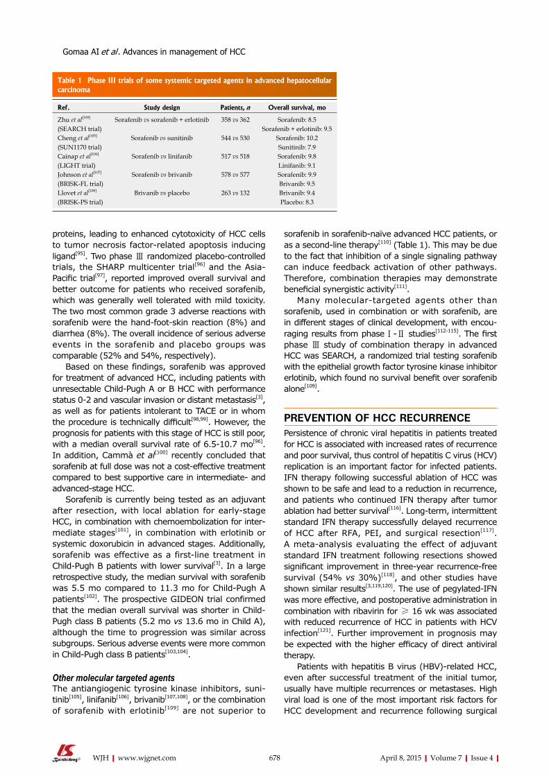
678 April 8, 2015|Volume 7|Issue 4|WJH|www.wjgnet.com
proteins, leading to enhanced cytotoxicity of HCC cells to tumor necrosis factor-related apoptosis inducing ligand[95]. Two phase Ⅲ randomized placebo-controlled trials, the SHARP multicenter trial[96] and the Asia-Pacific trial[97], reported improved overall survival and better outcome for patients who received sorafenib, which was generally well tolerated with mild toxicity. The two most common grade 3 adverse reactions with sorafenib were the hand-foot-skin reaction (8%) and diarrhea (8%). The overall incidence of serious adverse events in the sorafenib and placebo groups was comparable (52% and 54%, respectively).
Based on these findings, sorafenib was approved for treatment of advanced HCC, including patients with unresectable Child-Pugh A or B HCC with performance status 0-2 and vascular invasion or distant metastasis[3], as well as for patients intolerant to TACE or in whom the procedure is technically difficult[98,99]. However, the prognosis for patients with this stage of HCC is still poor, with a median overall survival rate of 6.5-10.7 mo[96]. In addition, Cammà et al[100] recently concluded that sorafenib at full dose was not a cost-effective treatment compared to best supportive care in intermediate- and advanced-stage HCC.
Sorafenib is currently being tested as an adjuvant after resection, with local ablation for early-stage HCC, in combination with chemoembolization for inter-mediate stages[101], in combination with erlotinib or systemic doxorubicin in advanced stages. Additionally, sorafenib was effective as a first-line treatment in Child-Pugh B patients with lower survival[3]. In a large retrospective study, the median survival with sorafenib was 5.5 mo compared to 11.3 mo for Child-Pugh A patients[102]. The prospective GIDEON trial confirmed that the median overall survival was shorter in Child-Pugh class B patients (5.2 mo vs 13.6 mo in Child A), although the time to progression was similar across subgroups. Serious adverse events were more common in Child-Pugh class B patients[103,104].
Other molecular targeted agentsThe antiangiogenic tyrosine kinase inhibitors, suni-tinib[105], linifanib[106], brivanib[107,108], or the combination of sorafenib with erlotinib[109] are not superior to
sorafenib in sorafenib-naïve advanced HCC patients, or as a second-line therapy[110] (Table 1). This may be due to the fact that inhibition of a single signaling pathway can induce feedback activation of other pathways. Therefore, combination therapies may demonstrate beneficial synergistic activity[111].
Many molecular-targeted agents other than sorafenib, used in combination or with sorafenib, are in different stages of clinical development, with encou-raging results from phase Ⅰ-Ⅱ studies[112-115]. The first phase Ⅲ study of combination therapy in advanced HCC was SEARCH, a randomized trial testing sorafenib with the epithelial growth factor tyrosine kinase inhibitor erlotinib, which found no survival benefit over sorafenib alone[109].
PREVENTION OF HCC RECURRENCEPersistence of chronic viral hepatitis in patients treated for HCC is associated with increased rates of recurrence and poor survival, thus control of hepatitis C virus (HCV) replication is an important factor for infected patients. IFN therapy following successful ablation of HCC was shown to be safe and lead to a reduction in recurrence, and patients who continued IFN therapy after tumor ablation had better survival[116]. Long-term, intermittent standard IFN therapy successfully delayed recurrence of HCC after RFA, PEI, and surgical resection[117]. A meta-analysis evaluating the effect of adjuvant standard IFN treatment following resections showed significant improvement in three-year recurrence-free survival (54% vs 30%)[118], and other studies have shown similar results[3,119,120]. The use of pegylated-IFN was more effective, and postoperative administration in combination with ribavirin for ≥ 16 wk was associated with reduced recurrence of HCC in patients with HCV infection[121]. Further improvement in prognosis may be expected with the higher efficacy of direct antiviral therapy.
Patients with hepatitis B virus (HBV)-related HCC, even after successful treatment of the initial tumor, usually have multiple recurrences or metastases. High viral load is one of the most important risk factors for HCC development and recurrence following surgical
Ref. Study design Patients, n Overall survival, mo
Zhu et al[109] (SEARCH trial)
Sorafenib vs sorafenib + erlotinib 358 vs 362 Sorafenib: 8.5Sorafenib + erlotinib: 9.5
Cheng et al[105]
(SUN1170 trial)Sorafenib vs sunitinib 544 vs 530 Sorafenib: 10.2
Sunitinib: 7.9Cainap et al[106]
(LIGHT trial)Sorafenib vs linifanib 517 vs 518 Sorafenib: 9.8
Linifanib: 9.1Johnson et al[107]
(BRISK-FL trial)Sorafenib vs brivanib 578 vs 577 Sorafenib: 9.9
Brivanib: 9.5Llovet et al[108]
(BRISK-PS trial)Brivanib vs placebo 263 vs 132 Brivanib: 9.4
Placebo: 8.3
Table 1 Phase III trials of some systemic targeted agents in advanced hepatocellular carcinoma
Gomaa AI et al . Advances in management of HCC
679 April 8, 2015|Volume 7|Issue 4|WJH|www.wjgnet.com
resection[122]. Similar to HCV, antiviral therapy for HBV following curative HCC ablation improved patient survival and decreased HCC recurrence. In their study, Hann et al[123] followed patients for 12 years who underwent local tumor ablation with or without concomitant antiviral therapy with lamivudine. Although initially there was no difference between the treatment groups with respect to tumor size (all ≤ 7 cm), levels of AFP and albumin, antiviral therapy was significantly associated with increased median survival (36 mo vs 16 mo)[123].
No other modality has demonstrated equivalent effectiveness for decreasing recurrence after curative treatment of HCC as antiviral therapy has for viral hepatitis-related tumors. Chemoembolization[124], internal radiation[125,126], immune therapies[127], retinoids[128], and the heparanase inhibitor PI-88[129] have been investigated as methods of reducing postoperative recurrence; however, none can be recommended as a preoperative/postoperative adjuvant/neo-adjuvant therapy for improving prognosis and diminishing the incidence of recurrence following curative therapy.
MULTIDISCIPLINARY TEAMHCC has diverse presentations that are compounded by the status of liver disease, and the multiple treatment options available make choosing the first line of treatment for a given patient a difficult task. Treatment of HCC patients should be undertaken by a multidisciplinary team that includes all the specialties involved in delivering the different therapies. In addition, simultaneous or sequential multi-modal therapies for patients with HCC show promise for improving patient outcome, further emphasizing the importance of a multidisciplinary approach to HCC management.
The multidisciplinary team should include hepa-tologists, medical and surgical oncologists, transplant surgeons, diagnostic and interventional radiologists, radiation oncologists, and pathologists[130]. All members should play an active role, as their expertise is required to provide optimal care for patients with HCC. The hepatologist should assess underlying liver disease, identify patients at risk for HCC, and monitor for early detection. Hepatologists are essential for managing liver disease and its complications, arranging for and monitoring treatment, and referring eligible patients for LTx. Oncologists are responsible for assigning systemic or targeted therapy as initial treatment or adjuvant therapy, and for managing associated side effects. The diagnostic radiologist makes and confirms the diagnosis, stages the tumor, its spread and vascular invasion, and assesses the radiologic response to treatment. The interventional radiologist delivers ablative therapy in early stages, and palliative therapy for intermediate-stage tumors. The hepatobiliary surgeon evaluates for and performs resection or transplantation. The pathologist assesses the grade of tumor differentiation,
stage of progression, and evaluates tissue markers. This multidisciplinary team also involves nurses, supportive care specialists, and palliative physicians[130].
MULTI-MODAL THERAPIESWith the multidisciplinary approach, various treatments are being delivered simultaneously or sequentially, as first- or second-line therapies, to improve patient outcome.
Transplantation and locoregional treatmentPatients whose tumors exceed the Milan criteria can undergo locoregional treatment (TACE or RFA) to down-stage the tumor to within the Milan criteria to allow LTx. Two prospective studies showed similar survival after LTx for patients with successfully down-staged HCC compared with those who initially met the Milan criteria[131,132].
Neo-adjuvant therapies for patients while on the waiting list are used in most centers. Systemic and interventional treatments are used to bridge patients in order to control disease and prevent tumor progression when the waiting time exceeds 6 mo[133,134]. Percutaneous treatments are more cost-effective than surgical resection[135]. Moreover, a poor response to TACE before transplantation is an indicator of post-transplantation recurrence[136].
Surgery and sorafenibSorafenib following curative surgery in a phase Ⅱ trial including 30 patients resulted in a lower tumor recurrence rate compared to surgery alone (33.3% vs 73.6%)[137].
TACE and ablative therapyCombining PEI with TACE has been shown to be effective for unresectable HCC[138]. The three-year survival rate was longer in patients with large and unresectable HCC treated with a combination of TACE and PEI than with TACE alone (22% vs 4%, respectively).
Combining RFA with TACE was evaluated in a RCT for patients with tumors between 3 and 5 cm[139]. The local tumor progression rate was significantly lower with combined treatment compared to RFA only (6% vs 39%).
Sorafenib and locoregional treatment There are more than 20 clinical trials in progress evaluating locoregional treatments combined with molecular-targeted agents, and some have demonstrated promising results[140-142]. A large phase Ⅲ, randomized, placebo-controlled trial (the STORM trial) evaluating sorafenib as an adjuvant therapy after curative treatment (resection or local ablation) is ongoing[143].
Sorafenib and TACE Following TACE, the tumor microenvironment becomes
Gomaa AI et al . Advances in management of HCC
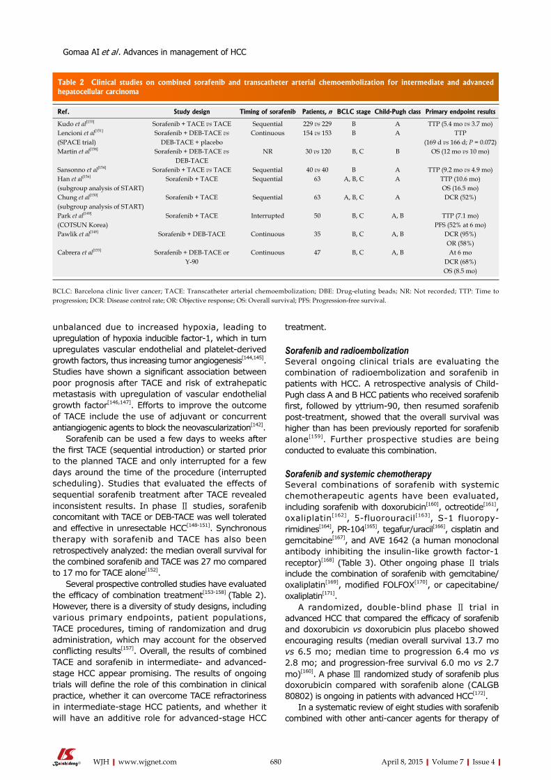
680 April 8, 2015|Volume 7|Issue 4|WJH|www.wjgnet.com
unbalanced due to increased hypoxia, leading to upregulation of hypoxia inducible factor-1, which in turn upregulates vascular endothelial and platelet-derived growth factors, thus increasing tumor angiogenesis[144,145]. Studies have shown a significant association between poor prognosis after TACE and risk of extrahepatic metastasis with upregulation of vascular endothelial growth factor[146,147]. Efforts to improve the outcome of TACE include the use of adjuvant or concurrent antiangiogenic agents to block the neovascularization[142].
Sorafenib can be used a few days to weeks after the first TACE (sequential introduction) or started prior to the planned TACE and only interrupted for a few days around the time of the procedure (interrupted scheduling). Studies that evaluated the effects of sequential sorafenib treatment after TACE revealed inconsistent results. In phase Ⅱ studies, sorafenib concomitant with TACE or DEB-TACE was well tolerated and effective in unresectable HCC[148-151]. Synchronous therapy with sorafenib and TACE has also been retrospectively analyzed: the median overall survival for the combined sorafenib and TACE was 27 mo compared to 17 mo for TACE alone[152].
Several prospective controlled studies have evaluated the efficacy of combination treatment[153-158] (Table 2). However, there is a diversity of study designs, including various primary endpoints, patient populations, TACE procedures, timing of randomization and drug administration, which may account for the observed conflicting results[157]. Overall, the results of combined TACE and sorafenib in intermediate- and advanced-stage HCC appear promising. The results of ongoing trials will define the role of this combination in clinical practice, whether it can overcome TACE refractoriness in intermediate-stage HCC patients, and whether it will have an additive role for advanced-stage HCC
treatment.
Sorafenib and radioembolizationSeveral ongoing clinical trials are evaluating the combination of radioembolization and sorafenib in patients with HCC. A retrospective analysis of Child-Pugh class A and B HCC patients who received sorafenib first, followed by yttrium-90, then resumed sorafenib post-treatment, showed that the overall survival was higher than has been previously reported for sorafenib alone[159]. Further prospective studies are being conducted to evaluate this combination.
Sorafenib and systemic chemotherapySeveral combinations of sorafenib with systemic chemotherapeutic agents have been evaluated, including sorafenib with doxorubicin[160], octreotide[161], oxaliplatin[162], 5-fluorouracil[163], S-1 fluoropy-rimidines[164], PR-104[165], tegafur/uracil[166], cisplatin and gemcitabine[167], and AVE 1642 (a human monoclonal antibody inhibiting the insulin-like growth factor-1 receptor)[168] (Table 3). Other ongoing phase Ⅱ trials include the combination of sorafenib with gemcitabine/oxaliplatin[169], modified FOLFOX[170], or capecitabine/oxaliplatin[171].
A randomized, double-blind phase Ⅱ trial in advanced HCC that compared the efficacy of sorafenib and doxorubicin vs doxorubicin plus placebo showed encouraging results (median overall survival 13.7 mo vs 6.5 mo; median time to progression 6.4 mo vs 2.8 mo; and progression-free survival 6.0 mo vs 2.7 mo)[160]. A phase Ⅲ randomized study of sorafenib plus doxorubicin compared with sorafenib alone (CALGB 80802) is ongoing in patients with advanced HCC[172].
In a systematic review of eight studies with sorafenib combined with other anti-cancer agents for therapy of
Ref. Study design Timing of sorafenib Patients, n BCLC stage Child-Pugh class Primary endpoint results
Kudo et al[155] Sorafenib + TACE vs TACE Sequential 229 vs 229 B A TTP (5.4 mo vs 3.7 mo)Lencioni et al[151]
(SPACE trial)Sorafenib + DEB-TACE vs
DEB-TACE + placeboContinuous 154 vs 153 B A TTP
(169 d vs 166 d; P = 0.072)Martin et al[158] Sorafenib + DEB-TACE vs
DEB-TACENR 30 vs 120 B, C B OS (12 mo vs 10 mo)
Sansonno et al[154] Sorafenib + TACE vs TACE Sequential 40 vs 40 B A TTP (9.2 mo vs 4.9 mo)Han et al[156]
(subgroup analysis of START)Sorafenib + TACE Sequential 63 A, B, C A TTP (10.6 mo)
OS (16.5 mo)Chung et al[150]
(subgroup analysis of START)Sorafenib + TACE Sequential 63 A, B, C A DCR (52%)
Park et al[149]
(COTSUN Korea)Sorafenib + TACE Interrupted 50 B, C A, B TTP (7.1 mo)
PFS (52% at 6 mo)Pawlik et al[148] Sorafenib + DEB-TACE Continuous 35 B, C A, B DCR (95%)
OR (58%)Cabrera et al[153] Sorafenib + DEB-TACE or
Y-90Continuous 47 B, C A, B At 6 mo
DCR (68%)OS (8.5 mo)
Table 2 Clinical studies on combined sorafenib and transcatheter arterial chemoembolization for intermediate and advanced hepatocellular carcinoma
BCLC: Barcelona clinic liver cancer; TACE: Transcatheter arterial chemoembolization; DBE: Drug-eluting beads; NR: Not recorded; TTP: Time to progression; DCR: Disease control rate; OR: Objective response; OS: Overall survival; PFS: Progression-free survival.
Gomaa AI et al . Advances in management of HCC
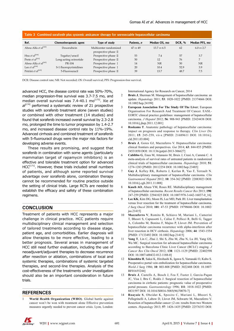
681 April 8, 2015|Volume 7|Issue 4|WJH|www.wjgnet.com
advanced HCC, the disease control rate was 50%-70%, median progression-free survival was 3.7-7.5 mo, and median overall survival was 7.4-40.1 mo[173]. Xie et al[174] performed a systematic review of 21 prospective studies with sorafenib treatment alone (seven studies) or combined with other treatment (14 studies) and found that sorafenib increased overall survival by 2.3-2.8 mo, prolonged the time to tumor progression by 1.4-2.7 mo, and increased disease control rate by 11%-19%. Advanced cirrhosis and combined treatment of sorafenib with 5-fluorouracil drugs were the major risk factors for developing adverse events.
These results are promising, and suggest that sorafenib in combination with some agents (particularly mammalian target of rapamycin inhibitors) is an effective and tolerable treatment option for advanced HCC[171]. However, these trials included small numbers of patients, and although some reported survival advantage over sorafenib alone, combination therapy cannot be recommended for routine practice outside the setting of clinical trials. Large RCTs are needed to establish the efficacy and safety of these combination regimens.
CONCLUSIONTreatment of patients with HCC represents a major challenge in clinical practice. HCC patients require multidisciplinary clinical management and selection of tailored treatments according to disease stage, patient age, and comorbidities. Earlier diagnosis will allow therapies to be more effective, leading to a better prognosis. Several areas in management of HCC still need further evaluation, including the use of neoadjuvant/adjuvant therapies to decrease recurrence after resection or ablation, combinations of local and systemic therapies, combinations of systemic targeted therapies, and second-line therapies. Analysis of the cost-effectiveness of the treatments under investigation should also be an important consideration in future trials.
REFERENCES1 World Health Organization (WHO). Global battle against
cancer won’t be won with treatment alone Effective prevention measures urgently needed to prevent cancer crisis. Lyon, London:
International Agency for Research on Cancer, 20142 Bruix J, Sherman M. Management of hepatocellular carcinoma: an
update. Hepatology 2011; 53: 1020-1022 [PMID: 21374666 DOI: 10.1002/hep.24199]
3 European Association For The Study Of The Liver; European Organisation For Research And Treatment Of Cancer. EASL-EORTC clinical practice guidelines: management of hepatocellular carcinoma. J Hepatol 2012; 56: 908-943 [PMID: 22424438 DOI: 10.1016/j.jhep.2011.12.001]
4 Roskams T. Anatomic pathology of hepatocellular carcinoma: impact on prognosis and response to therapy. Clin Liver Dis 2011; 15: 245-259, vii-x [PMID: 21689611 DOI: 10.1016/j.cld.2011.03.004]
5 Bruix J, Gores GJ, Mazzaferro V. Hepatocellular carcinoma: clinical frontiers and perspectives. Gut 2014; 63: 844-855 [PMID: 24531850 DOI: 10.1136/gutjnl-2013-306627]
6 Cabibbo G, Enea M, Attanasio M, Bruix J, Craxì A, Cammà C. A meta-analysis of survival rates of untreated patients in randomized clinical trials of hepatocellular carcinoma. Hepatology 2010; 51: 1274-1283 [PMID: 20112254 DOI: 10.1002/hep.23485]
7 Guy J, Kelley RK, Roberts J, Kerlan R, Yao F, Terrault N. Multidisciplinary management of hepatocellular carcinoma. Clin Gastroenterol Hepatol 2012; 10: 354-362 [PMID: 22083023 DOI: 10.1016/j.cgh.2011.11.008]
8 Kaseb AO, Abaza YM, Roses RE. Multidisciplinary management of hepatocellular carcinoma. Recent Results Cancer Res 2013; 190: 247-259 [PMID: 22941025 DOI: 10.1007/978-3-642-16037-0_16]
9 Lee KK, Kim DG, Moon IS, Lee MD, Park JH. Liver transplantation versus liver resection for the treatment of hepatocellular carcinoma. J Surg Oncol 2010; 101: 47-53 [PMID: 19798686 DOI: 10.1002/jso.21415]
10 Mazzaferro V, Romito R, Schiavo M, Mariani L, Camerini T, Bhoori S, Capussotti L, Calise F, Pellicci R, Belli G, Tagger A, Colombo M, Bonino F, Majno P, Llovet JM. Prevention of hepatocellular carcinoma recurrence with alpha-interferon after liver resection in HCV cirrhosis. Hepatology 2006; 44: 1543-1554 [PMID: 17133492 DOI: 10.1002/hep.21415]
11 Yang T, Lin C, Zhai J, Shi S, Zhu M, Zhu N, Lu JH, Yang GS, Wu MC. Surgical resection for advanced hepatocellular carcinoma according to Barcelona Clinic Liver Cancer (BCLC) staging. J Cancer Res Clin Oncol 2012; 138: 1121-1129 [PMID: 22402598 DOI: 10.1007/s00432-012-1188-0]
12 Kinoshita H, Sakai K, Hirohashi K, Igawa S, Yamasaki O, Kubo S. Preoperative portal vein embolization for hepatocellular carcinoma. World J Surg 1986; 10: 803-808 [PMID: 3022488 DOI: 10.1007/BF01655244]
13 Bruix J, Castells A, Bosch J, Feu F, Fuster J, Garcia-Pagan JC, Visa J, Bru C, Rodés J. Surgical resection of hepatocellular carcinoma in cirrhotic patients: prognostic value of preoperative portal pressure. Gastroenterology 1996; 111: 1018-1022 [PMID: 8831597 DOI: 10.1016/S0016-5085(96)70070-7]
14 Roayaie S, Obeidat K, Sposito C, Mariani L, Bhoori S, Pellegrinelli A, Labow D, Llovet JM, Schwartz M, Mazzaferro V. Resection of hepatocellular cancer ≤2 cm: results from two Western centers. Hepatology 2013; 57: 1426-1435 [PMID: 22576353 DOI:
Ref. Chemotherapeutic agent Type of study Patients, n Median OS, mo DCR, % Median PFS, mo
Abou-Alfa et al[160] Doxorubicin Multicenter randomized prospective phase Ⅱ
47 vs 49 13.7 vs 6.5 62 6.0 vs 2.7
Hsu et al[166] Tegafur/uracil Prospective phase Ⅱ 53 7.4 57 3.7Prete et al[161] Long-acting octreotide Prospective phase Ⅱ 50 12 76 7Abou-Alfa et al[165] PR-104 Prospective phase Ⅰ 14 NR 50 NRLee et al[164] S-1 fluoropyrimidines Prospective phase Ⅰ 20 10.4 52.9 3.9Petrini et al[163] 5-Fluorouracil Prospective phase Ⅱ 39 13.7 48.7 7.5
Table 3 Combined sorafenib plus systemic anticancer therapy for unresectable hepatocellular carcinoma
DCR: Disease control rate; NR: Not recorded; OS: Overall survival; PFS: Progression-free survival.
Gomaa AI et al . Advances in management of HCC
682 April 8, 2015|Volume 7|Issue 4|WJH|www.wjgnet.com
10.1002/hep.25832]15 Cucchetti A, Ercolani G, Vivarelli M, Cescon M, Ravaioli M,
Ramacciato G, Grazi GL, Pinna AD. Is portal hypertension a contraindication to hepatic resection? Ann Surg 2009; 250: 922-928 [PMID: 19855258 DOI: 10.1097/SLA.0b013e3181b977a5]
16 Pai M, Jiao LR, Khorsandi S, Canelo R, Spalding DR, Habib NA. Liver resection with bipolar radiofrequency device: Habib 4X. HPB (Oxford) 2008; 10: 256-260 [PMID: 18773112 DOI: 10.1080/13651820802167136]
17 Gagner M, Rogula T, Selzer D. Laparoscopic liver resection: benefits and controversies. Surg Clin North Am 2004; 84: 451-462 [PMID: 15062655 DOI: 10.1016/j.suc.2003.11.002]
18 Li N, Wu YR, Wu B, Lu MQ. Surgical and oncologic outcomes following laparoscopic versus open liver resection for hepato-cellular carcinoma: A meta-analysis. Hepatol Res 2012; 42: 51-59 [PMID: 21988222 DOI: 10.1111/j.1872-034X.2011.00890.x]
19 Kim BW, Kim YB, Wang HJ, Kim MW. Risk factors for immediate post-operative fatal recurrence after curative resection of hepatocellular carcinoma. World J Gastroenterol 2006; 12: 99-104 [PMID: 16440425]
20 Nault JC, De Reyniès A, Villanueva A, Calderaro J, Rebouissou S, Couchy G, Decaens T, Franco D, Imbeaud S, Rousseau F, Azoulay D, Saric J, Blanc JF, Balabaud C, Bioulac-Sage P, Laurent A, Laurent-Puig P, Llovet JM, Zucman-Rossi J. A hepatocellular carcinoma 5-gene score associated with survival of patients after liver resection. Gastroenterology 2013; 145: 176-187 [PMID: 23567350 DOI: 10.1053/j.gastro.2013.03.051]
21 Mazzaferro V, Chun YS, Poon RT, Schwartz ME, Yao FY, Marsh JW, Bhoori S, Lee SG. Liver transplantation for hepatocellular carcinoma. Ann Surg Oncol 2008; 15: 1001-1007 [PMID: 18236119 DOI: 10.1245/s10434-007-9559-5]
22 de Lope CR , Tremosini S, Forner A, Reig M, Bruix J. Management of HCC. J Hepatol 2012; 56 Suppl 1: S75-S87 [PMID: 22300468 DOI: 10.1016/S0168-8278(12)60009-9]
23 Mazzaferro V, Llovet JM, Miceli R, Bhoori S, Schiavo M, Mariani L, Camerini T, Roayaie S, Schwartz ME, Grazi GL, Adam R, Neuhaus P, Salizzoni M, Bruix J, Forner A, De Carlis L, Cillo U, Burroughs AK, Troisi R, Rossi M, Gerunda GE, Lerut J, Belghiti J, Boin I, Gugenheim J, Rochling F, Van Hoek B, Majno P. Predicting survival after liver transplantation in patients with hepatocellular carcinoma beyond the Milan criteria: a retrospective, exploratory analysis. Lancet Oncol 2009; 10: 35-43 [PMID: 19058754 DOI: 10.1016/S1470-2045(08)70284-5]
24 Mazzaferro V, Bhoori S, Sposito C, Bongini M, Langer M, Miceli R, Mariani L. Milan criteria in liver transplantation for hepatocellular carcinoma: an evidence-based analysis of 15 years of experience. Liver Transpl 2011; 17 Suppl 2: S44-S57 [PMID: 21695773 DOI: 10.1002/lt.22365]
25 Farinati F, Sergio A, Baldan A, Giacomin A, Di Nolfo MA, Del Poggio P, Benvegnu L, Rapaccini G, Zoli M, Borzio F, Giannini EG, Caturelli E, Trevisani F. Early and very early hepatocellular carcinoma: when and how much do staging and choice of treatment really matter? A multi-center study. BMC Cancer 2009; 9: 33 [PMID: 19171074 DOI: 10.1186/1471-2407-9-33]
26 Raj A, McCall J, Gane E. Validation of the “Metroticket” predictor in a cohort of patients transplanted for predominantly HBV-related hepatocellular carcinoma. J Hepatol 2011; 55: 1063-1068 [PMID: 21354447 DOI: 10.1016/j.jhep.2011.01.052]
27 Schwartz M, Dvorchik I, Roayaie S, Fiel MI, Finkelstein S, Marsh JW, Martignetti JA, Llovet JM. Liver transplantation for hepatocellular carcinoma: extension of indications based on molecular markers. J Hepatol 2008; 49: 581-588 [PMID: 18602719 DOI: 10.1016/j.jhep.2008.03.032]
28 Toso C , Mentha G, Majno P. Selection of patients with hepatocellular carcinoma before liver transplantation: need to combine alpha-fetoprotein with morphology? Hepatobiliary Pancreat Dis Int 2010; 9: 460-461 [PMID: 20943453]
29 Duvoux C, Roudot-Thoraval F, Decaens T, Pessione F, Badran H, Piardi T, Francoz C, Compagnon P, Vanlemmens C, Dumortier J, Dharancy S, Gugenheim J, Bernard PH, Adam R, Radenne S,
Muscari F, Conti F, Hardwigsen J, Pageaux GP, Chazouillères O, Salame E, Hilleret MN, Lebray P, Abergel A, Debette-Gratien M, Kluger MD, Mallat A, Azoulay D, Cherqui D. Liver transplantation for hepatocellular carcinoma: a model including α-fetoprotein improves the performance of Milan criteria. Gastroenterology 2012; 143: 986-984.e3; quiz e14-15 [PMID: 22750200 DOI: 10.1053/j.gastro.2012.05.052]
30 Vibert E, Azoulay D, Hoti E, Iacopinelli S, Samuel D, Salloum C, Lemoine A, Bismuth H, Castaing D, Adam R. Progression of alphafetoprotein before liver transplantation for hepatocellular carcinoma in cirrhotic patients: a critical factor. Am J Transplant 2010; 10: 129-137 [PMID: 20070666 DOI: 10.1111/j.1600-6143.2009.02750.x]
31 Toso C, Asthana S, Bigam DL, Shapiro AM, Kneteman NM. Reassessing selection criteria prior to liver transplantation for hepatocellular carcinoma utilizing the Scientific Registry of Transplant Recipients database. Hepatology 2009; 49: 832-838 [PMID: 19152426 DOI: 10.1002/hep.22693]
32 Livraghi T, Festi D, Monti F, Salmi A, Vettori C. US-guided percutaneous alcohol injection of small hepatic and abdominal tumors. Radiology 1986; 161: 309-312 [PMID: 3020612 DOI: 10.1148/radiology.161.2.3020612]
33 Rossi S, Di Stasi M, Buscarini E, Quaretti P, Garbagnati F, Squassante L, Paties CT, Silverman DE, Buscarini L. Percutaneous RF interstitial thermal ablation in the treatment of hepatic cancer. AJR Am J Roentgenol 1996; 167: 759-768 [PMID: 8751696 DOI: 10.2214/ajr.167.3.8751696]
34 Seki T, Wakabayashi M, Nakagawa T, Itho T, Shiro T, Kunieda K, Sato M, Uchiyama S, Inoue K. Ultrasonically guided percutaneous microwave coagulation therapy for small hepatocellular carcinoma. Cancer 1994; 74: 817-825 [PMID: 8039109]
35 Pacella CM, Bizzarri G, Magnolfi F, Cecconi P, Caspani B, Anelli V, Bianchini A, Valle D, Pacella S, Manenti G, Rossi Z. Laser thermal ablation in the treatment of small hepatocellular carcinoma: results in 74 patients. Radiology 2001; 221: 712-720 [PMID: 11719667 DOI: 10.1148/radiol.2213001501]
36 Ross WB, Horton M, Bertolino P, Morris DL. Cryotherapy of liver tumours--a practical guide. HPB Surg 1995; 8: 167-173 [PMID: 7547619]
37 Lencioni R. Loco-regional treatment of hepatocellular carcinoma. Hepatology 2010; 52: 762-773 [PMID: 20564355 DOI: 10.1002/hep.23725]
38 Kuang M, Lu MD, Xie XY, Xu HX, Xu ZF, Liu GJ, Yin XY, Huang JF, Lencioni R. Ethanol ablation of hepatocellular carcinoma Up to 5.0 cm by using a multipronged injection needle with high-dose strategy. Radiology 2009; 253: 552-561 [PMID: 19709992 DOI: 10.1148/radiol.2532082021]
39 Livraghi T, Meloni F, Di Stasi M, Rolle E, Solbiati L, Tinelli C, Rossi S. Sustained complete response and complications rates after radiofrequency ablation of very early hepatocellular carcinoma in cirrhosis: Is resection still the treatment of choice? Hepatology 2008; 47: 82-89 [PMID: 18008357 DOI: 10.1002/hep.21933]
40 Orlando A, Leandro G, Olivo M, Andriulli A, Cottone M. Radiofrequency thermal ablation vs. percutaneous ethanol injection for small hepatocellular carcinoma in cirrhosis: meta-analysis of randomized controlled trials. Am J Gastroenterol 2009; 104: 514-524 [PMID: 19174803 DOI: 10.1038/ajg.2008.80]
41 Cho YK, Kim JK, Kim MY, Rhim H, Han JK. Systematic review of randomized trials for hepatocellular carcinoma treated with percutaneous ablation therapies. Hepatology 2009; 49: 453-459 [PMID: 19065676 DOI: 10.1002/hep.22648]
42 Shen A, Zhang H, Tang C, Chen Y, Wang Y, Zhang C, Wu Z. Systematic review of radiofrequency ablation versus percutaneous ethanol injection for small hepatocellular carcinoma up to 3 cm. J Gastroenterol Hepatol 2013; 28: 793-800 [PMID: 23432154 DOI: 10.1111/jgh.12162]
43 Chen MS, Li JQ, Zheng Y, Guo RP, Liang HH, Zhang YQ, Lin XJ, Lau WY. A prospective randomized trial comparing percutaneous local ablative therapy and partial hepatectomy for small hepatocellular carcinoma. Ann Surg 2006; 243: 321-328 [PMID:
Gomaa AI et al . Advances in management of HCC
683 April 8, 2015|Volume 7|Issue 4|WJH|www.wjgnet.com
16495695]44 Cho YK, Kim JK, Kim WT, Chung JW. Hepatic resection versus
radiofrequency ablation for very early stage hepatocellular carcinoma: a Markov model analysis. Hepatology 2010; 51: 1284-1290 [PMID: 20099299 DOI: 10.1002/hep.23466]
45 Peng ZW, Lin XJ, Zhang YJ, Liang HH, Guo RP, Shi M, Chen MS. Radiofrequency ablation versus hepatic resection for the treatment of hepatocellular carcinomas 2 cm or smaller: a retrospective comparative study. Radiology 2012; 262: 1022-1033 [PMID: 22357902 DOI: 10.1148/radiol.11110817]
46 Takayama T, Makuuchi M, Kojiro M, Lauwers GY, Adams RB, Wilson SR, Jang HJ, Charnsangavej C, Taouli B. Early hepatocellular carcinoma: pathology, imaging, and therapy. Ann Surg Oncol 2008; 15: 972-978 [PMID: 18236118 DOI: 10.1245/s10434-007-9685-0]
47 Wang JH, Wang CC, Hung CH, Chen CL, Lu SN. Survival comparison between surgical resection and radiofrequency ablation for patients in BCLC very early/early stage hepatocellular carcinoma. J Hepatol 2012; 56: 412-418 [PMID: 21756858 DOI: 10.1016/j.jhep.2011.05.020]
48 Cucchetti A, Piscaglia F, Cescon M, Colecchia A, Ercolani G, Bolondi L, Pinna AD. Cost-effectiveness of hepatic resection versus percutaneous radiofrequency ablation for early hepatocellular carcinoma. J Hepatol 2013; 59: 300-307 [PMID: 23603669 DOI: 10.1016/j.jhep.2013.04.009]
49 Shibata T, Iimuro Y, Yamamoto Y, Maetani Y, Ametani F, Itoh K, Konishi J. Small hepatocellular carcinoma: comparison of radio-frequency ablation and percutaneous microwave coagulation therapy. Radiology 2002; 223: 331-337 [PMID: 11997534]
50 Ellestad LE, Carre W, Muchow M, Jenkins SA, Wang X, Cogburn LA, Porter TE. Gene expression profiling during cellular differentiation in the embryonic pituitary gland using cDNA microarrays. Physiol Genomics 2006; 25: 414-425 [PMID: 16493019]
51 Goldberg SN, Grassi CJ, Cardella JF, Charboneau JW, Dodd GD, Dupuy DE, Gervais D, Gillams AR, Kane RA, Lee FT, Livraghi T, McGahan J, Phillips DA, Rhim H, Silverman SG. Image-guided tumor ablation: standardization of terminology and reporting criteria. Radiology 2005; 235: 728-739 [PMID: 15845798 DOI: 10.1148/radiol.2353042205]
52 Ferrari FS, Megliola A, Scorzelli A, Stella A, Vigni F, Drudi FM, Venezia D. Treatment of small HCC through radiofrequency ablation and laser ablation. Comparison of techniques and long-term results. Radiol Med 2007; 112: 377-393 [PMID: 17447018 DOI: 10.1007/s11547-007-0148-2]
53 Orlacchio A, Bazzocchi G, Pastorelli D, Bolacchi F, Angelico M, Almerighi C, Masala S, Simonetti G. Percutaneous cryoablation of small hepatocellular carcinoma with US guidance and CT monitoring: initial experience. Cardiovasc Intervent Radiol 2008; 31: 587-594 [PMID: 18236104 DOI: 10.1007/s00270-008-9293-9]
54 Ei S, Hibi T, Tanabe M, Itano O, Shinoda M, Kitago M, Abe Y, Yagi H, Okabayashi K, Sugiyama D, Wakabayashi G, Kitagawa Y. Cryoablation Provides Superior Local Control of Primary Hepatocellular Carcinomas of & gt; 2 cm Compared with Radiofrequency Ablation and Microwave Coagulation Therapy: An Underestimated Tool in the Toolbox. Ann Surg Oncol 2015; 22: 1294-1300 [PMID: 25287439 DOI: 10.1245/s10434-014-4114-7]
55 Wang C, Wang H, Yang W, Hu K, Xie H, Hu KQ, Bai W, Dong Z, Lu Y, Zeng Z, Lou M, Wang H, Gao X, Chang X, An L, Qu J, Li J, Yang Y. Multicenter randomized controlled trial of percutaneous cryoablation versus radiofrequency ablation in hepatocellular carcinoma. Hepatology 2014; Epub ahead of print [PMID: 25284802 DOI: 10.1002/hep.27548]
56 Lo CM, Ngan H, Tso WK, Liu CL, Lam CM, Poon RT, Fan ST, Wong J. Randomized controlled trial of transarterial lipiodol chemoembolization for unresectable hepatocellular carcinoma. Hepatology 2002; 35: 1164-1171 [PMID: 11981766 DOI: 10.1053/jhep.2002.33156]
57 Tsochatzis EA, Germani G, Burroughs AK. Transarterial chemoembolization, transarterial chemotherapy, and intra-arterial chemotherapy for hepatocellular carcinoma treatment. Semin Oncol 2010; 37: 89-93 [PMID: 20494700 DOI: 10.1053/j.seminon
col.2010.03.007]58 Lin S, Hoffmann K, Schemmer P. Treatment of hepatocellular
carcinoma: a systematic review. Liver Cancer 2012; 1: 144-158 [PMID: 24159579 DOI: 10.1159/000343828]
59 Kudo M, Han KH, Kokudo N, Cheng AL, Choi BI, Furuse J, Izumi N, Park JW, Poon RT, Sakamoto M. Liver Cancer Working Group report. Jpn J Clin Oncol 2010; 40 Suppl 1: i19-i27 [PMID: 20870915 DOI: 10.1093/jjco/hyq123]
60 Zhang ZM, Guo JX, Zhang ZC, Jiang N, Zhang ZY, Pan LJ. Therapeutic options for intermediate-advanced hepatocellular carcinoma. World J Gastroenterol 2011; 17: 1685-1689 [PMID: 21483627 DOI: 10.3748/wjg.v17.i13.1685]
61 Forner A, Llovet JM, Bruix J. Chemoembolization for intermediate HCC: is there proof of survival benefit? J Hepatol 2012; 56: 984-986 [PMID: 22008737 DOI: 10.1016/j.jhep.2011.08.017]
62 Bruix J, Sherman M. Management of hepatocellular carcinoma. Hepatology 2005; 42: 1208-1236 [PMID: 16250051 DOI: 10.1002/hep.20933]
63 Oliveri RS, Wetterslev J, Gluud C. Transarterial (chemo)-embolisation for unresectable hepatocellular carcinoma. Cochrane Database Syst Rev 2011; 16: CD004787 [PMID: 21412886 DOI: 10.1002/14651858.CD004787.pub2]
64 Cabibbo G, Genco C, Di Marco V, Barbara M, Enea M, Parisi P, Brancatelli G, Romano P, Craxì A, Cammà C. Predicting survival in patients with hepatocellular carcinoma treated by transarterial chemoembolisation. Aliment Pharmacol Ther 2011; 34: 196-204 [PMID: 21564144 DOI: 10.1111/j.1365-2036.2011.04694.x]
65 Dhanasekaran R, Kooby DA, Staley CA, Kauh JS, Khanna V, Kim HS. Prognostic factors for survival in patients with unresectable hepatocellular carcinoma undergoing chemoembolization with doxorubicin drug-eluting beads: a preliminary study. HPB (Oxford) 2010; 12: 174-180 [PMID: 20590884 DOI: 10.1111/j.1477-2574.2009.00138.x]
66 Sawhney S, Montano-Loza AJ, Salat P, McCarthy M, Kneteman N, Meza-Junco J, Owen R. Transarterial chemoembolization in patients with hepatocellular carcinoma: predictors of survival. Can J Gastroenterol 2011; 25: 426-432 [PMID: 21912767]
67 Bolondi L, Burroughs A, Dufour JF, Galle PR, Mazzaferro V, Piscaglia F, Raoul JL, Sangro B. Heterogeneity of patients with intermediate (BCLC B) Hepatocellular Carcinoma: proposal for a subclassification to facilitate treatment decisions. Semin Liver Dis 2012; 32: 348-359 [PMID: 23397536 DOI: 10.1055/s-0032-1329906]
68 Varela M, Real MI, Burrel M, Forner A, Sala M, Brunet M, Ayuso C, Castells L, Montañá X, Llovet JM, Bruix J. Chemoembolization of hepatocellular carcinoma with drug eluting beads: efficacy and doxorubicin pharmacokinetics. J Hepatol 2007; 46: 474-481 [PMID: 17239480 DOI: 10.1016/j.jhep.2006.10.020]
69 Lammer J, Malagari K, Vogl T, Pilleul F, Denys A, Watkinson A, Pitton M, Sergent G, Pfammatter T, Terraz S, Benhamou Y, Avajon Y, Gruenberger T, Pomoni M, Langenberger H, Schuchmann M, Dumortier J, Mueller C, Chevallier P, Lencioni R. Prospective randomized study of doxorubicin-eluting-bead embolization in the treatment of hepatocellular carcinoma: results of the PRECISION V study. Cardiovasc Intervent Radiol 2010; 33: 41-52 [PMID: 19908093 DOI: 10.1007/s00270-009-9711-7]
70 Lencioni R, Llovet JM. Modified RECIST (mRECIST) assessment for hepatocellular carcinoma. Semin Liver Dis 2010; 30: 52-60 [PMID: 20175033 DOI: 10.1055/s-0030-1247132]
71 Raoul JL, Sangro B, Forner A, Mazzaferro V, Piscaglia F, Bolondi L, Lencioni R. Evolving strategies for the management of intermediate-stage hepatocellular carcinoma: available evidence and expert opinion on the use of transarterial chemoembolization. Cancer Treat Rev 2011; 37: 212-220 [PMID: 20724077 DOI: 10.1016/j.ctrv.2010.07.006]
72 Piscaglia F, Bolondi L. The intermediate hepatocellular carcinoma stage: Should treatment be expanded? Dig Liver Dis 2010; 42 Suppl 3: S258-S263 [PMID: 20547312 DOI: 10.1016/S1590-8658(10)60514-2]
73 Sieghart W, Hucke F, Pinter M, Graziadei I, Vogel W, Müller C, Heinzl H, Trauner M, Peck-Radosavljevic M. The ART of
Gomaa AI et al . Advances in management of HCC
684 April 8, 2015|Volume 7|Issue 4|WJH|www.wjgnet.com
decision making: retreatment with transarterial chemoembolization in patients with hepatocellular carcinoma. Hepatology 2013; 57: 2261-2273 [PMID: 23316013 DOI: 10.1002/hep.26256]
74 Hucke F, Sieghart W, Pinter M, Graziadei I, Vogel W, Müller C, Heinzl H, Waneck F, Trauner M, Peck-Radosavljevic M. The ART-strategy: sequential assessment of the ART score predicts outcome of patients with hepatocellular carcinoma re-treated with TACE. J Hepatol 2014; 60: 118-126 [PMID: 24012941 DOI: 10.1016/j.jhep.2013.08.022]
75 Salem R, Mazzaferro V, Sangro B. Yttrium 90 radioembolization for the treatment of hepatocellular carcinoma: biological lessons, current challenges, and clinical perspectives. Hepatology 2013; 58: 2188-2197 [PMID: 23512791 DOI: 10.1002/hep.26382]
76 Sangro B, Iñarrairaegui M, Bilbao JI. Radioembolization for hepatocellular carcinoma. J Hepatol 2012; 56: 464-473 [PMID: 21816126 DOI: 10.1016/j.jhep.2011.07.012]
77 Kim YH , Kim do Y. Yttrium-90 radioembolization for hepatocellular carcinoma: what we know and what we need to know. Oncology 2013; 84 Suppl 1: 34-39 [PMID: 23428856 DOI: 10.1159/000345887]
78 Salem R, Lewandowski RJ, Mulcahy MF, Riaz A, Ryu RK, Ibrahim S, Atassi B, Baker T, Gates V, Miller FH, Sato KT, Wang E, Gupta R, Benson AB, Newman SB, Omary RA, Abecassis M, Kulik L. Radioembolization for hepatocellular carcinoma using Yttrium-90 microspheres: a comprehensive report of long-term outcomes. Gastroenterology 2010; 138: 52-64 [PMID: 19766639 DOI: 10.1053/j.gastro.2009.09.006]
79 Sangro B, Carpanese L, Cianni R, Golfieri R, Gasparini D, Ezziddin S, Paprottka PM, Fiore F, Van Buskirk M, Bilbao JI, Ettorre GM, Salvatori R, Giampalma E, Geatti O, Wilhelm K, Hoffmann RT, Izzo F, Iñarrairaegui M, Maini CL, Urigo C, Cappelli A, Vit A, Ahmadzadehfar H, Jakobs TF, Lastoria S. Survival after yttrium-90 resin microsphere radioembolization of hepatocellular carcinoma across Barcelona clinic liver cancer stages: a European evaluation. Hepatology 2011; 54: 868-878 [PMID: 21618574 DOI: 10.1002/hep.24451]
80 Salem R, Lewandowski RJ, Kulik L, Wang E, Riaz A, Ryu RK, Sato KT, Gupta R, Nikolaidis P, Miller FH, Yaghmai V, Ibrahim SM, Senthilnathan S, Baker T, Gates VL, Atassi B, Newman S, Memon K, Chen R, Vogelzang RL, Nemcek AA, Resnick SA, Chrisman HB, Carr J, Omary RA, Abecassis M, Benson AB, Mulcahy MF. Radioembolization results in longer time-to-progression and reduced toxicity compared with chemoembolization in patients with hepatocellular carcinoma. Gastroenterology 2011; 140: 497-507.e2 [PMID: 21044630 DOI: 10.1053/j.gastro.2010.10.049]
81 Louis C, Dewas S, Mirabel X, Lacornerie T, Adenis A, Bonodeau F, Lartigau E. Stereotactic radiotherapy of hepatocellular carcinoma: preliminary results. Technol Cancer Res Treat 2010; 9: 479-487 [PMID: 20815419 DOI: 10.1177/153303461000900506]
82 Blomgren H, Lax I, Näslund I, Svanström R. Stereotactic high dose fraction radiation therapy of extracranial tumors using an accelerator. Clinical experience of the first thirty-one patients. Acta Oncol 1995; 34: 861-870 [PMID: 7576756 DOI: 10.3109/02841869509127197]
83 Herfarth KK, Debus J, Lohr F, Bahner ML, Rhein B, Fritz P, Höss A, Schlegel W, Wannenmacher MF. Stereotactic single-dose radiation therapy of liver tumors: results of a phase I/II trial. J Clin Oncol 2001; 19: 164-170 [PMID: 11134209]
84 Wulf J, Guckenberger M, Haedinger U, Oppitz U, Mueller G, Baier K, Flentje M. Stereotactic radiotherapy of primary liver cancer and hepatic metastases. Acta Oncol 2006; 45: 838-847 [PMID: 16982548 DOI: 10.1080/02841860600904821]
85 Lai CL, Wu PC, Chan GC, Lok AS, Lin HJ. Doxorubicin versus no antitumor therapy in inoperable hepatocellular carcinoma. A prospective randomized trial. Cancer 1988; 62: 479-483 [PMID: 2839280]
86 Fuchs CS, Clark JW, Ryan DP, Kulke MH, Kim H, Earle CC, Vincitore M, Mayer RJ, Stuart KE. A phase II trial of gemcitabine in patients with advanced hepatocellular carcinoma. Cancer 2002; 94: 3186-3191 [PMID: 12115351 DOI: 10.1002/cncr.10607]
87 Yeo W, Mok TS, Zee B, Leung TW, Lai PB, Lau WY, Koh J, Mo FK, Yu SC, Chan AT, Hui P, Ma B, Lam KC, Ho WM, Wong HT, Tang A, Johnson PJ. A randomized phase III study of doxorubicin versus cisplatin/interferon alpha-2b/doxorubicin/fluorouracil (PIAF) combination chemotherapy for unresectable hepatocellular carcinoma. J Natl Cancer Inst 2005; 97: 1532-1538 [PMID: 16234567 DOI: 10.1093/jnci/dji315]
88 Nowak AK, Stockler MR, Chow PK, Findlay M. Use of tamoxifen in advanced-stage hepatocellular carcinoma. A systematic review. Cancer 2005; 103: 1408-1414 [PMID: 15744746 DOI: 10.1002/cncr.20963]
89 Llovet JM, Bruix J. Systematic review of randomized trials for unresectable hepatocellular carcinoma: Chemoembolization improves survival. Hepatology 2003; 37: 429-442 [PMID: 12540794 DOI: 10.1053/jhep.2003.50047]
90 Barbare JC, Bouché O, Bonnetain F, Raoul JL, Rougier P, Abergel A, Boige V, Denis B, Blanchi A, Pariente A, Milan C, Bedenne L. Randomized controlled trial of tamoxifen in advanced hepatocellular carcinoma. J Clin Oncol 2005; 23: 4338-4346 [PMID: 15994145 DOI: 10.1200/JCO.2005.05.470]
91 Chow PK, Tai BC, Tan CK, Machin D, Win KM, Johnson PJ, Soo KC. High-dose tamoxifen in the treatment of inoperable hepatocellular carcinoma: A multicenter randomized controlled trial. Hepatology 2002; 36: 1221-1226 [PMID: 12395333 DOI: 10.1053/jhep.2002.36824]
92 Leung TW, Patt YZ, Lau WY, Ho SK, Yu SC, Chan AT, Mok TS, Yeo W, Liew CT, Leung NW, Tang AM, Johnson PJ. Complete pathological remission is possible with systemic combination chemotherapy for inoperable hepatocellular carcinoma. Clin Cancer Res 1999; 5: 1676-1681 [PMID: 10430068]
93 Cervello M, McCubrey JA, Cusimano A, Lampiasi N, Azzolina A, Montalto G. Targeted therapy for hepatocellular carcinoma: novel agents on the horizon. Oncotarget 2012; 3: 236-260 [PMID: 22470194]
94 Chang YS, Adnane J, Trail PA, Levy J, Henderson A, Xue D, Bortolon E, Ichetovkin M, Chen C, McNabola A, Wilkie D, Carter CA, Taylor IC, Lynch M, Wilhelm S. Sorafenib (BAY 43-9006) inhibits tumor growth and vascularization and induces tumor apoptosis and hypoxia in RCC xenograft models. Cancer Chemother Pharmacol 2007; 59: 561-574 [PMID: 17160391 DOI: 10.1007/s00280-006-0393-4]
95 Nojiri K, Sugimoto K, Shiraki K, Tameda M, Inagaki Y, Ogura S, Kasai C, Kusagawa S, Yoneda M, Yamamoto N, Takei Y, Nobori T, Ito M. Sorafenib and TRAIL have synergistic effect on hepatocellular carcinoma. Int J Oncol 2013; 42: 101-108 [PMID: 23123700 DOI: 10.3892/ijo.2012.1676]
96 Llovet JM, Ricci S, Mazzaferro V, Hilgard P, Gane E, Blanc JF, de Oliveira AC, Santoro A, Raoul JL, Forner A, Schwartz M, Porta C, Zeuzem S, Bolondi L, Greten TF, Galle PR, Seitz JF, Borbath I, Häussinger D, Giannaris T, Shan M, Moscovici M, Voliotis D, Bruix J. Sorafenib in advanced hepatocellular carcinoma. N Engl J Med 2008; 359: 378-390 [PMID: 18650514 DOI: 10.1056/NEJMoa0708857]
97 Cheng AL, Kang YK, Chen Z, Tsao CJ, Qin S, Kim JS, Luo R, Feng J, Ye S, Yang TS, Xu J, Sun Y, Liang H, Liu J, Wang J, Tak WY, Pan H, Burock K, Zou J, Voliotis D, Guan Z. Efficacy and safety of sorafenib in patients in the Asia-Pacific region with advanced hepatocellular carcinoma: a phase III randomised, double-blind, placebo-controlled trial. Lancet Oncol 2009; 10: 25-34 [PMID: 19095497 DOI: 10.1016/S1470-2045(08)70285-7]
98 Kudo M, Tateishi R, Yamashita T, Ikeda M, Furuse J, Ikeda K, Kokudo N, Izumi N, Matsui O. Current status of hepatocellular carcinoma treatment in Japan: case study and discussion-voting system. Clin Drug Investig 2012; 32 Suppl 2: 37-51 [PMID: 22873626 DOI: 10.2165/1163024-S0-000000000-00000]
99 Kudo M, Ueshima K, Arizumi T. Real-life clinical practice with sorafenib in advanced hepatocellular carcinoma: a single-center experience. Dig Dis 2012; 30: 609-616 [PMID: 23258103 DOI: 10.1159/000343091]
100 Cammà C, Cabibbo G, Petta S, Enea M, Iavarone M, Grieco A, Gasbarrini A, Villa E, Zavaglia C, Bruno R, Colombo M, Craxì
Gomaa AI et al . Advances in management of HCC
685 April 8, 2015|Volume 7|Issue 4|WJH|www.wjgnet.com
A. Cost-effectiveness of sorafenib treatment in field practice for patients with hepatocellular carcinoma. Hepatology 2013; 57: 1046-1054 [PMID: 23299720 DOI: 10.1002/hep.26221]
101 Dufour JF, Hoppe H, Heim MH, Helbling B, Maurhofer O, Szucs-Farkas Z, Kickuth R, Borner M, Candinas D, Saar B. Continuous administration of sorafenib in combination with transarterial chemoembolization in patients with hepatocellular carcinoma: results of a phase I study. Oncologist 2010; 15: 1198-1204 [PMID: 21036880 DOI: 10.1634/theoncologist.2010-0180]
102 Pinter M, Sieghart W, Graziadei I, Vogel W, Maieron A, Königsberg R, Weissmann A, Kornek G, Plank C, Peck-Radosavljevic M. Sorafenib in unresectable hepatocellular carcinoma from mild to advanced stage liver cirrhosis. Oncologist 2009; 14: 70-76 [PMID: 19144684 DOI: 10.1634/theoncologist.2008-0191]
103 Lencioni R, Kudo M, Ye SL, Bronowicki JP, Chen XP, Dagher L, Furuse J, Geschwind JF, Ladrón de Guevara L, Papandreou C, Sanyal AJ, Takayama T, Yoon SK, Nakajima K, Cihon F, Heldner S, Marrero JA. First interim analysis of the GIDEON (Global Investigation of therapeutic decisions in hepatocellular carcinoma and of its treatment with sorafeNib) non-interventional study. Int J Clin Pract 2012; 66: 675-683 [PMID: 22698419 DOI: 10.1111/j.1742-1241.2012.02940.x]
104 Lencioni R, Kudo M, Ye SL, Bronowicki JP, Chen XP, Dagher L, Furuse J, Geschwind JF, de Guevara LL, Papandreou C, Takayama T, Yoon SK, Nakajima K, Lehr R, Heldner S, Sanyal AJ. GIDEON (Global Investigation of therapeutic DEcisions in hepatocellular carcinoma and Of its treatment with sorafeNib): second interim analysis. Int J Clin Pract 2014; 68: 609-617 [PMID: 24283303 DOI: 10.1111/ijcp.12352]
105 Cheng AL, Kang YK, Lin DY, Park JW, Kudo M, Qin S, Chung HC, Song X, Xu J, Poggi G, Omata M, Pitman Lowenthal S, Lanzalone S, Yang L, Lechuga MJ, Raymond E. Sunitinib versus sorafenib in advanced hepatocellular cancer: results of a randomized phase III trial. J Clin Oncol 2013; 31: 4067-4075 [PMID: 24081937 DOI: 10.1200/JCO.2012.45.8372]
106 Cainap C, Qin S, Huang WT, Chung IJ, Pan H, Cheng Y, Kudo M, Kang YK, Chen PJ, Toh HC, Gorbunova V, Eskens F, Qian J, McKee MD, Ricker JL, Carlson DM, El Nowiem S. Phase III trial of linifanib versus sorafenib in patients with advanced hepatocellular carcinoma (HCC). J Clin Oncol 2013; 31 Suppl 4: abstr 249
107 Johnson PJ, Qin S, Park JW, Poon RT, Raoul JL, Philip PA, Hsu CH, Hu TH, Heo J, Xu J, Lu L, Chao Y, Boucher E, Han KH, Paik SW, Robles-Aviña J, Kudo M, Yan L, Sobhonslidsuk A, Komov D, Decaens T, Tak WY, Jeng LB, Liu D, Ezzeddine R, Walters I, Cheng AL. Brivanib versus sorafenib as first-line therapy in patients with unresectable, advanced hepatocellular carcinoma: results from the randomized phase III BRISK-FL study. J Clin Oncol 2013; 31: 3517-3524 [PMID: 23980084 DOI: 10.1200/JCO.2012.48.4410]
108 Llovet JM, Decaens T, Raoul JL, Boucher E, Kudo M, Chang C, Kang YK, Assenat E, Lim HY, Boige V, Mathurin P, Fartoux L, Lin DY, Bruix J, Poon RT, Sherman M, Blanc JF, Finn RS, Tak WY, Chao Y, Ezzeddine R, Liu D, Walters I, Park JW. Brivanib in patients with advanced hepatocellular carcinoma who were intolerant to sorafenib or for whom sorafenib failed: results from the randomized phase III BRISK-PS study. J Clin Oncol 2013; 31: 3509-3516 [PMID: 23980090 DOI: 10.1200/JCO.2012.47.3009]
109 Zhu AX, Rosmorduc O, Evans TR, Ross PJ, Santoro A, Carrilho FJ, Bruix J, Qin S, Thuluvath PJ, Llovet JM, Leberre MA, Jensen M, Meinhardt G, Kang YK. SEARCH: A Phase III, Randomized, Double-Blind, Placebo-Controlled Trial of Sorafenib Plus Erlotinib in Patients With Advanced Hepatocellular Carcinoma. J Clin Oncol 2015; 33: 559-566 [PMID: 25547503 DOI: 10.1200/JCO.2013.53.7746]
110 Zhu AX, Kudo M, Assenat E, Cattan S, Kang YK, Lim HY, Poon RT, Blanc JF, Vogel A, Chen CL, Dorval E, Peck-Radosavljevic M, Santoro A, Daniele B, Furuse J, Jappe A, Perraud K, Anak O, Sellami DB, Chen LT. Effect of everolimus on survival in advanced hepatocellular carcinoma after failure of sorafenib: the EVOLVE-1 randomized clinical trial. JAMA 2014; 312: 57-67
[PMID: 25058218 DOI: 10.1001/jama.2014.7189]111 O’Reilly KE, Rojo F, She QB, Solit D, Mills GB, Smith D, Lane H,
Hofmann F, Hicklin DJ, Ludwig DL, Baselga J, Rosen N. mTOR inhibition induces upstream receptor tyrosine kinase signaling and activates Akt. Cancer Res 2006; 66: 1500-1508 [PMID: 16452206 DOI: 10.1158/0008-5472.CAN-05-2925]
112 Lim HY, Yen CJ, Tak WY, Heo J, Choi HJ, Lin CY, Yoon JH, Hsu C, Rau KM, Poon RTP, Yeo W, Park JW, Tay MH, Hsieh WS, Kappeler C, Rajagopalan P, Krissel H. A phase II trial of MEK inhibitor BAY 86-9766 in combination with sorafenib as first-line systemic treatment for patients with unresectable hepatocellular carcinoma (HCC). J Clin Oncol 2012; 30 Suppl 15: abstr4103
113 Choo SP, Ng QS, Chen WJJ, CK, Yong WP, Wang LZ, Koh TS, Goh BC, Thng CH, Huynh HT, Zee YK, Low L, Toh HC. A phase I/II study of AZD6244 in combination with sorafenib in advanced hepatocellular carcinoma. J Clin Oncol 2012; 30 Suppl 15: abstr4100
114 Finn RS, Poon RT, Yau T, Klümpen HJ, Chen LT, Kang YK, Kim TY, Gomez-Martin C, Rodriguez-Lope C, Kunz T, Paquet T, Brandt U, Sellami D, Bruix J. Phase I study investigating everolimus combined with sorafenib in patients with advanced hepatocellular carcinoma. J Hepatol 2013; 59: 1271-1277 [PMID: 23928403 DOI: 10.1016/j.jhep.2013.07.029]
115 Faivre SJ, Fartoux L, Bouattour M, Bumsel F, Dreyer C, Raymond E, Rosmorduc O. A phase I study of AVE1642, a human monoclonal antibody-blocking insulin-like growth factor-1 receptor (IGF-1R), given as a single agent and in combination with sorafenib as first-line therapy in patients with advanced hepatocellular carcinoma (HCC). J Clin Oncol 2011; 29 Suppl 4: abstr270
116 Ishikawa T. Secondary prevention of recurrence by interferon therapy after ablation therapy for hepatocellular carcinoma in chronic hepatitis C patients. World J Gastroenterol 2008; 14: 6140-6144 [PMID: 18985803 DOI: 10.3748/wjg.14.6140]
117 Sakaguchi Y, Kudo M, Fukunaga T, Minami Y, Chung H, Kawasaki T. Low-dose, long-term, intermittent interferon-alpha-2b therapy after radical treatment by radiofrequency ablation delays clinical recurrence in patients with hepatitis C virus-related hepatocellular carcinoma. Intervirology 2005; 48: 64-70 [PMID: 15785092 DOI: 10.1159/000082097]
118 Shen YC , Hsu C, Chen LT, Cheng CC, Hu FC, Cheng AL. Adjuvant interferon therapy after curative therapy for hepatocellular carcinoma (HCC): a meta-regression approach. J Hepatol 2010; 52: 889-894 [PMID: 20395009 DOI: 10.1016/j.jhep.2009.12.041]
119 Miyake Y, Takaki A, Iwasaki Y, Yamamoto K. Meta-analysis: interferon-alpha prevents the recurrence after curative treatment of hepatitis C virus-related hepatocellular carcinoma. J Viral Hepat 2010; 17: 287-292 [PMID: 19732321 DOI: 10.1111/j.1365-2893.2009.01181]
120 Singal AG, Waljee AK, Shiffman M, Bacon BR, Schoenfeld PS. Meta-analysis: re-treatment of genotype I hepatitis C nonresponders and relapsers after failing interferon and ribavirin combination therapy. Aliment Pharmacol Ther 2010; 32: 969-983 [PMID: 20937042 DOI: 10.1111/j.1365-2036.2010.04427]
121 Hsu YC, Ho HJ, Wu MS, Lin JT, Wu CY. Postoperative peg-interferon plus ribavirin is associated with reduced recurrence of hepatitis C virus-related hepatocellular carcinoma. Hepatology 2013; 58: 150-157 [PMID: 23389758 DOI: 10.1002/hep.26300]
122 Tan YJ. Hepatitis B virus infection and the risk of hepatocellular carcinoma. World J Gastroenterol 2011; 17: 4853-4857 [PMID: 22171125 DOI: 10.3748/wjg.v17.i44.4853]
123 Hann HW, Coben R, Brown D, Needleman L, Rosato E, Min A, Hann RS, Park KB, Dunn S, DiMarino AJ. A long-term study of the effects of antiviral therapy on survival of patients with HBV-associated hepatocellular carcinoma (HCC) following local tumor ablation. Cancer Med 2014; 3: 390-396 [PMID: 24519810 DOI: 10.1002/cam4.197]
124 Yamasaki S, Hasegawa H, Kinoshita H, Furukawa M, Imaoka S, Takasaki K, Kakumoto Y, Saitsu H, Yamada R, Oosaki Y, Arii S, Okamoto E, Monden M, Ryu M, Kusano S, Kanematsu T, Ikeda
Gomaa AI et al . Advances in management of HCC
686 April 8, 2015|Volume 7|Issue 4|WJH|www.wjgnet.com
K, Yamamoto M, Saoshiro T, Tsuzuki T. A prospective randomized trial of the preventive effect of pre-operative transcatheter arterial embolization against recurrence of hepatocellular carcinoma. Jpn J Cancer Res 1996; 87: 206-211 [PMID: 8609071 DOI: 10.1111/j.1349-7006.1996.tb03160.x]
125 Lau WY, Leung TW, Ho SK, Chan M, Machin D, Lau J, Chan AT, Yeo W, Mok TS, Yu SC, Leung NW, Johnson PJ. Adjuvant intra-arterial iodine-131-labelled lipiodol for resectable hepatocellular carcinoma: a prospective randomised trial. Lancet 1999; 353: 797-801 [PMID: 10459961 DOI: 10.1016/S0140-6736(98)06475-7]
126 Boucher E, Corbinais S, Rolland Y, Bourguet P, Guyader D, Boudjema K, Meunier B, Raoul JL. Adjuvant intra-arterial injection of iodine-131-labeled lipiodol after resection of hepatocellular carcinoma. Hepatology 2003; 38: 1237-1241 [PMID: 14578862 DOI: 10.1053/jhep.2003.50473]
127 Takayama T, Sekine T, Makuuchi M, Yamasaki S, Kosuge T, Yamamoto J, Shimada K, Sakamoto M, Hirohashi S, Ohashi Y, Kakizoe T. Adoptive immunotherapy to lower postsurgical recurrence rates of hepatocellular carcinoma: a randomised trial. Lancet 2000; 356: 802-807 [PMID: 11022927 DOI: 10.1016/S0140-6736(00)02654-4]
128 Muto Y, Moriwaki H, Ninomiya M, Adachi S, Saito A, Takasaki KT, Tanaka T, Tsurumi K, Okuno M, Tomita E, Nakamura T, Kojima T. Prevention of second primary tumors by an acyclic retinoid, polyprenoic acid, in patients with hepatocellular carcinoma. Hepatoma Prevention Study Group. N Engl J Med 1996; 334: 1561-1567 [PMID: 8628336]
129 Liu CJ, Lee PH, Lin DY, Wu CC, Jeng LB, Lin PW, Mok KT, Lee WC, Yeh HZ, Ho MC, Yang SS, Lee CC, Yu MC, Hu RH, Peng CY, Lai KL, Chang SS, Chen PJ. Heparanase inhibitor PI-88 as adjuvant therapy for hepatocellular carcinoma after curative resection: a randomized phase II trial for safety and optimal dosage. J Hepatol 2009; 50: 958-968 [PMID: 19303160 DOI: 10.1016/j.jhep.2008.12.023]
130 Barone C, Koeberle D, Metselaar H, Parisi G, Sansonno D, Spinzi G. Multidisciplinary approach for HCC patients: hepatology for the oncologists. Ann Oncol 2013; 24 Suppl 2: ii15-ii23 [PMID: 23715939 DOI: 10.1093/annonc/mdt053]
131 Cucchetti A, Cescon M, Bigonzi E, Piscaglia F, Golfieri R, Ercolani G, Cristina Morelli M, Ravaioli M, Daniele Pinna A. Priority of candidates with hepatocellular carcinoma awaiting liver transplantation can be reduced after successful bridge therapy. Liver Transpl 2011; 17: 1344-1354 [PMID: 21837731 DOI: 10.1002/lt.22397]
132 Yao FY, Kerlan RK, Hirose R, Davern TJ, Bass NM, Feng S, Peters M, Terrault N, Freise CE, Ascher NL, Roberts JP. Excellent outcome following down-staging of hepatocellular carcinoma prior to liver transplantation: an intention-to-treat analysis. Hepatology 2008; 48: 819-827 [PMID: 18688876 DOI: 10.1002/hep.22412]
133 Belghiti J, Carr BI, Greig PD, Lencioni R, Poon RT. Treatment before liver transplantation for HCC. Ann Surg Oncol 2008; 15: 993-1000 [PMID: 18236111 DOI: 10.1245/s10434-007-9787-8]
134 Schwartz M, Roayaie S, Uva P. Treatment of HCC in patients awaiting liver transplantation. Am J Transplant 2007; 7: 1875-1881 [PMID: 17532747]
135 Llovet JM, Mas X, Aponte JJ, Fuster J, Navasa M, Christensen E, Rodés J, Bruix J. Cost effectiveness of adjuvant therapy for hepatocellular carcinoma during the waiting list for liver transplantation. Gut 2002; 50: 123-128 [PMID: 11772979]
136 Bouchard-Fortier A, Lapointe R, Perreault P, Bouchard L, Pomier-Layrargues G. Transcatheter arterial chemoembolization of hepatocellular carcinoma as a bridge to liver transplantation: a retrospective study. Int J Hepatol 2011; 2011: 974514 [PMID: 21994880 DOI: 10.4061/2011/974514]
137 Lee KT, Wang SR. The impact of sorafenib on early recurrence of HCC after hepatic surgery. ILCA 2012: The International Liver Cancer Association’s 6th Annual Conference; 2012 Sep 14-16; Berlin, Germany. Berlin: International Liver Cancer Association, 2012: abstr 197
138 Lubienski A, Bitsch RG, Schemmer P, Grenacher L, Düx M, Kauffmann GW. [Long-term results of interventional treatment
of large unresectable hepatocellular carcinoma (HCC): significant survival benefit from combined transcatheter arterial chemoembolization (TACE) and percutaneous ethanol injection (PEI) compared to TACE monotherapy]. Rofo 2004; 176: 1794-1802 [PMID: 15573291]
139 Morimoto M, Numata K, Kondou M, Nozaki A, Morita S, Tanaka K. Midterm outcomes in patients with intermediate-sized hepatocellular carcinoma: a randomized controlled trial for determining the efficacy of radiofrequency ablation combined with transcatheter arterial chemoembolization. Cancer 2010; 116: 5452-5460 [PMID: 20672352]
140 Liapi E, Geschwind JF. Combination of local transcatheter arterial chemoembolization and systemic anti-angiogenic therapy for unresectable hepatocellular carcinoma. Liver Cancer 2012; 1: 201-215 [PMID: 24159585 DOI: 10.1159/000343835]
141 Weintraub JL, Salem R. Treatment of hepatocellular carcinoma combining sorafenib and transarterial locoregional therapy: state of the science. J Vasc Interv Radiol 2013; 24: 1123-1134 [PMID: 23562168 DOI: 10.1016/j.jvir.2013.01.494]
142 Kim HY, Park JW. Clinical trials of combined molecular targeted therapy and locoregional therapy in hepatocellular carcinoma: past, present, and future. Liver Cancer 2014; 3: 9-17 [PMID: 24804173 DOI: 10.1159/000343854]
143 Printz C. Clinical trials of note. Sorafenib as adjuvant treatment in the prevention of disease recurrence in patients with hepatocellular carcinoma (HCC) (STORM). Cancer 2009; 115: 4646 [PMID: 19806596 DOI: 10.1002/cncr.24673]
144 Li X, Feng GS, Zheng CS, Zhuo CK, Liu X. Expression of plasma vascular endothelial growth factor in patients with hepatocellular carcinoma and effect of transcatheter arterial chemoembolization therapy on plasma vascular endothelial growth factor level. World J Gastroenterol 2004; 10: 2878-2882 [PMID: 15334691]
145 Wang B, Xu H, Gao ZQ, Ning HF, Sun YQ, Cao GW. Increased expression of vascular endothelial growth factor in hepatocellular carcinoma after transcatheter arterial chemoembolization. Acta Radiol 2008; 49: 523-529 [PMID: 18568538 DOI: 10.1080/02841850801958890]
146 Shim JH, Park JW, Kim JH, An M, Kong SY, Nam BH, Choi JI, Kim HB, Lee WJ, Kim CM. Association between increment of serum VEGF level and prognosis after transcatheter arterial chemoembolization in hepatocellular carcinoma patients. Cancer Sci 2008; 99: 2037-2044 [PMID: 19016764 DOI: 10.1111/j.1349-7006.2008.00909.x]
147 Sergio A, Cristofori C, Cardin R, Pivetta G, Ragazzi R, Baldan A, Girardi L, Cillo U, Burra P, Giacomin A, Farinati F. Transcatheter arterial chemoembolization (TACE) in hepatocellular carcinoma (HCC): the role of angiogenesis and invasiveness. Am J Gastroenterol 2008; 103: 914-921 [PMID: 18177453 DOI: 10.1111/j.1572-0241.2007.01712.x]
148 Pawlik TM, Reyes DK, Cosgrove D, Kamel IR, Bhagat N, Geschwind JF. Phase II trial of sorafenib combined with concurrent transarterial chemoembolization with drug-eluting beads for hepatocellular carcinoma. J Clin Oncol 2011; 29: 3960-3967 [PMID: 21911714 DOI: 10.1200/JCO.2011.37.1021]
149 Park JW, Koh YH, Kim HB, Kim HY, An S, Choi JI, Woo SM, Nam BH. Phase II study of concurrent transarterial chemoembolization and sorafenib in patients with unresectable hepatocellular carcinoma. J Hepatol 2012; 56: 1336-1342 [PMID: 22314421 DOI: 10.1016/j.jhep.2012.01.006]
150 Chung YH, Han G, Yoon JH, Yang J, Wang J, Shao GL, Kim BI, Lee TY, Chao Y. Interim analysis of START: Study in Asia of the combination of TACE (transcatheter arterial chemoembolization) with sorafenib in patients with hepatocellular carcinoma trial. Int J Cancer 2013; 132: 2448-2458 [PMID: 23129123 DOI: 10.1002/ijc.27925]
151 Lencioni R, Llovet JM, Han G, Tak WY, Yang J, Leberre M, Niu W, Nicholson K, Meinhardt G, Bruix J. Sorafenib or placebo in combination with transarterial chemoembolization (TACE) with doxorubicin-eluting beads (DEBDOX) for intermediate stage hepatocellular carcinoma (HCC): phase II, randomized, double-blind SPACE trial. J Clin Oncol 2012; 30 Suppl 4: LBA154
Gomaa AI et al . Advances in management of HCC
687 April 8, 2015|Volume 7|Issue 4|WJH|www.wjgnet.com
152 Qu XD, Chen CS, Wang JH, Yan ZP, Chen JM, Gong GQ, Liu QX, Luo JJ, Liu LX, Liu R, Qian S. The efficacy of TACE combined sorafenib in advanced stages hepatocellullar carcinoma. BMC Cancer 2012; 12: 263 [PMID: 22721173 DOI: 10.1186/1471-2407-12-263]
153 Cabrera R, Pannu DS, Caridi J, Firpi RJ, Soldevila-Pico C, Morelli G, Clark V, Suman A, George TJ, Nelson DR. The combination of sorafenib with transarterial chemoembolisation for hepatocellular carcinoma. Aliment Pharmacol Ther 2011; 34: 205-213 [PMID: 21605146 DOI: 10.1111/j.1365-2036.2011.04697.x]
154 Sansonno D, Lauletta G, Russi S, Conteduca V, Sansonno L, Dammacco F. Transarterial chemoembolization plus sorafenib: a sequential therapeutic scheme for HCV-related intermediate-stage hepatocellular carcinoma: a randomized clinical trial. Oncologist 2012; 17: 359-366 [PMID: 22334456 DOI: 10.1634/theoncologist.2011-0313]
155 Kudo M, Imanaka K, Chida N, Nakachi K, Tak WY, Takayama T, Yoon JH, Hori T, Kumada H, Hayashi N, Kaneko S, Tsubouchi H, Suh DJ, Furuse J, Okusaka T, Tanaka K, Matsui O, Wada M, Yamaguchi I, Ohya T, Meinhardt G, Okita K. Phase III study of sorafenib after transarterial chemoembolisation in Japanese and Korean patients with unresectable hepatocellular carcinoma. Eur J Cancer 2011; 47: 2117-2127 [PMID: 21664811 DOI: 10.1016/j.ejca.2011.05.007]
156 Han G, Yang J, Shao G, Teng G, Wang M, Yang J, Liu Z, Feng G, Yang R, Lu L, Chao Y, Wang J. Sorafenib in combination with transarterial chemoembolization in Chinese patients with hepatocellular carcinoma: a subgroup interim analysis of the START trial. Future Oncol 2013; 9: 403-410 [PMID: 23469975 DOI: 10.2217/fon.13.11]
157 Hsu C, Po-Ching-Liang S, Hu FC, Cheng AL. Perspectives on the design of clinical trials combining transarterial chemoembolization and molecular targeted therapy. Liver Cancer 2012; 1: 168-176 [PMID: 24159581 DOI: 10.1159/000343830]
158 Martin RC, Keck G, Robbins K, Strnad B, Dubel G, Longares J, Padr R, Narayanan G. Evaluation of sorafenib in combination with doxorubicin-loaded DC bead as a combination treatment option for HCC. Gastrointestinal Cancers Symposium, 2010: abstr 216
159 Gadani S, Mahvash A, Avritscher R, Chasen B, Kaseb A, Murthy R. Yttirum-90 resin microspheres as an adjunct to sorafenib in patients with unresectable HCC: A retrospective study for evaluation of survival benefit and adverse events. J Vasc Interv Radiol 2013; 24: S35 [DOI: 10.1016/j.jvir.2013.01.075]
160 Abou-Alfa GK, Johnson P, Knox JJ, Capanu M, Davidenko I, Lacava J, Leung T, Gansukh B, Saltz LB. Doxorubicin plus sorafenib vs doxorubicin alone in patients with advanced hepatocellular carcinoma: a randomized trial. JAMA 2010; 304: 2154-2160 [PMID: 21081728 DOI: 10.1001/jama.2010.1672]
161 Prete SD, Montella L, Caraglia M, Maiorino L, Cennamo G, Montesarchio V, Piai G, Febbraro A, Tarantino L, Capasso E, Palmieri G, Guarrasi R, Bianco M, Mamone R, Savastano C, Pisano A, Vincenzi B, Sabia A, D’Agostino A, Faiola V, Addeo R. Sorafenib plus octreotide is an effective and safe treatment in advanced hepatocellular carcinoma: multicenter phase II So.LAR. study. Cancer Chemother Pharmacol 2010; 66: 837-844 [PMID: 20041325 DOI: 10.1007/s00280-009-1226-z]
162 Yau T, Chan P, Cheung FY, Lee AS, Yau TK, Choo SP, Lau J, Wong JS, Fan ST, Poon RT. Phase II trial of sorafenib with capecitabine and oxaliplatin (SECOX) in patients with locally advanced or metastatic hepatocellular carcinoma. European Journal of Cancer Supplements 2009; 7: 20-21 [DOI: 10.1016/
S1359-6349(09)72082-8]163 Petrini I, Lencioni M, Ricasoli M, Iannopollo M, Orlandini
C, Oliveri F, Bartolozzi C, Ricci S. Phase II trial of sorafenib in combination with 5-fluorouracil infusion in advanced hepatocellular carcinoma. Cancer Chemother Pharmacol 2012; 69: 773-780 [PMID: 22033636 DOI: 10.1007/s00280-011-1753-2]
164 Lee SJ, Lee J, Park SH, Park JO, Park YS, Kang WK, Lee J, Yim DS, Lim HY. Phase 1 trial of S-1 in combination with sorafenib for patients with advanced hepatocellular carcinoma. Invest New Drugs 2012; 30: 1540-1547 [PMID: 21695438 DOI: 10.1007/s10637-011-9706-5]
165 Abou-Alfa GK, Chan SL, Lin CC, Chiorean EG, Holcombe RF, Mulcahy MF, Carter WD, Patel K, Wilson WR, Melink TJ, Gutheil JC, Tsao CJ. PR-104 plus sorafenib in patients with advanced hepatocellular carcinoma. Cancer Chemother Pharmacol 2011; 68: 539-545 [PMID: 21594722 DOI: 10.1007/s00280-011-1671-3]
166 Hsu CH, Shen YC, Lin ZZ, Chen PJ, Shao YY, Ding YH, Hsu C, Cheng AL. Phase II study of combining sorafenib with metronomic tegafur/uracil for advanced hepatocellular carcinoma. J Hepatol 2010; 53: 126-131 [PMID: 20416968 DOI: 10.1016/j.jhep.2010.01.035]
167 Giuliana FAR, Addeo R, Febbraio A, Rizzi D, Macello E, del Prete S, Pisconti S, Fico M, Colucci G. Sorafenib plus cisplatin and gemcitabine in the treatment of advanced hepatocellular carcinoma: a phase II study by the Grupo Oncologico Dell’Italia Meridonale (PROT. GOIM 2705). Cancer Treat Rev 2010; 36 Suppl 4: S96
168 Abstracts of the American Association for the Study of Liver Diseases 61st Annual Meeting and Postgraduate Course. October 29-November 2, 2010. Boston, Massachusetts, USA. Hepatology 2010; 52 Suppl: 320A-1291A [PMID: 20949695]
169 National Cancer Institute (NCI). Sorafenib Tosylate With or Without Gemcitabine Hydrochloride and Oxaliplatin in Treating Patients With Locally Advanced, Unresectable, or Metastatic Liver Cancer. 2009. In: ClinicalTrials.gov [Internet]. Bethesda (MD): National Library of Medicine (US). [Cited 2014 October]. Available from: URL: http: //clinicaltrials.gov/ct2/show/NCT00941967 NLM Identifier: NCT00941967
170 Zhu AX. Sorafenib mFOLFOX for Hepatocellular Carcinoma. In: ClinicalTrials.gov [Internet]. Bethesda (MD): National Library of Medicine (US). 2013. [Cited 2014 October]. Available from: URL: http: //clinicaltrials.gov/ct2/show/NCT01775501 NLM Identifier: NCT01775501
171 The University of Hong Kong. Sorafenib With Capecitabine and Oxaliplatin for Advanced or Metastatic Hepatocellular Carcinoma (SECOX). In: ClinicalTrials.gov [Internet]. Bethesda (MD): National Library of Medicine (US). 2008. [Cited 2014 October]. Available from: URL: http: //clinicaltrials.gov/ct2/show/NCT00752063 NLM Identifier: NCT00752063.
172 Dollinger M. Sorafenib Plus Doxorubicin Versus Sorafenib Alone for the Treatment of Advanced Hepatocellular Carcinoma: a Randomized Phase II Trial (SoraDox). In: ClinicalTrials.gov [Internet]. Bethesda (MD): National Library of Medicine (US). 2011. [Cited 2014 October]. Available from: URL: http: //clinicaltrials.gov/ct2/show/NCT01272557 NLM Identifier: NCT01272557
173 Abdel-Rahman O, Fouad M. Sorafenib-based combination as a first line treatment for advanced hepatocellular carcinoma: a systematic review of the literature. Crit Rev Oncol Hematol 2014; 91: 1-8 [PMID: 24457121 DOI: 10.1016/j.critrevonc.2013.12.013]
174 Xie B, Wang DH, Spechler SJ. Sorafenib for treatment of hepatocellular carcinoma: a systematic review. Dig Dis Sci 2012; 57: 1122-1129 [PMID: 22451120 DOI: 10.1007/s10620-012-2136-1]
P- Reviewer: Hann HW, Sun XY S- Editor: Tian YL L- Editor: A E- Editor: Liu SQ
Gomaa AI et al . Advances in management of HCC
Vincenzo La Mura, Antonio Nicolini, Giulia Tosetti, Massimo Primignani
Vincenzo La Mura, Antonio Nicolini, Giulia Tosetti, Massimo Primignani, Fondazione IRCCS, Ca’ Granda, Ospedale Maggiore Policlinico, 20100 Milano, ItalyAuthor contributions: La Mura V contributed to review concept and design; La Mura V and Tosetti G contributed to drafting of the manuscript; Nicolini A and Primignani M contributed to critical revision of the manuscript for important intellectual content. Conflict-of-interest: There are no conflict of interest to declare related with the manuscript.Open-Access: This article is an open-access article which was selected by an in-house editor and fully peer-reviewed by external reviewers. It is distributed in accordance with the Creative Commons Attribution Non Commercial (CC BY-NC 4.0) license, which permits others to distribute, remix, adapt, build upon this work non-commercially, and license their derivative works on different terms, provided the original work is properly cited and the use is non-commercial. See: http://creativecommons.org/licenses/by-nc/4.0/Correspondence to: Vincenzo La Mura, MD, PhD, Fondazione IRCCS, Ca’ Granda, Ospedale Maggiore Policlinico, via F. Sforza, 20100 Milano, Italy. [email protected]: +39-02-5035432Fax: +39-02-50320410Received: August 29, 2014Peer-review started: August 29, 2014First decision: October 14, 2014Revised: November 11, 2014Accepted: January 9, 2015Article in press: January 12, 2015Published online: April 8, 2015
AbstractPortal hypertension is the main prognostic factor in cirrhosis. The recent emergence of potent antiviral drugs and new algorithm of treatment for the management of complications due to portal hypertension have sensibly changed our perception of cirrhosis that can be now considered as a multistage liver disease whose mortality risk can be reduced by a tailored approach
for any stage of risk. Experts recommend to move toward a pathophysiological classification of cirrhosis that considers both structural and functional changes. The hepatic venous pressure gradient HVPG, is the reference gold standard to estimate the severity of portal hypertension in cirrhosis. It correlates with both structural and functional changes that occur in cirrhosis and carries valuable prognostic information to stratify the mortality risk. This article provides a general overview of the pathophysiology and natural course of cirrhosis and portal hypertension. We propose a simplified classification of cirrhosis based on low, intermediate and high mortality stage. The prognostic information provided by HVPG is presented according to each stage. A comparison with prognostic models based on clinical and endoscopic variables is discussed in order to evidence the additional contribute given by HVPG on top of other clinical and instrumental variables widely used in clinical practice.
Key words: Cirrhosis; Portal hypertension; Hepatic venous pressure gradient; Variceal bleeding; Prognosis
© The Author(s) 2015. Published by Baishideng Publishing Group Inc. All rights reserved.
Core tip: Recently a pathophysiological classification of cirrhosis has been strongly encouraged. Hepatic venous pressure gradient (HVPG) measurement is the most reliable tool to estimate the severity of portal hypertension in cirrhosis but several methodological concerns have limited its use in clinical practice. The article summarizes the results published about the prognostic value of HVPG and originally offers a critical revision of the information provided by this hemodynamic parameter on top of the most widely used models based on non-hemodynamic parameters.
La Mura V, Nicolini A, Tosetti G, Primignani M. Cirrhosis and
MINIREVIEWS
Submit a Manuscript: http://www.wjgnet.com/esps/Help Desk: http://www.wjgnet.com/esps/helpdesk.aspxDOI: 10.4254/wjh.v7.i4.688
688 April 8, 2015|Volume 7|Issue 4|WJH|www.wjgnet.com
World J Hepatol 2015 April 8; 7(4): 688-695ISSN 1948-5182 (online)
© 2015 Baishideng Publishing Group Inc. All rights reserved.
Cirrhosis and portal hypertension: The importance of risk stratification, the role of hepatic venous pressure gradient measurement
portal hypertension: The importance of risk stratification, the role of hepatic venous pressure gradient measurement. World J Hepatol 2015; 7(4): 688-695 Available from: URL: http://www.wjgnet.com/1948-5182/full/v7/i4/688.htm DOI: http://dx.doi.org/10.4254/wjh.v7.i4.688
INTRODUCTION Advanced chronic hepatitis, whatever the etiology, accounts for the development of regenerative nodules surrounded by fibrotic septa that are the histological hallmark of liver cirrhosis[1]. These architectural changes are associated with a relevant increase of intra-hepatic resistance to portal blood flow and, as a consequence, an increase of portal pressure[2]. In particular, a portal pressure gradient (the difference between the portal pressure and the inferior cava vein pressure) greater than 5 mmHg defines the condition of portal hypertension[3]. The hepatic venous pressure gradient (HVPG) (the difference between wedged and free hepatic venous pressure measurement) accurately reflects the portal pressure gradient in the most common causes of cirrhosis[4,5].
No specific symptoms are associated with chronic hepatitis, even in the early phases of cirrhosis, up to the development of clinical significant portal hyper-tension (CSPH, HVPG ≥ 10 mmHg)[6]. Ascites and gastroesophageal varices are the most frequent manifestations of CSPH. Other complications as variceal bleeding (VB), spontaneous bacterial peritonitis (SBP), and infections other than SBP, hepato-renal syndrome (HRS) and hepatic encephalopathy (HE) parallel the severity of portal hypertension and substantially worsen the prognosis. As a consequence, cirrhosis should no longer be considered “per se” an end-stage liver disease, but a multi-stage disease with different outcomes according to the mortality risk[7]. By contrast hepato-cellular carcinoma (HCC) can occur at any stage of liver disease although the severity of portal hypertension influences the therapeutic options[8].
In the last years, the development of very effective antiviral drugs and new therapeutic strategies to prevent or manage acute complications of portal hypertension have changed our general perception of cirrhosis[9]. At present, cirrhosis should be regarded as a disease whose mortality risk can be significantly reduced by a specific tailored approach for any stage of risk[7].
This article is focused on the clinical implications of cirrhosis as a multistage liver disease with low, intermediate and high mortality risk (Table 1) and discusses the use of HVPG measurement as a surrogate indicator of clinical outcome for any stage.
THE PATHOPHYSIOLOGY OF CIRRHOSIS (SHORT OVERVIEW) Portal hypertension is the main driving factor in the
natural history of cirrhosis[10]. In the earliest stages of the disease, portal pressure is determined by the increase of liver resistance to portal blood flow caused by histological changes of the liver, like fibrosis and nodules[6-11]. On top of these histological features, there is an over-activation of contractile elements at the portal and sinusoidal level (e.g., hepatic stellate cells, hepatic myofibroblasts, vascular smooth muscle cells) that weakens the ability of the liver to accommodate the vascular tone to portal blood flow[12]. Along with the progression of the disease, extrahepatic vascular changes occur[13]. Indeed, arterial vasodilation and neo-angiogenesis at the splanchnic circulation creates a condition of “effective hypovolemia” featured by a discrepancy between the relatively low blood volume flowing into the great central vessels and the increased vascular filling capacity. Effective hypovolemia is the trigger of a hyperkinetic syndrome that causes portal venous blood flow increase[2]. Hallmarks of the hyperkinetic syndrome are a high cardiac output and a low peripheral vascular resistance. Along with these changes, an activation of neuro-humoral vasoactive systems occurs that, finally, causes hydro-electrolyte retention by the kidney[14]. The final consequence is a major increase of blood volume flowing through the splanchnic circulation. The inability of the liver to accommodate sinusoidal resistance to this high blood flow fosters a further increase of portal pressure that determines fluid escape into the peritoneal cavity (ascites) and induces the development of collateral vessels (varices). In the latest stages of cirrhosis, the extreme consequences of vasodilation and hydro-electrolyte imbalance are type-2 hepato-renal syndrome (HSR-2) and/or hyponatremia[15,16]. Recently, a reduction of cardiac output, and the consequent reversal of the hyperdynamic syndrome into a (relative) hypodynamic circulatory state has been shown to trigger type-1 HRS[17]. Given the importance of splanchnic vasodilation in the genesis of effective hypovolemia and the hyperdynamic circulation, and the consequent further increase of portal pressure, any superimposed condition able to further reduce the vascular tone and/or the cardiac function is, potentially, life-threatening, as it could irreversibly impair organ perfusion. This may occur either in the presence of SBP (or, potentially, other infections), or in the case of large volume paracentesis without adequate volume expansion with albumin (post-paracentesis circulatory dysfunction)[18-22]. All in all, along with the development of severe portal hypertension, the control of vascular homeostasis and organ perfusion becomes frail and this is among the main pathogenic mechanisms underlying the high mortality rate of acute on chronic liver failure[23]. The occurrence of hepato-cellular insufficiency is another important pathogenic mechanism clinically manifested in advanced stages of cirrhosis. We know that portal pressure reduction can ameliorate liver function[24]. Further emergent factors such as cytokine release[25], the prothrombotic imbalance described in cirrhosis[26],
689 April 8, 2015|Volume 7|Issue 4|WJH|www.wjgnet.com
La Mura V et al . The prognostic contribution of HVPG measurement
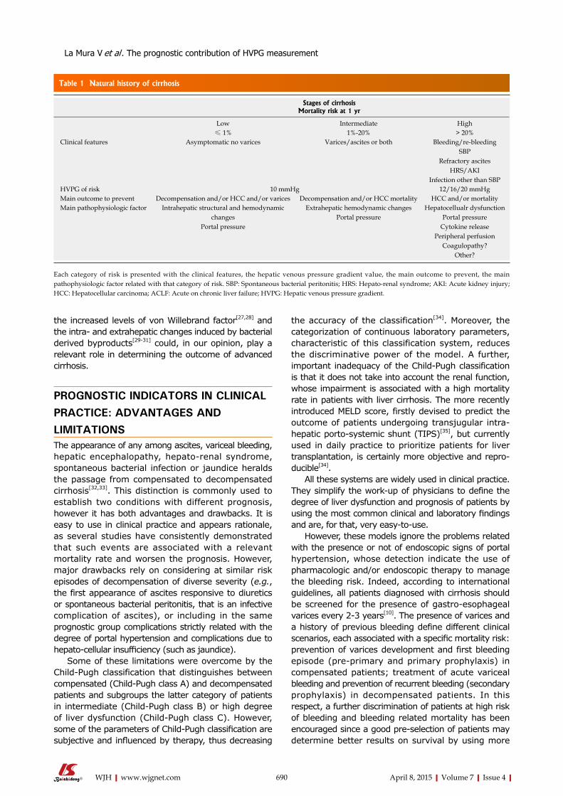
the increased levels of von Willebrand factor[27,28] and the intra- and extrahepatic changes induced by bacterial derived byproducts[29-31] could, in our opinion, play a relevant role in determining the outcome of advanced cirrhosis.
PROGNOSTIC INDICATORS IN CLINICAL PRACTICE: ADVANTAGES AND LIMITATIONSThe appearance of any among ascites, variceal bleeding, hepatic encephalopathy, hepato-renal syndrome, spontaneous bacterial infection or jaundice heralds the passage from compensated to decompensated cirrhosis[32,33]. This distinction is commonly used to establish two conditions with different prognosis, however it has both advantages and drawbacks. It is easy to use in clinical practice and appears rationale, as several studies have consistently demonstrated that such events are associated with a relevant mortality rate and worsen the prognosis. However, major drawbacks rely on considering at similar risk episodes of decompensation of diverse severity (e.g., the first appearance of ascites responsive to diuretics or spontaneous bacterial peritonitis, that is an infective complication of ascites), or including in the same prognostic group complications strictly related with the degree of portal hypertension and complications due to hepato-cellular insufficiency (such as jaundice).
Some of these limitations were overcome by the Child-Pugh classification that distinguishes between compensated (Child-Pugh class A) and decompensated patients and subgroups the latter category of patients in intermediate (Child-Pugh class B) or high degree of liver dysfunction (Child-Pugh class C). However, some of the parameters of Child-Pugh classification are subjective and influenced by therapy, thus decreasing
the accuracy of the classification[34]. Moreover, the categorization of continuous laboratory parameters, characteristic of this classification system, reduces the discriminative power of the model. A further, important inadequacy of the Child-Pugh classification is that it does not take into account the renal function, whose impairment is associated with a high mortality rate in patients with liver cirrhosis. The more recently introduced MELD score, firstly devised to predict the outcome of patients undergoing transjugular intra-hepatic porto-systemic shunt (TIPS)[35], but currently used in daily practice to prioritize patients for liver transplantation, is certainly more objective and repro-ducible[34].
All these systems are widely used in clinical practice. They simplify the work-up of physicians to define the degree of liver dysfunction and prognosis of patients by using the most common clinical and laboratory findings and are, for that, very easy-to-use.
However, these models ignore the problems related with the presence or not of endoscopic signs of portal hypertension, whose detection indicate the use of pharmacologic and/or endoscopic therapy to manage the bleeding risk. Indeed, according to international guidelines, all patients diagnosed with cirrhosis should be screened for the presence of gastro-esophageal varices every 2-3 years[10]. The presence of varices and a history of previous bleeding define different clinical scenarios, each associated with a specific mortality risk: prevention of varices development and first bleeding episode (pre-primary and primary prophylaxis) in compensated patients; treatment of acute variceal bleeding and prevention of recurrent bleeding (secondary prophylaxis) in decompensated patients. In this respect, a further discrimination of patients at high risk of bleeding and bleeding related mortality has been encouraged since a good pre-selection of patients may determine better results on survival by using more
690 April 8, 2015|Volume 7|Issue 4|WJH|www.wjgnet.com
Stages of cirrhosis Mortality risk at 1 yr
Low Intermediate High≤ 1% 1%-20% > 20%
Clinical features Asymptomatic no varices Varices/ascites or both Bleeding/re-bleeding SBP
Refractory ascitesHRS/AKI
Infection other than SBPHVPG of risk 10 mmHg 12/16/20 mmHgMain outcome to prevent Decompensation and/or HCC and/or varices Decompensation and/or HCC mortality HCC and/or mortalityMain pathophysiologic factor Intrahepatic structural and hemodynamic
changesPortal pressure
Extrahepatic hemodynamic changesPortal pressure
Hepatocellualr dysfunctionPortal pressure
Cytokine releasePeripheral perfusion
Coagulopathy?Other?
Table 1 Natural history of cirrhosis
Each category of risk is presented with the clinical features, the hepatic venous pressure gradient value, the main outcome to prevent, the main pathophysiologic factor related with that category of risk. SBP: Spontaneous bacterial peritonitis; HRS: Hepato-renal syndrome; AKI: Acute kidney injury; HCC: Hepatocellular carcinoma; ACLF: Acute on chronic liver failure; HVPG: Hepatic venous pressure gradient.
La Mura V et al . The prognostic contribution of HVPG measurement
691 April 8, 2015|Volume 7|Issue 4|WJH|www.wjgnet.com
Low risk cirrhosisWith the term of low risk cirrhosis we defined the condition of asymptomatic cirrhosis without neither gastroesophageal varices nor other gastro-intestinal signs of portal hypertension with a mortality rate at one year less than 1%. In this context the main clinical outcome to be predicted is the appearance of the first decompensation, that, often, is the appearance of ascites, but another clinical outcome to predict is the development of varices at risk of bleeding. At this stage, HVPG significantly reflects structural and functional changes occurring in the liver[6,11]. However, above the threshold of 10 mmHg, the high correlation of HVPG with intrahepatic changes is blunted[50] and the degree of portal pressure is determined by the concomitant activation of extrahepatic hemodynamic and humoral changes that foster a state of hyperdynamic circula-tion[13]. A large longitudinal study including patients with compensated cirrhosis without varices demonstrated that patients with a HVPG < 10 mmHg carry a neg-ligible risk of developing varices and a very low risk of decompensation where a HVPG ≥ 10 mmHg is associated with 28% rate of varices development and 20% of first decompensation at two years[51,52]. This demonstrates that patients with asymptomatic cirrhosis, classically defined as compensated and/or Child-Pugh class A, are a heterogeneous population at different risk of becoming symptomatic. However, opportunely designed studies are warranted to translate this prognostic information into the decision making process. Interesting, from the same cohort of patients, it was also demonstrated that HVPG ≥ 10 mmHg is associated with a 6-fold increase of HCC risk[53]. In this context, 10 mmHg was, again, the threshold able to identify the candidates to liver resection at risk of decompensation. Such valuable prognostic information allows considering HVPG as the strongest surrogate marker of clinical outcome to guide the decision making in this setting.
Intermediate risk cirrhosisThe diagnosis of varices, with or without ascites, or ascites alone is the hallmark of CSPH. The need at this step is to predict the risk of portal hypertension related complications (e.g., bleeding, SBP, refractory ascites, etc.), and the hemodynamic response to non-selective beta-blockers (NSBBs), the mainstay of pharmacologic treatment to reduce portal pressure. We know that esophageal varices, whatever the diameter, are at risk of bleeding for HVPG ≥ 12 mmHg[54,55]. Moreover a second hemodynamic study that demonstrates the achievement of a HVPG below this value or a reduction ≥ 20% of the basal value is highly specific of a good clinical response to NSBBs treatment[3]. At this stage, a reduction of HVPG also correlates with a reduced risk of SBP or bacteremia[42]. Importantly, the prognostic contribution provided by HVPG appears to be independent by the degree of liver insufficiency and the
aggressive strategies of treatment[36].In recent years, the availability of non-invasive tools
to diagnose cirrhosis, together with the development of potent antiviral drugs able to reduce fibrosis, has significantly increased the proportion of patients with chronic liver disease diagnosed in the asymptomatic stage of cirrhosis with the ultimate result that the surveillance schedule for the presence of varices should be revised in order to reduce the number of unnecessary endoscopies performed in daily clinical practice[37]. Another clinical outcome that sensibly influences the outcome of patients with cirrhosis is the possibility of predicting the response to pharmacologic therapy to prevent variceal bleeding/rebleeding. Unfortunately, the low intra- and inter-observer reproducibility of the endoscopic features does not allow recommending the use of this technique systematically[38].
In summary, the natural history of cirrhosis is strongly dependent on portal hypertension related complications. The use of several clinical and/or instru-mental signs, in particular, upper gastro-intestinal endoscopy, characterizes the work-up of physicians to predict and manage the risk of portal hypertension related complications in several stages of risk mainly based on varices, ascites and bleeding[39]. However, recently, a pathophysiological classification of cirrhosis has been evoked to promote a system that better reflects the dynamic state of cirrhosis and provides a useful tool to predict outcomes and individualize therapy[7].
THE ADDITIONAL PROGNOSTIC INFORMATION PROVIDED BY HVPG Given the importance of portal hypertension in the natural history of patients with cirrhosis, it would be expected that HVPG measurement hold prognostic information in this setting. Theoretically, the use of HVPG as a prognostic tool has several advantages: (1) it is an objective and continuous variable; (2) it changes in presence of specific therapeutic interventions and/or an improvement of liver function[40-42]; and (3) it has been widely studied in cross sectional-, longitudinal-studies, randomized controlled trials and metanalysis to consider it as one of the most reliable surrogate markers of clinical outcome in Hepatology[43]. However, there are several disadvantages limiting its use[3,44,45]: (1) the costs associated with the methodology; (2) theneed of trained physicians to get a reliable measurement; and (3) the relatively invasiveness of the methodology. Since its introduction, evidence has been accumulated that HVPG correlates with the degree of liver dys-function[46-49]. Due for that, it has been recommended to test the prognostic value of the HVPG in the context of multivariable analysis in order to quantify the additional prognostic information provided on top of that achieving by common clinical and instrumental parameters.
La Mura V et al . The prognostic contribution of HVPG measurement
692 April 8, 2015|Volume 7|Issue 4|WJH|www.wjgnet.com
bleeding risk assessed by endoscopy markers of risk such as variceal diameter and the presence of red wale markers of risk (North Italian Endoscopic Club)[56], thus suggesting that the degree of portal hypertension is the main determinant of prognosis, and its reduction should be the principal aim at this stage of the disease[41,42]. One limitation of monitoring response to NSBBs treatment by HVPG is that two measurements are needed. This doubles the costs, but also the invasive procedures for the patients. Moreover, not all patients classified as non-responders to NSBBs will bleed in the follow-up, while some patients can bleed before the second HVPG measurement[57]. To overcome these limitations, a single HVPG measurement by testing the acute hemodynamic response to intra-venous propranolol has been proposed and proved to correlate with the chronic response to NSBBs. Moreover, such single measurement seemed to be able to predict the prognosis of candidates to NSBBs treatment more accurately than the HVPG response to NSBBs requiring two hemodynamic sessions[58,59]. Further clinical studies to confirm the role of acute HVPG response to propranolol in the decision making are waited. High risk cirrhosisThis stage includes all patients experiencing a first or further episode of decompensation at high mortality risk such as VB, refractory ascites, SBP, HRS, hyponatremia and HE. Recently, a metanalysis[60] showed that any kind of bacterial infection is dramatically associated with a 67% mortality rate at one year, even though patients overcome the infection. The role of a prognostic marker, at this stage, is to help in identifying, among patients with decompensation, those at the highest mortality risk in order to implement more aggressive therapeutic strategies.
In several studies HVPG disclosed an independent predictive value on mortality in decompensated patients[39]. Ripoll et al[61] in a series of 393 patients, mostly with previous decompensation, showed that the HVPG, independently by MELD, had an overall effect of 3% increase of mortality for each 1 mmHg of HVPG increase. Unfortunately, the addition of the HVPG to the MELD score augmented very little the discriminative power of the MELD score alone to rank the mortality risk of decompensated patients. Then, although portal hypertension can further increase at this advanced stage of cirrhosis, hepato-cellular dysfunction, and, probably, other factors related to acute on chronic liver failure, like inflammatory cytokine release or the impairment of cardiac function and peripheral organ perfusion, probably play a major prognostic role in this clinical setting.
Another important aspect is that the relationship between HVPG and the risk of death could be non-linear. This can be evidenced by considering dichotomization of the HVPG value. In this respect, several studies found an increased mortality risk in patients with HVPG beyond the threshold of 16 mmHg[48,62-64]. The biological rationale behind this finding is intriguing but still unclear. The
threshold of 16 mmHg would allow an early detection of patients at high mortality risk, and is a promising threshold to be tested in the clinical decision process.
Acute variceal bleeding is the complication of advanced cirrhosis with the highest mortality reduction achieved in the last decades, from 40%-50% to 10%-20%[65]. In acute variceal bleeding, an HVPG ≥ 20 mmHg identifies patients at high risk of early rebleeding and bleeding related mortality[66]. Recently, two randomized trials have shown that the reduction of portal pressure achieved by an early TIPS (within 72 h from bleeding) significantly decreases the mortality rate of “high risk patients”[67,68] (those with a HVPG ≥ 20 mmHg or patient in Child-Pugh class C or in class B who have persistent bleeding at endoscopy). The positive effect on survival achieved by TIPS in patients at high risk of mortality during acute variceal bleeding confirms the importance of the degree of portal pressure in this clinical setting.
Beyond HVPG measurement: Liver/spleen stiffness Several clinical studies have tried to demonstrate that portal pressure can be indirectly estimated by also non-invasive techniques, in particular, measurement of liver stiffness and, more recently, splenic stiffness by transient elastography[69,70]. Moreover, several attempts have been made in order to test the ability of these parameters (alone or in combination with other clinical variables) to predict the presence of varices, the risk of decompensation, the presence of CSPH in order to facilitate the sub-classification of cirrhosis by a non-invasive methodology[71,72].
Liver stiffness is mainly determined by fibrosis that plays a determinant role in the pathogenesis of portal hypertension in the earliest stages of cirrhosis. Due for that, it shows a good correlation with HVPG only before extrahepatic hemodynamic changes determine the increase of portal pressure over the threshold of CSPH. The accuracy of this parameter to detect CSPH is high, good for the detection of varices, however the cut-offs proposed to predict these clinical conditions differs from one study to another[69]. In our opinion this sensibly reduces the possibility of introducing liver stiffness measurement in the clinical decision process neither allows to detecting the response to pharmacological reduction of portal pressure. Theoretically, splenic stiffness would be a better candidate for the non-invasive assessment of portal pressure since spleen congestion and fibrosis are both effects related with portal pressure increase[73]. At now the most interesting results have been achieved in hepatitis c virus positive cirrhotic patients. In this clinical subset splenic stiffness has been showed promising results for the non-invasive assessment of portal hypertension, the detection of varices, the prediction of the first decompensation[74,75].
CONCLUSIONThe introduction of potent antiviral drugs and the definition of new algorithms of treatments to prevent
La Mura V et al . The prognostic contribution of HVPG measurement
693 April 8, 2015|Volume 7|Issue 4|WJH|www.wjgnet.com
and manage complications due to portal hypertension has revealed that cirrhosis is a multi-stage disease featured by different survival risks. The HVPG mea-surement is, at present, the most reliable tool for clinicians to predict the clinical outcome and dictate the decision making in several complications. HVPG is particularly promising in low risk cirrhosis for any complication related to portal hypertension and for HCC screening and management. HVPG is a useful tool to predict the response to NSBBs in primary and secondary prevention of variceal bleeding, respectively, in intermediate and high risk cirrhosis. The occurrence of critical complications of portal hypertension and the increasing hepato-cellular insufficiency in this stage of high mortality risk apparently reduce the prognostic contribution of HVPG measurement. Probably, at this clinical stage, other clinical variables may substitute HVPG with a good level of accuracy.
Liver/spleen stiffness are new non-invasive emerging tools to, potentially, estimate the degree of portal hypertension. They might resemble in several aspects the HVPG measurement with the additional advantage of a higher tolerability and reduced costs. However, further studies are needed before a non-invasive “portal sphygmomanometer” can substitute the prognostic information provided by HVPG in cirrhosis.
REFERENCES1 Tsochatzis EA, Bosch J, Burroughs AK. Liver cirrhosis. Lancet
2014; 383: 1749-1761 [PMID: 24480518 DOI: 10.1016/S0140-6736(14)60121-5]
2 Bosch J, Abraldes JG, Fernández M, García-Pagán JC. Hepatic endothelial dysfunction and abnormal angiogenesis: new targets in the treatment of portal hypertension. J Hepatol 2010; 53: 558-567 [PMID: 20561700 DOI: 10.1016/j.jhep.2010.03.021]
3 Bosch J, Abraldes JG, Berzigotti A, García-Pagan JC. The clinical use of HVPG measurements in chronic liver disease. Nat Rev Gastroenterol Hepatol 2009; 6: 573-582 [PMID: 19724251 DOI: 10.1038/nrgastro.2009.149]
4 Boyer TD, Triger DR, Horisawa M, Redeker AG, Reynolds TB. Direct transhepatic measurement of portal vein pressure using a thin needle. Comparison with wedged hepatic vein pressure. Gastroenterology 1977; 72: 584-589 [PMID: 838210 DOI: 10.1016/S0016-5085(77)80136-4]
5 Perelló A, Escorsell A, Bru C, Gilabert R, Moitinho E, García-Pagán JC, Bosch J. Wedged hepatic venous pressure adequately reflects portal pressure in hepatitis C virus-related cirrhosis. Hepatology 1999; 30: 1393-1397 [PMID: 10573517 DOI: 10.1002/hep.510300628]
6 Nagula S, Jain D, Groszmann RJ, Garcia-Tsao G. Histological-hemodynamic correlation in cirrhosis-a histological classification of the severity of cirrhosis. J Hepatol 2006; 44: 111-117 [PMID: 16274836 DOI: 10.1016/j.jhep.2005.07.036]
7 Garcia-Tsao G, Friedman S, Iredale J, Pinzani M. Now there are many (stages) where before there was one: In search of a pathophysiological classification of cirrhosis. Hepatology 2010; 51: 1445-1449 [PMID: 20077563 DOI: 10.1002/hep.23478]
8 European Association For The Study Of The Liver; European Organisation For Research And Treatment Of Cancer. EASL-EORTC clinical practice guidelines: management of hepatocellular carcinoma. J Hepatol 2012; 56: 908-943 [PMID: 22424438 DOI: 10.1016/j.jhep.2011.12.001]
9 Albilllos A, Garcia-Tsao G. Classification of cirrhosis: the clinical use of HVPG measurements. Dis Markers 2011; 31: 121-128
[PMID: 22045397 DOI: 10.3233/DMA-2011-0834]10 de Franchis R. Revising consensus in portal hypertension: report
of the Baveno V consensus workshop on methodology of diagnosis and therapy in portal hypertension. J Hepatol 2010; 53: 762-768 [PMID: 20638742 DOI: 10.1016/j.jhep.2010.06.004]
11 Sethasine S, Jain D, Groszmann RJ, Garcia-Tsao G. Quantitative histological-hemodynamic correlations in cirrhosis. Hepatology 2012; 55: 1146-1153 [PMID: 22109744 DOI: 10.1002/hep.24805]
12 Iwakiri Y, Groszmann RJ. Vascular endothelial dysfunction in cirrhosis. J Hepatol 2007; 46: 927-934 [PMID: 17391799 DOI: 10.1016/j.jhep.2007.02.006]
13 Iwakiri Y, Groszmann RJ. The hyperdynamic circulation of chronic liver diseases: from the patient to the molecule. Hepatology 2006; 43: S121-S131 [PMID: 16447289 DOI: 10.1002/hep.20993]
14 Salerno F, Guevara M, Bernardi M, Moreau R, Wong F, Angeli P, Garcia-Tsao G, Lee SS. Refractory ascites: pathogenesis, definition and therapy of a severe complication in patients with cirrhosis. Liver Int 2010; 30: 937-947 [PMID: 20492521 DOI: 10.1111/j.1478-3231.2010.02272.x]
15 Schrier RW, Arroyo V, Bernardi M, Epstein M, Henriksen JH, Rodés J. Peripheral arterial vasodilation hypothesis: a proposal for the initiation of renal sodium and water retention in cirrhosis. Hepatology 1988; 8: 1151-1157 [PMID: 2971015 DOI: 10.1002/hep.1840080532]
16 Porcel A, Díaz F, Rendón P, Macías M, Martín-Herrera L, Girón-González JA. Dilutional hyponatremia in patients with cirrhosis and ascites. Arch Intern Med 2002; 162: 323-328 [PMID: 11822925 DOI: 10.1001/archinte.162.3.323]
17 Ruiz-del-Arbol L, Urman J, Fernández J, González M, Navasa M, Monescillo A, Albillos A, Jiménez W, Arroyo V. Systemic, renal, and hepatic hemodynamic derangement in cirrhotic patients with spontaneous bacterial peritonitis. Hepatology 2003; 38: 1210-1218 [PMID: 14578859 DOI: 10.1053/jhep.2003.50447]
18 Ginès P, Arroyo V. Paracentesis in the management of cirrhotic ascites. J Hepatol 1993; 17 Suppl 2: S14-S18 [PMID: 8491965 DOI: 10.1016/S0168-8278(05)80449-0]
19 Ginès A, Fernández-Esparrach G, Monescillo A, Vila C, Domènech E, Abecasis R, Angeli P, Ruiz-Del-Arbol L, Planas R, Solà R, Ginès P, Terg R, Inglada L, Vaqué P, Salerno F, Vargas V, Clemente G, Quer JC, Jiménez W, Arroyo V, Rodés J. Randomized trial comparing albumin, dextran 70, and polygeline in cirrhotic patients with ascites treated by paracentesis. Gastroenterology 1996; 111: 1002-1010 [PMID: 8831595 DOI: 10.1016/S0016-5085(96)70068-9]
20 Sort P, Navasa M, Arroyo V, Aldeguer X, Planas R, Ruiz-del-Arbol L, Castells L, Vargas V, Soriano G, Guevara M, Ginès P, Rodés J. Effect of intravenous albumin on renal impairment and mortality in patients with cirrhosis and spontaneous bacterial peritonitis. N Engl J Med 1999; 341: 403-409 [PMID: 10432325 DOI: 10.1056/NEJM199908053410603]
21 Fernández J, Monteagudo J, Bargallo X, Jiménez W, Bosch J, Arroyo V, Navasa M. A randomized unblinded pilot study comparing albumin versus hydroxyethyl starch in spontaneous bacterial peritonitis. Hepatology 2005; 42: 627-634 [PMID: 16108036 DOI: 10.1002/hep.20829]
22 Guevara M, Terra C, Nazar A, Solà E, Fernández J, Pavesi M, Arroyo V, Ginès P. Albumin for bacterial infections other than spontaneous bacterial peritonitis in cirrhosis. A randomized, controlled study. J Hepatol 2012; 57: 759-765 [PMID: 22732511 DOI: 10.1016/j.jhep.2012.06.013]
23 Moreau R, Jalan R, Gines P, Pavesi M, Angeli P, Cordoba J, Durand F, Gustot T, Saliba F, Domenicali M, Gerbes A, Wendon J, Alessandria C, Laleman W, Zeuzem S, Trebicka J, Bernardi M, Arroyo V. Acute-on-chronic liver failure is a distinct syndrome that develops in patients with acute decompensation of cirrhosis. Gastroenterology 2013; 144: 1426-1437, 1437.e1-9 [PMID: 23474284 DOI: 10.1053/j.gastro.2013.02.042]
24 Abraldes JG, Albillos A, Bañares R, Turnes J, González R, García-Pagán JC, Bosch J. Simvastatin lowers portal pressure in patients with cirrhosis and portal hypertension: a randomized
La Mura V et al . The prognostic contribution of HVPG measurement
694 April 8, 2015|Volume 7|Issue 4|WJH|www.wjgnet.com
controlled trial. Gastroenterology 2009; 136: 1651-1658 [PMID: 19208350 DOI: 10.1053/j.gastro.2009.01.043]
25 Cazzaniga M, Dionigi E, Gobbo G, Fioretti A, Monti V, Salerno F. The systemic inflammatory response syndrome in cirrhotic patients: relationship with their in-hospital outcome. J Hepatol 2009; 51: 475-482 [PMID: 19560225 DOI: 10.1016/j.jhep.2009.04.017]
26 Tripodi A, Mannucci PM. The coagulopathy of chronic liver disease. N Engl J Med 2011; 365: 147-156 [PMID: 21751907 DOI: 10.1056/NEJMra1011170]
27 La Mura V, Reverter JC, Flores-Arroyo A, Raffa S, Reverter E, Seijo S, Abraldes JG, Bosch J, García-Pagán JC. Von Willebrand factor levels predict clinical outcome in patients with cirrhosis and portal hypertension. Gut 2011; 60: 1133-1138 [PMID: 21427197 DOI: 10.1136/gut.2010.235689]
28 Ferlitsch M, Reiberger T, Hoke M, Salzl P, Schwengerer B, Ulbrich G, Payer BA, Trauner M, Peck-Radosavljevic M, Ferlitsch A. von Willebrand factor as new noninvasive predictor of portal hypertension, decompensation and mortality in patients with liver cirrhosis. Hepatology 2012; 56: 1439-1447 [PMID: 22532296 DOI: 10.1002/hep.25806]
29 Tandon P, Garcia-Tsao G. Bacterial infections, sepsis, and multiorgan failure in cirrhosis. Semin Liver Dis 2008; 28: 26-42 [PMID: 18293275 DOI: 10.1055/s-2008-1040319]
30 Bellot P, García-Pagán JC, Francés R, Abraldes JG, Navasa M, Pérez-Mateo M, Such J, Bosch J. Bacterial DNA translocation is associated with systemic circulatory abnormalities and intrahepatic endothelial dysfunction in patients with cirrhosis. Hepatology 2010; 52: 2044-2052 [PMID: 20979050 DOI: 10.1002/hep.23918]
31 La Mura V, Pasarín M, Meireles CZ, Miquel R, Rodríguez-Vilarrupla A, Hide D, Gracia-Sancho J, García-Pagán JC, Bosch J, Abraldes JG. Effects of simvastatin administration on rodents with lipopolysaccharide-induced liver microvascular dysfunction. Hepatology 2013; 57: 1172-1181 [PMID: 23184571 DOI: 10.1002/hep.26127]
32 Saunders JB, Walters JR, Davies AP, Paton A. A 20-year prospective study of cirrhosis. Br Med J (Clin Res Ed) 1981; 282: 263-266 [PMID: 6779978 DOI: 10.1136/bmj.282.6260.263]
33 Ginés P, Quintero E, Arroyo V, Terés J, Bruguera M, Rimola A, Caballería J, Rodés J, Rozman C. Compensated cirrhosis: natural history and prognostic factors. Hepatology 1987; 7: 122-128 [PMID: 3804191 DOI: 10.1002/hep.1840070124]
34 Pagliaro L. MELD: the end of Child-Pugh classification? J Hepatol 2002; 36: 141-142 [PMID: 11804679 DOI: 10.1016/S0168-8278(01)00302-6]
35 Malinchoc M, Kamath PS, Gordon FD, Peine CJ, Rank J, ter Borg PC. A model to predict poor survival in patients undergoing transjugular intrahepatic portosystemic shunts. Hepatology 2000; 31: 864-871 [PMID: 10733541 DOI: 10.1053/he.2000.5852]
36 Garcia-Tsao G, Bosch J. Management of varices and variceal hemorrhage in cirrhosis. N Engl J Med 2010; 362: 823-832 [PMID: 20200386 DOI: 10.1056/NEJMra0901512]
37 Rudler M, Benosman H, Lebray P, Nghiem D, Ngo Y, Munteanu M. Screening for esophageal varices in patients newly diagnosed with cirrhosis in 2011: 84% of upper gastrointestinal endoscopies are futile. Hepatology 2011; 54 (Suppl): 935A [DOI: 10.1002/hep.24666]
38 Winkfield B, Aubé C, Burtin P, Calès P. Inter-observer and intra-observer variability in hepatology. Eur J Gastroenterol Hepatol 2003; 15: 959-966 [PMID: 12923367 DOI: 10.1097/00042737-200309000-00004]
39 D’Amico G, Garcia-Tsao G, Pagliaro L. Natural history and prognostic indicators of survival in cirrhosis: a systematic review of 118 studies. J Hepatol 2006; 44: 217-231 [PMID: 16298014 DOI: 10.1016/j.jhep.2005.10.013]
40 Vorobioff J, Groszmann RJ, Picabea E, Gamen M, Villavicencio R, Bordato J, Morel I, Audano M, Tanno H, Lerner E, Passamonti M. Prognostic value of hepatic venous pressure gradient measurements in alcoholic cirrhosis: a 10-year prospective study. Gastroenterology 1996; 111: 701-709 [PMID: 8780575 DOI: 10.1053/gast.1996.v111.pm8780575]
41 Abraldes JG, Tarantino I, Turnes J, Garcia-Pagan JC, Rodés J,
Bosch J. Hemodynamic response to pharmacological treatment of portal hypertension and long-term prognosis of cirrhosis. Hepatology 2003; 37: 902-908 [PMID: 12668985 DOI: 10.1053/jhep.2003.50133]
42 Turnes J, Garcia-Pagan JC, Abraldes JG, Hernandez-Guerra M, Dell’Era A, Bosch J. Pharmacological reduction of portal pressure and long-term risk of first variceal bleeding in patients with cirrhosis. Am J Gastroenterol 2006; 101: 506-512 [PMID: 16542287]
43 Gluud C, Brok J, Gong Y, Koretz RL. Hepatology may have problems with putative surrogate outcome measures. J Hepatol 2007; 46: 734-742 [PMID: 17316871]
44 Groszmann RJ, Wongcharatrawee S. The hepatic venous pressure gradient: anything worth doing should be done right. Hepatology 2004; 39: 280-282 [PMID: 14767976 DOI: 10.1002/hep.20062]
45 La Mura V, Abraldes JG, Berzigotti A, Erice E, Flores-Arroyo A, García-Pagán JC, Bosch J. Right atrial pressure is not adequate to calculate portal pressure gradient in cirrhosis: a clinical-hemodynamic correlation study. Hepatology 2010; 51: 2108-2116 [PMID: 20512998 DOI: 10.1002/hep.23612]
46 Braillon A, Cales P, Valla D, Gaudy D, Geoffroy P, Lebrec D. Influence of the degree of liver failure on systemic and splanchnic haemodynamics and on response to propranolol in patients with cirrhosis. Gut 1986; 27: 1204-1209 [PMID: 3781335 DOI: 10.1136/gut.27.10.1204]
47 Gluud C, Henriksen JH, Nielsen G. Prognostic indicators in alcoholic cirrhotic men. Hepatology 1988; 8: 222-227 [PMID: 3258578 DOI: 10.1002/hep.1840080205]
48 Stanley AJ, Robinson I, Forrest EH, Jones AL, Hayes PC. Haemodynamic parameters predicting variceal haemorrhage and survival in alcoholic cirrhosis. QJM 1998; 91: 19-25 [PMID: 9519209]
49 Le Moine O, Hadengue A, Moreau R, Sogni P, Soupison T, Yang S, Hartleb M, Lebrec D. Relationship between portal pressure, esophageal varices, and variceal bleeding on the basis of the stage and cause of cirrhosis. Scand J Gastroenterol 1997; 32: 731-735 [PMID: 9246716 DOI: 10.3109/00365529708996526]
50 Vizzutti F, Arena U, Romanelli RG, Rega L, Foschi M, Colagrande S, Petrarca A, Moscarella S, Belli G, Zignego AL, Marra F, Laffi G, Pinzani M. Liver stiffness measurement predicts severe portal hypertension in patients with HCV-related cirrhosis. Hepatology 2007; 45: 1290-1297 [PMID: 17464971 DOI: 10.1002/hep.21665]
51 Groszmann RJ, Garcia-Tsao G, Bosch J, Grace ND, Burroughs AK, Planas R, Escorsell A, Garcia-Pagan JC, Patch D, Matloff DS, Gao H, Makuch R. Beta-blockers to prevent gastroesophageal varices in patients with cirrhosis. N Engl J Med 2005; 353: 2254-2261 [PMID: 16306522 DOI: 10.1056/NEJMoa044456]
52 Ripoll C, Groszmann R, Garcia-Tsao G, Grace N, Burroughs A, Planas R, Escorsell A, Garcia-Pagan JC, Makuch R, Patch D, Matloff DS, Bosch J. Hepatic venous pressure gradient predicts clinical decompensation in patients with compensated cirrhosis. Gastroenterology 2007; 133: 481-488 [PMID: 17681169]
53 Ripoll C, Groszmann RJ, Garcia-Tsao G, Bosch J, Grace N, Burroughs A, Planas R, Escorsell A, Garcia-Pagan JC, Makuch R, Patch D, Matloff DS. Hepatic venous pressure gradient predicts development of hepatocellular carcinoma independently of severity of cirrhosis. J Hepatol 2009; 50: 923-928 [PMID: 19303163 DOI: 10.1016/j.jhep.2009.01.014]
54 Garcia-Tsao G, Groszmann RJ, Fisher RL, Conn HO, Atterbury CE, Glickman M. Portal pressure, presence of gastroesophageal varices and variceal bleeding. Hepatology 1985; 5: 419-424 [PMID: 3873388]
55 Casado M, Bosch J, García-Pagán JC, Bru C, Bañares R, Bandi JC, Escorsell A, Rodríguez-Láiz JM, Gilabert R, Feu F, Schorlemer C, Echenagusia A, Rodés J. Clinical events after transjugular intrahepatic portosystemic shunt: correlation with hemodynamic findings. Gastroenterology 1998; 114: 1296-1303 [PMID: 9609767 DOI: 10.1016/S0016-5085(98)70436-6]
56 North Italian Endoscopic Club for the Study and Treatment of Esophageal Varices. Prediction of the first variceal hemorrhage in patients with cirrhosis of the liver and esophageal varices. A
La Mura V et al . The prognostic contribution of HVPG measurement
695 April 8, 2015|Volume 7|Issue 4|WJH|www.wjgnet.com
prospective multicenter study. N Engl J Med 1988; 319: 983-989 [PMID: 3262200 DOI: 10.1056/NEJM198810133191505]
57 Thalheimer U, Bosch J, Burroughs AK. How to prevent varices from bleeding: shades of grey--the case for nonselective beta blockers. Gastroenterology 2007; 133: 2029-2036 [PMID: 18054573]
58 Villanueva C, Aracil C, Colomo A, Hernández-Gea V, López-Balaguer JM, Alvarez-Urturi C, Torras X, Balanzó J, Guarner C. Acute hemodynamic response to beta-blockers and prediction of long-term outcome in primary prophylaxis of variceal bleeding. Gastroenterology 2009; 137: 119-128 [PMID: 19344721 DOI: 10.1053/j.gastro.2009.03.048]
59 La Mura V, Abraldes JG, Raffa S, Retto O, Berzigotti A, García-Pagán JC, Bosch J. Prognostic value of acute hemodynamic response to i.v. propranolol in patients with cirrhosis and portal hypertension. J Hepatol 2009; 51: 279-287 [PMID: 19501930 DOI: 10.1016/j.jhep.2009.04.015]
60 Arvaniti V, D’Amico G, Fede G, Manousou P, Tsochatzis E, Pleguezuelo M, Burroughs AK. Infections in patients with cirrhosis increase mortality four-fold and should be used in determining prognosis. Gastroenterology 2010; 139: 1246-1256, 1256.e1-5 [PMID: 20558165 DOI: 10.1053/j.gastro.2010.06.019]
61 Ripoll C, Bañares R, Rincón D, Catalina MV, Lo Iacono O, Salcedo M, Clemente G, Núñez O, Matilla A, Molinero LM. Influence of hepatic venous pressure gradient on the prediction of survival of patients with cirrhosis in the MELD Era. Hepatology 2005; 42: 793-801 [PMID: 16175621 DOI: 10.1002/hep.20871]
62 Merkel C, Bolognesi M, Sacerdoti D, Bombonato G, Bellini B, Bighin R, Gatta A. The hemodynamic response to medical treatment of portal hypertension as a predictor of clinical effectiveness in the primary prophylaxis of variceal bleeding in cirrhosis. Hepatology 2000; 32: 930-934 [PMID: 11050041]
63 Merkel C, Bolognesi M, Bellon S, Zuin R, Noventa F, Finucci G, Sacerdoti D, Angeli P, Gatta A. Prognostic usefulness of hepatic vein catheterization in patients with cirrhosis and esophageal varices. Gastroenterology 1992; 102: 973-979 [PMID: 1537533]
64 Berzigotti A, Rossi V, Tiani C, Pierpaoli L, Zappoli P, Riili A, Serra C, Andreone P, Morelli MC, Golfieri R, Rossi C, Magalotti D, Zoli M. Prognostic value of a single HVPG measurement and Doppler-ultrasound evaluation in patients with cirrhosis and portal hypertension. J Gastroenterol 2011; 46: 687-695 [PMID: 21213113 DOI: 10.1007/s00535-010-0360-z]
65 Chalasani N, Kahi C, Francois F, Pinto A, Marathe A, Bini EJ, Pandya P, Sitaraman S, Shen J. Improved patient survival after acute variceal bleeding: a multicenter, cohort study. Am J Gastroenterol 2003; 98: 653-659 [PMID: 12650802 DOI: 10.1016/S0002-9270(02)06016-1]
66 Moitinho E, Escorsell A, Bandi JC, Salmerón JM, García-Pagán JC, Rodés J, Bosch J. Prognostic value of early measurements of
portal pressure in acute variceal bleeding. Gastroenterology 1999; 117: 626-631 [PMID: 10464138 DOI: 10.1016/S0016-5085(99)70455-5]
67 Monescillo A, Martínez-Lagares F, Ruiz-del-Arbol L, Sierra A, Guevara C, Jiménez E, Marrero JM, Buceta E, Sánchez J, Castellot A, Peñate M, Cruz A, Peña E. Influence of portal hypertension and its early decompression by TIPS placement on the outcome of variceal bleeding. Hepatology 2004; 40: 793-801 [PMID: 15382120 DOI: 10.1002/hep.20386]
68 García-Pagán JC, Caca K, Bureau C, Laleman W, Appenrodt B, Luca A, Abraldes JG, Nevens F, Vinel JP, Mössner J, Bosch J. Early use of TIPS in patients with cirrhosis and variceal bleeding. N Engl J Med 2010; 362: 2370-2379 [PMID: 20573925 DOI: 10.1056/NEJMoa0910102]
69 Castera L, Pinzani M, Bosch J. Non invasive evaluation of portal hypertension using transient elastography. J Hepatol 2012; 56: 696-703 [PMID: 21767510 DOI: 10.1016/j.jhep.2011.07.005]
70 Singh S, Eaton JE, Murad MH, Tanaka H, Iijima H, Talwalkar JA. Accuracy of spleen stiffness measurement in detection of esophageal varices in patients with chronic liver disease: systematic review and meta-analysis. Clin Gastroenterol Hepatol 2014; 12: 935-45.e4 [PMID: 24055985 DOI: 10.1016/j.cgh.2013.09.013]
71 Berzigotti A, Seijo S, Arena U, Abraldes JG, Vizzutti F, García-Pagán JC, Pinzani M, Bosch J. Elastography, spleen size, and platelet count identify portal hypertension in patients with compensated cirrhosis. Gastroenterology 2013; 144: 102-111.e1 [PMID: 23058320 DOI: 10.1053/j.gastro.2012.10.001]
72 Augustin S, Millán L, González A, Martell M, Gelabert A, Segarra A, Serres X, Esteban R, Genescà J. Detection of early portal hypertension with routine data and liver stiffness in patients with asymptomatic liver disease: a prospective study. J Hepatol 2014; 60: 561-569 [PMID: 24211744 DOI: 10.1016/j.jhep.2013.10.027]
73 Abraldes JG, Reverter E, Berzigotti A. Spleen stiffness: toward a noninvasive portal sphygmomanometer? Hepatology 2013; 57: 1278-1280 [PMID: 23339063 DOI: 10.1002/hep.26239]
74 Colecchia A, Montrone L, Scaioli E, Bacchi-Reggiani ML, Colli A, Casazza G, Schiumerini R, Turco L, Di Biase AR, Mazzella G, Marzi L, Arena U, Pinzani M, Festi D. Measurement of spleen stiffness to evaluate portal hypertension and the presence of esophageal varices in patients with HCV-related cirrhosis. Gastroenterology 2012; 143: 646-654 [PMID: 22643348 DOI: 10.1053/j.gastro.2012.05.035]
75 Colecchia A, Colli A, Casazza G, Mandolesi D, Schiumerini R, Reggiani LB, Marasco G, Taddia M, Lisotti A, Mazzella G, Di Biase AR, Golfieri R, Pinzani M, Festi D. Spleen stiffness measurement can predict clinical complications in compensated HCV-related cirrhosis: a prospective study. J Hepatol 2014; 60: 1158-1164 [PMID: 24607624 DOI: 10.1016/j.jhep.2014.02.024]
P- Reviewer: Qin JM, Zhu X S- Editor: Song XX L- Editor: A E- Editor: Liu SQ
La Mura V et al . The prognostic contribution of HVPG measurement
(miRNAs), their growth factors and their downstream agents is required for the initiation and completion of pathogenesis in the liver. miRNAs are thought to exert a profound effect on almost every aspect of liver biology and pathology. Accumulating evidence indicates that several miRNAs are involved in the hepatitis B virus (HBV) life cycle and infectivity, in addition to HBV-associated liver diseases including fibrosis, cirrhosis and hepatocellular carcinoma (HCC). In turn, HBV can modulate the expression of several cellular miRNAs, thus promoting a favorable environment for its replication and survival. In this review, we focused on the involve-ment of host cellular miRNAs that are directly and indirectly associated with HBV RNA or HBV associated transcription factors. Exploring different facets of the interactions among miRNA, HBV and HCV infections, and the carcinogenesis and progress of HCC, could facilitate the development of novel and effective treatment approaches for liver disease.
Key words: Hepatitis B virus; Gene expression; Gene replication; Transcription; MicroRNA
© The Author(s) 2015. Published by Baishideng Publishing Group Inc. All rights reserved.
Core tip: A large number of studies have demonstrated that the synergistic collaboration of a number of microRNAs (miRNAs), their growth factors and their downstream agents is required for the initiation and completion of pathogenesis in the liver. miRNAs are thought to exert a profound effect on almost every aspect of biology and pathology. In this review, we focused on the miRNAs that play an important role in hepatitis B virus replication and gene expression, and summarized the involvement of host cellular miRNAs that are directly and indirectly associated with hepatitis B virus (HBV) RNA or HBV associated transcription factors.
Mizuguchi Y, Takizawa T, Uchida E. Host cellular microRNA
Yoshiaki Mizuguchi, Toshihiro Takizawa, Eiji Uchida
Yoshiaki Mizuguchi, Eiji Uchida, Department of Surgery, Nippon Medical School Hospital, Bunkyo-Ku, Tokyo 113-8603, JapanToshihiro Takizawa, Department of Anatomy, Nippon Medical School, Bunkyo-Ku, Tokyo 113-8603, JapanAuthor contributions: Takizawa T and Uchida E contributed equally to this work for generating the figures and revising the manuscript; Mizuguchi Y contributed to the writing of the manuscript.Conflict-of-interest: The authors certify that they have no affiliations with or involvement in any organization or entity with any financial interest (such as honoraria; educational grants; participation in speakers’ bureaus; membership, employment, consultancies, stock ownership, or other equity interest; and expert testimony or patent-licensing arrangements), or non-financial interest (such as personal or professional relationships, affiliations, knowledge or beliefs) in the subject matter or materials discussed in this manuscript. Open-Access: This article is an open-access article which was selected by an in-house editor and fully peer-reviewed by external reviewers. It is distributed in accordance with the Creative Commons Attribution Non Commercial (CC BY-NC 4.0) license, which permits others to distribute, remix, adapt, build upon this work non-commercially, and license their derivative works on different terms, provided the original work is properly cited and the use is non-commercial. See: http://creativecommons.org/licenses/by-nc/4.0/Correspondence to: Yoshiaki Mizuguchi, MD, PhD, Department of Surgery, Nippon Medical School Hospital, 1-1-5 Sendagi, Bunkyo-Ku, Tokyo 113-8603, Japan. [email protected]: +81-3-38222131Fax: + 81-3-58146135 Received: September 17, 2014Peer-review started: September 20, 2014First decision: October 28, 2014Revised: November 28, 2014Accepted: January 15, 2015 Article in press: January 19, 2015Published online: April 8, 2015
AbstractA large number of studies have demonstrated that the synergistic collaboration of a number of microRNAs
MINIREVIEWS
Submit a Manuscript: http://www.wjgnet.com/esps/Help Desk: http://www.wjgnet.com/esps/helpdesk.aspxDOI: 10.4254/wjh.v7.i4.696
696 April 8, 2015|Volume 7|Issue 4|WJH|www.wjgnet.com
World J Hepatol 2015 April 8; 7(4): 696-702ISSN 1948-5182 (online)
© 2015 Baishideng Publishing Group Inc. All rights reserved.
Host cellular microRNA involvement in the control of hepatitis B virus gene expression and replication
involvement in the control of hepatitis B virus gene expression and replication. World J Hepatol 2015; 7(4): 696-702 Available from: URL: http://www.wjgnet.com/1948-5182/full/v7/i4/696.htm DOI: http://dx.doi.org/10.4254/wjh.v7.i4.696
INTRODUCTIONMicroRNAs (miRNAs) participate in crucial biological processes, including development, differentiation, apoptosis, and proliferation[1,2], by either inhibiting target mRNA translation or inducing its degradation by pairing with complementary sequences within the 3’-untranslated regions (3’-UTRs) of targeted transcripts at the post-transcriptional and/or translational level[3]. There are more than 2500 homo sapiens miRNAs (1881 precursors, 2588 mature; data taken from miRbase 21; http://www.mirbase.org/), each of which can influence hundreds of gene transcripts[4]. Several miRNAs can regulate a specific mRNA, engendering substantial complexity with respect to their capacity to modulate fundamental biological processes.
MiRNAs are crucial in normal liver development[5] and biological processes, including liver differentiation, hepatocyte development, and many metabolic functions.
Recent studies have demonstrated that the profiling of miRNAs facilitates the classification of various tumors (whereas mRNA profiles are relatively inaccurate), which suggests that miRNAs possess a high potential for facilitating cancer diagnosis[6]. Another study using sequencing demonstrated that a set of miRNAs account for the majority of the differences in miRNA profiles among cell lineages and tissues[7]. A large number of miRNAs are expected to be involved in critical aspects of liver physiology, because the liver serves as an endocrine and exocrine organ with numerous functions, including carbohydrate, lipid and amino-acid metabolism, urea synthesis, detoxification of drugs and toxic endogenous compounds, bile production and plasma protein secretion[8]. The dysregulation of tissue and serum miRNA expression has also been observed in the context of specific liver pathologies, including hepatitis, cirrhosis, and liver cancers, as well as chronic cholestasis[9].
MiRNAs are typically transcribed from genes in the nucleus by RNA polymerase Ⅱ into initial transcripts of either monocistronic or polycistronic primary-miRNAs (pri-miRNAs; Figure 1). These pri-miRNAs are further processed via the canonical pathway (by Drosha-DGCR8)[10] into hairpin-shaped precursor miRNAs (pre-miRNAs). These pre-miRNAs are exported via Exportin-5/RanGTP[11] from the nucleus into the cytoplasm, where they undergo cleavage by the RNase Ⅲ enzyme known as Dicer, as well as TBRP[12], which produces an imperfect miRNA duplex. This duplex then splits, thereby generating a single-stranded mature miRNA that is loaded onto the RNA-induced silencing complex[12], which is then guided to the target mRNA through interactions with members of the Argonaute family. Due to
complementarity with its target gene sequence, mature miRNA principally induces either translational repression or mRNA degradation[13]. However, several reports have indicated that miRNAs and their associated protein complexes (micro-ribonucleoproteins or microRNPs) can also post-transcriptionally stimulate gene expression via direct and indirect mechanisms[14,15]. miRNAs primarily regulate mRNAs by interacting with their 5’ end (5p) and 3’-UTR, although it has recently been suggested that the miRNA target sites may be located in the 5’-UTR or even at simultaneous 5’-UTR and 3’-UTR interaction sites[16].
In the following section, we review recent advances in the understanding of the involvement of miRNA in the control of hepatitis B virus (HBV) gene expression and replication.
HBV INFECTIONThe estimated number of new hepatocellular carcinoma (HCC) cases rose to 564300, with 548600 of these patients dying (i.e., 97.2% of those receiving a diagnosis of HCC). The World Health Organization estimates that 2 billion people worldwide have been infected with the HBV, which represents the most common cause of HCC and, furthermore, that 350 million people have chronic-type HBV infection[9]. Because the current therapeutic options for HCC patients are limited, there is an urgent need to analyze the molecular oncogenic mechanisms of HCC, to determine novel targets for specific systemic therapy and to detect novel biomarkers for early diagnosis. The biology and lifecycle of the HBV remains to be elucidated, but it is presumed to involve the following processes[17]: (1) entry into hepatocytes via the fusion of viral and cellular membranes, such that the viral capsid is transported into the nucleus; (2) the HBV-relaxed circular genome is released into the nucleus and converted to 1-50 covalently closed circular DNA (cccDNA); (3) 3.2 kb cccDNA contains four major open reading frames of the pre-core/core gene, the polymerase gene, the preS1/L-, preS2/M-, and Surface/S- gene, and the X gene. The genes are transcribed into subgenomic RNA (sgRNA) and pregenomic RNA (pgRNA), HBV surface antigen, HBV core protein, viral reverse DNA polymerase, and X protein, as well as pgRNA; (4) following nuclear export, the pgRNA is translated into the core protein and viral polymerase. The sgRNA is translated into regulatory X-protein and three envelope proteins; and (5) complex formation and reverse transcription of pgRNA produces an RNA-containing nucleocapsid. RNA-containing nucleocapsids mature into DNA-containing nucleocapsids within the cytoplasm.
Accumulating evidence indicates that several miRNAs are involved in the HBV life cycle and infectivity, in addition to HBV-associated liver diseases including fibrosis, cirrhosis and HCC. In turn, HBV can modulate the expression of several cellular miRNAs, thus pro-moting a favorable environment for its replication and
697 April 8, 2015|Volume 7|Issue 4|WJH|www.wjgnet.com
Mizuguchi Y et al . miRNA in HBV gene expression and replication
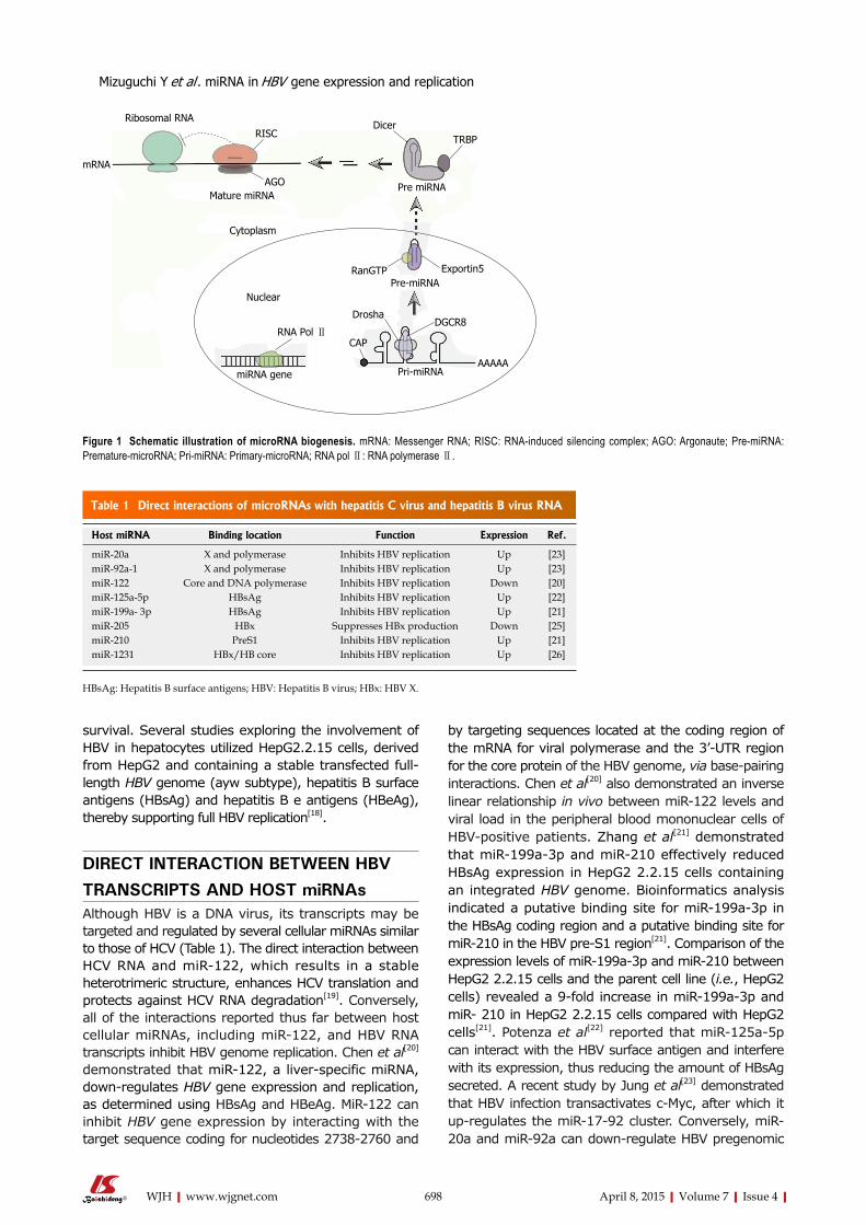
survival. Several studies exploring the involvement of HBV in hepatocytes utilized HepG2.2.15 cells, derived from HepG2 and containing a stable transfected full-length HBV genome (ayw subtype), hepatitis B surface antigens (HBsAg) and hepatitis B e antigens (HBeAg), thereby supporting full HBV replication[18].
DIRECT INTERACTION BETWEEN HBV TRANSCRIPTS AND HOST miRNAsAlthough HBV is a DNA virus, its transcripts may be targeted and regulated by several cellular miRNAs similar to those of HCV (Table 1). The direct interaction between HCV RNA and miR-122, which results in a stable heterotrimeric structure, enhances HCV translation and protects against HCV RNA degradation[19]. Conversely, all of the interactions reported thus far between host cellular miRNAs, including miR-122, and HBV RNA transcripts inhibit HBV genome replication. Chen et al[20] demonstrated that miR-122, a liver-specific miRNA, down-regulates HBV gene expression and replication, as determined using HBsAg and HBeAg. MiR-122 can inhibit HBV gene expression by interacting with the target sequence coding for nucleotides 2738-2760 and
by targeting sequences located at the coding region of the mRNA for viral polymerase and the 3’-UTR region for the core protein of the HBV genome, via base-pairing interactions. Chen et al[20] also demonstrated an inverse linear relationship in vivo between miR-122 levels and viral load in the peripheral blood mononuclear cells of HBV-positive patients. Zhang et al[21] demonstrated that miR-199a-3p and miR-210 effectively reduced HBsAg expression in HepG2 2.2.15 cells containing an integrated HBV genome. Bioinformatics analysis indicated a putative binding site for miR-199a-3p in the HBsAg coding region and a putative binding site for miR-210 in the HBV pre-S1 region[21]. Comparison of the expression levels of miR-199a-3p and miR-210 between HepG2 2.2.15 cells and the parent cell line (i.e., HepG2 cells) revealed a 9-fold increase in miR-199a-3p and miR- 210 in HepG2 2.2.15 cells compared with HepG2 cells[21]. Potenza et al[22] reported that miR-125a-5p can interact with the HBV surface antigen and interfere with its expression, thus reducing the amount of HBsAg secreted. A recent study by Jung et al[23] demonstrated that HBV infection transactivates c-Myc, after which it up-regulates the miR-17-92 cluster. Conversely, miR-20a and miR-92a can down-regulate HBV pregenomic
698 April 8, 2015|Volume 7|Issue 4|WJH|www.wjgnet.com
Ribosomal RNARISC
mRNA
Mature miRNAAGO
DicerTRBP
Pre miRNA
Cytoplasm
Exportin5RanGTPPre-miRNA
Nuclear
RNA Pol Ⅱ
miRNA gene Pri-miRNAAAAAA
DGCR8Drosha
CAP
Figure 1 Schematic illustration of microRNA biogenesis. mRNA: Messenger RNA; RISC: RNA-induced silencing complex; AGO: Argonaute; Pre-miRNA: Premature-microRNA; Pri-miRNA: Primary-microRNA; RNA pol Ⅱ: RNA polymerase Ⅱ.
Table 1 Direct interactions of microRNAs with hepatitis C virus and hepatitis B virus RNA
Host miRNA Binding location Function Expression Ref.
miR-20a X and polymerase Inhibits HBV replication Up [23]miR-92a-1 X and polymerase Inhibits HBV replication Up [23]miR-122 Core and DNA polymerase Inhibits HBV replication Down [20]miR-125a-5p HBsAg Inhibits HBV replication Up [22]miR-199a- 3p HBsAg Inhibits HBV replication Up [21]miR-205 HBx Suppresses HBx production Down [25]miR-210 PreS1 Inhibits HBV replication Up [21]miR-1231 HBx/HB core Inhibits HBV replication Up [26]
HBsAg: Hepatitis B surface antigens; HBV: Hepatitis B virus; HBx: HBV X.
Mizuguchi Y et al . miRNA in HBV gene expression and replication
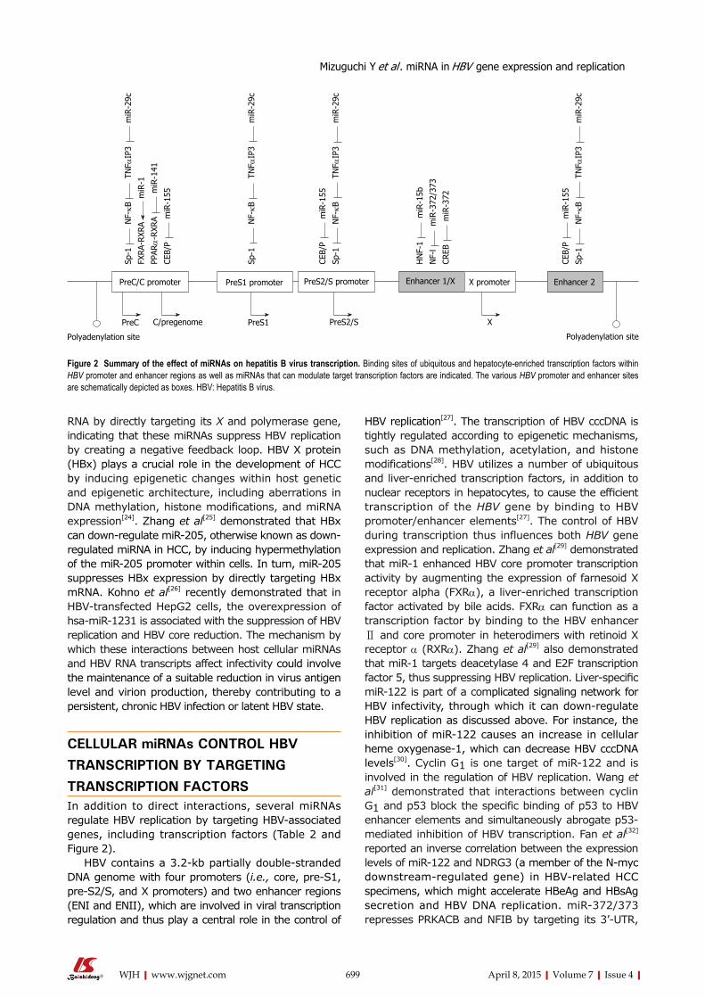
699 April 8, 2015|Volume 7|Issue 4|WJH|www.wjgnet.com
HBV replication[27]. The transcription of HBV cccDNA is tightly regulated according to epigenetic mechanisms, such as DNA methylation, acetylation, and histone modifications[28]. HBV utilizes a number of ubiquitous and liver-enriched transcription factors, in addition to nuclear receptors in hepatocytes, to cause the efficient transcription of the HBV gene by binding to HBV promoter/enhancer elements[27]. The control of HBV during transcription thus influences both HBV gene expression and replication. Zhang et al[29] demonstrated that miR-1 enhanced HBV core promoter transcription activity by augmenting the expression of farnesoid X receptor alpha (FXRα), a liver-enriched transcription factor activated by bile acids. FXRα can function as a transcription factor by binding to the HBV enhancer Ⅱ and core promoter in heterodimers with retinoid X receptor α (RXRα). Zhang et al[29] also demonstrated that miR-1 targets deacetylase 4 and E2F transcription factor 5, thus suppressing HBV replication. Liver-specific miR-122 is part of a complicated signaling network for HBV infectivity, through which it can down-regulate HBV replication as discussed above. For instance, the inhibition of miR-122 causes an increase in cellular heme oxygenase-1, which can decrease HBV cccDNA levels[30]. Cyclin G1 is one target of miR-122 and is involved in the regulation of HBV replication. Wang et al[31] demonstrated that interactions between cyclin G1 and p53 block the specific binding of p53 to HBV enhancer elements and simultaneously abrogate p53-mediated inhibition of HBV transcription. Fan et al[32] reported an inverse correlation between the expression levels of miR-122 and NDRG3 (a member of the N-myc downstream-regulated gene) in HBV-related HCC specimens, which might accelerate HBeAg and HBsAg secretion and HBV DNA replication. miR-372/373 represses PRKACB and NFIB by targeting its 3’-UTR,
RNA by directly targeting its X and polymerase gene, indicating that these miRNAs suppress HBV replication by creating a negative feedback loop. HBV X protein (HBx) plays a crucial role in the development of HCC by inducing epigenetic changes within host genetic and epigenetic architecture, including aberrations in DNA methylation, histone modifications, and miRNA expression[24]. Zhang et al[25] demonstrated that HBx can down-regulate miR-205, otherwise known as down-regulated miRNA in HCC, by inducing hypermethylation of the miR-205 promoter within cells. In turn, miR-205 suppresses HBx expression by directly targeting HBx mRNA. Kohno et al[26] recently demonstrated that in HBV-transfected HepG2 cells, the overexpression of hsa-miR-1231 is associated with the suppression of HBV replication and HBV core reduction. The mechanism by which these interactions between host cellular miRNAs and HBV RNA transcripts affect infectivity could involve the maintenance of a suitable reduction in virus antigen level and virion production, thereby contributing to a persistent, chronic HBV infection or latent HBV state.
CELLULAR miRNAs CONTROL HBV TRANSCRIPTION BY TARGETING TRANSCRIPTION FACTORSIn addition to direct interactions, several miRNAs regulate HBV replication by targeting HBV-associated genes, including transcription factors (Table 2 and Figure 2).
HBV contains a 3.2-kb partially double-stranded DNA genome with four promoters (i.e., core, pre-S1, pre-S2/S, and X promoters) and two enhancer regions (ENI and ENII), which are involved in viral transcription regulation and thus play a central role in the control of
PreC/C promoter PreS1 promoter PreS2/S promoter Enhancer 1/X X promoter Enhancer 2
Polyadenylation site
PreC C/pregenome PreS1 PreS2/S X
Polyadenylation site
Sp-1
N
F-kB
T
NF α
IP3
m
iR-2
9c
FXRA
-RXR
A m
iR-1
PPAR
α-R
XRA
m
iR-1
41
CEB/
P m
iR-1
55
Sp-1
N
F-kB
T
NF α
IP3
m
iR-2
9c
CEB/
P m
iR-1
55
Sp-1
N
F-kB
T
NF α
IP3
m
iR-2
9c
HN
F-1
m
iR-1
5b
NF-
l m
iR-3
72/3
73
CREB
m
iR-3
72
CEB/
P m
iR-1
55
Sp-1
N
F-kB
T
NF α
IP3
m
iR-2
9c
Figure 2 Summary of the effect of miRNAs on hepatitis B virus transcription. Binding sites of ubiquitous and hepatocyte-enriched transcription factors within HBV promoter and enhancer regions as well as miRNAs that can modulate target transcription factors are indicated. The various HBV promoter and enhancer sites are schematically depicted as boxes. HBV: Hepatitis B virus.
Mizuguchi Y et al . miRNA in HBV gene expression and replication
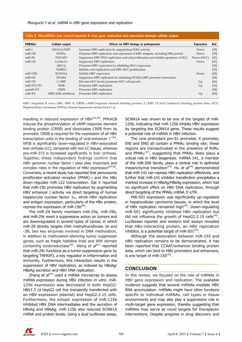
700 April 8, 2015|Volume 7|Issue 4|WJH|www.wjgnet.com
resulting in reduced expression of HBV[33,34]. PPKACB induces the phosphorylation of cAMP-response element binding protein (CREB) and dissociates CREB from its promoter. CREB is required for the expression of all HBV transcription units in the binding of viral enhancer Ⅰ[35].NFIB is significantly down-regulated in HBV-associated liver cirrhosis (LC), compared with non-LC tissues, whereas pre-miR-372 is increased significantly in liver cirrhosis. Together, these independent findings confirm that HBV genomic nuclear factor Ⅰ sites play important and complex roles in the regulation of HBV expression[36-38]. Conversely, a recent study has reported that peroxisome proliferator-activated receptor (PPAR)-γ and the HBx down-regulate miR-122 transcription. Dai X reported that miR-15b promotes HBV replication by augmenting HBV enhancer Ⅰ activity via direct targeting of human hepatocyte nuclear factor 1α, while HBV replication and antigen expression, particularly of the HBx protein, repress the expression of miR-15b[39].
The miR-29 family members miR-29a, miR-29b, and miR-29c exert a suppressive action on tumors and are downregulated in several types of cancer, in which miR-29 directly targets DNA methyltransferase 3A and -3B, two key enzymes involved in DNA methylation, in addition to methylation-silencing tumor suppressor genes, such as fragile histidine triad and WW domain containing oxidoreductase[40]. Wang et al[41] reported that miR-29c functions as a tumor-suppressive gene by targeting TNFAIP3, a key regulator in inflammation and immunity. Furthermore, this interaction results in the suppression of HBV replication, as indexed by HBsAg/HBeAg secretion and HBV DNA replication.
Zhang et al[42] used a miRNA microarray to assess miRNA expression during HBV infection in vitro. miR-125b expression was decreased in both HepG2-HBV1.3 (a HepG2 cell line transiently transfected with an HBV expression plasmid) and HepG2.2.15 cells. Furthermore, the ectopic expression of miR-125b inhibited HBV DNA intermediates and the secretion of HBsAg and HBeAg. miR-125b also reduced SCNN1A mRNA and protein levels. Using a dual luciferase assay,
SCNN1A was shown to be one of the targets of miR-125b, indicating that miR-125b inhibits HBV expression by targeting the SCNN1A gene. These results suggest a potential role of miRNA in HBV infection.
The core promoters pre-S1 promoter, X promoter, ENI and ENII all contain a PPARα binding site; these regions are transactivated in the presence of RXRα and PPARα[27], suggesting that PPARα likely plays a critical role in HBV biogenesis. miRNA 141, a member of the miR-200 family, plays a central role in epithelial mesenchymal transition[43]. Hu et al[44] demonstrated that miR-141 can repress HBV replication effectively, and further that miR-141 inhibitor transfection precipitates a marked increase in HBsAg/HBeAg expression, which had no significant effect on HBV DNA replication, through direct targeting of the PPARα mRNA 3’-UTR.
MiR-501 expression was significantly up-regulated in hepatocellular carcinoma tissues, in which the level of HBV replication remained high[45]. Down-regulating miR-501 significantly inhibited HBV replication but did not influence the growth of HepG2.2.15 cells[45]. Luciferase reporter and western blot assays revealed that HBx-interacting protein, an HBV replication inhibitor, is a potential target of miR-501[45].
Although the association between miR-155 and HBV replication remains to be demonstrated, it has been reported that CCAAT/enhancer binding protein beta, which can bind to HBV promoters and enhancers, is one target of miR-155[46].
CONCLUSIONIn this review, we focused on the role of miRNAs in HBV gene expression and replication. The available evidence suggests that several miRNAs mediate HBV RNA accumulation. miRNAs might have other functions specific to individual miRNAs, cell types or tissue environments and may also play a suppressive role in multi-target gene expression, thereby suggesting that miRNAs may serve as novel targets for therapeutic interventions. Despite progress in drug discovery and
MiRNAs Cellular targets Effects on HBV biology or pathogenesis Expression Ref.
miR-1 HDAC4/E2F5 Increases HBV replication by augmenting FXRA activity Down [29]miR-15b HNF1α Promotes HBV replication and expression of HBV antigens, including HBx protein Down [39]miR-29c TNFAIP3 Suppresses HBV DNA replication and cell proliferation and inhibits apoptosis of HCC Down (HCC) [41]miR-122 Cyclin G1 Suppresses HBV replication Down [31]
(HO-1) Promotes HBV expression by inhibiting HO-1 expression [30]NDRG3 Inhibits viral replication and HBV HCC proliferation [32]
miR-125b SCNN1A Inhibits HBV expression Down [42]miR-141 PPARA Suppresses HBV replication by inhibiting PPARA-HBV promoter interaction [44]miR-155 C/EBP Elevates HCC levels/promotes HCC cell growth Up [46]miR-372/373 NFIB Promotes HBV replication Up [33]upmiR-372 CREB Promotes HBV replication [34]miR-501 HBIP (HBx inhibitor) Promotes HBV replication Up [45]
Table 2 MicroRNAs that control hepatitis B virus gene replication and expression through cellular targets
HBV: Hepatitis B virus; HBx: HBV X; CREB: cAMP-response element binding protein; C/EBP: CCAAT/enhancer binding protein beta; HCC: Hepatocellular carcinoma; HNF1α: Human hepatocyte nuclear factor 1 α.
Mizuguchi Y et al . miRNA in HBV gene expression and replication
701 April 8, 2015|Volume 7|Issue 4|WJH|www.wjgnet.com
development, as evidenced by novel protease inhibitors and polymerase inhibitors, the clinical use of direct-acting antivirals is limited due to poor compliance and rapid-onset drug resistance. The use of imatinib mesylate, a platelet-derived growth factor receptor, and other tyrosine kinase inhibitors, as molecular targets against HCC progression, has been proposed[47,48]; however, clinical trials have indicated a lack of efficacy using this approach[49]. The development of more effective, cost efficient, and better-tolerated novel treatments is crucial to control the development of hepatitis, cirrhosis, and hepatocellular carcinoma. Exploring different facets of the interactions among miRNA, HBV and HCV infections, and the carcinogenesis and progression of HCC could facilitate the development of novel and effective treatment approaches for liver disease.
Although relatively poorly understood in the context of human cancers, evidence continues to accumulate indicating that long non-coding RNAs play a crucial role in regulating numerous developmental and biological genomic pathways[50].
REFERENCES1 Bartel DP. MicroRNAs: genomics, biogenesis, mechanism,
and function. Cell 2004; 116: 281-297 [PMID: 14744438 DOI: 10.1016/S0092-8674(04)00045-5]
2 Harfe BD. MicroRNAs in vertebrate development. Curr Opin Genet Dev 2005; 15: 410-415 [PMID: 15979303 DOI: 10.1016/j.gde.2005.06.012]
3 Rajewsky N. microRNA target predictions in animals. Nat Genet 2006; 38 Suppl: S8-13 [PMID: 16736023 DOI: 10.1038/ng1798]
4 Lim LP, Lau NC, Garrett-Engele P, Grimson A, Schelter JM, Castle J, Bartel DP, Linsley PS, Johnson JM. Microarray analysis shows that some microRNAs downregulate large numbers of target mRNAs. Nature 2005; 433: 769-773 [PMID: 15685193 DOI: 10.1038/nature03315]
5 Si-Tayeb K, Lemaigre FP, Duncan SA. Organogenesis and development of the liver. Dev Cell 2010; 18: 175-189 [PMID: 20159590 DOI: 10.1016/j.devcel.2010.01.011]
6 Lu J, Getz G, Miska EA, Alvarez-Saavedra E, Lamb J, Peck D, Sweet-Cordero A, Ebert BL, Mak RH, Ferrando AA, Downing JR, Jacks T, Horvitz HR, Golub TR. MicroRNA expression profiles classify human cancers. Nature 2005; 435: 834-838 [PMID: 15944708 DOI: 10.1038/nature03702]
7 Landgraf P, Rusu M, Sheridan R, Sewer A, Iovino N, Aravin A, Pfeffer S, Rice A, Kamphorst AO, Landthaler M, Lin C, Socci ND, Hermida L, Fulci V, Chiaretti S, Foà R, Schliwka J, Fuchs U, Novosel A, Müller RU, Schermer B, Bissels U, Inman J, Phan Q, Chien M, Weir DB, Choksi R, De Vita G, Frezzetti D, Trompeter HI, Hornung V, Teng G, Hartmann G, Palkovits M, Di Lauro R, Wernet P, Macino G, Rogler CE, Nagle JW, Ju J, Papavasiliou FN, Benzing T, Lichter P, Tam W, Brownstein MJ, Bosio A, Borkhardt A, Russo JJ, Sander C, Zavolan M, Tuschl T. A mammalian microRNA expression atlas based on small RNA library sequencing. Cell 2007; 129: 1401-1414 [PMID: 17604727 DOI: 10.1016/j.cell.2007.04.040]
8 Chen Y, Verfaillie CM. MicroRNAs: the fine modulators of liver development and function. Liver Int 2014; 34: 976-990 [PMID: 24517588 DOI: 10.1111/liv.12496]
9 Mizuguchi Y, Mishima T, Yokomuro S, Arima Y, Kawahigashi Y, Shigehara K, Kanda T, Yoshida H, Uchida E, Tajiri T, Takizawa T. Sequencing and bioinformatics-based analyses of the microRNA transcriptome in hepatitis B-related hepatocellular carcinoma. PLoS One 2011; 6: e15304 [PMID: 21283620 DOI: 10.1371/journal.pone.0015304]
10 Lau NC, Lim LP, Weinstein EG, Bartel DP. An abundant class of tiny RNAs with probable regulatory roles in Caenorhabditis elegans. Science 2001; 294: 858-862 [PMID: 11679671 DOI: 10.1126/science.1065062]
11 Lund E, Güttinger S, Calado A, Dahlberg JE, Kutay U. Nuclear export of microRNA precursors. Science 2004; 303: 95-98 [PMID: 14631048 DOI: 10.1126/science.1090599]
12 Hutvágner G, Zamore PD. A microRNA in a multiple-turnover RNAi enzyme complex. Science 2002; 297: 2056-2060 [PMID: 12154197 DOI: 10.1126/science.1073827]
13 He L, Hannon GJ. MicroRNAs: small RNAs with a big role in gene regulation. Nat Rev Genet 2004; 5: 522-531 [PMID: 15211354 DOI: 10.1038/nrg1379]
14 Vasudevan S, Tong Y, Steitz JA. Switching from repression to activation: microRNAs can up-regulate translation. Science 2007; 318: 1931-1934 [PMID: 18048652 DOI: 10.1126/science.1149460]
15 Vasudevan S. Posttranscriptional upregulation by microRNAs. Wiley Interdiscip Rev RNA 2012; 3: 311-330 [PMID: 22072587 DOI: 10.1002/wrna.121]
16 Lee I, Ajay SS, Yook JI, Kim HS, Hong SH, Kim NH, Dhanasekaran SM, Chinnaiyan AM, Athey BD. New class of microRNA targets containing simultaneous 5’-UTR and 3’-UTR interaction sites. Genome Res 2009; 19: 1175-1183 [PMID: 19336450 DOI: 10.1101/gr.089367.108]
17 Urban S, Schulze A, Dandri M, Petersen J. The replication cycle of hepatitis B virus. J Hepatol 2010; 52: 282-284 [PMID: 20056291 DOI: 10.1016/j.jhep.2009.10.031]
18 Sells MA, Chen ML, Acs G. Production of hepatitis B virus particles in Hep G2 cells transfected with cloned hepatitis B virus DNA. Proc Natl Acad Sci USA 1987; 84: 1005-1009 [PMID: 3029758 DOI: 10.1073/pnas.84.4.1005]
19 Li X, Yang W, Ye W, Jin L, He J, Lou L. microRNAs: novel players in hepatitis C virus infection. Clin Res Hepatol Gastroenterol 2014; 38: 664-675 [PMID: 24875730 DOI: 10.1016/j.clinre.2014.04.008]
20 Chen Y, Shen A, Rider PJ, Yu Y, Wu K, Mu Y, Hao Q, Liu Y, Gong H, Zhu Y, Liu F, Wu J. A liver-specific microRNA binds to a highly conserved RNA sequence of hepatitis B virus and negatively regulates viral gene expression and replication. FASEB J 2011; 25: 4511-4521 [PMID: 21903935 DOI: 10.1096/fj.11-187781]
21 Zhang GL, Li YX, Zheng SQ, Liu M, Li X, Tang H. Suppression of hepatitis B virus replication by microRNA-199a-3p and micro-RNA-210. Antiviral Res 2010; 88: 169-175 [PMID: 20728471 DOI: 10.1016/j.antiviral.2010.08.008]
22 Potenza N, Papa U, Mosca N, Zerbini F, Nobile V, Russo A. Human microRNA hsa-miR-125a-5p interferes with expression of hepatitis B virus surface antigen. Nucleic Acids Res 2011; 39: 5157-5163 [PMID: 21317190 DOI: 10.1093/nar/gkr067]
23 Jung YJ, Kim JW, Park SJ, Min BY, Jang ES, Kim NY, Jeong SH, Shin CM, Lee SH, Park YS, Hwang JH, Kim N, Lee DH. c-Myc-mediated overexpression of miR-17-92 suppresses replication of hepatitis B virus in human hepatoma cells. J Med Virol 2013; 85: 969-978 [PMID: 23532756 DOI: 10.1002/jmv.23534]
24 Tian Y, Yang W, Song J, Wu Y, Ni B. Hepatitis B virus X protein-induced aberrant epigenetic modifications contributing to human hepatocellular carcinoma pathogenesis. Mol Cell Biol 2013; 33: 2810-2816 [PMID: 23716588 DOI: 10.1128/MCB.00205-13]
25 Zhang T, Zhang J, Cui M, Liu F, You X, Du Y, Gao Y, Zhang S, Lu Z, Ye L, Zhang X. Hepatitis B virus X protein inhibits tumor suppressor miR-205 through inducing hypermethylation of miR-205 promoter to enhance carcinogenesis. Neoplasia 2013; 15: 1282-1291 [PMID: 24339740]
26 Kohno T, Tsuge M, Murakami E, Hiraga N, Abe H, Miki D, Imamura M, Ochi H, Hayes CN, Chayama K. Human microRNA hsa-miR-1231 suppresses hepatitis B virus replication by targeting core mRNA. J Viral Hepat 2014; 21: e89-e97 [PMID: 24835118 DOI: 10.1111/jvh.12240]
27 Quasdorff M, Protzer U. Control of hepatitis B virus at the level of transcription. J Viral Hepat 2010; 17: 527-536 [PMID: 20546497 DOI: 10.1111/j.1365-2893.2010.01315.x]
28 Liu WH, Yeh SH, Chen PJ. Role of microRNAs in hepatitis B virus
Mizuguchi Y et al . miRNA in HBV gene expression and replication
702 April 8, 2015|Volume 7|Issue 4|WJH|www.wjgnet.com
replication and pathogenesis. Biochim Biophys Acta 2011; 1809: 678-685 [PMID: 21565290 DOI: 10.1016/j.bbagrm.2011.04.008]
29 Zhang X, Zhang E, Ma Z, Pei R, Jiang M, Schlaak JF, Roggendorf M, Lu M. Modulation of hepatitis B virus replication and hepatocyte differentiation by MicroRNA-1. Hepatology 2011; 53: 1476-1485 [PMID: 21520166 DOI: 10.1002/hep.24195]
30 Qiu L, Fan H, Jin W, Zhao B, Wang Y, Ju Y, Chen L, Chen Y, Duan Z, Meng S. miR-122-induced down-regulation of HO-1 negatively affects miR-122-mediated suppression of HBV. Biochem Biophys Res Commun 2010; 398: 771-777 [PMID: 20633528 DOI: 10.1016/j.bbrc.2010.07.021]
31 Wang S, Qiu L, Yan X, Jin W, Wang Y, Chen L, Wu E, Ye X, Gao GF, Wang F, Chen Y, Duan Z, Meng S. Loss of microRNA 122 expression in patients with hepatitis B enhances hepatitis B virus replication through cyclin G(1) -modulated P53 activity. Hepatology 2012; 55: 730-741 [PMID: 22105316 DOI: 10.1002/hep.24809]
32 Fan CG, Wang CM, Tian C, Wang Y, Li L, Sun WS, Li RF, Liu YG. miR-122 inhibits viral replication and cell proliferation in hepatitis B virus-related hepatocellular carcinoma and targets NDRG3. Oncol Rep 2011; 26: 1281-1286 [PMID: 21725618 DOI: 10.3892/or.2011.1375]
33 Guo H, Liu H, Mitchelson K, Rao H, Luo M, Xie L, Sun Y, Zhang L, Lu Y, Liu R, Ren A, Liu S, Zhou S, Zhu J, Zhou Y, Huang A, Wei L, Guo Y, Cheng J. MicroRNAs-372/373 promote the expression of hepatitis B virus through the targeting of nuclear factor I/B. Hepatology 2011; 54: 808-819 [PMID: 21608007 DOI: 10.1002/hep.24441]
34 Wang J, Liu X, Wu H, Ni P, Gu Z, Qiao Y, Chen N, Sun F, Fan Q. CREB up-regulates long non-coding RNA, HULC expression through interaction with microRNA-372 in liver cancer. Nucleic Acids Res 2010; 38: 5366-5383 [PMID: 20423907 DOI: 10.1093/nar/gkq285]
35 Kim BK, Lim SO, Park YG. Requirement of the cyclic adenosine monophosphate response element-binding protein for hepatitis B virus replication. Hepatology 2008; 48: 361-373 [PMID: 18615500 DOI: 10.1002/hep.22359]
36 Pineau P, Volinia S, McJunkin K, Marchio A, Battiston C, Terris B, Mazzaferro V, Lowe SW, Croce CM, Dejean A. miR-221 overexpression contributes to liver tumorigenesis. Proc Natl Acad Sci USA 2010; 107: 264-269 [PMID: 20018759 DOI: 10.1073/pnas.0907904107]
37 Shaul Y, Ben-Levy R, De-Medina T. High affinity binding site for nuclear factor I next to the hepatitis B virus S gene promoter. EMBO J 1986; 5: 1967-1971 [PMID: 3463507]
38 Ben-Levy R, Faktor O, Berger I, Shaul Y. Cellular factors that interact with the hepatitis B virus enhancer. Mol Cell Biol 1989; 9: 1804-1809 [PMID: 2725524]
39 Dai X, Zhang W, Zhang H, Sun S, Yu H, Guo Y, Kou Z, Zhao G, Du L, Jiang S, Zhang J, Li J, Zhou Y. Modulation of HBV replication by microRNA-15b through targeting hepatocyte nuclear factor 1α. Nucleic Acids Res 2014; 42: 6578-6590 [PMID: 24705650 DOI: 10.1093/nar/gku260]
40 Fabbri M, Garzon R, Cimmino A, Liu Z, Zanesi N, Callegari E, Liu S, Alder H, Costinean S, Fernandez-Cymering C, Volinia S, Guler G, Morrison CD, Chan KK, Marcucci G, Calin GA, Huebner K, Croce CM. MicroRNA-29 family reverts aberrant methylation in lung cancer by targeting DNA methyltransferases 3A and 3B. Proc Natl Acad Sci USA 2007; 104: 15805-15810 [PMID: 17890317 DOI: 10.1073/pnas.0707628104]
41 Wang CM, Wang Y, Fan CG, Xu FF, Sun WS, Liu YG, Jia JH. miR-29c targets TNFAIP3, inhibits cell proliferation and induces apoptosis in hepatitis B virus-related hepatocellular carcinoma. Biochem Biophys Res Commun 2011; 411: 586-592 [PMID: 21763284 DOI: 10.1016/j.bbrc.2011.06.191]
42 Zhang Z, Chen J, He Y, Zhan X, Zhao R, Huang Y, Xu H, Zhu Z, Liu Q. miR-125b inhibits hepatitis B virus expression in vitro through targeting of the SCNN1A gene. Arch Virol 2014; 159: 3335-3343 [PMID: 25173609 DOI: 10.1007/s00705-014-2208-y]
43 Mizuguchi Y, Isse K, Specht S, Lunz JG, Corbitt N, Takizawa T, Demetris AJ. Small proline rich protein 2a in benign and malignant liver disease. Hepatology 2014; 59: 1130-1143 [PMID: 24123265 DOI: 10.1002/hep.26889]
44 Hu W, Wang X, Ding X, Li Y, Zhang X, Xie P, Yang J, Wang S. MicroRNA-141 represses HBV replication by targeting PPARA. PLoS One 2012; 7: e34165 [PMID: 22479552 DOI: 10.1371/annotation/cbbe9454-0b72-44b3-a972-10dcaf22db68]
45 Jin J, Tang S, Xia L, Du R, Xie H, Song J, Fan R, Bi Q, Chen Z, Yang G, Liu J, Shi Y, Fan D. MicroRNA-501 promotes HBV replication by targeting HBXIP. Biochem Biophys Res Commun 2013; 430: 1228-1233 [PMID: 23266610 DOI: 10.1016/j.bbrc.2012.12.071]
46 Wang B, Majumder S, Nuovo G, Kutay H, Volinia S, Patel T, Schmittgen TD, Croce C, Ghoshal K, Jacob ST. Role of microRNA-155 at early stages of hepatocarcinogenesis induced by choline-deficient and amino acid-defined diet in C57BL/6 mice. Hepatology 2009; 50: 1152-1161 [PMID: 19711427 DOI: 10.1002/hep.23100]
47 Treiber G, Wex T, Schleyer E, Troeger U, Hosius C, Malfertheiner P. Imatinib for hepatocellular cancer--focus on pharmacokinetic/pharmacodynamic modelling and liver function. Cancer Lett 2008; 260: 146-154 [PMID: 18083304 DOI: 10.1016/j.canlet.2007.10.041]
48 Höpfner M, Schuppan D, Scherübl H. Growth factor receptors and related signalling pathways as targets for novel treatment strategies of hepatocellular cancer. World J Gastroenterol 2008; 14: 1-14 [PMID: 18176955 DOI: 10.3748/wjg.14.1]
49 Eckel F, von Delius S, Mayr M, Dobritz M, Fend F, Hosius C, Schleyer E, Schulte-Frohlinde E, Schmid RM, Lersch C. Pharmacokinetic and clinical phase II trial of imatinib in patients with impaired liver function and advanced hepatocellular carcinoma. Oncology 2005; 69: 363-371 [PMID: 16319507 DOI: 10.1159/000089990]
50 Gupta RA, Shah N, Wang KC, Kim J, Horlings HM, Wong DJ, Tsai MC, Hung T, Argani P, Rinn JL, Wang Y, Brzoska P, Kong B, Li R, West RB, van de Vijver MJ, Sukumar S, Chang HY. Long non-coding RNA HOTAIR reprograms chromatin state to promote cancer metastasis. Nature 2010; 464: 1071-1076 [PMID: 20393566 DOI: 10.1038/nature08975]
P- Reviewer: De Minicis S, Sugimura H, Vespasiani-Gentilucci U S- Editor: Tian YL L- Editor: A E- Editor: Liu SQ
Mizuguchi Y et al . miRNA in HBV gene expression and replication
Sebastián Marciano, Silvia M Borzi, Melisa Dirchwolf, Ezequiel Ridruejo, Manuel Mendizabal, Fernando Bessone, María E Sirotinsky, Diego H Giunta, Julieta Trinks, Pablo A Olivera, Omar A Galdame, Marcelo O Silva, Hugo A Fainboim, Adrián C Gadano
Sebastián Marciano, Omar A Galdame, Adrián C Gadano, Liver Unit, Hospital Italiano de Buenos Aires, Buenos Aires, ArgentinaSilvia M Borzi, Hepatology Section, Hospital R. Rossi, La Plata, Buenos Aires, ArgentinaMelisa Dirchwolf, Hugo A Fainboim, Liver Infectious Disease Unit, Hospital F.J Muñiz, Uspallata 2272, Buenos Aires, ArgentinaEzequiel Ridruejo, Hepatology Section, Department of Medicine, Centro de Educación Médica e Investigaciones Clínicas Norberto Quirno “CEMIC”, Avenida Galván 4102, Buenos Aires, ArgentinaEzequiel Ridruejo, Manuel Mendizabal, Marcelo O Silva, Hepatology and Liver Transplant Unit, Hospital Universitario Austral, Buenos Aires, ArgentinaFernando Bessone, Hepatology Unit, Sanatorio del Parque, Rosario, ArgentinaMaría E Sirotinsky, HEPATOSUR group, Comodoro Rivadavia, Chubut, ArgentinaDiego H Giunta, Internal Medicine Research Unit, Hospital Italiano de Buenos Aires, Buenos Aires, ArgentinaJulieta Trinks, Basic Science and Experimental Medicine Institute, Hospital Italiano de Buenos Aires, Buenos Aires, ArgentinaPablo A Olivera, Internal Medicine, Centro de Educación Médica e Investigaciones Clínicas Norberto Quirno “CEMIC”, Buenos Aires, ArgentinaAuthor contributions: Marciano S studied design, data acquisition, analyze the data, wrote the manuscript; Borzi SM, Dirchwolf M, Ridruejo E, Mendizabal M, Bessone F, Sirotinsky ME, Olivera PA, Galdame OA, Silva MO, Fainboim HA and Gadano AC contributed equally in data acquisition, concept design and reviewing of the manuscript; Giunta DH contributed to statistical analyses, concept design and reviewing of the manuscript; Trinks J processed INFL3 polimorfisms, conceived design and reviewed of the manuscript. Ethics approval: The study was reviewed and approved by the Hospital Italiano de Buenos Aires Institutional Review Board.Informed consent: All study participants, or their legal guardian, provided informed written consent prior to study enrollment. Conflict-of-interest: The authors have no conflict of interest to declare.Data sharing: None.
Open-Access: This article is an open-access article which was selected by an in-house editor and fully peer-reviewed by external reviewers. It is distributed in accordance with the Creative Commons Attribution Non Commercial (CC BY-NC 4.0) license, which permits others to distribute, remix, adapt, build upon this work non-commercially, and license their derivative works on different terms, provided the original work is properly cited and the use is non-commercial. See: http://creativecommons.org/licenses/by-nc/4.0/Correspondence to: Sebastián Marciano, MD, Liver Unit, Hospital Italiano de Buenos Aires, Juan D. Peron 4190, C1181ACH Capital Federal, Buenos Aires, Argentina. [email protected]: +54-11-49590200-5370Fax: +54-11-49590346Received: August 29, 2014Peer-review started: August 30, 2014First decision: November 1, 2014Revised: November 5, 2014Accepted: January 18, 2015Article in press: January 20, 2015Published online: April 8, 2015
AbstractAIM: To evaluate pre-treatment factors associated with sustained virological response (SVR) in patients with hepatitis C virus (HCV) genotype 3 treated with peginterferon and ribavirin (RBV).
METHODS: We retrospectively analyzed treatment naive, mono-infected HCV genotype 3 patients treated with peginterferon and RBV. Exclusion criteria included presence of other liver disease, alcohol consumption and African American or Asian ethnicity. The variables collected and compared between patients who achieved an SVR and patients who did not were as follows: gender, age, fibrosis stage, diabetes, body mass index,
Submit a Manuscript: http://www.wjgnet.com/esps/Help Desk: http://www.wjgnet.com/esps/helpdesk.aspxDOI: 10.4254/wjh.v7.i4.703
703 April 8, 2015|Volume 7|Issue 4|WJH|www.wjgnet.com
World J Hepatol 2015 April 8; 7(4): 703-709ISSN 1948-5182 (online)
© 2015 Baishideng Publishing Group Inc. All rights reserved.
Pre-treatment prediction of response to peginterferon plus ribavirin in chronic hepatitis C genotype 3
Retrospective Study
ORIGINAL ARTICLE
steatosis, INFL3 polymorphism, pre-treatment HCV-RNA, type of peginterferon, RBV dose and adherence.
RESULTS: A total of 107 patients treated between June, 2004 and March, 2013 were included. Mean treatment duration was 25.1 (± 1.8) wk. Overall, 58% (62/107) of the patients achieved an SVR and 42% (45/107) did not. In the multivariate logistic regression analysis, pre-treatment HCV-RNA ≥ 600000 UI/mL (OR = 0.375, 95%CI: 0.153-0.919, P = 0.032) and advanced fibrosis (OR = 0.278, 95%CI: 0.113-0.684, P = 0.005) were significantly associated with low SVR rates. In patients with pre-treatment HCV-RNA ≥ 600000 UI/mL and advanced fibrosis, the probability of achieving an SVR was 29% (95%CI: 13.1-45.2). In patients with pre-treatment HCV-RNA < 600000 UI/mL and mild to moderate fibrosis, the probability of achieving an SVR was 81% (95%CI: 68.8-93.4).
CONCLUSION: In patients with HCV genotype 3 infections the presence of advance fibrosis and high pre-treatment viral load might be associated with poor response to peginterferon plus RBV. These patients could benefit the most from new direct antiviral agents-based regimes.
Key words: Sustained virological response; Direct antiviral agents; Sofosbuvir; Cirrhosis; Viral load
© The Author(s) 2015. Published by Baishideng Publishing Group Inc. All rights reserved.
Core tip: Our study evaluates pre-treatment factors associated with sustained virological response in patients with hepatitis C virus genotype 3 treated with peginterferon plus ribavirin. We identified a sub-group of patients with high pre-treatment viral load and advanced fibrosis whose chance of achieving a sustained virological response is as low as 29%. We believe these patients should be prioritized to access new treatment strategies.
Marciano S, Borzi SM, Dirchwolf M, Ridruejo E, Mendizabal M, Bessone F, Sirotinsky ME, Giunta DH, Trinks J, Olivera PA, Galdame OA, Silva MO, Fainboim HA, Gadano AC. Pre-treatment prediction of response to peginterferon plus ribavirin in chronic hepatitis C genotype 3. World J Hepatol 2015; 7(4): 703-709 Available from: URL: http://www.wjgnet.com/1948-5182/full/v7/i4/703.htm DOI: http://dx.doi.org/10.4254/wjh.v7.i4.703
INTRODUCTIONHepatitis C virus (HCV) is a major health problem affecting more than 180 million people worldwide[1]. It is estimated that at least 350000 HCV infected people die annually due to liver-related causes[2].
Six genotypes of HCV have been identified. In Latin America, genotype 1 is the most prevalent, followed by genotypes 2 and 3[3,4]. In some areas of this region
the prevalence of HCV genotype 3 (HCV-3) is as high as 30%[3].
Traditionally, HCV-2 and HCV-3 have been grouped together as “easy to treat” genotypes. However we now know that sustained virological response (SVR) rates of HCV-3 patients treated with peginterferon plus ribavirin (RBV) are sub-optimal. The global SVR rates for HCV-3 patients treated with peginterferon plus RBV are around 65%-70%[5,6]. Since these data mainly arise from randomized trials, SVR rates are expected to be lower in real-life patients with adverse factors[7].
Several host and viral factors have an impact on the SVR rate of patients infected with HCV-3 treated with peginterferon plus RBV. Pre-treatment factors that have been proposed to have a negative impact on SVR are advanced fibrosis or cirrhosis, male gender, non-Caucasian race, high body weight, diabetes mellitus, and high pre-treatment HCV-RNA[7-12]. More recently, INFL3 (formerly IL28B) polymorphisms were evaluated, but a clear association between the favorable INFL3 genotypes and SVR could not be established[13-15].
A major limitation of the studies that assessed predictors of SVR in HCV-3 patients lies in the fact that they evaluated HCV-2 and HCV-3 together, and difficulties arise when trying to draw conclusions for HCV-3 individually.
By the end of 2013, sofosbuvir was approved for the treatment of chronic HCV-3, being the standard of care in a minority of countries at the moment this manuscript was submitted. However, in regions like Latin America and Middle East/Africa, payer-related barriers were reported[16], which might hamper adequate treatment delivery.
When new treatments become globally available, a careful selection of candidates will be mandatory, particularly for cost-related reasons. Therefore, it will be necessary to identify patients at higher risk of fibrosis progression and treatment failure to current therapies.
Thus, we decided to conduct this study that eva-luates pre-treatment variables associated with SVR in patients with chronic HCV-3 treated with peginterferon plus RBV.
MATERIALS AND METHODSThis is a retrospective multicenter study performed in 7 Liver Units from Argentina.
The centers involved in the study were, Hospital Italiano from Buenos Aires, Hospital R. Rossi from La Plata, Hospital F. J. Muñiz, Centro de Educación Médica e Investigaciones Clínicas Norberto Quirno (CEMIC), Hospital Universitario Austral, Sanatorio Parque from Rosario, and the HEPATOSUR Group representing the Patagonia.
The survey was discussed by all the participating centers. The local institutional review board of each center approved the study. The study was conducted
704 April 8, 2015|Volume 7|Issue 4|WJH|www.wjgnet.com
Marciano S et al . Predicting treatment response in HCV-3
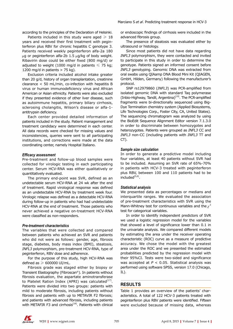
according to the principles of the Declaration of Helsinki.Patients included in this study were aged ≥ 18
years and received their first treatment with pegin-terferon plus RBV for chronic hepatitis C genotype 3. Patients received weekly peginterferon alfa-2a 180 μg or peginterferon alfa-2b 1.5 μg/kg of body weight. Ribavirin dose could be either fixed (800 mg/d) or adjusted to weight (1000 mg/d in patients ≤ 75 kg; 1200 mg/d in patients > 75 kg).
Exclusion criteria included alcohol intake greater than 20 g/d, history of organ transplantation, creatinine clearance < 50 mL/min, co-infection with hepatitis B virus or human immunodeficiency virus and African American or Asian ethnicity. Patients were also excluded if they presented evidence of other liver disease, such as autoimmune hepatitis, primary biliary cirrhosis, sclerosing cholangitis, Wilson’s disease or alfa-1-antitrypsin deficiency.
Each center provided detailed information of patients included in the study. Patient management and treatment candidacy were determined in each center. All data records were checked for missing values and inconsistencies, queries were sent to all participating institutions, and corrections were made at the data coordinating center, namely Hospital Italiano.
Efficacy assessmentPre-treatment and follow-up blood samples were collected for virologic testing in each participating center. Serum HCV-RNA was either qualitatively or quantitatively evaluated.
The primary end-point was SVR, defined as an undetectable serum HCV-RNA at 24 wk after the end of treatment. Rapid virological response was defined as an undetectable HCV-RNA by treatment week four. Virologic relapse was defined as a detectable HCV-RNA during follow-up in patients who had had undetectable HCV-RNA at the end of treatment. Those patients who never achieved a negative on-treatment HCV-RNA were classified as non-responders.
Pre-treatment characteristics The variables that were collected and compared between patients who achieved an SVR and patients who did not were as follows: gender, age, fibrosis stage, diabetes, body mass index (BMI), steatosis, INFL3 polymorphism, pre-treatment HCV-RNA, type of peginterferon, RBV dose and adherence.
For the purpose of this study, high HCV-RNA was defined as ≥ 600000 UI/mL.
Fibrosis grade was staged either by biopsy or Transient Elastography (Fibroscan®). In patients without fibrosis evaluation, the aspartate aminotransferase to Platelet Ration Index (APRI) was calculated[17]. Patients were divided into two groups: patients with mild to moderate fibrosis, including patients without fibrosis and patients with up to METAVIR F2 fibrosis; and patients with advanced fibrosis, including patients with METAFIR F3 and cirrhosis[18]. Patients with clinical
or endoscopic findings of cirrhosis were included in the advanced fibrosis group.
The presence of steatosis was evaluated either by ultrasound or histology.
Since most patients did not have data regarding INFL3 polymorphism, they were contacted and invited to participate in this study in order to determine the genotype. Patients signed an informed consent before INFL3 genotyping. Genomic DNA was extracted from oral swabs using QIAamp DNA Blood Mini Kit (QIAGEN, GmbH, Hilden, Germany) following the manufacturer’s protocol.
SNP rs12979860 (INFL3) was PCR-amplified from isolated genomic DNA with standard Taq polymerase (Inbio-Highway, Tandil, Argentina)[19]. The PCR-amplified fragments were bi-directionally sequenced using Big-Dye Termination chemistry system (Applied Biosystems, Life Technologies Corp., Foster City, CA, United States). The sequencing chromatogram was analyzed by using the BioEdit Sequence Alignment Editor version 7.1.3.0 in order to discriminate between homozygotes and heterozygotes. Patients were grouped as INFL3 CC and INFL3 non-CC (including patients with INFL3 TT and CT).
Sample size calculationIn order to generate a predictive model including four variables, at least 40 patients without SVR had to be included. Assuming an SVR rate of 65%-70% in patients with HCV-3 treated with peginterferon plus RBV, between 100 and 110 patients had to be included[5,6].
Statistical analysisWe presented data as percentages or medians and interquartile ranges. We evaluated the association of pre-treatment characteristics with SVR using the Mann-Whitney test for continuous variables and the χ 2 test for categorical variables.
In order to identify independent predictors of SVR we used a logistic regression model for the variables that showed a level of significance lower than 0.1 in the univariate analysis. We compared different models by estimating the area under the receiver operating characteristic (ROC) curve as a measure of predictive accuracy. We chose the model with the greatest area under the ROC and we presented the estimated probabilities predicted by the regression model with their 95%CI. Tests were two-sided and significance was accepted at P < 0.05. Statistical analysis was performed using software SPSS, version 17.0 (Chicago, IL).
RESULTSTable 1 provides an overview of the patients’ char-acteristics. A total of 122 HCV-3 patients treated with peginterferon plus RBV patients were identified. Fifteen were excluded because of missing data, whereas
705 April 8, 2015|Volume 7|Issue 4|WJH|www.wjgnet.com
Marciano S et al . Predicting treatment response in HCV-3
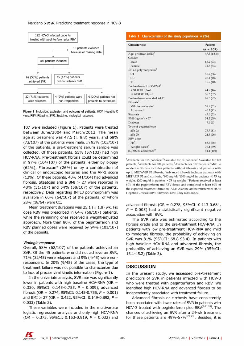
706 April 8, 2015|Volume 7|Issue 4|WJH|www.wjgnet.com
advanced fibrosis (OR = 0.278, 95%CI: 0.113-0.684, P = 0.005) had a statistically significant negative association with SVR.
The SVR rate was estimated according to the fibrosis grade and to the pre-treatment HCV-RNA. In patients with low pre-treatment HCV-RNA and mild to moderate fibrosis, the probability of achieving an SVR was 81% (95%CI: 68.8-93.4). In patients with high baseline HCV-RNA and advanced fibrosis, the probability of achieving an SVR was 29% (95%CI: 13.1-45.2) (Table 3).
DISCUSSIONIn the present study, we assessed pre-treatment predictors of SVR in patients infected with HCV-3 who were treated with peginterferon and RBV. We identified high HCV-RNA and advanced fibrosis to be independently associated with treatment failure.
Advanced fibrosis or cirrhosis have consistently been associated with lower rates of SVR in patients with HCV-3 treated with peginterferon plus RBV[9-11,20]. The chances of achieving an SVR after a 24-wk treatment for these patients are 49%-57%[21-23]. Besides, it is
107 were included (Figure 1). Patients were treated between June/2004 and March/2013. The mean age at treatment was 47.5 (± 8.8) years, and 68% (73/107) of the patients were male. In 93% (103/107) of the patients, a pre-treatment serum sample was collected. Of those patients, 55% (57/103) had high HCV-RNA. Pre-treatment fibrosis could be determined in 97% (104/107) of the patients, either by biopsy (62%), Fibroscan® (26%) or by a combination of clinical or endoscopic features and the APRI score (12%). Of these patients, 40% (41/104) had advanced fibrosis. Steatosis and a BMI > 27 were reported in 48% (51/107) and 54% (58/107) of the patients, respectively. Data regarding INFL3 polymorphism was available in 60% (64/107) of the patients, of whom 28% (18/64) were CC.
Mean treatment durations was 25.1 (± 1.8) wk. Fix dose RBV was prescribed in 64% (68/107) patients, while the remaining ones received a weight-adjusted approach. More than 80% of the peginterferon and RBV planned doses were received by 94% (101/107) of the patients.
Virologic responseOverall, 58% (62/107) of the patients achieved an SVR. Of the 45 patients who did not achieve an SVR, 71% (32/45) were relapsers and 9% (4/45) were non-responders. In 20% (9/45) of the cases, the type of treatment failure was not possible to characterize due to lack of precise viral kinetic information (Figure 1).
In the univariate analysis, SVR rate was significantly lower in patients with high baseline HCV-RNA (OR = 0.330, 95%CI: 0.145-0.755, P = 0.009), advanced fibrosis (OR = 0.274, 95%CI: 0.145-0.755, P = 0.001) and BMI > 27 (OR = 0.422, 95%CI: 0.149-0.892, P = 0.033) (Table 2).
These variables were included in the multivariate logistic regression analysis and only high HCV-RNA (OR = 0.375, 95%CI: 0.153-0.919, P = 0.032) and
Table 1 Characteristics of the study population n (%)
Characteristic Patients
(n = 107)Age, yr (mean ± SD)1 47.5 (± 8.8)Gender Male 68.2 (73) Female 31.8 (34)INFL3 polymorphism2
CT 56.2 (36) CC 28.1 (18) TT 15.7 (10)Pre treatment HCV-RNA3
< 600000 UI/mL 44.7 (46) ≥ 6000000 UI/mL 55.3 (57)Pre treatment elevated ALT4 88.5 (92)Fibrosis5
Mild to moderate6 59.8 (61) Advanced7 40.2 (41)Steatosis 47.6 (51)BMI (kg/m2) > 27 54.2 (58)Diabetes 5.6 (6)Type of peginterferon alfa 2a 75.7 (81) alfa 2b 24.3 (26)RBV dose Fix8 63.6 (68) Weight-Based9 36.4 (39)80/80/80 adherence10 94.4 (101)
1Available for 105 patients; 2Available for 64 patients; 3Available for 103 patients; 4Available for 104 patients; 5Available for 102 patients; 6Mild to moderate fibrosis includes patients without fibrosis and patients with up to METAVIR F2 fibrosis; 7Advanced fibrosis includes patients with METAFIR F3 and cirrhosis; 8800 mg/d; 91000 mg/d in patients ≤ 75 kg weight, 1200 mg/d in patients > 75 kg weight; 10Patients received at least 80% of the peginterferon and RBV doses, and completed at least 80% of the expected treatment duration. ALT: Alanine aminotransferase; HCV: Hepatitis C virus; RBV: Ribavirin; BMI: Body mass index.
122 HCV-3 infected patients treated with peginterferon plus RBV
15 patients excluded because of missing data
107 patients included
62 (58%) patients achieved SVR
45 (42%) patients did not achieve SVR
32 (71%) patients were relapsers
4 (9%) patients were non-responders
9 (20%) patients not possible to determine
Figure 1 Inclusion, exclusion and outcome of patients. HCV: Hepatitis C virus; RBV: Ribavirin; SVR: Sustained virological response.
Marciano S et al . Predicting treatment response in HCV-3
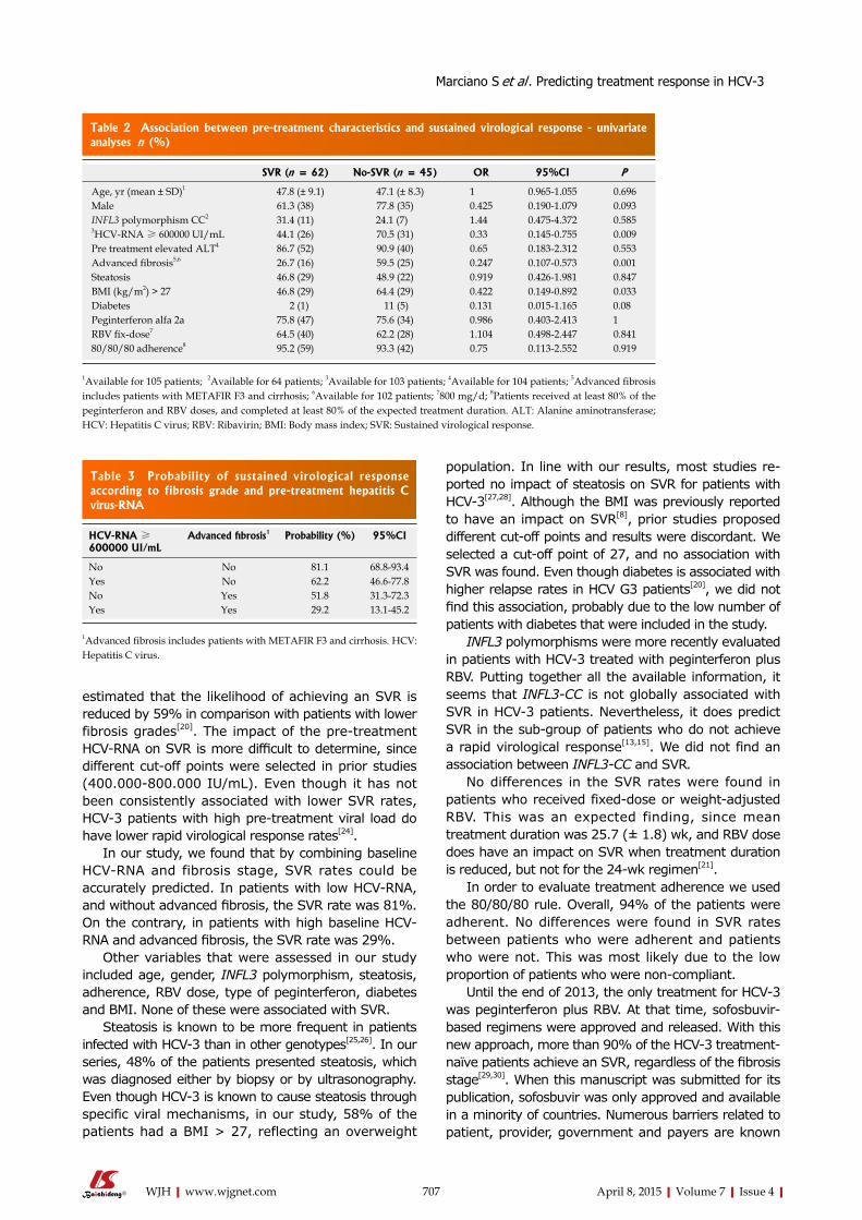
707 April 8, 2015|Volume 7|Issue 4|WJH|www.wjgnet.com
estimated that the likelihood of achieving an SVR is reduced by 59% in comparison with patients with lower fibrosis grades[20]. The impact of the pre-treatment HCV-RNA on SVR is more difficult to determine, since different cut-off points were selected in prior studies (400.000-800.000 IU/mL). Even though it has not been consistently associated with lower SVR rates, HCV-3 patients with high pre-treatment viral load do have lower rapid virological response rates[24].
In our study, we found that by combining baseline HCV-RNA and fibrosis stage, SVR rates could be accurately predicted. In patients with low HCV-RNA, and without advanced fibrosis, the SVR rate was 81%. On the contrary, in patients with high baseline HCV-RNA and advanced fibrosis, the SVR rate was 29%.
Other variables that were assessed in our study included age, gender, INFL3 polymorphism, steatosis, adherence, RBV dose, type of peginterferon, diabetes and BMI. None of these were associated with SVR.
Steatosis is known to be more frequent in patients infected with HCV-3 than in other genotypes[25,26]. In our series, 48% of the patients presented steatosis, which was diagnosed either by biopsy or by ultrasonography. Even though HCV-3 is known to cause steatosis through specific viral mechanisms, in our study, 58% of the patients had a BMI > 27, reflecting an overweight
population. In line with our results, most studies re-ported no impact of steatosis on SVR for patients with HCV-3[27,28]. Although the BMI was previously reported to have an impact on SVR[8], prior studies proposed different cut-off points and results were discordant. We selected a cut-off point of 27, and no association with SVR was found. Even though diabetes is associated with higher relapse rates in HCV G3 patients[20], we did not find this association, probably due to the low number of patients with diabetes that were included in the study.
INFL3 polymorphisms were more recently evaluated in patients with HCV-3 treated with peginterferon plus RBV. Putting together all the available information, it seems that INFL3-CC is not globally associated with SVR in HCV-3 patients. Nevertheless, it does predict SVR in the sub-group of patients who do not achieve a rapid virological response[13,15]. We did not find an association between INFL3-CC and SVR.
No differences in the SVR rates were found in patients who received fixed-dose or weight-adjusted RBV. This was an expected finding, since mean treatment duration was 25.7 (± 1.8) wk, and RBV dose does have an impact on SVR when treatment duration is reduced, but not for the 24-wk regimen[21].
In order to evaluate treatment adherence we used the 80/80/80 rule. Overall, 94% of the patients were adherent. No differences were found in SVR rates between patients who were adherent and patients who were not. This was most likely due to the low proportion of patients who were non-compliant.
Until the end of 2013, the only treatment for HCV-3 was peginterferon plus RBV. At that time, sofosbuvir-based regimens were approved and released. With this new approach, more than 90% of the HCV-3 treatment-naïve patients achieve an SVR, regardless of the fibrosis stage[29,30]. When this manuscript was submitted for its publication, sofosbuvir was only approved and available in a minority of countries. Numerous barriers related to patient, provider, government and payers are known
Table 2 Association between pre-treatment characteristics and sustained virological response - univariate analyses n (%)
SVR (n = 62) No-SVR (n = 45) OR 95%CI P
Age, yr (mean ± SD)1 47.8 (± 9.1) 47.1 (± 8.3) 1 0.965-1.055 0.696Male 61.3 (38) 77.8 (35) 0.425 0.190-1.079 0.093INFL3 polymorphism CC2 31.4 (11) 24.1 (7) 1.44 0.475-4.372 0.5853HCV-RNA ≥ 600000 UI/mL 44.1 (26) 70.5 (31) 0.33 0.145-0.755 0.009Pre treatment elevated ALT4 86.7 (52) 90.9 (40) 0.65 0.183-2.312 0.553Advanced fibrosis5,6 26.7 (16) 59.5 (25) 0.247 0.107-0.573 0.001Steatosis 46.8 (29) 48.9 (22) 0.919 0.426-1.981 0.847BMI (kg/m2) > 27 46.8 (29) 64.4 (29) 0.422 0.149-0.892 0.033Diabetes 2 (1) 11 (5) 0.131 0.015-1.165 0.08Peginterferon alfa 2a 75.8 (47) 75.6 (34) 0.986 0.403-2.413 1RBV fix-dose7 64.5 (40) 62.2 (28) 1.104 0.498-2.447 0.84180/80/80 adherence8 95.2 (59) 93.3 (42) 0.75 0.113-2.552 0.919
1Available for 105 patients; 2Available for 64 patients; 3Available for 103 patients; 4Available for 104 patients; 5Advanced fibrosis includes patients with METAFIR F3 and cirrhosis; 6Available for 102 patients; 7800 mg/d; 8Patients received at least 80% of the peginterferon and RBV doses, and completed at least 80% of the expected treatment duration. ALT: Alanine aminotransferase; HCV: Hepatitis C virus; RBV: Ribavirin; BMI: Body mass index; SVR: Sustained virological response.
Table 3 Probability of sustained virological response according to fibrosis grade and pre-treatment hepatitis C virus-RNA
HCV-RNA ≥ 600000 UI/mL
Advanced fibrosis1 Probability (%) 95%CI
No No 81.1 68.8-93.4Yes No 62.2 46.6-77.8No Yes 51.8 31.3-72.3Yes Yes 29.2 13.1-45.2
1Advanced fibrosis includes patients with METAFIR F3 and cirrhosis. HCV: Hepatitis C virus.
Marciano S et al . Predicting treatment response in HCV-3
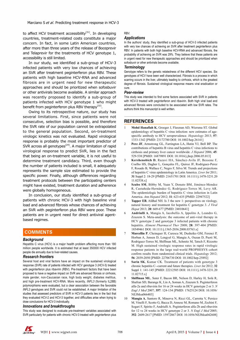
708 April 8, 2015|Volume 7|Issue 4|WJH|www.wjgnet.com
to affect HCV treatment accessibility[31]. In developing countries, treatment-related costs constitute a major concern. In fact, in some Latin American countries, after more than three years of the release of Boceprevir and Telaprevir for the treatments of HCV genotype 1, accessibility is still limited.
In our study, we identified a sub-group of HCV-3 infected patients with very low chances of achieving an SVR after treatment peginterferon plus RBV. These patients with high baseline HCV-RNA and advanced fibrosis are in urgent need for new therapeutic approaches and should be prioritized when sofosbuvir or other antivirals become available. A similar approach was recently proposed to identify a sub-group of patients infected with HCV genotype 1 who might benefit from peginterferon plus RBV therapy[32].
Owing to its retrospective nature, our study has several limitations. First, since patients were not consecutive, selection bias is possible, and therefore the SVR rate of our population cannot be extrapolated to the general population. Second, on-treatment virologic kinetics was not evaluated. Rapid virological response is probably the most important predictor of SVR across all genotypes[24]. A major limitation of rapid virological response to predict SVR, lies in the fact that being an on-treatment variable, it is not useful to determine treatment candidacy. Third, even though the number of patients included is small, it adequately represents the sample size estimated to provide the specific power. Finally, although differences regarding treatment protocols between the participating centers might have existed, treatment duration and adherence were globally homogeneous.
In conclusion, our study identified a sub-group of patients with chronic HCV-3 with high baseline viral load and advanced fibrosis whose chances of achieving an SVR with peginterferon plus RBV were poor. These patients are in urgent need for direct antiviral agent-based regimes.
COMMENTSBackgroundHepatitis C virus (HCV) is a major health problem affecting more than 180 million people worldwide. It is estimated that at least 350000 HCV infected people die annually due to liver-related causes.Research frontiersSeveral host and viral factors have an impact on the sustained virological response (SVR) rate of patients infected with HCV genotype 3 (HCV-3) treated with peginterferon plus ribavirin (RBV). Pre-treatment factors that have been proposed to have a negative impact on SVR are advanced fibrosis or cirrhosis, male gender, non-Caucasian race, high body weight, diabetes mellitus, and high pre-treatment HCV-RNA. More recently, INFL3 (formerly IL28B) polymorphisms were evaluated, but a clear association between the favorable INFL3 genotypes and SVR could not be established. A major limitation of the studies that assessed predictors of SVR in HCV-3 patients lies in the fact that they evaluated HCV-2 and HCV-3 together, and difficulties arise when trying to draw conclusions for HCV-3 individually.Innovations and breakthroughsThis study was designed to evaluate pre-treatment variables associated with SVR particularly for patients with chronic HCV-3 treated with peginterferon plus
RBV. ApplicationsIn the authors’ study, they identified a sub-group of HCV-3 infected patients with very low chances of achieving an SVR after treatment peginterferon plus RBV. In patients with both high baseline HCV-RNA and advanced fibrosis, the probability of achieving an SVR was 29%. They believe that these patients are in urgent need for new therapeutic approaches and should be prioritized when sofosbuvir or other antivirals become available.TerminologyGenotype refers to the genetic relatedness of the different HCV species. Six genotypes of HCV have been well characterized. Fibrosis is a process in which scarring occurs in the liver, ultimately leading to cirrhosis, which is the greatest degree of fibrosis. Sustained virological response means viral eradication or cure.Peer-reviewThis study was intended to find some factors associated with SVR in patients with HCV-3 treated with peginterferon and ribavirin. Both high viral load and advanced fibrosis were concluded to be associated with low SVR rates. The authors think this manuscript is well written.
REFERENCES1 Mohd Hanafiah K, Groeger J, Flaxman AD, Wiersma ST. Global
epidemiology of hepatitis C virus infection: new estimates of age-specific antibody to HCV seroprevalence. Hepatology 2013; 57: 1333-1342 [PMID: 23172780 DOI: 10.1002/hep.26141]
2 Perz JF, Armstrong GL, Farrington LA, Hutin YJ, Bell BP. The contributions of hepatitis B virus and hepatitis C virus infections to cirrhosis and primary liver cancer worldwide. J Hepatol 2006; 45: 529-538 [PMID: 16879891 DOI: 10.1016/j.jhep.2006.05.013]
3 Kershenobich D, Razavi HA, Sánchez-Avila JF, Bessone F, Coelho HS, Dagher L, Gonçales FL, Quiroz JF, Rodriguez-Perez F, Rosado B, Wallace C, Negro F, Silva M. Trends and projections of hepatitis C virus epidemiology in Latin America. Liver Int 2011; 31 Suppl 2: 18-29 [PMID: 21651701 DOI: 10.1111/j.1478-3231.2011.02538.x]
4 Szabo SM, Bibby M, Yuan Y, Donato BM, Jiménez-Mendez R, Castañeda-Hernández G, Rodríguez-Torres M, Levy AR. The epidemiologic burden of hepatitis C virus infection in Latin America. Ann Hepatol 2012; 11: 623-635 [PMID: 22947522]
5 Tapper EB, Afdhal NH. Is 3 the new 1: perspectives on virology, natural history and treatment for hepatitis C genotype 3. J Viral Hepat 2013; 20: 669-677 [PMID: 24010641]
6 Andriulli A, Mangia A, Iacobellis A, Ippolito A, Leandro G, Zeuzem S. Meta-analysis: the outcome of anti-viral therapy in HCV genotype 2 and genotype 3 infected patients with chronic hepatitis. Aliment Pharmacol Ther 2008; 28: 397-404 [PMID: 18549461 DOI: 10.1111/j.1365-2036.2008.03763.x]
7 Marcellin P, Cheinquer H, Curescu M, Dusheiko GM, Ferenci P, Horban A, Jensen D, Lengyel G, Mangia A, Ouzan D, Puoti M, Rodriguez-Torres M, Shiffman ML, Schmitz M, Tatsch F, Rizzetto M. High sustained virologic response rates in rapid virologic response patients in the large real-world PROPHESYS cohort confirm results from randomized clinical trials. Hepatology 2012; 56: 2039-2050 [PMID: 22706730 DOI: 10.1002/hep.25892]
8 Sarin SK, Kumar CK. Treatment of patients with genotype 3 chronic hepatitis C--current and future therapies. Liver Int 2012; 32 Suppl 1: 141-145 [PMID: 22212585 DOI: 10.1111/j.1478-3231.2011.02715.x]
9 Shiffman ML, Suter F, Bacon BR, Nelson D, Harley H, Solá R, Shafran SD, Barange K, Lin A, Soman A, Zeuzem S. Peginterferon alfa-2a and ribavirin for 16 or 24 weeks in HCV genotype 2 or 3. N Engl J Med 2007; 357: 124-134 [PMID: 17625124 DOI: 10.1056/NEJMoa066403]
10 Mangia A, Santoro R, Minerva N, Ricci GL, Carretta V, Persico M, Vinelli F, Scotto G, Bacca D, Annese M, Romano M, Zechini F, Sogari F, Spirito F, Andriulli A. Peginterferon alfa-2b and ribavirin for 12 vs 24 weeks in HCV genotype 2 or 3. N Engl J Med 2005; 352: 2609-2617 [PMID: 15972867 DOI: 10.1056/NEJMoa042608]
COMMENTS
Marciano S et al . Predicting treatment response in HCV-3
709 April 8, 2015|Volume 7|Issue 4|WJH|www.wjgnet.com
11 Yu ML, Dai CY, Huang JF, Hou NJ, Lee LP, Hsieh MY, Chiu CF, Lin ZY, Chen SC, Hsieh MY, Wang LY, Chang WY, Chuang WL. A randomised study of peginterferon and ribavirin for 16 versus 24 weeks in patients with genotype 2 chronic hepatitis C. Gut 2007; 56: 553-559 [PMID: 16956917 DOI: 10.1136/gut.2006.102558]
12 Shah SR, Patel K, Marcellin P, Foster GR, Manns M, Kottilil S, Healey L, Pulkstenis E, Subramanian GM, McHutchison JG, Sulkowski MS, Zeuzem S, Nelson DR. Steatosis is an independent predictor of relapse following rapid virologic response in patients with HCV genotype 3. Clin Gastroenterol Hepatol 2011; 9: 688-693 [PMID: 21640198 DOI: 10.1016/j.cgh.2011.04.029]
13 Mangia A, Thompson AJ, Santoro R, Piazzolla V, Tillmann HL, Patel K, Shianna KV, Mottola L, Petruzzellis D, Bacca D, Carretta V, Minerva N, Goldstein DB, McHutchison JG. An IL28B polymorphism determines treatment response of hepatitis C virus genotype 2 or 3 patients who do not achieve a rapid virologic response. Gastroenterology 2010; 139: 821-827, 827.e1 [PMID: 20621700 DOI: 10.1053/j.gastro.2010.05.079]
14 Sarrazin C, Susser S, Doehring A, Lange CM, Müller T, Schlecker C, Herrmann E, Lötsch J, Berg T. Importance of IL28B gene polymorphisms in hepatitis C virus genotype 2 and 3 infected patients. J Hepatol 2011; 54: 415-421 [PMID: 21112657 DOI: 10.1016/j.jhep.2010.07.041]
15 Moghaddam A, Melum E, Reinton N, Ring-Larsen H, Verbaan H, Bjøro K, Dalgard O. IL28B genetic variation and treatment response in patients with hepatitis C virus genotype 3 infection. Hepatology 2011; 53: 746-754 [PMID: 21374656 DOI: 10.1002/hep.24154]
16 McGowan CE, Monis A, Bacon BR, Mallolas J, Goncales FL, Goulis I, Poordad F, Afdhal N, Zeuzem S, Piratvisuth T, Marcellin P, Fried MW. A global view of hepatitis C: physician knowledge, opinions, and perceived barriers to care. Hepatology 2013; 57: 1325-1332 [PMID: 23315914 DOI: 10.1002/hep.26246]
17 Lin ZH, Xin YN, Dong QJ, Wang Q, Jiang XJ, Zhan SH, Sun Y, Xuan SY. Performance of the aspartate aminotransferase-to-platelet ratio index for the staging of hepatitis C-related fibrosis: an updated meta-analysis. Hepatology 2011; 53: 726-736 [PMID: 21319189 DOI: 10.1002/hep.24105]
18 Bedossa P, Poynard T. An algorithm for the grading of activity in chronic hepatitis C. The METAVIR Cooperative Study Group. Hepatology 1996; 24: 289-293 [PMID: 8690394 DOI: 10.1002/hep.510240201]
19 Ito K, Higami K, Masaki N, Sugiyama M, Mukaide M, Saito H, Aoki Y, Sato Y, Imamura M, Murata K, Nomura H, Hige S, Adachi H, Hino K, Yatsuhashi H, Orito E, Kani S, Tanaka Y, Mizokami M. The rs8099917 polymorphism, when determined by a suitable genotyping method, is a better predictor for response to pegylated alpha interferon/ribavirin therapy in Japanese patients than other single nucleotide polymorphisms associated with interleukin-28B. J Clin Microbiol 2011; 49: 1853-1860 [PMID: 21389156 DOI: 10.1128/JCM.02139-10]
20 Shoeb D, Rowe IA, Freshwater D, Mutimer D, Brown A, Moreea S, Sood R, Marley R, Sabin CA, Foster GR. Response to antiviral therapy in patients with genotype 3 chronic hepatitis C: fibrosis but not race encourages relapse. Eur J Gastroenterol Hepatol 2011; 23: 747-753 [PMID: 21691208 DOI: 10.1097/MEG.0b013e3283488aba]
21 Zeuzem S, Rizzetto M, Ferenci P, Shiffman ML. Management of hepatitis C virus genotype 2 or 3 infection: treatment optimization on the basis of virological response. Antivir Ther 2009; 14: 143-154 [PMID: 19430089]
22 Cornberg M, Razavi HA, Alberti A, Bernasconi E, Buti M,
Cooper C, Dalgard O, Dillion JF, Flisiak R, Forns X, Frankova S, Goldis A, Goulis I, Halota W, Hunyady B, Lagging M, Largen A, Makara M, Manolakopoulos S, Marcellin P, Marinho RT, Pol S, Poynard T, Puoti M, Sagalova O, Sibbel S, Simon K, Wallace C, Young K, Yurdaydin C, Zuckerman E, Negro F, Zeuzem S. A systematic review of hepatitis C virus epidemiology in Europe, Canada and Israel. Liver Int 2011; 31 Suppl 2: 30-60 [PMID: 21651702 DOI: 10.1111/j.1478-3231.2011.02539.x]
23 Lagging M, Langeland N, Pedersen C, Färkkilä M, Buhl MR, Mørch K, Dhillon AP, Alsiö A, Hellstrand K, Westin J, Norkrans G. Randomized comparison of 12 or 24 weeks of peginterferon alpha-2a and ribavirin in chronic hepatitis C virus genotype 2/3 infection. Hepatology 2008; 47: 1837-1845 [PMID: 18454508 DOI: 10.1002/hep.22253]
24 Fried MW, Hadziyannis SJ, Shiffman ML, Messinger D, Zeuzem S. Rapid virological response is the most important predictor of sustained virological response across genotypes in patients with chronic hepatitis C virus infection. J Hepatol 2011; 55: 69-75 [PMID: 21145856 DOI: 10.1016/j.jhep.2010.10.032]
25 Leandro G, Mangia A, Hui J, Fabris P, Rubbia-Brandt L, Colloredo G, Adinolfi LE, Asselah T, Jonsson JR, Smedile A, Terrault N, Pazienza V, Giordani MT, Giostra E, Sonzogni A, Ruggiero G, Marcellin P, Powell EE, George J, Negro F. Relationship between steatosis, inflammation, and fibrosis in chronic hepatitis C: a meta-analysis of individual patient data. Gastroenterology 2006; 130: 1636-1642 [PMID: 16697727 DOI: 10.1053/j.gastro.2006.03.014]
26 Rubbia-Brandt L, Quadri R, Abid K, Giostra E, Malé PJ, Mentha G, Spahr L, Zarski JP, Borisch B, Hadengue A, Negro F. Hepatocyte steatosis is a cytopathic effect of hepatitis C virus genotype 3. J Hepatol 2000; 33: 106-115 [PMID: 10905593]
27 Patton HM, Patel K, Behling C, Bylund D, Blatt LM, Vallée M, Heaton S, Conrad A, Pockros PJ, McHutchison JG. The impact of steatosis on disease progression and early and sustained treatment response in chronic hepatitis C patients. J Hepatol 2004; 40: 484-490 [PMID: 15123364 DOI: 10.1016/j.jhep.2003.11.004]
28 Rodriguez-Torres M, Govindarajan S, Diago M, Morgan T, Anand B, Barange K, Suter F, Lin A, Hooper G, Shiffman M. Hepatic steatosis in patients with chronic hepatitis C virus genotype 2 or 3 does not affect viral response in patients treated with peginterferon alpha-2a (40KD) (PEGASYS) plus ribavirin (COPEGUS) for 16 or 24 weeks. Liver Int 2009; 29: 237-241 [PMID: 18710427 DOI: 10.1111/j.1478-3231.2008.01859.x]
29 Zeuzem S, Dusheiko GM, Salupere R, Mangia A, Flisiak R, Hyland RH, Illeperuma A, Svarovskaia E, Brainard DM, Symonds WT, Subramanian GM, McHutchison JG, Weiland O, Reesink HW, Ferenci P, Hézode C, Esteban R. Sofosbuvir and ribavirin in HCV genotypes 2 and 3. N Engl J Med 2014; 370: 1993-2001 [PMID: 24795201 DOI: 10.1056/NEJMoa1316145]
30 European Association for Study of Liver. EASL Clinical Practice Guidelines: management of hepatitis C virus infection. J Hepatol 2014; 60: 392-420 [PMID: 24331294 DOI: 10.1016/j.jhep.2013.11.003]
31 Mcgowan CE, Fried MW. Barriers to hepatitis C treatment. Liver International 2012; 57: 151-156 [DOI: 10.1111/j.1478-3231.2011.02706.x]
32 Andriulli A, Nardi A, Di Marco V, Ippolito AM, Gavrila C, Aghemo A, Di Paolo D, Squadrito G, Grassi E, Calvaruso V, Valvano MR, Brancaccio G, Craxi A, Angelico M. An a priori prediction model of response to peginterferon plus ribavirin dual therapy in naïve patients with genotype 1 chronic hepatitis C. Dig Liver Dis 2014; 46: 818-825 [PMID: 24953209 DOI: 10.1016/j.dld.2014.05.015]
P- Reviewer: Lakatos PL, Lisotti A, Luo GH S- Editor: Tian YL L- Editor: A E- Editor: Liu SQ
Marciano S et al . Predicting treatment response in HCV-3
out, reviewing all papers with specific words in the title or abstract, and excluding appropriately. Of 283 papers that were reviewed, 22 papers reporting 53 cases in total were included in the analyses.
RESULTS: No predominant etiology for endotipsitis was identified, but gram-positive organisms were more common among early-onset infections (P < 0.01). A higher mortality rate was associated with Staphylococcus aureus and Candida spp infections (P < 0.01). There was no trend in choice of antibiotic based on the microorganisms isolated and treatment varied from the guidelines of other vegetative prosthetic infections. In endotipsitis “high risk” organisms have been identified, emphasizing the importance of ensuring optimal antimicrobial therapy and adjunctive management strategies.
CONCLUSION: Higher mortality rate was associated with Staphylococcus aureus and Candida spp infections. A prospective multicenter trial is needed before specific treatment can be recommended.
Key words: Transjugular intrahepatic portosystemic shunts; Transjugular intrahepatic portosystemic shunt infection; Persistent bacteremia; Tipsitis; Antimicrobial therapy
© The Author(s) 2015. Published by Baishideng Publishing Group Inc. All rights reserved.
Core tip: We present a case of a rare disease entity called endotipsitis, vegetative infection of a transjugular intrahepatic portosystemic shunt (TIPS). This case has the longest latency, from insertion to infection, reported in the literature. The literature review supplementing this case report demonstrates an association between onset of infection from TIPS insertion and the etiological agent causing the disease. Furthermore, we demonstrate significantly poorer outcomes in specific infections.
Annalan MD Navaratnam, Matthew Grant, David B Banach
Annalan MD Navaratnam, Department of Infectious Disease Epidemiology, Imperial College London, W2 1PG London, United KingdomMatthew Grant, David B Banach, Department of Infectious Diseases, Yale School of Medicine, New Haven, CT 06510, United StatesAuthor contributions: Navaratnam AMD and Grant M conceived the idea and drafted the manuscript; Navaratnam AMD carried out the literature review and data collection; Navaratnam AMD, Grant M and Banach DB analyzed the data; all authors revised, read and approved the final manuscript. Conflict-of-interest: The authors declare that they have no conflict of interests.Data sharing: No additional data are available.Open-Access: This article is an open-access article which was selected by an in-house editor and fully peer-reviewed by external reviewers. It is distributed in accordance with the Creative Commons Attribution Non Commercial (CC BY-NC 4.0) license, which permits others to distribute, remix, adapt, build upon this work non-commercially, and license their derivative works on different terms, provided the original work is properly cited and the use is non-commercial. See: http://creativecommons.org/licenses/by-nc/4.0/Correspondence to: Dr. Annalan MD Navaratnam, Department of Infectious Disease Epidemiology, Imperial College London, St Mary's Campus, Norfolk Place, W2 1PG London, United Kingdom. [email protected]: +44-20-759443287Received: August 1, 2014Peer-review started: August 2, 2014First decision: November 14, 2014Revised: December 15, 2014Accepted: January 30, 2015 Article in press: February 2, 2015Published online: April 8, 2015
AbstractAIM: To investigate the etiology and management of a poorly understood complication of transjugular intrahepatic portosystemic shunt; “endotipsitis”.
METHODS: A MEDLINE database search was carried
SYSTEMATIC REVIEWS
Submit a Manuscript: http://www.wjgnet.com/esps/Help Desk: http://www.wjgnet.com/esps/helpdesk.aspxDOI: 10.4254/wjh.v7.i4.710
710 April 8, 2015|Volume 7|Issue 4|WJH|www.wjgnet.com
World J Hepatol 2015 April 8; 7(4): 710-716ISSN 1948-5182 (online)
© 2015 Baishideng Publishing Group Inc. All rights reserved.
Endotipsitis: A case report with a literature review on an emerging prosthetic related infection
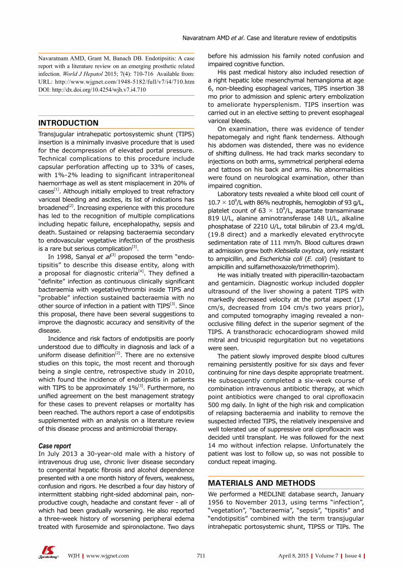
Navaratnam AMD, Grant M, Banach DB. Endotipsitis: A case report with a literature review on an emerging prosthetic related infection. World J Hepatol 2015; 7(4): 710-716 Available from: URL: http://www.wjgnet.com/1948-5182/full/v7/i4/710.htm DOI: http://dx.doi.org/10.4254/wjh.v7.i4.710
INTRODUCTIONTransjugular intrahepatic portosystemic shunt (TIPS) insertion is a minimally invasive procedure that is used for the decompression of elevated portal pressure. Technical complications to this procedure include capsular perforation affecting up to 33% of cases, with 1%-2% leading to significant intraperitoneal haemorrhage as well as stent misplacement in 20% of cases[1]. Although initially employed to treat refractory variceal bleeding and ascites, its list of indications has broadened[2]. Increasing experience with this procedure has led to the recognition of multiple complications including hepatic failure, encephalopathy, sepsis and death. Sustained or relapsing bacteraemia secondary to endovascular vegetative infection of the prosthesis is a rare but serious complication[3].
In 1998, Sanyal et al[3] proposed the term “endo-tipsitis” to describe this disease entity, along with a proposal for diagnostic criteria[4]. They defined a “definite” infection as continuous clinically significant bacteraemia with vegetative/thrombi inside TIPS and “probable” infection sustained bacteraemia with no other source of infection in a patient with TIPS[3]. Since this proposal, there have been several suggestions to improve the diagnostic accuracy and sensitivity of the disease.
Incidence and risk factors of endotipsitis are poorly understood due to difficulty in diagnosis and lack of a uniform disease definition[2]. There are no extensive studies on this topic, the most recent and thorough being a single centre, retrospective study in 2010, which found the incidence of endotipsitis in patients with TIPS to be approximately 1%[3]. Furthermore, no unified agreement on the best management strategy for these cases to prevent relapses or mortality has been reached. The authors report a case of endotipsitis supplemented with an analysis on a literature review of this disease process and antimicrobial therapy.
Case reportIn July 2013 a 30-year-old male with a history of intravenous drug use, chronic liver disease secondary to congenital hepatic fibrosis and alcohol dependence presented with a one month history of fevers, weakness, confusion and rigors. He described a four day history of intermittent stabbing right-sided abdominal pain, non-productive cough, headache and constant fever - all of which had been gradually worsening. He also reported a three-week history of worsening peripheral edema treated with furosemide and spironolactone. Two days
before his admission his family noted confusion and impaired cognitive function.
His past medical history also included resection of a right hepatic lobe mesenchymal hemangioma at age 6, non-bleeding esophageal varices, TIPS insertion 38 mo prior to admission and splenic artery embolization to ameliorate hypersplenism. TIPS insertion was carried out in an elective setting to prevent esophageal variceal bleeds.
On examination, there was evidence of tender hepatomegaly and right flank tenderness. Although his abdomen was distended, there was no evidence of shifting dullness. He had track marks secondary to injections on both arms, symmetrical peripheral edema and tattoos on his back and arms. No abnormalities were found on neurological examination, other than impaired cognition.
Laboratory tests revealed a white blood cell count of 10.7 × 10⁹/L with 86% neutrophils, hemoglobin of 93 g/L, platelet count of 63 × 109/L, aspartate transaminase 819 U/L, alanine aminotransferase 148 U/L, alkaline phosphatase of 2210 U/L, total bilirubin of 23.4 mg/dL (19.8 direct) and a markedly elevated erythrocyte sedimentation rate of 111 mm/h. Blood cultures drawn at admission grew both Klebsiella oxytoca, only resistant to ampicillin, and Escherichia coli (E. coli) (resistant to ampicillin and sulfamethoxazole/trimethoprim).
He was initially treated with piperacillin-tazobactam and gentamicin. Diagnostic workup included doppler ultrasound of the liver showing a patent TIPS with markedly decreased velocity at the portal aspect (17 cm/s, decreased from 104 cm/s two years prior), and computed tomography imaging revealed a non-occlusive filling defect in the superior segment of the TIPS. A transthoracic echocardiogram showed mild mitral and tricuspid regurgitation but no vegetations were seen.
The patient slowly improved despite blood cultures remaining persistently positive for six days and fever continuing for nine days despite appropriate treatment. He subsequently completed a six-week course of combination intravenous antibiotic therapy, at which point antibiotics were changed to oral ciprofloxacin 500 mg daily. In light of the high risk and complication of relapsing bacteraemia and inability to remove the suspected infected TIPS, the relatively inexpensive and well tolerated use of suppressive oral ciprofloxacin was decided until transplant. He was followed for the next 14 mo without infection relapse. Unfortunately the patient was lost to follow up, so was not possible to conduct repeat imaging.
MATERIALS AND METHODSWe performed a MEDLINE database search, January 1956 to November 2013, using terms “infection”, “vegetation”, “bacteraemia”, “sepsis”, “tipsitis” and “endotipsitis” combined with the term transjugular intrahepatic portosystemic shunt, TIPSS or TIPs. The
711 April 8, 2015|Volume 7|Issue 4|WJH|www.wjgnet.com
Navaratnam AMD et al . Case and literature review of endotipsitis
titles and abstracts of all papers that appeared on this search were reviewed and excluded accordingly. All data was entered in Microsoft Excel spreadsheet software. Univariate analysis was performed using a χ 2 or Fisher’s exact test, as appropriate. Data was analyzed in SPSS, version 16.0[4]. P-values less than 0.05 were considered statistically significant.
RESULTSOur literature search produced 283 papers, 246 of which were excluded on the basis of their title and abstract. Of the 37 remaining articles, another 15 were excluded after reviewing the contents of the full article. There were 22 papers that reported a sum of 53 cases of “endotipsitis”; 15 case reports and 7 case series (Figure 1). Based on the demographics of the cases, including, age, gender, co-morbidities and aetiological agent, it was possible to exclude any duplications. Analysis includes the case reported by the authors.
Patient characteristics The mean age was 54.3 years (range 30-69 years; 14 cases unrecorded), with a predominance of males (70.4% vs 17%). The most common underlying disease process was alcoholic liver disease (n = 32), 6 of which were also positive for hepatitis B (HBV) (n = 1), hepatitis C virus (HCV) (n = 4) or both (n = 1). HCV cirrhosis (n = 10) was the second most common etiology of chronic liver disease, followed by cryptogenic cirrhosis (n = 9), HBV (n = 1), primary sclerosing cholangitis (n = 1) and one case was not recorded. There was no record of whether viral hepatitis positive cases were undergoing treatment at the time of diagnosis of endotipsitis.
MicrobiologyOf the 54 cases (including our case report); there were 30 monomicrobial gram-positive infections [of which 14 were Enterococcal spp and 7 were Staphylococcus aureus (S. aureus)], 11 monomicrobial gram-negative
infections (of which 6 were E. coli), 5 polymicrobial and 6 Candida spp infections (4 Candida glabrata and 2 Candida albicans). The mean onset of endotipsitis from insertion of TIPS was 277 d, with bacterial being 304 d, and the longest overall being our case report 1158 d (range 6-158 d; 3 unrecorded). Among monomicrobial bacterial infections gram-positive organisms were significantly more common in early-onset infections (less than 120 d following TIPS insertion) than late-onset infections [18/19, (94.7%) vs 9/18 (50%) P < 0.01]. The average duration of bacteraemia was 43 d (range 3-20 d; 14 unrecorded).
TreatmentOf the 54 cases reported, 30 (56%) were treated successfully with antimicrobials, 4 (7%) underwent transplantation and 17 (31%) died (Table 1). The average duration of treatment for fungemia and bacteremia was 28 (range 6-51 wk, 3 unrecorded) and 13.5 (range 1-101 wk; 10 unrecorded) wk respectively. Of those with bacteremia, 25 cases were treated with a single antibiotic (19 cases resolved; 2 outcome not recorded), where as 14 were managed with combi-nation antibiotic therapy (1 case patient received 6 different antibiotics; 8 cases resolved). Comparing gram-positive and gram-negative infections, there was no significant difference in mortality [9/30 (30%) vs 4/12 (33%), P = 0.57]. Of the 39 antibiotic therapies recorded for bacterial endotipsitis, 7 cases used an aminoglycoside (gentamicin) and 2 used rifampin. None of the staphylococcal cases were treated with gentamicin.
The organisms associated with the highest mortality rates were S. aureus (63% mortality) and Candida spp (67% mortality). Patients with infections caused by S. aureus and Candida spp had significantly higher mortality than those infections caused by all other organisms [9/14 (64.3%) vs 8/36 (22.2%) P < 0.01]. There was no difference in mortality rate between early and late-onset infections [8/22 (36.4%) vs 7/23 (30.4%) P = 0.67].
DISCUSSIONThe authors have reported a case of Klebsiella oxytoca and E. coli polymicrobial endotipsitis with the longest latency of time between TIPS insertion and TIPS infection ever reported. A case of endotipsitis with similar bacteriology was reported previously but was managed quite differently (ceftriaxone monotherapy for 4 wk). Our literature review reviews the largest number of reported endotipsitis cases to date and provides a window into a rare disease, heterogeneous in both its microbiologic etiology and management approaches.
Endotipsitis is suspected in the setting of unex-plained bacteremia in a patient with TIPS, supported by abnormalities in TIPS flow or the presence of vegetation
712 April 8, 2015|Volume 7|Issue 4|WJH|www.wjgnet.com
283 papers
37 papers
246 papers excluded after reviewing title and abstract
15 excluded after reviewing full article
22 papers
53 cases
Figure 1 Number of papers excluded and include from the results of the literature review.
Navaratnam AMD et al . Case and literature review of endotipsitis
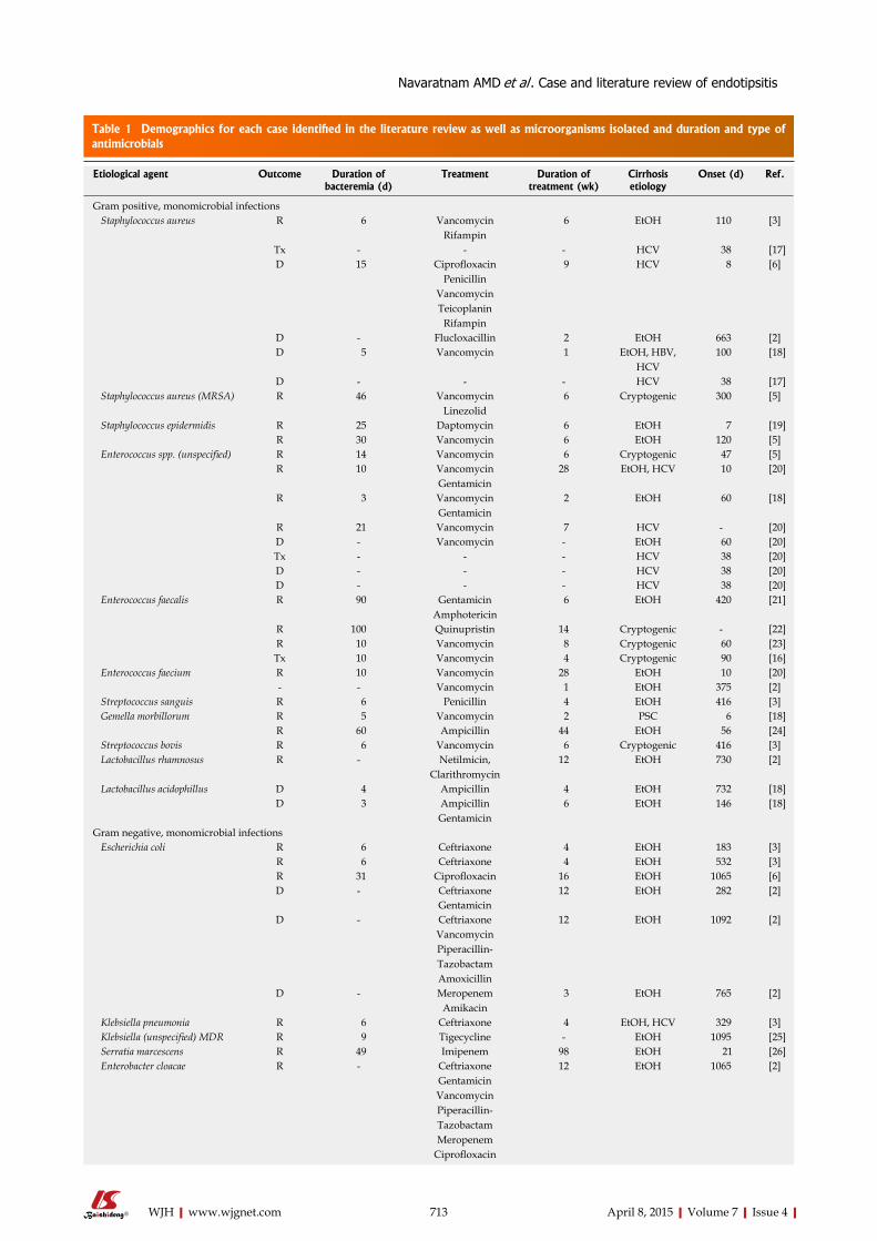
713 April 8, 2015|Volume 7|Issue 4|WJH|www.wjgnet.com
Etiological agent Outcome Duration of bacteremia (d)
Treatment Duration of treatment (wk)
Cirrhosis etiology
Onset (d) Ref.
Gram positive, monomicrobial infections Staphylococcus aureus R 6 Vancomycin
Rifampin 6 EtOH 110 [3]
Tx - - - HCV 38 [17]D 15 Ciprofloxacin
Penicillin Vancomycin Teicoplanin
Rifampin
9 HCV 8 [6]
D - Flucloxacillin 2 EtOH 663 [2]D 5 Vancomycin 1 EtOH, HBV,
HCV 100 [18]
D - - - HCV 38 [17] Staphylococcus aureus (MRSA) R 46 Vancomycin
Linezolid 6 Cryptogenic 300 [5]
Staphylococcus epidermidis R 25 Daptomycin 6 EtOH 7 [19]R 30 Vancomycin 6 EtOH 120 [5]
Enterococcus spp. (unspecified) R 14 Vancomycin 6 Cryptogenic 47 [5]R 10 Vancomycin
Gentamicin28 EtOH, HCV 10 [20]
R 3 VancomycinGentamicin
2 EtOH 60 [18]
R 21 Vancomycin 7 HCV - [20]D - Vancomycin - EtOH 60 [20]Tx - - - HCV 38 [20]D - - - HCV 38 [20]D - - - HCV 38 [20]
Enterococcus faecalis R 90 Gentamicin Amphotericin
6 EtOH 420 [21]
R 100 Quinupristin 14 Cryptogenic - [22]R 10 Vancomycin 8 Cryptogenic 60 [23]Tx 10 Vancomycin 4 Cryptogenic 90 [16]
Enterococcus faecium R 10 Vancomycin 28 EtOH 10 [20]- - Vancomycin 1 EtOH 375 [2]
Streptococcus sanguis R 6 Penicillin 4 EtOH 416 [3] Gemella morbillorum R 5 Vancomycin 2 PSC 6 [18]
R 60 Ampicillin 44 EtOH 56 [24] Streptococcus bovis R 6 Vancomycin 6 Cryptogenic 416 [3] Lactobacillus rhamnosus R - Netilmicin,
Clarithromycin12 EtOH 730 [2]
Lactobacillus acidophillus D 4 Ampicillin 4 EtOH 732 [18]D 3 Ampicillin
Gentamicin 6 EtOH 146 [18]
Gram negative, monomicrobial infections Escherichia coli R 6 Ceftriaxone 4 EtOH 183 [3]
R 6 Ceftriaxone 4 EtOH 532 [3]R 31 Ciprofloxacin 16 EtOH 1065 [6]D - Ceftriaxone 12 EtOH 282 [2]
GentamicinD - Ceftriaxone
VancomycinPiperacillin-Tazobactam Amoxicillin
12 EtOH 1092 [2]
D - MeropenemAmikacin
3 EtOH 765 [2]
Klebsiella pneumonia R 6 Ceftriaxone 4 EtOH, HCV 329 [3] Klebsiella (unspecified) MDR R 9 Tigecycline - EtOH 1095 [25] Serratia marcescens R 49 Imipenem 98 EtOH 21 [26] Enterobacter cloacae R - Ceftriaxone
GentamicinVancomycinPiperacillin-Tazobactam MeropenemCiprofloxacin
12 EtOH 1065 [2]
Table 1 Demographics for each case identified in the literature review as well as microorganisms isolated and duration and type of antimicrobials
Navaratnam AMD et al . Case and literature review of endotipsitis
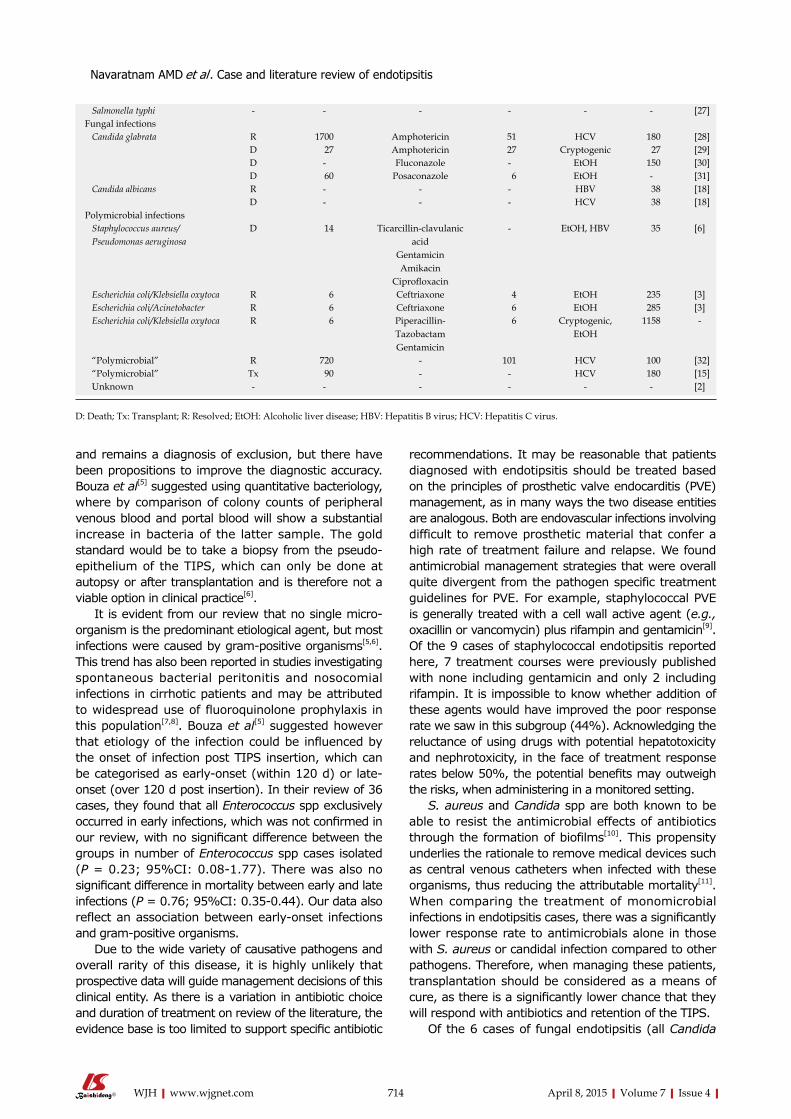
714 April 8, 2015|Volume 7|Issue 4|WJH|www.wjgnet.com
and remains a diagnosis of exclusion, but there have been propositions to improve the diagnostic accuracy. Bouza et al[5] suggested using quantitative bacteriology, where by comparison of colony counts of peripheral venous blood and portal blood will show a substantial increase in bacteria of the latter sample. The gold standard would be to take a biopsy from the pseudo-epithelium of the TIPS, which can only be done at autopsy or after transplantation and is therefore not a viable option in clinical practice[6].
It is evident from our review that no single micro-organism is the predominant etiological agent, but most infections were caused by gram-positive organisms[5,6]. This trend has also been reported in studies investigating spontaneous bacterial peritonitis and nosocomial infections in cirrhotic patients and may be attributed to widespread use of fluoroquinolone prophylaxis in this population[7,8]. Bouza et al[5] suggested however that etiology of the infection could be influenced by the onset of infection post TIPS insertion, which can be categorised as early-onset (within 120 d) or late-onset (over 120 d post insertion). In their review of 36 cases, they found that all Enterococcus spp exclusively occurred in early infections, which was not confirmed in our review, with no significant difference between the groups in number of Enterococcus spp cases isolated (P = 0.23; 95%CI: 0.08-1.77). There was also no significant difference in mortality between early and late infections (P = 0.76; 95%CI: 0.35-0.44). Our data also reflect an association between early-onset infections and gram-positive organisms.
Due to the wide variety of causative pathogens and overall rarity of this disease, it is highly unlikely that prospective data will guide management decisions of this clinical entity. As there is a variation in antibiotic choice and duration of treatment on review of the literature, the evidence base is too limited to support specific antibiotic
recommendations. It may be reasonable that patients diagnosed with endotipsitis should be treated based on the principles of prosthetic valve endocarditis (PVE) management, as in many ways the two disease entities are analogous. Both are endovascular infections involving difficult to remove prosthetic material that confer a high rate of treatment failure and relapse. We found antimicrobial management strategies that were overall quite divergent from the pathogen specific treatment guidelines for PVE. For example, staphylococcal PVE is generally treated with a cell wall active agent (e.g., oxacillin or vancomycin) plus rifampin and gentamicin[9]. Of the 9 cases of staphylococcal endotipsitis reported here, 7 treatment courses were previously published with none including gentamicin and only 2 including rifampin. It is impossible to know whether addition of these agents would have improved the poor response rate we saw in this subgroup (44%). Acknowledging the reluctance of using drugs with potential hepatotoxicity and nephrotoxicity, in the face of treatment response rates below 50%, the potential benefits may outweigh the risks, when administering in a monitored setting.
S. aureus and Candida spp are both known to be able to resist the antimicrobial effects of antibiotics through the formation of biofilms[10]. This propensity underlies the rationale to remove medical devices such as central venous catheters when infected with these organisms, thus reducing the attributable mortality[11]. When comparing the treatment of monomicrobial infections in endotipsitis cases, there was a significantly lower response rate to antimicrobials alone in those with S. aureus or candidal infection compared to other pathogens. Therefore, when managing these patients, transplantation should be considered as a means of cure, as there is a significantly lower chance that they will respond with antibiotics and retention of the TIPS.
Of the 6 cases of fungal endotipsitis (all Candida
Salmonella typhi - - - - - - [27]Fungal infections Candida glabrata R 1700 Amphotericin 51 HCV 180 [28]
D 27 Amphotericin 27 Cryptogenic 27 [29]D - Fluconazole - EtOH 150 [30]D 60 Posaconazole 6 EtOH - [31]
Candida albicans R - - - HBV 38 [18]D - - - HCV 38 [18]
Polymicrobial infections Staphylococcus aureus/ Pseudomonas aeruginosa
D 14 Ticarcillin-clavulanic acid
GentamicinAmikacin
Ciprofloxacin
- EtOH, HBV 35 [6]
Escherichia coli/Klebsiella oxytoca R 6 Ceftriaxone 4 EtOH 235 [3] Escherichia coli/Acinetobacter R 6 Ceftriaxone 6 EtOH 285 [3] Escherichia coli/Klebsiella oxytoca R 6 Piperacillin-
TazobactamGentamicin
6 Cryptogenic, EtOH
1158 -
“Polymicrobial” R 720 - 101 HCV 100 [32] “Polymicrobial” Tx 90 - - HCV 180 [15] Unknown - - - - - - [2]
D: Death; Tx: Transplant; R: Resolved; EtOH: Alcoholic liver disease; HBV: Hepatitis B virus; HCV: Hepatitis C virus.
Navaratnam AMD et al . Case and literature review of endotipsitis
715 April 8, 2015|Volume 7|Issue 4|WJH|www.wjgnet.com
spp), only 2 responded to antifungal treatment. Similarly, Candida spp are the most common cause of fungal infective endocarditis (IE), with these patients more likely to have prosthetic intravascular devices[12]. Baddley et al[12] found persistently positive blood cultures were associated with this group as well as a significantly higher rate of mortality when compared to non-fungal cases of IE[13]. With the accumulating evidence of dismal outcomes in patients with candidal endotipsitis, strong consideration for urgent transplantation needs to be contemplated in appropriate patients[14,15].
In patients who are ineligible for transplantation aggressive medical therapy and potentially chronic suppressive antibiotics should be considered acknow-ledging the prognosis is poor. In general, the role and timing of liver transplantation in the setting of endotipsitis is poorly understood. In our review we identified four patients who underwent transplantation. Mizrahi et al[1] did report clearance of blood cultures prior to transplant in their 2 cases[16,17]. Jawaid et al[15] described a case with repeat episodes of bacteraemia, 1 mo after TIPS insertion, with 9 different microorganisms isolated from blood cultures over the period of 11 mo. Willner et al[16] reported persistent polymicrobial bacteraemia 4 d after a TIPS revision, which resulted in a transplant 3 mo later (after 3 episodes of bacteraemia). It unclear whether the patients had negative blood cultures leading up to their transplant. In both cases, a biliary-venous fistula was not identified until after the assessment of the explant. Among patients with suspected endotipsitis and persistent or relapsing bacteremia, biliary-venous fistula should be considered and in this setting transplantation would be warranted to achieve cure.
Optimal treatment of endotipsitis remains elusive given the difficulty of studying a rare disease without a broadly accepted definition and caused by a variety of microbes. Our data suggests that infections caused by S. aureus and Candida spp is associated with particularly poor outcomes. Based on the treatment of PVE, addition of gentamicin and rifampin to cell wall active antimicrobials may improve outcomes of S. aureus endotipsitis cases. Transplantation should be strongly considered in the setting of relapsing disease and infections caused by difficult to eradicate pathogens. More data on the epidemiology and management outcomes of endotipsitis will be needed to determine the most appropriate prevention and treatment strategies.
COMMENTSBackground“Endotipsitis” is a poorly understood complication of transjugular intrahepatic portosystemic shunt (TIPS) procedures. Sustained or relapsing bacteraemia secondary to endovascular vegetative infection of the prosthesis, “endotipsitis”, is a rare but serious complication. The authors report a case with the longest latency from insertion to infection in the literature, supplemented by a review of the literature, in particular the management of this insidious and variable disease process.Research frontiersIncidence and risk factors of endotipsitis are poorly understood due to difficulty
in diagnosis and lack of a uniform disease definition. Furthermore, no unified agreement on the best management strategy for these cases to prevent relapses or mortality has been reached.Innovations and breakthroughsThe authors’ data reflects an association between early-onset infections and gram-positive organisms. It also suggests that infections caused by Staphylococcus aureus (S. aureus) and Candida spp are associated with particularly poor outcomes. The evidence base is too limited to support specific antibiotic recommendations, but it may be reasonable to treat based on the principles of prosthetic valve endocarditis (PVE) management. ApplicationsTransplantation should be strongly considered in those with infections associated with particularly poor outcomes, such as Candida spp. Based on the treatment of PVE, addition of gentamicin and rifampin to cell wall active antimicrobials may improve outcomes of S. aureus endotipsitis cases.TerminologyTIPS insertion is a minimally invasive procedure that is used for the decom-pression of elevated portal pressure. Although there is no universally accepted definition of “endotipsitis”, it is generally agreed that it can be defined as “definite” infection (continuous clinically significant bacteraemia with vegetative/thrombi inside TIPS) and “probable” infection (sustained bacteraemia with no other source of infection in a patient with TIPS).Peer-reviewThe manuscript presents an interesting case of “endotipsitis” and reviews the literature on a rare disease. In this paper Navaratnam et al present a case of “endotipsitis” and performed a review of the existing literature on this field. They identified 22 papers reporting 54 patients with endotipsitis. The great majority had monomicrobial infections (gram positive agents were the majority). Infections with S. aureus and Candida spp were associated higher mortality. No homogeneous management was applied for the treatment of this condition and guidelines on antibiotics use are usually derivate from the treatment of endocarditis.
REFERENCES1 Mizrahi M, Adar T, Shouval D, Bloom AI, Shibolet O. Endotipsitis-
persistent infection of transjugular intrahepatic portosystemic shunt: pathogenesis, clinical features and management. Liver Int 2010; 30: 175-183 [PMID: 19929905 DOI: 10.1111/j.1478-3231]
2 Kochar N, Tripathi D, Arestis NJ, Ireland H, Redhead DN, Hayes PC. Tipsitis: incidence and outcome-a single centre experience. Eur J Gastroenterol Hepatol 2010; 22: 729-735 [PMID: 20440117 DOI: 10.1097/MEG.0b013e3282fd6917]
3 Sanyal AJ, Reddy KR. Vegetative infection of transjugular intra-hepatic portosystemic shunts. Gastroenterology 1998; 115: 110-115 [PMID: 9649465 DOI: 10.1016/S0016-5085(98)70371-3]
4 Fisher RA. On the interpretation of Chi-square from contingency tables and the calculation of P. J R Stat Soc 1922; 85: 87-94 [DOI: 10.2307/2340521]
5 Bouza E, Muñoz P, Rodríguez C, Grill F, Rodríguez-Créixems M, Bañares R, Fernández J, García-Pagán JC. Endotipsitis: an emerging prosthetic-related infection in patients with portal hypertension. Diagn Microbiol Infect Dis 2004; 49: 77-82 [PMID: 15183855 DOI: 10.1016/j.diagmicrobio.2004.03.006]
6 Armstrong PK, MacLeod C. Infection of transjugular intrahepatic portosystemic shunt devices: three cases and a review of the literature. Clin Infect Dis 2003; 36: 407-412 [PMID: 12567297 DOI: 10.1086/346156]
7 Fernández J, Navasa M, Gómez J, Colmenero J, Vila J, Arroyo V, Rodés J. Bacterial infections in cirrhosis: epidemiological changes with invasive procedures and norfloxacin prophylaxis. Hepatology 2002; 35: 140-148 [PMID: 11786970 DOI: 10.1053/jhep.2002.30082]
8 Gould FK, Denning DW, Elliott TS, Foweraker J, Perry JD, Prendergast BD, Sandoe JA, Spry MJ, Watkin RW. Guidelines for the diagnosis and antibiotic treatment of endocarditis in adults: a report of the Working Party of the British Society for Antimicrobial Chemotherapy. J Antimicrob Chemother 2012; 67: 269-289 [PMID: 22086858 DOI: 10.1093/jac/dkr450]
9 Harriott MM, Noverr MC. Candida albicans and Staphylococcus
COMMENTS
Navaratnam AMD et al . Case and literature review of endotipsitis
716 April 8, 2015|Volume 7|Issue 4|WJH|www.wjgnet.com
aureus form polymicrobial biofilms: effects on antimicrobial resistance. Antimicrob Agents Chemother 2009; 53: 3914-3922 [PMID: 19564370 DOI: 10.1128/AAC.00657-09]
10 Steinbach WJ, Perfect JR, Cabell CH, Fowler VG, Corey GR, Li JS, Zaas AK, Benjamin DK. A meta-analysis of medical versus surgical therapy for Candida endocarditis. J Infect 2005; 51: 230-247 [PMID: 16230221 DOI: 10.1016/j.jinf.2004.10.016]
11 Falcone M, Barzaghi N, Carosi G, Grossi P, Minoli L, Ravasio V, Rizzi M, Suter F, Utili R, Viscoli C, Venditti M; Italian Study on Endocarditis. Candida infective endocarditis: report of 15 cases from a prospective multicenter study. Medicine (Baltimore) 2009; 88: 160-168 [PMID: 19440119 DOI: 10.1097/MD.0b013e3181a693f8]
12 Baddley JW, Benjamin DK, Patel M, Miró J, Athan E, Barsic B, Bouza E, Clara L, Elliott T, Kanafani Z, Klein J, Lerakis S, Levine D, Spelman D, Rubinstein E, Tornos P, Morris AJ, Pappas P, Fowler VG, Chu VH, Cabell C; International Collaboration on Endocarditis-Prospective Cohort Study Group (ICE-PCS). Candida infective endocarditis. Eur J Clin Microbiol Infect Dis 2008; 27: 519-529 [PMID: 18283504 DOI: 10.1007/s10096-008-0466-x]
13 Mora-Duarte J, Betts R, Rotstein C, Colombo AL, Thompson-Moya L, Smietana J, Lupinacci R, Sable C, Kartsonis N, Perfect J. Comparison of caspofungin and amphotericin B for invasive candidiasis. N Engl J Med 2002; 347: 2020-2029 [PMID: 12490683 DOI: 10.1056/NEJMoa021585]
14 Kullberg BJ, Sobel JD, Ruhnke M, Pappas PG, Viscoli C, Rex JH, Cleary JD, Rubinstein E, Church LW, Brown JM, Schlamm HT, Oborska IT, Hilton F, Hodges MR. Voriconazole versus a regimen of amphotericin B followed by fluconazole for candidaemia in non-neutropenic patients: a randomised non-inferiority trial. Lancet 2005; 366: 1435-1442 [PMID: 16243088 DOI: 10.1016/S0140-6736(05)67490-9]
15 Jawaid Q, Saeed ZA, Di Bisceglie AM, Brunt EM, Ramrakhiani S, Varma CR, Solomon H. Biliary-venous fistula complicating transjugular intrahepatic portosystemic shunt presenting with recurrent bacteremia, jaundice, anemia and fever. Am J Transplant 2003; 3: 1604-1607 [PMID: 14629294 DOI: 10.1046/j.1600-6135.2003.00267.x]
16 Willner IR, El-Sakr R, Werkman RF, Taylor WZ, Riely CA. A fistula from the portal vein to the bile duct: an unusual complication of transjugular intrahepatic portosystemic shunt. Am J Gastroenterol 1998; 93: 1952-1955 [PMID: 9772063 DOI: 10.1111/j.1572-0241.1998.00553.x]
17 Mizrahi M, Roemi L, Shouval D, Adar T, Korem M, Moses A, Bloom A, Shibolet O. Bacteremia and “Endotipsitis” following transjugular intrahepatic portosystemic shunting. World J Hepatol 2011; 3: 130-136 [PMID: 21731907 DOI: 10.4254/wjh.v3.i5.130]
18 DeSimone JA, Beavis KG, Eschelman DJ, Henning KJ. Sustained bacteremia associated with transjugular intrahepatic portosystemic shunt (TIPS). Clin Infect Dis 2000; 30: 384-386 [PMID: 10671346 DOI: 10.1086/313653]
19 Colston JM, Scarborough M, Collier J, Bowler IC. High-dose daptomycin monotherapy cures Staphylococcus epidermidis ‘endotipsitis’ after failure of conventional therapy. BMJ Case Rep
2013; 2013: pii: bcr2013009529 [PMID: 23595199 DOI: 10.1136/bcr-2013-009529]
20 Brown RS, Brumage L, Yee HF, Lake JR, Roberts JP, Somberg KA. Enterococcal bacteremia after transjugular intrahepatic portosystemic shunts (TIPS). Am J Gastroenterol 1998; 93: 636-639 [PMID: 9576462 DOI: 10.1111/j.1572-0241.1998.180_b.x]
21 Passeron A, Mihaïla-Amrouche L, Perreira Rocha E, Wyplosz B, Capron L. [Recurrent enterococcal bacteremia associated with a transjugular intrahepatic protosystemic shunt]. Gastroenterol Clin Biol 2004; 28: 1284-1286 [PMID: 15671940 DOI: 10.1016/S0399-8320(04)95222-0]
22 Zaman MM, Recco R, Tejwani U, Scuto TJ, Ahmed S, Hypolite A, Jayaraman G. Case of vancomycin-resistant Enterococcus faecium infection associated with a transjugular intrahepatic portosystemic shunt that was treated with quinupristin/dalfopristin after bacteremia persisted with alatrofloxacin therapy. Clin Infect Dis 1999; 29: 954-955 [PMID: 10589932 DOI: 10.1086/520480]
23 Eversman D, Chalasani N. A case of infective endotipsitis. Gastroenterology 1999; 117: 514 [PMID: 10465639 DOI: 10.1053/gast.1999.0029900514a]
24 Medina-Gens L, López J, Manzanedo B, Pintado V. [Endotipsitis due to Gemella morbillorum]. Enferm Infecc Microbiol Clin 2007; 25: 419-420 [PMID: 17583662 DOI: 10.1157/13106974]
25 Aggarwal S, Park J. Endotipsitis: a diagnostic challenge. J Postgrad Med 2011; 57: 134 [PMID: 21654138 DOI: 10.4103/0022-3859.81875]
26 Marques N, Sá R, Coelho F, da Cunha S, Meliço-Silvestre A. Spondylodiscitis associated with recurrent Serratia bacteremia due to a transjugular intrahepatic portosystemic shunt (TIPS): a case report. Braz J Infect Dis 2007; 11: 525-527 [PMID: 17962882 DOI: 10.1590/S1413-86702007000500016]
27 Barrio J, Castiella A, Von Wichman MA, Cosme A, López P, Arenas JI. [Spontaneous bacteremia due to Salmonella hadar in liver cirrhosis with transjugular intrahepatic portosystemic shunt]. Gastroenterol Hepatol 1999; 22: 79-81 [PMID: 10193091]
28 Brickey TW, Trotter JF, Johnson SP. Torulopsis glabrata fungemia from infected transjugular intrahepatic portosystemic shunt stent. J Vasc Interv Radiol 2005; 16: 751-752 [PMID: 15872333 DOI: 10.1097/01.RVI.0000153587.52276.06]
29 Darwin P, Mergner W, Thuluvath P. Torulopsis glabrata fungemia as a complication of a clotted transjugular intrahepatic portosystemic shunt. Liver Transpl Surg 1998; 4: 89-90 [PMID: 9457972 DOI: 10.1002/lt.500040112]
30 Schiano TD, Atillasoy E, Fiel MI, Wolf DC, Jaffe D, Cooper JM, Jonas ME, Bodenheimer HC, Min AD. Fatal fungemia resulting from an infected transjugular intrahepatic portosystemic shunt stent. Am J Gastroenterol 1997; 92: 709-710 [PMID: 9128335]
31 Anstead GM, Martinez M, Graybill JR. Control of a Candida glabrata prosthetic endovascular infection with posaconazole. Med Mycol 2006; 44: 273-277 [PMID: 16702108 DOI: 10.1080/13693780500049152]
32 Suhocki PV, Smith AD, Tendler DA, Sexton DJ. Treatment of TIPS/biliary fistula-related endotipsitis with a covered stent. J Vasc Interv Radiol 2008; 19: 937-939 [PMID: 18503911 DOI: 10.1016/j.jvir.2008.01.026]
P- Reviewer: De Ponti F, Narciso-Schiavon JL, Procopet B S- Editor: Tian YL L- Editor: A E- Editor: Liu SQ
Navaratnam AMD et al . Case and literature review of endotipsitis
Published online: April 8, 2015
AbstractLiver transplantation (LT) is a widely-accepted, definitive therapy of irreversible liver diseases including hepatitis C, alcoholic liver disease and metabolic liver disease. After transplantation, patients generally use a variety of immunosuppressive medications for the rest of their lives to prevent rejection of transplanted liver. Mortality after LT is mainly caused by recurrence of alcoholic hepatitis which is mostly seen in the patients who resume heavy drinking. On the other hand, de-novo malignancies after LT are not seldom. Skin cancers make up 13.5% of the de-novo malignancies seen in these patients. Malignancies tend to affect survival earlier in the course with a 53% risk of death at 5 years after diagnosis. We aimed to report a case who underwent LT secondary to alcoholic liver disease and developed squamous cell carcinoma of the skin eighteen years after transplantation. In summary, transplant recipients are recommended to be educated on self examination for skin cancer; health care providers should be further suspicious during routine dermatological examinations of the transplant patients and biopsies of possible lesions for skin cancer is warranted even many years after transplantation.
Key words: Alcoholic liver disease; Skin cancer; Non-squamous; Liver transplantation; Sirolimus
© The Author(s) 2015. Published by Baishideng Publishing Group Inc. All rights reserved.
Core tip: We presented a case who underwent liver transplantation due to alcoholic liver disease and developed a skin cancer after 18 years of follow-up, which is exceptionally rare as malignancies tend to
Ozan Unlu, Emir Charles Roach, Alexis Okoh, May Olayan, Bulent Yilmaz, Didem Uzunaslan, Abdullah Shatnawei
Ozan Unlu, Didem Uzunaslan, Department of Pathobiology, Cleveland Clinic, Cleveland, OH 44106, United StatesEmir Charles Roach, Abdullah Shatnawei, Department of Gastroenterology, Cleveland Clinic, Cleveland, OH 44106, United StatesAlexis Okoh, Department of Endocrine Surgery, Cleveland Clinic, Cleveland, OH 44106, United StatesMay Olayan, Department of Internal Medicine, Fairview Hospital, Cleveland Clinic, Cleveland, OH 44106, United StatesBulent Yilmaz, Department of Gastroenterology, Hacettepe University School of Medicine, 06230 Ankara, TurkeyAuthor contributions: All authors contributed significantly to the work, had read and revised the manuscript.Ethics approval: Cleveland Clinic Publication Guidance for IRB review and HIPAA Compliance allow researchers and physician publish case reports involving three or less patients without and IRB review as long as all the patients involved in the study provide written informed consent forms.Informed consent: The patient in this study provided informed written consent prior to study enrollment.Conflict-of-interest: We certify that there is no conflict of interest with any commercial, personal, political, intellectual, religious or any other kind of organization regarding the material discussed in the manuscript.Open-Access: This article is an open-access article which was selected by an in-house editor and fully peer-reviewed by external reviewers. It is distributed in accordance with the Creative Commons Attribution Non Commercial (CC BY-NC 4.0) license, which permits others to distribute, remix, adapt, build upon this work non-commercially, and license their derivative works on different terms, provided the original work is properly cited and the use is non-commercial. See: http://creativecommons.org/licenses/by-nc/4.0/Correspondence to: Emir Charles Roach, MD, Department of Gastroenterology, Cleveland Clinic, 9500 Euclid Avenue, Cleveland, OH 44106, United States. [email protected]: +1-216-8488870Received: November 10, 2014 Peer-review started: November 11, 2014First decision: December 12, 2014Revised: December 27, 2014 Accepted: January 15, 2015Article in press: January 19, 2015
CASE REPORT
Submit a Manuscript: http://www.wjgnet.com/esps/Help Desk: http://www.wjgnet.com/esps/helpdesk.aspxDOI: 10.4254/wjh.v7.i4.717
717 April 8, 2015|Volume 7|Issue 4|WJH|www.wjgnet.com
World J Hepatol 2015 April 8; 7(4): 717-720ISSN 1948-5182 (online)
© 2015 Baishideng Publishing Group Inc. All rights reserved.
Skin cancer in immunosuppressed transplant patients: Vigilance matters
affect survival earlier in the course with a 53% risk of death at 5 years after diagnosis.
Unlu O, Roach EC, Okoh A, Olayan M, Yilmaz B, Uzunaslan D, Shatnawei A. Skin cancer in immunosuppressed transplant patients: Vigilance matters. World J Hepatol 2015; 7(4): 717-720 Available from: URL: http://www.wjgnet.com/1948-5182/full/v7/i4/717.htm DOI: http://dx.doi.org/10.4254/wjh.v7.i4.717
INTRODUCTIONAlcoholic liver disease (ALD) is known to be the number one cause of cirrhosis in western countries. Twenty percent of the liver transplants in United States and forty per-cent in Europe are performed due to ALD which makes it the second most common indication for liver transplantation (LT), a definitive treatment option for patients with cirrhosis and end stage liver disease[1-3].
Patient survival rates after LT for alcoholic cirrhosis have been reported to be 73%-86% at 5 years after diagnosis[4,5]. Mortality after LT is mainly caused by recurrence of ALD and alcoholic hepatitis which are mostly seen in the patients who resume heavy drinking. Other causes of mortality in patients who stay abstinent are cardiovascular diseases or events, infection and malignancies. Of note is the high incidence of de-novo malignancies after LT. This has been associated with the use of post LT immunosuppressants and the history of heavy alcohol use. Other reported risk factors for de-novo malignancies include older age, male gender, and Epstein Barr virus reactivation or infection for lymphoproliferative malignancy, and exposure to sun for non-melanoma skin cancer. Skin cancers make up the 13.5% of the de-novo malignancies seen in the patients with LT[6-8]. Malignancies tend to affect survival early after LT with a 53% risk of death at 5 years after diagnosis[9].
Herein, we present a case underwent LT (ALD etiology) who is followed up for 18 years without develop-ment of any malignancy. The patient consequently developed skin cancer at different sites 7 years after change of immunosuppressive regimen.
A high index of clinical suspicion together with routine dermatological examinations and biopsies of possible lesions for skin cancer is warranted in LT patients even eighteen years after transplant.
CASE REPORTThis is a 74-year-old male patient, who was under follow up for LT due to ALD at our center for 18 years. He was first diagnosed with cirrhosis secondary to ALD in 1993 and had a liver transplant two years later. Early post-transplant immunosuppressive treatment regimen included mycophenolate and tacrolimus for which he showed moderate clinical response. Afterwards, he
developed calcineurin inhibitor induced end stage renal insufficiency and switched to sirolimus from tacrolimus in 2003 and a combination therapy with mycophenolate was continued.
Previous medical and surgical history included uncomplicated diabetes mellitus, hyperlipidemia and hypertension for 15 years and aortic valve replacement due to vascular thrombosis that occurred over the course of treatment. His co-morbidities were under control with antihypertensives and lipid lowering statins and his renal insufficiency related anemia was managed with erythropoietin.
Two years after treatment with sirolimus was started, the patient had recurring acneiform eruptions with pustules on his face, ears, and scalp. He was therefore referred to a dermatology unit for further evaluation and follow up.
A definitive diagnosis consistent with squamous cell carcinoma of the skin was made five years later. In September 2010, the patient underwent a resection of a tumor from his left temple, sacrificing the frontal branch of left facial nerve. He had multiple surgeries including left superficial parotidectomy due to relapses at different sites such as left auricle, preauricular area and occipitofrontal region of the scalp. Histological evaluations after each surgery revealed surgical margins wider than 6 mm. Identification of atypical tumor cells and keratinous pearls in microscopical evaluation supported the diagnosis. Despite multiple surgeries, chemotherapy and radiotherapy, patient was diagnosed with stage 4 squamous cell carcinoma due to metastatic lesions in his brain in 2012, and died in February 2013.
DISCUSSIONPatient survival rates after LT for alcoholic cirrhosis have been reported to be 81%-92%, 78%-86%, and 73%-86% at 1, 3, and 5 years respectively[4,5]. In an European Liver Transplant Registry which enrolled patients between 1988 and 2009, survival rates were reported as 73% and 59% for 5 and 10 years of follow-up respectively. These rates were shown to be higher compared to non-alcoholic etiology associated liver transplants, thus confirming ALD as an acceptable indication for LT[4,5,10].
Malignancy has been shown to significantly affect survival in LT patients with about 38% and 53% risk of death at 1 and 5 years after diagnosis[9]. Among LT recipients who survive the first year after trans-plantation, De novo malignancy is reported to account for 30%-40% of all deaths[9,11]. Although, intensive surveillance protocols in the post-transplant period have been shown to improve survival by detection of malignancy, clear guidelines including the frequency of work-up have not been developed yet. In this report, we describe the case of an LT patient who was followed up for 18 years after LT due to ALD. Renal insufficiency that occurred secondary to immunosuppressive therapy with
718 April 8, 2015|Volume 7|Issue 4|WJH|www.wjgnet.com
Unlu O et al . Skin cancer after liver transplantation
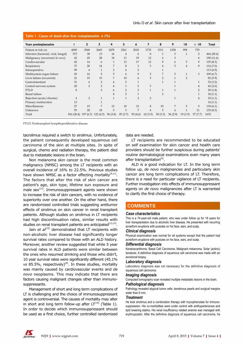
tacrolimus required a switch to sirolimus. Unfortunately, the patient consequently developed squamous cell carcinoma of the skin at multiple sites. In spite of surgical, chemo and radiation therapy, the patient died due to metastatic lesions in the brain.
Non melanoma skin cancer is the most common malignancy (NMSC) among the LT recipients with an overall incidence of 16% to 22.5%. Previous studies have shown NMSC as a factor effecting mortality[9,11]. The factors that alter the risk of skin cancer are patient’s age, skin type, lifetime sun exposure and male sex[12]. Immunosuppressant agents were shown to increase the risk of skin cancers, with no evidence of superiority over one another. On the other hand, there are randomized controlled trials suggesting antitumor effects of sirolimus on skin cancer in renal transplant patients. Although studies on sirolimus in LT recipients had high discontinuation rates, similar results with studies on renal transplant patients are anticipated[13,14].
Jain et al[15] demonstrated that LT recipients with non-alcoholic liver disease had significantly longer survival rates compared to those with an ALD history. Moreover, another review suggested that while 5 year survival rates in ALD patients were similar between the ones who resumed drinking and those who didn’t,10 year survival rates were significantly different (45.1% vs 85.5%, respectively)[8]. In these studies, mortality was mainly caused by cardiovascular events and de novo neoplasms. This may indicate that there are factors causing malignant changes other than immuno-suppressants.
Management of short and long term complications of LT is challenging and the choice of immunosuppressant agent is controversial. The causes of mortality may alter in short and long term follow-up after LT[16] (Table 1). In order to decide which immunosuppressant should be used as a first choice, further controlled randomized
data are needed. LT recipients are recommended to be educated
on self examination for skin cancer and health care providers should be further suspicious during patients’ routine dermatological examinations even many years after transplantation[6].
ALD is a good indication for LT. In the long term follow up, de novo malignancies and particularly skin cancer are long term complications of LT. Therefore, there is a need for particular vigilance of LT recipients. Further investigation into effects of immunosuppressant agents on de novo malignancies after LT is warranted to clarify the first choice of therapy.
COMMENTSCase characteristicsThis is a 74-year-old male patient, who was under follow up for 18 years for liver transplantation due to alcoholic liver disease. He presented with recurring acneiform eruptions with pustules on his face, ears, and scalp.Clinical diagnosisPhysical examination was normal for all systems except that the patient had acneiform eruptions with pustules on his face, ears, and scalp.Differential diagnosisKeratoacanthoma, Basal Cell Carcinoma, Malignant melanoma, Solar (actinic) keratosis. A definitive diagnosis of squamous cell carcinoma was made with an excisional biopsy. Laboratory diagnosisLaboratory diagnosis was not necessary for the definitive diagnosis of squamous cell carcinoma. Imaging diagnosisComputed tomography scan revealed multiple metastatic lesions in the brain.Pathological diagnosisPathology revealed atypical tumor cells, keratinous pearls and surgical margins wider than 6 mm.Treatment He took sirolimus and a combination therapy with mycophenolate for immuno-suppression. His co-morbidities were under control with antihypertensives and lipid lowering statins. His renal insufficiency related anemia was managed with erythropoietin. After the definitive diagnosis of squamous cell carcinoma, he
719 April 8, 2015|Volume 7|Issue 4|WJH|www.wjgnet.com
Years postimplantation 1 2 3 4 5 6 7 8 9 10 > 10 Total
Patient at risk (n) 4000 2940 2665 2478 2261 2018 1732 1511 1238 958 735Infection (bacterial, viral, fungal) 372 38 13 16 4 6 8 1 3 1 2 464 (28.4)Malignancy (recurrent/de novo) 42 45 28 18 11 19 12 6 3 6 190 (11.6)Cardiovascular 42 14 6 1 13 17 13 9 6 5 9 135 (8.3)Respiratory 37 20 14 7 8 3 3 4 5 4 9 114 (7.0)Intraoperative 99 4 1 2 4 2 1 113 (6.9)Multisystem organ failure 45 16 9 5 6 9 5 7 3 1 3 109 (6.7)Liver failure (recurrent) 21 15 15 7 10 6 3 2 1 2 82 (5.9)Gastrointestinal 31 6 2 4 5 1 1 1 1 52 (3.2)Central nervous system 20 2 2 4 1 5 3 1 4 42 (2.6)PTLD 8 5 6 2 3 1 1 1 2 29 (1.8)Renal failure 8 5 1 3 1 18 (1.1)Rejection (acute/chronic) 4 2 1 1 1 2 2 1 4 18 (1.1)Primary nonfunction 13 1 14 (1.1)Miscellaneous 27 15 7 12 10 12 8 10 7 5 5 118 (6.1)Unknown 55 20 18 5 5 7 4 5 4 1 11 135 (8.3)Total 816 (20.4) 197 (6.7) 122 (4.5) 96 (3.8) 85 (3.7) 93 (4.6) 62 (3.5) 50 (3.3) 36 (2.9) 19 (1.9) 57 (7.7) 1633
Table 1 Causes of death after liver transplantation n (%)
PTLD: Posttransplant lympthoproliferative disease.
COMMENTS
Unlu O et al . Skin cancer after liver transplantation
720 April 8, 2015|Volume 7|Issue 4|WJH|www.wjgnet.com
[PMID: 23370710 DOI: 10.1097/TP.0b013e31827afb3a]6 Chandok N, Watt KD. Burden of de novo malignancy in the liver
transplant recipient. Liver Transpl 2012; 18: 1277-1289 [PMID: 22887956 DOI: 10.1002/lt.23531]
7 Ducroux E, Boillot O, Ocampo MA, Decullier E, Roux A, Dumortier J, Kanitakis J, Jullien D, Euvrard S. Skin cancers after liver transplantation: retrospective single-center study on 371 recipients. Transplantation 2014; 98: 335-340 [PMID: 24621534 DOI: 10.1097/TP.0000000000000051]
8 Iruzubieta P, Crespo J, Fábrega E. Long-term survival after liver transplantation for alcoholic liver disease. World J Gastroenterol 2013; 19: 9198-9208 [PMID: 24409048 DOI: 10.3748/wjg.v19.i48.9198]
9 Aberg F, Pukkala E, Höckerstedt K, Sankila R, Isoniemi H. Risk of malignant neoplasms after liver transplantation: a population-based study. Liver Transpl 2008; 14: 1428-1436 [PMID: 18825704 DOI: 10.1002/lt.21475]
10 Varma V, Webb K, Mirza DF. Liver transplantation for alcoholic liver disease. World J Gastroenterol 2010; 16: 4377-4393 [PMID: 20845504]
11 Herrero JI, España A, Quiroga J, Sangro B, Pardo F, Alvárez-Cienfuegos J, Prieto J. Nonmelanoma skin cancer after liver transplantation. Study of risk factors. Liver Transpl 2005; 11: 1100-1106 [PMID: 16123952]
12 Mithoefer AB, Supran S, Freeman RB. Risk factors associated with the development of skin cancer after liver transplantation. Liver Transpl 2002; 8: 939-944 [PMID: 12360438]
13 Alberú J, Pascoe MD, Campistol JM, Schena FP, Rial Mdel C, Polinsky M, Neylan JF, Korth-Bradley J, Goldberg-Alberts R, Maller ES. Lower malignancy rates in renal allograft recipients converted to sirolimus-based, calcineurin inhibitor-free immunotherapy: 24-month results from the CONVERT trial. Transplantation 2011; 92: 303-310 [PMID: 21792049 DOI: 10.1097/TP.0b013e3182247ae2]
14 Mathew T, Kreis H, Friend P. Two-year incidence of malignancy in sirolimus-treated renal transplant recipients: results from five multicenter studies. Clin Transplant 2004; 18: 446-449 [PMID: 15233824]
15 Jain A, DiMartini A, Kashyap R, Youk A, Rohal S, Fung J. Long-term follow-up after liver transplantation for alcoholic liver disease under tacrolimus. Transplantation 2000; 70: 1335-1342 [PMID: 11087149]
16 Kashyap R, Jain A, Reyes J, Demetris AJ, Elmagd KA, Dodson SF, Marsh W, Madariaga V, Mazariegos G, Geller D, Bonham CA, Cacciarelli T, Fontes P, Starzl TE, Fung JJ. Causes of death after liver transplantation in 4000 consecutive patients: 2 to 19 year follow-up. Transplant Proc 2001; 33: 1482-1483 [PMID: 11267383]
P- Reviewer: Negosanti L, Qin JM S- Editor: Ji FF L- Editor: A E- Editor: Liu SQ
had multiple surgeries and took chemotherapy and radiotherapy.Related reportsDe novo malignancies and non melanoma skin cancers were shown to develop in liver transplant patients under immunosuppressive therapy. However, the case is highly unique to develop a squamous cell carcinoma after 18 years of follow up.Term explanation A focus of central keratinization found within concentric layers of abnormal squamous cells, occurring in squamous cell carcinoma. Also called epithelial pearl.Experiences and lessonsTransplant recipients are recommended to be educated on self examination for skin cancer; health care providers should be further suspicious during routine dermatological examinations of the transplant patients and biopsies of possible lesions for skin cancer is warranted even many years after transplantation. Peer-reviewThe article reports a case of a patient who developed a squamous cell carcinoma 7 years after liver transplant following the change of immunosuppressive therapy. It is interesting.
REFERENCES1 Berg CL, Steffick DE, Edwards EB, Heimbach JK, Magee JC,
Washburn WK, Mazariegos GV. Liver and intestine transplantation in the United States 1998-2007. Am J Transplant 2009; 9: 907-931 [PMID: 19341415 DOI: 10.1111/j.1600-6143.2009.02567.x]
2 Burra P, Senzolo M, Adam R, Delvart V, Karam V, Germani G, Neuberger J. Liver transplantation for alcoholic liver disease in Europe: a study from the ELTR (European Liver Transplant Registry). Am J Transplant 2010; 10: 138-148 [PMID: 19951276 DOI: 10.1111/j.1600-6143.2009.02869.x]
3 Singal AK, Chaha KS, Rasheed K, Anand BS. Liver transplantation in alcoholic liver disease current status and controversies. World J Gastroenterol 2013; 19: 5953-5963 [PMID: 24106395 DOI: 10.3748/wjg.v19.i36.5953]
4 Adam R, Karam V, Delvart V, O’Grady J, Mirza D, Klempnauer J, Castaing D, Neuhaus P, Jamieson N, Salizzoni M, Pollard S, Lerut J, Paul A, Garcia-Valdecasas JC, Rodríguez FS, Burroughs A. Evolution of indications and results of liver transplantation in Europe. A report from the European Liver Transplant Registry (ELTR). J Hepatol 2012; 57: 675-688 [PMID: 22609307 DOI: 10.1016/j.jhep.2012.04.015]
5 Singal AK, Guturu P, Hmoud B, Kuo YF, Salameh H, Wiesner RH. Evolving frequency and outcomes of liver transplantation based on etiology of liver disease. Transplantation 2013; 95: 755-760
Unlu O et al . Skin cancer after liver transplantation
safe reconstruction of the artery with suitable graft is essential. Arterial reconstruction with autologous saphenous vein graft is the preferred method practiced routinely. However the right hepatic artery reconstruction has also been carried out with several other vessels like gastroduodenal artery, right gastroepiploic artery or the splenic artery. We report a case of 63-year-old man presenting with history of progressive jaundice, pruritus and impaired appetite. Following various imaging modalities including computed tomography, endoscopic retrograde cholangiopancreatography, magnetic resonance cholangiopancreatography, intra-ductal ultrasound extrahepatic bile duct cancer was diagnosed; however, none of those detected vessel invasion. Intraoperatively, right hepatic artery invasion was revealed. Right hepatic artery was resected and reconstructed with a graft harvested from the first jejunal artery (JA). Postoperative outcome was satisfactory with a long-term graft patency. First JA can be a reliable graft option for right hepatic artery reconstruction.
Key words: Common bile duct cancer; Right hepatic artery; Arterial reconstruction; Jejunal artery; Arterial graft
© The Author(s) 2015. Published by Baishideng Publishing Group Inc. All rights reserved.
Core tip: Common bile duct (CBD) cancers frequently invade the surrounding vessels like the right hepatic artery (RHA). The arterial excision with tumor free margins followed by the reconstruction remains the mainstay treatment for a better outcome and long-term survival. Various grafts including saphenous vein, splenic artery, right gastroepiploic artery or gastroduodenal artery have been practiced for RHA reconstruction. In our case, the RHA invasion by CBD cancer was detected intra-operatively. We performed the RHA reconstruction using the autologous first jejunal artery (JA) graft. The use of first JA graft during RHA reconstruction seems
Bibek Aryal, Teruo Komokata, Jun Kadono, Hiroyuki Motodaka, Tetsuya Ueno, Akira Furoi, Yutaka Imoto
Bibek Aryal, Jun Kadono, Hiroyuki Motodaka, Tetsuya Ueno, Yutaka Imoto, Cardiovascular and Gastroenterological Surgery, Graduate School of Medical and Dental Sciences, Kagoshima University, Kagoshima-shi, Kagoshima 890-8520, JapanTeruo Komokata, Department of Surgery, Kagoshima Medical Center, National Hospital Organization, Kagoshima-shi, Kagoshima 892-0853, JapanAkira Furoi, Department of Surgery, Kirishima Medical Center, Kirishima-shi, Kagoshima 899-5112, JapanAuthor contributions: Aryal B, Komokata T and Kadono J followed the case and designed the report; Motodaka H involved in in-patient care and collected the patient’s clinical data; Ueno T assisted the vascular reconstruction; Aryal B wrote the paper; Kadono J, Komokata T, Furoi A and Imoto Y reviewed the paper; Imoto Y approved the paper.Conflict-of-interest: None of the authors in the manuscript has any conflict of interest to declare.Open-Access: This article is an open-access article which was selected by an in-house editor and fully peer-reviewed by external reviewers. It is distributed in accordance with the Creative Commons Attribution Non Commercial (CC BY-NC 4.0) license, which permits others to distribute, remix, adapt, build upon this work non-commercially, and license their derivative works on different terms, provided the original work is properly cited and the use is non-commercial. See: http://creativecommons.org/licenses/by-nc/4.0/Correspondence to: Teruo Komokata, MD, PhD, Department of Surgery, Kagoshima Medical Center, National Hospital Organization, 8-1 Shiroyamacho, Kagoshima-shi, Kagoshima 892-0853, Japan. [email protected]: +81-99-2231151Fax: +81-99-269246Received: December 17, 2014Peer-review started: December 18, 2014First decision: December 27, 2014Revised: January 9, 2015Accepted: February 10, 2015 Article in press: February 12, 2015Published online: April 8, 2015
AbstractCommon bile duct cancer invading right hepatic artery is sometimes diagnosed intraoperatively. Excision and
CASE REPORT
Submit a Manuscript: http://www.wjgnet.com/esps/Help Desk: http://www.wjgnet.com/esps/helpdesk.aspxDOI: 10.4254/wjh.v7.i4.721
721 April 8, 2015|Volume 7|Issue 4|WJH|www.wjgnet.com
World J Hepatol 2015 April 8; 7(4): 721-724ISSN 1948-5182 (online)
© 2015 Baishideng Publishing Group Inc. All rights reserved.
First jejunal artery, an alternative graft for right hepatic artery reconstruction
to be technically feasible leading to an acceptable outcome.
Aryal B, Komokata T, Kadono J, Motodaka H, Ueno T, Furoi A, Imoto Y. First jejunal artery, an alternative graft for right hepatic artery reconstruction. World J Hepatol 2015; 7(4): 721-724 Available from: URL: http://www.wjgnet.com/1948-5182/full/v7/i4/721.htm DOI: http://dx.doi.org/10.4254/wjh.v7.i4.721
INTRODUCTIONCommon bile duct (CBD) cancers invading hepatic artery are sometimes diagnosed intraoperatively. Resection of the artery should be considered with efforts to perform a safe reconstruction procedure. A good reconstruction procedure is expected to preserve the blood flow and ensure long-term patency. Arterial reconstruction with an autologous saphenous vein graft (SVG) has been the method of choice for decades. Hepatic artery reconstruction has also been carried out with gastroduodenal artery[1], right gastroepiploic artery (RGEPA)[2,3] or splenic artery[4].
Here, we report a case of advanced extrahepatic bile duct cancer invading right hepatic artery (RHA). Subtotal stomach preserving pancreaticoduodenectomy (SSPPD) accompanied by resection of RHA was performed. Arterial reconstruction was performed with the auto-logous first jejunal artery (JA) graft. Postoperative course was satisfactory with no complications. The first JA can be considered as an alternative graft for RHA reconstruction.
CASE REPORTA 63-year-old male patient presented to a private hospital with a history of progressive jaundice, pruritus, and loss of appetite. He underwent computed tomography (CT) scan where bile duct stenosis from superior (Bs) to inferior (Bi) duct was evident. The patient was referred to our department for further evaluation and management.
We carried out diagnostic imagings; endoscopic retrograde cholangiopancreatography (ERCP), mag-netic resonance cholangiopancreatography (MRCP), intraductal ultrasound (IDUS). MRCP displayed bile duct cancer from Bs to middle duct. ERCP showed an irregular stricture extending from Bs to Bi (Figure 1A). IDUS revealed pancreatic invasion of the tumor. However, there was no evidence of tumor invasion to the portal vein and RHA (Figure 1B) nor the CT showed any signs of RHA invasion (Figure 2). Preoperative diagnosis of CBD cancer with positive pancreatic invasion was made and SSPPD was proposed.
Following opening of the peritoneal cavity, ascites, liver metastasis and peritoneal dissemination were ruled out. We could palpate the hard tumor in the CBD. While scrutinizing around the hepatoduodenal
ligament, we perceived that the bile duct cancer had directly invaded RHA. No invasion or metastasis to duodenum, portal vein, gall bladder, common hepatic artery or other surrounding structures were observed. After dividing RHA and CBD, we resected RHA, 2.8 cm in length. Frozen section revealed cancer cells in adventitia of RHA extending to 15 mm in depth with grossly negative stump’s margins.
The back flow from the cut end of the RHA was noted to be poor, so the collateral circulation between RHA and left hepatic artery in the hilar plate was considered to be inadequate. Preoperatively we were not prepared for RHA resection; hence, saphenous vein had not been arranged for reconstruction. We selected the first JA as an alternate graft. A graft of 2 cm from first JA was harvested after transection of jejunum. We interposed the JA in an end-to-end fashion with 7-0 prolene using interrupted suturing technique (Figure 3). Doppler blood flow study displayed a good flow in the graft at a rate of 120 mL/min. SSPPD was completed with pancreatogastrostomy and hepatico-jejunostomy. Total intraoperative blood loss was 2420 mL and time of reconstruction of RHA with first JA was about an hour.
Anticoagulant therapy with heparin was started from fourth postoperative day for a week, and aspirin was continued until 6 mo after operation. Histological examination showed moderately differentiated tubular adenocarcinoma with pancreatic invasion; stage Ⅲ-B (T3N1M0) (Union for International Cancer Control/UICC classification). No postoperative complications were noted. Following operation, the patient was put on adjuvant chemotherapy with Gemcitabine. After 2 years of surgery, the JA graft was noted to be patent on CT examination with no signs of stenosis. Unfortunately, the patient had evidence of recurrence with multiple para aortic lymph node metastases and died 3 years after the surgery.
DISCUSSIONThe pancreatic or biliary cancers potentially invade hepatic artery either by direct extension or by lymphatic metastasis, and the invasion is sometimes detected intraoperatively. Additionally, during radical resection of the pancreatic or biliary tumors the hepatic artery may require excision to achieve tumor-free margins. Various methods for reconstruction of the hepatic artery have already been described: interposition of venous [greater saphenous vein (GSV)] and prosthetic grafts; transposition of the native arteries to the distal stump of the hepatic artery, i.e., splenic artery, right gastroepiploic artery, and even primary repair to avoid prosthetics with combined major visceral resections[5-9]. The suitable technique should be adapted in regard to the specific situation posed by the resection.
In our case, preoperative imagings including IDUS didn’t clue the hepatic artery invasion, and by the
722 April 8, 2015|Volume 7|Issue 4|WJH|www.wjgnet.com
Aryal B et al . First jejunal artery graft for right hepatic artery reconstruction
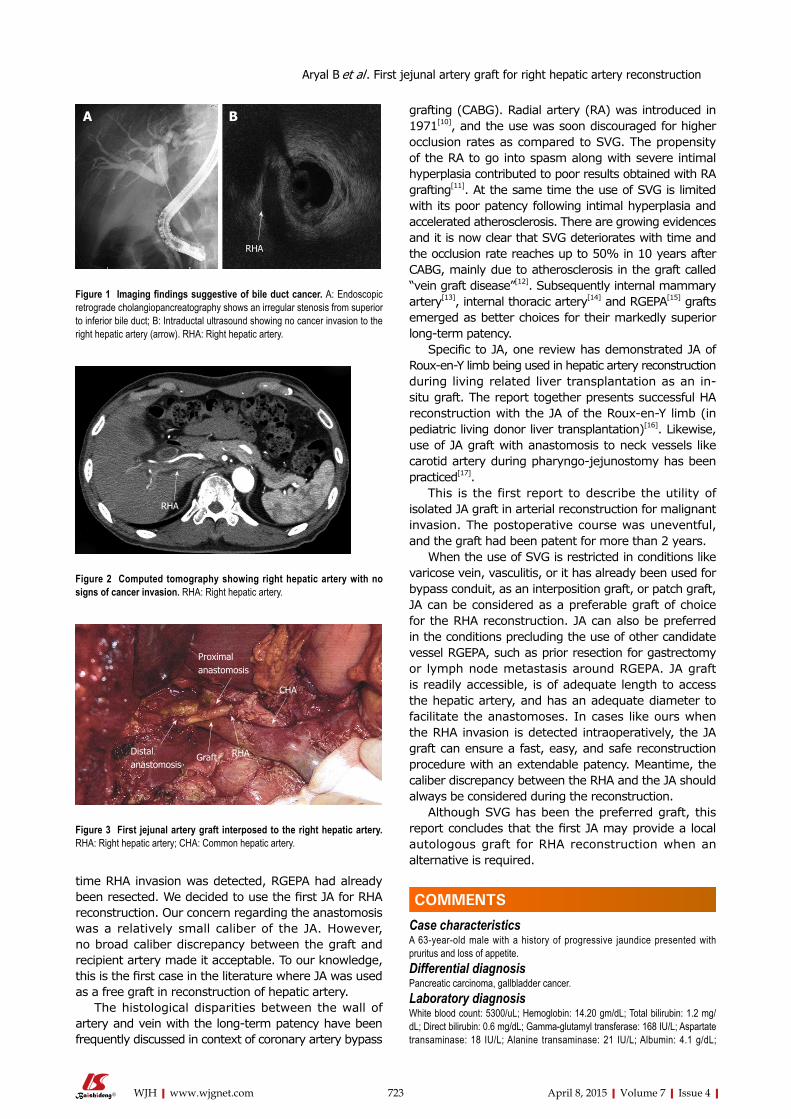
time RHA invasion was detected, RGEPA had already been resected. We decided to use the first JA for RHA reconstruction. Our concern regarding the anastomosis was a relatively small caliber of the JA. However, no broad caliber discrepancy between the graft and recipient artery made it acceptable. To our knowledge, this is the first case in the literature where JA was used as a free graft in reconstruction of hepatic artery.
The histological disparities between the wall of artery and vein with the long-term patency have been frequently discussed in context of coronary artery bypass
grafting (CABG). Radial artery (RA) was introduced in 1971[10], and the use was soon discouraged for higher occlusion rates as compared to SVG. The propensity of the RA to go into spasm along with severe intimal hyperplasia contributed to poor results obtained with RA grafting[11]. At the same time the use of SVG is limited with its poor patency following intimal hyperplasia and accelerated atherosclerosis. There are growing evidences and it is now clear that SVG deteriorates with time and the occlusion rate reaches up to 50% in 10 years after CABG, mainly due to atherosclerosis in the graft called “vein graft disease”[12]. Subsequently internal mammary artery[13], internal thoracic artery[14] and RGEPA[15] grafts emerged as better choices for their markedly superior long-term patency.
Specific to JA, one review has demonstrated JA of Roux-en-Y limb being used in hepatic artery reconstruction during living related liver transplantation as an in-situ graft. The report together presents successful HA reconstruction with the JA of the Roux-en-Y limb (in pediatric living donor liver transplantation)[16]. Likewise, use of JA graft with anastomosis to neck vessels like carotid artery during pharyngo-jejunostomy has been practiced[17].
This is the first report to describe the utility of isolated JA graft in arterial reconstruction for malignant invasion. The postoperative course was uneventful, and the graft had been patent for more than 2 years.
When the use of SVG is restricted in conditions like varicose vein, vasculitis, or it has already been used for bypass conduit, as an interposition graft, or patch graft, JA can be considered as a preferable graft of choice for the RHA reconstruction. JA can also be preferred in the conditions precluding the use of other candidate vessel RGEPA, such as prior resection for gastrectomy or lymph node metastasis around RGEPA. JA graft is readily accessible, is of adequate length to access the hepatic artery, and has an adequate diameter to facilitate the anastomoses. In cases like ours when the RHA invasion is detected intraoperatively, the JA graft can ensure a fast, easy, and safe reconstruction procedure with an extendable patency. Meantime, the caliber discrepancy between the RHA and the JA should always be considered during the reconstruction.
Although SVG has been the preferred graft, this report concludes that the first JA may provide a local autologous graft for RHA reconstruction when an alternative is required.
COMMENTSCase characteristicsA 63-year-old male with a history of progressive jaundice presented with pruritus and loss of appetite. Differential diagnosisPancreatic carcinoma, gallbladder cancer.Laboratory diagnosisWhite blood count: 5300/uL; Hemoglobin: 14.20 gm/dL; Total bilirubin: 1.2 mg/dL; Direct bilirubin: 0.6 mg/dL; Gamma-glutamyl transferase: 168 IU/L; Aspartate transaminase: 18 IU/L; Alanine transaminase: 21 IU/L; Albumin: 4.1 g/dL;
723 April 8, 2015|Volume 7|Issue 4|WJH|www.wjgnet.com
Figure 1 Imaging findings suggestive of bile duct cancer. A: Endoscopic retrograde cholangiopancreatography shows an irregular stenosis from superior to inferior bile duct; B: Intraductal ultrasound showing no cancer invasion to the right hepatic artery (arrow). RHA: Right hepatic artery.
A B
RHA
Figure 2 Computed tomography showing right hepatic artery with no signs of cancer invasion. RHA: Right hepatic artery.
RHA
Figure 3 First jejunal artery graft interposed to the right hepatic artery. RHA: Right hepatic artery; CHA: Common hepatic artery.
Distal anastomosis
Graft RHA
CHA
Proximalanastomosis
COMMENTS
Aryal B et al . First jejunal artery graft for right hepatic artery reconstruction
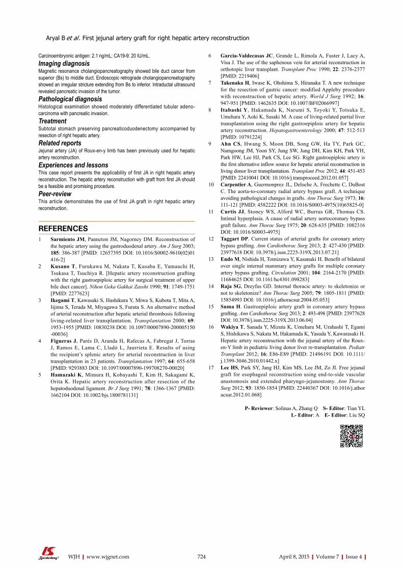
724 April 8, 2015|Volume 7|Issue 4|WJH|www.wjgnet.com
6 Garcia-Valdecasas JC, Grande L, Rimola A, Fuster J, Lacy A, Visa J. The use of the saphenous vein for arterial reconstruction in orthotopic liver transplant. Transplant Proc 1990; 22: 2376-2377 [PMID: 2219406]
7 Takenaka H, Iwase K, Ohshima S, Hiranaka T. A new technique for the resection of gastric cancer: modified Appleby procedure with reconstruction of hepatic artery. World J Surg 1992; 16: 947-951 [PMID: 1462635 DOI: 10.1007/BF02066997]
8 Itabashi Y, Hakamada K, Narumi S, Toyoki Y, Totsuka E, Umehara Y, Aoki K, Sasaki M. A case of living-related partial liver transplantation using the right gastroepiploic artery for hepatic artery reconstruction. Hepatogastroenterology 2000; 47: 512-513 [PMID: 10791224]
9 Ahn CS, Hwang S, Moon DB, Song GW, Ha TY, Park GC, Namgoong JM, Yoon SY, Jung SW, Jung DH, Kim KH, Park YH, Park HW, Lee HJ, Park CS, Lee SG. Right gastroepiploic artery is the first alternative inflow source for hepatic arterial reconstruction in living donor liver transplantation. Transplant Proc 2012; 44: 451-453 [PMID: 22410041 DOI: 10.1016/j.transproceed.2012.01.057]
10 Carpentier A, Guermonprez JL, Deloche A, Frechette C, DuBost C. The aorta-to-coronary radial artery bypass graft. A technique avoiding pathological changes in grafts. Ann Thorac Surg 1973; 16: 111-121 [PMID: 4582222 DOI: 10.1016/S0003-4975(10)65825-0]
11 Curtis JJ, Stoney WS, Alford WC, Burrus GR, Thomas CS. Intimal hyperplasia. A cause of radial artery aortocoronary bypass graft failure. Ann Thorac Surg 1975; 20: 628-635 [PMID: 1082316 DOI: 10.1016/S0003-4975]
12 Taggart DP. Current status of arterial grafts for coronary artery bypass grafting. Ann Cardiothorac Surg 2013; 2: 427-430 [PMID: 23977618 DOI: 10.3978/j.issn.2225-319X.2013.07.21]
13 Endo M, Nishida H, Tomizawa Y, Kasanuki H. Benefit of bilateral over single internal mammary artery grafts for multiple coronary artery bypass grafting. Circulation 2001; 104: 2164-2170 [PMID: 11684625 DOI: 10.1161/hc4301.098283]
14 Raja SG, Dreyfus GD. Internal thoracic artery: to skeletonize or not to skeletonize? Ann Thorac Surg 2005; 79: 1805-1811 [PMID: 15854993 DOI: 10.1016/j.athoracsur.2004.05.053]
15 Suma H. Gastroepiploic artery graft in coronary artery bypass grafting. Ann Cardiothorac Surg 2013; 2: 493-498 [PMID: 23977628 DOI: 10.3978/j.issn.2225-319X.2013.06.04]
16 Wakiya T, Sanada Y, Mizuta K, Umehara M, Urahashi T, Egami S, Hishikawa S, Nakata M, Hakamada K, Yasuda Y, Kawarasaki H. Hepatic artery reconstruction with the jejunal artery of the Roux-en-Y limb in pediatric living donor liver re-transplantation. Pediatr Transplant 2012; 16: E86-E89 [PMID: 21496191 DOI: 10.1111/j.1399-3046.2010.01442.x]
17 Lee HS, Park SY, Jang HJ, Kim MS, Lee JM, Zo JI. Free jejunal graft for esophageal reconstruction using end-to-side vascular anastomosis and extended pharyngo-jejunostomy. Ann Thorac Surg 2012; 93: 1850-1854 [PMID: 22440367 DOI: 10.1016/j.athoracsur.2012.01.068]
P- Reviewer: Solinas A, Zhang Q S- Editor: Tian YL L- Editor: A E- Editor: Liu SQ
Carcinoembryonic antigen: 2.1 ng/mL; CA19-9: 20 IU/mL.Imaging diagnosisMagnetic resonance cholangiopancreatography showed bile duct cancer from superior (Bs) to middle duct. Endoscopic retrograde cholangiopancreatography showed an irregular stricture extending from Bs to inferior. Intraductal ultrasound revealed pancreatic invasion of the tumor.Pathological diagnosisHistological examination showed moderately differentiated tubular adeno-carcinoma with pancreatic invasion.TreatmentSubtotal stomach preserving pancreaticoduodenectomy accompanied by resection of right hepatic artery. Related reportsJejunal artery (JA) of Roux-en-y limb has been previously used for hepatic artery reconstruction. Experiences and lessonsThis case report presents the applicability of first JA in right hepatic artery reconstruction. The hepatic artery reconstruction with graft from first JA should be a feasible and promising procedure. Peer-reviewThis article demonstrates the use of first JA graft in right hepatic artery reconstruction.
REFERENCES1 Sarmiento JM, Panneton JM, Nagorney DM. Reconstruction of
the hepatic artery using the gastroduodenal artery. Am J Surg 2003; 185: 386-387 [PMID: 12657395 DOI: 10.1016/S0002-9610(02)01416-2]
2 Kusano T, Furukawa M, Nakata T, Kusaba E, Yamauchi H, Tsukasa T, Tsuchiya R. [Hepatic artery reconstruction grafting with the right gastroepiploic artery for surgical treatment of upper bile duct cancer]. Nihon Geka Gakkai Zasshi 1990; 91: 1749-1751 [PMID: 2277623]
3 Ikegami T, Kawasaki S, Hashikura Y, Miwa S, Kubota T, Mita A, Iijima S, Terada M, Miyagawa S, Furuta S. An alternative method of arterial reconstruction after hepatic arterial thrombosis following living-related liver transplantation. Transplantation 2000; 69: 1953-1955 [PMID: 10830238 DOI: 10.1097/00007890-200005150-00036]
4 Figueras J, Parés D, Aranda H, Rafecas A, Fabregat J, Torras J, Ramos E, Lama C, Lladó L, Jaurrieta E. Results of using the recipient’s splenic artery for arterial reconstruction in liver transplantation in 23 patients. Transplantation 1997; 64: 655-658 [PMID: 9293883 DOI: 10.1097/00007890-199708270-00020]
5 Hamazaki K, Mimura H, Kobayashi T, Kim H, Sakagami K, Orita K. Hepatic artery reconstruction after resection of the hepatoduodenal ligament. Br J Surg 1991; 78: 1366-1367 [PMID: 1662104 DOI: 10.1002/bjs.1800781131]
Aryal B et al . First jejunal artery graft for right hepatic artery reconstruction

© 2015 Baishideng Publishing Group Inc. All rights reserved.
Published by Baishideng Publishing Group Inc8226 Regency Drive, Pleasanton, CA 94588, USA
Telephone: +1-925-223-8242Fax: +1-925-223-8243
E-mail: [email protected] Desk: http://www.wjgnet.com/esps/helpdesk.aspx
http://www.wjgnet.com


![(Tokyo Tram)] roo( (Tokyo Tram)] (Rose) 737— (Flower](https://img.pdfslide.tips/doc/110x75/61d94880de8d1111764df0e3/tokyo-tram-roo-tokyo-tram-rose-737-flower-.jpg)