Embed Size (px)
Citation preview

Chen et al. BMC Cardiovascular Disorders (2020) 20:48 https://doi.org/10.1186/s12872-020-01344-0
RESEARCH ARTICLE Open Access
Zero-fluoroscopy approach for ablation of
supraventricular tachycardia using theEnsite NavX system: a multicenterexperience Guangzhi Chen1, Yan Wang1* , Riccardo Proietti2, Xunzhang Wang3, Feifan Ouyang4, Chang Sheng Ma5,Rong Hui Yu5, Chunxia Zhao1, Kezhong Ma6, Jie Qiu1, Qigong Liu1 and Dao Wen Wang1*Abstract
Background: Three-dimensional electroanatomic mapping systems have demonstrated a significant reduction inradiation exposure during radiofrequency catheter ablation procedures. We aimed to investigate the safety,feasibility and efficacy of a completely zero-fluoroscopy approach for catheter ablation of supraventriculartachycardia using the Ensite NavX navigation system compared with a conventional fluoroscopy approach.
Methods: A multicenter prospective non-randomized registry study was performed in seven centers from January2013 to February 2018. Consecutive patients referred for catheter ablation of supraventricular tachycardia wereassigned either to a completely zero-fluoroscopic approach (ZF) or conventional fluoroscopy approach (CF)according to the operator’s preference. Patients with atrial tachycardia were excluded.
Results: Totally, 1020 patients were enrolled in ZF group; 2040 patients ablated by CF approach were selected forcontrols. There was no significant difference between the zero-fluoroscopy group and conventional fluoroscopygroup as to procedure time (60.3 ± 20.3 vs. 59.7 ± 22.6 min, P = 0.90), immediate success rate of procedure (98.8% vs.99.2%, P = 0.22), arrhythmia recurrence (0.4% vs. 0.5%, P = 0.85), total success rate of procedure (98.4% vs. 98.8%, P =0.39) or complications (1.1% vs. 1.5%, P = 0.41). Compared with the conventional fluoroscopy approach, the zero-fluoroscopy approach provided similar outcomes without compromising the safety or efficacy of the procedure.
Conclusion: The completely zero-fluoroscopy approach demonstrated safety and efficacy comparable to aconventional fluoroscopy approach for catheter ablation of supraventricular tachycardia, and mitigated radiationexposure to both patients and operators.
Trial registration: clinicaltrials.gov Identifier: NCT03042078; first registered February 3, 2017; retrospectivelyregistered.
Keywords: Supraventricular tachycardia, Radiofrequency ablation, Zero-fluoroscopy, Radiation exposure
© The Author(s). 2020 Open Access This article is distributed under the terms of the Creative Commons Attribution 4.0International License (http://creativecommons.org/licenses/by/4.0/), which permits unrestricted use, distribution, andreproduction in any medium, provided you give appropriate credit to the original author(s) and the source, provide a link tothe Creative Commons license, and indicate if changes were made. The Creative Commons Public Domain Dedication waiver(http://creativecommons.org/publicdomain/zero/1.0/) applies to the data made available in this article, unless otherwise stated.
* Correspondence: [email protected]; [email protected] of Cardiology, Department of Internal Medicine, Tongji Hospital,Tongji Medical College, Huazhong University of Science and Technology,Wuhan 430030, People’s Republic of ChinaFull list of author information is available at the end of the article

Chen et al. BMC Cardiovascular Disorders (2020) 20:48 Page 2 of 10
BackgroundOver the past two decades, the cardiac electrophysiologyfield has undergone great changes and development.Catheter ablation (CA) has become the gold standardfor the treatment of symptomatic and recurrent supra-ventricular tachycardia (SVT). The success rate for thisprocedure is greater than 90% for supraventriculartachycardia such as atrioventricular nodal reentranttachycardia (AVNRT) and atrioventricular re-entranttachycardia (AVRT) [1]. Radiofrequency catheter abla-tion (RFCA) procedures are traditionally performedunder the guidance of fluoroscopy that is a highlyeffective way to navigate catheters and to monitor theirlocation [2]. However, fluoroscopy requires the adminis-tration of ionizing radiation, which carries non-negligible stochastic and deterministic effects on healthfor both patients and staffs [3]. These effects are cumu-lative and give rise to great concerns especially in ayounger population, which highlights the importance ofreducing radiation exposure during cardiac electrophysi-ology procedures [4]. Thus, the American College ofCardiology recommends the adoption of the “ALARA”(as low as reasonably achievable) principle in all inter-ventional laboratories [5].In recent years, non-fluoroscopic three-dimensional
electroanatomic mapping systems (such as Ensite NavXand Carto) have been introduced to facilitate catheterablation procedures [6]. Some studies have demon-strated these systems enable ablation of supraventriculartachycardia (SVT) substrates with significantly decreasedradiation exposure to both patients and staffs comparedto conventional fluoroscopy-guided ablation [7–10].However, there are only a few experiences published ofusing three-dimensional electroanatomic mapping sys-tems with a completely zero-fluoroscopy (ZF) approach.Therefore, the aim of the present study was to explore the
safety, feasibility, and efficacy of a completely zero-fluoroscopy (ZF) approach for catheter ablation of supraven-tricular tachycardia (SVT) guided by the Ensite NavX systemcompared with a conventional fluoroscopy (CF) approach.
MethodsStudy designThis prospective non-randomized registry study was per-formed in seven geographically diverse Chinesearrhythmia centers. It was approved by the institutionalethical review board of Tongji hospital, Tongji MedicalCollege, Huazhong University of Science and Technol-ogy according to the guidelines for good clinical practiceand the Declaration of Helsinki.Between January 2013 and February 2018, consecutive
patients with supraventricular tachycardia, includingAVNRT and AVRT, were enrolled in this study. The pa-tients with atrial tachycardia were excluded. Routinely,
except manifest pre-excitation syndrome, all patients withsupraventricular tachycardia were arranged to undergotransesophageal electrophysiological study (TEEPS) beforethe procedure. Eligible participants were assigned to eitherthe ZF or CF group according to the operator’s preference.Since more operators utilized conventional approach, thesample ratio of ZF to CF was about 1:2.The ZF approach was performed using Ensite NavX sys-
tem as the only guidance (St Jude Medical, St. Paul, MN,USA) and the operating staffs did not wear lead protec-tion. The CF approach utilized fluoroscopy imaging withor without three-dimensional mapping system.Cardiac magnetic resonance imaging, computed tom-
ography, and intracardiac echocardiography were notused during the procedure. Independent operators work-ing in seven centers participated in this study.Written informed consents were obtained from all pa-
tients before the procedure. All the patients underwentroutine blood biochemistry, electrocardiography, chestX-ray imaging, and cardiac echocardiography before theprocedure. Antiarrhythmic drugs were discontinued fora minimum of five half-lives prior to the procedure.
Operative proceduresAll procedures were performed under local anesthesiaand without sedation.
Zero-fluoroscopy (ZF) approachThe Ensite NavX system was used for catheter position-ing and mapping. Catheters were generally first insertedinto the heart through the femoral veins in biplane viewsthat were used to visualize the path of the catheters.Two quadripolar catheters were introduced via the fem-oral vein and positioned in the right ventricular apexand at the His bundle, respectively. A steerable decapo-lar electrode was then placed in the coronary sinus (CS)via the femoral vein. Once the catheter was inserted intothe CS, system optimization and respiratory compensa-tion were performed. A general right atrial geometry wasconstructed from the placed catheters during positioningand the electrophysiology study was routinely performedin accordance with standard protocols (Figs. 1 and 2).Standard protocols and procedures, depending on the
arrhythmic substrate, were used for all ablation proce-dures [11]. For arrhythmias originating in a right heartchamber, the ablation catheter was introduced via a fem-oral vein. The mapping points were as follows: (1) forAVNRT, the slow pathway was ablated (Fig. 3); (2) for aright accessory pathway, the tricuspid valve was labeledusing five to six white points according to electrophysio-logical characteristics before the ablation (Fig. 4). For leftaccessory pathways, the operator selected a retrogradeaortic approach via the femoral artery to obtain accessto the left side of heart. The positions of the aortic root

Fig. 1 A J-shaped wire was placed into a subclavian vein. A correct insertion was verified by the characteristic interference signal, which wasproduced by rotating the J-shaped wire when the first catheter had been positioned at the middle of right atrium. The white arrows indicate thevertical magenta line associated with the interference signal. Abbreviations: LAO, left anterior oblique view; RAO, right anterior oblique view
Chen et al. BMC Cardiovascular Disorders (2020) 20:48 Page 3 of 10
and left sided His bundle were labeled using one or twoblue or yellow points, respectively, and the accessorypathway was tagged along the electrode placed in thecoronary sinus or other regions of the mitral annulus(Fig. 5).
Fig. 2 Stepwise approach to placement of the steerable-curve electrode inquadripolar electrode was placed into the apex of right ventricle (as shownmarked by yellow dot. a First, the steerable electrode was advanced to thepeak of tricuspid valve. b Secondly, it was flexed toward the right ventriclesinus. d Fourthly, the electrode was extended gently. e Finally, the tip of elblank arrow showed the track of catheter tip in RAO and LAO, respectively
Conventional fluoroscopy (CF) approachAfter local anesthesia, diagnostic electrode catheterswere introduced via the femoral, right internal jugular,or subclavian veins; and were positioned in the highright atrium, right ventricle, bundle of His, and coronary
to a coronary sinus via the femoral vein. Generally, a fixed-curvein blue bands); then His potential was recorded by the electrode andposition about one centimeter below the position of his potential or. c Thirdly, it was rotated backward to locate the orifice of coronaryectrode was advanced distally into coronary sinus. The solid arrow and

Fig. 3 The virtual geometry of the targeted area in the right atrium was reconstructed during ablation of AVNRT. Upper panel: the ablationcatheter was placed at the His bundle with the tip of the catheter shown in green, and the area of the His bundle is labeled with yellow dots,and the yellow and blue bands show the electrodes placed in the coronary sinus and in the right ventricle, respectively; typical slow pathwaypotential was mapped and marked with a blue dot and finally power delivery at 35 watts , which was marked as red dot, led to slow junctionalbeats. Lower panel: the white arrows show the His bundle electrogram recorded by the ablation catheter. AVNRT, atrioventricular nodal reentranttachycardia; the abbreviations are as in Fig. 1
Fig. 4 A patient with two manifest right sided accessory pathways was ablated using the ZF approach. Upper panel showed that the tricuspidvalve was labeled with white dots and the site of the His bundle was labeled with yellow dots in RAO and LAO. The ablation catheter was placedat the first accessory pathway at about nine o’clock during tachycardia. Left lower panel showed the electrogram recording at the first target site.Right lower panel showed the second accessory pathway at 5 o’clock marked with red dot. Other abbreviations are as in Fig. 1
Chen et al. BMC Cardiovascular Disorders (2020) 20:48 Page 4 of 10

Fig. 5 A concealed left accessory pathway was mapped and ablated using the ZF approach. The two white arrows in lower panel shows theendocardial electrogram recorded by the ablation catheter (green tip) placed in the targeted ablation area through aorta via retrograde access.The bottom of noncoronary aortic cusp was marked with a blue dot and His potential was marked with a yellow dot. The solid white arrowmarks the small “A” wave during sinus rhythm. The dashed white arrow indicates the earliest “A” wave fused in the “V” wave during rightventricular pacing. Abbreviations are as in Fig. 1
Chen et al. BMC Cardiovascular Disorders (2020) 20:48 Page 5 of 10
sinus. The ablation catheter was introduced to the heartvia the femoral vein. Ventricular and atrial stimulationprotocols were used to assess conduction properties andarrhythmia inducibility. Standard protocols and proce-dures, depending on the arrhythmic substrate, were usedfor all ablation procedures [12]. Fluoroscopy was usedthroughout all phases of the procedure, including con-firmation of the guidewire position, electrophysiologicalstudies, mapping, and ablation.
Data collectionAll preoperative, operative, and follow-up data were collectedand stored in excel spreadsheets by independent researchersand other independent researchers made follow-up.The following data were collected on each patient:
clinical and demographic variables (age, sex, bodyweight, height, arrhythmia type); procedure-related vari-ables (procedure date, assigned group, ablation methodand route, procedure time, fluoroscopy time, number oflesions, total ablation time, immediate success rate, com-plications, and recurrences during follow-up).Procedure time (in minutes) was defined as the inter-
val from the beginning of local anesthesia to extractionof all femoral venous sheaths at the end of the proced-ure. Fluoroscopy time (in minutes) was defined as thetotal duration of exposure during the procedure. Totalablation time was calculated in seconds but time for ten-tative ablation was not taken into account. Number oflesions was defined as the number of ablation sites and
tentative ablation was excluded. Routinely, tentative ab-lation would be terminated after 5 to 10 s of power de-livery when pathway was not blocked. Proceduralsuccess for AVNRT was defined as the absence of indu-cible tachycardia either under basal condition or underisoproterenol stimulation. Procedural success for AVRTwas defined as the non-inducibility of tachycardia, lossof pre-excitation (if manifest), loss of retrogradeaccessory pathway conduction and transient atrioven-tricular block induced by intravenous adenosine. Com-plications were defined as pseudoaneurysm, arterial-venous fistula, pneumothorax, second- or third-degreeatrioventricular block, cardiac tamponade, or other ser-ious complications requiring intervention.
Follow-upAn independent technician performed follow-up at 1month, 3 month, and 12 months after the ablation pro-cedure. Generally, all the patients underwent physicalexamination and echocardiography at one month, andECG each time during the follow-up. Any symptoms orsigns related to complications or recurrence was re-corded. An ECG and electrophysiology study would beperformed to rule out recurrence when the patients hadsuspicious symptoms or signs.
Statistical analysisData are shown as the mean ± standard deviation,whereas categorical data are expressed as numbers and

Chen et al. BMC Cardiovascular Disorders (2020) 20:48 Page 6 of 10
percentages. Student’s t-tests, one-way analysis of vari-ance, Chi-square tests, and Fisher’s exact tests were usedto compare differences among groups. All analyses wereperformed using Statistical Package for the Social Sci-ences (SPSS) version 13.0 (IBM Inc., Armonk, NY,U.S.A.). All P values < 0.05 were considered statisticallysignificant.
ResultsPatient characteristicsFinally, 1020 cases were enrolled for ablation using ZFapproach; 2040 consecutive cases ablated by CF ap-proach were selected as control. Distributions of age,gender, weight, height, and the number of redo caseshad no significance between the two groups. Both ZFand CF groups were well balanced with regard to demo-graphics, clinical baseline characteristics, and the typesof arrhythmia (Tables 1 and 2). The proportion ofAVNRT and special, multiple, septal, right free wall andleft free wall accessory pathway, and accessory pathwayplus AVNRT were comparable and had no significancebetween ZF group and CF group (Table 2).As shown in Tables 1 and 2, the baseline characteris-
tics, the types of arrhythmias, and the rate of redo casesin CF group were well balanced with those in ZF group.The mean follow-up period was 9.7 ± 4.0 months.
Procedural featuresFluoroscopy use
ZF group Among the 1020 cases enrolled in ZF group:(1) there were 1015 cases (99.5%) who completed elec-trophysiology study without fluoroscopy; (2) four cases(0.4%) were excluded because of atrial tachycardia or
Table 1 Baseline characteristics of the enrolled patients in allgroups
ZF (n = 1020) CF (n = 2040) Total cohort (n = 3060)
Mean age (years) 45.4 ± 15.4 45.2 ± 15.3 45.3 ± 15.4
Weight (kg) 62.7 ± 11.8 64.5 ± 11.6 63.8 ± 11.7
Height (cm) 163.6 ± 8.4 165.9 ± 8.2 164.9 ± 8.3
Male patients 459 (45.0%) 908 (44.5%) 1367 (44.7%)
With AT/AF 4 (0.4%) 7 (0.3%) 11 (0.4%)
EPS Only 15 (1.5%) 35 (1.7%) 50 (1.6%)
SVT 1001 (98.1%) 1998 (97.9%) 2999 (98.0%)
Give upa 3 (0.3%) 5 (0.2%) 8 (0.3%)
Ablation 998 (97.8%) 1993 (97.7%) 2991 (97.7%)
Redo cases 5 (0.5%) 12 (0.6%) 17 (0.6%)
Abbreviations: AT atrial tachycardia, AF atrial flutter, EPS electrophysiologicalstudy, ZF zero-fluoroscopy, CF conventional fluoroscopy, SVTsupraventricular tachycardiaasome patients refused to receive ablation owing to the possible risk afterelectrophysiology study
atrial flutter; (3) fifteen cases (1.5%) could not identifyablation target and only underwent electrophysiologystudy; (3) three young patients (0.3%) with para-Hispathway finally refused to take ablation after electro-physiology study because the risk of atrioventricularblock; (4) finally, 998 cases (97.8%) were planned for fur-ther ablation (Table 1).Among the 998 cases planned for ablation, 7 cases
(0.7%) switched to CF group because of the need ofangiography; five of them were due to severe tortuousvessels; two of them were due to pathway suspected tobe originated from diverticulum, although one of themactually was originated from the anterior margin of cor-onary sinus orifice. There were 99.3% (991/998) of pa-tients who completed ablation without fluoroscopy(Table 3).
CF group All the procedures in CF group were per-formed fluoroscopy. The mean total fluoroscopy timewas 6.9 ± 5.9 min in the CF group (Table 3).
Number of ablated lesionsThe ZF approach required fewer lesions for ablationthan did the CF approach (3.6 ± 2.9 vs. 4.5 ± 3.5; P <0.01) (Table 3).
Procedure timeAs shown in Table 3, the procedure time of ZF and CFgroup was 60.3 ± 20.3 and 59.7 ± 22.6 min respectively(Table 3). The results were similar and had no signifi-cance between two groups (P = 0.90).
Success and recurrence ratesImmediate ablation success was achieved in 986 (98.8%)of the ZF approach patients and in 1978 (99.2%) of theCF approach patients (Table 3). The ZF and CF ap-proaches had similar rates of total success (98.4% vs.98.8%, P = 0.39) and arrhythmia recurrence (0.4% vs.0.5%, P = 0.85) (Table 3).
ComplicationsOver the course of the study a total of forty complica-tions were observed. The total complication rates weresimilar in the two groups (1.1% vs. 1.5%, P = 0.41). In theZF group, there were six pseudoaneurysms, four arterial-venous fistulas, and one case of 2nd or 3rd degree atrio-ventricular block. In the CF group, there were fourteenpseudoaneurysms, six arterial-venous fistulas, six caseswith pneumothorax, two cases of 2nd or 3rd degreeatrioventricular block, and one cardiac tamponade.There were no serious complications that required thor-acic surgery. The occurrence of pneumothorax washigher in CF group compared with the ZF group (0.3%vs. 0.0%, P = 0.01). The higher incidence of

Table 2 Classification of supraventricular tachycardia in study patients
ZF (n = 1020) CF (n = 2040) Total cohort (n = 3060)
SVT 1001 (98.1%) 1998 (97.9%) 2999 (98.0%)
1.AVNRT 556 (54.5%) 1052 (51.6%) 1608 (52.5%)
2.AP
1) Septal 75 (7.4%) 160 (7.8%) 235 (7.7%)
2) Left free wall 261 (25.6%) 568 (27.8%) 829 (27.1%)
3) Right free wall 88 (8.6%) 173 (8.5%) 261 (8.5%)
4) Multiple 16 (1.6%) 31 (1.5%) 47 (1.5%)
3.AP + AVNRT 5 (0.5%) 14 (0.7%) 19 (0.6%)
(Special AP)
(Para His) 6 (0.6%) 15 (0.7%) 21 (0.7%)
(Venous) 2 (0.2%) 5 (0.2%) 7 (0.2%)
Abbreviations: AP accessory pathway, AVNRT atrial ventricular node reentrant tachycardia, AVRT atrial ventricular reentrant tachycardia
Chen et al. BMC Cardiovascular Disorders (2020) 20:48 Page 7 of 10
pneumothorax in CF group might be due to more casesof subclavian access whereas almost all the cases in ZFgroup just use femoral access. No specific complicationswere related to zero-fluoroscopy technique (Table 4).
The learning curve of the ZF approachFigure 6a showed the average procedure time of the twoapproaches for the ablation of supraventricular arrhyth-mias according to procedure quartile, 1st to 20th, 21stto 40th, and 41st to 60th cases, and that of all cases. Theaverage procedure time of the ZF approach was about10 min longer than that of the CF approach during theablation of 1st to 20th, 21st to 40th cases. No statisticallysignificant reduction in average procedure time was ob-served between ZF approach group and CF approachgroup.Figure 6b showed linear trend of the procedure time
for the first 120 ablations performed using zero-fluoroscopy approach. The procedure time showed a
Table 3 Ablation outcome of the zero-fluoroscopy (ZF)approach compared with those of the conventional fluoroscopy(CF) approach
ZF (n = 998) CF (n = 1993) p-value
Number of lesionsa 3.6 ± 2.9 4.5 ± 3.5 < 0.01
Ablation time (seconds) 274.4 ± 207.1 301.5 ± 247.5 0.13
Fluoroscopy time (minutes) 0 6.9 ± 5.9 < 0.01
Procedure time (minutes) 60.3 ± 20.3 59.7 ± 22.6 0.90
Switch to CF (n, %) 7 (0.7%) NA NA
Complete ZF (n, %) 991 (99.3%) NA NA
Immediate failure (n, %) 5 (0.5%) 15 (0.8%) 0.43
Immediate success (n, %) 986 (98.8%) 1978 (99.2%) 0.22
Recurrence (n, %) 4 (0.4%) 9 (0.5%) 0.85
Total success (n, %) 982 (98.4%) 1969 (98.8%) 0.39
Abbreviations: NA not applicable; other abbreviations were seen as in Table 1atentative ablations of less than 10 s were not included
gradually reduction trend and nearly reduced to averageprocedure time about after 60 to 80 cases of operation.
DiscussionThe present study investigated the safety, efficiency, andefficacy of a zero-fluoroscopy approach compared with aconventional fluoroscopy approach. The zero-fluoroscopy approach used the Ensite NavX system asthe sole imaging modality for guiding procedures duringthe ablation of supraventricular tachyarrhythmia includ-ing AVNRT and AVRT. Our results demonstrated thatthe zero-fluoroscopy approach had equivalent safety, ef-ficiency, and efficacy as the conventional approach,which utilized fluoroscopy with or without three dimen-sional mapping systems [13].As we all know, fluoroscopic radiation is harmful to
both medical staff and patients, particularly in children,pregnant women, and some specific adults. Various ef-forts and measures have been explored for reducing ra-diation exposure including adjustments in fluoroscopictechnique, the use of lead protective garments, and theintroduction of additional imaging technologies, such aselectro-anatomical mapping or ultrasound [14]. Optimal
Table 4 Complications in zero-fluoroscopy (ZF) andconventional fluoroscopy (CF) groups
Complications ZF (n = 998) CF (n = 1993) p-value
Pseudoaneurysm, n 6 (0.6%) 14 (0.7%) 0.75
Arterial-venous fistula, n 4 (0.4%) 6 (0.3%) 0.66
Pneumothorax, n 0 (0.0%) 6 (0.3%) 0.01
II-III degree of AVB, n 1 (0.1%) 2 (0.1%) 0.99
Cardiac tamponade, n 0 (0.0%) 1 (0.1%) 0.32
Thoracic surgery, n 0 (0.0%) 0 (0.0%) NA
Total (n, %) 11 (1.1%) 29 (1.5%) 0.41
Abbreviations: AVB atrial ventricular block, NA not applicable; otherabbreviations were seen as in Table 1
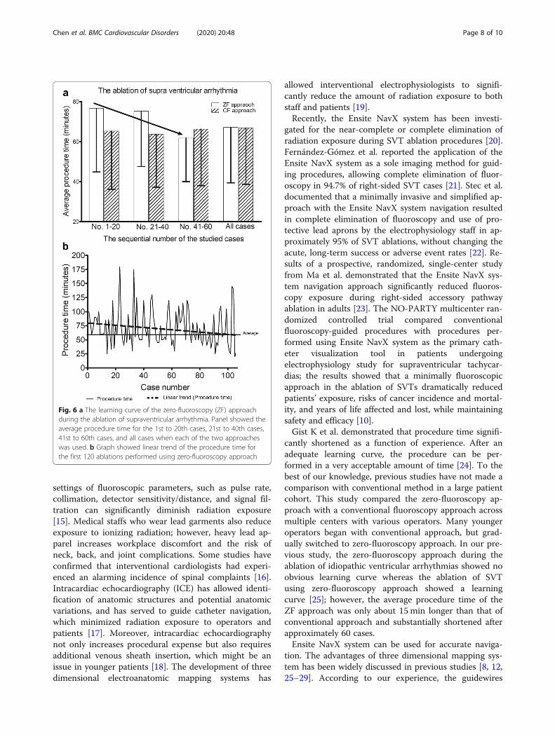
Fig. 6 a The learning curve of the zero-fluoroscopy (ZF) approachduring the ablation of supraventricular arrhythmia. Panel showed theaverage procedure time for the 1st to 20th cases, 21st to 40th cases,41st to 60th cases, and all cases when each of the two approacheswas used. b Graph showed linear trend of the procedure time forthe first 120 ablations performed using zero-fluoroscopy approach
Chen et al. BMC Cardiovascular Disorders (2020) 20:48 Page 8 of 10
settings of fluoroscopic parameters, such as pulse rate,collimation, detector sensitivity/distance, and signal fil-tration can significantly diminish radiation exposure[15]. Medical staffs who wear lead garments also reduceexposure to ionizing radiation; however, heavy lead ap-parel increases workplace discomfort and the risk ofneck, back, and joint complications. Some studies haveconfirmed that interventional cardiologists had experi-enced an alarming incidence of spinal complaints [16].Intracardiac echocardiography (ICE) has allowed identi-fication of anatomic structures and potential anatomicvariations, and has served to guide catheter navigation,which minimized radiation exposure to operators andpatients [17]. Moreover, intracardiac echocardiographynot only increases procedural expense but also requiresadditional venous sheath insertion, which might be anissue in younger patients [18]. The development of threedimensional electroanatomic mapping systems has
allowed interventional electrophysiologists to signifi-cantly reduce the amount of radiation exposure to bothstaff and patients [19].Recently, the Ensite NavX system has been investi-
gated for the near-complete or complete elimination ofradiation exposure during SVT ablation procedures [20].Fernández-Gómez et al. reported the application of theEnsite NavX system as a sole imaging method for guid-ing procedures, allowing complete elimination of fluor-oscopy in 94.7% of right-sided SVT cases [21]. Stec et al.documented that a minimally invasive and simplified ap-proach with the Ensite NavX system navigation resultedin complete elimination of fluoroscopy and use of pro-tective lead aprons by the electrophysiology staff in ap-proximately 95% of SVT ablations, without changing theacute, long-term success or adverse event rates [22]. Re-sults of a prospective, randomized, single-center studyfrom Ma et al. demonstrated that the Ensite NavX sys-tem navigation approach significantly reduced fluoros-copy exposure during right-sided accessory pathwayablation in adults [23]. The NO-PARTY multicenter ran-domized controlled trial compared conventionalfluoroscopy-guided procedures with procedures per-formed using Ensite NavX system as the primary cath-eter visualization tool in patients undergoingelectrophysiology study for supraventricular tachycar-dias; the results showed that a minimally fluoroscopicapproach in the ablation of SVTs dramatically reducedpatients’ exposure, risks of cancer incidence and mortal-ity, and years of life affected and lost, while maintainingsafety and efficacy [10].Gist K et al. demonstrated that procedure time signifi-
cantly shortened as a function of experience. After anadequate learning curve, the procedure can be per-formed in a very acceptable amount of time [24]. To thebest of our knowledge, previous studies have not made acomparison with conventional method in a large patientcohort. This study compared the zero-fluoroscopy ap-proach with a conventional fluoroscopy approach acrossmultiple centers with various operators. Many youngeroperators began with conventional approach, but grad-ually switched to zero-fluoroscopy approach. In our pre-vious study, the zero-fluoroscopy approach during theablation of idiopathic ventricular arrhythmias showed noobvious learning curve whereas the ablation of SVTusing zero-fluoroscopy approach showed a learningcurve [25]; however, the average procedure time of theZF approach was only about 15 min longer than that ofconventional approach and substantially shortened afterapproximately 60 cases.Ensite NavX system can be used for accurate naviga-
tion. The advantages of three dimensional mapping sys-tem has been widely discussed in previous studies [8, 12,25–29]. According to our experience, the guidewires

Chen et al. BMC Cardiovascular Disorders (2020) 20:48 Page 9 of 10
must be withdrawn before model construction usingEnsite NavX system for guidance since the guidewirewill lead to the distortion of the impedance based field.Additionally, there are some issues that should be ad-dressed just prior to power delivery at a high risk area,such as modifying the slow pathway during the ablationof AVNRT. Firstly, respiratory validation and recalibra-tion by a technician should be repeated, and the exactlocation of the His potential should be routinely re-confirmed. Secondly, the patient should avoid largerespiratory excursions and should avoid significantmovement. Thirdly, the surface patch used for a systemreference should be placed in an inter-scapular area, par-ticularly in obese patients with significant abdominalmovement during respiration.In this study, only seven cases (0.7%) finally switched into
conventional fluoroscopy approach as fluoroscopic angiog-raphy was required. The most common reason for theswitch was severe tortuous vessels; those were usually inelderly patients over seventies. Actually, zero-fluoroscopyapproach still can be succeed by advancing a long sheath ifa stiff long guidewire can pass through the tortuous vessel.The less common reason was pathways suspected to beoriginated from a coronary sinus or a diverticulum; most ofthe patients showed an initial negative delta wave on lead IIin surface ECG electrocardiogram; although one of themeventually were not inside the coronary sinus but were lo-cated at the superior margin of coronary sinus orifice.We also found that noviciate was more rapidly familiar
with zero-fluoroscopy navigation than experienced oper-ators. Hence, we believe that the establishing of themethod, habit, and systemic training are critical factorsfor familiar applying zero-fluoroscopy technique.Our study has several limitations. First, our study was
not a randomized study. Second, we did not compareother navigation systems (e.g. Carto system) versus theconventional fluoroscopy approach in this study. Finally,we acknowledge that using the Ensite NavX system addscertain procedural costs.
ConclusionsZero-fluoroscopy ablation of supraventricular tachycar-dia guided by a three dimensional electroanatomic sys-tem is as effective and safe as a conventional fluoroscopyapproach with comparable procedure duration.Further research is needed to establish the techniques
and applicability of a zero-fluoroscopy approach formore complex arrhythmias. Meanwhile, continuedtechnological advances may help realize the dream of acompletely zero-fluoroscopy approach for arrhythmiasablation procedures.
AbbreviationsAF: Atrial flutter; ALARA: As low as reasonably achievable; AP: Accessorypathway; AT: Atrial tachycardia; AVB: Atrial ventricular block;
AVNRT: Atrioventricular nodal reentry tachycardia; AVRT: Atrioventricularreciprocating tachycardia; CA: Catheter ablation; CF: Conventionalfluoroscopy; CS: Coronary sinus; EPS: Electrophysiological study;ICE: Intracardiac echocardiography; LAO: Left anterior oblique view; RA: Rightatrium; RAO: Right anterior oblique view; RFCA: Radiofrequency catheterablation; SVT: Supraventricular tachycardia; 3D: Three-dimensional;TEEPS: Transesophageal electrophysiological study; ZF: Zero-fluoroscopy
AcknowledgementsWe thank Jiangtao Wu and Lun Zhuge for their works in figure preparation.
Authors’ contributionsGZC, YW, CXZ, JQ and DWW were involved in the study conception anddesign, data analysis and interpretation. GZC, YW, RHY, CXZ, KZM, JQ andQGL acquired the data. GZC and YW drafted the manuscript. RP, XZW, FFOand CSM were involved in the study conception and design, revised themanuscript. All authors have reviewed the article and made substantialrevisions to improve the scientific credibility of the content. All authorsapproved the final version to be published.
FundingThis work was supported by funds from the Science and TechnologyDepartment of Hubei Province (No.2015CFA077) and the National NaturalScience Foundation of China (Nos. 81400369; 81570308). There was no roleof the funding body in the design of the study and collection, analysis, andinterpretation of data and in writing the manuscript.
Availability of data and materialsThe datasets used and analyzed during the current study are available fromthe corresponding author on reasonable request.
Ethics approval and consent to participateThis study was performed in compliance with the guidelines for goodclinical practice and the Declaration of Helsinki and was approved by theinstitutional ethical review board of Tongji hospital, Tongji Medical College,Huazhong University of Science and Technology. Written informed consentswere obtained from all patients before the procedure.
Consent for publicationNot applicable.
Competing interestsThe authors declare that they have no competing interests.
Author details1Division of Cardiology, Department of Internal Medicine, Tongji Hospital,Tongji Medical College, Huazhong University of Science and Technology,Wuhan 430030, People’s Republic of China. 2Department of Cardiac, Thoracic,and Vascular Sciences, via Giustiniani 2, 35121 Padua, Italy. 3Heart Institute,Cedars Sinai Medical Center, Los Angeles, CA, USA. 4Asklepios Klinik St.Georg, Hamburg, Germany. 5Department of Cardiology, Beijing AnzhenHospital, Capital Medical University, Beijing 100029, People’s Republic ofChina. 6Department of Cardiology, Xiangyang Central Hospital, Xiangyang441021, People’s Republic of China.
Received: 27 July 2019 Accepted: 16 January 2020
References1. Page RL, Joglar JA, Caldwell MA, Calkins H, Conti JB, Deal BJ, Estes NA III,
Field ME, Goldberger ZD, Hammill SC, et al. 2015 ACC/AHA/HRS guidelinefor the management of adult patients with supraventricular tachycardia:executive summary: a report of the American College of Cardiology/American Heart Association task force on clinical practice guidelines andthe Heart Rhythm Society. Heart Rhythm. 2016;13(4):e92–135.
2. Papagiannis J, Beissel DJ, Krause U, Cabrera M, Telishevska M, Seslar S,Johnsrude C, Anderson C, Tisma-Dupanovic S, Connelly D, et al.Atrioventricular Nodal Reentrant Tachycardia in Patients With CongenitalHeart Disease: Outcome After Catheter Ablation. Circ ArrhythmElectrophysiol. 2017;10(7):e004869.

Chen et al. BMC Cardiovascular Disorders (2020) 20:48 Page 10 of 10
3. Duran A, Hian SK, Miller DL, Le Heron J, Padovani R, Vano E.Recommendations for occupational radiation protection in interventionalcardiology. Catheter Cardiovasc Interv. 2013;82(1):29–42.
4. Picano E, Vano E, Rehani MM, Cuocolo A, Mont L, Bodi V, Bar O, Maccia C,Pierard L, Sicari R, et al. The appropriate and justified use of medicalradiation in cardiovascular imaging: a position document of the ESCassociations of cardiovascular imaging, percutaneous cardiovascularinterventions and electrophysiology. Eur Heart J. 2014;35(10):665–72.
5. Limacher MC, Douglas PS, Germano G, Laskey WK, Lindsay BD, McKetty MH,Moore ME, Park JK, Prigent FM, Walsh MN. ACC expert consensusdocument. Radiation safety in the practice of cardiology. American Collegeof Cardiology. J Am Coll Cardiol. 1998;31(4):892–913.
6. LaPage MJ, Saul JP. Update on rhythm mapping and catheter navigation.Curr Opin Cardiol. 2011;26(2):79–85.
7. Christoph M, Wunderlich C, Moebius S, Forkmann M, Sitzy J, Salmas J, MayerJ, Huo Y, Piorkowski C, Gaspar T. Fluoroscopy integrated 3D mappingsignificantly reduces radiation exposure during ablation for a widespectrum of cardiac arrhythmias. Europace. 2015;17(6):928–37.
8. Giaccardi M, Del Rosso A, Guarnaccia V, Ballo P, Mascia G, Chiodi L, Colella A.Near-zero x-ray in arrhythmia ablation using a 3-dimensional electroanatomicmapping system: a multicenter experience. Heart Rhythm. 2016;13(1):150–6.
9. Seizer P, Bucher V, Frische C, Heinzmann D, Gramlich M, Muller I, Henning A,Hofbeck M, Kerst G, Gawaz M, et al. Efficacy and safety of zero-fluoroscopyablation for supraventricular tachycardias. Use of optional contact forcemeasurement for zero-fluoroscopy ablation in a clinical routine setting.Herz. 2016;41(3):241–5.
10. Casella M, Dello Russo A, Pelargonio G, Del Greco M, Zingarini G, Piacenti M,Di Cori A, Casula V, Marini M, Pizzamiglio F, et al. Near zerO fluoroscopicexPosure during catheter ablAtion of supRavenTricular arrhYthmias: the NO-PARTY multicentre randomized trial. Europace. 2016;18(10):1565–72.
11. Casella M, Pelargonio G, Dello Russo A, Riva S, Bartoletti S, Santangeli P,Scara A, Sanna T, Proietti R, Di Biase L, et al. "Near-zero" fluoroscopicexposure in supraventricular arrhythmia ablation using the EnSite NavXmapping system: personal experience and review of the literature. J IntervCard Electrophysiol. 2011;31(2):109–18.
12. See J, Amora JL, Lee S, Lim P, Teo WS, Tan BY, Ho KL, Lee CW, Ching CK.Non-fluoroscopic navigation systems for radiofrequency catheter ablationfor supraventricular tachycardia reduce ionising radiation exposure. SingapMed J. 2016;57(7):390–5.
13. Bibas L, Levi M, Essebag V. Diagnosis and management of supraventriculartachycardias. Cmaj. 2016;188(17–18):E466–73.
14. Duran A, Hian SK, Miller DL, Le Heron J, Padovani R, Vano E. A summary ofrecommendations for occupational radiation protection in interventionalcardiology. Catheter Cardiovasc Interv. 2013;81(3):562–7.
15. Hill KD, Einstein AJ. New approaches to reduce radiation exposure. TrendsCardiovasc Med. 2016;26(1):55–65.
16. Chen G, Sun G, Xu R, Chen X, Yang L, Bai Y, Yang S, Guo P, Zhang Y, ZhaoC, et al. Zero-fluoroscopy catheter ablation of severe drug-resistantarrhythmia guided by Ensite NavX system during pregnancy: two casereports and literature review. Medicine (Baltimore). 2016;95(32):e4487.
17. George JC, Varghese V, Mogtader A. Intracardiac echocardiography: evolvinguse in interventional cardiology. J Ultrasound Med. 2014;33(3):387–95.
18. Njeim M, Desjardins B, Bogun F. Multimodality imaging for guiding EPablation procedures. JACC Cardiovasc Imaging. 2016;9(7):873–86.
19. Anselmino M, Sillano D, Casolati D, Ferraris F, Scaglione M, Gaita F. A newelectrophysiology era: zero fluoroscopy. J Cardiovasc Med (Hagerstown).2013;14(3):221–7.
20. Yang L, Sun G, Chen X, Chen G, Yang S, Guo P, Wang Y, Wang DW. Meta-analysis of zero or near-zero fluoroscopy use during ablation of cardiacarrhythmias. Am J Cardiol. 2016;118(10):1511–8.
21. Fernandez-Gomez JM, Morina-Vazquez P, Morales Edel R, Venegas-GameroJ, Barba-Pichardo R, Carranza MH. Exclusion of fluoroscopy use in catheterablation procedures: six years of experience at a single center. J CardiovascElectrophysiol. 2014;25(6):638–44.
22. Stec S, Sledz J, Mazij M, Ras M, Ludwik B, Chrabaszcz M, Sledz A, Banasik M,Bzymek M, Mlynarczyk K, et al. Feasibility of implementation of a "simplified,no-X-ray, no-lead apron, two-catheter approach" for ablation ofsupraventricular arrhythmias in children and adults. J CardiovascElectrophysiol. 2014;25(8):866–74.
23. Ma Y, Qiu J, Yang Y, Tang A. Catheter ablation of right-sided accessorypathways in adults using the three-dimensional mapping system: a
randomized comparison to the conventional approach. PLoS One. 2015;10(6):e0128760.
24. Gist K, Tigges C, Smith G, Clark J. Learning curve for zero-fluoroscopycatheter ablation of AVNRT: early versus late experience. Pacing ClinElectrophysiol. 2011;34(3):264–8.
25. Wang Y, Chen GZ, Yao Y, Bai Y, Chu HM, Ma KZ, Liew R, Liu H, Zhong GQ,Xue YM, et al. Ablation of idiopathic ventricular arrhythmia using zero-fluoroscopy approach with equivalent efficacy and less fatigue: amulticenter comparative study. Medicine (Baltimore). 2017;96(6):e6080.
26. Ozyilmaz I, Ergul Y, Akdeniz C, Ozturk E, Tanidir IC, Tuzcu V. Catheterablation of idiopathic ventricular tachycardia in children using the EnSiteNavX system with/without fluoroscopy. Cardiol Young. 2014;24(5):886–92.
27. Tuzcu V, Gul EE, Karacan M, Kamali H, Celik N, Akdeniz C. Comparison of 6-mm versus 8-mm-tip Cryoablation catheter for the treatment ofAtrioventricular nodal reentrant tachycardia in children: a prospective study.Pediatr Cardiol. 2017;38(6):1220–5.
28. Gaita F, Guerra PG, Battaglia A, Anselmino M. The dream of near-zero X-raysablation comes true. Eur Heart J. 2016;37(36):2749–55.
29. Tsuchiya T. Three-dimensional mapping of cardiac arrhythmias - string ofpearls. Circ J. 2012;76(3):572–81.
Publisher’s NoteSpringer Nature remains neutral with regard to jurisdictional claims inpublished maps and institutional affiliations.


![NIH Public Access - UNILBIB_301DB2EA945E.P001/REF.pdf · Ablation and Procedural Parameters The ablation parameters (average power, temperature [endo ± epi] and impedance, duration](https://img.pdfslide.tips/doc/110x75/5e8086bdbc41707fd7327d66/nih-public-access-unil-bib301db2ea945ep001refpdf-ablation-and-procedural.jpg)