Embed Size (px)
Citation preview
Zhang, FR; Huang, W; Chen, SM; Sun, LD; Liu, H; Li, Y; Cui, Y;Yan, XX; Yang, HT; Yang, RD; Chu, TS; Zhang, C; Zhang, L; Han,JW; Yu, GQ; Quan, C; Yu, YX; Zhang, Z; Shi, BQ; Zhang, LH;Cheng, H; Wang, CY; Lin, Y; Zheng, HF; Fu, XA; Zuo, XB; Wang,Q; Long, H; Sun, YP; Cheng, YL; Tian, HQ; Zhou, FS; Liu, HX;Lu, WS; He, SM; Du, WL; Shen, M; Jin, QY; Wang, Y; Low, HQ;Erwin, T; Yang, NH; Li, JY; Zhao, X; Jiao, YL; Mao, LG; Yin, G;Jiang, ZX; Wang, XD; Yu, JP; Hu, ZH; Gong, CH; Liu, YQ; Liu,RY; Wang, DM; Wei, D; Liu, JX; Cao, WK; Cao, HZ; Li, YP; Yan,WG; Wei, SY; Wang, KJ; Hibberd, ML; Yang, S; Zhang, XJ; Liu, JJ(2009) Genomewide association study of leprosy. The New Englandjournal of medicine, 361 (27). pp. 2609-18. ISSN 0028-4793 DOI:https://doi.org/10.1056/NEJMoa0903753
Downloaded from: http://researchonline.lshtm.ac.uk/2875457/
DOI: 10.1056/NEJMoa0903753
Usage Guidelines
Please refer to usage guidelines at http://researchonline.lshtm.ac.uk/policies.html or alterna-tively contact [email protected].
Available under license: http://creativecommons.org/licenses/by-nc-nd/2.5/
T h e n e w e ngl a nd j o u r na l o f m e dic i n e
n engl j med 361;27 nejm.org december 31, 2009 2609
original article
Genomewide Association Study of LeprosyFu-Ren Zhang, M.D., Ph.D., Wei Huang, Ph.D., Shu-Min Chen, M.D., Ph.D.,
Liang-Dan Sun, M.D., Ph.D., Hong Liu, M.D., Yi Li, Ph.D., Yong Cui, M.D., Ph.D., Xiao-Xiao Yan, M.D., Hai-Tao Yang, M.D., Rong-De Yang, M.D.,
Tong-Sheng Chu, M.D., Chi Zhang, M.D., Lin Zhang, M.D., Jian-Wen Han, M.D., Gong-Qi Yu, B.S., Cheng Quan, M.D., Yong-Xiang Yu, B.S., Zheng Zhang, M.D.,
Ben-Qing Shi, M.D., Lian-Hua Zhang, M.D., Hui Cheng, M.D., Chang-Yuan Wang, M.D., Yan Lin, M.D., Hou-Feng Zheng, M.D., Xi-An Fu, M.D.,
Xian-Bo Zuo, M.S., Qiang Wang, M.D., Heng Long, M.D., Yi-Ping Sun, M.D., Yi-Lin Cheng, M.S., Hong-Qing Tian, M.D., Fu-Sheng Zhou, B.S., Hua-Xu Liu, M.D., Ph.D., Wen-Sheng Lu, M.D., Su-Min He, M.D., Wen-Li Du, B.S., Min Shen, B.S., Qi-Yi Jin, B.S., Ying Wang, Ph.D.,
Hui-Qi Low, B.S., Tantoso Erwin, B.S., Ning-Han Yang, B.S., Jin-Yong Li, M.D., Xin Zhao, M.D., Yue-Lin Jiao, M.D., Li-Guo Mao, M.D., Gang Yin, M.D.,
Zhen-Xia Jiang, M.D., Xiao-Dong Wang, M.D., Jing-Ping Yu, M.D., Zong-Hou Hu, M.D., Cui-Hua Gong, M.D., Yu-Qiang Liu, M.D., Rui-Yu Liu, M.D.,
De-Min Wang, M.D., Dong Wei, M.D., Jin-Xian Liu, M.D., Wei-Kun Cao, M.D., Hong-Zhong Cao, M.D., Yong-Ping Li, M.D., Wei-Guo Yan, M.D., Shi-Yu Wei, M.D., Kui-Jun Wang, M.D., Martin L. Hibberd, Ph.D.,
Sen Yang, M.D., Ph.D., Xue-Jun Zhang, M.D., Ph.D., and Jian-Jun Liu, Ph.D.
The authors’ affiliations are listed in the Supplementary Appendix, available with the full text of this article at NEJM.org. Address reprint requests to Dr. Fu-Ren Zhang at Shandong Provincial Institute of Dermatology and Venereology, Shan-dong Academy of Medical Science, 57, Ji-yan Lu, Jinan, Shandong 250022, China, or at [email protected]; to Dr. Xue-Jun Zhang at the Institute of Derma-tology and Department of Dermatology at No. 1 Hospital, Anhui Medical Univer-sity, Hefei, Anhui 230022, China, or at [email protected]; or to Dr. Jian-Jun Liu at the Department of Human Genet-ics, Genome Institute of Singapore, Sin-gapore 138672, Singapore, or at [email protected].
This article (10.1056/NEJMoa0903753) was published on December 16, 2009, at NEJM.org.
N Engl J Med 2009;361:2609-18.Copyright © 2009 Massachusetts Medical Society.
A BS TR AC T
BACKGROUND
The narrow host range of Mycobacterium leprae and the fact that it is refractory to growth in culture has limited research on and the biologic understanding of lep-rosy. Host genetic factors are thought to influence susceptibility to infection as well as disease progression.
METHODS
We performed a two-stage genomewide association study by genotyping 706 pa-tients and 1225 controls using the Human610-Quad BeadChip (Illumina). We then tested three independent replication sets for an association between the presence of leprosy and 93 single-nucleotide polymorphisms (SNPs) that were most strongly associated with the disease in the genomewide association study. Together, these replication sets comprised 3254 patients and 5955 controls. We also carried out tests of heterogeneity of the associations (or lack thereof) between these 93 SNPs and disease, stratified according to clinical subtype (multibacillary vs. paucibacillary).
RESULTS
We observed a significant association (P<1.00×10⁻¹⁰) between SNPs in the genes CCDC122, C13orf31, NOD2, TNFSF15, HLA-DR, and RIPK2 and a trend toward an as-sociation (P = 5.10×10⁻⁵) with a SNP in LRRK2. The associations between the SNPs in C13orf31, LRRK2, NOD2, and RIPK2 and multibacillary leprosy were stronger than the associations between these SNPs and paucibacillary leprosy.
CONCLUSIONS
Variants of genes in the NOD2-mediated signaling pathway (which regulates the in-nate immune response) are associated with susceptibility to infection with M. leprae.
The New England Journal of Medicine Downloaded from nejm.org at LONDON SCH HYGIENE & TROPICAL MED on October 13, 2016. For personal use only. No other uses without permission.
Copyright © 2009 Massachusetts Medical Society. All rights reserved.
T h e n e w e ngl a nd j o u r na l o f m e dic i n e
n engl j med 361;27 nejm.org december 31, 20092610
Leprosy is a chronic infectious dis-ease caused by Mycobacterium leprae. It affects the skin and peripheral nerves and can cause
irreversible impairment of nerve function and con-sequent chronic disabilities.1 Despite a dramatic decrease in its prevalence over the past two de-cades (largely due to the worldwide introduction of multidrug therapy in 1982),2 leprosy remains a major public health problem and one of the most important preventable disabilities in many devel-oping countries.3 It is therefore particularly unfor-tunate that research into the mechanisms under-lying infection and clinical sequelae has been limited by the fact that M. leprae infects only hu-mans and cannot be cultured in vitro.4
The clinical disease of leprosy develops in a minority of infected persons,5 and it manifests as a spectrum of disease symptoms that result from interactions between the host’s immune response and the bacterium. Tuberculoid and lepromatous leprosy are at opposite ends of the spectrum, each being associated with a relatively stable immune status of the host. “Borderline” categories of the disease, characterized by a variety of clinical manifestations, are associated with an unstable immune response to the bacilli.6
The unusually low diversity of genomic se-quences among M. leprae strains makes it unlikely that differences in susceptibility or clinical mani-festation are governed by the strain of M. leprae or variation within each strain.7 Therefore, the immunologic response of the host is thought to play a critical role; multibacillary infection is as-sociated with a type 2 helper T (Th2) cell re-sponse, whereas paucibacillary infection is asso-ciated with an immune response mediated by type 1 helper T (Th1) cells.8
Host genetic factors have been implicated in susceptibility to leprosy in studies of familial clus-tering, studies of twins, complex segregation analyses, and tests of association with the HLA genes.9-13 Markers in several genes and genomic regions (e.g., HLA-DR [the gene encoding major histocompatibility complex class II DR], PARK2–PACRG [genes encoding proteins related to Parkin-son’s disease], LTA [the gene encoding lympho-toxin alpha], and chromosome 10p13) have been reported to be associated with susceptibility to leprosy or the development of a particular clinical form of the disease, but few of these associations have been replicated.14-17 We performed a genome-
wide association study involving large numbers of patients with leprosy and unaffected persons (controls).
Me thods
We carried out a genomewide association study of leprosy in a “discovery” set of 706 affected patients and 1225 unaffected controls, all of whom were Han Chinese from eastern China. The first repli-cation set consisted of Han Chinese from eastern China, and the second and third replication sets were made up of Han Chinese as well as persons from minority, non-Han ethnic groups (including the Chung, Miao, Yízú, and other smaller groups) from southern China.
Leprosy was diagnosed on the basis of consen-sus by at least two dermatologists. From medical records, we determined the clinical subtype of the disease, whether there was a family history of lep-rosy, and the age at onset of disease. The controls did not have a history of leprosy, autoimmune, or systemic disorders or a family history of leprosy (among first-, second-, or third-degree relatives). Patients and controls self-reported their age, sex, and ethnic group on a questionnaire. All partici-pants reported that they were free of infection by M. tuberculosis and chronic infection by other agents (with the exception of M. leprae in the case pa-tients). Patients and controls were matched accord-ing to ethnic origin and geographic region of recruitment. All participants provided written in-formed consent, and the study was approved by local institutional ethics committees (see the Sup-plementary Appendix, available with the full text of this article at NEJM.org).
We carried out the genomewide association study using Human610-Quad BeadChip (Illumina) and the follow-up genotyping using the iPLEX system (Sequenom) and the TaqMan assay (Ap-plied Biosystems). We tested for population strat-ification in the discovery set using a method based on principal-components analysis and tested for the presence of genotype–phenotype associations using the Cochran–Armitage trend test with and without correction for population stratification. We also carried out heterogeneity analyses of the 93 single-nucleotide polymorphisms (SNPs) with the strongest associations with disease suscepti-bility in the genomewide association study to de-termine whether these associations were dispro-
The New England Journal of Medicine Downloaded from nejm.org at LONDON SCH HYGIENE & TROPICAL MED on October 13, 2016. For personal use only. No other uses without permission.
Copyright © 2009 Massachusetts Medical Society. All rights reserved.
Genomewide Association Study of Leprosy
n engl j med 361;27 nejm.org december 31, 2009 2611
portionately driven by the presence or absence of family history of leprosy, presence or absence of disability from leprosy, the age at onset of the disease, or its clinical subtype. More information on the samples, genotyping, quality control, and statistical analyses is provided in the Supplemen-tary Appendix.
R esult s
Genomewide Association Analysis
After filtering the data obtained by genomewide association study, for purposes of quality control, a total of 491,883 SNPs from 706 case patients and 1225 controls remained and were subjected to sta-tistical analysis (see the Supplementary Appendix). Principal-components analysis, using the 206 HapMap reference samples, confirmed that all par-ticipants were of Chinese ancestry (Fig. 1 in the Supplementary Appendix), although the case pa-tients and controls showed some genetic stratifi-cation (Fig. 2 in the Supplementary Appendix). To minimize the effect of population stratifica-tion, we tested for the presence of genotype–phe-notype associations using two approaches. First, we analyzed the genomewide genotypes of the 706 case patients and 1225 controls using the Cochran–Armitage trend test with correction for population stratification based on principal-com-ponents analysis.18 Second, we tested the geno-types for an association with affected status, with-out correction for population stratification, after removing the 711 genetically unmatched controls (Fig. 3 in the Supplementary Appendix). (The sum-mary statistics of the full data set obtained by means of the genomewide association analysis can be obtained from the National Center for Biotech-nology Information’s database of genotypes and phenotypes [dbGaP; www.ncbi.nlm.nih.gov/gap], accession number phs000217.v1.p1.)
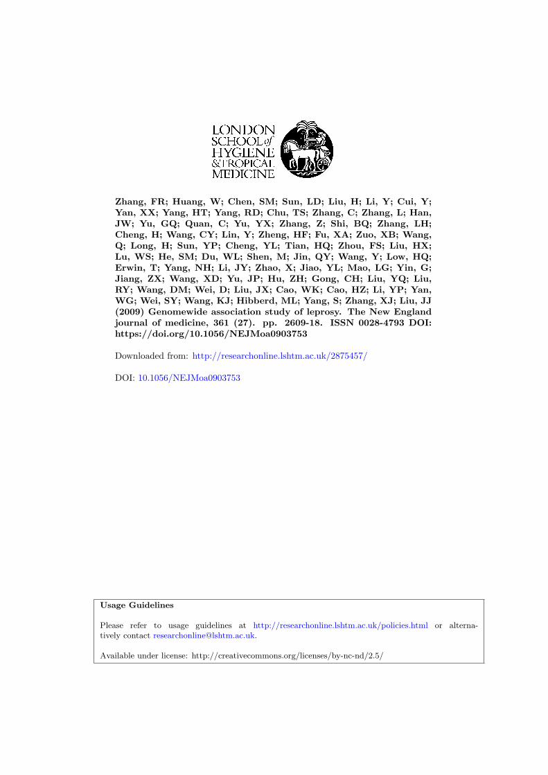
The results of these two analyses indicated that there was no overall inflation of the associations with leprosy because of population stratification (Fig. 4 in the Supplementary Appendix). Moreover, the results of the two analyses were generally con-sistent, suggesting a strong association within the major histocompatibility complex (MHC) region (on chromosome 6p21) and additional associations at chromosome 16q12 (rs9302752; P = 1.42×10⁻⁹; odds ratio for leprosy, 2.28) and chromosome 13q14 (rs3764147; P = 4.06×10⁻⁷; odds ratio, 1.97)
(Fig. 1 and Table 1). The P values yielded by both analyses showed a deviation from the null distri-bution of no association after the SNPs within the MHC region were removed from the analyses, suggesting that the observed P values within the tail of the distribution are smaller than those ex-pected on the basis of chance and therefore prob-ably reflect true genetic associations (Fig. 4 in the Supplementary Appendix).
We observed two associations with leprosy within the MHC region. One was within the HLA-B–HLA-C locus (encoding MHC, classes I, B and C), at which the most strongly associated SNP was rs9264868 (P = 1.96×10⁻⁴; odds ratio, 2.12), and the other was within the HLA-DR–DQ locus (en-coding MHC, class II, DR and DQ), at which the most strongly associated SNP was rs9271366 (P = 1.94×10⁻¹⁷; odds ratio, 2.35) (Fig. 5 in the Supplementary Appendix). After controlling for the genetic effect of rs9271366, the association within the HLA-B–HLA-C locus remained signifi-cant (Table 2 in the Supplementary Appendix), suggesting that these two associations are inde-pendent of each other.
Tests of Replication
We genotyped 93 SNPs — those that showed the strongest association with leprosy in the genome-wide association study — in samples from three replication sets: two consisting of Han Chinese and one of Chinese minority groups — collective-ly, 3254 case patients and 5955 controls (Table 1). In addition to these tests of association carried out using each of the three replication sets, we carried out a combined analysis of the results obtained by means of targeted genotyping of the samples in the replication sets and the genomewide geno-typing of the samples in the discovery set. (With respect to the discovery set, we used results from the second analysis, in which we used the small-er group of matched control samples.)
With respect to evaluating the MHC region in the replication sets, we genotyped two SNPs: rs602875 at the HLA-DR–DQ locus (P = 3.47×10⁻⁴; odds ratio, 0.58) (since rs9271366, also at this lo-cus and with a stronger association, was refrac-tory to genotyping) and rs9264868 at the HLA-B–C locus (P = 1.96×10⁻⁴; odds ratio, 2.12) (Fig. 5 in the Supplementary Appendix). The results of the combined analysis strongly support an association between rs602875 and susceptibility to leprosy
The New England Journal of Medicine Downloaded from nejm.org at LONDON SCH HYGIENE & TROPICAL MED on October 13, 2016. For personal use only. No other uses without permission.
Copyright © 2009 Massachusetts Medical Society. All rights reserved.

T h e n e w e ngl a nd j o u r na l o f m e dic i n e
n engl j med 361;27 nejm.org december 31, 20092612
6 col33p9
–Log
10 P
Val
ue
20
18
19
17
16
14
13
1
15
9
11
10
8
12
7
6
4
3
5
2
01 2 4 6 8 10 12 14 16 18 20 219 11 133 5 7 15 17 19 22
A
AUTHOR:
FIGURE:
RETAKE:
SIZE
4-C H/TLine Combo
Revised
AUTHOR, PLEASE NOTE: Figure has been redrawn and type has been reset.
Please check carefully.
1st2nd3rd
Zhang
1 of 2
ARTIST:
TYPE:
MRL
12-31-09JOB: 36127 ISSUE:
Chromosome
–Log
10 P
Val
ue
0
1
2
3
4
5
6
7
8
9
10
11
12
13
14
15
16
17
1 2 4 6 8 10 12 14 16 18 20 219 11 133 5 7 15 17 19 22
B
Chromosome
Figure 1. Results of Genomewide Association Analysis.
The −log10 genomewide P values, as calculated with the use of the Cochran–Armitage trend test, are shown. Panel A shows the P values, after EIGENSTRAT correction, calculated on the basis of data from 491,883 polymorphic single-nucleotide polymorphisms (SNPs) identified in the 706 patients with leprosy and 1225 controls (total, 1931 sam-ples). Panel B shows the P values calculated on the basis of data from 492,109 polymorphic SNPs in the 706 pa-tients and the 514 genetically matched controls (total, 1220 samples). The red horizontal lines indicate P = 1.00×10⁻⁵, the threshold used for a trend toward genomewide significance.
The New England Journal of Medicine Downloaded from nejm.org at LONDON SCH HYGIENE & TROPICAL MED on October 13, 2016. For personal use only. No other uses without permission.
Copyright © 2009 Massachusetts Medical Society. All rights reserved.
Genomewide Association Study of Leprosy
n engl j med 361;27 nejm.org december 31, 2009 2613
(P = 5.33×10⁻²⁷; odds ratio, 0.67) but not between rs9264868 and the disease (P = 2.33×10⁻³; odds ra-tio, 1.14).
The associations with susceptibility to disease were replicated for two SNPs (rs42490 and rs40457) within RIPK2 (the gene encoding receptor-inter-acting serine–threonine kinase 2, on chromosome 8q21), three SNPs (rs4574921, rs10114470, and rs6478108) within TNFSF15 (the gene encoding tumor necrosis factor [ligand] superfamily mem-ber 15, on chromosome 9q32), two SNPs (rs3764147 and rs10507522) within C13orf31 (the gene en-coding chromosome 13 open reading frame 31, on chromosome 13q14), two SNPs (rs9533634 and rs3088362) within CCDC122 (the gene encoding
coiled-coil domain containing 122, on chromo-some 13q14), and two SNPs (rs9302752 and rs7194886) within NOD2 (the gene encoding nu-cleotide-binding oligomerization domain contain-ing 2, on chromosome 16q12) (Table 2). At least two SNPs in each of these five genes showed sig-nificant association (P<1.00×10⁻¹⁰ for all analy-ses combined) with affected status. To investigate the independence of the multiple associations ob-served within each of the five genes, we performed conditional association analyses, in which the ge-netic effect of the most strongly associated SNP at each locus was controlled. These analyses re-vealed at least two independently associated SNPs, located in different blocks of linkage disequilib-
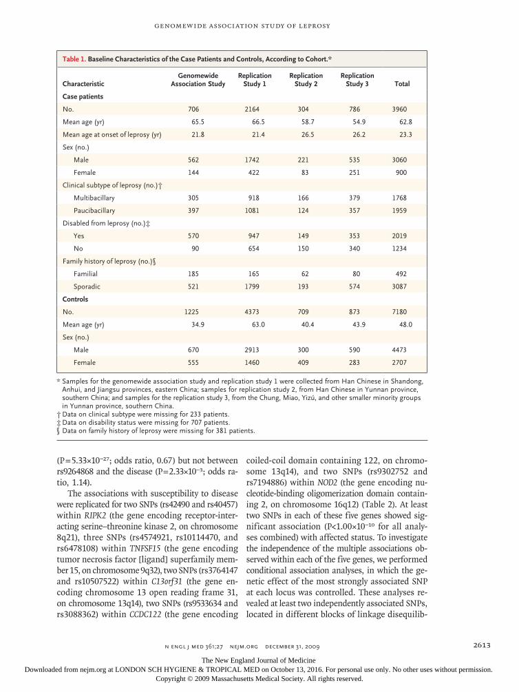
Table 1. Baseline Characteristics of the Case Patients and Controls, According to Cohort.*
CharacteristicGenomewide
Association StudyReplication
Study 1Replication
Study 2Replication
Study 3 Total
Case patients
No. 706 2164 304 786 3960
Mean age (yr) 65.5 66.5 58.7 54.9 62.8
Mean age at onset of leprosy (yr) 21.8 21.4 26.5 26.2 23.3
Sex (no.)
Male 562 1742 221 535 3060
Female 144 422 83 251 900
Clinical subtype of leprosy (no.)†
Multibacillary 305 918 166 379 1768
Paucibacillary 397 1081 124 357 1959
Disabled from leprosy (no.)‡
Yes 570 947 149 353 2019
No 90 654 150 340 1234
Family history of leprosy (no.)§
Familial 185 165 62 80 492
Sporadic 521 1799 193 574 3087
Controls
No. 1225 4373 709 873 7180
Mean age (yr) 34.9 63.0 40.4 43.9 48.0
Sex (no.)
Male 670 2913 300 590 4473
Female 555 1460 409 283 2707
* Samples for the genomewide association study and replication study 1 were collected from Han Chinese in Shandong, Anhui, and Jiangsu provinces, eastern China; samples for replication study 2, from Han Chinese in Yunnan province, southern China; and samples for the replication study 3, from the Chung, Miao, Yizú, and other smaller minority groups in Yunnan province, southern China.
† Data on clinical subtype were missing for 233 patients.‡ Data on disability status were missing for 707 patients.§ Data on family history of leprosy were missing for 381 patients.
The New England Journal of Medicine Downloaded from nejm.org at LONDON SCH HYGIENE & TROPICAL MED on October 13, 2016. For personal use only. No other uses without permission.
Copyright © 2009 Massachusetts Medical Society. All rights reserved.
T h e n e w e ngl a nd j o u r na l o f m e dic i n e
n engl j med 361;27 nejm.org december 31, 20092614
rium (Table 2 in the Supplementary Appendix) and with low pairwise r2 values (<0.3) at each locus (Fig. 6 in the Supplementary Appendix).
The results indicate a trend toward an asso-ciation between the SNP rs1873613 in LRRK2 (the gene encoding leucine-rich repeat kinase 2, on chromosome 12q12) and susceptibility to leprosy (Table 2). Inclusion of the replication samples strengthened the evidence for an association for this SNP (P = 5.10×10⁻⁵ for all analyses combined; odds ratio, 0.86). Joint analysis of the 1931 sam-ples (including the 711 unmatched controls) in the genomewide association study and those in all three replication sets also supported an association (P = 3.68×10⁻⁵; odds ratio, 0.86), with an even stronger association from joint analysis of all the Han samples (from 3174 case patients and 6307 controls) (P = 5.68×10⁻⁶; odds ratio, 0.82).
The results for the other 77 SNPs included in
the replication analyses are summarized in Ta-ble 1 in the Supplementary Appendix.
Analysis of Subgroups of Patients
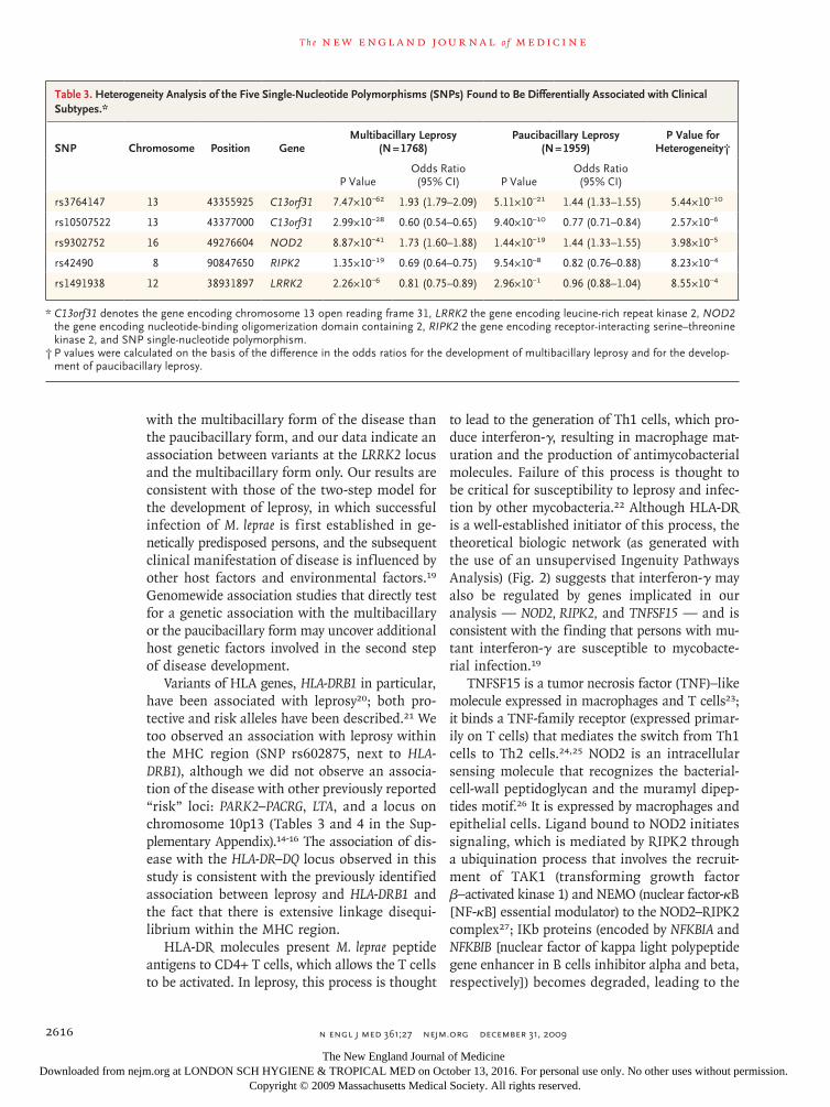
The subgroup analysis of the multibacillary and paucibacillary clinical subtypes of leprosy revealed significant evidence for heterogeneity at five SNPs (rs3764147, rs10507522, rs9302752, rs42490, and rs1491938) within four genes (C13orf31, LRRK2, NOD2, and RIPK2). The associations of these SNPs were stronger with the multibacillary form of lep-rosy than with the paucibacillary form, and the difference in the strength of association was sig-nificant (defined as P<0.05 after correction for multiple testing for the 16 SNPs listed in Table 2) (Table 3). The rs1491938 variant (in LRRK2) showed a significant association with the multibacillary form (P = 2.26×10⁻⁶; odds ratio, 0.81) but not the paucibacillary form (P = 2.96×10⁻¹; odds ratio, 0.96).
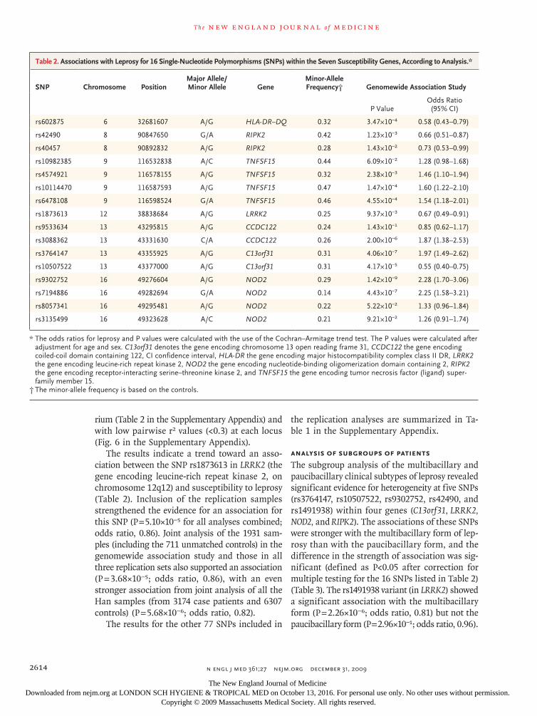
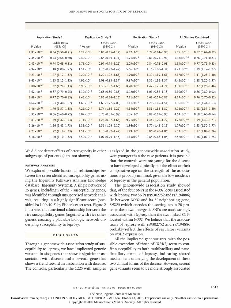
Table 2. Associations with Leprosy for 16 Single-Nucleotide Polymorphisms (SNPs) within the Seven Susceptibility Genes, According to Analysis.*
SNP Chromosome PositionMajor Allele/ Minor Allele Gene
Minor-Allele Frequency† Genomewide Association Study
P ValueOdds Ratio (95% CI)
rs602875 6 32681607 A/G HLA-DR–DQ 0.32 3.47×10⁻⁴ 0.58 (0.43–0.79)
rs42490 8 90847650 G/A RIPK2 0.42 1.23×10⁻³ 0.66 (0.51–0.87)
rs40457 8 90892832 A/G RIPK2 0.28 1.43×10⁻² 0.73 (0.53–0.99)
rs10982385 9 116532838 A/C TNFSF15 0.44 6.09×10⁻² 1.28 (0.98–1.68)
rs4574921 9 116578155 A/G TNFSF15 0.32 2.38×10⁻³ 1.46 (1.10–1.94)
rs10114470 9 116587593 A/G TNFSF15 0.47 1.47×10⁻⁴ 1.60 (1.22–2.10)
rs6478108 9 116598524 G/A TNFSF15 0.46 4.55×10⁻⁴ 1.54 (1.18–2.01)
rs1873613 12 38838684 A/G LRRK2 0.25 9.37×10⁻³ 0.67 (0.49–0.91)
rs9533634 13 43295815 A/G CCDC122 0.24 1.43×10⁻¹ 0.85 (0.62–1.17)
rs3088362 13 43331630 C/A CCDC122 0.26 2.00×10⁻⁶ 1.87 (1.38–2.53)
rs3764147 13 43355925 A/G C13orf31 0.31 4.06×10⁻⁷ 1.97 (1.49–2.62)
rs10507522 13 43377000 A/G C13orf31 0.31 4.17×10⁻⁵ 0.55 (0.40–0.75)
rs9302752 16 49276604 A/G NOD2 0.29 1.42×10⁻⁹ 2.28 (1.70–3.06)
rs7194886 16 49282694 G/A NOD2 0.14 4.43×10⁻⁷ 2.25 (1.58–3.21)
rs8057341 16 49295481 A/G NOD2 0.22 5.22×10⁻² 1.33 (0.96–1.84)
rs3135499 16 49323628 A/C NOD2 0.21 9.21×10⁻² 1.26 (0.91–1.74)
* The odds ratios for leprosy and P values were calculated with the use of the Cochran–Armitage trend test. The P values were calculated after adjustment for age and sex. C13orf31 denotes the gene encoding chromosome 13 open reading frame 31, CCDC122 the gene encoding coiled-coil domain containing 122, CI confidence interval, HLA-DR the gene encoding major histocompatibility complex class II DR, LRRK2 the gene encoding leucine-rich repeat kinase 2, NOD2 the gene encoding nucleotide-binding oligomerization domain containing 2, RIPK2 the gene encoding receptor-interacting serine–threonine kinase 2, and TNFSF15 the gene encoding tumor necrosis factor (ligand) super-family member 15.
† The minor-allele frequency is based on the controls.
The New England Journal of Medicine Downloaded from nejm.org at LONDON SCH HYGIENE & TROPICAL MED on October 13, 2016. For personal use only. No other uses without permission.
Copyright © 2009 Massachusetts Medical Society. All rights reserved.
Genomewide Association Study of Leprosy
n engl j med 361;27 nejm.org december 31, 2009 2615
We did not detect effects of heterogeneity in other subgroups of patients (data not shown).
Pathway Analysis
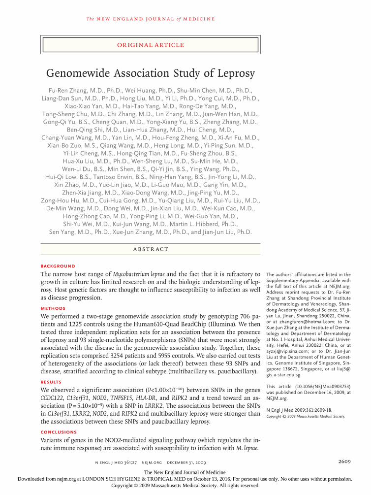
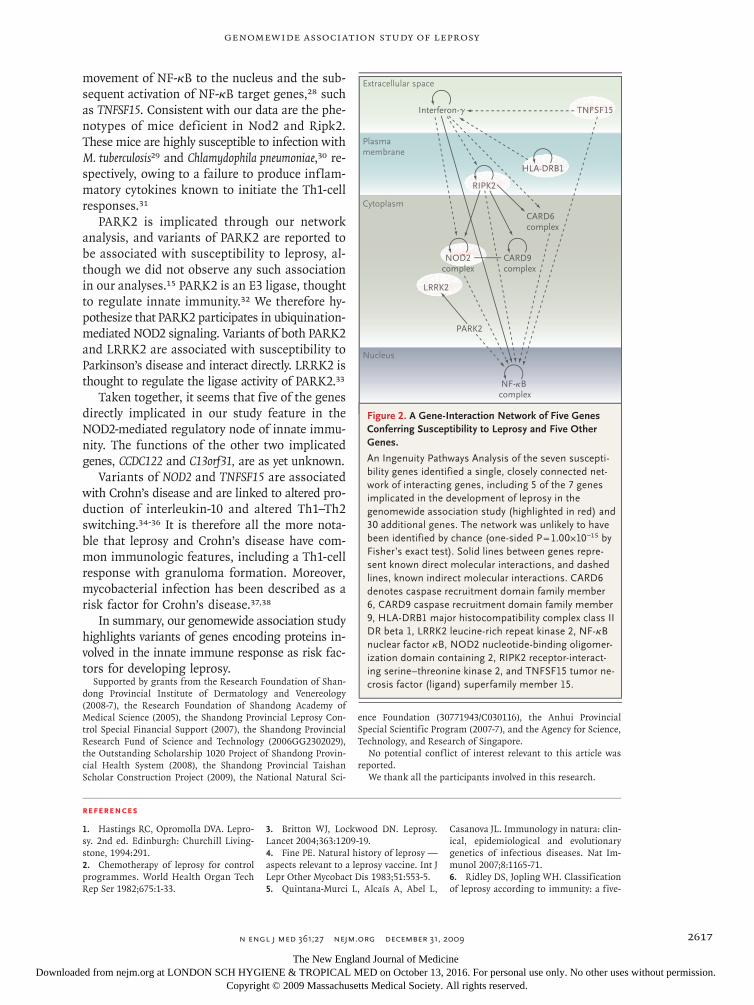
We explored possible functional relationships be-tween the seven identified susceptibility genes us-ing the Ingenuity Pathways Analysis knowledge database (Ingenuity Systems). A single network of 35 genes, including 5 of the 7 susceptibility genes, was identified through unsupervised network anal-ysis, resulting in a highly significant score (one-sided P = 1.00×10⁻¹⁵ by Fisher’s exact test). Figure 2 illustrates the functional relationship between the five susceptibility genes (together with five other genes), creating a plausible biologic network un-derlying susceptibility to leprosy.
Discussion
Through a genomewide association study of sus-ceptibility to leprosy, we have implicated genetic variants in six genes that show a significant as-sociation with disease and a seventh gene that shows a trend toward an association with disease. The controls, particularly the 1225 with samples
analyzed in the genomewide association study, were younger than the case patients. It is possible that the controls were too young for the disease to have developed clinically but the effect of their comparative age on the strength of the associa-tions is probably minimal, given the low incidence of leprosy in the general population.
The genomewide association study showed that, of the four SNPs at the NOD2 locus associated with leprosy, two SNPs (rs9302752 and rs7194886) lie between NOD2 and its 5′ neighboring gene, SNX20 (which encodes the sorting nexin 20 pro-tein); these two intergenic SNPs are more strongly associated with leprosy than the two linked SNPs located within NOD2. We believe that the associa-tions of leprosy with rs9302752 and rs7194886 probably reflect the effects of regulatory variants on NOD2 expression.
All the implicated gene variants, with the pos-sible exception of those of LRRK2, seem to con-fer susceptibility to both multibacillary and pauc-ibacillary forms of leprosy, indicating shared mechanisms underlying the development of these two clinical forms of the disease. However, several gene variants seem to be more strongly associated
Replication Study 1 Replication Study 2 Replication Study 3 All Studies Combined
P ValueOdds Ratio (95% CI) P Value
Odds Ratio (95% CI) P Value
Odds Ratio (95% CI) P Value
Odds Ratio (95% CI)
8.81×10⁻²² 0.64 (0.59–0.71) 3.29×10⁻¹ 0.85 (0.65–1.12) 6.32×10⁻³ 0.77 (0.64–0.93) 5.35×10⁻²⁷ 0.67 (0.62–0.72)
2.45×10⁻¹³ 0.74 (0.68–0.80) 2.40×10⁻¹ 0.88 (0.69–1.11) 1.21×10⁻² 0.83 (0.71–0.96) 1.38×10⁻¹⁶ 0.76 (0.71–0.81)
2.45×10⁻¹⁰ 0.74 (0.68–0.81) 6.79×10⁻¹ 0.97 (0.74–1.26) 2.03×10⁻² 0.84 (0.72–0.98) 1.34×10⁻¹² 0.77 (0.72–0.83)
4.94×10⁻⁵ 1.18 (1.09–1.27) 1.54×10⁻¹ 1.16 (0.92–1.47) 5.66×10⁻² 1.16 (1.00–1.34) 8.73×10⁻⁸ 1.19 (1.12–1.27)
9.23×10⁻⁹ 1.27 (1.17–1.37) 2.29×10⁻² 1.29 (1.02–1.63) 1.79×10⁻⁵ 1.39 (1.19–1.61) 2.17×10⁻¹⁶ 1.31 (1.23–1.40)
6.63×10⁻⁸ 1.25 (1.15–1.35) 4.95×10⁻¹ 1.08 (0.85–1.37) 9.87×10⁻⁵ 1.35 (1.16–1.57) 5.42×10⁻¹⁴ 1.28 (1.20–1.37)
1.80×10⁻¹¹ 1.32 (1.21–1.43) 3.95×10⁻² 1.30 (1.02–1.66) 8.20×10⁻⁷ 1.47 (1.26–1.71) 3.39×10⁻²¹ 1.37 (1.28–1.46)
3.62×10⁻³ 0.87 (0.79–0.95) 1.39×10⁻³ 0.65 (0.50–0.85) 8.92×10⁻¹ 1.01 (0.86–1.18) 5.10×10⁻⁵ 0.86 (0.80–0.92)
9.48×10⁻⁸ 0.77 (0.70–0.85) 2.45×10⁻¹ 0.85 (0.64–1.15) 7.31×10⁻⁵ 0.69 (0.57–0.83) 4.77×10⁻¹² 0.76 (0.70–0.82)
6.64×10⁻²³ 1.53 (1.40–1.67) 4.69×10⁻⁴ 1.60 (1.22–2.09) 1.11×10⁻² 1.26 (1.05–1.51) 1.36×10⁻³¹ 1.52 (1.41–1.63)
1.46×10−37 1.70 (1.57–1.85) 7.29×10⁻⁶ 1.74 (1.36–2.22) 4.54×10⁻⁸ 1.55 (1.32–1.82) 3.72×10−54 1.68 (1.57–1.80)
3.32×10⁻¹⁸ 0.66 (0.60–0.72) 3.07×10⁻² 0.75 (0.57–0.98) 1.05×10⁻² 0.81 (0.69–0.95) 4.64×10⁻²⁴ 0.68 (0.63–0.74)
3.83×10⁻²⁸ 1.59 (1.47–1.73) 7.11×10⁻² 1.26 (0.97–1.63) 9.21×10⁻⁵ 1.44 (1.20–1.72) 3.77×10−40 1.59 (1.49–1.71)
5.26×10⁻¹⁸ 1.56 (1.41–1.73) 1.31×10⁻² 1.51 (1.09–2.10) 1.86×10⁻⁷ 1.77 (1.42–2.19) 1.77×10⁻³⁰ 1.63 (1.50–1.77)
2.23×10⁻⁵ 1.22 (1.11–1.33) 4.51×10⁻¹ 1.10 (0.82–1.47) 1.49×10⁻¹ 0.86 (0.70–1.06) 5.53×10⁻⁵ 1.17 (1.09–1.26)
8.16×10⁻⁵ 1.20 (1.10–1.32) 5.59×10⁻¹ 1.07 (0.79–1.44) 1.13×10⁻¹ 0.84 (0.68–1.04) 2.52×10⁻⁴ 1.16 (1.07–1.25)
The New England Journal of Medicine Downloaded from nejm.org at LONDON SCH HYGIENE & TROPICAL MED on October 13, 2016. For personal use only. No other uses without permission.
Copyright © 2009 Massachusetts Medical Society. All rights reserved.
T h e n e w e ngl a nd j o u r na l o f m e dic i n e
n engl j med 361;27 nejm.org december 31, 20092616
with the multibacillary form of the disease than the paucibacillary form, and our data indicate an association between variants at the LRRK2 locus and the multibacillary form only. Our results are consistent with those of the two-step model for the development of leprosy, in which successful infection of M. leprae is first established in ge-netically predisposed persons, and the subsequent clinical manifestation of disease is influenced by other host factors and environmental factors.19 Genomewide association studies that directly test for a genetic association with the multibacillary or the paucibacillary form may uncover additional host genetic factors involved in the second step of disease development.
Variants of HLA genes, HLA-DRB1 in particular, have been associated with leprosy20; both pro-tective and risk alleles have been described.21 We too observed an association with leprosy within the MHC region (SNP rs602875, next to HLA-DRB1), although we did not observe an associa-tion of the disease with other previously reported “risk” loci: PARK2–PACRG, LTA, and a locus on chromosome 10p13 (Tables 3 and 4 in the Sup-plementary Appendix).14-16 The association of dis-ease with the HLA-DR–DQ locus observed in this study is consistent with the previously identified association between leprosy and HLA-DRB1 and the fact that there is extensive linkage disequi-librium within the MHC region.
HLA-DR molecules present M. leprae peptide antigens to CD4+ T cells, which allows the T cells to be activated. In leprosy, this process is thought
to lead to the generation of Th1 cells, which pro-duce interferon-γ, resulting in macrophage mat-uration and the production of antimycobacterial molecules. Failure of this process is thought to be critical for susceptibility to leprosy and infec-tion by other mycobacteria.22 Although HLA-DR is a well-established initiator of this process, the theoretical biologic network (as generated with the use of an unsupervised Ingenuity Pathways Analysis) (Fig. 2) suggests that interferon-γ may also be regulated by genes implicated in our analysis — NOD2, RIPK2, and TNFSF15 — and is consistent with the finding that persons with mu-tant interferon-γ are susceptible to mycobacte-rial infection.19
TNFSF15 is a tumor necrosis factor (TNF)–like molecule expressed in macrophages and T cells23; it binds a TNF-family receptor (expressed primar-ily on T cells) that mediates the switch from Th1 cells to Th2 cells.24,25 NOD2 is an intracellular sensing molecule that recognizes the bacterial-cell-wall peptidoglycan and the muramyl dipep-tides motif.26 It is expressed by macrophages and epithelial cells. Ligand bound to NOD2 initiates signaling, which is mediated by RIPK2 through a ubiquination process that involves the recruit-ment of TAK1 (transforming growth factor β–activated kinase 1) and NEMO (nuclear factor-κB [NF-κB] essential modulator) to the NOD2–RIPK2 complex27; IKb proteins (encoded by NFKBIA and NFKBIB [nuclear factor of kappa light polypeptide gene enhancer in B cells inhibitor alpha and beta, respectively]) becomes degraded, leading to the
Table 3. Heterogeneity Analysis of the Five Single-Nucleotide Polymorphisms (SNPs) Found to Be Differentially Associated with Clinical Subtypes.*
SNP Chromosome Position GeneMultibacillary Leprosy
(N = 1768)Paucibacillary Leprosy
(N = 1959)P Value for
Heterogeneity†
P ValueOdds Ratio (95% CI) P Value
Odds Ratio (95% CI)
rs3764147 13 43355925 C13orf31 7.47×10−62 1.93 (1.79–2.09) 5.11×10⁻²¹ 1.44 (1.33–1.55) 5.44×10⁻¹⁰
rs10507522 13 43377000 C13orf31 2.99×10⁻²⁸ 0.60 (0.54–0.65) 9.40×10⁻¹⁰ 0.77 (0.71–0.84) 2.57×10⁻⁶
rs9302752 16 49276604 NOD2 8.87×10−41 1.73 (1.60–1.88) 1.44×10⁻¹⁹ 1.44 (1.33–1.55) 3.98×10⁻⁵
rs42490 8 90847650 RIPK2 1.35×10⁻¹⁹ 0.69 (0.64–0.75) 9.54×10⁻⁸ 0.82 (0.76–0.88) 8.23×10⁻⁴
rs1491938 12 38931897 LRRK2 2.26×10⁻⁶ 0.81 (0.75–0.89) 2.96×10⁻¹ 0.96 (0.88–1.04) 8.55×10⁻⁴
* C13orf31 denotes the gene encoding chromosome 13 open reading frame 31, LRRK2 the gene encoding leucine-rich repeat kinase 2, NOD2 the gene encoding nucleotide-binding oligomerization domain containing 2, RIPK2 the gene encoding receptor-interacting serine–threonine kinase 2, and SNP single-nucleotide polymorphism.
† P values were calculated on the basis of the difference in the odds ratios for the development of multibacillary leprosy and for the develop-ment of paucibacillary leprosy.
The New England Journal of Medicine Downloaded from nejm.org at LONDON SCH HYGIENE & TROPICAL MED on October 13, 2016. For personal use only. No other uses without permission.
Copyright © 2009 Massachusetts Medical Society. All rights reserved.
Genomewide Association Study of Leprosy
n engl j med 361;27 nejm.org december 31, 2009 2617
movement of NF-κB to the nucleus and the sub-sequent activation of NF-κB target genes,28 such as TNFSF15. Consistent with our data are the phe-notypes of mice deficient in Nod2 and Ripk2. These mice are highly susceptible to infection with M. tuberculosis29 and Chlamydophila pneumoniae,30 re-spectively, owing to a failure to produce inflam-matory cytokines known to initiate the Th1-cell responses.31
PARK2 is implicated through our network analysis, and variants of PARK2 are reported to be associated with susceptibility to leprosy, al-though we did not observe any such association in our analyses.15 PARK2 is an E3 ligase, thought to regulate innate immunity.32 We therefore hy-pothesize that PARK2 participates in ubiquination-mediated NOD2 signaling. Variants of both PARK2 and LRRK2 are associated with susceptibility to Parkinson’s disease and interact directly. LRRK2 is thought to regulate the ligase activity of PARK2.33
Taken together, it seems that five of the genes directly implicated in our study feature in the NOD2-mediated regulatory node of innate immu-nity. The functions of the other two implicated genes, CCDC122 and C13orf31, are as yet unknown.
Variants of NOD2 and TNFSF15 are associated with Crohn’s disease and are linked to altered pro-duction of interleukin-10 and altered Th1–Th2 switching.34-36 It is therefore all the more nota-ble that leprosy and Crohn’s disease have com-mon immunologic features, including a Th1-cell response with granuloma formation. Moreover, mycobacterial infection has been described as a risk factor for Crohn’s disease.37,38
In summary, our genomewide association study highlights variants of genes encoding proteins in-volved in the innate immune response as risk fac-tors for developing leprosy.
Supported by grants from the Research Foundation of Shan-dong Provincial Institute of Dermatology and Venereology (2008-7), the Research Foundation of Shandong Academy of Medical Science (2005), the Shandong Provincial Leprosy Con-trol Special Financial Support (2007), the Shandong Provincial Research Fund of Science and Technology (2006GG2302029), the Outstanding Scholarship 1020 Project of Shandong Provin-cial Health System (2008), the Shandong Provincial Taishan Scholar Construction Project (2009), the National Natural Sci-
ence Foundation (30771943/C030116), the Anhui Provincial Special Scientific Program (2007-7), and the Agency for Science, Technology, and Research of Singapore.
No potential conflict of interest relevant to this article was reported.
We thank all the participants involved in this research.
Extracellular space
Plasmamembrane
Cytoplasm
Nucleus
Interferon-γ
NOD2complex
CARD9complex
CARD6complex
LRRK2
PARK2
TNFSF15
RIPK2
HLA-DRB1
NF-κB complex
2
Phimister
11/19/09
AUTHOR PLEASE NOTE:Figure has been redrawn and type has been reset
Please check carefully
Author
Fig #
Title
ME
DEArtist
Issue date
COLOR FIGURE
Draft 2Zhang
Knoper
12/31/09
Gene interaction network
Figure 2. A Gene-Interaction Network of Five Genes Conferring Susceptibility to Leprosy and Five Other Genes.
An Ingenuity Pathways Analysis of the seven suscepti-bility genes identified a single, closely connected net-work of interacting genes, including 5 of the 7 genes implicated in the development of leprosy in the genomewide association study (highlighted in red) and 30 additional genes. The network was unlikely to have been identified by chance (one-sided P = 1.00×10⁻¹⁵ by Fisher’s exact test). Solid lines between genes repre-sent known direct molecular interactions, and dashed lines, known indirect molecular interactions. CARD6 denotes caspase recruitment domain family member 6, CARD9 caspase recruitment domain family member 9, HLA-DRB1 major histocompatibility complex class II DR beta 1, LRRK2 leucine-rich repeat kinase 2, NF-κB nuclear factor κB, NOD2 nucleotide-binding oligomer-ization domain containing 2, RIPK2 receptor-interact-ing serine–threonine kinase 2, and TNFSF15 tumor ne-crosis factor (ligand) superfamily member 15.
References
Hastings RC, Opromolla DVA. Lepro-1. sy. 2nd ed. Edinburgh: Churchill Living-stone, 1994:291.
Chemotherapy of leprosy for control 2. programmes. World Health Organ Tech Rep Ser 1982;675:1-33.
Britton WJ, Lockwood DN. Leprosy. 3. Lancet 2004;363:1209-19.
Fine PE. Natural history of leprosy — 4. aspects relevant to a leprosy vaccine. Int J Lepr Other Mycobact Dis 1983;51:553-5.
Quintana-Murci L, Alcaïs A, Abel L, 5.
Casanova JL. Immunology in natura: clin-ical, epidemiological and evolutionary genetics of infectious diseases. Nat Im-munol 2007;8:1165-71.
Ridley DS, Jopling WH. Classification 6. of leprosy according to immunity: a five-
The New England Journal of Medicine Downloaded from nejm.org at LONDON SCH HYGIENE & TROPICAL MED on October 13, 2016. For personal use only. No other uses without permission.
Copyright © 2009 Massachusetts Medical Society. All rights reserved.
n engl j med 361;27 nejm.org december 31, 20092618
Genomewide Association Study of Leprosy
group system. Int J Lepr Other Mycobact Dis 1966;34:255-73.
Monot M, Honoré N, Garnier T, et al. 7. On the origin of leprosy. Science 2005; 308:1040-2.
Modlin RL. Th1-Th2 paradigm: in-8. sights from leprosy. J Invest Dermatol 1994;102:828-32.
Shields ED, Russell DA, Pericak-Vance 9. MA. Genetic epidemiology of the suscep-tibility to leprosy. J Clin Invest 1987;79: 1139-43.
Chakravartti MR, Vogel F. A twin 10. study on leprosy. Vol. 1 of Topics in hu-man genetics. Stuttgart, Germany: Georg Thieme Verlag, 1973.
Abel L, Demenais F. Detection of ma-11. jor genes for susceptibility to leprosy and its subtypes in a Caribbean island: De-sirade island. Am J Hum Genet 1988; 42:256-66.
Abel L, Vu DL, Oberti J, et al. Complex 12. segregation analysis of leprosy in south-ern Vietnam. Genet Epidemiol 1995;12:63-82.
Todd JR, West BC, McDonald JC. Hu-13. man leukocyte antigen and leprosy: study in northern Louisiana and review. Rev In-fect Dis 1990;12:63-74.
Siddiqui MR, Meisner S, Tosh K, et al. 14. A major susceptibility locus for leprosy in India maps to chromosome 10p13. Nat Genet 2001;27:439-41.
Mira MT, Alcaïs A, Nguyen VT, et al. 15. Susceptibility to leprosy is associated with PARK2 and PACRG. Nature 2004;427: 636-40.
Alcaïs A, Alter A, Antoni G, et al. 16. Stepwise replication identifies a low-pro-ducing lymphotoxin-alpha allele as a ma-jor risk factor for early-onset leprosy. Nat Genet 2007;39:517-22.
Moraes MO, Cardoso CC, Vanderborght 17. PR, Pacheco AG. Genetics of host response in leprosy. Lepr Rev 2006;77:189-202.
Price AL, Patterson NJ, Plenge RM, 18.
Weinblatt ME, Shadick NA, Reich D. Prin-cipal components analysis corrects for stratification in genome-wide association studies. Nat Genet 2006;38:904-9.
Casanova JL, Abel L. Genetic dissec-19. tion of immunity to mycobacteria: the hu-man model. Annu Rev Immunol 2002; 20:581-620.
Ottenhoff TH, de Vries RR. HLA class 20. II immune response and suppression genes in leprosy. Int J Lepr Other Mycobact Dis 1987;55:521-34.
Geluk A, Ottenhoff TH. HLA and lep-21. rosy in the pre and postgenomic eras. Hum Immunol 2006;67:439-45.
Ottenhoff TH, Verreck FA, Hoeve MA, 22. van de Vosse E. Control of human host immunity to mycobacteria. Tuberculosis (Edinb) 2005;85:53-64.
Croft M. The role of TNF superfamily 23. members in T-cell function and diseases. Nat Rev Immunol 2009;9:271-85.
Meylan F, Davidson TS, Kahle E, et al. 24. The TNF-family receptor DR3 is essential for diverse T cell-mediated inflammatory diseases. Immunity 2008;29:79-89.
Murphy KM, Reiner SL. The lineage 25. decisions of helper T cells. Nat Rev Im-munol 2002;2:933-44.
Franchi L, Warner N, Viani K, Nuñez 26. G. Function of Nod-like receptors in mi-crobial recognition and host defense. Im-munol Rev 2009;227:106-28.
Hitotsumatsu O, Ahmad RC, Tavares R, 27. et al. The ubiquitin-editing enzyme A20 restricts nucleotide-binding oligomeriza-tion domain containing 2-triggered sig-nals. Immunity 2008;28:381-90.
Hasegawa M, Fujimoto Y, Lucas PC, et 28. al. A critical role of RICK/RIP2 polyubiq-uitination in Nod-induced NF-kappaB ac-tivation. EMBO J 2008;27:373-83.
Divangahi M, Mostowy S, Coulombe 29. F, et al. NOD2-deficient mice have im-paired resistance to Mycobacterium tu-berculosis infection through defective in-
nate and adaptive immunity. J Immunol 2008;181:7157-65.
Shimada K, Chen S, Dempsey PW, et 30. al. The NOD/RIP2 pathway is essential for host defenses against Chlamydophila pneumoniae lung infection. PLoS Pathog 2009;5(4):e1000379.
Moreira LO, El Kasmi KC, Smith AM, et 31. al. The TLR2-MyD88-NOD2-RIPK2 signal-ling axis regulates a balanced pro-inflam-matory and IL-10-mediated anti-inflamma-tory cytokine response to Gram-positive cell walls. Cell Microbiol 2008;10:2067-77.
Schurr E, Alcaïs A, de Léséleuc L, Abel 32. L. Genetic predisposition to leprosy: a major gene reveals novel pathways of immunity to Mycobacterium leprae. Semin Immunol 2006;18:404-10.
Smith WW, Pei Z, Jiang H, et al. Leu-33. cine-rich repeat kinase 2 (LRRK2) inter-acts with parkin, and mutant LRRK2 in-duces neuronal degeneration. Proc Natl Acad Sci U S A 2005;102:18676-81.
Cho JH. The genetics and immuno-34. pathogenesis of inflammatory bowel dis-ease. Nat Rev Immunol 2008;8:458-66.
Noguchi E, Homma Y, Kang X, Netea 35. MG, Ma XA. Crohn’s disease-associated NOD2 mutation suppresses transcription of human IL10 by inhibiting activity of the nuclear ribonucleoprotein hnRNP-A1. Nat Immunol 2009;10:471-9.
Thiébaut R, Kotti S, Jung C, et al. 36. TNFSF15 polymorphisms are associated with susceptibility to inflammatory bowel disease in a new European cohort. Am J Gastroenterol 2009;104:384-91.
Behr MA, Schurr E. Mycobacteria in 37. Crohn’s disease: a persistent hypothesis. Inflamm Bowel Dis 2006;12:1000-4.
Pierce ES. Where are all the Mycobac-38. terium avium subspecies paratuberculosis in patients with Crohn’s disease? PLoS Pathog 2009;5(3):e1000234.Copyright © 2009 Massachusetts Medical Society.
The New England Journal of Medicine Downloaded from nejm.org at LONDON SCH HYGIENE & TROPICAL MED on October 13, 2016. For personal use only. No other uses without permission.
Copyright © 2009 Massachusetts Medical Society. All rights reserved.