Embed Size (px)
Citation preview
Zooming into π-Stacked Manifolds of Nucleobases: Ionized States ofDimethylated Uracil Dimers
Anna A. Zadorozhnaya and Anna I. Krylov*Department of Chemistry, UniVersity of Southern California, Los Angeles, California 90089-0482
ReceiVed: NoVember 2, 2009; ReVised Manuscript ReceiVed: December 17, 2009
The electronic structure of 1,3-dimethyluracil and its dimer is characterized by ab initio calculations. Themethylation eliminates the H-bonded isomers and allows one to focus on the π-stacked manifold. In theneutral species, methylation increases the binding energy by 3-4 kcal/mol and reduces the lowest ionizationenergy (IE) by 0.6 eV. Other valence IEs are also red-shifted and the relative state ordering is the same asin uracil; however, the magnitude of the effect varies from 0.37 to 0.86 eV. The largest shifts are observedfor the states with large contributions from lone pairs of nitrogens, which are primary substitution sites. Theeffect of stacking interactions on IEs is similar in methylated and non-methylated dimers: the lowest IE isred-shifted by 0.37 and 0.35 eV relative to the respective monomers. The splittings between other pairs ofdimer states derived from the in-phase and out-of-phase combinations of the monomers’ states are also similarto non-methylated uracil dimers, except for the states that include a large weight of nitrogen lone pairs.Because of the nonuniform effect on both monomers’ levels and the shifts, the relative order of the ionizedstates in the dimer changes, relative to that of the non-methylated uracil dimer. The ionized stacked isomersshow two different relaxation patternssseveral isomers form structures with the delocalized hole stabilizedby the orbital overlap, whereas others relax to the structures with the localized hole stabilized by electrostaticinteractions. Electronic spectra of the ionized species at the neutral and cation geometries are presented anddiscussed.
1. Introduction
Gas-phase clusters of nucleobases play an important role asmodel systems for studying properties of DNA.1–5 Owing totheir small size, high-level computational methods can beapplied to obtain an accurate and detailed description of theirelectronic structure. The theoretical predictions can be testedby gas-phase experiments. These studies, which allow us tofocus on the intrinsic properties of DNA’s building blocks andtheir interactions,5 are a prerequisite for investigating the effectsof more complex environments (backbone, counterions, solvent,etc.) on the properties of these molecules. Moreover, theyprovide benchmark data for calibrating more approximatecomputational methods that can tackle larger systems. Also, newexperimental techniques can be probed and developed usingthese small model systems.
Recently, we characterized the electronic structure of theionized uracil dimers,6,7 adenine and thymine homo- andheterodimers,8 and cytosine dimers.9 Calculations6–9 and VUVmeasurements8,9 demonstrated that noncovalent interactionslower vertical ionization energies (VIEs) by as much as 0.7 eV(in cytosine dimers). Interestingly, the magnitude and origin ofthe effect are different for different isomers. The largest dropin IEs was observed in the symmetric stacked and nonsymmetricH-bonded dimers. In the former case, the IE is lowered due tothe hole delocalization over the two fragments and the changedepends on the overlap between the fragments’ molecularorbitals (MOs). In the latter case, the overlap does not play animportant rolesthe hole, which is localized on one of thefragments, is stabilized by the electrostatic interactions with the“neutral” fragment. In this case, the magnitude of the IE dropis determined by the magnitude and orientation of the dipolemoment of the spectator fragment. The changes of IEs due to
H-bonding in the symmetric H-bonded dimers were found tobe smaller.8,9 Because cytosine and thymine are more polar thanadenine, their dimers (both stacked and H-bonded) exhibit largershifts than the dimers of adenine. Thus, noncovalent interactionsseem to reduce the gaps in IEs of purines and pyrimidines, whichmay play an important role in hole migration through DNA.
Comparison of the computed IEs for different isomers withthe experimental photoionization efficiency curves suggestedthat multiple isomers are present in the beam prepared bythermal desorption,8,9 in agreement with previous experimentaland computational studies.2,3,10 Numerous isomers of the gas-phase dimers and their nonthermal populations complicate theinterpretation of experimental measurements. One can reducethe number of isomers by using methylated species that do notform H-bonded dimers.11–14 By removing the entire H-bondedmanifold, one can zoom into stacked isomers representing aπ-stacking structural motif so prominently present in DNA.However, methylation may affect the monomer’s properties,15,16
and also change the interactions in the stacked dimers. Thus, itis important to quantify the differences between methylatedspecies and their non-methylated analogues.
Our studies of uracil dimers6,7 focused on their electronicspectroscopy, which may be used to discriminate between theisomers and to monitor ionization-induced dynamics. Forexample, π-stacked isomers feature intense charge-resonance(CR) bands, which are very sensitive to the relative orientationof the fragments and the overlap of the fragments’ molecularorbitals (FMOs).17–24 In the lowest-energy π-stacked isomer ofthe uracil dimer, the CR band appears at 0.52 eV at the geometryof the neutral dimer, and shifts to 1.25 eV upon structuralrelaxation of the cation. The symmetric H-bonded isomers haveless intense CR bands at lower energies, and they disappear
J. Phys. Chem. A 2010, 114, 2001–2009 2001
10.1021/jp910440d 2010 American Chemical SocietyPublished on Web 01/07/2010
upon ionization-induced proton transfer.6,7 The t-shaped isomerfeatures higher-energy bands corresponding to charge-transfertransitions.
The goal of this work is to characterize the electronic structureof the lowest-energy methylated uracil dimers. We discuss thestructures and binding energies of the several lowest neutralisomers. The structural relaxation in the ionized systems andthe binding energies of the cations are also presented. Weinvestigate the effect of methylation on the ionized states ofthe monomer and dimers, and quantify the changes in IEs dueto π-stacking interactions. As the results below demonstrate,the methylation results in nonuniform red shifts in the mono-mer’s levels, and also affects the magnitude of the splittingsbetween the dimer states, resulting in a different state orderingin the dimer. Finally, we present electronic spectra calculationsfor the lowest-energy methylated dimer. As in the case of non-methylated species, the hole delocalization gives rise to theintense CR band characteristic of the ionized dimer.
2. Theoretical Methods and Computational Details
Electronic structure calculations of dimer cations are chal-lenging owing to the open-shell character of these species, asdiscussed in detail in our previous studies.6–8,21,25 The commonproblems include symmetry breaking, spin-contamination, andinverse symmetry breaking due to self-interaction error.
Within the wave function formalism, these systems are bestdescribed by the equation-of-motion coupled-cluster method forionization potentials, EOM-IP-CCSD or simply IP-CCSD,22,26–29
and by its less expensive configuration interaction approxima-tion, IP-CISD.30 EOM-IP-CCSD and IP-CISD describe prob-lematic doublet wave functions as ionized states derived froma well-behaved closed-shell wave function; i.e., the target open-shell wave functions are generated by a Koopmans-like excita-tion operator R acting on the reference wave function:
where Ψ0(N) is the wave function of the N-electron neutralsystem and R consists of 1h and 2h1p (1 hole and 2 hole 1particle, respectively) operators generating (N - 1)-electrondeterminants from the N-electron reference. In the more accurateIP-CCSD method, Ψ0 is a correlated CCSD wave function,whereas Ψ0 in IP-CISD is just a single Slater determinant.Amplitudes of R are found by diagonalization of the similarity-transformed (IP-CCSD) or bare (IP-CISD) Hamiltonian.
In DFT methods, self-interaction error can be mitigated byincluding long-range Hartree-Fock exchange.31–33 We employedthe ωB97X-D functional,34 which also includes empiricaldispersion terms.35
In this study, we employed a variety of ab initio techniques.The structures were obtained as follows. For the monomer, weemployed the RI-MP2/cc-pVTZ and IP-CISD/6-31(+)G*methods36,37 in the neutral and the cation optimizations, respec-tively. Different starting geometries were used in optimizationsincluding the Cs and C1 conformers with different angles ofrotation of the CH3 groups. We found that both the neutral andthe ionized 1,3-dimethyluracil have Cs structures in which onlythe hydrogens of the CH3 groups lie out of plane.
By considering the two main factors contributing to thestability of the stacked dimers, i.e., electrostatic interactions andsteric repulsion, five starting geometries were generated for theoptimization, which employed a DFT-D method with theωB97X-D functional,34 the 6-311(+,+)G(2d,2p) basis set,38 and
the EML(75,302) grid. The basis set and grid combination waschosen based on the numerical tests, which showed thatcalculations with smaller bases, e.g., 6-311(+,+)G**, andsmaller grids fail to reproduce the degeneracy of enantiomericstructures. Tight convergence criteria were enforced in alloptimizations, with the gradient and energy tolerance set to 3× 10⁻⁵ and 1.2 × 10⁻⁴, respectively, and the maximum energychange 1 × 10⁻⁷. For the only symmetric isomer, we carriedout additional optimization without the symmetry constraint,which proved that the minimum-energy structure is indeed Ci
symmetric.The same level of theory was used in the dimer cation
optimizations. We used the neutral structures as the startinggeometries. All cation optimizations employed the spin-unrestricted references. The spin-contamination of the doubletKohn-Sham determinant was low with the typical ⟨S2⟩ valueswithin the 0.76-0.77 range. Just like in the neutrals, the Ci
symmetry of the only symmetric isomer was tested by additionaloptimizations without the Ci constraint.
The dissociation and ionization energies and the electronicspectra of the cations were then calculated with the IP-CCSDmethod and a moderate 6-31(+)G* basis set. In the monomercalculations, we also employed a larger 6-311(+)G** basis toinvestigate the basis set effect on ionization energies. Coreelectrons were frozen in the single-point IP-CCSD energy andspectra calculations.
Optimized geometries, relevant total energies, and harmonicfrequencies are given in the Supporting Information. The dataon the non-methylated uracil monomer and dimer to which wefrequently refer in this work are from refs 6 and 7. Allcalculations were performed using the Q-CHEM electronicstructure program.39
3. Results and Discussion
3.1. Potential Energy Surface of the Neutral Dimers:Structures and Energetics. Nucleobase dimers form numerousisomers,2,3,10 which can be described as the stacked, t-shaped,and H-bonded structures. Three representative isomers from eachmanifold have been characterized in our recent study of theuracil dimer.7 The H-bonded structure corresponds to the globalenergy minimum in non-methylated species.
Methylation at nitrogens reduces the polarity of the molecule,eliminates hydrogens that can participate in H-bonding, andintroduces bulky groups. These factors destabilize the t-shapedand H-bonded structures of the 1,3-dimethyluracil dimers. Themolecular dynamics study by Hobza and co-workers11 showedthat the potential energy surface (PES) of the 1,3-dimethyluracildimers is dominated by the stacked structures, the t-shapedisomers lying 5-6 kcal/mol higher in energy and H-bondedisomers being unstable. Our calculations using ωB97X-D/6-311(+)G** found a H-bonded-like structure (which is betterdescribed as a van der Waals dimer) about 10 kcal/mol abovethe stacked manifold. Thus, we focus on the stacked isomersof the 1,3-dimethyluracil dimer.
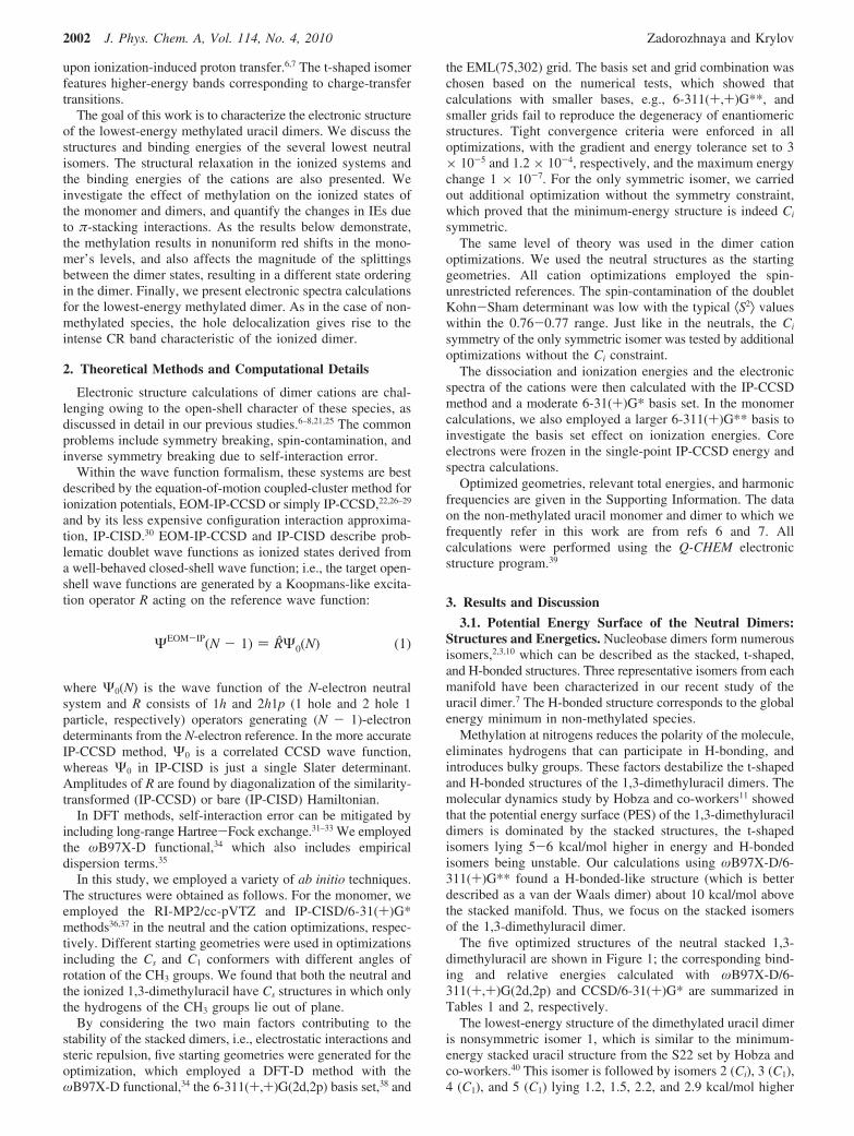
The five optimized structures of the neutral stacked 1,3-dimethyluracil are shown in Figure 1; the corresponding bind-ing and relative energies calculated with ωB97X-D/6-311(+,+)G(2d,2p) and CCSD/6-31(+)G* are summarized inTables 1 and 2, respectively.
The lowest-energy structure of the dimethylated uracil dimeris nonsymmetric isomer 1, which is similar to the minimum-energy stacked uracil structure from the S22 set by Hobza andco-workers.40 This isomer is followed by isomers 2 (Ci), 3 (C1),4 (C1), and 5 (C1) lying 1.2, 1.5, 2.2, and 2.9 kcal/mol higher
ΨEOM-IP(N - 1) ) RΨ0(N) (1)
2002 J. Phys. Chem. A, Vol. 114, No. 4, 2010 Zadorozhnaya and Krylov
in energy, respectively. The energy gaps between the isomersare very small: the five isomers lie in just the 2.9 kcal/mol range,and some of them are nearly degenerate, i.e., separated by 0.3kcal/mol. These energy differences are of the order of kT(298.18
K) ) 0.6 kcal/mol, which suggests significant populations ofall of these isomers at the standard laboratory conditions. Thedense π-stacked manifold and structural motifs are similar tostacked thymine dimers,8 where five isomers lie within 2.2 kcal/mol. Interestingly, no low-energy stacked isomers were identi-fied for dimers of another pyrimidine, cytosine dimer.9
The binding energies of the neutral stacked 1,3-dimethyluracildimers lie in the range 10.9-13.8 kcal/mol, as computed byDFT-D. For the lowest-energy isomer, we also computed theCCSD/6-31(+)G* value. The resulting binding energy of 15.9kcal/mol is in good agreement with 13.8 kcal/mol computedwith ωB97X-D/6-311(+)G**. On the basis of our results foruracil,7 using a larger basis set in CCSD calculations lowersthe CCSD binding energy and improves the agreement betweenthe methods.
The binding energy of the lowest-energy isomer (13.8 kcal/mol) is larger than that of the stacked non-methylated uracildimer for which De ) 10.5 kcal/mol (these are DFT-D values,but a similar trend is observed for the CCSD/6-31+G* bindingenergies, which are 15.9 and 12.2 kcal/mol). For comparison,the binding energies of the lowest stacked thymine and adeninehomodimers are 12.5 and 10.6 kcal/mol, respectively.8
An increase in binding energy upon methylation is somewhatsurprising, as methylated uracil is less polar than uracil (theRI-MP2/cc-pVTZ dipole moments are 4.19 D versus 4.02 D),and therefore, one may expect weaker electrostatic interactionbetween the fragments in the 1,3-dimethyluracil dimer. How-ever, this difference appears to be too small, and localelectrostatic interactions play a more important role. The analysisof the structures reveals that the (NCH2)Hδ+ · · ·Oδ-(C) distancein the stacked 1,3-dimethyluracil dimer is shorter than the(N)Hδ+ · · ·Oδ- distance in the stacked uracil dimer, which resultsin stronger electrostatic interaction between the fragments inthe former complex. A tighter structure of the methylated dimeris also counterintuitive because of the presence of the bulkymethyl groups. The observed increase in binding energy uponsubstitution is consistent with the results of Sherrill andco-workers,41,42 who demonstrated that the electrostatic consid-erations alone are not sufficient to explain the changes in bindingin π-stacked systems upon substitution and that differentialchanges in dispersion interactions play an important role.
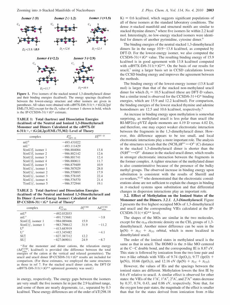
3.2. Effect of Methylation on the Ionized States of theMonomer and the Dimers. 3.2.1. 1,3-Dimethyluracil. Figure2 presents the five highest occupied MOs of 1,3-dimethyluraciland uracil and the corresponding VIEs calculated at the IP-CCSD/6-311(+)G** level.
The shapes of the MOs are similar in the two molecules,except for the σCH electronic density on the CH3 groups of 1,3-dimethyluracil. Another minor difference can be seen in thelp(N) + πCC + πCO orbital, which is more localized indimethylated uracil.
The order of the ionized states in methylated uracil is thesame as that in uracil. The HOMO is the π-like MO centeredat the C-C double bond, and the corresponding IE is 8.87 eV.This state is followed by ionization from the two lone pair andtwo π-like orbitals with VIEs of 9.74 (lp(O1)), 9.77 (lp(O) +lp(N)), 10.66 (lp(O2)), and 12.16 eV (lp(N) + πCC + πCO).
However, the values of IEs and the spacings between theionized states are different. Methylation lowers the first IE by0.6 eV relative to uracil. A similar effect is observed for otherstates: the VIEs of the 12A′, 12A′′, 22A′, and 22A′′ states decreaseby 0.37, 0.74, 0.43, and 0.86 eV, respectively. Note that, forthe oxygen lone-pair states, the magnitude of the effect is smallerthan that for the states derived from ionization from π-like
Figure 1. Five isomers of the stacked neutral 1,3-dimethyluracil dimerand their binding energies (kcal/mol). The energy spacings (kcal/mol)between the lowest-energy structure and other isomers are given inparentheses. All values were obtained with ωB97X-D/6-311(+,+)G(2d,2p)/EML(75,302) except for the De value of isomer 1 shown in bold, whichis the IP-CCSD/6-31(+)G* estimate.
TABLE 1: Total (hartree) and Dissociation Energies(kcal/mol) of the Neutral and Ionized 1,3-DimethyluracilMonomer and Dimers Calculated at the ωB97X-D/6-311(+,+)G(2d,2p)/EML(75,302) Level of Theory
complex EDFT-Dtot De
DFT-D
mU0 -493.431022mU+ -493.111429S(mU)2
0, isomer 1 -986.884084 13.8S(mU)2
0, isomer 2 -986.882142 12.6S(mU)2
0, isomer 3 -986.881741 12.4S(mU)2
0, isomer 4 -986.880611 11.7S(mU)2
0, isomer 5 -986.879409 10.9S(mU)2
+, isomer 1 -986.587029 28.0S(mU)2
+, isomer 2 -986.570893 17.9S(mU)2
+, isomer 3 -986.578185 22.4S(mU)2
+, isomer 4 -986.576570 21.4T(mU)2
+, isomer 5 -986.572944 19.1
TABLE 2: Total (hartree) and Dissociation Energies(kcal/mol) of the Neutral and Ionized 1,3-Dimethyluracil andIts Dimer (Lowest-Energy Isomer) Calculated at theIP-CCSD/6-31(+)G* Level of Theorya
complex ECCSDtot De
CCSD ∆ErelaxCCSD
mU0 -492.032033mU+ -491.715681 -3.8S(mU)2
0, isomer 1 -984.089466 15.9S(mU)2
+, isomer 1 -983.798612 31.9 -11.2U0 -413.683919U+ -413.345482 -4.1SU2
0 -827.387312 12.2SU2
+ -827.069011 24.9 -8.7
a For the monomer and dimer cations, the relaxation energy(∆E
relaxCCSD, kcal/mol) is provided (the difference between the total
energies of the cation at the vertical and relaxed geometries). Theuracil and uracil dimer IP-CCSD/6-31(+)G* results are included forcomparison. (For these estimates, we employed the same structuresas those in ref 7. For the stacked uracil dimer cation, the DFT-D/ωB97X-D/6-311(+)G** optimized geometry was used.)
Zooming into π-Stacked Manifolds of Nucleobases J. Phys. Chem. A, Vol. 114, No. 4, 2010 2003
orbitals. The largest shifts are observed for the states with largecontributions from lone pairs of nitrogens, which are primarysubstitution sites. As a result, the 12A′ and 12A′′ states that areseparated by 0.4 eV in uracil become almost degenerate in 1,3-dimethyluracil.
The IEs are lowered due to electron-donating CH3 groupsincreasing the electron density in the ring (destabilization ofthe respective MOs) and due to a larger size of the methylatedspecies contributing to hole stabilization. The effect is largerin the states derived from ionization from delocalized π-orbitals,in which the CH3 group donates the electron density to theπ-system via the lp(N) component, whereas the in-plane lp(O)orbitals are affected less.
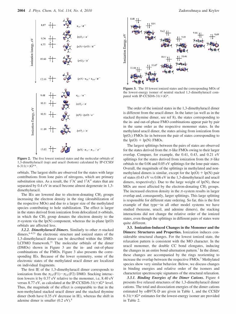
3.2.2. Dimethyluracil Dimers. Similarly to other π-stackeddimers,6–8,21 the electronic structure and ionized states of the1,3-dimethyluracil dimer can be described within the DMO-LCFMO framework.21 The molecular orbitals of the dimer(DMOs) shown in Figure 3 are the in- and out-of-phasecombinations of the FMOs. Figure 3 also presents the corre-sponding IEs. Because of the lower symmetry, some of theelectronic states of the methylated uracil dimer are localizedon individual fragments.
The first IE of the 1,3-dimethyluracil dimer corresponds toionization from the πCC(F1)-πCC(F2) DMO. Stacking interac-tion lowers it by 0.37 eV relative to the monomer, i.e, 8.40 eVversus 8.77 eV, as calculated at the IP-CCSD/6-31(+)G* level.Thus, the magnitude of the effect is comparable to that in thenon-methylated stacked uracil dimer and the stacked thyminedimer (both have 0.35 eV decrease in IE), whereas the shift inadenine dimer is smaller (0.2 eV).8
The order of the ionized states in the 1,3-dimethyluracil dimeris different from the uracil dimer. In the latter (as well as in thestacked thymine dimer, see ref 8), the states corresponding tothe in- and out-of-phase FMO combinations appear pair by pairin the same order as the respective monomer states. In themethylated uracil dimer, the states arising from ionization fromlp(O1) FMOs lie in between the pair of states corresponding tothe lp(O) + lp(N) FMOs.
The largest splittings between the pairs of states are observedfor the states derived from the π-like FMOs owing to their largeroverlap. Compare, for example, the 0.41, 0.43, and 0.21 eVsplittings for the states derived from ionization from the π-likeorbitals to the 0.06 and 0.05 eV splittings for the lone-pair states.Overall, the magnitude of the splittings in methylated and non-methylated dimers is similar, except for the lp(O) + lp(N) pairof states (0.43 eV vs 0.06 eV in the 1,3-dimethyluracil and uracildimers, respectively). Due to the large weight of lp(N), theseMOs are most affected by the electron-donating CH3 groups.The increased electron density in the π-system results in largeroverlap and, consequently, larger splittings. This large splittingis responsible for different state ordering. So far, this is the firstexample of that typesin all other model systems we havestudied (benzene, uracil, and adenine dimers), the stackinginteractions did not change the relative order of the ionizedstates, even though the splittings in different pairs of states werequite different.
3.3. Ionization-Induced Changes in the Monomer and theDimers: Structures and Properties. Ionization induces con-siderable structural changes. For the lowest ionized state, therelaxation pattern is consistent with the MO character. In theuracil monomer, the double CC bond elongates, inducingthe changes in an entire bond-alternation pattern.7 In the dimer,these changes are accompanied by the rings reorienting toincrease the overlap between the respective FMOs.7 Methylatedspecies show very similar behavior. Below, we discuss changesin binding energies and relative order of the isomers andcharacterize spectroscopic signatures of the structural relaxation.
3.3.1. Binding Energies of the Dimer Cations. Figure 4presents five relaxed structures of the 1,3-dimethyluracil dimercations. The total and dissociation energies of the dimer cationsestimated by ωB97X-D are given in Table 1, and the CCSD/6-31(+)G* estimates for the lowest-energy isomer are providedin Table 2.
Figure 2. The five lowest ionized states and the molecular orbitals of1,3-dimethyluracil (top) and uracil (bottom) calculated by IP-CCSD/6-311(+)G**.
Figure 3. The 10 lowest ionized states and the corresponding MOs ofthe lowest-energy isomer of neutral stacked 1,3-dimethyluracil com-puted with IP-CCSD/6-31(+)G*.
2004 J. Phys. Chem. A, Vol. 114, No. 4, 2010 Zadorozhnaya and Krylov
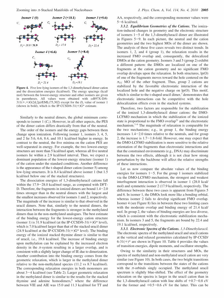
Similarly to the neutral dimers, the global minimum corre-sponds to isomer 1 (C1). However, in all other aspects, the PESof the dimer cation differs drastically from that of the neutral.
The order of the isomers and the energy gaps between themchange upon ionization. Following isomer 1, isomers 3, 4, 5,and 2 lie 5.6, 6.6, 8.4, and 10.1 kcal/mol higher in energy. Incontrast to the neutral, the five minima on the cation PES arewell-separated in energy. For example, the two lowest-energystructures are more than 5 kcal/mol apart, whereas all five neutralisomers lie within a 2.9 kcal/mol interval. Thus, we expect adominant population of the lowest-energy structure (isomer 1)of the cation under the standard conditions. Another differenceis the appearance of the t-shaped dimer cation (isomer 5) amonglow-lying structures. It is 8.4 kcal/mol above isomer 1 (but 1.5kcal/mol below one of the stacked structures).
The dissociation energies of 1,3-dimethyluracil cations fallwithin the 17.9-28.0 kcal/mol range, as computed with DFT-D. Therefore, the fragments in ionized dimers are bound 1.4-2.0times stronger than in the neutral dimers with the largest andthe smallest increases observed for isomers 1 and 2, respectively.The magnitude of the increase is similar to that observed in theuracil dimers. Note that, similarly to the neutral dimers, theinteraction between the fragments is stronger in the methylateddimers than in the non-methylated analogues. The best estimateof the binding energy for the lowest-energy cation structure(isomer 1) is 31.9 kcal/mol (at the IP-CCSD/6-31(+)G* level),which is 7.0 kcal/mol larger than that of the stacked uracil dimer(24.9 kcal/mol at the IP-CCSD/6-31(+)G* level). The bindingenergy of the ionized stacked thymine dimer is similar to thatof uracil, i.e., 19.8 kcal/mol. The increase of binding energyupon methylation can be explained by the increased electrondensity in the π-system resulting in a larger overlap, and isconsistent with a slightly larger change of IE due to dimerization.Another contribution into the binding energy comes from thegeometric relaxation, which is larger in the methylated dimerrelative to the non-methylated species (11.2 vs 8.7 kcal/mol).The corresponding relaxation energies in both monomers areabout 3-4 kcal/mol (see Table 2). Larger geometric relaxationin the methylated dimer is similar to the results for the stackedthymine and adenine homodimers,8 where the differencebetween VIE and AIE was 15.0 and 11.3 kcal/mol for TT and
AA, respectively, and the corresponding monomer values were5-6 kcal/mol.
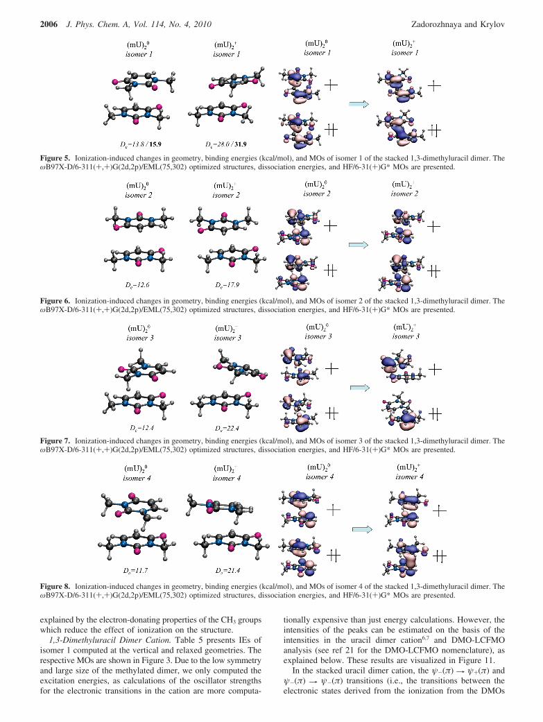
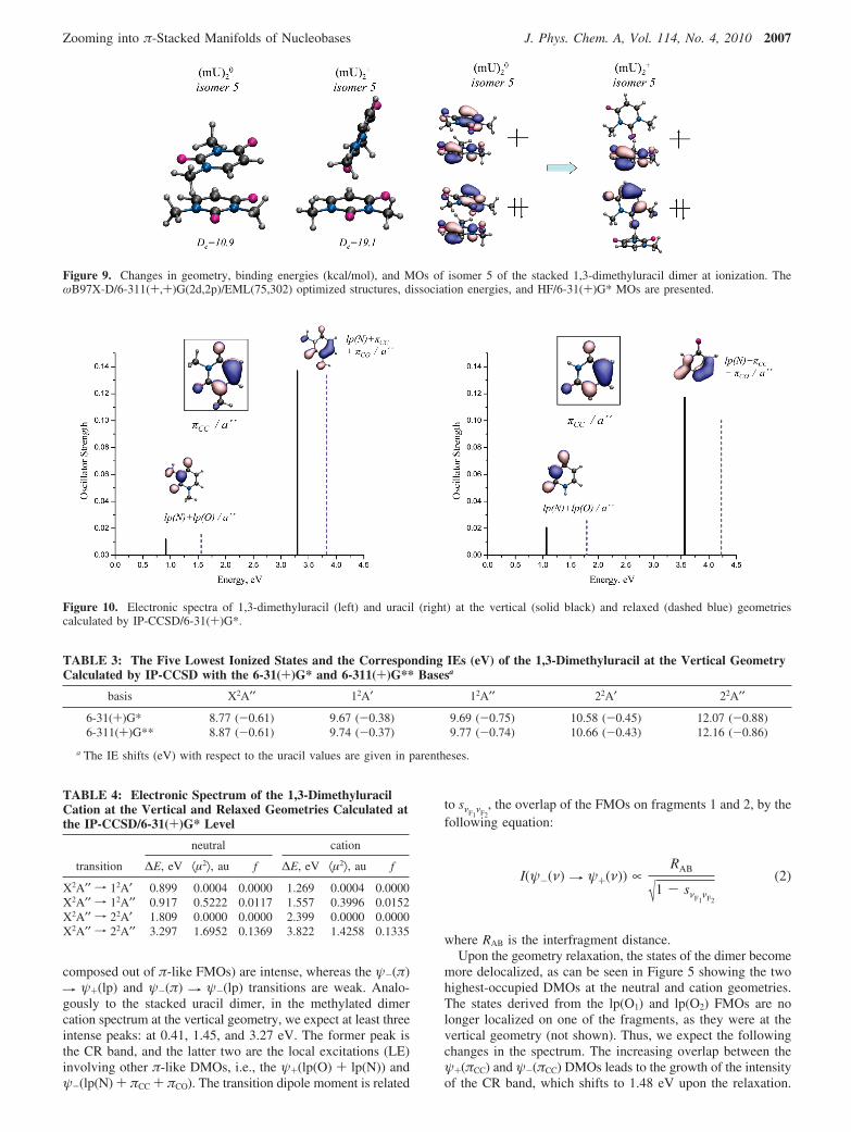
3.3.2. Equilibrium Geometries of the Cations. The ioniza-tion-induced changes in geometry and the electronic structureof isomers 1-5 of the 1,3-dimethyluracil dimer are illustratedin Figures 5-9. In each picture, the neutral and the cationgeometries and the two highest MOs of the dimer are shown.The analysis of these five cases reveals two distinct trends. Inisomers 1, 2, and 4 (group 1), the relaxation results in theincreased FMO overlap and, consequently, the delocalizedDMOs at the cation geometry. Isomers 3 and 5 (group 2) exhibita different pattern: the DMOs are localized on one of thefragments at the cation geometry and no significant FMOoverlap develops upon the relaxation. In both structures, lp(O)of one of the fragments moves toward the hole centered on theπCC MO of the other fragment. Thus, group 2 cations arestabilized by the favorable electrostatic interaction of thelocalized hole and the negative charge on lp(O). This motif,which is similar to the t-shaped uracil dimer,7 demonstrates thatelectrostatic interactions can be competitive with the holedelocalization effects even in the stacked systems.
Therefore, two factors are responsible for the stabilizationof the ionized 1,3-dimethyluracil dimer cations: the DMO-LCFMO mechanism in which the stabilization of the ionizedstate is proportional to the FMO overlap21 and the electrostaticmechanism.7–9 The magnitude of relaxation is comparable forthe two mechanisms; e.g., in group 1, the binding energyincreases 1.4-2.0 times relative to the neutrals, and for group2, the increase is 1.7-1.8-fold. However, one may expect thatthe DMO-LCFMO stabilization is more sensitive to the relativeorientation of the fragments than electrostatic interactions andthat the constrained environments (e.g., DNA) may discriminatebetween the two effects, although it is not clear how strongperturbation by the backbone will affect the relative strengthsof these interactions.
Let us now compare the absolute values of the bindingenergies for isomers 1-5. For the group 1 isomers stabilizedvia the DMO-LCFMO mechanism, the strongest and weakestinterfragment interaction is observed in isomer 1 (28.0 kcal/mol) and symmetric isomer 2 (17.9 kcal/mol), respectively. Thedifference between these two cases is apparent from Figures 5and 6. In isomer 1, the DMOs look more like a bonding orbital,whereas isomer 2 fails to develop significant FMO overlap.Isomer 4 (see Figure 8) lies in between these two limiting caseswith the moderate overlap and binding energy of 21.4 kcal/mol. In group 2, the values of binding energies are less diverse,which is consistent with the electrostatic stabilization mecha-nism. In isomers 3 and 5, the fragments are bound by 22.4 and19.1 kcal/mol, respectively.
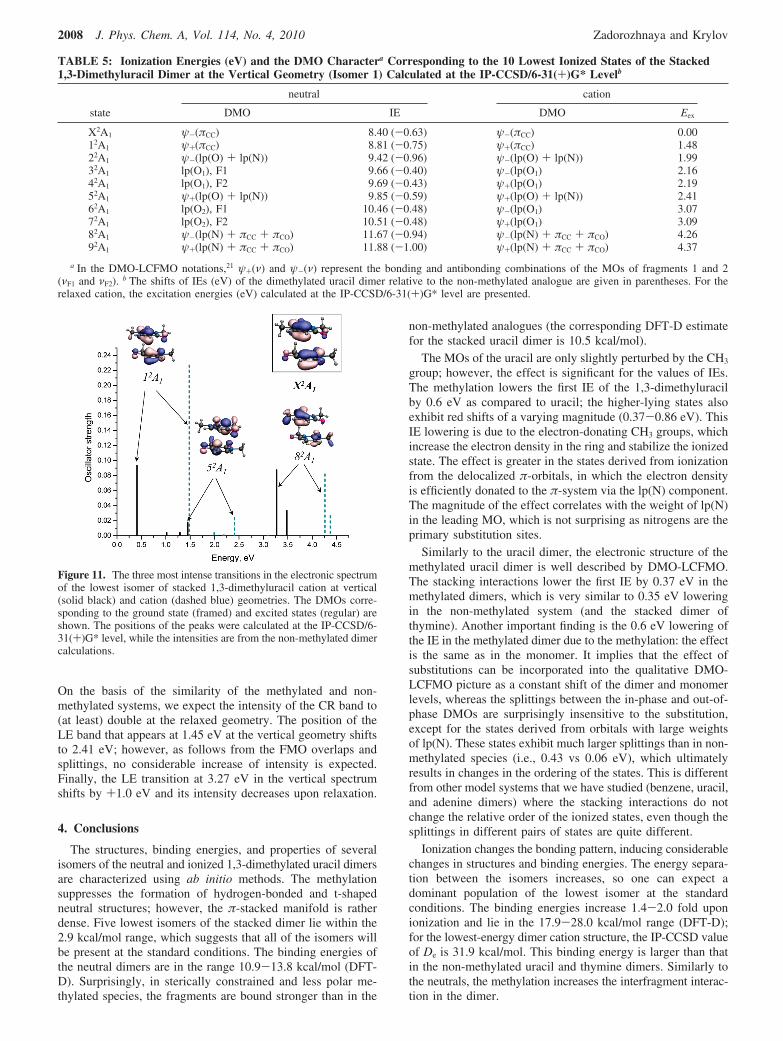
3.3.3. Electronic Spectra of the Cations. 1,3-Dimethyluracil.The electronic spectra of the methylated uracil and uracil cationsat the vertical and relaxed geometries calculated by IP-CCSD/6-31(+)* are shown in Figure 10. Table 4 provides the valuesof transition energies, dipole moments, and oscillator strengths.
Owing to the similarity in their structures and MOs, thespectra of methylated and non-methylated uracil cation are verysimilar (see Figure 10). In both cases, the two bright transitionscorrespond to the transitions between the states of the cationswith the π-orbitals singly occupied. The methylated uracilspectrum is slightly blue-shifted. The effect of the geometryrelaxation on the spectra is larger in the uracil cation than inthe 1,3-dimethyluracil cation with line shifts of +0.7-0.8 eVfor the former and +0.5-0.6 eV for the latter. This can be
Figure 4. Five low-lying isomers of the 1,3-dimethyluracil dimer cationand the dissociation energies (kcal/mol). The energy spacings (kcal/mol) between the lowest-energy structure and other isomers are givenin parentheses. All values were obtained with ωB97X-D/6-311(+,+)G(2d,2p)/EML(75,302) except for the De value of isomer 1(shown in bold), which is the IP-CCSD/6-31(+)G* estimate.
Zooming into π-Stacked Manifolds of Nucleobases J. Phys. Chem. A, Vol. 114, No. 4, 2010 2005
explained by the electron-donating properties of the CH3 groupswhich reduce the effect of ionization on the structure.
1,3-Dimethyluracil Dimer Cation. Table 5 presents IEs ofisomer 1 computed at the vertical and relaxed geometries. Therespective MOs are shown in Figure 3. Due to the low symmetryand large size of the methylated dimer, we only computed theexcitation energies, as calculations of the oscillator strengthsfor the electronic transitions in the cation are more computa-
tionally expensive than just energy calculations. However, theintensities of the peaks can be estimated on the basis of theintensities in the uracil dimer cation6,7 and DMO-LCFMOanalysis (see ref 21 for the DMO-LCFMO nomenclature), asexplained below. These results are visualized in Figure 11.
In the stacked uracil dimer cation, the ψ-(π) f ψ+(π) andψ-(π) f ψ-(π) transitions (i.e., the transitions between theelectronic states derived from the ionization from the DMOs
Figure 5. Ionization-induced changes in geometry, binding energies (kcal/mol), and MOs of isomer 1 of the stacked 1,3-dimethyluracil dimer. TheωB97X-D/6-311(+,+)G(2d,2p)/EML(75,302) optimized structures, dissociation energies, and HF/6-31(+)G* MOs are presented.
Figure 6. Ionization-induced changes in geometry, binding energies (kcal/mol), and MOs of isomer 2 of the stacked 1,3-dimethyluracil dimer. TheωB97X-D/6-311(+,+)G(2d,2p)/EML(75,302) optimized structures, dissociation energies, and HF/6-31(+)G* MOs are presented.
Figure 7. Ionization-induced changes in geometry, binding energies (kcal/mol), and MOs of isomer 3 of the stacked 1,3-dimethyluracil dimer. TheωB97X-D/6-311(+,+)G(2d,2p)/EML(75,302) optimized structures, dissociation energies, and HF/6-31(+)G* MOs are presented.
Figure 8. Ionization-induced changes in geometry, binding energies (kcal/mol), and MOs of isomer 4 of the stacked 1,3-dimethyluracil dimer. TheωB97X-D/6-311(+,+)G(2d,2p)/EML(75,302) optimized structures, dissociation energies, and HF/6-31(+)G* MOs are presented.
2006 J. Phys. Chem. A, Vol. 114, No. 4, 2010 Zadorozhnaya and Krylov
composed out of π-like FMOs) are intense, whereas the ψ-(π)f ψ+(lp) and ψ-(π) f ψ-(lp) transitions are weak. Analo-gously to the stacked uracil dimer, in the methylated dimercation spectrum at the vertical geometry, we expect at least threeintense peaks: at 0.41, 1.45, and 3.27 eV. The former peak isthe CR band, and the latter two are the local excitations (LE)involving other π-like DMOs, i.e., the ψ+(lp(O) + lp(N)) andψ-(lp(N) + πCC + πCO). The transition dipole moment is related
to sνF1νF2
, the overlap of the FMOs on fragments 1 and 2, by thefollowing equation:
where RAB is the interfragment distance.Upon the geometry relaxation, the states of the dimer become
more delocalized, as can be seen in Figure 5 showing the twohighest-occupied DMOs at the neutral and cation geometries.The states derived from the lp(O1) and lp(O2) FMOs are nolonger localized on one of the fragments, as they were at thevertical geometry (not shown). Thus, we expect the followingchanges in the spectrum. The increasing overlap between theψ+(πCC) and ψ-(πCC) DMOs leads to the growth of the intensityof the CR band, which shifts to 1.48 eV upon the relaxation.
Figure 9. Changes in geometry, binding energies (kcal/mol), and MOs of isomer 5 of the stacked 1,3-dimethyluracil dimer at ionization. TheωB97X-D/6-311(+,+)G(2d,2p)/EML(75,302) optimized structures, dissociation energies, and HF/6-31(+)G* MOs are presented.
Figure 10. Electronic spectra of 1,3-dimethyluracil (left) and uracil (right) at the vertical (solid black) and relaxed (dashed blue) geometriescalculated by IP-CCSD/6-31(+)G*.
TABLE 3: The Five Lowest Ionized States and the Corresponding IEs (eV) of the 1,3-Dimethyluracil at the Vertical GeometryCalculated by IP-CCSD with the 6-31(+)G* and 6-311(+)G** Basesa
basis X2A′′ 12A′ 12A′′ 22A′ 22A′′
6-31(+)G* 8.77 (-0.61) 9.67 (-0.38) 9.69 (-0.75) 10.58 (-0.45) 12.07 (-0.88)6-311(+)G** 8.87 (-0.61) 9.74 (-0.37) 9.77 (-0.74) 10.66 (-0.43) 12.16 (-0.86)
a The IE shifts (eV) with respect to the uracil values are given in parentheses.
TABLE 4: Electronic Spectrum of the 1,3-DimethyluracilCation at the Vertical and Relaxed Geometries Calculated atthe IP-CCSD/6-31(+)G* Level
neutral cation
transition ∆E, eV ⟨µ2⟩, au f ∆E, eV ⟨µ2⟩, au f
X2A′′ f 12A′ 0.899 0.0004 0.0000 1.269 0.0004 0.0000X2A′′ f 12A′′ 0.917 0.5222 0.0117 1.557 0.3996 0.0152X2A′′ f 22A′ 1.809 0.0000 0.0000 2.399 0.0000 0.0000X2A′′ f 22A′′ 3.297 1.6952 0.1369 3.822 1.4258 0.1335
I(ψ-(ν) f ψ+(ν)) ∝RAB
√1 - sνF1νF2
(2)
Zooming into π-Stacked Manifolds of Nucleobases J. Phys. Chem. A, Vol. 114, No. 4, 2010 2007
On the basis of the similarity of the methylated and non-methylated systems, we expect the intensity of the CR band to(at least) double at the relaxed geometry. The position of theLE band that appears at 1.45 eV at the vertical geometry shiftsto 2.41 eV; however, as follows from the FMO overlaps andsplittings, no considerable increase of intensity is expected.Finally, the LE transition at 3.27 eV in the vertical spectrumshifts by +1.0 eV and its intensity decreases upon relaxation.
4. Conclusions
The structures, binding energies, and properties of severalisomers of the neutral and ionized 1,3-dimethylated uracil dimersare characterized using ab initio methods. The methylationsuppresses the formation of hydrogen-bonded and t-shapedneutral structures; however, the π-stacked manifold is ratherdense. Five lowest isomers of the stacked dimer lie within the2.9 kcal/mol range, which suggests that all of the isomers willbe present at the standard conditions. The binding energies ofthe neutral dimers are in the range 10.9-13.8 kcal/mol (DFT-D). Surprisingly, in sterically constrained and less polar me-thylated species, the fragments are bound stronger than in the
non-methylated analogues (the corresponding DFT-D estimatefor the stacked uracil dimer is 10.5 kcal/mol).
The MOs of the uracil are only slightly perturbed by the CH3
group; however, the effect is significant for the values of IEs.The methylation lowers the first IE of the 1,3-dimethyluracilby 0.6 eV as compared to uracil; the higher-lying states alsoexhibit red shifts of a varying magnitude (0.37-0.86 eV). ThisIE lowering is due to the electron-donating CH3 groups, whichincrease the electron density in the ring and stabilize the ionizedstate. The effect is greater in the states derived from ionizationfrom the delocalized π-orbitals, in which the electron densityis efficiently donated to the π-system via the lp(N) component.The magnitude of the effect correlates with the weight of lp(N)in the leading MO, which is not surprising as nitrogens are theprimary substitution sites.
Similarly to the uracil dimer, the electronic structure of themethylated uracil dimer is well described by DMO-LCFMO.The stacking interactions lower the first IE by 0.37 eV in themethylated dimers, which is very similar to 0.35 eV loweringin the non-methylated system (and the stacked dimer ofthymine). Another important finding is the 0.6 eV lowering ofthe IE in the methylated dimer due to the methylation: the effectis the same as in the monomer. It implies that the effect ofsubstitutions can be incorporated into the qualitative DMO-LCFMO picture as a constant shift of the dimer and monomerlevels, whereas the splittings between the in-phase and out-of-phase DMOs are surprisingly insensitive to the substitution,except for the states derived from orbitals with large weightsof lp(N). These states exhibit much larger splittings than in non-methylated species (i.e., 0.43 vs 0.06 eV), which ultimatelyresults in changes in the ordering of the states. This is differentfrom other model systems that we have studied (benzene, uracil,and adenine dimers) where the stacking interactions do notchange the relative order of the ionized states, even though thesplittings in different pairs of states are quite different.
Ionization changes the bonding pattern, inducing considerablechanges in structures and binding energies. The energy separa-tion between the isomers increases, so one can expect adominant population of the lowest isomer at the standardconditions. The binding energies increase 1.4-2.0 fold uponionization and lie in the 17.9-28.0 kcal/mol range (DFT-D);for the lowest-energy dimer cation structure, the IP-CCSD valueof De is 31.9 kcal/mol. This binding energy is larger than thatin the non-methylated uracil and thymine dimers. Similarly tothe neutrals, the methylation increases the interfragment interac-tion in the dimer.
TABLE 5: Ionization Energies (eV) and the DMO Charactera Corresponding to the 10 Lowest Ionized States of the Stacked1,3-Dimethyluracil Dimer at the Vertical Geometry (Isomer 1) Calculated at the IP-CCSD/6-31(+)G* Levelb
neutral cation
state DMO IE DMO Eex
X2A1 ψ-(πCC) 8.40 (-0.63) ψ-(πCC) 0.0012A1 ψ+(πCC) 8.81 (-0.75) ψ+(πCC) 1.4822A1 ψ-(lp(O) + lp(N)) 9.42 (-0.96) ψ-(lp(O) + lp(N)) 1.9932A1 lp(O1), F1 9.66 (-0.40) ψ-(lp(O1) 2.1642A1 lp(O1), F2 9.69 (-0.43) ψ+(lp(O1) 2.1952A1 ψ+(lp(O) + lp(N)) 9.85 (-0.59) ψ+(lp(O) + lp(N)) 2.4162A1 lp(O2), F1 10.46 (-0.48) ψ-(lp(O1) 3.0772A1 lp(O2), F2 10.51 (-0.48) ψ+(lp(O1) 3.0982A1 ψ-(lp(N) + πCC + πCO) 11.67 (-0.94) ψ-(lp(N) + πCC + πCO) 4.2692A1 ψ+(lp(N) + πCC + πCO) 11.88 (-1.00) ψ+(lp(N) + πCC + πCO) 4.37
a In the DMO-LCFMO notations,21 ψ+(ν) and ψ-(ν) represent the bonding and antibonding combinations of the MOs of fragments 1 and 2(νF1 and νF2). b The shifts of IEs (eV) of the dimethylated uracil dimer relative to the non-methylated analogue are given in parentheses. For therelaxed cation, the excitation energies (eV) calculated at the IP-CCSD/6-31(+)G* level are presented.
Figure 11. The three most intense transitions in the electronic spectrumof the lowest isomer of stacked 1,3-dimethyluracil cation at vertical(solid black) and cation (dashed blue) geometries. The DMOs corre-sponding to the ground state (framed) and excited states (regular) areshown. The positions of the peaks were calculated at the IP-CCSD/6-31(+)G* level, while the intensities are from the non-methylated dimercalculations.
2008 J. Phys. Chem. A, Vol. 114, No. 4, 2010 Zadorozhnaya and Krylov
The relaxation of the cation structures is governed by twodistinct mechanisms: the hole delocalization (and the FMOoverlap) and the electrostatic stabilization (interaction of thelp(O) with the localized hole).
Finally, we presented electronic spectra of the ionized species.Significant changes in the spectra upon relaxation can beexploited to monitor the ionization-induced dynamics in dim-ethylated uracils. At the vertical geometry, there are three intensetransitions: at 0.41, 1.45, and 3.27 eV, the CR band at 0.41 eVand LE at 1.45 eV being the most intense. Upon relaxation,these bands are blue-shifted, and their intensities change to 1.48(CR), 2.41 (LE), and 4.26(LE) eV. The CR band at 1.48 eV isexpected be the most intense and can be used to monitor therelaxed stacked dimer cation formation.
Acknowledgment. We are grateful to Dr. Ksenia Bravayafor her insightful remarks and critical reading of the manuscript.This work was conducted in the framework of the iOpenShellCenter for Computational Studies of Electronic Structure andSpectroscopy of Open-Shell and Electronically Excited Species(iopenshell.usc.edu) supported by the National Science Founda-tion through the CRIF:CRF CHE-0625419 + 0624602 +0625237 grant, and is also supported by the CHE-0616271 grant.
Supporting Information Available: Tables showing theoptimized geometries of neutral and ionized 1,3-dimethyluraciland stacked 1,3-dimethyluracil dimers, as well as the relevantenergies. This material is available free of charge via the Internetat http://pubs.acs.org.
References and Notes
(1) Nir, E.; Kleinermanns, K.; de Vries, M. S. Nature 2000, 408, 949.(2) Sponer, J.; Leszczynski, J.; Hobza, P. Biopolymers 2002, 61, 3.(3) Saigusa, H. Photochem. Photobiol. 2006, 7, 197.(4) de Vries, M. S.; Hobza, P. Annu. ReV. Phys. Chem. 2007, 58, 585.(5) de Vries, M. S. In Radiation induced molecular phenomena in
nucleic acids; Shukla, M., Leszczynski, J., Eds.; Springer: Berlin, 2008; p323.
(6) Golubeva, A. A.; Krylov, A. I. Phys. Chem. Chem. Phys. 2009,11, 1303.
(7) Zadorozhnaya, A. A.; Krylov, A. I. J. Chem. Theory Comput.,submitted for publication, 2009.
(8) Bravaya, K. B.; Kostko, O.; Ahmed, M.; Krylov, A. I. Phys. Chem.Chem. Phys. DOI: 10.1039/b919930f.
(9) Kostko, O.; Bravaya, K. B.; Krylov, A. I.; Ahmed, M. Phys. Chem.Chem. Phys., submitted for publication, 2009.
(10) Muller-Dethlefs, K.; Hobza, P. Chem. ReV. 2000, 100, 143.(11) Kratochvıl, M.; Engkvist, O.; Vacek, J.; Jungwirth, P.; Hobza, P.
Phys. Chem. Chem. Phys. 2000, 2, 2419.(12) Kabelac, M.; Hobza, P. J. Phys. Chem. B 2001, 105, 5804.(13) Kabelac, M.; Hobza, P. Chem.sEur. J. 2001, 7, 2067.
(14) Plutzer, C.; Hunig, I.; Kleinermanns, K. Phys. Chem. Chem. Phys.2003, 5, 1158.
(15) Satzger, H.; Townsend, D.; Stolow, A. Chem. Phys. Lett. 2006,430, 144.
(16) He, Y.; Wu, C.; Kong, W. J. Phys. Chem. A 2004, 108, 943.(17) Mulliken, R. S.; Person, W. B. Molecular Complexes; Wiley-
Interscience: 1969.(18) Badger, B.; Brocklehurst, B. Trans. Faraday Soc. 1969, 65, 2576.(19) Badger, B.; Brocklehurst, B. Trans. Faraday Soc. 1969, 65, 2582.(20) Badger, B.; Brocklehurst, B. Trans. Faraday Soc. 1969, 65, 2588.(21) Pieniazek, P. A.; Krylov, A. I.; Bradforth, S. E. J. Chem. Phys.
2007, 127, 044317.(22) Pieniazek, P. A.; Bradforth, S. E.; Krylov, A. I. J. Chem. Phys.
2008, 129, 074104.(23) Pieniazek, P. A.; VandeVondele, J.; Jungwirth, P.; Krylov, A. I.;
Bradforth, S. E. J. Phys. Chem. A 2008, 112, 6159.(24) Pieniazek, P. A.; Sundstrom, E. J.; Bradforth, S. E.; Krylov, A. I.
J. Phys. Chem. A 2009, 113, 4423.(25) Pieniazek, P. A.; Arnstein, S. A.; Bradforth, S. E.; Krylov, A. I.;
Sherrill, C. D. J. Chem. Phys. 2007, 127, 164110.(26) Sinha, D.; Mukhopadhyay, D.; Mukherjee, D. Chem. Phys. Lett.
1986, 129, 369.(27) Pal, S.; Rittby, M.; Bartlett, R. J.; Sinha, D.; Mukherjee, D. Chem.
Phys. Lett. 1987, 137, 273.(28) Stanton, J. F.; Gauss, J. J. Chem. Phys. 1994, 101, 8938.(29) Nooijen, M.; Bartlett, R. J. J. Chem. Phys. 1995, 102, 3629.(30) Golubeva, A. A.; Pieniazek, P. A.; Krylov, A. I. J. Chem. Phys.
2009, 130, 124113.(31) Iikura, H.; Tsuneda, T.; Yanai, T.; Hirao, K. J. Chem. Phys. 2001,
115, 3540.(32) Baer, R.; Neuhauser, D. Phys. ReV. Lett. 2005, 94, 043002.(33) Chai, J.-D.; Head-Gordon, M. J. Chem. Phys. 2008, 128, 084106.(34) Chai, J.-D.; Head-Gordon, M. Phys. Chem. Chem. Phys. 2008, 10,
6615.(35) Grimme, S. J. Comput. Chem. 2004, 25, 1463.(36) Hehre, W. J.; Ditchfield, R.; Pople, J. A. J. Chem. Phys. 1972, 56,
2257.(37) Dunning, T. H. J. Chem. Phys. 1989, 90, 1007.(38) Krishnan, R.; Binkley, J. S.; Seeger, R.; Pople, J. A. J. Chem. Phys.
1980, 72, 650.(39) Shao, Y.; Molnar, L. F.; Jung, Y.; Kussmann, J.; Ochsenfeld, C.;
Brown, S.; Gilbert, A. T. B.; Slipchenko, L. V.; Levchenko, S. V.; O’Neil,D. P.; Distasio, R. A., Jr.; Lochan, R. C.; Wang, T.; Beran, G. J. O.; Besley,N. A.; Herbert, J. M.; Lin, C. Y.; Van Voorhis, T.; Chien, S. H.; Sodt, A.;Steele, R. P.; Rassolov, V. A.; Maslen, P.; Korambath, P. P.; Adamson,R. D.; Austin, B.; Baker, J.; Bird, E. F. C.; Daschel, H.; Doerksen, R. J.;Drew, A.; Dunietz, B. D.; Dutoi, A. D.; Furlani, T. R.; SGwaltney. R.;Heyden, A.; Hirata, S.; Hsu, C.-P.; Kedziora, G. S.; Khalliulin, R. Z.;Klunziger, P.; Lee, A. M.; Liang, W. Z.; Lotan, I.; Nair, N.; Peters, B.;Proynov, E. I.; Pieniazek, P. A.; Rhee, Y. M.; Ritchie, J.; Rosta, E.; Sherrill,C. D.; Simmonett, A. C.; Subotnik, J. E.; Woodcock, H. L., III; Zhang,W.; Bell, A. T.; Chakraborty, A. K.; Chipman, D. M.; Keil, F. J.; Warshel,A.; Herhe, W. J.; Schaefer, H. F., III; Kong, J.; Krylov, A. I.; Gill, P. M. W.;HeadGordon, M. Phys. Chem. Chem. Phys. 2006, 8, 3172.
(40) Jurecka, P.; Sponer, J.; Cerny, J.; Hobza, P. Phys. Chem. Chem.Phys. 2006, 8, 1985.
(41) Sinnokrot, M. O.; Sherrill, C. D. J. Phys. Chem. A 2003, 107, 8377.(42) Sinnokrot, M. O.; Sherrill, C. D. J. Am. Chem. Soc. 2004, 126,
7690.
JP910440D
Zooming into π-Stacked Manifolds of Nucleobases J. Phys. Chem. A, Vol. 114, No. 4, 2010 2009


![Radical Cation π‐Dimers of Conjugated Oligomers as ... › contents › ... · transport through molecular wires has been pointed out.[43-47] This intimate relationship was deduced](https://img.pdfslide.tips/doc/110x75/5f0c70957e708231d43568ca/radical-cation-adimers-of-conjugated-oligomers-as-a-contents-a-.jpg)


![Formation of Long, Multicenter π [TCNE] 2 Dimers in 2 ...diposit.ub.edu/dspace/bitstream/2445/154509/1/678270.pdfWhile dimers dissociate at room temperature, they are stable at 175](https://img.pdfslide.tips/doc/110x75/60d0ab48f09c2e68e856dea2/formation-of-long-multicenter-tcne-2-dimers-in-2-while-dimers-dissociate.jpg)