57
6 Anhang
6.1 Literaturangaben
1 Alessandri C, Basili S, Violi F, Ferroni P, Gazzaniga PP, Cordova C and C.O.B.H. group:Hypercoagulability state in patients with Chronic Obstructive pulmonary disease. Thromb.Haemost. 72 (1994) 343-346
2 Altman R, Scazziota A, Rouvier J, Gurfinkel E, Favaloro R, Perrone S, Fareed J: Coagulationand fibrinolytic parameters in patients with pulmonary hypertension. Clin. Cardiol. 19 (1996)549-554
3 Anders O, Burstein C, Bitter S, Kundt M, Freund M: Aktivierungs- und Umsatzmarker derHämostase im Verlauf der akuten tiefen Beinvenenthrombose. Haemost. 15 (1995) 87-91
4 Barthels M, Möller,W, Oestereich C: Thrombin/Antithrombin III Komplex (TAT)Plasminogen-Aktivator-Inhibitor (PAI): Neue Erkenntnisse zur klinischen Relevanz In:Wisser H: Neue Methoden in der Labordiagnostik. Medizinische Verlagsgesellschaft,Marburg 1990 S. 121-132
5 Barthels M, Poliwoda H: Gerinnungsanalysen. 4. Aufl., Thieme, Stuttgart New York 19936 Bauer KA, Weiss LM, Sparrow D, Vokonas PS, Rosenberg RD: Ageing-associated changes
in indices of thrombin generation and protein C activation in humans. Normative AgeingStudy. J. Clin. Invest. 80 (1987) 1527-1534
7 Bounameaux H, Slosmann D, de Moerloose P, Reber G: Laboratory diagnosis of pulmonaryembolism: value of increased levels of plasma d-dimer and thrombin-antithrombin IIIcomplexes. Biomed. Pharmacother. 43 (1989) 385-388
8 Brantley ML, Lester DP, Miller BH: Clinical features and history of the destructive lungdisease associated with alpha-1-antitrypsin deficiency of adults with pulmonary symptoms.Am. Rev. Resp. Div. 138 (1988) 327-336
9 Bratzel D, Seiler FR: Struktur und Biologie von Interleukinen. Haemost. 10 (1990) 52-6310 Brus F, van Oevern W, Okken A, Oetomo SB: Activation of the plasma clotting, fibrinolytic
and Kinin-Kallikrein system in preterm infants with severe idiopathic respiratory distresssyndrome. Ped. Res. 36 (1994) 647-653
11 Buhl R, Meyer A, Vogelmeyer C: Oxidant-protease interaction in the lung. Chest 110 (1996)Supplement 1
12 Camner P: Minireview: How important is mucociliary clearance?. Exp. Lung. Res. 14 (1988)423-429
13 CAPRIE Steering Committee: A randomised, blinded, trial of clopidogrel versus aspirin inpatients at risk of ischaemic events (CAPRIE). Lancet 348 (1996) 1329-1339
14 Carmassi F, Morale M, de Negri F, Puccetti R, Pistelli F, Mariani G, Pazzagli M, Palla A,Giuntini C: Thrombin-antithrombin III complexes as an additional diagnostic aid inpulmonary embolism. Haemostasis 26 (1996) 16-22
15 Carson JL, Terrin ML, Duff A, Kelley MA: Pulmonary embolism and mortality in patientswith COPD. Chest 110 (1996) 1212-1219
16 Chan CK, Matthay RA: Pulmonary thromboembolism. In: Stein (Ed): Internal medicine,Mosby-Year Book, 4th ed., St. Louis, 1994, 1748-1754
17 Chapmann HA, Allen CL, Stone OL: Abnormalities in pathways of alveolar fibrin turnoveramong patients with interstitial lung disease. Am Rev Respir Dis 133 (1986) 437-443
18 Chrispin AR, Norman AP: The systematic evaluation of the chest radiograph in cysticfibrosis. Pediatr., Radiol. 2 (1974) 101-107
19 Conway EM, Bauer KA, Barzegar S, Rosenberg RD: Suppression of hemostatic systemactivation by oral anticoagulants in the blood of patients with thrombotic diatheses. J. Clin.Invest. (1987) 1535-44

58
20 Costabel U: Interstitielle Lungenkrankheiten. Pneumologie 50 (1996) 611-61421 Dahl R, Venge P: Activation of blood coagulation during inhalation challenge tests. Allergy
36 (1981) 129-13322 Demers C, Ginsberg JS, Johnston M, Brill-Edwards P, Panju A: D-Dimer and Thrombin-
Antithrombin III complexes in patients with clinically suspected pulmonary embolism.Thromb. Haemost. 67 (1992) 408-412
23 Deutsche Atemwegsliga: Vorschläge zur Diagnostik und Therapie des chronischen Corpulmonale. Pneumologie 48 (1994) 287-291
24 Dokter G, Lindemann H, Tümmler B, Wunderlich P, Dittrich-Weber P: Mukoviszidose. 2.Aufl., Thieme Stuttgart, New York, 1997
25 Frank K, Gurtner HP, Kneussl M, Lang I, Mlczloch J: Aminorexinduzierte plexogenepulmonale Arteriopathie: 25 Jahre danach!. Z. Kardiol.82 (1993) 568-572
26 Fuchimukai T, Fujiwara T, Takahashi A, Enhorning G: Artificial pulmonary surfactantinhibited by proteins. Appl. Physiol. 62 (1987) 429-437
27 Fulmer JD: Interstitial lung disease. In: Stein (Ed): Internal medicine, Mosby-Year Book, 4th
ed., St. Louis, 1994, 1681-169228 Fuster V, Steele PM, Edwards WD, Gersh BJ., Mc Goon MD, Frye RL: Primary pulmonary
hypertension: natural history and the importance of thrombosis. Circulation 70 (1984) 580-587
29 Goldhaber SZ, Vaughan DE, Tumeh SS, Loscalzo J : Utility of cross linked fibrindegradation products in the diagnosis of pulmonary embolism. Am Heart J 116 (1988) 505-508
30 Goldhaber SZ: Strategies for diagnosis In: Goldhaber SZ (Ed.): Pulmonary embolism anddeep vein thrombosis. Saunders, Philadelphia 1985 S. 79-97
31 Gram J: Coronary artery disease, fibrinolysis and age. in: Tilsner V.(Hrsg.) HamburgerSymposium über Blutgerinnung 1994. Schattauer, Basel, 1995 S. 73-80
32 Gresele P, Todisco T, Merante F, Nenci GG: Platelet activation and allergic asthma. N. Engl.J. Med. 306 (1982) 549-554
33 Grimminger F, Walmrath HD, Seeger W, Lasch HG: Granulozyten-Endothel-Interaktion.Mechanismen der Abwehr und Autoaggression. Haemost. 14 (1994) 7-15
34 Günther A, Kalinowski M, Elssner A, Seeger W: Clot-embedded natural surfactant: kineticsof fibrinolysis and surface activity. Am. J. Physiol. 267 (1994) L618-L624
35 Hasday JD, Bachwich PR, Lynch JP, Sitrin RG: Procoagulant and plasminogen activatoractivities of bronchoalveolar fluid in patients with pulmonary sarcoidosis. Exp. Lung Res. 14(1988) 261-278
36 Häußinger K,Huber RM: Bronchialkarzinom (Lungenkrebs). Pneumologie 50 (1996) 599-603
37 Hawinger J, Niewiarowski S, Gurewich V, Thomas DP: Measurement of fibrinogen andfibrin degradation products in serum by staphylococcal clumping test. J. Lab. clin. Med. 75(1970) 93-98
38 Hawinger J:Adhesive interactions of blood cells and the vascular wall. In:Colmann R.W., Hirsh J., Marder V.J., Salzmann E.W.: Hemostasis and Thrombosis: Basicprinciples and clinical practice. J. B. Lippincott, Philadelphia, 1994, S.: 762-796
39 Heimburger N: Entzündungsreaktion und Hämostase. Haemost. 14 (1994) 1-640 Hoek JA, Nurmohamed MT, ten Cate JW, Buller HR, Knipscheer HC, Hamelynck J, Marti
RK, Sturk A: Thrombin-antithrombin III complexes in the prediction of deep vein thrombosisfollowing total hip replacement. Thromb. Haemost. 62 (1989) 1050-1058
41 Hunninghake GE, Crystal RG: Cigarette smoking and lung destruction: accumulation ofneutrophils in the lung of cigarette smokers. Am. Rev. Respir. Dis. 128 (1983) 833-838
4 Ind PW: Platelet and Clotting abnormalities in asthma. Clin. Exp. All. 21 (1991) 395-398

59
43 Kienast J M, Leppelmann M, van de Loo J: Hämostasefaktoren und koronare Herzkrankheit.Fibrinogen, Faktor VII und Plasminogenaktivator-Inhibitor. Haemost. 11 (1991) 172-180
44 Kirsten D: Sarkoidose. In: Konietzko N, Wendel H, Wiesner B (Hrsg.): Erkrankungen derLunge. Walter de Gruyter, Berlin, New York, 1995, S 348-356
45 Kluft C, Dooijewaard: Faktor XII (Hagemann-Faktor). Seine Rolle bei der Blutgerinnung undFibrinolyse. Haemost. 11 (1988) 11-17
46 Kluft C: Insulin resistance and haemostasis risk variables. in: Tilsner V., Mathias F.R.(Hrsg.):Gefäßsystem und Blutgerinnung, Schattauer, Basel 1995 S 59-67
47 Knauer KA, Lichtenstein LM, Adkinson F, Fish JE: Platelet activation during antigen inducedairways reactions in asthmatic subjects. Engl. J. Med. 304 (1981) 1404-1407
48 Kobayashi H, Gabazza EC, Taguchi O, Wada H, Takeya H, Hishioka J, Yasui H: Protein Canticoagulant system in patients with interstitial lung disease. Am J Respir Crit Care Med157 (1998) 1850 1854
49 Konietzko N, Fabel H: Lungenkrankheiten in Deutschland. Pneumologie 50 (1996) 574-57750 Konietzko N: Chronische Bronchitis. Pneumologie 50 (1996) 582-58451 Konietzko N: Lungenemphysem. Pneumologie 50 (1996) 585-58752 Lämmle B, Wuillemin WA, Huber J: Thromboembolism and bleeding tendency in congenital
Factor XII deficiency. Thromb. Haemost. 65 (1991) 117-12353 Leitha T, Speiser W, Dudczak R: Pulmonary embolism: Effiacy of D-Dimer and thrombin
antithrombin III complex determinations before lung scanning. Chest 100 (1991) 1536-154154 Lindemann H: Mucoviscidose / Cystische Fibrose. Pneumologie 50 (1996) 588-59155 Loskutoff DJ, Sawdey M, Mimuro J: Type 1 plasminogen activator inhibitor. Haemost.
Thrombos. 9 (1989) 8756 Magnussen H: Asthma bronchiale. Pneumologie 50 (1996) 578-58157 Matthias FR, Lasch HG: Interpretation gerinnungsanalytischer Befunde. Med. Welt 30 (1979)
64558 Metzger WJ, Hunninghake GW, Richerson HB: Late asthmatic responses: injury into
mechanisms and significance. Clin. Rev. Allergy 3 (1985) 145-16559 Müller-Berghaus G: Hämostatische Funktionen der Endothelzelle. Haemost. 17 (1997) 78-8560 Nakanishni M, Takayama M, Ishii H, Ogino H, Kawasaki M, Yoshida M, Yagawa K:
Thrombosis inducing activity - A factor which appears in plasma of patients with allergicasthma during attack. Int. Arch. Allergy Immunol. 102 (1993) 414-416
61 Nakanishni M, Yagawa K, Hayashi S, Ogino H, Ogata K, Jasunami J, Miygawa Y, Otha M:Plasma thrombosis-inducing activity in 120 patients with primary lung cancer. Oncology 48(1991) 297-300
62 Nakstad B, Lyberg T, Skjonsberg OH, Boye N: Local activation of coagulation andfibrinolysis systems in lung disease. Thrombosis research 57 (1990) 827-838
63 Niiazowa MK, Kapranov NI, Makatsariia AD: Clinical significance of disorders ofhaemostasis in children with chronic pneumonia and mucoviscidosis. Pediatriia 1 (1998) 27-30
64 Page C: The role of platelets in allergic in allergic disease. Clin. Exp. Allergy 20 (1990) 339-340
65 Pelzer H, Schwarz A, Heimburger M: Enzyme immunoassay for determination of humanthrombin-antithrombin III complex. Thromb Haemost 24 (1985) 142-147
66 Pelzer H, Schwarz A, Heimburger N: Determination of human thrombin antithrombin IIIcomplex in plasma with an enzyme-linked immunosorbent assay. Thromb. Haemost. 59(1988) 101-118
67 Pelzer H, Schwarz A, Stuber W: Determination of human prothrombin activation fragment1+2 in plasma with an antibody against a synthetic peptide. Haemost. 65 (1991) 153-164
68 Persson CG: Role of plasma exsudation in asthmatic airways. Lancet 2 (1986) 1126-1129

60
69 Prediletto R, Paoletti P, Fornai E, Perissinotto A, Petruzzelli S, Formichi B, Ruschi S, PallaA, Giannella-Neto A, GiuntiniC: Natural course of treated pulmonary embolism. Evaluationby perfusion lung szintigraphy, gas exchange and chest roentgenogramm. Chest 97 (1990)554-561
70 Pueringer RJ, Hunninghake GW: Sarcoidosis. In: Stein (Ed): Internal medicine, Mosby-YearBook, 4th ed., St. Louis, 1994, 1692-1697
71 Ratnoff OD Colopy JE: A familial haemorrhagic trait associated with deficiency of clotpromoting fraction of plasma. J. Clin. Invest. 34 (1955) 602-610
72 Rich S, Kaufmann E, Levy PS: The effect of the high doses of calcium-channel blockers onsurvival in primary pulmonary hypertension. N. Engl. J. Med. 327 (1992) 76-81
73 Rodeghiero F, Castaman G, Ruggeri M, Tosetto A: Thrombosis in subjects with homozygotusand heterozygotus factor XII deficiency. Thromb. Haemost. 67 (1992) 590-591
74 Rodriguez-Roisin R: Acute severe asthma: pathophysiology and pathobiology of gasexchange abnormalities. Eur. Resp. J. 10 (1997) 1359-1371
75 Rosenberg SA: A study of the ethiologigal basis of primary pulmonary hypertension. Am.Heart J. 68 (1964) 484-489
76 Rowbotham BJ, Egerton-Vernon J, Whitaker AN, Elms MJ, Bunce IH:Plasma cross-linked fibrin degradation products in pulmonary embolism. Thorax 45 (1990)684-687
77 Rubin AE, Smith JL, Patterson R: The bronchokonstrictor properties of platelet activatingfactor in humans. Am. Rev. Resp. Dis. 136 (1987) 1145-1151
78 Sandler DA, Martin JF: Autopsy proven pulmonary embolism in hospital patients: Are wedetecting enough deep vein thrombosis?. J R Soc Med 82 (1989) 203-205
79 Seitz R, Egbring R: Diagnostische Erfassung einer Aktivierung des Gerinnungssystems durchMessung der Antithrombinkomplexe In: Bruhn HD (Ed.): 6. Kongreß der Gesellschaft fürThrombose- und Hämostaseforschung. Schattauer, Stuttgart 1990 S. 109-123
80 Shi Q Ruitz JA, Perez LM: Detection of prothrombin activation with a two-site enzymeimmunoassay for the fragment F 1.2. Thromb. Haemost. 62 (1989) 165-178
81 Shwachmann H, Kulczycki LL: Long term study of one hundred and five patients with cysticfibrosis. Amer. J. Dis. Child. 96 (1958) 6-15
82 Sibille Y, Reynolds HY: Macrophages and polymorphnuclear neutrophils in lung defense andinjury. Am. Rev. Respir. Dis. 114 (1990) 471-501
83 Sill V: Lungenembolie, Lungeninfarkt. In: Konietzko N, Wendel H, Wiesner B (Hrsg.):Erkrankungen der Lunge. Walter de Gruyter, Berlin, New York, 1995, 598-610
84 Tani K, Yasuoka S, Ogura T: Significance of thrombin in bleomycin-induced pulmonaryfibrosis. Thromb. Haemost. 67 (1992) 408-412
85 Tani K, Yasuoka S, Ogura T: Significance of thrombin in bleomycin induced pulmonaryfibrosis. Tokushima J. exp. Med. 37 (1990) 39-48
86 The DVTENOX Study Group: Markers of haemostatic system activation in acute deepvenous thrombosis-evolution during the first days of heparin treatment.
87 Tubbs RR, Levin RD, Shirey EK, Hoffmann GC: Fibrinolysis in familial pulmonaryhypertension. Am J. Clin. Pathol. 71 (1979) 384-387
88 Ullmer WT: Definition: Bronchitis, Asthme, Emphysem, Small airways diseases. InBochumer Treff 1981. Gedon & Reuss, München 1982
89 Wagenvoort C A, Mulder P G: Thrombitic lesions in primary plexogenic arteriopathy. Chest103 / 3 (1993) 844-849
90 Wagenvoort CA, Wagenvoort N; Primary pulmonary Hypertension: a pathologic study of thelung vessels in 156 clinically diagnosed cases. Circulation 42 (1970) 1163-1184
91 Welsh CH, Hassel KL, Badesh DB, Kressin DC, Marlar RA: Coagulation and fibrinolyticprofiles in patients with severs pulmonary hypertension.Chest 110 (1996) 710-717

61
92 Zuborn K.H., Bruhn H.D.: Hämostaseologische Labordiagnostik beim Thrombosepatienten.Haemost. 13 (1993) 125-131
93 Zuborn KH, Bruhn H.D: Biochemische Marker der Gerinnungsaktivierung und derFibrinbildung als Risikoindikatoren. Haemost. 11 (1991) 200-231

62
6.2 Tabellen und Abbildungen
6.2.2 Athropometrische Daten
1 Allgemeine Merkmale der einzelnen GruppenTabelle 10: Geschlechtsverteilung
18 43.9% 23 56.1% 41
6 27.3% 16 72.7% 22
22 46.8% 25 53.2% 479 52.9% 8 47.1% 17
10 58.8% 7 41.2% 172 18.2% 9 81.8% 11
11 57.9% 8 42.1% 1933 84.6% 6 15.4% 39
111 52.1% 102 47.9% 213
Diagnoseintrinsic AsthmaallergischesAsthmaMucoviscidosechron. BronchitisLungenfibroseSarkoidoseLungenembolieKontrollgruppe
Gesamt
n Zeilen%männlich
n Zeilen%weiblich
Geschlecht
n
Gesamt
Tabelle 11: Altersverteilung
41 44.5 25.7 59.1 17 7522 26.6 23.4 38.6 17 5847 18.5 14.0 22.9 4 3517 57.6 44.2 68.3 40 7617 54.1 40.9 63.8 25 7311 34.5 27.2 43.2 26 6519 55.3 41.0 67.6 18 7639 44.3 34.1 51.7 13 75
213 36.5 23.2 54.1 4 76
Diagnoseintrinsic Asthmaallergisches AsthmaMucoviscidosechron. BronchitisLungenfibroseSarkoidoseLungenembolieKontrollgruppe
Gesamt
n Median25.
Perzentil75.
Perzentil Minimum Maximum
Alter zum Unt.-zeitpkt

63
Tabelle 12: Altersverteilung der Untersuchungsgruppen II
7 17.1% 29 70.7% 5 12.2% 41
9 40.9% 13 59.1% 22
43 91.5% 4 8.5% 47 11 64.7% 6 35.3% 17 14 82.4% 3 17.6% 17 11 100.0% 11
2 10.5% 12 63.2% 5 26.3% 191 2.7% 35 94.6% 1 2.7% 37
62 29.4% 129 61.1% 20 9.5% 211
Diagnoseintrinsic AsthmaallergischesAsthmaMucoviscidosechron. BronchitisLungenfibroseSarkoidoseLungenembolieKontrollgruppe
Gesamt
n Zeilen%unter 25
n Zeilen%25 bis 65
n Zeilen%über 65
Altersgruppe
n
Gesamt
Tabelle 13: Art der Betreuung innerhalb der Patientengruppen
29 70.712 29.341 100.03 13.6
19 86.422 100.046 97.91 2.1
47 100.08 47.19 52.9
17 100.014 82.43 17.6
17 100.011 100.011 100.019 100.019 100.0
ambulantstationär
Gültig
Gesamtambulantstationär
Gültig
Gesamtambulantstationär
Gültig
Gesamtambulantstationär
Gültig
Gesamtambulantstationär
Gültig
GesamtambulantGültig
GesamtambulantGültig
Gesamt
DiagnoseintrinsicAsthma
allergischesAsthma
Mukoviszidose
chron.Bronchitis
Lungenfibrose
Sarkoidose
Lungenembolie
Häufigkeit Prozent
Art der Betreuung

64
Tabelle 14: Aktivität der Erkrankung
30 73,2% 6 14,6% 5 12,2% 41
15 68,2% 6 27,3% 1 4,5% 22
45 95,7% 2 4,3% 476 35,3% 6 35,3% 5 29,4% 17
12 70,6% 4 23,5% 1 5,9% 1711 100,0% 1119 100,0% 19
138 79,3% 22 12,6% 14 8,0% 174
Diagnoseintrinsic AsthmaallergischesAsthmaMucoviscidosechron. BronchitisLungenfibroseSarkoidoseLungenembolie
Gesamt
n Zeilen%
gut
n Zeilen%
Z. n. Exacerbat.
n Zeilen%
ExacerbationZUSTAND
n
Gesamt
Klinischer Zustand der Patienten
Tabelle 15: Aktivität der Erkrankung (CF)
29 61.7
6 12.8
7 14.9
5 10.6
47 100.0
keine Parameterpatholog.ein Parameterpatholog.zwei Parameterpatholog.drei Parameterpatholog.
Gültig
Gesamt
Häufigkeit Prozent
paraklinische Entzündungszeichen der CF Patienten
Tabelle 16: Schweregrad der Erkrankung (CF)
37 10 12.0 4 29 8 1624 23 60.0 35 75 50 7029 18 18.8 14 23 16 2025 22 97.3 76 123 87 108
CN-SCORES-SCOREBMILSG
Gültig FehlendN
Median Minimum Maximum 25 75Perzentile
Krankheitszustand der CF Patienten

65
6.2.3 Gerinnungsanalysen der Patientengruppen
1. Statistische Basiszahlen
Tabelle 17: Statistische Basiszahlen der Parameter D-Dimere, Faktor XII a und PAI
32 ,7637 ,833323 4,93 5,8434 3,84 3,0721 ,47 ,2816 5,72 5,7020 3,43 1,5946 ,90 ,7339 3,13 3,7042 2,36 1,2815 1,30 1,2114 3,92 3,8614 3,94 2,4012 ,96 ,99
8 2,45 1,2710 4,71 2,9910 ,85 ,49
5 4,98 3,6811 5,65 3,0719 1,46 1,69
1 4,7918 3,51 2,1633 ,70 1,3435 4,05 4,2829 3,59 3,96
D - DimereFaktor XII aPAID - DimereFaktor XII aPAID - DimereFaktor XII aPAID - DimereFaktor XII aPAID - DimereFaktor XII aPAID - DimereFaktor XII aPAID - DimereFaktor XII aPAID - DimereFaktor XII aPAI
Diagnoseintrinsic Asthma
allergischesAsthma
Mucoviscidose
chron.Bronchitis
Lungenfibrose
Sarkoidose
Lungenembolie
Kontrollgruppe
N Mittelwert St.abw.
Normalwerte: TAT: < 4,0 µg/l F1+2: < 1,11 nmol/l D-Dimere: < 0,5 mg/lFaktor XII a: <= 3,0 ng/ml PAI: 0,3 U/ml - 3,5 U/ml

66
2. Häufigkeiten
Tabelle 18: Häufigkeitsverteilung des Auftretens von erhöhtem Faktor XIIa
13 56.5% 10 43.5% 238 50.0% 8 50.0% 16
31 79.5% 8 20.5% 398 57.1% 6 42.9% 146 75.0% 2 25.0% 81 20.0% 4 80.0% 5
1 100.0% 119 48.7% 20 51.3% 3986 59.3% 59 40.7% 145
intrinsic Asthmaallergisches AsthmaMucoviscidosechron. BronchitisLungenfibroseSarkoidoseLungenembolieKontrollgruppe
Gesamt
Anzahl Zeilen%
normal
Anzahl Zeilen%
erhöhtFaktor 12 a
Anzahl
Gesamt
normal: <= 3,0 ng/ml
Tabelle 19: Koinzidenz zwischen erhöhter TAT Konzentration und Auftretenvermehrter Prothrombinfragmente / alle Patienten
29 56,9% 76,3% 4 66,7% 10,5% 5 5,3% 13,2% 38 25,2%
18 35,3% 85,7% 1 16,7% 4,8% 2 2,1% 9,5% 21 13,9%
4 7,8% 4,3% 1 16,7% 1,1% 87 92,6% 94,6% 92 60,9%
51 100,0% 33,8% 6 100,0% 4,0% 94 100,0% 62,3% 151 100,0%
normalgeringerhöhtpatholog.erhöht
TAT
Gesamt
n Spalten% Zeilen%normal
n Spalten% Zeilen%gering erhöht
n Spalten% Zeilen%deutlich erhöht
Prothrombinfragmente F 1 u. 2
n Spalten%
Gesamt
TAT normal > 4,0 µg/l; gering erhöht: 4,0 µg/l - 25,0 µg/l; pathologisch erhöht: > 25,0µg/lF 1+2 normal: <= 1,11 nmol/l; gering erhöht: 1,11 nmol/l - 1,5 nmol/l; deutlich erhöht: > 1,5 nmol/l
Tabelle 20: Koinzidenz zwischen erhöhter TAT Konzentration und Auftretenvermehrter Prothrombinfragmente / Kontrollen
7 77.8% 87.5% 2 22.2% 9 90.0%1 100.0% 12.5% 1 10.0%
8 80.0% 100.0% 2 20.0% 10 100.0%
normalerhöht
TAT
Gesamt
n Zeilen% Spalten%
normal
n Zeilen%
gering erhöhtProthrombinfragmente F 1 u. 2
n Spalten%
Gesamt

67
Tabelle 21: Koinzidenz erhöhter F 1+2 mit erhöhtem TAT krankheitsbezogeneBetrachtungsweise
9 23,7% 2 5,3% 11 28,9%1 2,6% 1 2,6% 2 5,3%
25 65,8% 25 65,8%
10 26,3% 1 2,6% 27 71,1% 38 100,0%7 33,3% 1 4,8% 2 9,5% 10 47,6%2 9,5% 2 9,5%
9 42,9% 9 42,9%
9 42,9% 1 4,8% 11 52,4% 21 100,0%7 18,9% 7 18,9%8 21,6% 8 21,6%
3 8,1% 1 2,7% 18 48,6% 22 59,5%
18 48,6% 1 2,7% 18 48,6% 37 100,0%3 20,0% 1 6,7% 4 26,7%2 13,3% 1 6,7% 3 20,0%
1 6,7% 7 46,7% 8 53,3%
5 33,3% 2 13,3% 8 53,3% 15 100,0%1 10,0% 1 10,0%1 10,0% 1 10,0%
1 10,0% 7 70,0% 8 80,0%
2 20,0% 1 10,0% 7 70,0% 10 100,0%1 9,1% 2 18,2% 3 27,3%1 9,1% 2 18,2% 3 27,3%
5 45,5% 5 45,5%
2 18,2% 2 18,2% 7 63,6% 11 100,0%1 5,3% 1 5,3% 2 10,5%1 5,3% 1 5,3% 2 10,5%
15 78,9% 15 78,9%
2 10,5% 2 10,5% 15 78,9% 19 100,0%
normalerhöhtpatholog.erhöht
TAT
Gesamtnormalerhöhtpatholog.erhöht
TAT
Gesamtnormalerhöhtpatholog.erhöht
TAT
Gesamtnormalerhöhtpatholog.erhöht
TAT
Gesamtnormalerhöhtpatholog.erhöht
TAT
Gesamtnormalerhöhtpatholog.erhöht
TAT
Gesamtnormalerhöhtpatholog.erhöht
TAT
Gesamt
DiagnoseintrinsicAsthma
allergischesAsthma
Mucoviscidose
chron.Bronchitis
Lungenfibrose
Sarkoidose
Lungen-embolie
n % allernormal
n % allergering erhöht
n % allerdeutlich erhöht
Prothrombinfragmente F 1 u. 2
n % aller
Gesamt

68
Tabelle 22: Koinzidenz zwischen erhöhter TAT Konzentration und Auftretenvermehrter D-Dimere / Patienten
25 75,8% 29,4% 8 24,2% 16,0% 33 24,1%10 47,6% 11,8% 11 52,4% 22,0% 21 15,3%
50 60,2% 58,8% 31 37,3% 62,0% 2 2,4% 100,0% 83 60,6%
85 62,0% 100,0% 50 36,5% 100,0% 2 1,5% 100,0% 137 100,0%
normalerhöhtpatholog.erhöht
TAT
Gesamt
n Zeilen% Spalten%normal
n Zeilen% Spalten%gering erhöht
n Zeilen% Spalten%deutlich erhöht
D-Dimere
n Spalten%
Gesamt
TAT normal: > 4,0 µg/l; erhöht: 4,0 µg/l < TAT < 25,0 µg/l; patholog. erhöht: >= 25,0µg/lD-Dimere normal: < 0,5 mg/l; erhöht: 0,5 mg/l < D-Dimere < 4,0 mg/l; deutlich erhöht: >= 4,0 mg/l
Tabelle 23: Koinzidenz zwischen erhöhter TAT Konzentration und Auftretenvermehrter D-Dimere (ohne die Asthma-Gruppen)
11 12,5% 5 5,7% 16 18,2%7 8,0% 10 11,4% 17 19,3%
25 28,4% 28 31,8% 2 2,3% 55 62,5%
43 48,9% 43 48,9% 2 2,3% 88 100,0%
normalerhöhtpatholog.erhöht
TAT
Gesamt
Anzahl % allernormal
Anzahl % allergering erhöht
Anzahl % allerdeutlich erhöht
Fibrinspaltprodukte
Anzahl % aller
Gesamt
TAT normal: > 4,0 µg/l; erhöht: 4,0 µg/l - 25,0 µg/l; patholog. erhöht: >D-Dimere normal: < 0,5 mg/l; erhöht: 0,5 mg/l - 4,0 mg/l; deutlich erhöht: > 4,0 mg/l
69
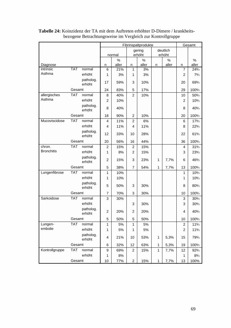
Tabelle 24: Koinzidenz der TA mit dem Auftreten erhöhter D-Dimere / krankheits-bezogene Betrachtungsweise im Vergleich zur Kontrollgruppe
6 21% 1 3% 7 24%1 3% 1 3% 2 7%
17 59% 3 10% 20 69%
24 83% 5 17% 29 100%8 40% 2 10% 10 50%2 10% 2 10%
8 40% 8 40%
18 90% 2 10% 20 100%4 11% 2 6% 6 17%4 11% 4 11% 8 22%
12 33% 10 28% 22 61%
20 56% 16 44% 36 100%2 15% 2 15% 4 31%1 8% 2 15% 3 23%
2 15% 3 23% 1 7,7% 6 46%
5 38% 7 54% 1 7,7% 13 100%1 10% 1 10%1 10% 1 10%
5 50% 3 30% 8 80%
7 70% 3 30% 10 100%3 30% 3 30%
3 30% 3 30%
2 20% 2 20% 4 40%
5 50% 5 50% 10 100%1 5% 1 5% 2 11%1 5% 1 5% 2 11%
4 21% 10 53% 1 5,3% 15 79%
6 32% 12 63% 1 5,3% 19 100%9 69% 2 15% 1 7,7% 12 92%1 8% 1 8%
10 77% 2 15% 1 7,7% 13 100%
normalerhöhtpatholog.erhöht
TAT
Gesamtnormalerhöhtpatholog.erhöht
TAT
Gesamtnormalerhöhtpatholog.erhöht
TAT
Gesamtnormalerhöhtpatholog.erhöht
TAT
Gesamtnormalerhöhtpatholog.erhöht
TAT
Gesamtnormalerhöhtpatholog.erhöht
TAT
Gesamtnormalerhöhtpatholog.erhöht
TAT
Gesamtnormalerhöht
TAT
Gesamt
DiagnoseintrinsicAsthma
allergischesAsthma
Mucoviscidose
chron.Bronchitis
Lungenfibrose
Sarkoidose
Lungen-embolie
Kontrollgruppe
n%
aller
normal
n%
aller
geringerhöht
n%
aller
deutlicherhöht
Fibrinspaltprodukte
n%
aller
Gesamt

70
Tabelle 25: Koinzidenz zw. erhöhter TAT Konzentration und Auftreten erhöhter FaktorXII a Werte / Pat.
13 56,5% 24,1% 10 43,5% 30,3% 23 26,4%12 100,0% 22,2% 12 13,8%
29 55,8% 53,7% 23 44,2% 69,7% 52 59,8%
54 62,1% 100,0% 33 37,9% 100,0% 87 100,0%
normalerhöhtpatholog.erhöht
TAT
Gesamt
n Zeilen% Spalten%normal
n Zeilen% Spalten%erhöht
Faktor 12 a
n Spalten%
Gesamt
TAT normal: > 4,0 µg/l; erhöht: 4,0 µg/l < TAT < 25,0 µg/l; patholog. erhöht: >=Faktor XII a normal <= 3 ng/ml

71
Tabelle 26: Koinzidenz der TA mit dem Auftreten erhöhter Faktor-XII a-Werte I /krankheitsbezogene Betrachtung
3 15% 1 5% 4 20%2 10% 2 10%
6 30% 8 40% 14 70%
11 55% 9 45% 20 100%3 20% 3 20% 6 40%2 13% 2 13%
3 20% 4 27% 7 47%
8 53% 7 47% 15 100%6 21% 1 3% 7 24%5 17% 5 17%
13 45% 4 14% 17 59%
24 83% 5 17% 29 100%1 8% 3 25% 4 33%3 25% 3 25%
3 25% 2 17% 5 42%
7 58% 5 42% 12 100%
3 60% 2 40% 5 100%
3 60% 2 40% 5 100% 2 40% 2 40%
1 20% 2 40% 3 60%
1 20% 4 80% 5 100%
1 100% 1 100%
1 100% 1 100%
normalerhöhtpatholog.erhöht
TAT
Gesamtnormalerhöhtpatholog.erhöht
TAT
Gesamtnormalerhöhtpatholog.erhöht
TAT
Gesamtnormalerhöhtpatholog.erhöht
TAT
Gesamtpatholog.erhöht
TAT
Gesamtnormalpatholog.erhöht
TAT
Gesamtpatholog.erhöht
TAT
Gesamt
Diagnoseintrinsic Asthma
allergischesAsthma
Mucoviscidose
chron.Bronchitis
Lungenfibrose
Sarkoidose
Lungenembolie
n % allernormal
n % allererhöht
Faktor 12 a
n % aller
Gesamt
72
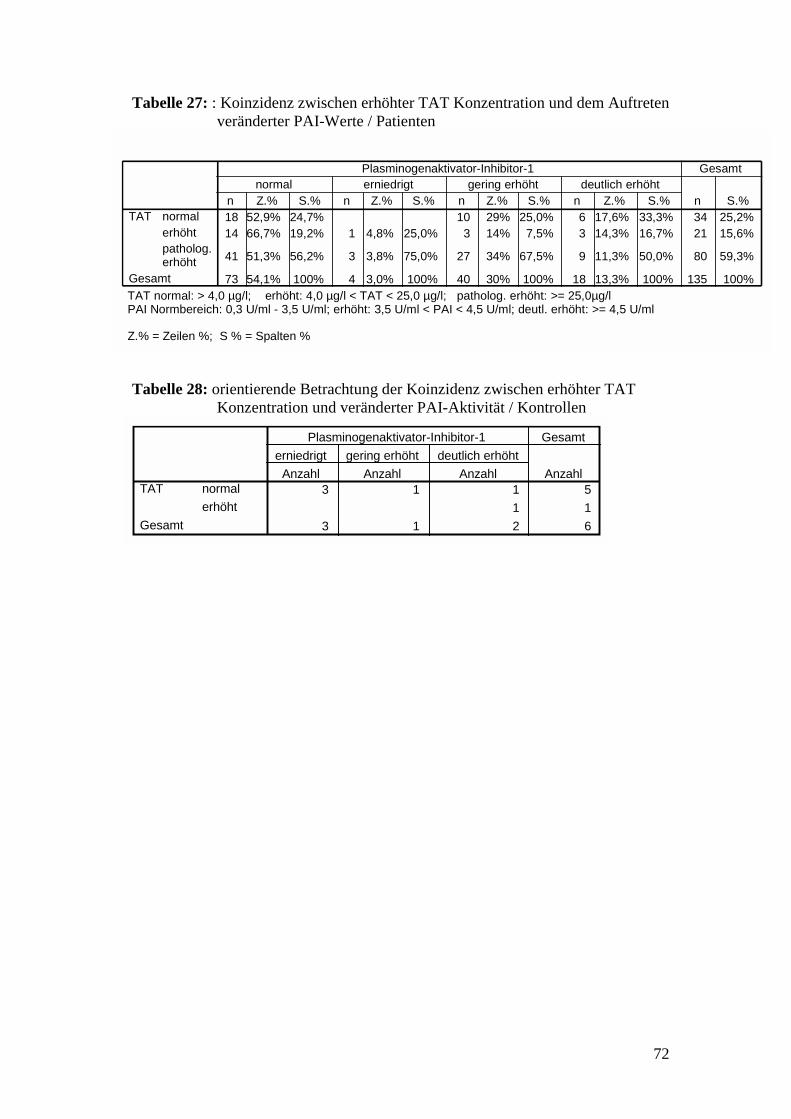
Tabelle 27: : Koinzidenz zwischen erhöhter TAT Konzentration und dem Auftretenveränderter PAI-Werte / Patienten
18 52,9% 24,7% 10 29% 25,0% 6 17,6% 33,3% 34 25,2%14 66,7% 19,2% 1 4,8% 25,0% 3 14% 7,5% 3 14,3% 16,7% 21 15,6%
41 51,3% 56,2% 3 3,8% 75,0% 27 34% 67,5% 9 11,3% 50,0% 80 59,3%
73 54,1% 100% 4 3,0% 100% 40 30% 100% 18 13,3% 100% 135 100%
normalerhöhtpatholog.erhöht
TAT
Gesamt
n Z.% S.%normal
n Z.% S.%erniedrigt
n Z.% S.%gering erhöht
n Z.% S.%deutlich erhöht
Plasminogenaktivator-Inhibitor-1
n S.%
Gesamt
TAT normal: > 4,0 µg/l; erhöht: 4,0 µg/l < TAT < 25,0 µg/l; patholog. erhöht: >= 25,0µg/lPAI Normbereich: 0,3 U/ml - 3,5 U/ml; erhöht: 3,5 U/ml < PAI < 4,5 U/ml; deutl. erhöht: >= 4,5 U/ml
Z.% = Zeilen %; S % = Spalten %
Tabelle 28: orientierende Betrachtung der Koinzidenz zwischen erhöhter TATKonzentration und veränderter PAI-Aktivität / Kontrollen
3 1 1 5 1 1
3 1 2 6
normalerhöht
TAT
Gesamt
Anzahlerniedrigt
Anzahlgering erhöht
Anzahldeutlich erhöht
Plasminogenaktivator-Inhibitor-1
Anzahl
Gesamt

73
Tabelle 29: Thrombinaktivierung und Veränderung der Fibrinolyse bei verschiedenenpulmonalen Erkrankungen / krankheitsbezogene Betrachtung
5 16% 4 13% 1 3% 10 31%1 3% 1 3% 2 6%
10 31% 3 9% 7 22% 20 63%
16 50% 8 25% 8 25% 32 100%5 25% 3 15% 2 10% 10 50%1 5% 1 5% 2 10%
5 25% 1 5% 2 10% 8 40%
11 55% 5 25% 4 20% 20 100%5 15% 1 3% 6 18%7 21% 1 3% 8 24%
14 42% 4 12% 1 3% 19 58%
26 79% 4 12% 2 6% 1 3% 33 100%1 8% 1 8% 2 17%2 17% 1 8% 3 25%
1 8% 2 17% 3 25% 1 8% 7 58%
4 33% 2 17% 5 42% 1 8% 12 100%1 11% 1 11%1 11% 1 11%
2 22% 1 11% 4 44% 7 78%
4 44% 1 11% 4 44% 9 100% 3 27% 3 27%
2 18% 1 9% 3 27%
1 9% 1 9% 3 27% 5 45%
3 27% 1 9% 7 64% 11 100%1 6% 1 6% 2 11%
1 6% 1 6% 2 11%
10 56% 1 6% 3 17% 14 78%
11 61% 2 11% 5 28% 18 100%
normalerhöhtpatholog.erhöht
TAT
Gesamtnormalerhöhtpatholog.erhöht
TAT
Gesamtnormalerhöhtpatholog.erhöht
TAT
Gesamtnormalerhöhtpatholog.erhöht
TAT
Gesamtnormalerhöhtpatholog.erhöht
TAT
Gesamtnormalerhöhtpatholog.erhöht
TAT
Gesamtnormalerhöhtpatholog.erhöht
TAT
Gesamt
DiagnoseintrinsicAsthma
allergischesAsthma
Mucoviscidose
chron.Bronchitis
Lungenfibrose
Sarkoidose
Lungen-embolie
n%
aller
normal
n%
aller
geringerhöht
n%
aller
deutlicherhöht
n%
aller
erniedrigt
Plasminogenaktivator-Inhibitor-1
n%
aller
Gesamt

74
Tabelle 30: Veränderung der Fibrinbildung und Fibrinolyse bei pulmonalenErkrankungen
42 29,8% 37 26,2% 2 1,4% 81 57,4%3 2,1% 3 2,1%
27 19,1% 13 9,2% 40 28,4%
13 9,2% 4 2,8% 17 12,1%
85 60,3% 54 38,3% 2 1,4% 141 100,0%
normalerniedrigtgeringerhöhtdeutlicherhöht
PAI
Gesamt
Anzahl Tabellen%normal
Anzahl Tabellen%gering erhöht
Anzahl Tabellen%deutlich erhöht
Fibrinspaltprodukte
Anzahl Tabellen%
Gesamt
Koinzidenz zwischen erhöhtem PAI und erhöhten D-Dimerenbezogen auf alle Patienten
PAI normal: 0,3 U/ml - 3,5 U/ml; erhöht: 3,5 - 4,5 U/ml; deutlich erhöht: > 4,5 U/mlD-Dimere normal: < 0,5 mg/l; erhöht: 0,5 - 4,0 mg/l; deutlich erhöht: > 4,0 mg/l

75
6.2.4 Tabellen für die Fehlerdiskussion
1. Alle Patienten
Tabelle 31: Einfluß krankheitsabhängiger und krankheitsunabhängiger Faktoren auf dieThrombinaktivierung bei Patienten mit pulmonalen Erkrankungen
29 24,2% 17 14,2% 74 61,7% 120
8 40,0% 1 5,0% 11 55,0% 20
1 9,1% 3 27,3% 7 63,6% 1138 25,2% 21 13,9% 92 60,9% 15111 22,0% 8 16,0% 31 62,0% 5025 28,4% 10 11,4% 53 60,2% 882 15,4% 3 23,1% 8 61,5% 13
38 25,2% 21 13,9% 92 60,9% 15117 25,0% 11 16,2% 40 58,8% 6821 25,3% 10 12,0% 52 62,7% 8338 25,2% 21 13,9% 92 60,9% 151
gut (amb.Kontr.)Z. n.Exacerbat.Exacerbation
ZUSTAND
Gesamtunter 2525 bis 65über 65
Altersgruppe
Gesamtmännlichweiblich
Geschlecht
Gesamt
Anzahl Zeilen%normal
Anzahl Zeilen%erhöht
Anzahl Zeilen%patholog. erhöht
TAT
Anzahl
Ges.
Einfluß der Merkmale Geschlecht, Alter, Zustand auf TAT / Häufigkeitsverteilung
Tabelle 32: Einfluß krankheitsabhängiger und krankheitsunabhängiger Faktoren aufden Fibrinumsatz bei Patienten mit pulmonalen Erkrankungen
81 65,3% 41 33,1% 2 1,6% 124
10 55,6% 8 44,4% 18
5 38,5% 8 61,5% 1396 61,9% 57 36,8% 2 1,3% 15538 65,5% 20 34,5% 5852 65,8% 26 32,9% 1 1,3% 796 33,3% 11 61,1% 1 5,6% 18
96 61,9% 57 36,8% 2 1,3% 15544 62,9% 25 35,7% 1 1,4% 7052 61,2% 32 37,6% 1 1,2% 8596 61,9% 57 36,8% 2 1,3% 155
gut (amb.Kontr.)Z. n.Exacerbat.Exacerbation
ZUSTAND
Gesamtunter 2525 bis 65über 65
Altersgruppe
Gesamtmännlichweiblich
Geschlecht
Gesamt
Anzahl Zeilen%
normal
Anzahl Zeilen%
gering erhöht
Anzahl Zeilen%
deutlich erhöht
Fibrinspaltprodukte
Anzahl
Ges.
Einfluß der Merkmale Geschlecht, Alter, Zustand auf D-Dimere / Häufigkeitsverteilung

76
2. Mukoviszidose- Gruppe / exemplarisch
Tabelle 33: Analyse des Einflusses des Geschlechts auf Gerinnungsparameter I
22 16,2 4 35 12 2114 17,6 14 21 16 2018 15,5 5 20 9 2011 60,0 35 70 50 7016 60,00 2,00 60,00 8,30 60,0016 5,30 ,43 10,00 ,69 10,0019 3,95 4,25 ,96 15,1022 ,77 ,56 ,50 2,3020 2,25 ,97 ,80 4,2025 19,3 6 29 15 2415 19,1 15 23 16 2119 8,0 4 29 8 1613 60,0 40 75 48 7321 32,00 2,00 60,00 10,30 60,0021 1,07 ,33 10,00 ,67 6,4820 2,36 2,99 ,50 14,4424 1,02 ,84 ,50 3,6022 2,45 1,53 ,20 6,40
AlterBMICN-SCORES-SCORETATF1+2Faktor XII aD - DimerePAIAlterBMICN-SCORES-SCORETATF1+2Faktor XII aD - DimerePAI
männlich
weiblich
AnzahlN
Mittelwert Median St.abw. Minimum Maximum 25 75Perzentile
Einfluß des Geschlechts auf die mittleren (medianen) Werte am Beispiel der CF Gruppe
Tabelle 34: Analyse des Einflusses des Geschlechts auf Gerinnungsparameter II
3 13,6
3 13,6
10 45,5
16 72,7
4 16,0
5 20,0
12 48,0
21 84,0
keine Thromb.Aktiv.Thromb. Aktiv.darstellb.deutl. Thromb.Aktiv.Gesamtkeine Thromb.Aktiv.Thromb. Aktiv.darstellb.deutl. Thromb.Aktiv.Gesamt
Geschlechtmännlich
weiblich
Häufigkeit Prozent
Einfluß des Geschlechts auf dieHäufigkeit des Auftretenserhöhter TAT- Werte am Beispiel der CF Gruppe
77
Tabelle 35: Analyse des Einflusses des Geschlechts auf Gerinnungsparameter III
7 31,89 40,9
16 72,711 44,010 40,021 84,0
normaldeutlich erhöhtGesamtnormaldeutlich erhöhtGesamt
Geschlechtmännlich
weiblich
Häufigkeit Prozent
Einfluß des Geschlechts auf dieHäufigkeit des Auftretenserhöhter F 1+2- Werte am Beispiel der CF Gruppe
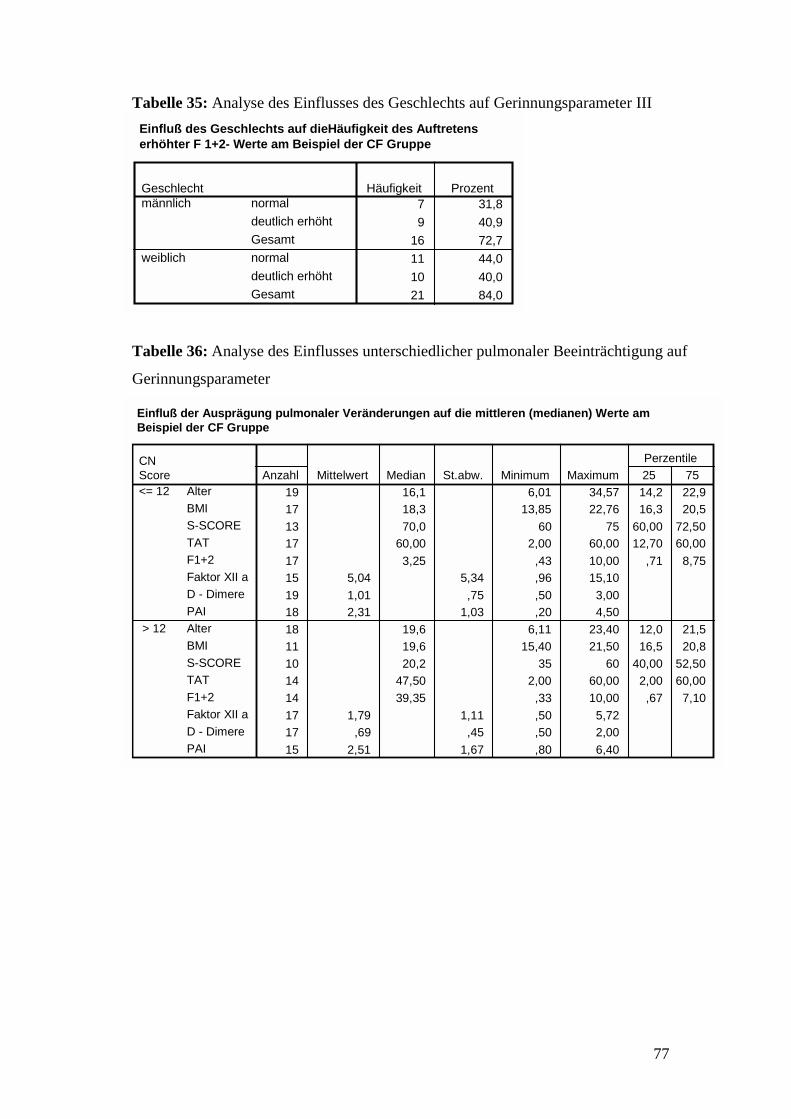
Tabelle 36: Analyse des Einflusses unterschiedlicher pulmonaler Beeinträchtigung auf
Gerinnungsparameter
19 16,1 6,01 34,57 14,2 22,917 18,3 13,85 22,76 16,3 20,513 70,0 60 75 60,00 72,5017 60,00 2,00 60,00 12,70 60,0017 3,25 ,43 10,00 ,71 8,7515 5,04 5,34 ,96 15,1019 1,01 ,75 ,50 3,0018 2,31 1,03 ,20 4,5018 19,6 6,11 23,40 12,0 21,511 19,6 15,40 21,50 16,5 20,810 20,2 35 60 40,00 52,5014 47,50 2,00 60,00 2,00 60,0014 39,35 ,33 10,00 ,67 7,1017 1,79 1,11 ,50 5,7217 ,69 ,45 ,50 2,0015 2,51 1,67 ,80 6,40
AlterBMIS-SCORETATF1+2Faktor XII aD - DimerePAIAlterBMIS-SCORETATF1+2Faktor XII aD - DimerePAI
CNScore<= 12
> 12
Anzahl Mittelwert Median St.abw. Minimum Maximum 25 75Perzentile
Einfluß der Ausprägung pulmonaler Veränderungen auf die mittleren (medianen) Werte amBeispiel der CF Gruppe

78
Tabelle 37: exemplarische Analyse des Einflusses unterschiedlicher klinisch beurteilter
Leistungsfähigkeit auf Gerinnungsparameter
3 20,0 18,6 24,6 18,6 ,3 17,8 16,2 20,5 16,2 ,3 6,0 4,0 8,0 4,0 ,3 60,00 12,60 60,00 12,60 ,3 5,46 ,72 10,00 ,72 ,1 1,86 1,86 1,86 , ,3 1,37 ,78 ,50 2,00 ,3 2,63 240 1,76 1,00 4,50 ,
13 16,6 6,0 34,6 13,5 23,613 17,0 13,9 22,8 16,4 20,212 8,0 6,0 15,0 6,3 10,011 60,00 2,00 60,00 12,80 60,0011 3,25 ,43 10,00 ,70 10,0013 3,95 4,90 ,96 15,1013 1,02 ,80 ,50 3,0012 2,52 2, ,92 1,10 4,20
8 19,6 8,7 23,3 11,1 22,68 20,3 15,4 21,5 16,3 21,28 20,0 16,0 29,0 16,3 20,07 30,10 2,00 60,00 2,00 60,007 1,50 ,49 10,00 ,60 4,428 1,55 ,32 1,16 2,217 ,70 ,45 ,50 1,706 2,05 1,03 ,90 3,80
AlterBMICN-SCORETATF1+2Faktor XII aD - DimerePAIAlterBMICN-SCORETATF1+2Faktor XII aD - DimerePAIAlterBMICN-SCORETATF1+2Faktor XII aD - DimerePAI
Shwachmannscoregut bis sehrgut
leicht krank
mittelschwerbisschwerkrank
Anzahl Mittelwert Median St.abw. Minimum Maximum 25 75Perzentile
Einfluß des klinischen Zustandes auf die mittleren (medianen) Werte am Beispiel der CF Gruppe
Recommended