107
RPCV (2015) 110 (593-594) 107-109
A clinical, sonographic, histopathological and hormonal evaluation of a buck with gynaecomastia and galactorrhea
Avaliação clínica, ultrassonográfica, histopatológica e hormonal de um bode com ginecomastia e galactorréia
Michelle S. Araujo*, Marianne C. Dias, Ana Paula Bertoni, Leonardo D. da Costa, Nereu C. Prestes
Faculdade de Medicina Veterinária e Zootecnica, Universidade Estadual Paulista, Campus Botucatu, São Paulo, Brasil.
Summary: A Saanem buck with gynaecomastia and galactorrhea was examined. Limited mobility, a low body score, normodipsia, normorexia, normoquezia and normouria were observed upon general clinical examination. The breasts showed an excessive increase in size, consistent floating and a translucent watery dis-charge after milking. Sonographic examinations of the breasts and testes were performed, showing no alteration in testes and rounded areas with mixed echogenicity and hyperechoic walls in breasts. Histopathological evaluation diagnosed moderate testicular dege-neration and breast cystic hyperplasia. Blood samples were col-lected to measure the levels of estradiol and testosterone by che-miluminescence method, and for karyotyping analysis. Bilateral mastectomy and orchiectomy were performed. Because this dise-ase presents many cases lacking a clear etiology, it is necessary to conduct further studies to avoid the occurrence of the disease.
Keywords: goats, hormone levels, mammary hyperplasia, mastec-tomy
Resumo: Foi examinado um bode da raça Saanem apresentando ginecomastia associada à galactorréia. Foram observados durante o exame clínico geral mobilidade limitada, baixo escore corporal, normodipsia, normorexia, normoquezia e normouria. As mamas apresentavam-se excessivamente aumentadas, com consistência flutuante e secreção aquosa translúcida após ordenha. Exames utrassonográficos das mamas e dos testículos foram realizados, sem alterações nos testículos e áreas arredondadas de ecogenicida-de mista e paredes hiperecóicas nas mamas. A avaliação histopato-lógica diagnosticou moderada degeneração teticular e hiperplasia mamária cística. Amostras de sangue foram coletadas para medir os níveis de estradiol e testosterone pelo método de quimiolumi-nescência, e para o exame de cariotipagem. Foi realizada mastec-tomia e orquiectomia bilaterais. Devido à essa doença apresentar diversas causas de etiologia não esclarecida, torna-se necessária a realização de mais estudos para evitar a ocorrência dessa doença.
Palavras-chave: caprinos, hiperplasia mamária, mastectomia, ní-
veis hormonais.
Introduction
Gynaecomastia associated with galactorrhea is defi-ned as a benign development of the mammary glands
in males with concomitant production of milk. In go-ats, it is a rare, anomalous occurrence with a reported production of up to 1 liter of milk per day (Basrur and Basrur, 2004, Wang et al., 2009).
This disease has been observed in fertile males and hermaphroditic animals. Its suggested causes include chromosomal abnormalities, familial predispositions, hormonal changes and mechanical effects (Wooldridge et al., 1999). The genetic background and environmen-tal factors that lead to the occurrence of gynaecomastia in goats have not been determined (Wang et al., 2009).
This report aims to assist the diagnosis and the un-derstanding of the etiology of gynaecomastia asso-ciated with galactorrhea in a Saanen buck, as the few cases previously reported in the literature are not suffi-cient to clarify the etiology of this disease.
Case Report
A five-year-old Saanen buck was brought to the Veterinary Hospital of the Faculty of Veterinary Medicine and Animal Science at UNESP – Botucatu. Approximately two years prior to its arrival at the hos-pital, the animal began to show a progressive increase in the size of the mammary glands, accompanied by milk production. The owner reported an initial increase in the size of the right udder followed by an increase in the size of the left udder. The buck belonged to the owner’s breeding stock and showed normal sexual ac-tivity despite breast enlargement, producing offspring during the previous breeding season.
Limited mobility due to breast enlargement, a low body score, normodipsia, normorexia, normoquezia and normouria were observed upon general clinical exami-nation. The breasts showed an excessive increase in size, consistent floating and a translucent watery discharge after milking. Upon examination of the genital tract, both testicles were mobile, atrophied, symmetric, ovoid, vertical, flaccid in consistency and showed no painful sensitivity to palpation. No alterations were observed in *Correspondência: [email protected]
Araujo M. et al. RPCV (2015) 110 (593-594) 107-109
108
the penis, prepuce or epididymis. Attempt was made to perform semen collection, but the animal showed no li-bido and for this reason no semen was collected.
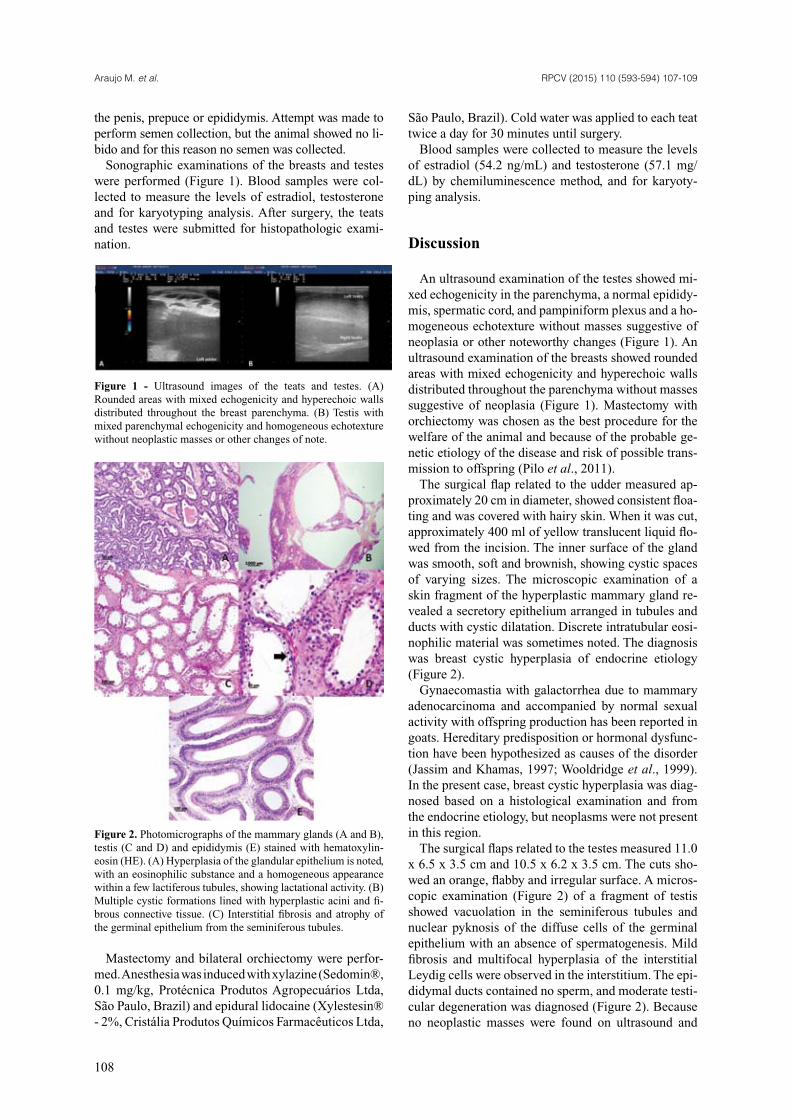
Sonographic examinations of the breasts and testes were performed (Figure 1). Blood samples were col-lected to measure the levels of estradiol, testosterone and for karyotyping analysis. After surgery, the teats and testes were submitted for histopathologic exami-nation.
Figure 1 - Ultrasound images of the teats and testes. (A) Rounded areas with mixed echogenicity and hyperechoic walls distributed throughout the breast parenchyma. (B) Testis with mixed parenchymal echogenicity and homogeneous echotexture without neoplastic masses or other changes of note.
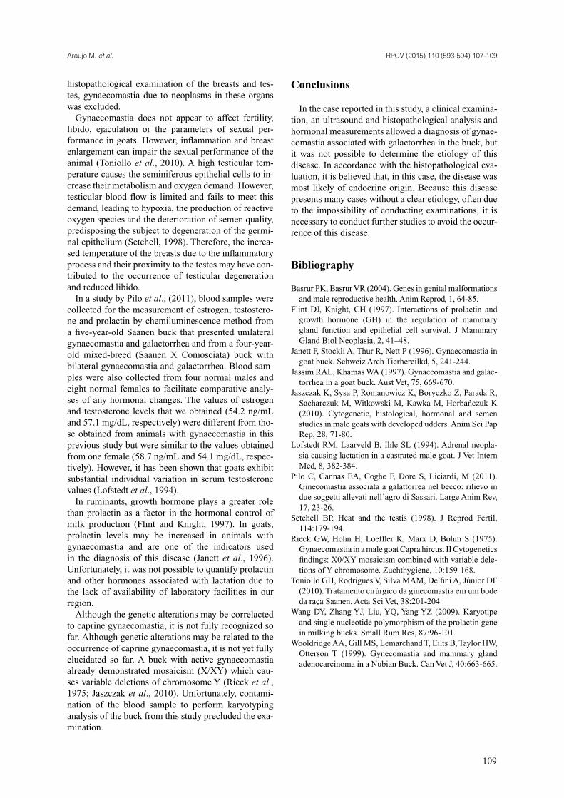
Figure 2. Photomicrographs of the mammary glands (A and B), testis (C and D) and epididymis (E) stained with hematoxylin-eosin (HE). (A) Hyperplasia of the glandular epithelium is noted, with an eosinophilic substance and a homogeneous appearance within a few lactiferous tubules, showing lactational activity. (B) Multiple cystic formations lined with hyperplastic acini and fi -brous connective tissue. (C) Interstitial fi brosis and atrophy of the germinal epithelium from the seminiferous tubules.
Mastectomy and bilateral orchiectomy were perfor-med. Anesthesia was induced with xylazine (Sedomin®, 0.1 mg/kg, Protécnica Produtos Agropecuários Ltda, São Paulo, Brazil) and epidural lidocaine (Xylestesin® - 2%, Cristália Produtos Químicos Farmacêuticos Ltda,
São Paulo, Brazil). Cold water was applied to each teat twice a day for 30 minutes until surgery.
Blood samples were collected to measure the levels of estradiol (54.2 ng/mL) and testosterone (57.1 mg/dL) by chemiluminescence method, and for karyoty-ping analysis.
Discussion
An ultrasound examination of the testes showed mi-xed echogenicity in the parenchyma, a normal epididy-mis, spermatic cord, and pampiniform plexus and a ho-mogeneous echotexture without masses suggestive of neoplasia or other noteworthy changes (Figure 1). An ultrasound examination of the breasts showed rounded areas with mixed echogenicity and hyperechoic walls distributed throughout the parenchyma without masses suggestive of neoplasia (Figure 1). Mastectomy with orchiectomy was chosen as the best procedure for the welfare of the animal and because of the probable ge-netic etiology of the disease and risk of possible trans-mission to offspring (Pilo et al., 2011).
The surgical fl ap related to the udder measured ap-proximately 20 cm in diameter, showed consistent fl oa-ting and was covered with hairy skin. When it was cut, approximately 400 ml of yellow translucent liquid fl o-wed from the incision. The inner surface of the gland was smooth, soft and brownish, showing cystic spaces of varying sizes. The microscopic examination of a skin fragment of the hyperplastic mammary gland re-vealed a secretory epithelium arranged in tubules and ducts with cystic dilatation. Discrete intratubular eosi-nophilic material was sometimes noted. The diagnosis was breast cystic hyperplasia of endocrine etiology (Figure 2).
Gynaecomastia with galactorrhea due to mammary adenocarcinoma and accompanied by normal sexual activity with offspring production has been reported in goats. Hereditary predisposition or hormonal dysfunc-tion have been hypothesized as causes of the disorder (Jassim and Khamas, 1997; Wooldridge et al., 1999). In the present case, breast cystic hyperplasia was diag-nosed based on a histological examination and from the endocrine etiology, but neoplasms were not present in this region.
The surgical fl aps related to the testes measured 11.0 x 6.5 x 3.5 cm and 10.5 x 6.2 x 3.5 cm. The cuts sho-wed an orange, fl abby and irregular surface. A micros-copic examination (Figure 2) of a fragment of testis showed vacuolation in the seminiferous tubules and nuclear pyknosis of the diffuse cells of the germinal epithelium with an absence of spermatogenesis. Mild fi brosis and multifocal hyperplasia of the interstitial Leydig cells were observed in the interstitium. The epi-didymal ducts contained no sperm, and moderate testi-cular degeneration was diagnosed (Figure 2). Because no neoplastic masses were found on ultrasound and
Araujo M. et al. RPCV (2015) 110 (593-594) 107-109
109
histopathological examination of the breasts and tes-tes, gynaecomastia due to neoplasms in these organs was excluded.
Gynaecomastia does not appear to affect fertility, libido, ejaculation or the parameters of sexual per-formance in goats. However, inflammation and breast enlargement can impair the sexual performance of the animal (Toniollo et al., 2010). A high testicular tem-perature causes the seminiferous epithelial cells to in-crease their metabolism and oxygen demand. However, testicular blood flow is limited and fails to meet this demand, leading to hypoxia, the production of reactive oxygen species and the deterioration of semen quality, predisposing the subject to degeneration of the germi-nal epithelium (Setchell, 1998). Therefore, the increa-sed temperature of the breasts due to the inflammatory process and their proximity to the testes may have con-tributed to the occurrence of testicular degeneration and reduced libido.
In a study by Pilo et al., (2011), blood samples were collected for the measurement of estrogen, testostero-ne and prolactin by chemiluminescence method from a five-year-old Saanen buck that presented unilateral gynaecomastia and galactorrhea and from a four-year-old mixed-breed (Saanen X Comosciata) buck with bilateral gynaecomastia and galactorrhea. Blood sam-ples were also collected from four normal males and eight normal females to facilitate comparative analy-ses of any hormonal changes. The values of estrogen and testosterone levels that we obtained (54.2 ng/mL and 57.1 mg/dL, respectively) were different from tho-se obtained from animals with gynaecomastia in this previous study but were similar to the values obtained from one female (58.7 ng/mL and 54.1 mg/dL, respec-tively). However, it has been shown that goats exhibit substantial individual variation in serum testosterone values (Lofstedt et al., 1994).
In ruminants, growth hormone plays a greater role than prolactin as a factor in the hormonal control of milk production (Flint and Knight, 1997). In goats, prolactin levels may be increased in animals with gynaecomastia and are one of the indicators used in the diagnosis of this disease (Janett et al., 1996). Unfortunately, it was not possible to quantify prolactin and other hormones associated with lactation due to the lack of availability of laboratory facilities in our region.
Although the genetic alterations may be correlacted to caprine gynaecomastia, it is not fully recognized so far. Although genetic alterations may be related to the occurrence of caprine gynaecomastia, it is not yet fully elucidated so far. A buck with active gynaecomastia already demonstrated mosaicism (X/XY) which cau-ses variable deletions of chromosome Y (Rieck et al., 1975; Jaszczak et al., 2010). Unfortunately, contami-nation of the blood sample to perform karyotyping analysis of the buck from this study precluded the exa-mination.
Conclusions
In the case reported in this study, a clinical examina-tion, an ultrasound and histopathological analysis and hormonal measurements allowed a diagnosis of gynae-comastia associated with galactorrhea in the buck, but it was not possible to determine the etiology of this disease. In accordance with the histopathological eva-luation, it is believed that, in this case, the disease was most likely of endocrine origin. Because this disease presents many cases without a clear etiology, often due to the impossibility of conducting examinations, it is necessary to conduct further studies to avoid the occur-rence of this disease.
Bibliography
Basrur PK, Basrur VR (2004). Genes in genital malformations and male reproductive health. Anim Reprod, 1, 64-85.
Flint DJ, Knight, CH (1997). Interactions of prolactin and growth hormone (GH) in the regulation of mammary gland function and epithelial cell survival. J Mammary Gland Biol Neoplasia, 2, 41–48.
Janett F, Stockli A, Thur R, Nett P (1996). Gynaecomastia in goat buck. Schweiz Arch Tierhereilkd, 5, 241-244.
Jassim RAL, Khamas WA (1997). Gynaecomastia and galac-torrhea in a goat buck. Aust Vet, 75, 669-670.
Jaszczak K, Sysa P, Romanowicz K, Boryczko Z, Parada R, Sacharczuk M, Witkowski M, Kawka M, Horbańczuk K (2010). Cytogenetic, histological, hormonal and semen studies in male goats with developed udders. Anim Sci Pap Rep, 28, 71-80.
Lofstedt RM, Laarveld B, Ihle SL (1994). Adrenal neopla-sia causing lactation in a castrated male goat. J Vet Intern Med, 8, 382-384.
Pilo C, Cannas EA, Coghe F, Dore S, Liciardi, M (2011). Ginecomastia associata a galattorrea nel becco: rilievo in due soggetti allevati nell´agro di Sassari. Large Anim Rev, 17, 23-26.
Setchell BP. Heat and the testis (1998). J Reprod Fertil, 114:179-194.
Rieck GW, Hohn H, Loeffler K, Marx D, Bohm S (1975). Gynaecomastia in a male goat Capra hircus. II Cytogenetics findings: X0/XY mosaicism combined with variable dele-tions of Y chromosome. Zuchthygiene, 10:159-168.
Toniollo GH, Rodrigues V, Silva MAM, Delfini A, Júnior DF (2010). Tratamento cirúrgico da ginecomastia em um bode da raça Saanen. Acta Sci Vet, 38:201-204.
Wang DY, Zhang YJ, Liu, YQ, Yang YZ (2009). Karyotipe and single nucleotide polymorphism of the prolactin gene in milking bucks. Small Rum Res, 87:96-101.
Wooldridge AA, Gill MS, Lemarchand T, Eilts B, Taylor HW, Otterson T (1999). Gynecomastia and mammary gland adenocarcinoma in a Nubian Buck. Can Vet J, 40:663-665.
Recommended