NeuroMolecular Medicine 115 Volume 2, 2002
Alpha-Synuclein and Presynaptic Function
Implications for Parkinson’s Disease
Simon Lykkebo and Poul Henning Jensen*
Department of Medical Biochemistry, Aarhus University, DK-8000 Aarhus-C, Denmark
Received April 17, 2002; Accepted May 16, 2002
Abstract
This article focuses on α-synuclein’s role in normal and pathological axonal and presynap-tic functions and its relationship to Parkinson’s disease. It is not possible to mention all the con-tributions to aspects of this area. Readers interested in α-synuclein’s relation to aggregation,Lewy lesions, and pathological modifications are referred to the many reviews (see Goldbergand Lansbury 2000; Galvin 2001a; Goedert 2001).
Index Entries: synuclein; synapse; axon; vesicles; Parkinson’s disease; Lewy body.
NeuroMolecular MedicineCopyright © 2002 Humana Press Inc.All rights of any nature whatsoever reserved.ISSN1535-1084/02/02:115–129/$20.00
*Author to whom all correspondence and reprint requests should be addressed. E-mail: [email protected]
α-Synuclein in Parkinson’s Disease
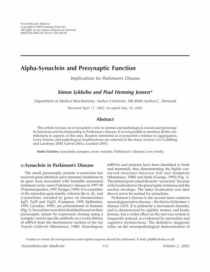
The small presynaptic protein α-synuclein hasreceived great attention since missense mutations inits gene were associated with heritable autosomaldominant early-onset Parkinson’s disease in 1997-98(Polymeropoulos, 1997; Krüger, 1998). It is a memberof the synuclein gene family, wherein the α-, β-, andγ-synucleins, encoded by genes on chromosomes4q21, 5q35 and 10q23, (Campion, 1995; Spillantini,1995; Lavedan, 1998), are predominant in humans(Fig. 1). Synucleins were first identified based on theirpresynaptic nature by expression cloning using asynaptic-vesicle-specific antibody on a vector libraryof mRNA from the electromotor nucleus of the rayTorpedo California (Maroteaux, 1988). Homologous
mRNAs and proteins have been identified in birdsand mammals, thus demonstrating the highly con-served structure between fish and mammals(Maroteaux, 1988) and birds (George, 1995) (Fig. 1).The initial report coined the term “synuclein” becauseof its localization to the presynaptic terminus and thenuclear envelope. The latter localization was latershown not to be normal for synucleins.
Parkinson’s disease is the second most commonneurodegenerative disease—the first is Alzheimer’sdisease (AD). It is primarily a movement disorder,and is characterized by rigidity, tremor, and bradykinesia, but a wider effect on the nervous system isfrequently noticed, as evidenced by autonomic andcognitive dysfunctions. The definitive diagnosisrelies on the neuropathological demonstration of
116 Lykkebo and Jensen
NeuroMolecular Medicine Volume 2, 2002
nerve-cell loss in the pars compacta of the substan-tia nigra in the brainstem, accompanied by the pres-ence of proteinaceous inclusions, Lewy bodies, andLewy neurites. Similar neurodegenerative changesare often found in other parts of the brain—e.g., thenucleus basalis of Meynert, hypothalamus, cingu-lated gyrus, entorhinal cortex, sympathetic ganglia,and peripheral parasympathetic neurons, wherethey may explain the autonomic and cognitivesymptomatology. For a recent review on clinical andpathophysiological aspects of Parkinson’s disease,see (Lang, 1998). The identification of two missense
mutations in the α-synuclein gene on chromo-some 4q21 causing Parkinson’s disease representedthe first demonstration of a single gene as causativefor the disease. Both the mutant Ala53Thr in thelarge Contoursi kindred (Polymeropoulos, 1997)and the Ala30Pro mutations in a German family(Krüger, 1998) cause an early-onset Parkinson’s dis-ease with a dominant heritability of high penetrancethat is indicative of a gain-of-toxic-function by themutant peptide. Mutations in the α-synuclein genehave subsequently turned out to be exceedingly rare. The identification of the mutations in the
Fig. 1. Sequence comparison among synucleins. (A) Alignment of human α-synuclein, β-synuclein and γ-synuclein protein sequences. Gray bars indicate the six KTKEGV consensus repeats in the α-synuclein sequence.Note the high degree of sequence identity within the N-terminal 62 residues of the gene products as compared totheir divergent C-termini. (B) Alignment of α-synuclein protein sequences from human, rat, bird, and fish synu-clein. Note the high degree of conservation of the α-synuclein protein in bird and mammals, and even with theunrelated fish synuclein. Dots indicate amino acids identical with the human α-synuclein sequence. Gaps areindicated with a wavy line (~). Alignments were performed using the MegAlign software from DNAstar Inc. Acces-sion numbers used for the alignments are: human α- (P37840), human β- (Q16143), human γ- (AAC27738), rat α- (P37377), bird α- (AAA93538), and fish synuclein (A60887).
Alpha-Synuclein and Presynaptic Function 117
NeuroMolecular Medicine Volume 2, 2002
α-synuclein gene prompted the immunohistochem-ical examination of brain tissue affected by Parkin-son’s disease, and this showed that α-synuclein inthe disease is accumulated in the Lewy bodies(Spillantini, 1997). More neurodegenerative diseasesare associated with the development of Lewy bodiesor similar inclusions in which α-synuclein is a prominent component, dementia with Lewy bodies,Lewy body variant of AD, motor neuron disease,Hallervorden-Spatz disease (for recent reviews seeDickson, 2001; Goedert, 2001) and multiple systematrophy, where the inclusions occur in the astrocytes(Gai, 1998). Accordingly, these diseases are collec-tively designated as α-synucleinopathies. Lewybodies were discovered by Frederic Lewy (Lewy,1912), and were for decades identified histochemi-cally by their affinity for the dye eosin. The sensi-tivity of their detection increased significantly withthe development of immunohistochemistry, forwhich ubiquitin antibodies have represented thestandard for their detection until recently. However,anti-α-synuclein antibodies have increased the sensitivity even further by demonstrating the exis-tence of subsets of ubiquitin-negative Lewy bodies(McKeith, 1999). Lewy bodies and Lewy neuritescontain large amounts of protein filaments (Forno,1987), and thus resemble the neuropathological
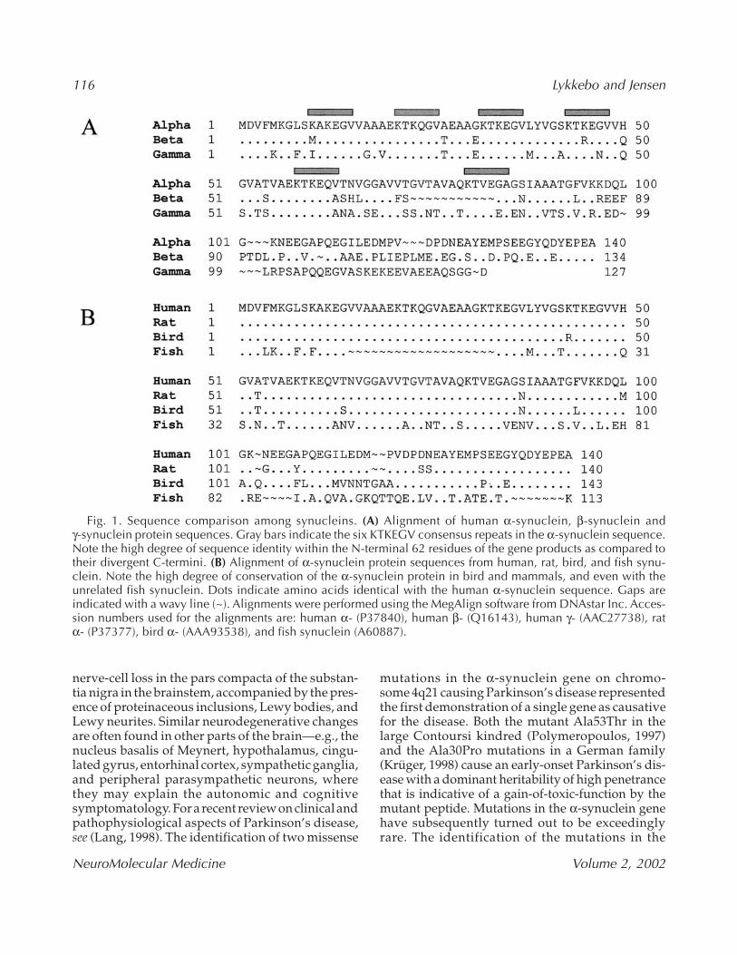
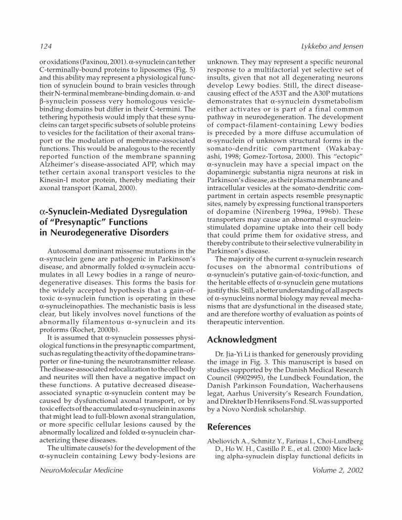
lesions in other late-onset neurodegenerative dis-eases, such as neuritic tangles in Alzheimer’s dis-ease and intranuclear inclusions in Huntington’sdisease. The filaments in Lewy bodies and glialcytoplasmic inclusions contain highly insoluble α-synuclein, which is partly modified by proteoly-sis, phosphorylation, and oxidations (Baba, 1998; Gai,1999; Giasson, 2000; Jensen, 2000; Fujiwara, 2002),and they resemble the filaments that can be formedin vitro from recombinant human α-synuclein basedon immuno-electronmicroscopical criteria (Conway,1998; Crowther, 1998; Giasson, 1999). This suggeststhat α-synuclein is the building block for the core fil-aments that may possess an additional coat of asso-ciated auxiliary cytoplasmic proteins. Structuralanalysis of in vitro-formed filaments have demon-strated that they possess a large content of β-foldedstructure and exhibit characteristics of classical amyloid filaments such as the β amyloid peptide(Aβ)-filaments in Alzheimer’s disease (Hashimoto,1998; Conway, 2000a). The accumulation of α-synuclein in the Lewy bodies and neurites repre-sents a dramatic relocalization of the protein from itsnormal presynaptic localization (Fig. 2). A less pro-nounced abnormal accumulation of β-synuclein andγ-synuclein in non-Lewy-body structures has alsobeen demonstrated in Parkinson’s disease, but no fil-
Fig. 2. Topological and structural changes in neuronal α-synuclein in Parkinson’s disease. α-synuclein is nor-mally a presynaptic protein. It is synthesized in the cell body and targeted to the nerve terminal by axonal trans-port. A dramatic relocalization from the nerve terminal to the cell body and neurites occurs in the degeneratingneurons during the course of Parkinson’s disease. A structural change takes place concomittant with this relocal-ization as β-folded amyloid-type α-synuclein filaments accumulates in the Lewy bodies and in the neurites. Colorimage available for viewing at www.humanapress.com
118 Lykkebo and Jensen
NeuroMolecular Medicine Volume 2, 2002
aments have been detected for these synucleins(Galvin, 1999). Concomitant with the α-synucleinrelocalization occurs the above-mentioned structuraltransition of some monomeric unfolded α-synucleininto β-folded filaments. A biochemical correlate tothe filamentous transition is a decreased solubilityofα-synuclein in isolated filaments and in brain tissuecontaining numerous α-synuclein-positive inclu-sions (Dickson 1999; Gai 1999; Jensen 2000). A sim-ilar decreased solubility has been demonstrated intransgenic mice expressing human α-synuclein anddisplaying pathological symptoms (Kahle, 2001).
The aggregation of recombinant human α-synuclein in vitro represents a nucleation-dependent aggregation process in which filamentformation is preceded by a lag phase where fila-mentogene oligomers build up (Conway, 1998;Wood, 1999). The oligomers represents an ill-definedstructural entity whose existence has been demon-strated biochemically and by atomic forcemicroscopy (Conway, 2000a, 2000b; Rochet, 2000;Volles, 2001). However, the oligomers’ ability to seedfilament growth emphasizes the importance of fac-tors that increase their concentration. Such factorsmay be subject to dynamic regulation in vivo andcomprise disease-causing mutations, C-terminaltruncation (Crowther, 1998, Jensen, 2000), oxidativemodifications (Hashimoto, 1999; Paik, 2000; Souza,2000), phosphorylations (Fujiwara, 2002), calciumbinding (Nielsen, 2001), and possibly certain α-synuclein binding proteins such as synphilin(Engelender, 1999).
The direct link between α-synuclein alterationsand the development of Parkinson’s disease—established by the pathogenic mutations in the α-synuclein gene, and the demonstration of anabnormal α-synuclein aggregation in all cases withLewy bodies, including Parkinson’s disease—havehad a tremendous impact by allowing directed molecular investigations of disease mechanisms.
Expression
The synucleins are abundant proteins that rep-resent about 0.1% of the total protein in the brain(Shibayama-Imazu, 1993). They are consideredneuron-specific proteins because their levels in theperipheral and central nervous system (CNS) arefar higher than in other tissues (Jakes, 1994; Ueda,1994; Lavedan, 1998), although significant expres-
sion does exist in other tissues. The synucleins are small acidic and cytosolic proteins of about125–140 amino acids with α-synuclein comprisedof 140 residues. The synuclein signature was imme-diately noticed after the first cloning as the pres-ence of repeats of 11 amino acids with the coreconsensus sequence KTKEGV (Maroteaux, 1988;Fig. 1B). These repeats extend over the N-terminalabout two-thirds of the peptide, and this region ishighly conserved between the different synucleinsand between species. The C-terminal one-third ishighly negatively charged and displays the mostvariable segment between the synucleins and con-trast, therefore, to the positively charged repeatregion (Fig. 1A).
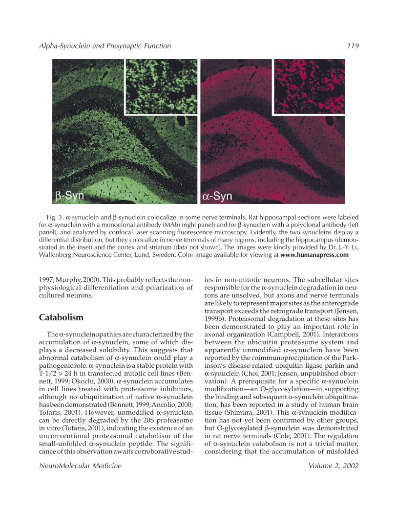
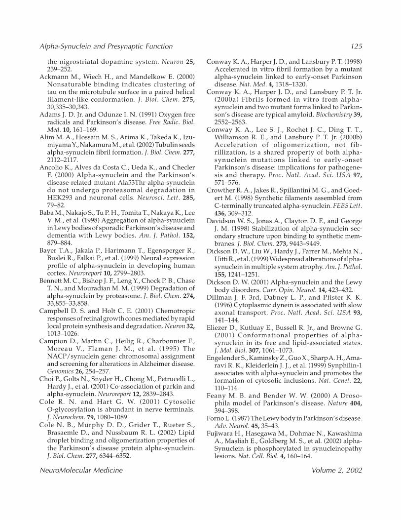
The developmental expression of α-synucleinbegins at an early stage in mammals (Hsu, 1998)and has been detected from gestational wk 15 and16 in the human substantia nigra and cortex (Bayer,1999; Galvin, 2001b). The level slowly rises duringthe gestational period, and increases more rapidlyduring the end of pregnancy and in the early post-partum period (Hsu, 1998; Petersen, 1999). The α-synuclein is initially localized in the cell body andproximal neurites, and the mature presynaptic local-ization in the neuropil is first acquired by about ges-tational wk 18 (Galvin, 2001b). Interestingly, theclose homologous β-synuclein and γ-synucleinare first expressed in the substantia nigra at aboutthe time when α-synuclein has acquired its synaptic localization (Galvin, 2001b). Moreover,they first reach their mature presynaptic localiza-tion after being present for several weeks in the cell body (Galvin, 2001b). This indicates either anasynchronous maturation of their respective trans-port mechanisms or expression in different neu-ronal populations. Coexpression of α-synuclein andβ-synuclein within the same synaptic terminal hasbeen demonstrated in vivo (Fig. 3) and in culturedneurons (Murphy 2000). The synaptic targeting ofα-synuclein displays a pattern very similar to synap-tophysin (Hsu, 1998; Galvin, 2001b), indicating aparallel maturation of the axonal transport mecha-nisms for the two proteins in vivo as compared tothe later maturation for synaptotagmin and synap-tobrevin (Galvin, 2001b). A discrepancy existsbetween the previously mentioned in vivo data andin vitro neuronal cell-culture experiments—thelatter demonstrate an earlier expression of andsynaptic translocation of synaptophysin andsynapsin-1 as compared to α-synuclein (Withers,
Alpha-Synuclein and Presynaptic Function 119
NeuroMolecular Medicine Volume 2, 2002
1997; Murphy, 2000). This probably reflects the non-physiological differentiation and polarization of cultured neurons.
Catabolism
The α-synucleinopathies are characterized by theaccumulation of α-synuclein, some of which dis-plays a decreased solubility. This suggests thatabnormal catabolism of α-synuclein could play apathogenic role. α-synuclein is a stable protein withT-1/2 > 24 h in transfected mitotic cell lines (Ben-nett, 1999; Okochi, 2000). α-synuclein accumulatesin cell lines treated with proteasome inhibitors,although no ubiquitination of native α-synucleinhas been demonstrated (Bennett, 1999; Ancolio, 2000;Tofaris, 2001). However, unmodified α-synucleincan be directly degraded by the 20S proteasome in vitro (Tofaris, 2001), indicating the existence of anunconventional proteasomal catabolism of thesmall-unfolded α-synuclein peptide. The signifi-cance of this observation awaits corroborative stud-
ies in non-mitotic neurons. The subcellular sitesresponsible for the α-synuclein degradation in neu-rons are unsolved, but axons and nerve terminalsare likely to represent major sites as the anterogradetransport exceeds the retrograde transport (Jensen,1999b). Proteasomal degradation at these sites hasbeen demonstrated to play an important role inaxonal organization (Campbell, 2001). Interactionsbetween the ubiquitin proteasome system and apparently unmodified α-synuclein have beenreported by the coimmunoprecipitation of the Park-inson’s disease-related ubiquitin ligase parkin andα-synuclein (Choi, 2001; Jensen, unpublished obser-vation). A prerequisite for a specific α-synucleinmodification—an O-glycosylation—in supportingthe binding and subsequent α-synuclein ubiquitina-tion, has been reported in a study of human braintissue (Shimura, 2001). This α-synuclein modifica-tion has not yet been confirmed by other groups, but O-glycosylated β-synuclein was demonstrated in rat nerve terminals (Cole, 2001). The regulation of α-synuclein catabolism is not a trivial matter, considering that the accumulation of misfolded
Fig. 3. α-synuclein and β-synuclein colocalize in some nerve terminals. Rat hippocampal sections were labeledfor α-synuclein with a monoclonal antibody (MAb) (right panel) and for β-synuclein with a polyclonal antibody (leftpanel), and analyzed by confocal laser scanning fluorescence microscopy. Evidently, the two synucleins display adifferential distribution, but they colocalize in nerve terminals of many regions, including the hippocampus (demon-strated in the inset) and the cortex and striatum (data not shown). The images were kindly provided by Dr. J.-Y. Li,Wallenberg Neuroscience Center, Lund, Sweden. Color image available for viewing at www.humanapress.com
120 Lykkebo and Jensen
NeuroMolecular Medicine Volume 2, 2002
α-synuclein is a key feature of more neurodegener-ative diseases. However, clarification of these issuesneeds much more work.
Axonal Transport
α-synuclein is almost completely confined topresynaptic spaces in the mature nervous system(Shibayama-Imazu, 1993; Jakes, 1994; George, 1995;Iwai, 1995; Kahle, 2000), thus demonstrating the existence of an efficient axonal transport mechanismsthat assures its clearance from its site of synthesis in the somato-dendritic compartment. Disturbancesherein may contribute to the development of thediverse group of α-synucleinopathies characterizedby ectopic accumulation of α-synuclein.
Axonal transport includes the anterograde move-ment of axonal and presynaptic constituents fromtheir site of synthesis in the cell body and the ret-rograde transport of vesicles and proteins, such asendocytosed growth factors. This transport has tra-ditionally been divided into rate components thatcharacterize groups of proteins and vesicular struc-tures based on their similar rate of transport. Theanterograde movement is categorized into threegroups; 1) fast transport, 2) slow component b, and3) slow component a, moving at rates about 100–400,2–4, and 0.2–1 mm/d, respectively (Hirokawa, 1994).By contrast, only one retrograde mode exists: a fastmovement similar in rate to the fast anterogradetransport. The fast axonal transport (FAT) is cat-alyzed by motor proteins that convert ATP hydrol-ysis into mechanical force (Hirokawa, 1998). Themotor proteins, kinesins as principal anterogrademotors and dynein as the prime retrograde motor—use the unidirectional axonal microtubules as tracksfor pulling their cargo, e.g. mitochondria and synap-tic-vesicle precursors. The Alzheimer’s disease-associated amyloid precursor protein (APP) wasrecently demonstrated to act as a linker that tethersa subset of axonal vesicles to kinesin-I (Kamal, 2000).The slow component b transports a heterogenousgroup of soluble proteins, whereas the insolubleproteins, considered to include axoskeletal compo-nents such as neurofilaments, migrate by the slowcomponent a (Hirokawa, 1994). The forces respon-sible for moving the slow components haveremained elusive for decades, although the idea ofa single transport system responsible for all axonaltransport was put forward as early as 1975 (Ochs,1975). Recent experiments supports this hypothe-
sis after video microscopical studies have demon-strated that cargos traditionally considered as“slow,” like neurofilament subunits, move rapidlyfor short distances followed by prolonged pauses(Shea, 2001). This indicates that the slow compo-nent transports constituents only temporarily asso-ciating with the motorproteins and their cargo, andwhose net velocity will depend of the affinitybetween the reactants—an affinity that may be sub-ject to dynamic regulation (e.g., by phosphoryla-tion) (Shea, 2001).
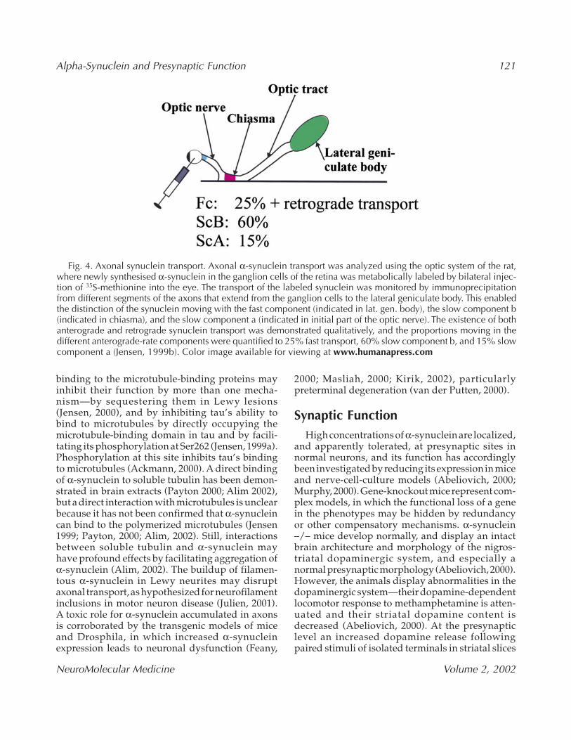
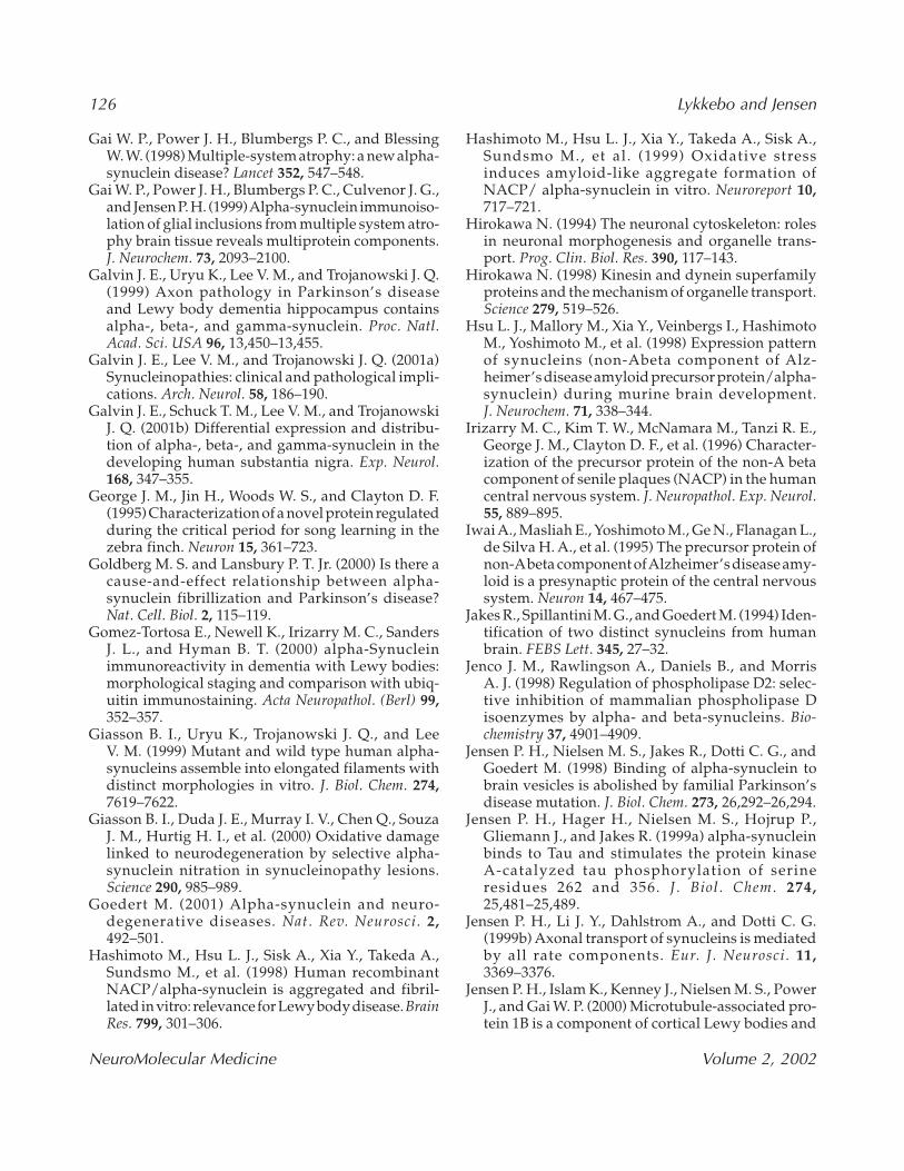
The axonal transport of synuclein in the CNS hasbeen analyzed directly using the rat optic nerve asa model (Jensen, 1998, 2000). This study, whichexamined both α- and β-synuclein, demonstratedthat each was transported by all the anterogradeand retrograde rate components (see Fig. 4). Theslow component b transported about 60% of themetabolically labeled synuclein, and the remainingportion was divided approximately evenly betweenthe fast component and the slow component a. Theproportion being transported retrograde was notquantified, but was demonstrated qualitatively bymicroscopic nerve-crush experiments. Proteins trans-ported by more than one rate component are notunprecedented—e.g., dynein and dynactin displaya pattern similar to synuclein with slow componentb dominating (Dillman, 1996). The involvement ofall the rate components in the axonal synuclein trans-port gives the synucleins ample opportunity to inter-act with many axonal constituents en route to thenerve terminals, such as motor proteins, vesicles(Jensen 1998, 2000), and axoskeletal regulators suchas soluble tau (Jensen, 1999a) and tubulin (Payton,2001; Alim, 2002). Apparently, synucleins can evenbecome incorporated into the axoskeleton, as radio-labeled synuclein was recovered from the axons 21 d after labeling of the nerve cells (Jensen, 2000).
There may be an association between disturbedaxonal α-synuclein transport and its accumulationin Lewy lesions, although this has not been proven.Partial transport defects will lead to an increasedtransit time for α-synuclein, and pending a contin-ued expression, a buildup in the cell body with subsequent increase in the local α-synucleinconcentration within the axon. An increased α-synuclein concentration in the stationary “pause”mode will favor interactions with axonal proteinssuch as the tubulins (Payton 2000; Alim 2002) andthe microtubule-stabilizing proteins MAP-1A,MAP-1B, MAP-4, and tau (Jensen, 1999a, 2000). The
Alpha-Synuclein and Presynaptic Function 121
NeuroMolecular Medicine Volume 2, 2002
binding to the microtubule-binding proteins mayinhibit their function by more than one mecha-nism—by sequestering them in Lewy lesions(Jensen, 2000), and by inhibiting tau’s ability to bind to microtubules by directly occupying themicrotubule-binding domain in tau and by facili-tating its phosphorylation at Ser262 (Jensen, 1999a).Phosphorylation at this site inhibits tau’s bindingto microtubules (Ackmann, 2000). A direct bindingof α-synuclein to soluble tubulin has been demon-strated in brain extracts (Payton 2000; Alim 2002),but a direct interaction with microtubules is unclearbecause it has not been confirmed that α-synucleincan bind to the polymerized microtubules (Jensen1999; Payton, 2000; Alim, 2002). Still, interactionsbetween soluble tubulin and α-synuclein may have profound effects by facilitating aggregation ofα-synuclein (Alim, 2002). The buildup of filamen-tous α-synuclein in Lewy neurites may disruptaxonal transport, as hypothesized for neurofilamentinclusions in motor neuron disease (Julien, 2001). A toxic role for α-synuclein accumulated in axonsis corroborated by the transgenic models of miceand Drosphila, in which increased α-synucleinexpression leads to neuronal dysfunction (Feany,
2000; Masliah, 2000; Kirik, 2002), particularlypreterminal degeneration (van der Putten, 2000).
Synaptic FunctionHigh concentrations of α-synuclein are localized,
and apparently tolerated, at presynaptic sites innormal neurons, and its function has accordinglybeen investigated by reducing its expression in miceand nerve-cell-culture models (Abeliovich, 2000;Murphy, 2000). Gene-knockout mice represent com-plex models, in which the functional loss of a genein the phenotypes may be hidden by redundancyor other compensatory mechanisms. α-synuclein–/– mice develop normally, and display an intactbrain architecture and morphology of the nigros-triatal dopaminergic system, and especially anormal presynaptic morphology (Abeliovich, 2000).However, the animals display abnormalities in thedopaminergic system—their dopamine-dependentlocomotor response to methamphetamine is atten-uated and their striatal dopamine content isdecreased (Abeliovich, 2000). At the presynapticlevel an increased dopamine release followingpaired stimuli of isolated terminals in striatal slices
Fig. 4. Axonal synuclein transport. Axonal α-synuclein transport was analyzed using the optic system of the rat,where newly synthesised α-synuclein in the ganglion cells of the retina was metabolically labeled by bilateral injec-tion of 35S-methionine into the eye. The transport of the labeled synuclein was monitored by immunoprecipitationfrom different segments of the axons that extend from the ganglion cells to the lateral geniculate body. This enabledthe distinction of the synuclein moving with the fast component (indicated in lat. gen. body), the slow component b(indicated in chiasma), and the slow component a (indicated in initial part of the optic nerve). The existence of bothanterograde and retrograde synuclein transport was demonstrated qualitatively, and the proportions moving in thedifferent anterograde-rate components were quantified to 25% fast transport, 60% slow component b, and 15% slowcomponent a (Jensen, 1999b). Color image available for viewing at www.humanapress.com
122 Lykkebo and Jensen
NeuroMolecular Medicine Volume 2, 2002
is observed (Abeliovich, 2000). This rather weakphenotype of the α-synuclein –/– mice does notallow a definition of α-synuclein’s functional role,although it indicates a link between α-synuclein anddopamine metabolism. This is corroborated by trans-fection studies, where increased α-synuclein alsoaffects the dopamine system in mice and rats, asdemonstrated by a loss of tyrosine hydroxylase-positive terminals and activity in striatum (Masliah,2000; Kirik, 2002). Dopamine is transported from the extracellular space into the cytosol by the dopa-mine transporter that is expressed specifically by thedopaminergic neurons. α-synuclein interacts directlywith the C-terminal part of the human dopaminetransporter and facilitates its translocation to theplasma membrane, thereby increasing dopamineuptake (Lee, 2001). In the cytosol, dopamine is con-sidered toxic because of its pro-oxidative properties(Adams, 1991), and it is efficiently removed from thecytosol into vesicles by the vesicular monoaminetransporter. The dopamine hypothesis states thatcytosolic dopamine, through its oxidative proper-ties, contributes to neurodegeneration, and it has theadvantage that it answers the question about theselective cellular vulnerability of the dopaminergicneurons in Parkinson’s disease. Candidates that favoran increased cytosolic dopamine concentrationsinclude increased activity of the plasma-membranedopamine transporter, where α-synuclein may con-tribute as stated here, or decreased function of thevesicular monoamine transporter. Detrimentaleffects of α-synuclein on the vesicular monoaminetransporter have not been reported, but a studywould be highly justified. Anti-sense oligonu-cleotide treatment represents another model forinvestigating the effect of the loss of a specific pro-tein. Arapid reduction in the gene product can oftenbe achieved, thereby possibly avoiding compen-satory mechanisms. Anti-sense oligonucleotidetreatment of cultured rat hippocampal neurons for 3 d resulted in a reduction of about 50% in α-synuclein protein without affecting the β-synuclein level, and this was accompanied by asignificant reduction of the distal pool of synapticvesicles and the synaptic-vesicle markers synapsin Iand synaptophysin (Murphy, 2000). Accordingly, α-synuclein may play a general role in the vesiclehomeostasis of nerve terminals that are not restrictedto dopaminergic neurons.
α-Synuclein associations with vesicles have beeninvestigated by methods such as immuno-
electronmicroscopy (Maroteaux, 1988; Iwai, 1995;Irizarry, 1996; Withers, 1997), biochemistry(Maroteaux, 1991; George, 1995; Kahle, 2000; Cole,2002; Lee, 2002) and fluorescence energy transfer(McLean, 2000), and these have led to the conclu-sion that α-synuclein is close to synaptic vesicles innerve terminals (Iwai, 1995), directly interactingwith brain vesicles (Jensen, 1998), and close to vesi-cles in cultured neurons (McLean, 2000). Moreover,the FAT of some α-synuclein in intact animals cor-roborates an interaction with vesicles (Jensen, 1998,1999b). Still, the fraction of α-synuclein being boundto vesicles, the characteristics of the interaction, andthe identity of the α-synuclein-containing vesiclesare a matter of debate, probably because thereversible nature of the binding leads to the disso-ciation of some α-synuclein during subcellular frac-tionation procedures. Synthetic liposomes havebeen widely used as models for investigating α-synuclein interaction with phospholipid bilayers(Davidson, 1998; Jo, 2000; Perrin, 2000; Eliezer, 2001;Narayanan, 2001; Volles, 2001), and this has allowedthe demonstration of the impact of membrane bind-ing on α-synucleins structure by circular dichroismspectroscopy. Through these means, a dramaticstructural transition was demonstrated from a pre-dominantly random coiled peptide in solution to astructure with a large α-helical content when boundto the membrane (Davidson, 1998; Jo, 2000; Perrin,2000; Eliezer, 2001; Narayanan, 2001; Volles, 2001).The interaction requires acidic phospholipids, andsome lipids facilitate the aggregation of α-synucleininto putative dimers and higher oligomers (David-son, 1998; Jo, 2000, Sharon, 2001). A pitfall in the interpretation of liposome studies has been uncov-ered as different phospholipid species support the binding of different segments of α-synuclein. Onestudy demonstrated a broad distribution of mem-brane-interacting elements over the N-terminal 103 amino-acid residues when using a certain lipidcomposition (Perrin, 2000) that contrasts with thecritical role of the N-terminal 30 in the interactionwith brain vesicles (Jensen, 1998). Liposomes pre-pared from a specific brain lipid extract, the Folchfraction I, require the same α-synuclein domain forbinding as brain vesicles and induce α-helical struc-ture (Lykkebo & Jensen, unpublished results). Thisfinding suggests that these liposomes may be ben-eficial for further studies on the functional effectsof α-synuclein binding to lipid bilayers and its inter-play with specific reconstituted vesicular proteins
Alpha-Synuclein and Presynaptic Function 123
NeuroMolecular Medicine Volume 2, 2002
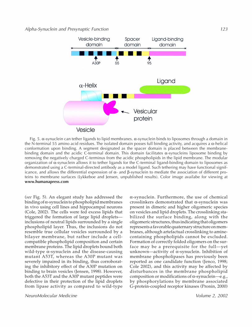
(see Fig. 5). An elegant study has addressed the binding of α-synuclein to phospholipid membranesin vivo using cell lines and hippocampal neurons(Cole, 2002). The cells were fed excess lipids thattriggered the formation of large lipid droplets—inclusions of neutral lipids surrounded by a singlephospholipid layer. Thus, the inclusions do notresemble true cellular vesicles surrounded by abilayer membrane, but rather include a cell-compatible phospholipid composition and certainmembrane proteins. The lipid droplets bound bothwild-type α-synuclein and the disease-causingmutant A53T, whereas the A30P mutant wasseverely impaired in its binding, thus corroborat-ing the inhibitory effect of the A30P mutation onbinding to brain vesicles (Jensen, 1998). However,both the A53T and the A30P mutant peptides weredefective in their protection of the lipid dropletsfrom lipase activity as compared to wild-type
α-synuclein. Furthermore, the use of chemicalcrosslinkers demonstrated that α-synuclein was present in dimeric and higher oligomeric species on vesicles and lipid droplets. The crosslinking sta-bilized the surface binding, along with theoligomeric structures, thus indicating that oligomersrepresents a favorable quaternary structure on mem-branes, although artefactual crosslinking to amine-containing phospholipids cannot be excluded.Formation of correctly folded oligomers on the sur-face may be a prerequisite for the full—yetunknown—activity of α-synuclein. Inhibition ofmembrane phospholipases has previously beenreported as one candidate function (Jenco, 1998;Cole 2002), and this activity may be affected by disturbances in the membrane phospholipid composition or modifications of α-synuclein—e.g.,by phosphorylations by membrane associated G-protein-coupled receptor kinases (Pronin, 2000)
Fig. 5. α-synuclein can tether ligands to lipid membranes. α-synuclein binds to liposomes through a domain inthe N-terminal 55 amino acid residues. The isolated domain posses full binding activity, and acquires a α-helicalconformation upon binding. A segment designated as the spacer domain is placed between the membrane-binding domain and the acidic C-terminal domain. This domain facilitates α-synucleins liposome binding byremoving the negatively charged C-terminus from the acidic phospholipids in the lipid membrane. The modularorganization of α-synuclein allows it to tether ligands for the C-terminal ligand-binding domain to liposomes asdemonstrated using a C-terminal-directed antibody as a model ligand. Such tethering may have functional signif-icance, and allows the differential expression of α- and β-synuclein to mediate the association of different pro-teins to membrane surfaces (Lykkeboe and Jensen, unpublished results). Color image availabe for viewing atwww.humanapress.com
124 Lykkebo and Jensen
NeuroMolecular Medicine Volume 2, 2002
or oxidations (Paxinou, 2001). α-synuclein can tetherC-terminally-bound proteins to liposomes (Fig. 5) and this ability may represent a physiological func-tion of synuclein bound to brain vesicles throughtheir N-terminal membrane-binding domain. α- andβ-synuclein possess very homologous vesicle-binding domains but differ in their C-termini. Thetethering hypothesis would imply that these synu-cleins can target specific subsets of soluble proteinsto vesicles for the facilitation of their axonal trans-port or the modulation of membrane-associatedfunctions. This would be analogous to the recentlyreported function of the membrane spanningAlzheimer’s disease-associated APP, which maytether certain axonal transport vesicles to theKinesin-I motor protein, thereby mediating theiraxonal transport (Kamal, 2000).
α-Synuclein-Mediated Dysregulationof “Presynaptic” Functions in Neurodegenerative Disorders
Autosomal dominant missense mutations in theα-synuclein gene are pathogenic in Parkinson’s disease, and abnormally folded α-synuclein accu-mulates in all Lewy bodies in a range of neuro-degenerative diseases. This forms the basis for the widely accepted hypothesis that a gain-of-toxic α-synuclein function is operating in these α-synucleinopathies. The mechanistic basis is lessclear, but likely involves novel functions of theabnormally filamentous α-synuclein and its proforms (Rochet, 2000b).
It is assumed that α-synuclein possesses physi-ological functions in the presynaptic compartment,such as regulating the activity of the dopamine trans-porter or fine-tuning the neurotransmitter release.The disease-associated relocalization to the cell bodyand neurites will then have a negative impact onthese functions. A putative decreased disease-associated synaptic α-synuclein content may becaused by dysfunctional axonal transport, or bytoxic effects of the accumulated α-synuclein in axonsthat might lead to full-blown axonal strangulation,or more specific cellular lesions caused by theabnormally localized and folded α-synuclein char-acterizing these diseases.
The ultimate cause(s) for the development of theα-synuclein containing Lewy body-lesions are
unknown. They may represent a specific neuronalresponse to a multifactorial yet selective set ofinsults, given that not all degenerating neuronsdevelop Lewy bodies. Still, the direct disease-causing effect of the A53T and the A30P mutationsdemonstrates that α-synuclein dysmetabolismeither activates or is part of a final common pathway in neurodegeneration. The developmentof compact-filament-containing Lewy bodies is preceded by a more diffuse accumulation of α-synuclein of unknown structural forms in thesomato-dendritic compartment (Wakabay-ashi, 1998; Gomez-Tortosa, 2000). This “ectopic” α-synuclein may have a special impact on thedopaminergic substantia nigra neurons at risk inParkinson’s disease, as their plasma membrane andintracellular vesicles at the somato-dendritic com-partment in certain aspects resemble presynapticsites, namely by expressing functional transportersof dopamine (Nirenberg 1996a, 1996b). These transporters may cause an abnormal α-synuclein-stimulated dopamine uptake into their cell bodythat could prime them for oxidative stress, andthereby contribute to their selective vulnerability inParkinson’s disease.
The majority of the current α-synuclein researchfocuses on the abnormal contributions of α-synuclein’s putative gain-of-toxic-function, andthe heritable effects of α-synuclein gene mutationsjustify this. Still, a better understanding of all aspectsof α-synucleins normal biology may reveal mecha-nisms that are dysfunctional in the diseased state,and are therefore worthy of evaluation as points oftherapeutic intervention.
Acknowledgment
Dr. Jia-Yi Li is thanked for generously providingthe image in Fig. 3. This manuscript is based onstudies supported by the Danish Medical ResearchCouncil (9902995), the Lundbeck Foundation, theDanish Parkinson Foundation, Wacherhausenslegat, Aarhus University’s Research Foundation,and Direktør Ib Henriksens Fond. SLwas supportedby a Novo Nordisk scholarship.
ReferencesAbeliovich A., Schmitz Y., Farinas I., Choi-Lundberg
D., Ho W. H., Castillo P. E., et al. (2000) Mice lack-ing alpha-synuclein display functional deficits in
Alpha-Synuclein and Presynaptic Function 125
NeuroMolecular Medicine Volume 2, 2002
the nigrostriatal dopamine system. Neuron 25,239–252.
Ackmann M., Wiech H., and Mandelkow E. (2000)Nonsaturable binding indicates clustering of tau on the microtubule surface in a paired helicalfilament-like conformation. J. Biol. Chem. 275,30,335–30,343.
Adams J. D. Jr. and Odunze I. N. (1991) Oxygen freeradicals and Parkinson’s disease. Free Radic. Biol.Med. 10, 161–169.
Alim M. A., Hossain M. S., Arima K., Takeda K., Izu-miyama Y., Nakamura M., et al. (2002) Tubulin seedsalpha-synuclein fibril formation. J. Biol. Chem. 277,2112–2117.
Ancolio K., Alves da Costa C., Ueda K., and CheclerF. (2000) Alpha-synuclein and the Parkinson’s disease-related mutant Ala53Thr-alpha-synucleindo not undergo proteasomal degradation inHEK293 and neuronal cells. Neurosci. Lett. 285,79–82.
Baba M., Nakajo S., Tu P. H., Tomita T., Nakaya K., LeeV. M., et al. (1998) Aggregation of alpha-synucleinin Lewy bodies of sporadic Parkinson’s disease anddementia with Lewy bodies. Am. J. Pathol. 152,879–884.
Bayer T.A., Jakala P., Hartmann T., Egensperger R.,Buslei R., Falkai P., et al. (1999) Neural expressionprofile of alpha-synuclein in developing humancortex. Neuroreport 10, 2799–2803.
Bennett M. C., Bishop J. F., Leng Y., Chock P. B., ChaseT. N., and Mouradian M. M. (1999) Degradation ofalpha-synuclein by proteasome. J. Biol. Chem. 274,33,855–33,858.
Campbell D. S. and Holt C. E. (2001) Chemotropicresponses of retinal growth cones mediated by rapidlocal protein synthesis and degradation. Neuron 32,1013–1026.
Campion D., Martin C., Heilig R., Charbonnier F.,Moreau V., Flaman J. M., et al. (1995) TheNACP/synuclein gene: chromosomal assignmentand screening for alterations in Alzheimer disease.Genomics 26, 254–257.
Choi P., Golts N., Snyder H., Chong M., Petrucelli L.,Hardy J., et al. (2001) Co-association of parkin andalpha-synuclein. Neuroreport 12, 2839–2843.
Cole R. N. and Hart G. W. (2001) Cytosolic O-glycosylation is abundant in nerve terminals. J. Neurochem. 79, 1080–1089.
Cole N. B., Murphy D. D., Grider T., Rueter S.,Brasaemle D., and Nussbaum R. L. (2002) Lipiddroplet binding and oligomerization properties ofthe Parkinson’s disease protein alpha-synuclein. J. Biol. Chem. 277, 6344–6352.
Conway K. A., Harper J. D., and Lansbury P. T. (1998)Accelerated in vitro fibril formation by a mutantalpha-synuclein linked to early-onset Parkinson disease. Nat. Med. 4, 1318–1320.
Conway K. A., Harper J. D., and Lansbury P. T. Jr.(2000a) Fibrils formed in vitro from alpha-synuclein and two mutant forms linked to Parkin-son’s disease are typical amyloid. Biochemistry 39,2552–2563.
Conway K. A., Lee S. J., Rochet J. C., Ding T. T.,Williamson R. E., and Lansbury P. T. Jr. (2000b)Acceleration of oligomerization, not fib-rillization, is a shared property of both alpha-synuclein mutations linked to early-onset Parkinson’s disease: implications for pathogene-sis and therapy. Proc. Natl. Acad. Sci. USA 97,571–576.
Crowther R. A., Jakes R., Spillantini M. G., and Goed-ert M. (1998) Synthetic filaments assembled fromC-terminally truncated alpha-synuclein. FEBS Lett.436, 309–312.
Davidson W. S., Jonas A., Clayton D. F., and George J. M. (1998) Stabilization of alpha-synuclein sec-ondary structure upon binding to synthetic mem-branes. J. Biol. Chem. 273, 9443–9449.
Dickson D. W., Liu W., Hardy J., Farrer M., Mehta N.,Uitti R., et al. (1999) Widespread alterations of alpha-synuclein in multiple system atrophy. Am. J. Pathol.155, 1241–1251.
Dickson D. W. (2001) Alpha-synuclein and the Lewybody disorders. Curr. Opin. Neurol. 14, 423–432.
Dillman J. F. 3rd, Dabney L. P., and Pfister K. K. (1996) Cytoplasmic dynein is associated with slowaxonal transport. Proc. Natl. Acad. Sci. USA 93,141–144.
Eliezer D., Kutluay E., Bussell R. Jr., and Browne G.(2001) Conformational properties of alpha-synuclein in its free and lipid-associated states. J. Mol. Biol. 307, 1061–1073.
Engelender S., Kaminsky Z., Guo X., Sharp A. H., Ama-ravi R. K., Kleiderlein J. J., et al. (1999) Synphilin-1associates with alpha-synuclein and promotes theformation of cytosolic inclusions. Nat. Genet. 22,110–114.
Feany M. B. and Bender W. W. (2000) A Droso-phila model of Parkinson’s disease. Nature 404,394–398.
Forno L. (1987) The Lewy body in Parkinson’s disease.Adv. Neurol. 45, 35–43.
Fujiwara H., Hasegawa M., Dohmae N., KawashimaA., Masliah E., Goldberg M. S., et al. (2002) alpha-Synuclein is phosphorylated in synucleinopathylesions. Nat. Cell. Biol. 4, 160–164.
126 Lykkebo and Jensen
NeuroMolecular Medicine Volume 2, 2002
Gai W. P., Power J. H., Blumbergs P. C., and BlessingW. W. (1998) Multiple-system atrophy: a new alpha-synuclein disease? Lancet 352, 547–548.
Gai W. P., Power J. H., Blumbergs P. C., Culvenor J. G.,and Jensen P. H. (1999) Alpha-synuclein immunoiso-lation of glial inclusions from multiple system atro-phy brain tissue reveals multiprotein components.J. Neurochem. 73, 2093–2100.
Galvin J. E., Uryu K., Lee V. M., and Trojanowski J. Q.(1999) Axon pathology in Parkinson’s disease and Lewy body dementia hippocampus contains alpha-, beta-, and gamma-synuclein. Proc. Natl.Acad. Sci. USA 96, 13,450–13,455.
Galvin J. E., Lee V. M., and Trojanowski J. Q. (2001a)Synucleinopathies: clinical and pathological impli-cations. Arch. Neurol. 58, 186–190.
Galvin J. E., Schuck T. M., Lee V. M., and TrojanowskiJ. Q. (2001b) Differential expression and distribu-tion of alpha-, beta-, and gamma-synuclein in thedeveloping human substantia nigra. Exp. Neurol.168, 347–355.
George J. M., Jin H., Woods W. S., and Clayton D. F.(1995) Characterization of a novel protein regulatedduring the critical period for song learning in thezebra finch. Neuron 15, 361–723.
Goldberg M. S. and Lansbury P. T. Jr. (2000) Is there acause-and-effect relationship between alpha-synuclein fibrillization and Parkinson’s disease?Nat. Cell. Biol. 2, 115–119.
Gomez-Tortosa E., Newell K., Irizarry M. C., SandersJ. L., and Hyman B. T. (2000) alpha-Synucleinimmunoreactivity in dementia with Lewy bodies:morphological staging and comparison with ubiq-uitin immunostaining. Acta Neuropathol. (Berl) 99,352–357.
Giasson B. I., Uryu K., Trojanowski J. Q., and Lee V. M. (1999) Mutant and wild type human alpha-synucleins assemble into elongated filaments withdistinct morphologies in vitro. J. Biol. Chem. 274,7619–7622.
Giasson B. I., Duda J. E., Murray I. V., Chen Q., SouzaJ. M., Hurtig H. I., et al. (2000) Oxidative damagelinked to neurodegeneration by selective alpha-synuclein nitration in synucleinopathy lesions. Science 290, 985–989.
Goedert M. (2001) Alpha-synuclein and neuro-degenerative diseases. Nat. Rev. Neurosci. 2,492–501.
Hashimoto M., Hsu L. J., Sisk A., Xia Y., Takeda A.,Sundsmo M., et al. (1998) Human recombinantNACP/alpha-synuclein is aggregated and fibril-lated in vitro: relevance for Lewy body disease. BrainRes. 799, 301–306.
Hashimoto M., Hsu L. J., Xia Y., Takeda A., Sisk A.,Sundsmo M., et al. (1999) Oxidative stress induces amyloid-like aggregate formation ofNACP/ alpha-synuclein in vitro. Neuroreport 10,717–721.
Hirokawa N. (1994) The neuronal cytoskeleton: rolesin neuronal morphogenesis and organelle trans-port. Prog. Clin. Biol. Res. 390, 117–143.
Hirokawa N. (1998) Kinesin and dynein superfamilyproteins and the mechanism of organelle transport.Science 279, 519–526.
Hsu L. J., Mallory M., Xia Y., Veinbergs I., HashimotoM., Yoshimoto M., et al. (1998) Expression patternof synucleins (non-Abeta component of Alz-heimer’s disease amyloid precursor protein/alpha-synuclein) during murine brain development. J. Neurochem. 71, 338–344.
Irizarry M. C., Kim T. W., McNamara M., Tanzi R. E.,George J. M., Clayton D. F., et al. (1996) Character-ization of the precursor protein of the non-A betacomponent of senile plaques (NACP) in the humancentral nervous system. J. Neuropathol. Exp. Neurol.55, 889–895.
Iwai A., Masliah E., Yoshimoto M., Ge N., Flanagan L.,de Silva H. A., et al. (1995) The precursor protein ofnon-Abeta component of Alzheimer’s disease amy-loid is a presynaptic protein of the central nervoussystem. Neuron 14, 467–475.
Jakes R., Spillantini M. G., and Goedert M. (1994) Iden-tification of two distinct synucleins from humanbrain. FEBS Lett. 345, 27–32.
Jenco J. M., Rawlingson A., Daniels B., and Morris A. J. (1998) Regulation of phospholipase D2: selec-tive inhibition of mammalian phospholipase Disoenzymes by alpha- and beta-synucleins. Bio-chemistry 37, 4901–4909.
Jensen P. H., Nielsen M. S., Jakes R., Dotti C. G., andGoedert M. (1998) Binding of alpha-synuclein tobrain vesicles is abolished by familial Parkinson’sdisease mutation. J. Biol. Chem. 273, 26,292–26,294.
Jensen P. H., Hager H., Nielsen M. S., Hojrup P., Gliemann J., and Jakes R. (1999a) alpha-synuclein binds to Tau and stimulates the protein kinase A-catalyzed tau phosphorylation of serine residues 262 and 356. J. Biol. Chem. 274,25,481–25,489.
Jensen P. H., Li J. Y., Dahlstrom A., and Dotti C. G.(1999b) Axonal transport of synucleins is mediatedby all rate components. Eur. J. Neurosci. 11,3369–3376.
Jensen P. H., Islam K., Kenney J., Nielsen M. S., PowerJ., and Gai W. P. (2000) Microtubule-associated pro-tein 1B is a component of cortical Lewy bodies and
Alpha-Synuclein and Presynaptic Function 127
NeuroMolecular Medicine Volume 2, 2002
binds alpha-synuclein filaments. J. Biol. Chem. 275,21,500–21,507.
Jensen P. H. and Gai W. P. (2001) Alpha-synuclein.Axonal transport, ligand interaction and neurode-generation. Adv. Exp. Med. Biol. 487, 129–134.
Jo E., McLaurin J., Yip C. M., St George-Hyslop P., andFraser P. E. (2000) alpha-Synuclein membrane inter-actions and lipid specificity. J. Biol. Chem. 275,34,328–34,334.
Julien J. P. (2001) Amyotrophic lateral sclerosis. unfold-ing the toxicity of the misfolded. Cell 104, 581–591.
Kahle P. J., Neumann M., Ozmen L., Muller V., Jacob-sen H., Schindzielorz A., et al. (2000) Subcellularlocalization of wild-type and Parkinson’s disease-associated mutant alpha-synuclein in human andtransgenic mouse brain. J. Neurosci. 20, 6365–6373.
Kahle P. J., Neumann M., Ozmen L., Muller V., OdoyS., Okamoto N., et al. (2001) Selective insolubilityof alpha-synuclein in human Lewy body diseasesis recapitulated in a transgenic mouse model. Am.J. Pathol. 159, 2215–2225.
Kamal A., Stokin G. B., Yang Z., Xia C. H., and Gold-stein L. S. (2000) Axonal transport of amyloid pre-cursor protein is mediated by direct binding to thekinesin light chain subunit of kinesin-I. Neuron 28,449–459.
Kirik D., Rosenblad C., Burger C., Lundberg C.,Johansen T. E., Muzyczka N., et al. (2002) Parkin-son-like neurodegeneration induced by targetedoverexpression of alpha-synuclein in the nigrostri-atal system. J. Neurosci. 22, 2780–2795.
Krüger R., Kuhn W., Muller T., Woitalla D., GraeberM., Kosel S., et al. (1998) Ala30Pro mutation in thegene encoding alpha-synuclein in Parkinson’s dis-ease. Nat. Genet. 18, 106–108.
Lang A. E. and Lozano A. M. (1998) Parkinson’s dis-ease. N. Engl. J. Med. 339, 1030–1053.
Lavedan C., Leroy E., Dehejia A., Buchholtz S., DutraA., Nussbaum R. L., et al. (1998) Identification, local-ization and characterization of the human gamma-synuclein gene. Hum. Genet. 103, 106–112.
Lee F. J., Liu F., Pristupa Z. B., and Niznik H. B. (2001)Direct binding and functional coupling of alpha-synuclein to the dopamine transporters acceler-ate dopamine-induced apoptosis. FASEB J. 15,916–926.
Lee H. J., Choi C., and Lee S. J. (2002) Membrane-boundalpha-synuclein has a high aggregation propensityand the ability to seed the aggregation of the cytoso-lic form. J. Biol. Chem. 277, 671–678.
Lewy F. H. (1912) Paralysis agitans, I PathologischeAnatomie, Handbuch der Neurologie, Band III,Springer Verlag, Germany, pp. 920–933.
Maroteaux L., Campanelli J. T., and Scheller R. H. (1988)Synuclein: a neuron-specific protein localized to thenucleus and presynaptic nerve terminal. J. Neurosci.8, 2804–2815.
Maroteaux L. and Scheller R. H. (1991) The rat brainsynucleins; family of proteins transiently associatedwith neuronal membrane. Brain Res. Mol. Brain Res.11, 335–343.
Masliah E., Rockenstein E., Veinbergs I., Mallory M.,Hashimoto M., Takeda A., et al. (2000) Dopamin-ergic loss and inclusion body formation in alpha-synuclein mice: implications for neurodegenerativedisorders. Science 287, 1265–1269.
McKeith I. G., Perry E. K., and Perry R. H. (1999) Reportof the second dementia with Lewy body internationalworkshop: diagnosis and treatment. Consortium onDementia with Lewy Bodies. Neurology 53, 902–905.
McLean P. J., Kawamata H., Ribich S., and Hyman B. T. (2000) Membrane association and protein con-formation of alpha-synuclein in intact neurons.Effect of Parkinson’s disease-linked mutations. J. Biol. Chem. 275, 8812–8816.
Murphy D. D., Rueter S. M., Trojanowski J. Q., and LeeV. M. (2000) Synucleins are developmentallyexpressed, and alpha-synuclein regulates the sizeof the presynaptic vesicular pool in primary hippo-campal neurons. J. Neurosci. 20, 3214–3220.
Nakajo S., Tsukada K., Kameyama H., Furuyama Y.,and Nakaya K. (1996) Distribution of phospho-neuroprotein 14 (PNP 14) in vertebrates: its levelsas determined by enzyme immunoassay. Brain Res.741, 180–184.
Narayanan V. and Scarlata S. (2001) Membrane binding and self-association of alpha-synucleins.Biochemistry 40, 9927–9934.
Nielsen M. S., Vorum H., Lindersson E., and Jensen P. H. (2001) Ca2+ binding to alpha-synuclein regu-lates ligand binding and oligomerization. J. Biol.Chem. 276, 22,680–22,684.
Nirenberg M. J., Vaughan R. A., Uhl G. R., Kuhar M. J., and Pickel V. M. (1996a) The dopamine trans-porter is localized to dendritic and axonal plasmamembranes of nigrostriatal dopaminergic neurons.J. Neurosci. 16, 436–447.
Nirenberg M. J., Chan J., Liu Y., Edwards R. H., and Pickel V. M. (1996b) Ultrastructural localiza-tion of the vesicular monoamine transporter-2 in midbrain dopaminergic neurons: potential sites for somatodendritic storage and release ofdopamine. J. Neurosci. 16, 4135–4145.
Ochs S. (1975) Retention and redistribution of proteinsin mammalian nerve fibres by axoplasmic trans-port. J. Physiol. 253, 459–475.
128 Lykkebo and Jensen
NeuroMolecular Medicine Volume 2, 2002
Okochi M., Walter J., Koyama A., Nakajo S., Baba M., Iwatsubo T., et al. (2000) Constitutive phos-phorylation of the Parkinson’s disease associatedalpha-synuclein. J. Biol. Chem. 275, 390–397.
Paik S. R., Shin H. J., and Lee J. H. (2000) Metal-catalyzed oxidation of alpha-synuclein in the presence of Copper(II) and hydrogen peroxide.Arch. Biochem. Biophys. 378, 269–277.
Paxinou E., Chen Q., Weisse M., Giasson B. I., NorrisE. H., Rueter S. M., et al. (2001) Induction of alpha-synuclein aggregation by intracellular nitrativeinsult. J. Neurosci. 21, 8053–8061.
Payton J. E., Perrin R. J., Clayton D. F., and George J. M. (2001) Protein-protein interactions of alpha-synuclein in brain homogenates and transfectedcells. Brain Res. Mol. Brain Res. 95, 138–145.
Perrin R. J., Woods W. S., Clayton D. F., and George J. M. (2000) Interaction of human alpha-Synucleinand Parkinson’s disease variants with phospho-lipids. Structural analysis using site-directed muta-genesis. J. Biol. Chem. 275, 34,393–34,398.
Petersen K., Olesen O. F., and Mikkelsen J. D. (1999)Developmental expression of alpha-synuclein in rathippocampus and cerebral cortex. Neuroscience 91,651–659.
Polymeropoulos M. H., Lavedan C., Leroy E., Ide S. E., Dehejia A., Dutra A., et al. (1997) Mutation inthe alpha-synuclein gene identified in families withParkinson’s disease. Science 276, 2045–2047.
Pronin A. N., Morris A. J., Surguchov A., and BenovicJ. L. (2000) Synucleins are a novel class of substratesfor G protein-coupled receptor kinases. J. Biol. Chem.275, 26,515–26,522.
Rochet J. C., Conway K. A., and Lansbury P. T. Jr. (2000a)Inhibition of fibrillization and accumulation of pre-fibrillar oligomers in mixtures of human and mousealpha-synuclein. Biochemistry 39, 10,619–10,626.
Rochet J. C. and Lansbury P. T. Jr. (2000b) Amyloid fib-rillogenesis: themes and variations. Curr. Opin.Struct. Biol. 10, 60–68.
Shah J. V. and Cleveland D. W. (2002) Slow axonaltransport: fast motors in the slow lane. Curr. Opin.Cell. Biol. 14, 58–62.
Sharon R., Goldberg M. S., Bar-Josef I., Betensky R. A.,Shen J., and Selkoe D. J. (2001) alpha-Synucleinoccurs in lipid-rich high molecular weight com-plexes, binds fatty acids, and shows homology tothe fatty acid-binding proteins. Proc. Natl. Acad. Sci.USA 98, 9110–9115.
Shea T. B. and Flanagan L. A. (2001) Kinesin, dynein andneurofilament transport. Trends Neurosci.24,644–648.
Shibayama-Imazu T., Okahashi I., Omata K., NakajoS., Ochiai H., Nakai Y., et al. (1993) Cell and tissue
distribution and developmental change of neuronspecific 14 kDa protein (phosphoneuroprotein 14).Brain Res. 622, 17–25.
Shimura H., Schlossmacher M. G., Hattori N., FroschM. P., Trockenbacher A., Schneider R., et al. (2001)Ubiquitination of a new form of alpha-synucleinby parkin from human brain: implications forParkinson’s disease. Science 293, 263–269.
Souza J. M., Giasson B. I., Chen Q., Lee V. M., andIschiropoulos H. (2000) Dityrosine cross-linkingpromotes formation of stable alpha-synucleinpolymers. Implication of nitrative and oxidativestress in the pathogenesis of neurodegen-erative synucleinopathies. J. Biol. Chem. 275,18,344–18,349.
Spillantini M. G., Divane A., and Goedert M. (1995)Assignment of human alpha-synuclein (SNCA) andbeta-synuclein (SNCB) genes to chromosomes 4q21and 5q35. Genomics 27, 379–381.
Spillantini M. G., Schmidt M. L., Lee V. M., TrojanowskiJ. Q., Jakes R., and Goedert M. (1997) Alpha-synuclein in Lewy bodies. Nature 388, 839–840.
Tofaris G. K., Layfield R., and Spillantini M. G. (2001) alpha-synuclein metabolism and aggre-gation is l inked to ubiquitin-independent degradation by the proteasome. FEBS Lett. 509,22–26.
Ueda K., Fukushima H., Masliah E., Xia Y., Iwai A.,Yoshimoto M., et al. (1993) Molecular cloning ofcDNA encoding an unrecognized component ofamyloid in Alzheimer disease. Proc. Natl. Acad. Sci.USA 90, 11,282–11,286.
Ueda K., Saitoh T., and Mori H. (1994) Tissue-dependent alternative splicing of mRNAfor NACP,the precursor of non-A beta component ofAlzheimer’s disease amyloid. Biochem. Biophys. Res.Commun. 205, 1366–1372.
Van der Putten H., Wiederhold K. H., Probst A., Barbieri S., Mistl C., Danner S., et al. (2000) Neu-ropathology in mice expressing human alpha-synuclein. J. Neurosci. 20, 6021–6029.
Volles M. J., Lee S. J., Rochet J. C., Shtilerman M. D.,Ding T. T., Kessler J. C., et al. (2001) Vesicle permeabilization by protofibrillar alpha-synuclein: implications for the pathogenesis andtreatment of Parkinson’s disease. Biochemistry 40,7812–7819.
Wakabayashi K., Hayashi S., Kakita A., Yamada M.,Toyoshima Y., Yoshimoto M., et al. (1998) Accumu-lation of alpha-synuclein/NACP is a cytopatho-logical feature common to Lewy body disease andmultiple system atrophy. Acta Neuropathol. (Berl) 96,445–452.
Alpha-Synuclein and Presynaptic Function 129
NeuroMolecular Medicine Volume 2, 2002
Weinreb P. H., Zhen W., Poon A. W., Conway K. A.,and Lansbury P. T. Jr. (1996) NACP, a protein impli-cated in Alzheimer ’s disease and learning, isnatively unfolded. Biochemistry 35, 13,709–13,715.
Withers G. S., George J. M., Banker G. A., and ClaytonD. F. (1997) Delayed localization of synelfin (synu-clein, NACP) to presynaptic terminals in cultured
rat hippocampal neurons. Brain Res. Dev. Brain Res.99, 87–94.
Wood S. J., Wypych J., Steavenson S., Louis J. C., CitronM., and Biere A. L. (1999) alpha-synuclein fibrillo-genesis is nucleation-dependent process. Implica-tions for the pathogenesis of Parkinson’s disease.J. Biol. Chem. 274, 19,509–19,512.
Recommended