Use of MicrobeamUse of Microbeamat JAEA Takasaki at JAEA Takasaki
Mitsuhiro FUKUDAResearch Center for Nuclear Physics(RCNP)
Osaka University
APAC2007 THZH102
AuthorsAuthorsTakasaki Advanced Radiation Research InstituteJapan Atomic Energy Agency
Tomihiro Kamiya, Masakazu Oikawa, Takahiro Satoh, Takuro Sakai, Satoshi Kurashima, Nobumasa Miyawaki, Susumu Okumura, Hirotsugu Kashiwagi,Watalu Yokota
Research Center for Nuclear Physics (RCNP)Osaka University
Mitsuhiro Fukuda, Kichiji Hatanaka, Tetsuhiko Yorita
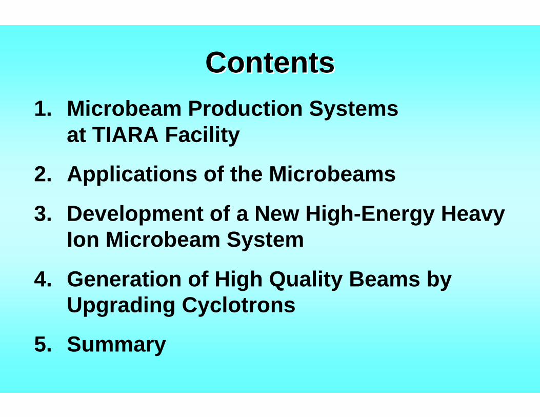
ContentsContents1. Microbeam Production Systems
at TIARA Facility
2. Applications of the Microbeams
3. Development of a New High-Energy Heavy Ion Microbeam System
4. Generation of High Quality Beams by Upgrading Cyclotrons
5. Summary
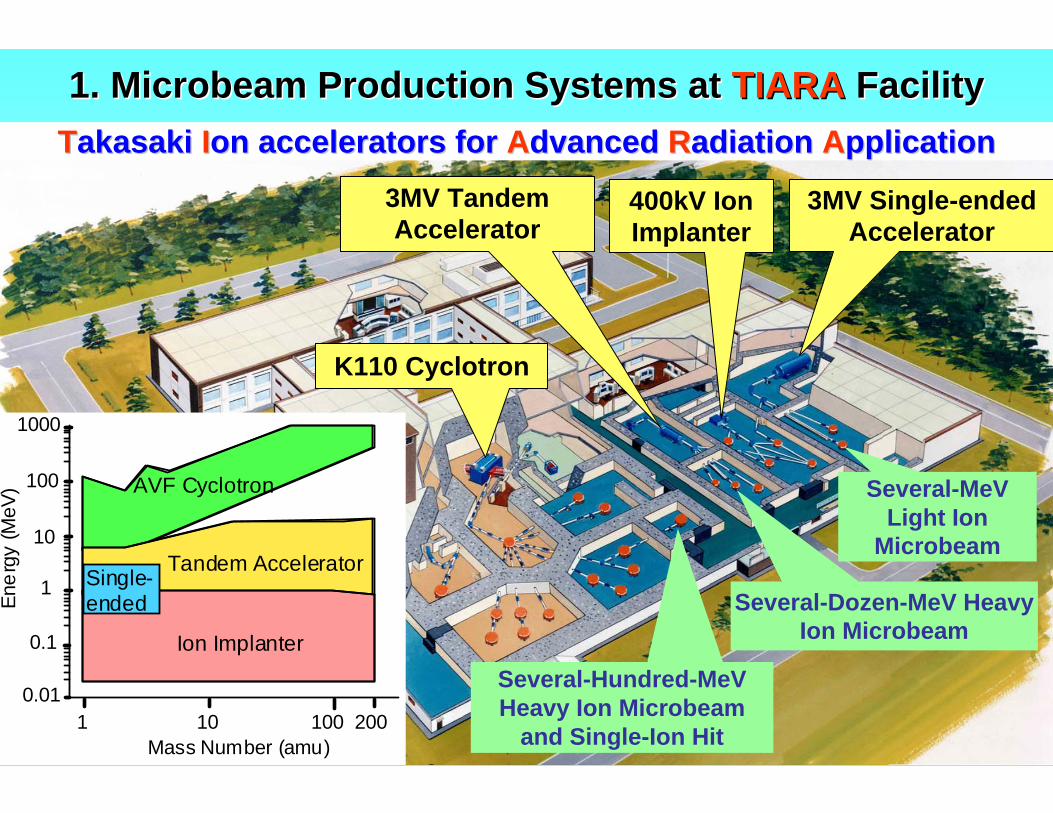
1. Microbeam Production Systems at 1. Microbeam Production Systems at TIARATIARA FacilityFacility
K110 Cyclotron
3MV Tandem Accelerator
3MV Single-ended Accelerator
Several-Hundred-MeVHeavy Ion Microbeam
and Single-Ion Hit
Several-Dozen-MeV Heavy Ion Microbeam
Several-MeVLight Ion
Microbeam
TTakasakiakasaki IIon accelerators foron accelerators for AAdvanceddvanced RRadiationadiation AApplicationpplication
Ener
gy (M
eV)
0.01
0.1
1
10
100
1000
AVF Cyclotron
Tandem Accelerator
Ion Implanter
Mass Number (amu)1
Single-ended
10 200100
400kV Ion Implanter
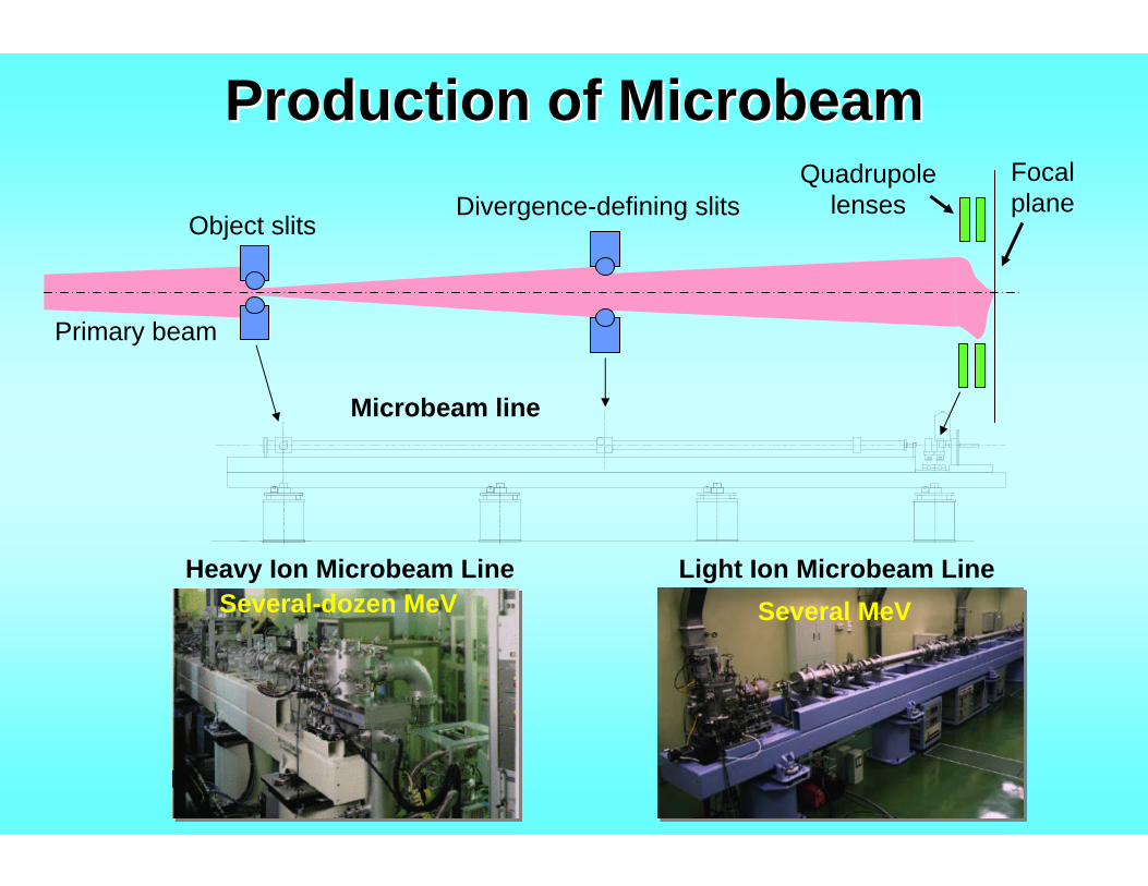
Production of MicrobeamProduction of Microbeam
Primary beam
Object slitsDivergence-defining slits
Quadrupole lenses
Focal plane
Microbeam line
Heavy Ion Microbeam Line Light Ion Microbeam LineSeveral-dozen MeV Several MeV
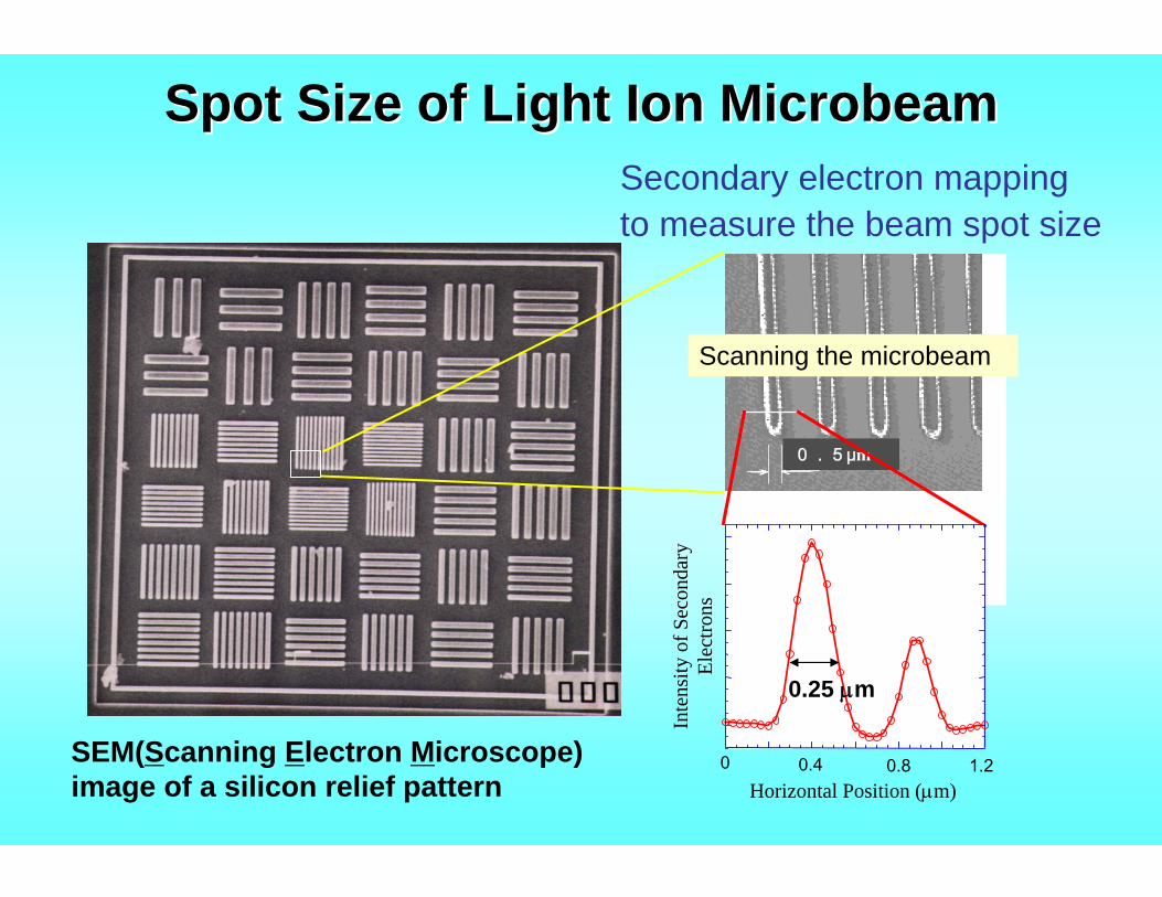
Spot Size of Light Ion Microbeam Spot Size of Light Ion Microbeam
0.5μm
SEM(Scanning Electron Microscope) image of a silicon relief pattern
0 0.4 0.8 1.2Horizontal Position (μm)
Inte
nsity
of S
econ
dary
El
ectro
ns
0.25 μm
Secondary electron mappingto measure the beam spot size
Scanning the microbeam
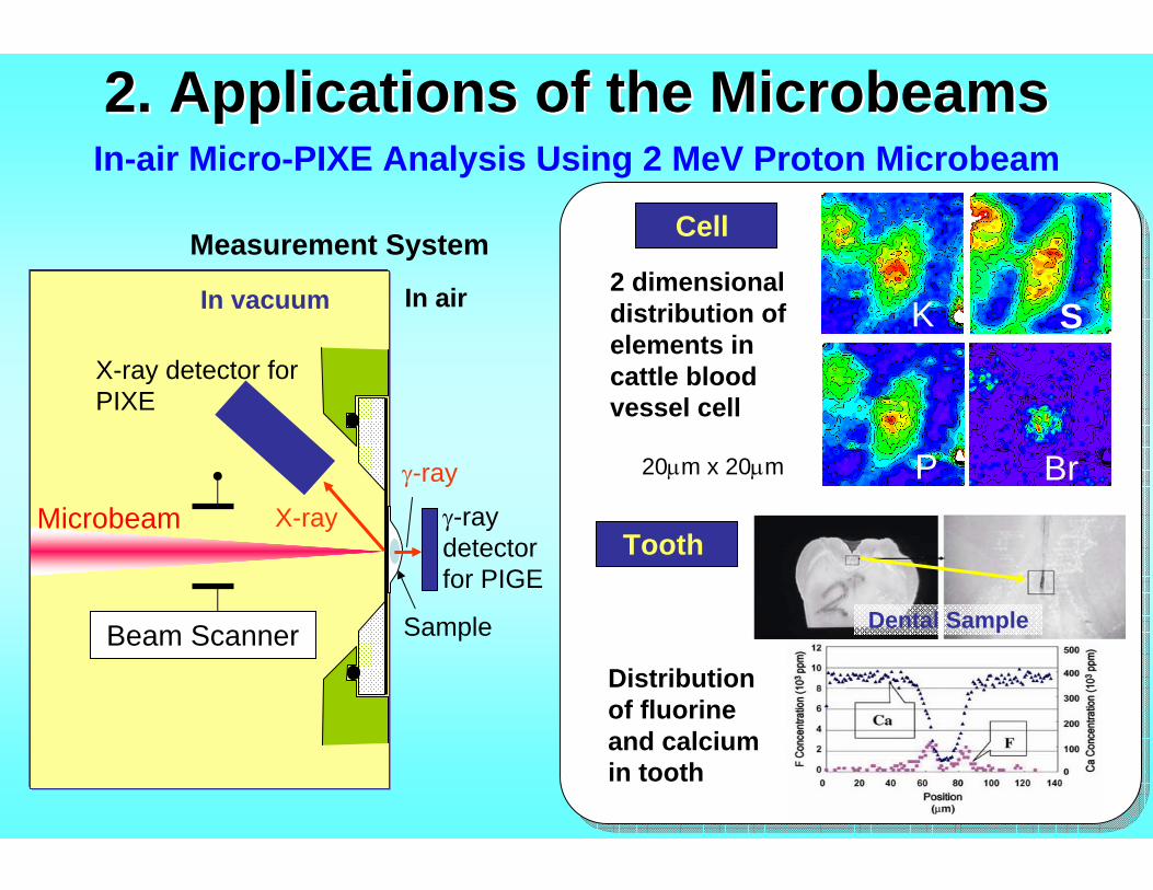
2. Applications of the 2. Applications of the MicrobeamsMicrobeams
2 dimensional distribution of elements in cattle blood vessel cell
20μm x 20μm
S
BrP
K
Cell
In vacuum In air
Sample
Microbeam
X-ray detector for PIXE
Beam Scanner
γ-ray detector for PIGE
Measurement System
X-ray
γ-ray
Tooth
Dental Sample
Distribution of fluorine and calcium in tooth
In-air Micro-PIXE Analysis Using 2 MeV Proton Microbeam
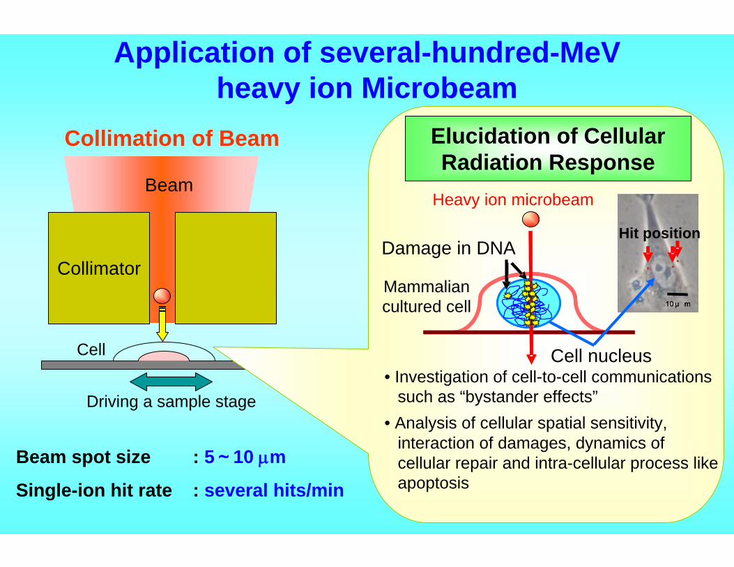
Application of several-hundred-MeVheavy ion Microbeam
Beam spot size : 5~10 μm
Single-ion hit rate : several hits/min
Collimator
Cell
Beam
Driving a sample stage
10 μ m核
イオンのヒット位置
Mammalian cultured cell
Heavy ion microbeam
10 μ m10 μ m
Elucidation of Cellular Radiation Response
Cell nucleus
Damage in DNA
• Investigation of cell-to-cell communications such as “bystander effects”
• Analysis of cellular spatial sensitivity, interaction of damages, dynamics of cellular repair and intra-cellular process like apoptosis
Hit position
Collimation of Beam
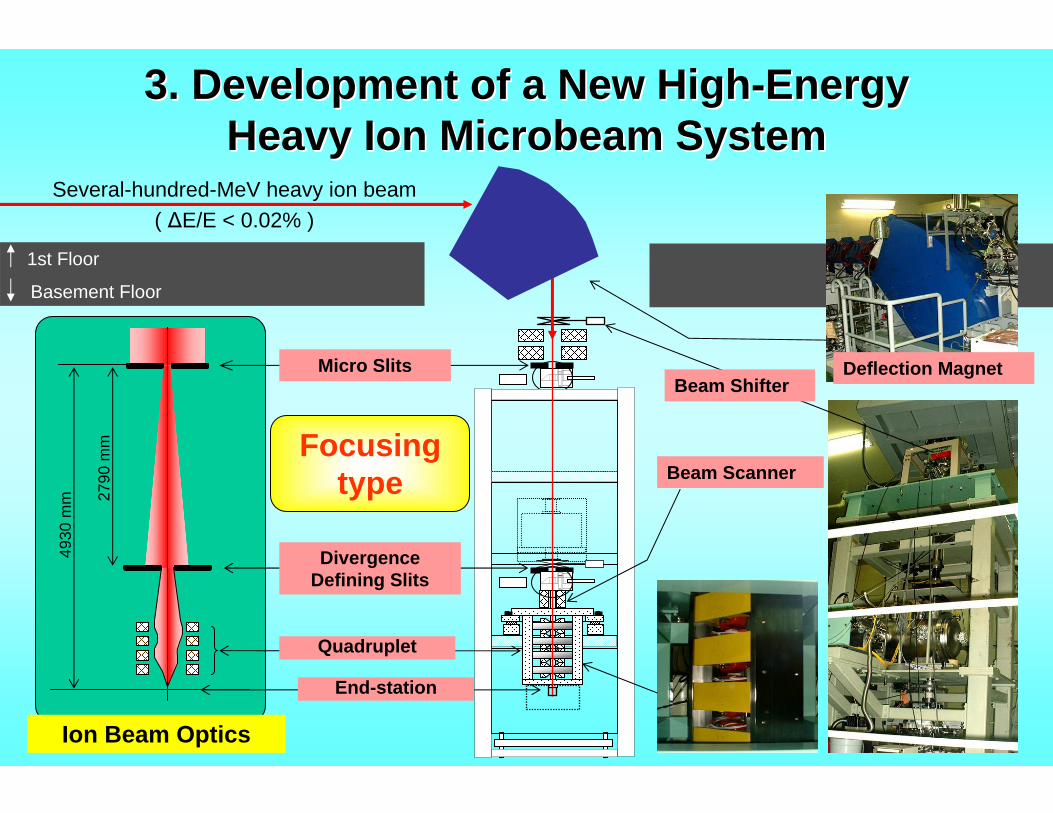
3. Development of a New High3. Development of a New High--EnergyEnergyHeavy Ion Microbeam SystemHeavy Ion Microbeam System
Micro Slits
2790
mm
Divergence Defining Slits
Quadruplet
basement floor level
Beam Shifter
Basement Floor
4930
mm
Deflection Magnet
Beam Scanner
Several-hundred-MeV heavy ion beam( ΔE/E < 0.02% )
Ion Beam Optics
1st Floor
End-station
Focusing type
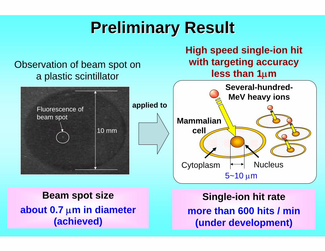
Preliminary ResultPreliminary Result
Several-hundred-MeV heavy ions
Mammalian cell
Nucleus
Beam spot sizeabout 0.7 μm in diameter
(achieved)
5~10 μmCytoplasm
High speed single-ion hit with targeting accuracy
less than 1μm
10 mm
Fluorescence ofbeam spot
Observation of beam spot on a plastic scintillator
applied to
Single-ion hit ratemore than 600 hits / min
(under development)
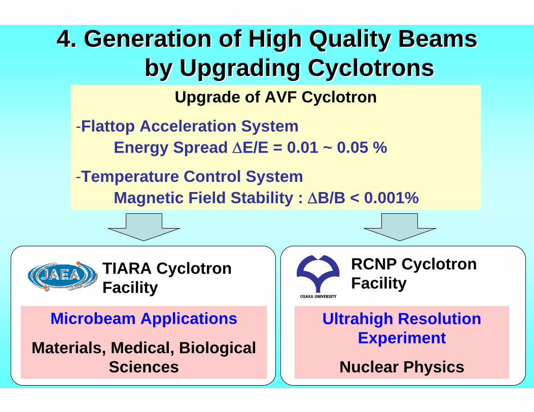
4. Generation of High Quality Beams4. Generation of High Quality Beamsby Upgrading Cyclotronsby Upgrading Cyclotrons
TIARA Cyclotron Facility
RCNP Cyclotron Facility
Microbeam Applications
Materials, Medical, Biological Sciences
Ultrahigh Resolution Experiment
Nuclear Physics
Upgrade of AVF Cyclotron
-Flattop Acceleration SystemEnergy Spread ΔE/E = 0.01 ~ 0.05 %
-Temperature Control SystemMagnetic Field Stability : ΔB/B < 0.001%
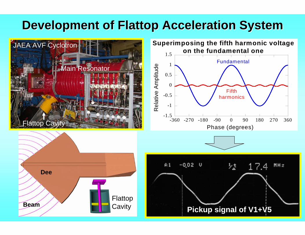
Development of Flattop Acceleration SystemDevelopment of Flattop Acceleration System
Flattop CavityBeam
Dee
Flattop Cavity
Main Resonator
JAEA AVF Cyclotron
Pickup signal of V1+V5
-1.5
-1
-0.5
0
0.5
1
1.5
-360 -270 -180 -90 0 90 180 270 360
Superimposing the fifth harmonic voltageon the fundamental one
Rel
ativ
e A
mpl
itude
Phase (degrees)
Fundamental
Fifthharmonics
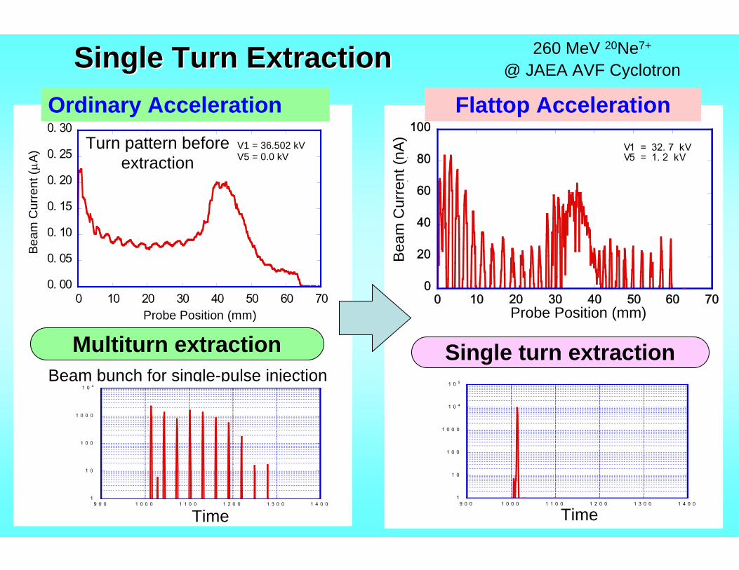
Single Turn ExtractionSingle Turn Extraction
Multiturn extraction Single turn extraction
0. 00
0. 05
0. 10
0. 15
0. 20
0. 25
0. 30
0 10 20 30 40 50 60 70
Beam
Cur
rent
(μA
)
Probe Position (mm)
V1 = 36.502 kVV5 = 0.0 kV
Ordinary Acceleration
Beam bunch for single-pulse injection
1
1 0
1 0 0
1 0 0 0
1 0 4
9 0 0 1 0 0 0 1 1 0 0 1 2 0 0 1 3 0 0 1 4 0 0
カウ
ント
時 間 ( 1 5 c h / 1 R F 周 期 )Time
0
20
40
60
80
100
0 10 20 30 40 50 60 70
フラットトップ ON
ビー
ム電
流(
nA)
プローブ位置(mm)
V1 = 32. 7 kVV5 = 1. 2 kV
Probe Position (mm)
Bea
m C
urre
nt (n
A)
1
1 0
1 0 0
1 0 0 0
1 0 4
1 0 5
9 0 0 1 0 0 0 1 1 0 0 1 2 0 0 1 3 0 0 1 4 0 0
カウ
ント
時 間 ( 1 5 c h / 1 R F 周 期 )Time
260 MeV 20Ne7+
@ JAEA AVF Cyclotron
Turn pattern before extraction
Flattop Acceleration
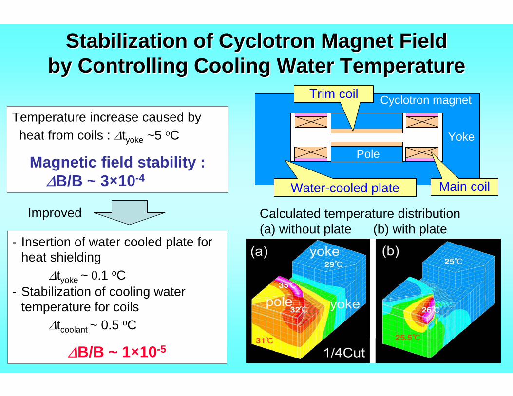
Stabilization of Cyclotron Magnet FieldStabilization of Cyclotron Magnet Fieldby Controlling Cooling Water Temperatureby Controlling Cooling Water Temperature
Temperature increase caused byheat from coils : Δtyoke ~5 oC
Magnetic field stability :ΔB/B ~ 3×10⁻⁴
- Insertion of water cooled plate for heat shielding
Δtyoke ~ 0.1 oC- Stabilization of cooling water
temperature for coils Δtcoolant ~ 0.5 oC
ΔB/B ~ 1×10⁻⁵
Calculated temperature distribution(a) without plate (b) with plate
¼ cut
Improved
Cyclotron magnet
Water-cooled plate
Trim coil
YokePole
Main coil
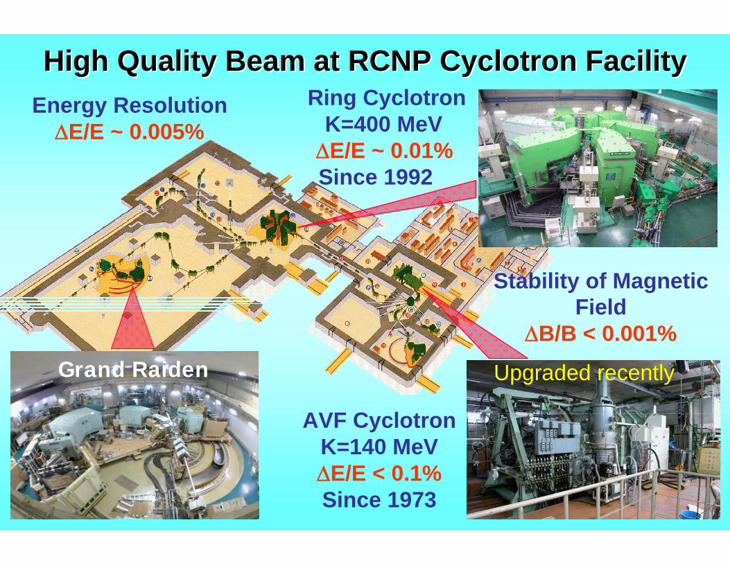
High Quality Beam at RCNP Cyclotron FacilityHigh Quality Beam at RCNP Cyclotron FacilityRing Cyclotron
K=400 MeVΔE/E ~ 0.01%Since 1992
Stability of Magnetic Field
ΔB/B < 0.001%Grand Raiden
Energy ResolutionΔE/E ~ 0.005%
AVF CyclotronK=140 MeVΔE/E < 0.1%Since 1973
Upgraded recently
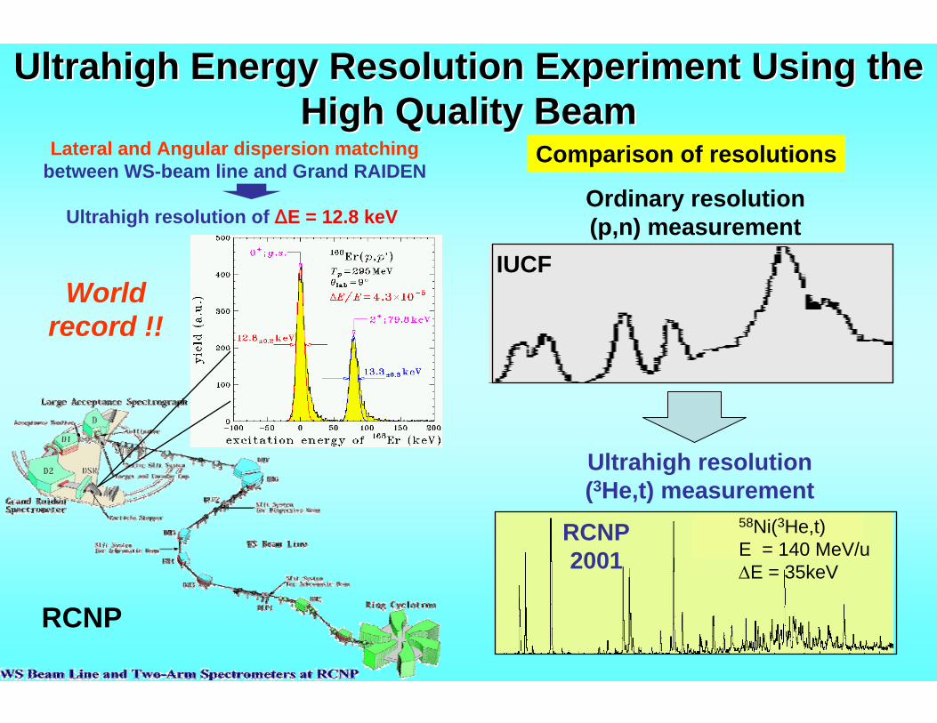
Ultrahigh Energy Resolution Experiment Using the Ultrahigh Energy Resolution Experiment Using the High Quality BeamHigh Quality Beam
Lateral and Angular dispersion matchingbetween WS-beam line and Grand RAIDEN
Ultrahigh resolution of ΔE = 12.8 keV
World record !!
58Ni(3He,t)E = 140 MeV/uΔE = 35keV
Ultrahigh resolution(3He,t) measurement
RCNP2001
IUCF
Ordinary resolution(p,n) measurement
Comparison of resolutions
RCNP
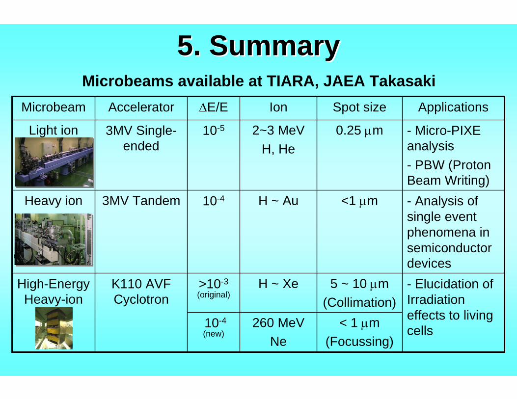
5. Summary5. Summary
- Elucidation of Irradiation effects to living cells
5 ~ 10 μm(Collimation)
H ~ Xe>10-3(original)
K110 AVF Cyclotron
High-Energy Heavy-ion
260 MeVNe
< 1 μm(Focussing)
10-4(new)
3MV Tandem
3MV Single-ended
Accelerator
- Analysis of single event phenomena in semiconductor devices
<1 μmH ~ Au10-4Heavy ion
- Micro-PIXE analysis- PBW (Proton Beam Writing)
0.25 μm2~3 MeVH, He
10-5Light ion
ApplicationsSpot sizeIonΔE/EMicrobeam
Microbeams available at TIARA, JAEA Takasaki
Recommended