Hindawi Publishing CorporationCase Reports in OrthopedicsVolume 2013, Article ID 472378, 4 pageshttp://dx.doi.org/10.1155/2013/472378
Case ReportIntra-Articular Giant Heterotopic Ossification following TotalKnee Arthroplasty for Charcot Arthropathy
Arata Nakajima,1,2 Shintaro Tsuge,3 Yasuchika Aoki,1 Masato Sonobe,1
Yoshifumi Shibata,3 Yu Sasaki,1 and Koichi Nakagawa1
1 Department of Orthopaedic Surgery, Toho University Sakura Medical Center, 564-1 Shimoshizu, Sakura, Chiba 285-8741, Japan2Department of Orthopedics and Rheumatology, Toho University Sakura Medical Center, 564-1 Shimoshizu, Sakura,Chiba 285-8741, Japan
3Department of Orthopaedic Surgery, Toho University Omori Medical Center, 6-11-1 Omori-nishi, Ota-ku, Tokyo 143-8541, Japan
Correspondence should be addressed to Arata Nakajima; [email protected]
Received 24 July 2013; Accepted 17 August 2013
Academic Editors: N. Kort, D. Saragaglia, and B. Unver
Copyright © 2013 Arata Nakajima et al.This is an open access article distributed under the Creative CommonsAttribution License,which permits unrestricted use, distribution, and reproduction in any medium, provided the original work is properly cited.
Although the Charcot arthropathy may be associated with serious complications, total knee arthroplasty (TKA) is the preferredchoice of treatment by patients. This case report presents an 80-year-old man with intra-articular giant heterotopic ossificationfollowing loosening of femoral and tibial implants and femoral condylar fracture. He had undergone TKA because of Charcotneuropathy seven years ago and had been doing well since. Immediately after a left knee sprain, he became unable to walk.Because he had developed a skin ulcer on his left calf where methicillin-resistant Staphylococcus aureuswas detected, we postponedrevision surgery until the ulcer was completely healed. While waiting, intra-articular bony fragments grew larger and formed giantheterotopic ossified masses. Eventually, the patient underwent revision surgery, and two major ossified masses were carefully andsuccessfully extirpated. It should be noted that intra-articular heterotopic giant ossification is a significant complication after TKAfor neuropathic arthropathy.
1. Introduction
Neuropathic arthropathy was described by Jean MartinCharcot (1825–1893) as the progressive destruction of boneand soft tissues in a patient with peripheral neuropathy.Charcot also noted the relationship between syphilis andsevere arthropathy in 1868 [1]. Diabetes mellitus is currentlythe most common cause of Charcot arthropathy, especially ofthe foot and ankle.
The diagnosis of Charcot arthropathy in the knee is rare.However, we can expect the increasing number of diabeticpatients living longer due to improvements in medicaltreatment to show an increasing incidence of neuropathicarthropathy as a late effect of peripheral neuropathy. Theoptimal treatment for Charcot arthropathy of the knee iscontroversial. Some investigators [2–4] consider it as anabsolute contraindication to total knee arthroplasty (TKA).
Recently, however, several studies [5] have shown satisfactoryresults from TKA.
In general, TKA for Charcot arthropathy is associatedwith high incidence of such serious complications as peripro-sthetic fractures, aseptic loosening, dislocation of the patellaand tibiofemoral joint, and deep infection [6–8]. How-ever, due to excellent functional recovery compared withconservative therapy and arthrodesis, TKA should not becontraindicated.
We present a case report of an 80-year-old man withintra-articular, giant, heterotopic ossification and a femoralcondylar fracture following loosening of femoral and tibialimplants. To date, the occurrence of intra-articular, giant,heterotopic ossification as a significant complication afterTKA for Charcot arthropathy has not been reported.
Our patient gave informed consent for data from his caseto be submitted for publication.
2 Case Reports in Orthopedics
(a) (b)
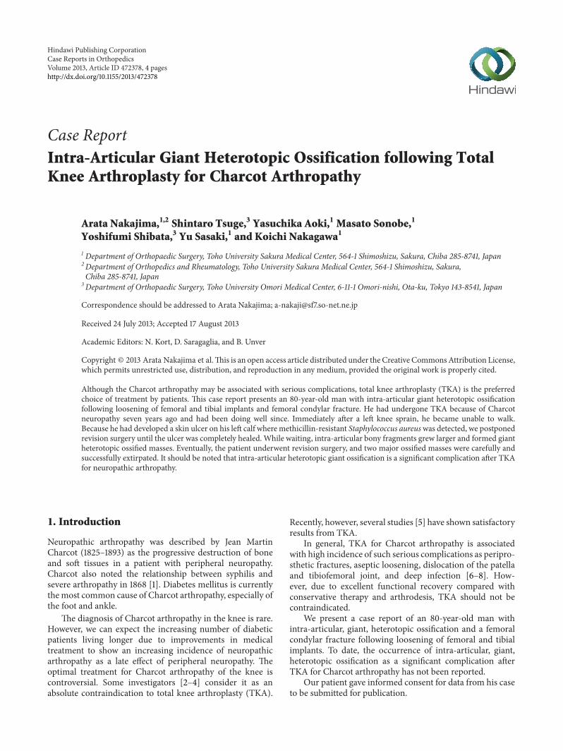
Figure 1: Radiographs of the left knee immediately after the patientsprained his knee and became unable to walk. An anteroposteriorradiograph shows a radiolucency 1mm in depth beneath the lateralaspect of the tibial component ((a), arrow), and the lateral viewindicates a condylar fracture ((b), arrow).
2. Case Report
An 80-year-old man presented with a primary complaintof left knee pain for five months. He had undergone TKAwith a posterior stabilized-type prosthesis (Scorpio Superflex,Stryker Orthopedics, Mahwah, NJ, USA) due to a neuro-pathic joint seven years ago and had been doing well since.Several days before his visit to our hospital, he had sprainedhis left knee and became unable to walk.
An anteroposterior radiograph showed a radiolucency1mm in depth beneath the lateral aspect of the tibial com-ponent (Figure 1(a), arrow), and the lateral view indicateda condylar fracture (Figure 1(b), arrow). We had planned toperform a revision TKA using a more strongly constrained-type knee prosthesis shortly thereafter, but this surgery hadto be postponed becausemethicillin-resistant staphylococcusaureus (MRSA) was detected in a skin ulcer on his left calf.Although his peripheral leukocyte count was within normallimits (6.8 × 10³/𝜇L), his serum C-reactive protein level waselevated to 5.3mg/dL (normal range <0.3mg/dL) and syn-ovial fluid bacterial cultures were negative. Stress radiographsfor the left knee taken before the primary TKA showed severejoint destruction and instability (Figures 2(a)–2(c)), stronglysuggesting neuropathic arthropathy. His hemoglobin A1cwas 5.4%, and both the serological test for syphilis andTreponema pallidum hemagglutination test were negative.MRI images of the cervical and thoracic spine were nor-mal, and syringomyelia was ruled out. However, he hadbeen diagnosed wiyh primary amyloidosis and followed byneurophysicians. Peripheral nerve conduction velocity wasseverely delayed, suggesting neuropathic arthropathy due toamyloidotic polyneuropathy.
The skin ulcer did not completely heal for three months,during which time the patient was hospitalized with hisleft knee immobilized in a long-leg brace and administered
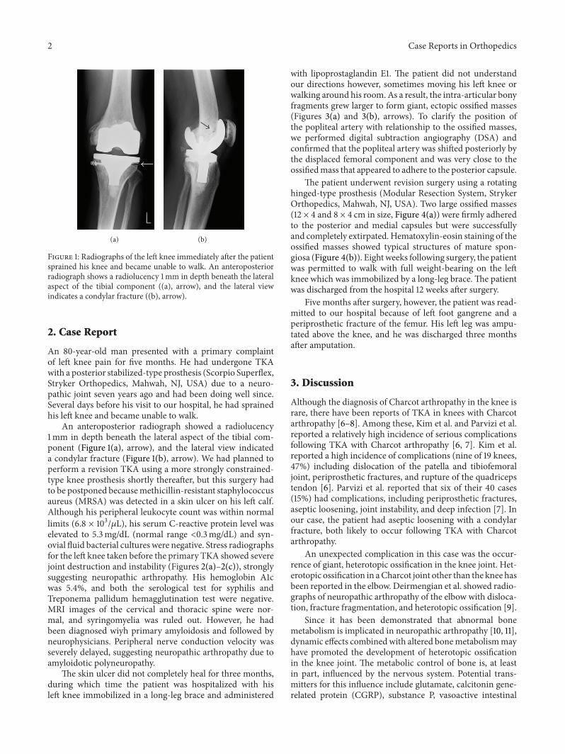
with lipoprostaglandin E1. The patient did not understandour directions however, sometimes moving his left knee orwalking around his room. As a result, the intra-articular bonyfragments grew larger to form giant, ectopic ossified masses(Figures 3(a) and 3(b), arrows). To clarify the position ofthe popliteal artery with relationship to the ossified masses,we performed digital subtraction angiography (DSA) andconfirmed that the popliteal artery was shifted posteriorly bythe displaced femoral component and was very close to theossifiedmass that appeared to adhere to the posterior capsule.
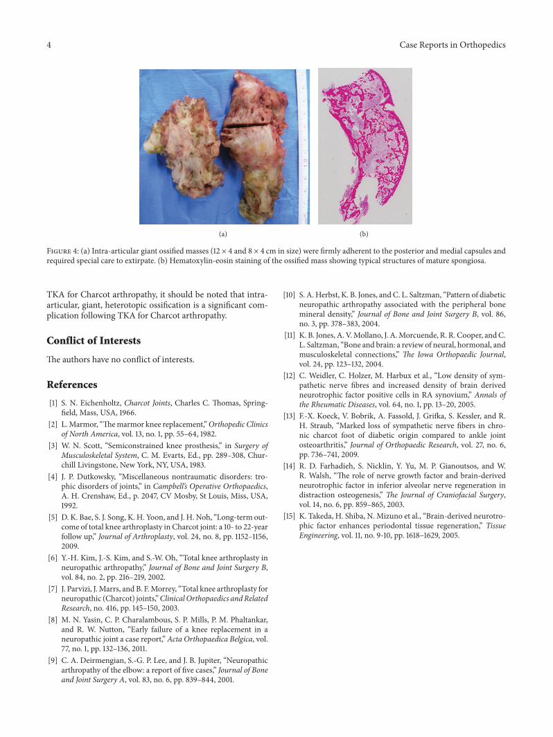
The patient underwent revision surgery using a rotatinghinged-type prosthesis (Modular Resection System, StrykerOrthopedics, Mahwah, NJ, USA). Two large ossified masses(12 × 4 and 8 × 4 cm in size, Figure 4(a)) were firmly adheredto the posterior and medial capsules but were successfullyand completely extirpated. Hematoxylin-eosin staining of theossified masses showed typical structures of mature spon-giosa (Figure 4(b)). Eight weeks following surgery, the patientwas permitted to walk with full weight-bearing on the leftknee which was immobilized by a long-leg brace. The patientwas discharged from the hospital 12 weeks after surgery.
Five months after surgery, however, the patient was read-mitted to our hospital because of left foot gangrene and aperiprosthetic fracture of the femur. His left leg was ampu-tated above the knee, and he was discharged three monthsafter amputation.
3. Discussion
Although the diagnosis of Charcot arthropathy in the knee israre, there have been reports of TKA in knees with Charcotarthropathy [6–8]. Among these, Kim et al. and Parvizi et al.reported a relatively high incidence of serious complicationsfollowing TKA with Charcot arthropathy [6, 7]. Kim et al.reported a high incidence of complications (nine of 19 knees,47%) including dislocation of the patella and tibiofemoraljoint, periprosthetic fractures, and rupture of the quadricepstendon [6]. Parvizi et al. reported that six of their 40 cases(15%) had complications, including periprosthetic fractures,aseptic loosening, joint instability, and deep infection [7]. Inour case, the patient had aseptic loosening with a condylarfracture, both likely to occur following TKA with Charcotarthropathy.
An unexpected complication in this case was the occur-rence of giant, heterotopic ossification in the knee joint. Het-erotopic ossification in aCharcot joint other than the knee hasbeen reported in the elbow. Deirmengian et al. showed radio-graphs of neuropathic arthropathy of the elbow with disloca-tion, fracture fragmentation, and heterotopic ossification [9].
Since it has been demonstrated that abnormal bonemetabolism is implicated in neuropathic arthropathy [10, 11],dynamic effects combinedwith altered bonemetabolismmayhave promoted the development of heterotopic ossificationin the knee joint. The metabolic control of bone is, at leastin part, influenced by the nervous system. Potential trans-mitters for this influence include glutamate, calcitonin gene-related protein (CGRP), substance P, vasoactive intestinal
Case Reports in Orthopedics 3
(a) (b) (c)
Figure 2: Stress radiographs of the left knee before the primary total knee arthroplasty (TKA) ((a) varus stress, (b) neutral, and (c) valgusstress). Severe joint destruction and instability are seen, indicating neuropathic arthropathy.
(a) (b)
Figure 3: Radiographs of the left knee threemonths after the patientbecame unable to walk. Intra-articular bony fragments are enlargedand form giant ectopic ossified masses ((a), (b), arrows). Note thatthe femoral component is displaced and shifted posteriorly (b).
peptide (VIP), pituitary adenylate cyclase activating polypep-tide (PACAP), leptin, and catecholamines. These factors areknown to be involved in bone formation, resorption, andremodeling [11], and imbalances in these factors may causeprogressive joint destruction and/or giant ectopic ossificationfollowing implant loosening and intra-articular fractures.
Neurotrophic factors, such as brain-derived neurotrophicfactor (BDNF) and nerve growth factor (NGF), are also pot-ential factors influencing bone metabolism. In the rheumaticjoint, a low density of sympathetic nerve fibers increaseddensity of BDNF-positive cells in the synovium have been
reported [12]. Koeck et al. reported that sympathetic nervefibers were significantly lower in patients with Charcot footcompared to those with osteoarthritis (OA), and that thesympathetic nerve repellent factor semaphorin 3Cwas highlyexpressed in inflamed tissue of Charcot patients [13]. Con-sidering these findings, BDNF and NGF, also known to bebone anabolic factors [14, 15], may be upregulated in Charcotarthropathy, leading to development of intra-articular giantectopic ossification.
In condylar fractures complicated with implant looseningin the Charcot joint, as found in our case, bony fragmentscould develop into the giant, heterotopic ossified masses thatwould make the revision surgery more difficult. Therefore,surgeons should perform revision surgery as soon as possiblefollowing implant loosening or periarticular fractures in theCharcot joint.
Our concern is that there may have already been septicloosening of the implants, when the patient presented toour hospital following his knee sprain. His C-reactive pro-tein serum levels were highly elevated (5.3mg/dL) beforethe revision TKA, and bacterial cultures taken from theperiprosthetic fracture site during amputation were positivefor MRSA. These factors mean that we cannot preclude thepossibility of existing bacterial infection in the patient’s kneebefore the revision surgery. However, in addition to negativeresults for the synovial fluid cultures before the revisionTKA, the elevated C-reactive protein serum levels returnedto normal 19 days after the revision surgery, indicating thatseptic loosening prior to the condylar fracture did not occur.
In conclusion, this case report is the first publishedaccount of the occurrence of giant, heterotopic ossificationassociated with an intra-articular fracture and implant loos-ening following TKA for neuropathic arthropathy. Althoughmany other complications have been reported to follow
4 Case Reports in Orthopedics
(a) (b)
Figure 4: (a) Intra-articular giant ossified masses (12 × 4 and 8 × 4 cm in size) were firmly adherent to the posterior and medial capsules andrequired special care to extirpate. (b) Hematoxylin-eosin staining of the ossified mass showing typical structures of mature spongiosa.
TKA for Charcot arthropathy, it should be noted that intra-articular, giant, heterotopic ossification is a significant com-plication following TKA for Charcot arthropathy.
Conflict of Interests
The authors have no conflict of interests.
References
[1] S. N. Eichenholtz, Charcot Joints, Charles C. Thomas, Spring-field, Mass, USA, 1966.
[2] L.Marmor, “Themarmor knee replacement,”Orthopedic Clinicsof North America, vol. 13, no. 1, pp. 55–64, 1982.
[3] W. N. Scott, “Semiconstrained knee prosthesis,” in Surgery ofMusculoskeletal System, C. M. Evarts, Ed., pp. 289–308, Chur-chill Livingstone, New York, NY, USA, 1983.
[4] J. P. Dutkowsky, “Miscellaneous nontraumatic disorders: tro-phic disorders of joints,” in Campbell’s Operative Orthopaedics,A. H. Crenshaw, Ed., p. 2047, CV Mosby, St Louis, Miss, USA,1992.
[5] D. K. Bae, S. J. Song, K. H. Yoon, and J. H. Noh, “Long-term out-come of total knee arthroplasty in Charcot joint: a 10- to 22-yearfollow up,” Journal of Arthroplasty, vol. 24, no. 8, pp. 1152–1156,2009.
[6] Y.-H. Kim, J.-S. Kim, and S.-W. Oh, “Total knee arthroplasty inneuropathic arthropathy,” Journal of Bone and Joint Surgery B,vol. 84, no. 2, pp. 216–219, 2002.
[7] J. Parvizi, J.Marrs, and B. F.Morrey, “Total knee arthroplasty forneuropathic (Charcot) joints,”ClinicalOrthopaedics andRelatedResearch, no. 416, pp. 145–150, 2003.
[8] M. N. Yasin, C. P. Charalambous, S. P. Mills, P. M. Phaltankar,and R. W. Nutton, “Early failure of a knee replacement in aneuropathic joint a case report,”Acta Orthopaedica Belgica, vol.77, no. 1, pp. 132–136, 2011.
[9] C. A. Deirmengian, S.-G. P. Lee, and J. B. Jupiter, “Neuropathicarthropathy of the elbow: a report of five cases,” Journal of Boneand Joint Surgery A, vol. 83, no. 6, pp. 839–844, 2001.
[10] S. A. Herbst, K. B. Jones, andC. L. Saltzman, “Pattern of diabeticneuropathic arthropathy associated with the peripheral bonemineral density,” Journal of Bone and Joint Surgery B, vol. 86,no. 3, pp. 378–383, 2004.
[11] K. B. Jones, A.V.Mollano, J. A.Morcuende, R. R. Cooper, andC.L. Saltzman, “Bone and brain: a review of neural, hormonal, andmusculoskeletal connections,” The Iowa Orthopaedic Journal,vol. 24, pp. 123–132, 2004.
[12] C. Weidler, C. Holzer, M. Harbux et al., “Low density of sym-pathetic nerve fibres and increased density of brain derivedneurotrophic factor positive cells in RA synovium,” Annals ofthe Rheumatic Diseases, vol. 64, no. 1, pp. 13–20, 2005.
[13] F.-X. Koeck, V. Bobrik, A. Fassold, J. Grifka, S. Kessler, and R.H. Straub, “Marked loss of sympathetic nerve fibers in chro-nic charcot foot of diabetic origin compared to ankle jointosteoarthritis,” Journal of Orthopaedic Research, vol. 27, no. 6,pp. 736–741, 2009.
[14] R. D. Farhadieh, S. Nicklin, Y. Yu, M. P. Gianoutsos, and W.R. Walsh, “The role of nerve growth factor and brain-derivedneurotrophic factor in inferior alveolar nerve regeneration indistraction osteogenesis,” The Journal of Craniofacial Surgery,vol. 14, no. 6, pp. 859–865, 2003.
[15] K. Takeda, H. Shiba, N. Mizuno et al., “Brain-derived neurotro-phic factor enhances periodontal tissue regeneration,” TissueEngineering, vol. 11, no. 9-10, pp. 1618–1629, 2005.

Submit your manuscripts athttp://www.hindawi.com
Stem CellsInternational
Hindawi Publishing Corporationhttp://www.hindawi.com Volume 2014
Hindawi Publishing Corporationhttp://www.hindawi.com Volume 2014
MEDIATORSINFLAMMATION
of
Hindawi Publishing Corporationhttp://www.hindawi.com Volume 2014
Behavioural Neurology
EndocrinologyInternational Journal of
Hindawi Publishing Corporationhttp://www.hindawi.com Volume 2014
Hindawi Publishing Corporationhttp://www.hindawi.com Volume 2014
Disease Markers
Hindawi Publishing Corporationhttp://www.hindawi.com Volume 2014
BioMed Research International
OncologyJournal of
Hindawi Publishing Corporationhttp://www.hindawi.com Volume 2014
Hindawi Publishing Corporationhttp://www.hindawi.com Volume 2014
Oxidative Medicine and Cellular Longevity
Hindawi Publishing Corporationhttp://www.hindawi.com Volume 2014
PPAR Research
The Scientific World JournalHindawi Publishing Corporation http://www.hindawi.com Volume 2014
Immunology ResearchHindawi Publishing Corporationhttp://www.hindawi.com Volume 2014
Journal of
ObesityJournal of
Hindawi Publishing Corporationhttp://www.hindawi.com Volume 2014
Hindawi Publishing Corporationhttp://www.hindawi.com Volume 2014
Computational and Mathematical Methods in Medicine
OphthalmologyJournal of
Hindawi Publishing Corporationhttp://www.hindawi.com Volume 2014
Diabetes ResearchJournal of
Hindawi Publishing Corporationhttp://www.hindawi.com Volume 2014
Hindawi Publishing Corporationhttp://www.hindawi.com Volume 2014
Research and TreatmentAIDS
Hindawi Publishing Corporationhttp://www.hindawi.com Volume 2014
Gastroenterology Research and Practice
Hindawi Publishing Corporationhttp://www.hindawi.com Volume 2014
Parkinson’s Disease
Evidence-Based Complementary and Alternative Medicine
Volume 2014Hindawi Publishing Corporationhttp://www.hindawi.com
Recommended