Chemical Carcinogenesis

Chemical Carcinogenesis Samuel M. Cohen, MD, PhD
University of Nebraska Medical Center Department of Pathology and Microbiology
Omaha, NE
Conflict of Interest Declaration • Consult for Numerous Companies • Research funded by NIH, Private Industry • FEMA Expert Panel
Outline • Basic Principles of Carcinogenesis • Carcinogenic Chemicals • Carcinogenicity Testing • Model of Carcinogenesis • Mode of Action/Human Relevance
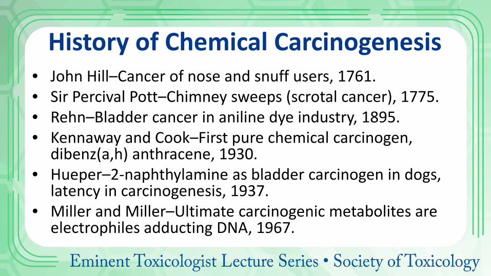
History of Chemical Carcinogenesis • John Hill–Cancer of nose and snuff users, 1761. • Sir Percival Pott–Chimney sweeps (scrotal cancer), 1775. • Rehn–Bladder cancer in aniline dye industry, 1895. • Kennaway and Cook–First pure chemical carcinogen,
dibenz(a,h) anthracene, 1930. • Hueper–2-naphthylamine as bladder carcinogen in dogs,
latency in carcinogenesis, 1937. • Miller and Miller–Ultimate carcinogenic metabolites are
electrophiles adducting DNA, 1967.
Cancer Requires Genetic Alterations • Usually occur as somatic alterations • Can occasionally be inherited • Multiple alterations are required
Inherited Diseases with High Tumor Incidence
• Retinoblastoma • Multiple polyposis coli • Thyroid medullary carcinoma • Multiple endocrine adenomas • Von Recklinghausen’s disease
Diseases Associated with Increased Cancer Risk • UV Radiation
– Albinism – Xeroderma pigmentosm
• Chromosome fragility syndrome – Bloom’s syndrome – Fanconi’s syndrome
• Immunodeficiencies – X-linked lymphoproliferative disease (XLP) – Ataxic-telangiectasia – Severe combined immunodeficiency – Wiskott-Aldrich Syndrome
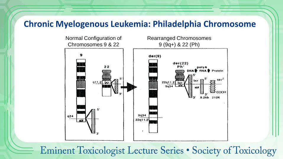
Chronic Myelogenous Leukemia: Philadelphia Chromosome Normal Configuration of Chromosomes 9 & 22
Rearranged Chromosomes 9 (9q+) & 22 (Ph)
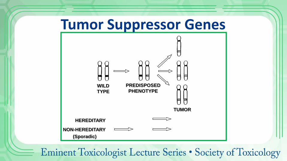
WILD TYPE
PREDISPOSED PHENOTYPE
TUMOR
HEREDITARY
NON-HEREDITARY (Sporadic)
Tumor Suppressor Genes
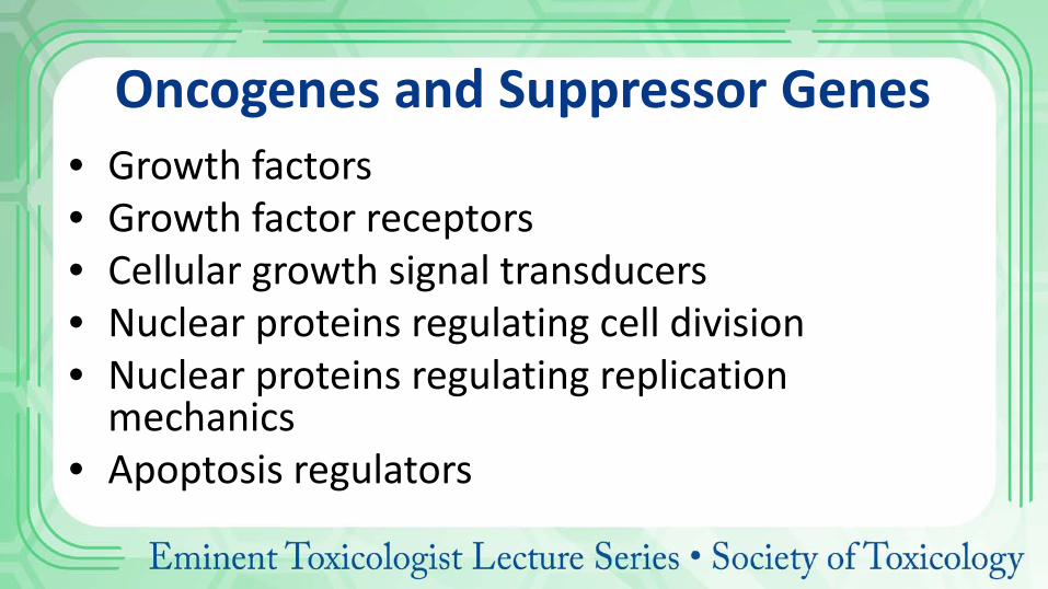
Oncogenes and Suppressor Genes • Growth factors • Growth factor receptors • Cellular growth signal transducers • Nuclear proteins regulating cell division • Nuclear proteins regulating replication
mechanics • Apoptosis regulators
Immunosurveillance and Cancer • Tumor specific transplantation antigens
– Viral specific in mice – No tumor specific antigens in humans
• Carcinogens are immunosuppressive
– Frequently only at doses >>carcinogenic dose
• Increased incidence of tumors in immunodeficient patients – Only a few specific types of cancer
• Neoplastic clone escapes immune surveillance
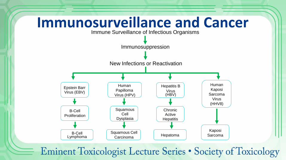
Epstein Barr Virus (EBV)
Human Papilloma
Virus (HPV)
Human Kaposi
Sarcoma Virus
(HHV8)
Hepatitis B Virus (HBV)
B-Cell Proliferation
B-Cell Lymphoma
Squamous Cell
Dysplasia
Squamous Cell Carcinoma
Chronic Active
Hepatitis
Hepatoma
Immune Surveillance of Infectious Organisms
Immunosuppression
New Infections or Reactivation
Kaposi Sarcoma
Immunosurveillance and Cancer
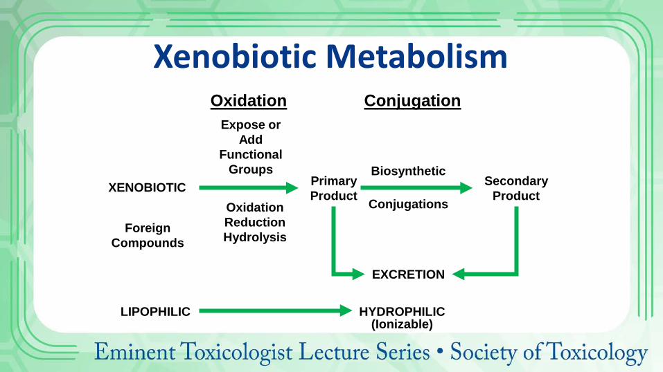
Oxidation Conjugation
XENOBIOTIC
LIPOPHILIC
EXCRETION
HYDROPHILIC (Ionizable)
Expose or Add
Functional Groups
Oxidation Reduction Hydrolysis
Primary Product
Secondary Product
Biosynthetic
Conjugations
Foreign Compounds
Xenobiotic Metabolism
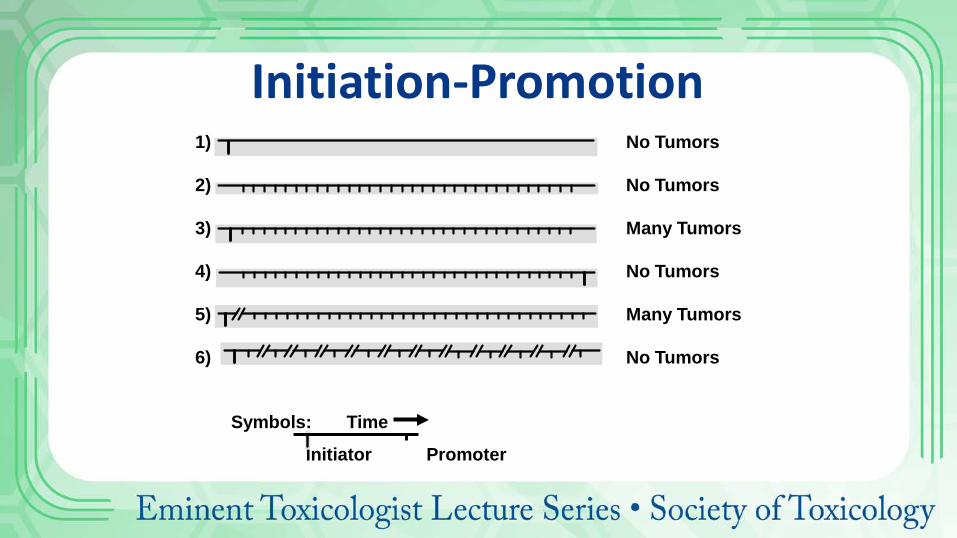
1) No Tumors
2) No Tumors
3) Many Tumors
4) No Tumors
5) Many Tumors
6) No Tumors
Symbols: Time
Initiator Promoter
Initiation-Promotion
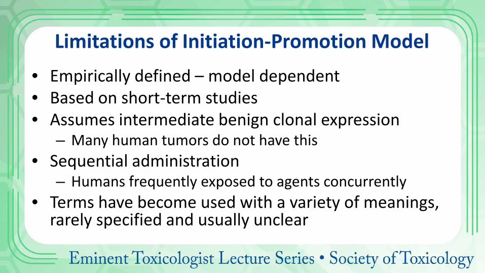
Limitations of Initiation-Promotion Model • Empirically defined – model dependent • Based on short-term studies • Assumes intermediate benign clonal expression
– Many human tumors do not have this • Sequential administration
– Humans frequently exposed to agents concurrently • Terms have become used with a variety of meanings,
rarely specified and usually unclear
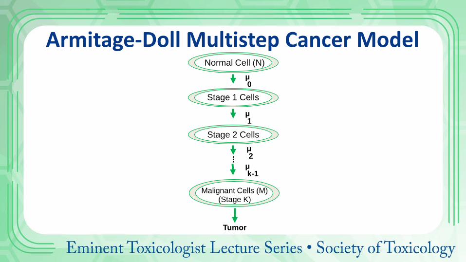
Armitage-Doll Multistep Cancer Model
. . .
μ 0
μ 1
μ 2 μ k-1
Tumor
Normal Cell (N)
Stage 1 Cells
Stage 2 Cells
Malignant Cells (M) (Stage K)
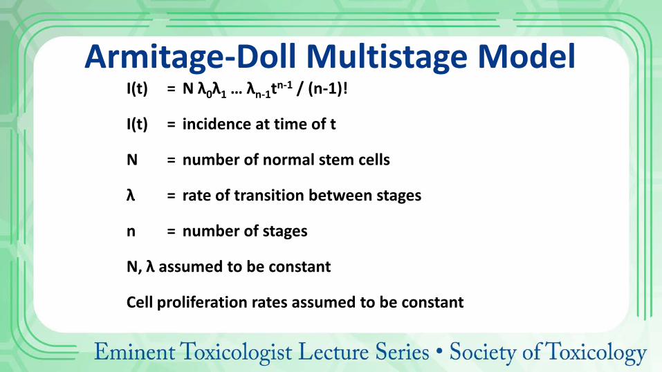
Armitage-Doll Multistage Model I(t) = N λ0λ1 … λn-1tn-1 / (n-1)!
I(t) = incidence at time of t
N = number of normal stem cells
λ = rate of transition between stages
n = number of stages
N, λ assumed to be constant
Cell proliferation rates assumed to be constant
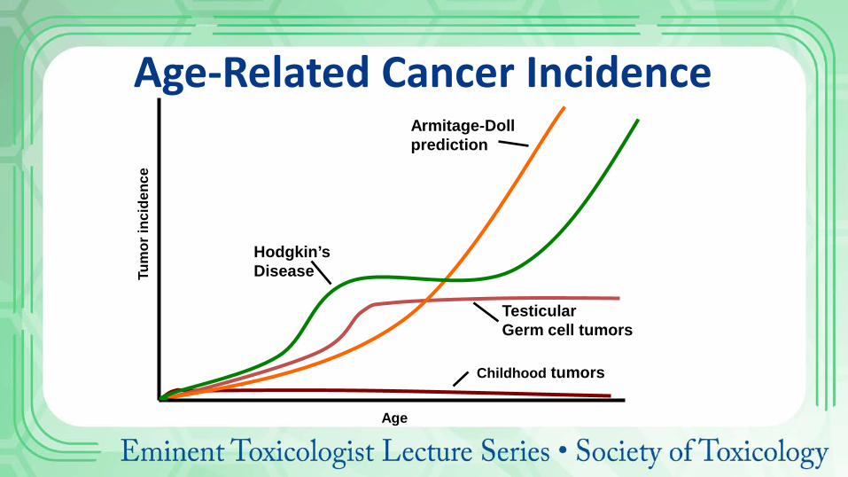
Childhood tumors
Testicular Germ cell tumors
Armitage-Doll prediction
Hodgkin’s Disease Tu
mor
inci
denc
e
Age
Age-Related Cancer Incidence
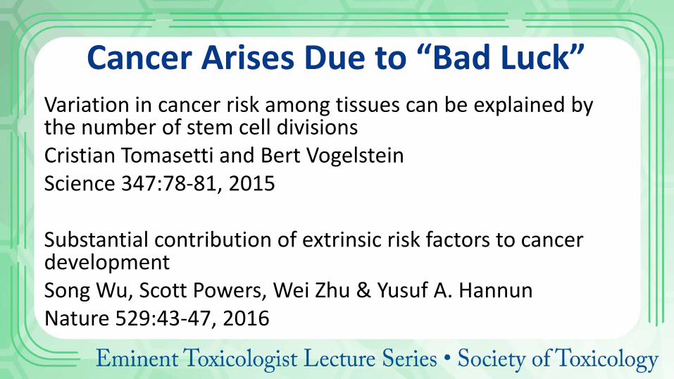
Cancer Arises Due to “Bad Luck” Variation in cancer risk among tissues can be explained by the number of stem cell divisions Cristian Tomasetti and Bert Vogelstein Science 347:78-81, 2015 Substantial contribution of extrinsic risk factors to cancer development Song Wu, Scott Powers, Wei Zhu & Yusuf A. Hannun Nature 529:43-47, 2016
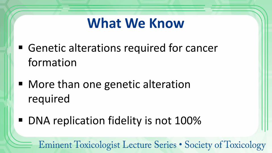
What We Know Genetic alterations required for cancer
formation
More than one genetic alteration required
DNA replication fidelity is not 100%
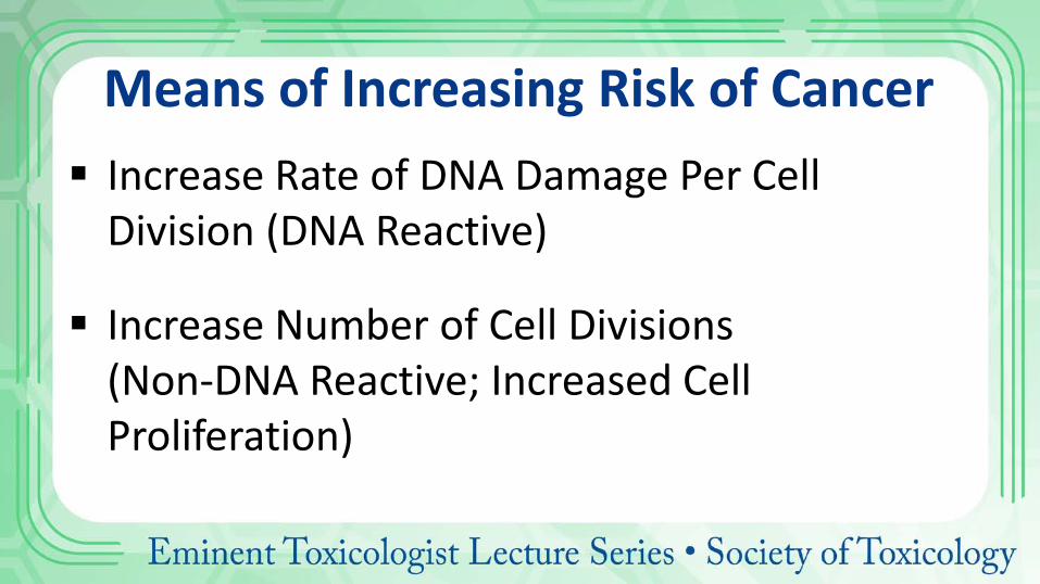
Means of Increasing Risk of Cancer Increase Rate of DNA Damage Per Cell
Division (DNA Reactive)
Increase Number of Cell Divisions (Non-DNA Reactive; Increased Cell Proliferation)
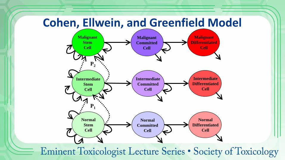
Cohen, Ellwein, and Greenfield Model Malignant
Differentiated Cell
Malignant Committed
Cell
Intermediate Differentiated
Cell
Intermediate Committed
Cell
Normal Stem Cell
Normal Differentiated
Cell
Normal Committed
Cell
Intermediate Stem Cell
P1
Malignant Stem Cell
P2
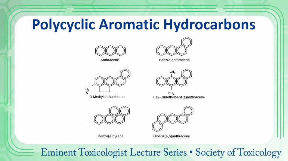
Polycyclic Aromatic Hydrocarbons
Anthracene Benz(a)anthracene
3-Methylcholanthrene 7,12-Dimethylbenz(a)anthracene
Benzo(a)pyrene Dibenz(a,h)anthracene
H3C
CH3
CH3

Aromatic and Heterocyclic Aromatic Amines
2-Naphthylamine 1-Naphthylamine
Benzidine (4,4’-diaminobiphenyl)
2-Aminoflourene
6-Aminochrysene NH2
NH2
NH2
NH2
H2 N
o-Toluidine
NH2
OCH3
H3C
p-Cresidine 4-Aminostilbene
NH2
6-Aminochrysene NH2
NH2
OCH3
H3C
p-Cresidine
NHCOCH3
OCH2CH3 Phenacetin
NH2
4-Aminobiphenyl
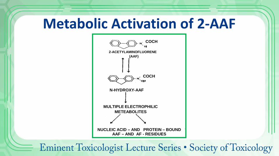
2-ACETYLAMINOFLUORENE (AAF)
N-HYDROXY-AAF
MULTIPLE ELECTROPHILIC METEABOLITES
NUCLEIC ACID – AND PROTEIN – BOUND AAF – AND AF - RESIDUES
COCH3
COCH3
Metabolic Activation of 2-AAF
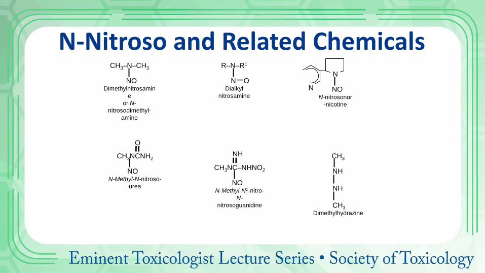
N-Nitroso and Related Chemicals CH3–N–CH3
NO
Dimethylnitrosamine
or N-nitrosodimethyl-
amine
R–N–R1
N O Dialkyl
nitrosamine N
N
NO N-nitrosonor
-nicotine
CH3NC–NHNO2
NO N-Methyl-N1-nitro-
N- nitrosoguanidine
NH CH3 NH NH CH3 Dimethylhydrazine
CH3NCNH2
NO N-Methyl-N-nitroso-
urea
O

Nitrate Nitrite
Secondary Amine
Nitrosamine H+
Endogenous Formation of N-Nitrosamines
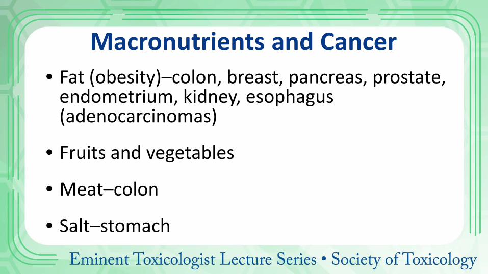
Macronutrients and Cancer • Fat (obesity)–colon, breast, pancreas, prostate,
endometrium, kidney, esophagus (adenocarcinomas)
• Fruits and vegetables
• Meat–colon
• Salt–stomach
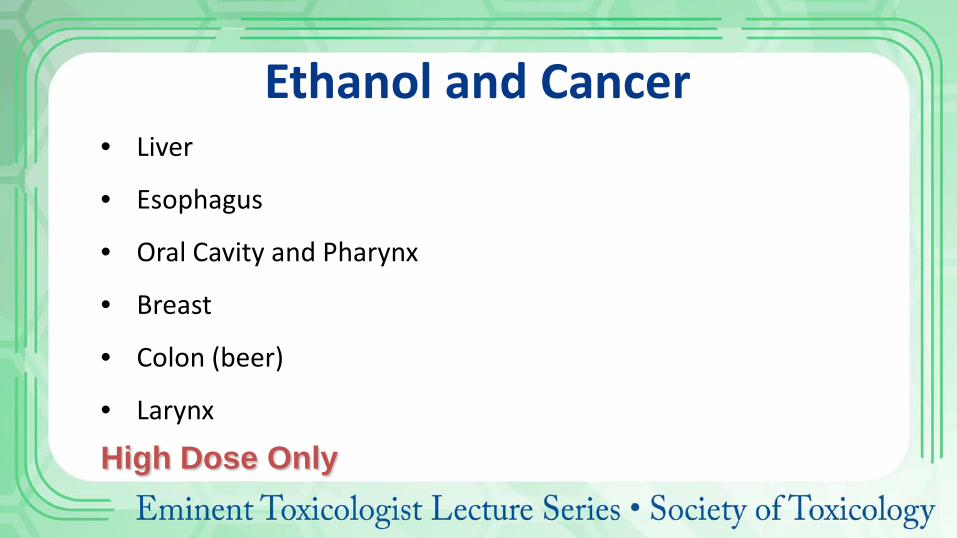
Ethanol and Cancer • Liver
• Esophagus
• Oral Cavity and Pharynx
• Breast
• Colon (beer)
• Larynx
High Dose Only
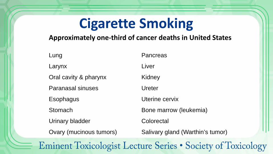
Cigarette Smoking Approximately one-third of cancer deaths in United States
Lung
Larynx
Oral cavity & pharynx
Paranasal sinuses
Esophagus
Stomach
Urinary bladder
Ovary (mucinous tumors)
Pancreas
Liver
Kidney
Ureter
Uterine cervix
Bone marrow (leukemia)
Colorectal
Salivary gland (Warthin’s tumor)
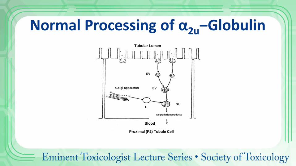
Normal Processing of α2u‒Globulin Tubular Lumen
Blood
Proximal (P2) Tubule Cell
Golgi apparatus
SL SL
Degradation products
EV
EV
L SL
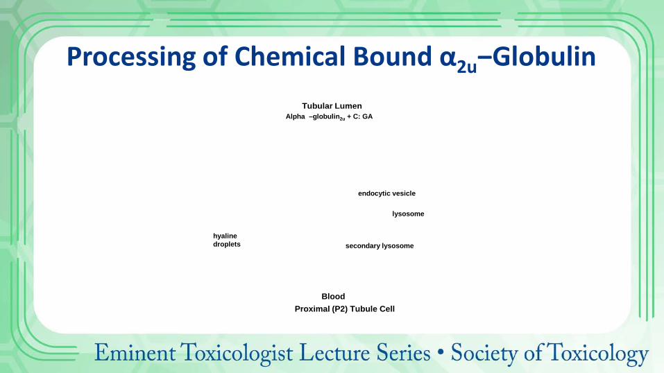
Blood Proximal (P2) Tubule Cell
Tubular Lumen Alpha –globulin2u + C: GA
hyaline droplets
secondary lysosome
lysosome
endocytic vesicle
Processing of Chemical Bound α2u‒Globulin
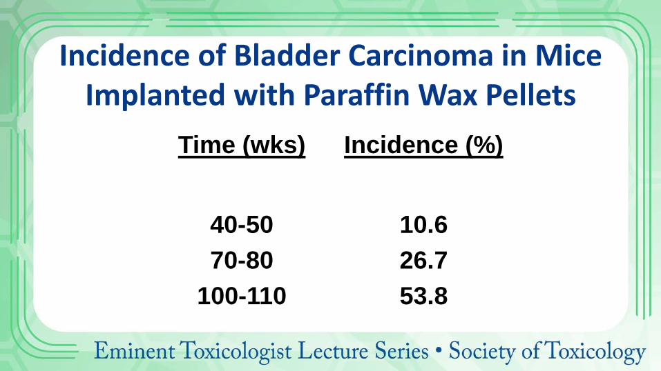
Incidence of Bladder Carcinoma in Mice Implanted with Paraffin Wax Pellets
Time (wks) Incidence (%)
40-50 70-80
100-110
10.6 26.7 53.8
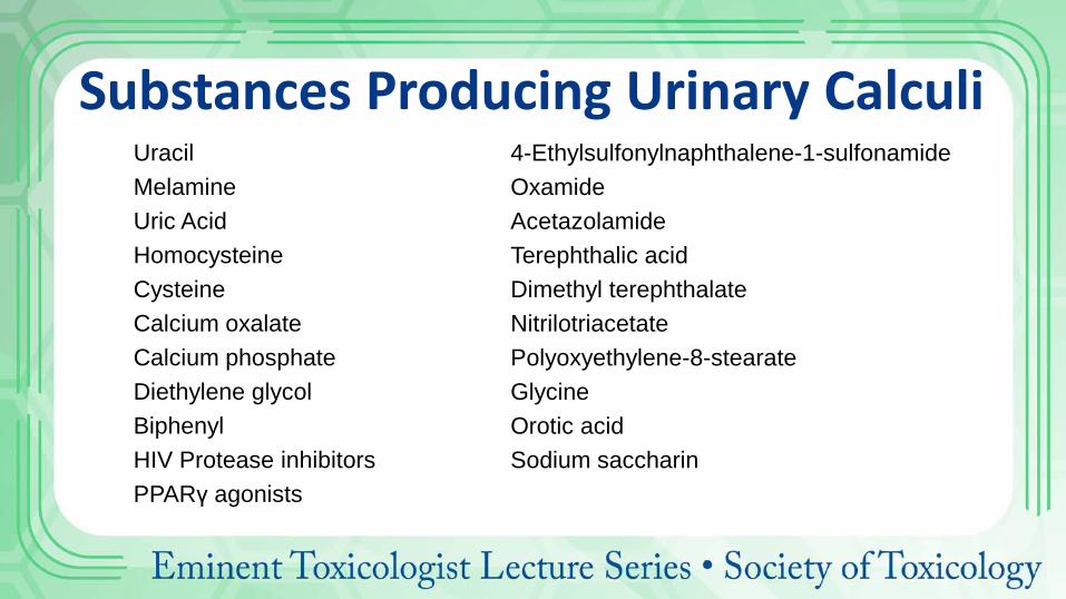
Substances Producing Urinary Calculi Uracil Melamine Uric Acid Homocysteine Cysteine Calcium oxalate Calcium phosphate Diethylene glycol Biphenyl HIV Protease inhibitors PPARγ agonists
4-Ethylsulfonylnaphthalene-1-sulfonamide Oxamide Acetazolamide Terephthalic acid Dimethyl terephthalate Nitrilotriacetate Polyoxyethylene-8-stearate Glycine Orotic acid Sodium saccharin
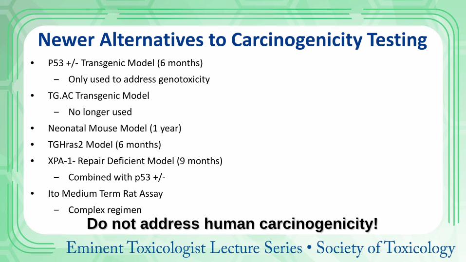
Newer Alternatives to Carcinogenicity Testing • P53 +/- Transgenic Model (6 months)
‒ Only used to address genotoxicity • TG.AC Transgenic Model
‒ No longer used • Neonatal Mouse Model (1 year) • TGHras2 Model (6 months) • XPA-1- Repair Deficient Model (9 months)
‒ Combined with p53 +/- • Ito Medium Term Rat Assay
‒ Complex regimen
Do not address human carcinogenicity!
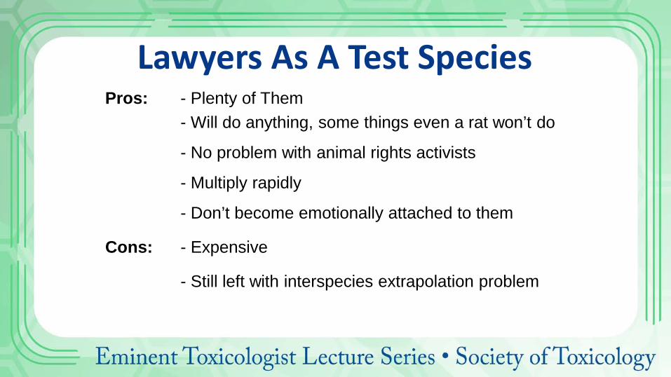
Lawyers As A Test Species Pros: - Plenty of Them - Will do anything, some things even a rat won’t do
- No problem with animal rights activists
- Multiply rapidly
- Don’t become emotionally attached to them
Cons: - Expensive
- Still left with interspecies extrapolation problem
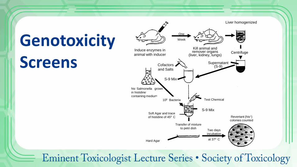
Genotoxicity Screens
Induce enzymes in animal with inducer
Kill animal and remover organs
(liver, kidney, lungs)
Cofactors and Salts
Supernatant (S-9)
Liver homogenized
S-9 Mix
his- Salmonella grown in histidine- containing medium
One
Week
Centrifuge
10β Bacteria Test Chemical
Soft Agar and trace of histidine of 450 C
S-9 Mix
Transfer of mixture to petri dish Two days
incubation
at 370 C
Revertant (his+) colonies counted
Hard Agar
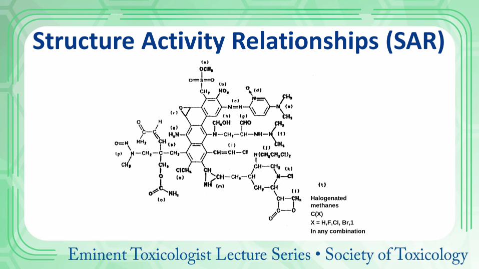
Structure Activity Relationships (SAR)
Halogenated methanes C(X)
X = H,F,CI, Br,1 In any combination
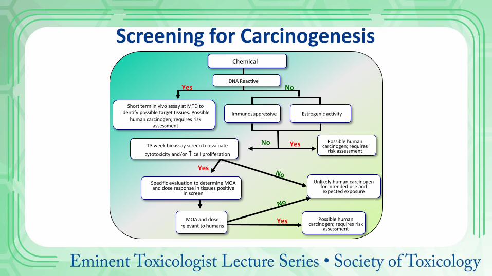
Chemical
Short term in vivo assay at MTD to identify possible target tissues. Possible
human carcinogen; requires risk assessment
Immunosuppressive Estrogenic activity
Yes Possible human carcinogen; requires
risk assessment
Specific evaluation to determine MOA and dose response in tissues positive
in screen
MOA and dose relevant to humans
Yes
Yes No DNA Reactive
Yes Possible human carcinogen; requires risk
assessment
Unlikely human carcinogen for intended use and expected exposure
Screening for Carcinogenesis
13 week bioassay screen to evaluate
cytotoxicity and/or cell proliferation
No
MODELS: ALL ARE WRONG, SOME ARE USEFUL.
GEORGE BOX
Basic Assumptions of Use of Bioassays for Human Risk Assessment
1.Carcinogenic effects at high doses will also occur at low doses (dose extrapolation).
2.Chemicals that cause cancer in rodents will cause cancer in humans (species extrapolation)
UNDERSTANDING COMPUTER TECHNOLOGY
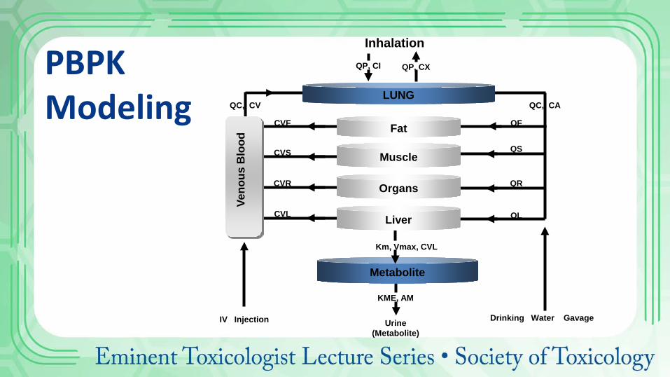
PBPK Modeling
Km, Vmax, CVL
CVL
CVR
CVS
CVF
QC, CV QC, CA
QF
QS
QR
QL
KME, AM
Urine (Metabolite)
Metabolite
LUNG
Organs
Muscle
Fat
Liver
Veno
us B
lood
IV Injection Drinking Water Gavage
Inhalation QP, CI QP, CX
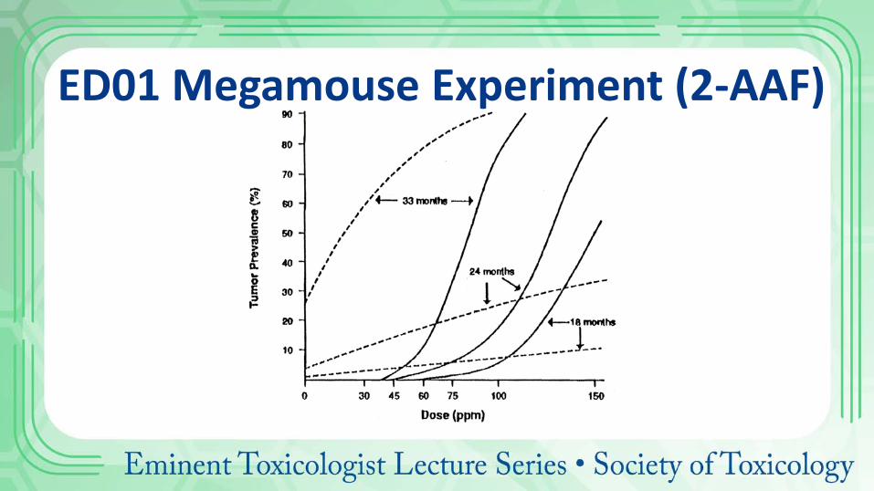
ED01 Megamouse Experiment (2-AAF)
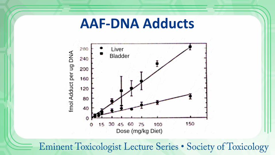
Liver Bladder
Dose (mg/kg Diet)
fmol
Add
uct p
er u
g D
NA
AAF-DNA Adducts
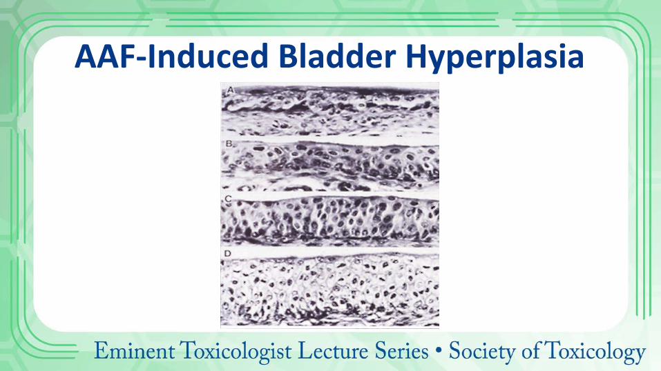
AAF-Induced Bladder Hyperplasia
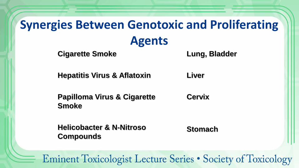
Synergies Between Genotoxic and Proliferating Agents
Cigarette Smoke Hepatitis Virus & Aflatoxin Papilloma Virus & Cigarette Smoke Helicobacter & N-Nitroso Compounds
Lung, Bladder Liver Cervix Stomach

Factors Influencing Carcinogenesis
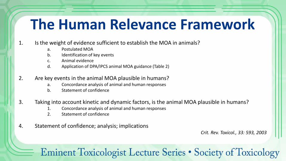
The Human Relevance Framework 1. Is the weight of evidence sufficient to establish the MOA in animals?
a. Postulated MOA b. Identification of key events c. Animal evidence d. Application of DPA/IPCS animal MOA guidance (Table 2)
2. Are key events in the animal MOA plausible in humans?
a. Concordance analysis of animal and human responses b. Statement of confidence
3. Taking into account kinetic and dynamic factors, is the animal MOA plausible in humans?
1. Concordance analysis of animal and human responses 2. Statement of confidence
4. Statement of confidence; analysis; implications
Crit. Rev. Toxicol., 33: 593, 2003
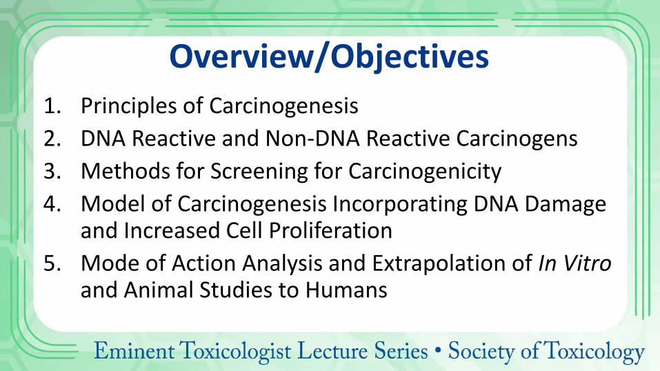
Overview/Objectives 1. Principles of Carcinogenesis 2. DNA Reactive and Non-DNA Reactive Carcinogens 3. Methods for Screening for Carcinogenicity 4. Model of Carcinogenesis Incorporating DNA Damage
and Increased Cell Proliferation 5. Mode of Action Analysis and Extrapolation of In Vitro
and Animal Studies to Humans
References 1. Cohen, SM. Evaluation of Possible Carcinogenic Risk to Humans Based on Liver
Tumors in Rodent Assays: The Two-Year Bioassay is No Longer Necessary. Toxicol. Pathol., 38: 487-501, 2010.
2. Cohen, SM and Arnold, LL. Chemical Carcinogenesis. Toxciol. Sci., 12 (Suppl. 1): 576-592, 2011.
3. Cohen, SM and Ellwein, LB. Cell Proliferation in Carcinogenesis. Science, 249: 1007-1011, 1990.
4. Cohen, SM and Ellwein, LB. Genetic Errors, Cell Proliferation and Carcinogenesis. Cancer Res., 51: 6493-6505, 1991.
5. Boobis, AR, et al. IPCS Framework for Analyzing the Relevance of a Cancer Mode of Action for Humans. Crit. Rev. Toxicol., 36: 781-792, 2006.
6. Embry, MR, et al. Risk Assessment in the 21st Century: Roadmap and Matrix. Crit. Rev. Toxicol., 44 (Suppl. 3): 6-16, 2014.
Chemical Carcinogenesis Samuel M. Cohen, MD, PhD
University of Nebraska Medical Center Department of Pathology and Microbiology
Omaha, NE
Recommended
![Sarcoidosis-Associated Hepatocellular Carcinoma · in the carcinogenesis of sarcoidosis-associated HCC, as in the carcinogenesis associated with viral hepatitis [13]. Actually, cases](https://img.pdfslide.tips/doc/110x75/5e21d512d044f5667706527f/sarcoidosis-associated-hepatocellular-in-the-carcinogenesis-of-sarcoidosis-associated.jpg)