ORIGINAL ARTICLE
Comparison study of single and concurrent administrationsof carbapenem, new quinolone, and macrolide against in vitronontypeable Haemophilus influenzae mature biofilms
Yusaku Uemura • Liang Qin • Kenji Gotoh •
Keisuke Ohta • Kei-ichiro Nakamura •
Hiroshi Watanabe
Received: 7 February 2013 / Accepted: 29 March 2013 / Published online: 20 April 2013
� Japanese Society of Chemotherapy and The Japanese Association for Infectious Diseases 2013
Abstract Nontypeable Haemophilus influenzae (NTHi) is
an opportunistic pathogen and a common cause of otitis
media in children, chronic bronchitis, and pneumonia in
patients with chronic obstructive pulmonary disease. Many
studies have reported that NTHi is capable of producing
biofilms, which may be one of the important factors
involved in chronic diseases and accelerating antimicrobial
resistance. Unfortunately, there is still no consensus about
the elimination of biofilms. In this study, concurrent
administrations of levofloxacin (LVFX)-imipenem (IPM)
and clarithromycin (CAM)-IPM, as well as the single
administration of IPM, LVFX, and CAM, were performed
to treat the mature biofilms produced by NTHi, respec-
tively. Biofilm inhibition was quantified using microtiter
biofilm assay (MBA), and relative biomass was calculated
as the ratio compared to that of untreated control biofilms.
The relative biomasses of biofilms treated with IPM,
LVFX-IPM, and CAM-IPM against a b-lactamase-negative
ampicillin-resistant strain was 1.10, 0.08, and 0.13 at 19
minimum inhibitory concentration (MIC), 0.90, 0.05, and
0.07 at 109 MIC, and 0.80, 0.06, and 0.07 at 1009 MIC,
respectively. Biofilms were also visually observed by
scanning electron microscopy, and a focused ion-beam
system showed that high concentrations of combined
administration strongly inhibited the biofilms, which was
consistent with the results of MBA. Our data demonstrated
the antibiofilm effect of concurrent administration against
mature NTHi biofilms, which indicated a rationale for the
potential use of concurrent administrations in diseases
involving chronic NTHi biofilms.
Keywords Biofilm � BLNAR � Concurrentadministration � Haemophilus influenzae
Introduction
Haemophilus influenzae is a fastidious gram-negative and
pleomorphic bacterium that initially colonizes the human
nasopharynx. Nontypeable H. influenzae (NTHi) is repor-
ted to cause otitis media, sinusitis in children, and is
associated with community-acquired pneumonia (CAP)
and acute exacerbations of chronic bronchitis [1]. Ampi-
cillin has long been used as the drug of first choice for the
treatment of infections caused by H. influenzae. Although
TEM-1 and ROB-1-type b-lactamase-positive ampicillin-
resistant (BLPAR) strains are responsible for almost all
isolates with decreased susceptibility to ampicillin, b-lac-tamase-nonproducing ampicillin-resistant (BLNAR) strains
have also increased in some countries recently [2–5].
Therefore, otitis media, paranasal sinusitis, and lower
respiratory tract infection caused by H. influenzae have
become more and more difficult to be cured with oral
antibiotics therapy [6].
Also, it has been reported that NTHi produces biofilms
that are associated with a variety of diseases such as
chronic obstructive pulmonary disease (COPD), cystic
fibrosis (CF), and middle ear infections [7, 8]. As is well
This study was presented in 110th American Society for
Microbiology General Meeting, San Diego, CA, USA, 26 May 2010.
Y. Uemura � L. Qin (&) � K. Gotoh � H. Watanabe
Division of Infectious Diseases, Department of Infectious
Medicine, Kurume University School of Medicine,
67 Asahi-Machi, Kurume, Fukuoka 830-0011, Japan
e-mail: [email protected]; [email protected]
K. Ohta � K. Nakamura
Division of Microscopic and Developmental Anatomy,
Department of Anatomy, Kurume University School
of Medicine, Fukuoka, Japan
123
J Infect Chemother (2013) 19:902–908
DOI 10.1007/s10156-013-0598-5
known, biofilms are defined as communities of microor-
ganisms that attach to a surface and are enveloped in a
hydrated polymeric matrix of their own synthesis [9, 10],
and bacteria within biofilms are more resistant to antibi-
otics than in the planktonic state [11]. Although many
studies have discussed biofilm treatment with single or
concurrent antibiotics, such as quinolones, macrolides,
aminoglycoside, penicillin, and cephems [12–16], there is
still no consensus about the treatment of biofilm diseases.
Here we evaluated the combinatory efficacy of a carbape-
nem with a new quinolone and a macrolide against NTHi
biofilms compared to each single administration.
Methods
Bacteria strains and culture conditions
Two H. influenzae strains were isolated from different
Japanese patients with respiratory tract infections. These
strains were reconstituted from frozen stocks and propa-
gated on chocolate agar or brain heart infusion (Becton-
Dickinson, USA) supplemented with 10 lg/ml hemin
(Sigma, USA) and b-nicotinamide adenine nucleotide (b-NAD) (Sigma) (sBHI) 5 % CO2 at 37 �C [17], and b-lac-tamase production was detected by means of a disc
impregnated with nitrocefin (Becton–Dickinson, USA).
Serotyping was performed by slide agglutination with
antisera purchased from Difco Laboratories (Detroit, MI,
USA).
Antimicrobial susceptibility test
Minimum inhibitory concentrations (MICs) were deter-
mined using the broth dilution method according to
guidelines from the Clinical and Laboratory Standards
Institute [18]. NTHi strains were tested against four anti-
biotics: ampicillin (ABPC) (Wako, Tokyo, Japan); imi-
penem (IPM) [Cmax, 40.1 (lg/ml; healthy adult, 0.5 g,
intravenous drip, 30 min) (Merck, USA); clarithromycin
(CAM) [Cmax, 1.16 (lg/ml; healthy adult, 200 mg, oral
administration (p.o.) (Abbot Japan, Japan)]; levofloxacin
(LVFX) [Cmax, 2.04 ± 0.21 (lg/ml; healthy adult, 200 mg,
p.o.) (Daiichi Sankyo, Japan)].
Genetic identification of antimicrobial
resistance-related genes
Polymerase chain reaction (PCR) was performed to iden-
tify antimicrobial resistance-related genes by using mixed
primers following the manufacturer’s manual (Wakunaga
Pharmaceutical, Hiroshima, Japan), as described previ-
ously [19, 20]. Briefly, we used P6 primers to amplify the
P6 gene that encodes the P6 membrane protein specific for
H. influenzae (198 bp); TEM-I primers to amplify a part of
the blaTEM-1 gene (458 bp); PBP3-S primers to identify an
Asn526 ? Lys amino acid substitution in the ftsI gene
(551 bp); and PBP3-BLN primers to identify an
Asn526 ? Lys and Ser385 ? Thr amino acid substitution
in the ftsI gene (465 bp).
Quantification of biofilms
NTHi biofilms were investigated by a modified microtiter
biofilm assay (MBA) [21]. Bacteria cells were suspended
in sBHI approximately at 0.1 OD490, and a 200-ll aliquotwas inoculated into a well of a 96-well microplate. Biofilm
formation was confirmed after 48 h culture, medium was
changed, and then 200 ll fresh sBHI with one of the fol-
lowing antibiotics (IPM, CAM, LVFX, or combined
administration of LVFX-IPM and CAM-IPM) was inocu-
lated into each tested well with a dose-dependent MIC
concentration (0.019MIC, 0.19MIC, 19MIC, 109MIC,
1009 MIC). After 2 h exposure to the antibiotics, all tested
wells were washed and refilled with 200 ll sBHI for con-tinuous culture. Antibiotics administration was repeated
every 12 h for a total of four times. After the last antimi-
crobial exposure, biofilms were stained with 1 % freshly
adjusted crystal violet (Merck, USA) at room temperature
for 15 min and vigorously washed with water three times.
After extraction with 230 ll 95 % ethanol, biofilms were
measured at OD595 with a microplate reader. Additionally,
intact biofilms (without any antibiotics exposure) were
measured as the baseline data. All studies were tested in
triplicate, and the average ± SD of each experiment was
calculated.
Preparations and conditions of scanning electron
microscopy
The KL-1 strain was suspended in sBHI at approximately
0.1 OD490. A 1-ml aliquot of bacteria solution was inocu-
lated into a flat bottle with glass coverslips inside and
statically cultured for 48 h at 37 �C with 5 % CO2. After
biofilm formation was confirmed, 1 ml fresh sBHI con-
taining one of the following antibiotics (IPM, CAM,
LVFX, combined administration of LVFX-IPM and CAM-
IPM) was inoculated into each tested bottle with a dose-
dependent MIC concentration (0.19 MIC, 19 MIC, 109
MIC). After 2 h exposure to the antibiotics, all tested
bottles were washed and refilled with 1 ml sBHI for con-
tinuous culture. Antibiotics administration was repeated
every 12 h for a total four times.
The glass coverslips were taken from the bottles and
incubated in 2.5 % glutaraldehyde for 1 h at room tem-
perature. After washing with 7.5 % sucrose (1 h), fixation
J Infect Chemother (2013) 19:902–908 903
123
was performed by incubation in 1 % OsO4 (2 % OsO4 1:1
7.5 % sucrose) for 1 h (4 �C). Specimens were continu-
ously dehydrated by critical-point drying, which the water
in the cells is replaced with graded ethanol from 50 % to
100 %, with use of 100 % t-butyl alcohol for freeze drying.
Samples were coated with gold in an ion-sputter coater [21,
22]. The specimens were observed with a scanning electron
microscopy (SEM) (S-800; Hitachi, Tokyo, Japan), as well
as a focused ion-beam system (FIB) (Quanta 3D; FEI,
USA) [23].
Statistical evaluation
Data were analyzed using the Student’s paired t test. A
P value\ 0.05 was considered statistically significant.
Results
Characteristics of the strains
According to the characteristics of resistant genes, KL-1
was confirmed as BLNAR, and KL-2 was a beta-lactamase-
nonproducing ampicillin-susceptible (BLNAS) strain: these
were verified as NTHi strains and without b-lactamase
production. The MICs values (lg/ml) for ABPC, IPM,
LVFX, and CAM against KL-1 were 16, 0.5, 0.032, and 8,
and those against KL-2 were 0.25, 1, 0.032, and 8,
respectively.
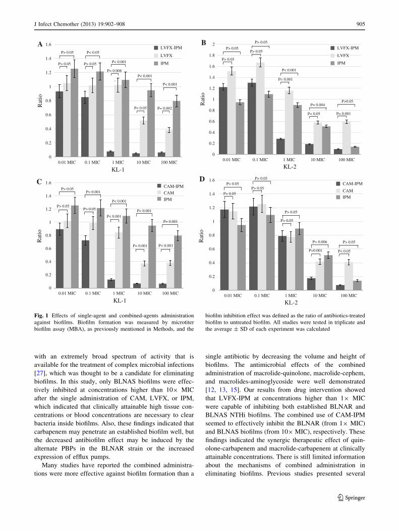
Effect of single and combination treatments against
biofilms
Biofilms were treated with IPM, LVFX, and CAM, as well
as the concurrent administration of LVFX-IPM and CAM-
IPM, respectively. Biofilm formation was measured by
MBA as previously mentioned in the Methods, and the
biofilm inhibition effect was defined as the ratio of anti-
biotics-treated biofilm to untreated biofilm. When effects of
single-agent administrations against the KL-1 biofilms
were investigated, LVFX and CAM showed a significant
biofilm inhibition effect compared to IPM from 109 MIC
administration. For KL-2, single-agent administration of
IPM, LVFX, and CAM showed high biofilm inhibition
effect from 109 MIC, respectively. IPM administration at
1009 MIC seems to be more significant to inhibit the
biofilm formation compared to LVFX and CAM
(P = 0.001 and P = 0.002). On the other hand, adminis-
tration of IPM, LVFX-IPM, and CAM-IPM against KL-1
biofilms showed a high biofilm inhibition effect with
results of 1.10, 0.08, and 0.13 at 19 MIC, 0.90, 0.05, and
0.07 at 109 MIC, and 0.80, 0.06, and 0.07 at 1009 MIC,
respectively. IPM-LVFX showed significant biofilm
inhibition effect against biofilms produced by both stains
from 19 MIC (Fig. 1a, b). CAM-LVFX also obtained a
similar effect against KL-1 biofilms (Fig. 1c); however,
there was no difference among single-agent administration
of CAM and IPM and combined administration of CAM-
IPM against KL-2 biofilms at 19 MIC (Fig. 1d).
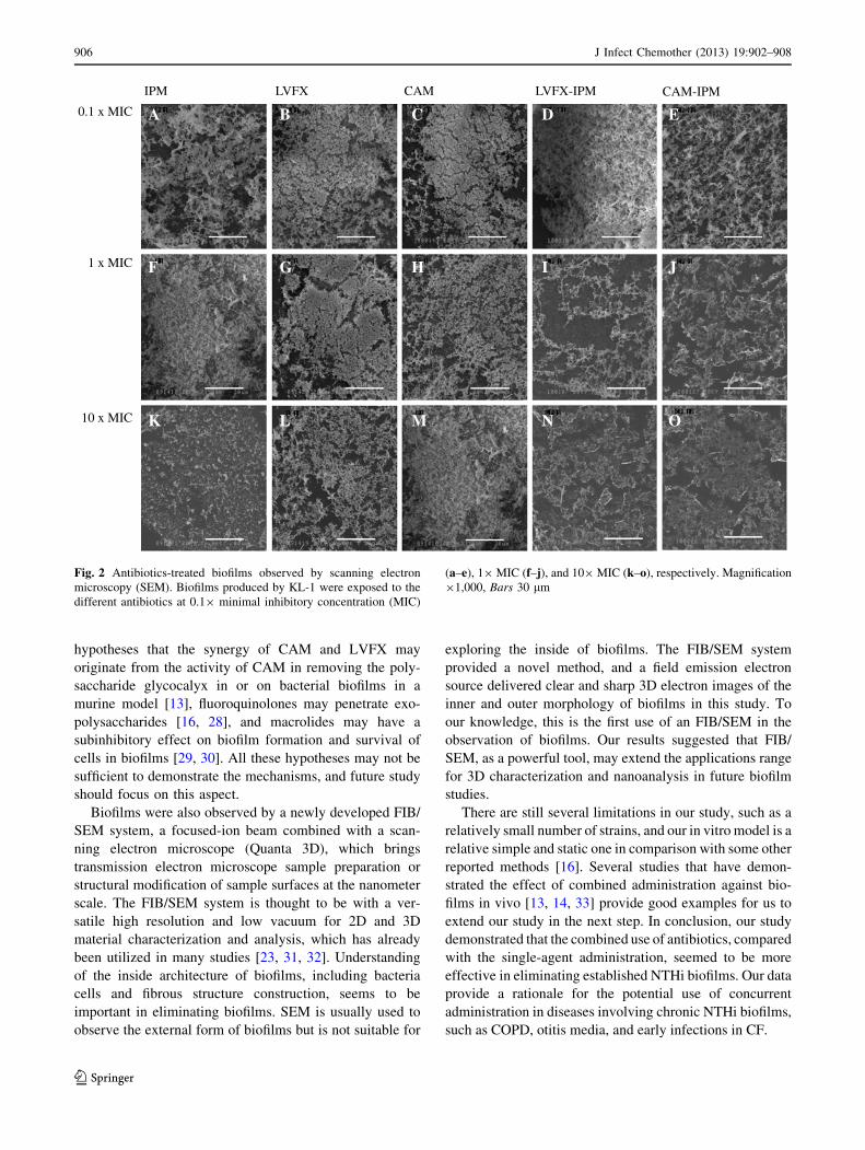
Architectures of biofilms
Biofilms of KL-1 were treated with different antibiotics
and visually observed by SEM. A large of amount of
biofilm materials was evident after exposure to the single-
agent administration of IPM, LVFX, and CAM at 0.19 and
19 MIC, as well as the combined administrations of
LVFX-IPM and CAM-IPM at 0.1 MIC (Fig. 2). Strands of
fibrin with many fewer bacteria cells were obviously
present in the biofilms exposed to the combined-agent
administration from 19 MIC (Fig. 2i, j, n, o). Fewer bac-
teria cells were seen in a fibrous matrix of biofilms treated
with IPM or LVFX compared to CAM at 109 MIC
(Fig. 2k–m).
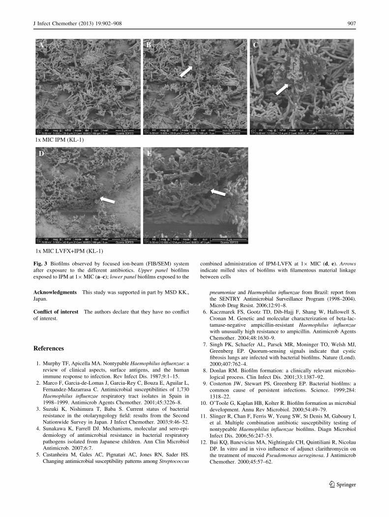
Biofilms treated with IPM and LVFX-IPM at 19 MIC
were also observed by the FIB/SEM system (Fig. 3). The
dense matrix contributing to the outer shapes of biofilms
were observed before milling the biofilms (Fig. 3a), and
after biofilms were sliced at the same site, the strands of
materials from interconnected cells were detected inside
the biofilms (Fig. 3b). Higher magnification showed fila-
mentous material links bacteria cells to each other
(Fig. 3c). Clusters of bacteria cells associated with the
flocculent materials on the surface were observed at low
magnification (Fig. 3d). At higher magnification, biofilms
were seen to be composed of large numbers of cells inti-
mately associated with a fibrous material resembling fibrin,
and cells of different sizes can be seen (Fig. 3e).
Discussion
Biofilms have become the major concern for clinicians in
the treatment of infectious diseases. It was reported that
bacteria in a biofilm can survive antibiotic concentrations
up to 1,000 fold higher than the same bacteria in a
planktonic state [24]. Some studies reported that azithro-
mycin (AZM) administered alone had no effect when
Pseudomonas aeruginosa biofilms became established, and
either CAM or LVFX alone had no statistical effect in a
biofilm-associated murine model [13, 25], but the use of
subinhibitory concentrations of AZM seemed to signifi-
cantly decrease biomass and maximal thickness in both
forming and established NTHi biofilms [26]. These results
indicated the efficacy of single antibiotic treatment for
biofilms is controversial. IPM became the first carbapenem
904 J Infect Chemother (2013) 19:902–908
123
with an extremely broad spectrum of activity that is
available for the treatment of complex microbial infections
[27], which was thought to be a candidate for eliminating
biofilms. In this study, only BLNAS biofilms were effec-
tively inhibited at concentrations higher than 109 MIC
after the single administration of CAM, LVFX, or IPM,
which indicated that clinically attainable high tissue con-
centrations or blood concentrations are necessary to clear
bacteria inside biofilms. Also, these findings indicated that
carbapenem may penetrate an established biofilm well, but
the decreased antibiofilm effect may be induced by the
alternate PBPs in the BLNAR strain or the increased
expression of efflux pumps.
Many studies have reported the combined administra-
tions were more effective against biofilm formation than a
single antibiotic by decreasing the volume and height of
biofilms. The antimicrobial effects of the combined
administration of macrolide-quinolone, macrolide-cephem,
and macrolides-aminoglycoside were well demonstrated
[12, 13, 15]. Our results from drug intervention showed
that LVFX-IPM at concentrations higher than 19 MIC
were capable of inhibiting both established BLNAR and
BLNAS NTHi biofilms. The combined use of CAM-IPM
seemed to effectively inhibit the BLNAR (from 19 MIC)
and BLNAS biofilms (from 109 MIC), respectively. These
findings indicated the synergic therapeutic effect of quin-
olone-carbapenem and macrolide-carbapenem at clinically
attainable concentrations. There is still limited information
about the mechanisms of combined administration in
eliminating biofilms. Previous studies presented several
0
0.2
0.4
0.6
0.8
1
1.2
1.4
1.6
0.01 MIC 0.1 MIC 1 MIC 10 MIC 100 MIC
LVFX-IPM
LVFX
IPM
0
0.2
0.4
0.6
0.8
1
1.2
1.4
1.6
1.8
2
0.01 MIC 0.1 MIC 1 MIC 10 MIC 100 MIC
LVFX-IPM
LVFX
IPM
0
0.2
0.4
0.6
0.8
1
1.2
1.4
1.6
0.01 MIC 0.1 MIC 1 MIC 10 MIC 100 MIC
CAM-IPM
CAM
IPM
0
0.2
0.4
0.6
0.8
1
1.2
1.4
1.6
0.01 MIC 0.1 MIC 1 MIC 10 MIC 100 MIC
CAM-IPM
CAM
IPM
KL-1 KL-2
A B
C D
Rat
ioR
atio
Rat
ioR
atio
KL-1 KL-2
P> 0.05
P> 0.05
P< 0.05
P> 0.05
P< 0.05
P< 0.001
P= 0.002
P= 0.008P< 0.001
P< 0.001
P> 0.05
P> 0.05
P> 0.05
P> 0.05
P< 0.05
P>0.05
P= 0.001
P= 0.001
P= 0.004
P< 0.001
P= 0.001 P< 0.001
P< 0.001
P> 0.05P> 0.05
P< 0.001
P< 0.001
P= 0.001P> 0.05
P= 0.001
P> 0.05
P> 0.05
P> 0.05
P> 0.05
P> 0.05
P> 0.05
P= 0.006
P=0.001
P> 0.05
P< 0.05
Fig. 1 Effects of single-agent and combined-agents administration
against biofilms. Biofilm formation was measured by microtiter
biofilm assay (MBA), as previously mentioned in Methods, and the
biofilm inhibition effect was defined as the ratio of antibiotics-treated
biofilm to untreated biofilm. All studies were tested in triplicate and
the average ± SD of each experiment was calculated
J Infect Chemother (2013) 19:902–908 905
123
hypotheses that the synergy of CAM and LVFX may
originate from the activity of CAM in removing the poly-
saccharide glycocalyx in or on bacterial biofilms in a
murine model [13], fluoroquinolones may penetrate exo-
polysaccharides [16, 28], and macrolides may have a
subinhibitory effect on biofilm formation and survival of
cells in biofilms [29, 30]. All these hypotheses may not be
sufficient to demonstrate the mechanisms, and future study
should focus on this aspect.
Biofilms were also observed by a newly developed FIB/
SEM system, a focused-ion beam combined with a scan-
ning electron microscope (Quanta 3D), which brings
transmission electron microscope sample preparation or
structural modification of sample surfaces at the nanometer
scale. The FIB/SEM system is thought to be with a ver-
satile high resolution and low vacuum for 2D and 3D
material characterization and analysis, which has already
been utilized in many studies [23, 31, 32]. Understanding
of the inside architecture of biofilms, including bacteria
cells and fibrous structure construction, seems to be
important in eliminating biofilms. SEM is usually used to
observe the external form of biofilms but is not suitable for
exploring the inside of biofilms. The FIB/SEM system
provided a novel method, and a field emission electron
source delivered clear and sharp 3D electron images of the
inner and outer morphology of biofilms in this study. To
our knowledge, this is the first use of an FIB/SEM in the
observation of biofilms. Our results suggested that FIB/
SEM, as a powerful tool, may extend the applications range
for 3D characterization and nanoanalysis in future biofilm
studies.
There are still several limitations in our study, such as a
relatively small number of strains, and our in vitro model is a
relative simple and static one in comparison with some other
reported methods [16]. Several studies that have demon-
strated the effect of combined administration against bio-
films in vivo [13, 14, 33] provide good examples for us to
extend our study in the next step. In conclusion, our study
demonstrated that the combined use of antibiotics, compared
with the single-agent administration, seemed to be more
effective in eliminating established NTHi biofilms. Our data
provide a rationale for the potential use of concurrent
administration in diseases involving chronic NTHi biofilms,
such as COPD, otitis media, and early infections in CF.
A B C D E
F G H I J1 x MIC
K L M N O
IPM LVFX CAM LVFX-IPM CAM-IPM
10 x MIC
0.1 x MIC
Fig. 2 Antibiotics-treated biofilms observed by scanning electron
microscopy (SEM). Biofilms produced by KL-1 were exposed to the
different antibiotics at 0.19 minimal inhibitory concentration (MIC)
(a–e), 19MIC (f–j), and 109MIC (k–o), respectively. Magnification
91,000, Bars 30 lm
906 J Infect Chemother (2013) 19:902–908
123
Acknowledgments This study was supported in part by MSD KK.,
Japan.
Conflict of interest The authors declare that they have no conflict
of interest.
References
1. Murphy TF, Apicella MA. Nontypable Haemophilus influenzae: a
review of clinical aspects, surface antigens, and the human
immune response to infection. Rev Infect Dis. 1987;9:1–15.
2. Marco F, Garcia-de-Lomas J, Garcia-Rey C, Bouza E, Aguilar L,
Fernandez-Mazarrasa C. Antimicrobial susceptibilities of 1,730
Haemophilus influenzae respiratory tract isolates in Spain in
1998–1999. Antimicrob Agents Chemother. 2001;45:3226–8.
3. Suzuki K, Nishimura T, Baba S. Current status of bacterial
resistance in the otolaryngology field: results from the Second
Nationwide Survey in Japan. J Infect Chemother. 2003;9:46–52.
4. Sunakawa K, Farrell DJ. Mechanisms, molecular and sero-epi-
demiology of antimicrobial resistance in bacterial respiratory
pathogens isolated from Japanese children. Ann Clin Microbiol
Antimicrob. 2007;6:7.
5. Castanheira M, Gales AC, Pignatari AC, Jones RN, Sader HS.
Changing antimicrobial susceptibility patterns among Streptococcus
pneumoniae and Haemophilus influenzae from Brazil: report from
the SENTRY Antimicrobial Surveillance Program (1998–2004).
Microb Drug Resist. 2006;12:91–8.
6. Kaczmarek FS, Gootz TD, Dib-Hajj F, Shang W, Hallowell S,
Cronan M. Genetic and molecular characterization of beta-lac-
tamase-negative ampicillin-resistant Haemophilus influenzae
with unusually high resistance to ampicillin. Antimicrob Agents
Chemother. 2004;48:1630–9.
7. Singh PK, Schaefer AL, Parsek MR, Moninger TO, Welsh MJ,
Greenberg EP. Quorum-sensing signals indicate that cystic
fibrosis lungs are infected with bacterial biofilms. Nature (Lond).
2000;407:762–4.
8. Donlan RM. Biofilm formation: a clinically relevant microbio-
logical process. Clin Infect Dis. 2001;33:1387–92.
9. Costerton JW, Stewart PS, Greenberg EP. Bacterial biofilms: a
common cause of persistent infections. Science. 1999;284:
1318–22.
10. O’Toole G, Kaplan HB, Kolter R. Biofilm formation as microbial
development. Annu Rev Microbiol. 2000;54:49–79.
11. Slinger R, Chan F, Ferris W, Yeung SW, St Denis M, Gaboury I,
et al. Multiple combination antibiotic susceptibility testing of
nontypeable Haemophilus influenzae biofilms. Diagn Microbiol
Infect Dis. 2006;56:247–53.
12. Bui KQ, Banevicius MA, Nightingale CH, Quintiliani R, Nicolau
DP. In vitro and in vivo influence of adjunct clarithromycin on
the treatment of mucoid Pseudomonas aeruginosa. J Antimicrob
Chemother. 2000;45:57–62.
1x MIC IPM (KL-1)
1x MIC LVFX+IPM (KL-1)
A B C
D E
Fig. 3 Biofilms observed by focused ion-beam (FIB/SEM) system
after exposure to the different antibiotics. Upper panel biofilms
exposed to IPM at 19MIC (a–c); lower panel biofilms exposed to the
combined administration of IPM-LVFX at 19 MIC (d, e). Arrowsindicate milled sites of biofilms with filamentous material linkage
between cells
J Infect Chemother (2013) 19:902–908 907
123
13. Yanagihara K, Tomono K, Sawai T, Kuroki M, Kaneko Y, Ohno
H, et al. Combination therapy for chronic Pseudomonas aeru-
ginosa respiratory infection associated with biofilm formation.
J Antimicrob Chemother. 2000;46:69–72.
14. Moreau-Marquis S, O’Toole GA, Stanton BA. Tobramycin and
FDA-approved iron chelators eliminate Pseudomonas aeruginosa
biofilms on cystic fibrosis cells. Am J Respir Cell Mol Biol.
2009;41:305–13.
15. Tre-Hardy M, Nagant C, El Manssouri N, Vanderbist F, Traore
H, Vaneechoutte M, et al. Efficacy of the combination of tobra-
mycin and a macrolide in an in vitro Pseudomonas aeruginosa
mature biofilm model. Antimicrob Agents Chemother. 2010;54:
4409–15.
16. Kaji C, Watanabe K, Apicella MA, Watanabe H. Antimicrobial
effect of fluoroquinolones for the eradication of nontypeable
Haemophilus influenzae isolates within biofilms. Tohoku J Exp
Med. 2008;214:121–8.
17. Anderson P, Peter G, Johnston RB Jr, Wetterlow LH, Smith DH.
Immunization of humans with polyribophosphate, the capsular
antigen of Hemophilus influenzae, type b. J Clin Invest.
1972;51:39–44.
18. CLSI. Methods for dilution antimicrobial susceptibility tests for
bacteria that grow aerobically: approved standard, seventh edi-
tion. M7–A7, vol. 26. Wayne: Clinical and Laboratory Standards
Institute; 2006.
19. Hasegawa K, Chiba N, Kobayashi R, Murayama SY, Iwata S,
Sunakawa K, et al. Rapidly increasing prevalence of beta-lacta-
mase-nonproducing, ampicillin-resistant Haemophilus influenzae
type b in patients with meningitis. Antimicrob Agents Chemo-
ther. 2004;48:1509–14.
20. Gotoh K, Qin L, Watanabe K, Anh DD, Huong Ple T, Anh NT,
et al. Prevalence of Haemophilus influenzae with resistant genes
isolated from young children with acute lower respiratory tract
infections in Nha Trang, Vietnam. J Infect Chemother. 2008;
14:349–53.
21. Kuroki R, Kawakami K, Qin L, Kaji C, Watanabe K, Kimura Y,
et al. Nosocomial bacteremia caused by biofilm-forming Bacillus
cereus and Bacillus thuringiensis. Intern Med. 2009;48:791–6.
22. Edwards JL, Shao JQ, Ault KA, Apicella MA. Neisseria gonor-
rhoeae elicits membrane ruffling and cytoskeletal rearrangements
upon infection of primary human endocervical and ectocervical
cells. Infect Immun. 2000;68:5354–63.
23. Ohta K, Sadayama S, Togo A, Higashi R, Tanoue R, Nakamura
K. Beam deceleration for block-face scanning electron micros-
copy of embedded biological tissue. Micron. 2012;43:612–20.
24. Hoiby N. Understanding bacterial biofilms in patients with cystic
fibrosis: current and innovative approaches to potential therapies.
J Cyst Fibrosis. 2002;1:249–54.
25. Gillis RJ, Iglewski BH. Azithromycin retards Pseudomonas
aeruginosa biofilm formation. J Clin Microbiol. 2004;42:5842–5.
26. Starner TD, Shrout JD, Parsek MR, Appelbaum PC, Kim G.
Subinhibitory concentrations of azithromycin decrease nontype-
able Haemophilus influenzae biofilm formation and diminish
established biofilms. Antimicrob Agents Chemother. 2008;52:
137–45.
27. Papp-Wallace KM, Endimiani A, Taracila MA, Bonomo RA.
Carbapenems: past, present, and future. Antimicrob Agents
Chemother. 2011;55:4943–60.
28. Roveta S, Schito AM, Marchese A, Schito GC. Activity of
moxifloxacin on biofilms produced in vitro by bacterial pathogens
involved in acute exacerbations of chronic bronchitis. Int J An-
timicrob Agents. 2007;30:415–21.
29. Favre-Bonte S, Kohler T, Van Delden C. Biofilm formation by
Pseudomonas aeruginosa: role of the C4-HSL cell-to-cell signal
and inhibition by azithromycin. J Antimicrob Chemother. 2003;
52:598–604.
30. Nalca Y, Jansch L, Bredenbruch F, Geffers R, Buer J, Haussler S.
Quorum-sensing antagonistic activities of azithromycin in Pseu-
domonas aeruginosa PAO1: a global approach. Antimicrob
Agents Chemother. 2006;50:1680–8.
31. Knott G, Rosset S, Cantoni M. Focussed ion beam milling and
scanning electron microscopy of brain tissue. J Vis Exp. 2011;
53:e2588.
32. Knott G, Marchman H, Wall D, Lich B. Serial section scanning
electron microscopy of adult brain tissue using focused ion beam
milling. J Neurosci. 2008;28:2959–64.
33. Furuhata M, Iwamura M, Baba S, Inoue M. Combined effect of
clarithromycin and imipenem/cilastatin against urinary biofilm
infection after pyeloplasty. Int J Urol. 2003;10:228–30.
908 J Infect Chemother (2013) 19:902–908
123
Recommended