Journ
alof
Cell
Scie
nce
COPI-mediated membrane trafficking is required forcytokinesis in Drosophila male meiotic divisions
Daishi Kitazawa, Masamitsu Yamaguchi, Hajime Mori and Yoshihiro H. Inoue*Insect Biomedical Research Center, Graduate School of Science and Technology, Kyoto Institute of Technology, Matsugasaki, Kyoto 606-8585,Japan
*Author for correspondence ([email protected])
Accepted 10 April 2012Journal of Cell Science 125, 3649–3660� 2012. Published by The Company of Biologists Ltddoi: 10.1242/jcs.103317
SummaryThe coatomer protein complex, COPI, mediates retrograde vesicle transport from the Golgi apparatus to the ER. Here, we investigated
the meiotic phenotype of Drosophila melanogaster spermatocytes expressing dsRNA of 52 genes encoding membrane-trafficking-related factors. We identified COPI as an essential factor for male meiosis. In Drosophila male meiotic divisions, COPI is localized inthe ER–Golgi intermediate compartment of tER–Golgi units scattered throughout the spermatocyte cytoplasm. Prior to chromosome
segregation, the vesicles assemble at the spindle pole periphery through a poleward movement, mediated by minus-end motor dyneinalong astral microtubules. At the end of each meiotic division, COPI-containing vesicles are equally partitioned between two daughtercells. Our present data strongly suggest that spermatocytes possess a regulatory mechanism for equal inheritance of several types ofmembrane vesicles. Using testis-specific knockdown of COPI subunits or the small GTPase Arf or mutations of the cCOP gene, we
examined the role of COPI in male meiosis. COPI depletion resulted in the failure of cytokinesis, through disrupted accumulation ofessential proteins and lipid components at the cleavage furrow region. Furthermore, it caused a reduction in the number of overlappingcentral spindle microtubules, which are essential for cytokinesis. Drosophila spermatocytes construct ER-based intracellular structures
associated with astral and spindle microtubules. COPI depletion resulted in severe disruption of these ER-based structures. Thus, wepropose that COPI plays an important role in Drosophila male meiosis, not only through vesicle transport to the cleavage furrow region,but also through the formation of ER-based structures.
Key words: Cell division, COPI, Cytokinesis, Drosophila melanogaster, Meiosis
IntroductionCytokinesis is the final step of cell division, and results from the
constriction of a contractile ring comprising F-actin and myosin
fibers (Satterwhite and Pollard, 1992). Microtubule bundles known
as the central spindle help position the actomyosin ring at the
equator (Glotzer, 2001; Cao and Wang, 1996; Inoue et al., 2004).
A protein complex called centralspindlin plays a key role in
initiating contractile ring formation (D’Avino et al., 2005;
Mishima and Glotzer, 2003). Completion of cytokinesis is
dependent not only on contractile ring formation, but also on
vesicle transport (Glotzer, 2001; Albertson et al., 2005; Montagnac
et al., 2008; Prekeris and Gould, 2008).
The delivery of membrane components to the cleavage furrow
region of the equatorial plasma membrane is thought to involve
two different vesicle transport pathways: the secretory pathway
and the endocytic pathway. The Drosophila melanogaster
secretory pathway involves several transport proteins. Syntaxin 5
(one of the SNAREs required for membrane fusion) is necessary
for male meiotic divisions (Xu et al., 2002). Lva (a Drosophila
ortholog of golgin) is essential for cellularization, which is a
cognate cytokinesis process in early embryos (Sisson et al., 2000).
An fws-encoded Drosophila Cog5 plays a role in intra-Golgi
vesicle transport, and is essential for cytokinesis in male meiotic
divisions (Farkas et al., 2003). Using proteomic analysis, Skop and
colleagues revealed that one-third of proteins accumulated in the
mammalian midbody were secretory or membrane-trafficking
proteins. RNAi experiments in Caenorhabditis elegans confirmed
the dependence of cytokinesis on these proteins (Skop et al., 2004).
The endocytic pathway involves intracellular transport through
early and recycling endosomes (Boucrot and Kirchhausen, 2007).
The small GTPase, Rab11, is important in regulating membrane
trafficking through recycling endosomes (Giansanti et al., 2007;
Prekeris and Gould, 2008). Giansanti and colleagues reported the
accumulation of Golgi-derived vesicles at the midzone in rab11
mutant spermatocytes, and the perturbation of membrane vesicle
insertion into the plasma membrane. Recruitment of Rab11 to the
cleavage furrow is dependent on the regulatory factors, Giotto
and Fwd (Giansanti et al., 2007). Nuf, a Rab11 effector, is
required for targeting of endosomal vesicles and F-actin through
the central spindle and furrow microtubules (Albertson et al.,
2008; Cao et al., 2008). Furthermore, in cell abscission, the
Rab11 interacting proteins, Fip3/Nuf and Cep55, are responsible
for targeting of recycling endosomes to the midbody (Simon
et al., 2008; Zhao et al., 2006). These recycling endosomes are
rich in Arf6 GTPase, which interacts with Rab11-mediated
membrane addition, but is not required for central spindle
formation, contractile ring assembly, or targeting of recycling
endosomes to the central spindle (Dyer et al., 2007). The
Drosophila bru gene encodes TRAPP (membrane-trafficking
transport protein particle) II complex, which is required for
Rab11 localization at the cleavage furrow, and ingression in
cytokinesis (Robinett et al., 2009).
Research Article 3649

Journ
alof
Cell
Scie
nce
To identify other cytokinesis factors, two independent largeRNAi screens were previously performed in S2 cultured cells
(Echard et al., 2004; Eggert et al., 2004). Several subunits of acoatomer protein complex, COPI, were commonly identified.The first screen identified five out of seven subunits as essentialcytokinesis factors. Consistent with these results, brefeldin A
(which inhibits coatomer assembly) disrupted the completion ofcytokinesis (Sisson et al., 2000; Skop et al., 2004).
Transported vesicles are coated with one of three types of
coatomer protein complex (COPI, COPII, or clathrin), accordingto cellular transport steps. COPI is a protein complex consistingof seven subunits (a, b, b9, c, d, e, and f). COPI-containing
vesicles return membrane components and selected proteins fromthe cis end of the Golgi apparatus to the ER (Kondylis andRabouille, 2009). In budding yeast, all COPI subunits except forf were shown to be essential genes. The seven subunits are
reported to be ubiquitously expressed in somatic and germlinecells during Drosophila development (Grieder et al., 2005).Mutants for cCOP or dCOP were lethal at the embryonic stages.
The cCOP mutant displayed marked morphogenetic defects ofthe epithelia and tracheal tubes (Grieder et al., 2008; Jayaramet al., 2008). Immunolocalization of the bCOP subunit in rat cells
by electron microscopy revealed the localization of COPI overthe Golgi apparatus, mainly at the cis side (Oprins et al., 1993). InDrosophila cells, bCOP and cCOP colocalize with the Golgistacks (Jayaram et al., 2008; Ripoche et al., 1994). COPI is
involved in the formation and/or maintenance of the Golgiapparatus (Kamena et al., 2008; Lippincott-Schwartz et al.,1998). A large-scale RNAi screen revealed that COPI is involved
not only in retrograde vesicle transport, but also in the regulationof lipid homeostasis (Beller et al., 2008). The screen identifiedseveral genes encoding subunits of COPI, but not of COPII or
clathrin, as essential regulators of lipid droplets. These lipiddroplets are ubiquitous lipid storage organelles, and may beimportant suppliers of vesicle components (Cermelli et al., 2006).
Thus, COPI may mediate the transport of lipid droplets out of theinternal stores. Mutants for the COG family, which regulatesCOPI-mediated transport through the Golgi cisternaes,demonstrated cytokinesis defects (Farkas et al., 2003). Thus, it
appears that COPI may be involved in cytokinesis. However, itsprecise role remains unclear. In the present study, we investigatedthe cytokinesis role of COPI in Drosophila male meiosis.
The Drosophila spermatocyte has frequently been used tocharacterize vesicle transport-related factors in cytokinesis(Fuller, 1993). The cell is the largest diploid cell to undergo
cell division. A cyst, which is a unit of 16 spermatocytes,simultaneously gives rise to 64 spermatids following twosuccessive meiotic divisions. Each onion-stage spermatid in acyst contains an equal-sized nucleus and mitochondrial-derived
Nebenkern, achieved through correct chromosome segregationand cytokinesis in germline cells, and by equal partitioning ofmitochondria (Fuller, 1993; Maines and Wasserman, 1997).
Phase-contrast microscopy facilitates the identification of subtledefects in meiotic divisions (Castrillon et al., 1993; Ichihara et al.,2007). The Drosophila spermatocyte possesses well-developed
membranous cellular structures, and therefore constitutes asuitable model for investigating the involvement of membranetrafficking in cell divisions (Bobinnec et al., 2003). Primary
spermatocytes posses particularly long astral microtubules(Fuller, 1993; Inoue et al., 2004; Inoue et al., 2011). Bycontrast, the spindle microtubules form within a nuclear space,
surrounded by a multilayer of parafusorial membranes (Fuller,1993; Tates, 1971). Thus, it is easy to distinguish spindle
microtubules from asters. Inoue and colleagues reported that amicrotubule-associated protein, Orbit/CLASP, accumulated onspindle microtubules, but not on asters (Inoue et al., 2004). Thus,
it appears that each type of microtubule has a differentbiochemical feature.
In the present study, we initially performed an RNAi screen to
identify essential genes for male meiosis among genes encodingmembrane trafficking-related factors. We identified COPIsubunits and regulatory proteins as essential cytokinesis factors.
Next, we examined the dynamics of COPI-containing vesiclesduring the progression of cell division in Drosophila malemeiosis. Prior to chromosome segregation, the vesicles assembleat the spindle pole periphery through a poleward movement,
mediated by minus-ended motor dynein along astralmicrotubules. At the end of each meiotic division, the vesiclesare equally partitioned between two daughter cells. We revealed
that COPI depletion resulted in the failure of cytokinesis, throughdisrupted accumulation of essential proteins and lipidcomponents at the cleavage furrow. Furthermore, it caused a
reduction in the number of overlapping central spindlemicrotubules, which are essential for cytokinesis. Drosophila
spermatocytes construct ER-based intracellular structuresassociated with astral and spindle microtubules. COPI depletion
resulted in severe disruption of these ER-based structures. Thus,we believe that COPI plays an important role in male meiosis, notonly through vesicle transport to the cleavage furrow region, but
also via the formation of intracellular structures.
ResultsIdentification of COPI subunits as essential factors forDrosophila male meiosis
Whole genome screen of Drosophila S2 cells previously identifiedgenes encoding certain subunits of the COPI complex as essentialfor cytokinesis (Echard et al., 2004; Eggert et al., 2004). Here, we
investigated the meiotic phenotype of Drosophila spermatocytes,by performing knockdown of 52 genes for membrane trafficking-related factors (including COPI subunits). Using the Drosophila-
targeted gene expression system, Gal4/UAS, we performedspermatocyte-specific knockdown of genes for the seven COPIsubunits, five arf genes encoding COPI assembly factors(supplementary material Table S1), and 40 membrane trafficking-
related genes. For induction of dsRNA for these genes, we mainlyused bam–Gal4 driver and UAS-RNAi stocks from the ViennaDrosophila RNAi Center. We observed that each wild-type,
onion-stage spermatid contained an equal-sized nucleus andmitochondrial-derived Nebenkern (Fig. 1A,B). Knockdown malesof COPI a, b, c, and f subunits exhibited abnormal spermatids, with
a higher ratio of nucleus to Nebenkern (Fig. 1A,C,D). Our geneticdata indicate the incorrect execution of cytokinesis. Spermatidsfrom knockdown males of all four subunits exhibited similar
meiotic phenotypes.
To confirm our observations, we examined male meioticphenotypes of mutants for the cCOP subunit (Fig. 1A,E).
Hypomorphic cCOP mutants died before the third instar larvalstage, because of the viability requirement for COP. We thereforeprovided cCOP expression by using an arm-Gal4 driver, which
induces ubiquitous UAS-dependent gene expression in somaticcells rather than in germline cells (Kirchner et al., 2008). Wegenerated arm-Gal4/+;cCop{XP}d06498 flies, with an XP element
Journal of Cell Science 125 (15)3650

Journ
alof
Cell
Scie
nce
integrated into the cCop locus. Somatic cells transcribed cCop
mRNA from the XP element, in a manner that was dependent on
arm-Gal4, whereas germ cells transcribed cCop mRNA less
abundantly. The arm-Gal4/+;cCop{XP}d06498 males displayed
meiotic defects in cytokinesis at a low frequency (Table 1). We
used a different insertional mutation, induced by a UAS-lacking
P element, P{SUPor-P}, as one of the cCop alleles (Table 1).
This mutation increased the frequency of abnormal cells
exhibiting cytokinesis phenotypes. Our genetic data from testis-
specific knockdown of COPI subunits, and the male meiotic
phenotype of cCop mutants, consistently indicated the
requirement of COPI for cytokinesis.
Furthermore, testis-specific knockdown of five Arf1 familymembers revealed the most severe cytokinesis phenotype in
onion-stage spermatids (Fig. 1F, 98%; n5820) from arf72A
knockdown males containing multiple normal-sized nuclei. Inarf79F knockdown males, cytokinesis defects were also observed
repeatedly, but less frequently (12%, n52412). Treatment ofmeiotic cells with the Arf1 inhibitors, brefeldin A or Exo1,influenced cytokinesis in meiotic divisions (Fig. 1F). Takentogether, our results indicate that the COPI complex assembled
by Arf1 is required for cytokinesis in Drosophila male meioticdivisions. We raised antibody against aCOP and confirmed that itrecognizes single polypeptides in testis extracts, which increased
in amount after overexpression of aCOP and significantlyreduced by expression of its dsRNA (Fig. 1G).
COPI localization in an ER–Golgi intermediate compartment
Prior to further investigation of the biological role of COPI in male
meiosis, we investigated the cellular distribution of the COPIcomplex in primary spermatocytes. Immunostaining of premeioticspermatocytes with antibody against aCOP showed that theantibody recognized 20–40 foci in the cells (supplementary
material Fig. S1). Moreover, aCOP knockdown spermatocytesexhibited considerably fewer foci (42% of normal control, n551)and reduced whole signal intensity (supplementary material Fig.
S2). The aCOP foci perfectly overlapped with the mRFP–cCOPfoci in spermatocytes (Fig. 2A). Thus, it is likely that Drosophila
aCOP forms part of the COPI complex with additional subunits, as
in other organisms (Hosobuchi et al., 1992; Duden et al., 1998).Hereafter, we consider foci stained with aCOP antibody to indicatethe presence of COPI.
In mammalian cultured cells, COPI co-localizes with the Golgiapparatus (Griffiths et al., 1995). We therefore examined whetherDrosophila COPI continuously co-localizes with several Golgi
markers in premeiotic spermatocytes. In Drosophila, as in yeastor worms, the Golgi apparatus exists as multiple stacks in thecytoplasm. In premeiotic spermatocytes, these Golgi stacksappeared as 20–40 horseshoe-shaped foci (Inoue et al., 2011).
We investigated the co-localization of COPI by using severalGolgi markers: GM130 for the cis side and Rab11 for the trans
side of the Golgi stacks; KDEL for the ER–Golgi intermediate
compartment (ERGIC); and Sec31 for the transitional ER (tER)sites (Fig. 2B). We observed that the COPI distribution perfectlyoverlapped with the ERGIC marker in premeiotic spermatocytes,
but was not identical to foci visualized using the other Golgimarkers (Fig. 2C–F). Given that COPI may play an importantrole in vesicle transport between the ER and Golgi membranes, it
is likely that most of the COPI is distributed in the ERGIC of theGolgi stacks.
Astral microtubules are required for equal partitioning ofCOPI-containing vesicles
Using the aCOP antibody (Fig. 1G), we examined the cellular
localization of COPI-containing vesicles during male meioticdivisions (supplementary material Fig. S3). We observed thatspermatocytes entering meiosis contained a twofold higher number
of COPI vesicles than did premeiotic spermatocytes (supplementarymaterial Figs S1, S3). Moreover, each vesicle diminished in size,implying their fragmentation into smaller pieces during prophase to
prometaphase. At each meiotic division, the vesicles were equallypartitioned between two daughter cells (supplementary material FigsS3, S4). At prophase, they were uniformly distributed throughout
Fig. 1. Loss of COPI subunits or inhibition of COPI assembly results in the
failure of cytokinesis in Drosophila male meiotic divisions.
(A) Quantification of cytokinesis defects in onion-stage spermatids from
control and bam–Gal4 males, knockdown males for COPI subunits and cCop
mutant males with the genotype arm-Gal4;ccop{XP}d06498/P{SUPor-P}G06383.
Testes from 15 males of each genotype were examined. (B–E) Phase-contrast
micrographs of onion-stage spermatids derived from control males (B), aCop
knockdown males (C), cCop knockdown males (D) and cCop mutant males
with the genotype arm-Gal4;ccop{XP}d06498/P{SUPor-P}G06383 (E) Scale bar:
10 mm. (F) Effects of two Arf1 inhibitors, brefeldin A (BFA) and Exo1, on
wild-type spermatids. Cells underwent meiotic divisions in the presence or
absence of the inhibitors. Spermatids from arf72A knockdown male are also
shown. (G) Western blots of the Drosophila aCOP subunit. The antibody
against aCOP recognized a single polypeptide of 138 kDa, which corresponded
to the predicted molecular mass of aCOP in western blot analysis using testis
extracts prepared from wild-type adult males. Overexpression of the aCop gene
in hsp-Gal4.UAS-aCop males resulted in accumulation of the polypeptide.
Conversely, extracts prepared from bam-Gal4.UAS-aCop IR males expressing
aCop dsRNA had less polypeptide.
Drosophila male meiosis requires COPI 3651

Journ
alof
Cell
Scie
nce
the cytoplasm. Prior to chromosome segregation, they were
assembled into two groups, each containing similar numbers of
vesicles. The vesicles were localized on astral microtubules, and
became comprehensively assembled at the spindle pole periphery
prior to metaphase. It appears that the vesicles are kept away from
the midzone, in order to achieve equal partitioning between two
daughter cells. During cytokinesis, they are redistributed throughout
the cytoplasm.
During the second meiotic division, we observed the same
distribution of vesicles. On completion of the meiotic divisions,
the COPI vesicles in each spermatid were assembled into a single
spherical structure, called an acroblast (supplementary material
Fig. S3F). Immunostaining of male meiotic cells with cCOP
antibody revealed the same distribution (supplementary material
Fig. S4). This characteristic distribution of COPI vesicles in male
meiotic cells was not observed during mitosis of Drosophila S2
cells or female meiotic cells (Fig. 3A). Such a characteristic
COPI distribution seems to be closely associated with astral
microtubules in male meiotic cells.
Drosophila spermatocytes possess particularly long astral
microtubules. By contrast, spindle microtubules form within a
nuclear space, surrounded by a multilayer of parafusorial
membranes (Fuller, 1993; Tates, 1971) (see supplementary
material Figs S3, S5). Thus, it is easy to distinguish spindle
microtubules from asters. We subsequently investigated the
influence of several mutations affecting microtubule structures,
on the distribution of COPI vesicles (Fig. 3B,C). Meiotic cells
from bTub85DD/+ males exhibited disrupted formation of astral
and spindle microtubules. The characteristic COPI distribution
was no longer present, and the vesicles were uniformly
distributed throughout the cytoplasm. Our results suggest that
COPI vesicle distribution is dependent on microtubule structures.
Meiotic cells from asl males may lack astral microtubules,
because asl is required for spindle pole formation (Bonaccorsi
et al., 1998). Fig. 3B shows the distribution of COPI vesicles
throughout the cytoplasm of asl mutants, including the central
region of metaphase-I-like cells. By contrast, the orbit7 mutation
influences central spindles and not astral microtubules (Inoue
Table 1. Quantification of meiotic defects in spermatids from cCOP mutant males with somatic rescue induced by arm–Gal4
GenotypeNo. of onion-stagespermatids scored
Nebenkern-to-nuclei ratio (% cells)
Macro/micronuclei (% cells)Normal
Abnormal
1:0 1:2 1:3 1:4 1:.5 Total
arm-GAL4 2820 100.0 0.0 0.0 0.0 0.0 0.0 0.0 0.0cCop{XP}d06498/+* 2721 100.0 0.0 0.0 0.0 0.0 0.0 0.0 0.0arm-Gal4/+;cCop{XP}d06498 821 99.5 0.0 0.5 0.0 0.0 0.0 0.5 2.6arm-Gal4/+;cCop{XP}d06498/P{SUPor-P}cCopKG06383 362 65.2 2.2 6.9 3.0 5.5 17.1 34.8 3.6
*cCop{XP}d06498 homozygotes are lethal.Testes from 15 males were scored per genotype.
Fig. 2. Immunodetection of the COPI complex and simultaneous localization of COPI with tER–Golgi marker proteins. (A) Simultaneous immunostaining
of aCOP and fluorescence of mRFP–cCOP. In the merged image (left panel), aCOP and cCOP are displayed in red and green, respectively. DNA is shown in
blue. Colocalization of aCOP with cCOP appears as yellow spots. (B) Schematic presentation of tER–Golgi components and their intracellular localization.
(C–F) Simultaneous immunostaining of premeiotic spermatocytes with the aCOP antibody and tER–Golgi markers at the S5–S6 stage of the growth phase.
Developmental stages of spermatocytes were determined by DAPI staining and phase-contrast observation, according to a previous study (Bonaccorsi et al., 2000).
aCOP immunofluorescence is shown in the middle column, and displayed in red in the left column. Immunofluorescence of the Golgi–tER site marker Sec31
(C), ER–Golgi intermediate compartment (ERGIC) marker KDEL (D), cis-side marker GM130 (E), and trans-side marker Rab11 (F) is displayed in the left
column, and visualized in green in the merged images. Scale bars: 10 mm.
Journal of Cell Science 125 (15)3652

Journ
alof
Cell
Scie
nce
et al., 2004). Thus, COPI distribution was maintained in an orbit7
metaphase-I-like cell (Fig. 3B). In comparison with the orbit7
mutants, the asl mutants contained markedly fewer cellsdisplaying normal COPI distribution (Fig. 3C).
Thus, our results indicate that microtubule structures areessential for the distribution of COPI vesicles during meiosis.Moreover, the equal partitioning of COPI vesicles is dependenton well-developed astral microtubules in male meiotic cells,
rather than on spindle microtubules.
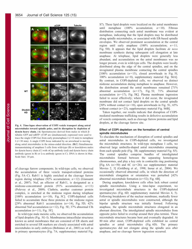
Requirement of dynein for COPI vesicle transport alongastral microtubules towards the spindle pole periphery
To understand the inheritance of COPI vesicles during malemeiosis, we investigated the movement of mRFP–cCop-labeledvesicles in a living spermatocyte by means of a time-lapse
experiment (Fig. 4A; supplementary material Movie 1). Theubiquitously distributed vesicles moved towards each spindlepole during prometaphase to metaphase. We observed the
movement of several COPI foci from the plus ends of asterstowards a spindle pole, along the microtubules. [All fociexamined (n550 foci in 26 cells) performed the movement.
Arrowhead in Fig. 4A]. These foci eventually converged aroundthe spindle poles on astral microtubules, until metaphase.
Sisson and colleagues proposed minus-ended motor dynein asa candidate for the transport of COPI vesicles (Sisson et al.,
2000). We observed the enrichment of dynein around the spindlepoles (Fig. 4B). Moreover, COPI vesicles were distributed on andover a dynein-accumulated region. In mutants for Dhc64C
encoding the dynein heavy chain, or Dhc64C knockdown
spermatocytes, the characteristic COPI distribution was notpresent (67% abnormality, n521 mutant cells, Fig. 4C).Furthermore, we observed Lva-containing vesicles, which may
correspond to Golgi stacks (Giansanti et al., 2007; Papoulas et al.,2005), dispersed throughout the cytoplasm. Our results suggestthat dynein is required for the transport of COPI and Lva-
containing vesicles, along astral microtubules towards the spindlepoles.
Defective accumulation of cleavage furrow proteins andlipid droplets in COPI-depleted spermatocytes
To understand the primary cytokinesis defects in COPI-depletedspermatocytes, we investigated perturbations in the accumulation
Fig. 3. Distribution of COPI-containing vesicles during male meiosis is affected in flies mutants for astral microtubules rather than for spindle
microtubules. (A) Immunolocalization of COPI vesicles in S2 cultured cells at metaphase and in metaphase I cells from wild-type females. Anti-tubulin
immunostaining is shown in green, anti-aCop immunostaining in red and DNA in blue. Note the random distribution of COPI vesicles throughout the cytoplasm of
dividing cells. (B) Immunofluorescence of the COPI vesicles (red) in metaphase I cells from wild-type and mutant males with abnormal microtubule structures. In
wild-type cells, the COPI vesicles appear to be localized on astral microtubules around two spindle poles. In a metaphase-I-like cell from a bTub85DD/+ male, the
COPI vesicles are distributed as far as the central region of the cell. In a metaphase-like cell from an asl1/asl2 male, with few or incorrectly oriented astral
microtubules, the distribution of COPI vesicles is compromised, and a subset of vesicles remains in the center of the cell. In the metaphase-I-like cell from an
orbit7 male, in which spindle microtubules fail to overlap each other at the plus ends, COPI vesicles accumulate around a spindle pole. Scale bars: 10 mm.
(C) Frequency of cells displaying normal distribution of COPI vesicles at metaphase I (M) or anaphase I (A). Spermatocytes from wild-type [M, 85.1% (n5141);
A, 96.4% (n5111)], bTub85DD/+ [M, 34.4% (n596); A, 54.1% (n5185)], asl1 [M, 33.8% (n5139); A, 32.7% (n587)], asl1/asl2 [M, 12.5% (n596); A, 8.2%
(n5107)], and orbit7 [M, 74.3% (n574); A, 81.8% (n5110)].
Drosophila male meiosis requires COPI 3653
Journ
alof
Cell
Scie
nce
of cleavage furrow components. In wild-type cells, we observed
the accumulation of three vesicle transport-related proteins
(Fig. 5A–C). Rab11 is highly enriched at the cleavage furrow
region during telophase (92% accumulation; n512) (Giansanti
et al., 2007). Nuf, an effector of Rab11, is designated as a
midzone-concentrated protein (91% accumulation; n511)
(Polevoy et al., 2009). Clathrin, another coatomer protein
complex, is enriched at the midzone region during telophase
[100% accumulation (n510), Fig. 5C]. COPI-depleted cells
failed to accumulate these three proteins at the midzone region
[29% abnormal Rab11 accumulation (n514), Fig. 5D; 42%
abnormal Nuf accumulation (n512), Fig. 5E; 37% abnormal Clh
accumulation (n511), Fig. 5F].
In wild-type male meiotic cells, we observed the accumulation
of lipid droplets (Fig. 5G–I). Membranous intracellular structures
known as astral membranes that are consisted of ER sheets and
enriched in several ER markers are closely associated with astral
microtubules in early embryos (Bobinnec et al., 2003) as well as
in primary spermatocytes (Fig. 7A; supplementary material Fig.
S7). These lipid droplets were localized on the astral membranes
until metaphase (100% accumulation; n510). Fibrous
distribution connecting each astral membrane was evident at
metaphase, indicating that the lipid droplets may be distributed
along spindle microtubules, or associated with ER-based spindle
envelopes. We observed prominent accumulation at the central
region until early anaphase (100% accumulation; n511;
Fig. 5H). It appears that the lipid droplets facilitate de novo
membrane synthesis during subsequent cell elongation at late
anaphase. At telophase, lipid droplets were generally less
abundant, and accumulation on the astral membranes was no
longer present, even in wild-type cells. The droplets were locally
distributed along the edge of the central spindles, and on the
invaginated plasma membrane contacting the central spindles
[100% accumulation (n515), closed arrowheads in Fig. 5I;
100% accumulation (n58), supplementary material Fig. S6A].
By contrast, in COPI-depleted cells, we observed no distinct
midzone accumulation during metaphase to anaphase. However,
the distribution around the astral membranes remained [71%
abnormal accumulation (n517), Fig. 5J; 71% abnormal
accumulation (n517), Fig. 5K]. Plasma membrane ingression
failed to occur effectively; where it did occur, the invaginated
membrane did not contact lipid droplets on the central spindle
[30% without contact (n510), open arrowheads in Fig. 5L; 65%
without contact (n523), supplementary material Fig. S6B].
Taken together, our results indicate that disruption of COPI-
mediated membrane trafficking results in defective accumulation
of vesicle components, such as cleavage furrow proteins and lipid
droplets, at the cleavage furrow site.
Effect of COPI depletion on the formation of central
spindle microtubules
To elucidate the mechanism of disruption of central spindles and
astral microtubules in COPI-depleted cells, we investigated
the microtubule structures. In wild-type metaphase I cells, we
observed large umbrella-shaped astral microtubules emanating
from each spindle pole (Fig. 3B; supplementary material Fig. S5).
The central spindles comprise bundles of interdigitated
microtubules formed between the separating homologous
chromosomes, and play a key role in contractile ring positioning
[Fig. 6A (n.50) and 6C (n526); see supplementary material
Movies 2, 3] (Goldberg et al., 1998; Inoue et al., 2004). We
occasionally observed abnormal cells, in which the direction of
microtubule elongation or orientation was perturbed [42%
abnormal microtubule formation (n552); Fig. 6B].
During anaphase, knockdown cells often lack robust central
spindle microtubules. Using a time-lapse experiment, we
investigated microtubule structures in the COPI-depleted
spermatocytes (Fig. 6D; supplementary material Movies 4, 5).
Following breakdown of the nuclear membrane, markedly fewer
astral or spindle microtubules were constructed, although the
bipolar spindle structure was initially formed. Following
anaphase, the spindle microtubule structures elongated in an
aberrant direction. The interdigitated spindle microtubules from
opposite poles failed to overlap around their plus termini. These
microtubule structures became bent and eventually degraded. At
80 min after onset, COPI-depleted cells exhibited no central
spindles or astral microtubules (Fig. 6D). The primary
spermatocytes did not elongate along the spindle axis after
anaphase, and no cleavage furrow ingression occurred.
Fig. 4. Time-lapse observation of COPI vesicle transport along astral
microtubules toward spindle poles, and its disruption by depletion of
dynein heavy chain. (A) Spermatocytes derived from males in which b-
tubulin–GFP and mRFP–cCOP are simultaneously expressed were used to
trace the single COPI foci from early prometaphase I (t50 min) to metaphase
I (t530 min). A single COPI focus indicated by an arrowhead is moving
along astral microtubules in the minus-ended direction. (B,C) Simultaneous
immunostaining of anaphase I cells from wild-type (B) or knockdown males
for dynein heavy chain (C) with aCop antibody (red) and dynein heavy chain
antibody (green in B) or Lva antibody (green in C). DNA is shown in blue.
Scale bars: 10 mm.
Journal of Cell Science 125 (15)3654

Journ
alof
Cell
Scie
nce
Next, we examined the formation of the anillin rings (known
as pre-contractile rings) in the knockdown spermatocytes
(Fig. 6E,F). The formation of anillin rings was frequently
incomplete (Fig. 6F) or absent (36% abnormal ring formation;
n536). Taken together, our results suggest that COPI depletion
affects the microtubule structures essential for cytokinesis.
Influence of COPI on central spindle microtubules,through the formation of ER-based cellular structures
To understand the mechanism by which COPI depletion results in
the formation of abnormal microtubule structures, we investigated
ER-based cellular structures. COPI mediates membrane trafficking
between the ER and the Golgi apparatus. In Drosophila meiotic
spermatocytes, the ER, recycling endosomes, and lipid droplets are
frequently associated with the astral membranes and spindle
envelopes (Bobinnec et al., 2003; Dorogova et al., 2009; Fuller,
1993; Tates, 1971). Our time-lapse experimental data confirmed
the overlapping of astral microtubules with ER-based astral
membranes during meiotic division (Fig. 7A). Furthermore, the
Rtnl1–GFP signal overlapped with the phase-dense intracellular
structures observed using phase-contrast microscope. The astral
membranes at the spindle pole periphery were covered by astral
microtubules, thereby supporting the microtubule structures from
inside. The central spindle microtubules were contiguous with the
spindle envelope surrounding the nuclear space in which
chromosome segregation occurs; some overlap with the spindle
envelope was evident [similar results were obtained
(n518), Fig. 7A; see supplementary material Movies 6, 7, 8;
supplementary material Fig. S7].
We subsequently obtained genetic evidence for a close
relationship between ER-based and microtubule structures in
spermatocytes. A dominant mutation for a testis-specific tubulin,
bTub85DD, inhibits microtubule polymerization (Kemphues et al.,
1980). We observed the disruption of ER-based astral membranes
and spindle envelopes in the bTub85DD/+ cells, but no
distribution of COPI vesicles around the centrosomes [63%
distinctly abnormal ER-based structures (n554), supplementary
material Fig. S8C]. Our findings suggest that ER-based and
microtubule structures interact considerably during male meiotic
divisions.
Using a time-lapse experiment, we investigated the way in
which aberrant ER-based structures are constructed in COPI-
depleted cells (39% distinctly abnormal ER-based structures
(n518; Fig. 7B; supplementary material Movies 9, 10, 11). In
normal spermatocytes, the ER formed concentric circles on the
outside of the nuclear membrane at prophase. The ER network
consisted of multi-layers of membranous sheets, visible as phase-
dense structures around the nuclear space. The reticular ER
structure was elongated and transformed into multiple layers of
membranes (known as spindle envelopes) surrounding the
Fig. 5. COPI depletion results in failure to accumulate vesicle components at the midzone of male meiotic cells. (A–F) Immunolocalization of three vesicle-
related proteins (Rab11, Nuf and clathrin), which are essential for membrane trafficking in cytokinesis. Wild-type (A–C) and aCop knockdown
(D–F) spermatocytes were examined. DNA is shown in blue, microtubules in green and immunofluorescence of the three proteins in red. Note that each protein
accumulated at the midzone of early telophase I cells (filled arrowheads) and around the centrosomes. COPI-depleted cells failed to accumulate each protein at the
midzone (open arrowheads). (G–L) Intracellular distribution of lipid droplets in living primary spermatocytes stained with Bodipy 493/503. In wild-type cells, the
Bodipy fluorescence can be seen on the ER-based astral membranes, and at the midzone of cells (arrowheads) during metaphase (G) to anaphase (H). The
accumulation on ribbon-like structures corresponding to ER-based structures is prominent at telophase; the midzone of the plasma membrane is budding inside, so
as to contact the cellular pool of lipid droplets before cytokinesis (I). In COPI-depleted cells, central accumulation at metaphase to anaphase is not present
(J,K); budding of the plasma membrane towards the internal lipid storage is initiated, but membrane ingression fails to contact the storage organelles (open
arrowhead in L). Scale bars: 10 mm.
Drosophila male meiosis requires COPI 3655

Journ
alof
Cell
Scie
nce
nuclear space. In COPI-depleted cells, the ER-based structure
was affected (Fig. 7B; supplementary material Fig. S8C).
Initially, the astral membranes and spindle envelopes were
indistinguishable from those of wild-type cells. However, as the
cells elongated at anaphase, the spindle envelopes became curved
in the middle and disconnected (arrowheads in Fig. 7B;
supplementary material Fig. S8).
Simultaneous observation confirmed the interaction of
microtubules and ER-based structures (Fig. 7A,B). In COPI-
depleted cells, microtubules exhibited aberrant organization
(Fig. 7B,C). At anaphase, spindle microtubules and ER-based
structures aligned side by side. In normal cells, characteristic
microtubule structures (termed exterior central spindles) are
constructed alongside the spindle envelope (Inoue et al., 2004). In
COPI-depleted cells, the orientation of the elongating central spindle
was lost, resulting in the generation of disorganized microtubule
structures. Interestingly, the disorganized microtubules overlappedwith the aberrant ER-based spindle envelopes visualized as phase-
dense structures (arrowheads in Fig. 7B). Moreover, no cleavagefurrow ingression occurred.
Taken together, our results suggest that COPI depletion
influences the formation of ER-based spindle envelopes, whichmutually interact with the central spindle microtubules essentialfor cytokinesis.
DiscussionIn the present study, our initial RNAi screen identified severalCOPI subunits and assembly regulators as essential factors forcytokinesis in Drosophila male meiosis. We subsequentlyelucidated the cellular function of COPI during Drosophila male
meiosis. We observed the localization of COPI in the ER–Golgiintermediate compartment of tER–Golgi units scatteredthroughout the spermatocyte cytoplasm. Prior to metaphase,
COPI-containing vesicles assemble at the spindle pole peripherythrough a poleward movement, mediated by astral microtubulesand minus-ended motor dynein. At the end of each meiotic
division, the vesicles are equally partitioned between two daughtercells. Our findings strongly suggest that spermatocytes possess aregulatory mechanism, to fulfill equal inheritance of several types
of membrane vesicles. Testis-specific knockdown of COPI, ormutations of the cCOP gene, resulted in defective cytokinesisduring male meiosis. COPI depletion disrupted the accumulationof vesicle components, such as cleavage furrow proteins and lipid
droplets, at the cleavage furrow zone. Furthermore, we observedaberrant central spindle microtubules. In COPI-depleted cells, theastral membrane and spindle envelope (associated with astral and
spindle microtubules, respectively) were severely disrupted. Wepropose that these ER-based structures are required as a structuralfoundation, to facilitate the formation of meiotic microtubule
structures. Thus, we believe that COPI plays an important role inDrosophila male meiosis, not only through vesicle transport to thecleavage furrow region, but also via the formation of intracellularstructures.
Equal partitioning of membrane vesicles duringDrosophila male meiosis
Giansanti et al. reported that Golgi-derived vesicles wereconcentrated around each spindle pole and excluded from the
equatorial region (Giansanti et al., 2007). In gio or fwd mutants,which encode vesicle transport factors, these vesicles continuedto be accumulated at the equator. The authors concluded that
accumulation of such vesicles was the consequence of failedincorporation into the membrane at the invaginating cleavagefurrow. However, this phenotype was not observed in othercytokinesis mutants, such as fws and pbl, which encode factors
for Golgi structure and function, and cleavage furrow formation,respectively (Farkas et al., 2003; Giansanti et al., 2006).Although Golgi stack components are required for Drosophila
cytokinesis, evidence for the direct fusion of Golgi-derivedvesicles with the furrow membrane remains elusive (Albertsonet al., 2008; Farkas et al., 2003; Xu et al., 2002). Our present
findings indicate the existence of a transport system, whichmediates equal partitioning not only of COPI vesicles, but also ofvesicles containing Lva (cis-Golgi components), during male
meiosis. Thus, we believe that abnormal distribution of Golgi-derived vesicles, and of other membranous organelles, resultsfrom the perturbation of vesicle transport.
Fig. 6. Abnormal meiotic microtubule structures required for
cytokinesis, and loss of contractile rings, in COPI-depleted
spermatocytes. (A,B) Fixed meiotic cells from wild-type (A) or aCop-
depleted testes (B), Wild-type spermatocytes exhibit robust microtubule
bundles, known as central spindle microtubules, in the middle of anaphase
cells. Microtubules are visualized in green; DNA is shown in blue. (C) Time-
lapse observation of meiotic microtubule structures, and phase-contrast
images of living wild-type spermatocytes. Microtubules were visualized by
expression of GFP–tubulin. The time-lapse images were continuously
collected from anaphase I, in which interior microtubules emanating from
each spindle pole made contact at the equator (t50 min), to the end of
meiosis I (t540 min). (D) Time-lapse observation of COPI-depleted
spermatocytes. (E,F) A contractile ring (visualized in red by anillin
immunostaining) is evident in telophase I cells from wild-type males (E) and
aCop knockdown males (F). Microtubules are shown in green, and DNA in
blue. In the knockdown spermatocytes, elongation of microtubules or their
orientation seems to be disturbed. In anaphase-I-like cells, the central spindle
microtubules are missing, whereas in non-constricted cells within a telophase
cyst, a disconnected contractile ring is formed. Scale bars: 10 mm.
Journal of Cell Science 125 (15)3656

Journ
alof
Cell
Scie
nce
On completion of meiosis during Drosophila spermatogenesis,
considerable cell elongation takes place synchronously in 64
spermatids within a cyst (Fuller, 1993; Inoue et al., 2011). It is
crucial for each spermatid to obtain sufficient numbers of
vesicles. Within spermatids, COPI and other Golgi components
comprise a single large Golgi apparatus, called an acroblast
(Farkas et al., 2003). A mammalian counterpart known as the
acrosome precursor (Moreno et al., 2000) is required for fusion
between the sperm and oval membranes. Thus, it is likely that
Drosophila spermatocytes possess a mechanism to ensure equal
partitioning of vesicles during male meiosis.
Dependence of COPI vesicle distribution on astral
microtubules and minus-ended motor dynein
Our present findings indicate that vesicle transport towards the
spindle pole periphery is mediated by minus-ended motor dynein.
Dynein was previously reported to form complexes with Lva-
containing vesicles, which may correspond to Golgi stacks
(Papoulas et al., 2005). It is reasonable to speculate that other
Golgi-derived vesicles are transported along astral microtubules by
motor dynein. In budding yeast, the actin cytoskeleton plays an
essential role in Golgi transport during mitosis (Arai et al., 2008).
We failed to observe co-localization of COPI vesicles with actin
networks in male meiotic cells (D.K. and Y.H.I., unpublished data).
It is likely that microtubule structures play a key role in the
inheritance of vesicles during male meiosis. Ordered vesicle
inheritance in mammalian male meiotic divisions has not been
demonstrated. In mammalian cultured cells, the Golgi ribbon is
fragmented into smaller pieces by the onset of mitosis (Rabouille
and Jolo, 2003; Shima et al., 1998). Wei and Seemann reported that
COPI vesicles accumulate around the centrosomes prior to
anaphase initiation (Wei and Seemann, 2009). Moreover, the
vesicles are transferred by the mitotic spindles during mitosis.
However, it is difficult to distinguish between spindles and asters.
By contrast, in Drosophila spermatocytes, the spindle microtubules
are formed within a nuclear space, surrounded by a multilayer of
parafusorial membranes (Fuller, 1993; Tates, 1971). Drosophila
spermatocytes possess particularly long astral microtubules, and it
is likely that these play an important role in the equal inheritance of
membrane vesicles during male meiosis.
Indispensability of Arf1-regulated, COPI-mediated
membrane trafficking during Drosophila male meiosis
In the present study, we have indicated that not only COPI
subunits, but also Arf1, which is a small GTPase required for
COPI assembly are essential for correct execution of cytokinesis
during Drosophila male meiosis. By contrast, in mammalian
cultured cells, inactivation of Arf1 is necessary for COPI and
clathrin disassembly, which normally occur prior to the initiation
of mitosis (Altan-Bonnet et al., 2003). The inhibition of COPI
disassembly causes defects not only in chromosome segregation,
but also in cleavage furrow ingression. Thus, inactivation of
vesicle transport complexes is believed to be indispensable for
mitotic entry. However, the meiotic phenotype of the Drosophila
knockdown cells indicates that the COPI complex plays an active
role in the progression of meiotic divisions. The significance of
COPI in cell division may be dependent on cell types. For smaller
cells to initiate cell division, it is important to shut down
cytoplasmic activities, such as protein synthesis and vesicle
transport (Hernandez-Verdun, 2011; Hernandez-Verdun et al.,
2002). By contrast, in Drosophila spermatocytes, membranous
cellular structures, such as multi-layers of parafusorial
Fig. 7. ER-based intracellular structures associated with astral or spindle microtubules are affected in COPI-depleted spermatocytes. (A) Time-lapse
observation of microtubules (second row) and ER-based structures (third row) in primary spermatocytes expressing mRFP–tubulin and GFP–Rtnl1 (an ER-
localizing enzyme). In merged images (first row), tubulin and Rtnl1 are shown in red and green, respectively. Simultaneous observation by phase-contrast
microscopy is shown in the fourth row (PC). The time-lapse images were continuously collected from mid-anaphase I (t50 min) to the end of meiosis I. Note the
association of astral and central spindle microtubules with astral membranes and spindle envelopes, respectively. The ER-based structures labeled by GFP–Rtnl1
overlapped with phase-dense structures. (B) Time-lapse observation of COPI-depleted spermatocytes. Bundles of central spindle microtubules were initially
constructed, but overlapping microtubules were rapidly degraded, and disappeared by the end of the experiment. Spindle envelopes were less abundant, and
became malformed and disappeared, in line with the degradation of microtubule structures (arrowheads). Scale bars: 10 mm.
Drosophila male meiosis requires COPI 3657

Journ
alof
Cell
Scie
nce
membranes, are well developed. The ER-based structures on
which COPI function depends undergo a massive alterationduring meiotic divisions (Fig. 7A). Spermatocytes are muchlarger than other somatic cells, and may need to maintain higher
COPI activity, not only at interphase, but also during celldivision. The involvement of membrane trafficking componentsin cell division was recently reported. The membranous spindlematrix, which has an ER feature, influences spindle morphology
during partially open mitosis in Drosophila early embryos, andalso during open mitosis in vertebrate cells (Zheng, 2010; Liu andZheng, 2009; Civelekoglu-Scholey et al., 2010). The endocytic
adaptor protein, epsin I, was also shown to regulate mitoticmembrane organization and spindle morphology (Liu and Zheng,2009).
Role of ER-based structures as a structural foundation toensure correct formation of the cell division apparatus
Our present findings indicate that meiotic microtubules and ER-
based structures mutually interact. Furthermore, the ER-basedastral membranes and spindle envelopes may serve as structuralfoundations, to ensure the correct formation of astral and spindlemicrotubules, respectively. Rebollo and Gonzalez observed that
colchicine treatment of primary spermatocytes undergoing malemeiosis influenced phase-dense structures corresponding to ER-based structures (Rebollo and Gonzalez, 2000). Barbosa et al.
reported that mutants of dd4 genes encoding spindle pole factorsled to abnormalities in spindle microtubules and phase-densestructures during male meiosis (Barbosa et al., 2000). Further
studies using live analysis of mutations affecting additionalmicrotubule structures are required to verify our hypothesisconcerning the mutual interaction of meiotic microtubules and
ER-based structures. The role of ER structures in microtubuleelongation may be compared to that of a splint in the regenerationof broken bones. In the COPI knockdown cells, the microtubulesmay have lacked support for elongation in the correct orientation.
Some microtubules can initiate elongation independently ofcentrosomes (Rebollo et al., 2004). In human, and alsoDrosophila cells, these noncentrosomal microtubules may be
dependent on seed proteins for elongation from the surface ofother microtubules (Rogers et al., 2008; Uehara et al., 2009).Thus, in Drosophila spermatocytes, advanced ER structures may
be useful for facilitating continuous and straight elongation of theparticularly long astral microtubules.
The microtubule assembly checkpoint in spermatocytesoperates less strictly than does the mitotic cycle (Rebollo and
Gonzalez, 2000). Moreover, Drosophila spermatocytes arenotable for their well-developed membranous structures(Stafstrom and Staehelin, 1984; Tates, 1971). Thus, the ER
structure may facilitate correct formation of the cell divisionapparatus, to compensate for the incomplete checkpoint system.
In summary, we have demonstrated that COPI depletion resultsin the failure of cytokinesis, through disrupted accumulation of
vesicles at the cleavage furrow region. Mutations for aDrosophila golgin ortholog, Lva (which mediates Golgitransport), resulted in a similar cytokinesis phenotype (Sisson
et al., 2000). The delivery of membrane components to thecleavage furrow region is thought to involve two differentpathways, the secretory pathway and the endocytic pathway.
COPI is related to Golgi functions, and therefore COPI-mediatedmembrane trafficking may be required for the delivery of vesiclecomponents required for cytokinesis, via the secretory pathway,
to the cleavage furrow zone. Nevertheless, we cannot exclude the
possibility that the failure of vesicle component accumulation in
COPI-depleted cells is a consequence, rather than a cause of
cytokinesis defects. Detailed observations of cleavage furrow
proteins in living meiotic cells are required to clarify this issue.
COPI is required for the construction of ER-based spindle
envelopes, which appear mutually to interact with the central
spindle microtubules. It is well known that these microtubule
structures and the contractile ring are interdependent (Giansanti
et al., 1998). Thus, it is possible to speculate that COPI depletion
eventually influences contractile ring formation, which is
essential for the initiation of cytokinesis.
Materials and MethodsDrosophila stocks
y w, or btubulin–GFP stocks that has been previously described (Inoue et al., 2004)were used as a wild-type control for cytological and time-lapse studies. Stockscontaining UAS-RNAi constructs of 52 genes, including genes for the seven COPIsubunits (aCop, bCop, b9Cop, cCop, dCop, eCop, and fCop), five arf genesencoding COPI assembly factors (arf79F, arf102F, arf51F, arf72A, and arf84F),and 40 other membrane trafficking-related factors, were obtained from the ViennaDrosophila RNAi Center. We used UAS-dir2; bam-Gal4::VP16 (a gift from T.Noguchi) or nanos-Gal4::VP16 for spermatocyte-specific gene induction orknockdown. The following stocks were obtained from the Bloomington StockCenter: P{sqh-EYFP-Golgi}, P{sqh-EYFP-ER}, nanos-Gal4::VP16, P{GAL4-
arm.S}4a P{GAL4-arm.S}4b, and P{SUPor-P}cCopKG06383. Protein trap stocksexpressing GFP–Rtnl1 (#G00071) and GFP–PDI (#G00198III) were obtained fromL. Cooley; cCop{XP}d06498 was supplied by the Exelixis Collection at Harvard;asl1 and asl2 were gifts from S. Bonaccorsi (Rebollo et al., 2004); and orbit7 was asdescribed previously (Inoue et al., 2004).
aCOP antibody and western blot analysis
A 1.3-kb BamHI–EcoRI fragment of Drosophila aCOP cDNA was inserted in-frame into expression vector pET24b (Novagen, Madison, WI, USA). Theresulting plasmid expresses a fusion protein of a polypeptide corresponding toamino acids 548–972 with a stretch of 14 amino acids at the NH3 terminus and astretch of 18 amino acids at the COOH terminus as linkers after the COOH 66Histag. The recombinant His–aCOP protein was purified by Ni-NTA Spin Kit(Qiagen, Valencia, CA, USA). Antiserum was prepared by injecting a guinea pig.Western blots were incubated with the antiserum diluted 1:5000, followed byincubation with HRP-conjugated anti-guinea pig IgG.
Live cell imaging of primary spermatocytes
To observe cytokinesis in living primary spermatocytes more easily, we addedsome modifications to the protocol described previously (Inoue et al., 2004). Toexamine dynamics of COPI in male meiosis, flies expressing mRFP–cCOP (a giftfrom N. Grieder) were used. Testes from the adult flies were dissected and cellswere laid out under mineral oil (Trinity Biotech, Bray, Ireland) in open chamberssurrounded by double-faced tape on clean glass coverslips without any pressure.Using this protocol, we have succeeded in continuous observation of primaryspermatocytes undergoing proper spindle formation and cytokinesis for at least anhour. For a drug treatment, we carried out short term in vitro culture of primaryspermatocytes according to methods described previously (Robinett et al., 2009)with minor modification. A testis complex attached with accessory gland,ejaculatory duct and pomp were dissected from adult males. A living testiscomplex in which ejaculatory pomp is actively contracting was selected andtransferred into a culture medium consisting of modified M3 medium withoutbicarbonates (Sigma-Aldrich, St Louis, MO, USA) containing 10% fetal calfserum (Sigma-Aldrich) and 50% male cell extracts. To inhibit COPI assembly,aCOPI inhibitor, brefeldin A (Sigma-Aldrich, St. Louis, MO, USA) or Exo1(Sigma-Aldrich) was directly added to the culture medium. The testis wasincubated in the culture medium for 14 hours before isolation of spermatocytes atroom temperature. For observation of lipid droplets, Bodipy 493/503 (4,4-difluoro-1,3,5,7,8-pentamethyl-4-bora-3a,4a-diaza-s-indacence; Molecular Probes, Eugene,OR, USA) or Nile red (Wako, Osaka, Japan) was used. Time-lapse imaging wasperformed on an Olympus IX81 fluorescence microscope (Olympus, Tokyo,Japan) outfitted with excitation, emission filter wheels (Olympus, Tokyo, Japan).Cells were imaged with a 406 lens. At each 30 sec time interval, near-simultaneous GFP and RFP fluorescence images were captured with a CCDcamera (Hamamatsu Photonics, Hamamatsu, Japan). Image acquisition wascontrolled through the Metamorph software package running on a PC. Testissquashes to evaluate onion-stage spermatids were made using previous protocols(Inoue et al., 2004) and viewed by phase-contrast microscopy.
Journal of Cell Science 125 (15)3658

Journ
alof
Cell
Scie
nce
ImmunofluorescenceTestis cells were fixed according to the method of Inoue and colleagues (Inoueet al., 2004). For immunostaining, anti-aCOP antibody was used at a 1:500dilution. The following primary antibodies were used: anti-GM130 (Abcam,Cambridge, MA, USA), anti–cCOP (a gift from Wieland) (Jayaram et al., 2008),anti-centrosomin (T. Kaufman), anti-anillin (D. Glover), anti-Lva (W. Sullivan)and anti-Clathrin (S. Kametaka). For anti-Rab11 (R. Choen) staining was carriedout by a method described previously (Giansanti et al., 2007). Microtubules werevisualized by immunostaining with anti-a-tubulin (DM1A: Sigma-Aldrich) orexpression of GFP–b-tubulin (Inoue et al., 2004). ER was visualized by expressionof GFP–Rtnl1 residing predominantly in the intracellular structure. All secondaryantibodies and DNA stains were commercially obtained. Images were processedand merged in pseudocolor using MetaMorph version 7.6 (Molecular Devices,Sunnyvale, CA, USA).
AcknowledgementsWe would like to thank S. Taketani and S. Miyata for technicaladvice on establishing antibodies. We are grateful to N. Grieder, S.Bonaccorsi, L. Cooley and DGRC, VDRC, BSC, for providing flystocks, and to F. Wieland, D. Glover, S. Kametaka, R. Cohen, andW. Sullivan for antibodies.
FundingThis work was supported by Grants-in-Aid for Scientific Research onPriority Area and for Scientific Research (C) [grant number23570004 to Y.H.I.]
Supplementary material available online at
http://jcs.biologists.org/lookup/suppl/doi:10.1242/jcs.103317/-/DC1
ReferencesAlbertson, R., Riggs, B. and Sullivan, W. (2005). Membrane traffic: a driving force in
cytokinesis. Trends Cell Biol. 15, 92-101.Albertson, R., Cao, J., Hsieh, T.-S. and Sullivan, W. (2008). Vesicles and actin are
targeted to the cleavage furrow via furrow microtubules and the central spindle.J. Cell Biol. 181, 777-790.
Altan-Bonnet, N., Phair, R. D., Polishchuk, R. S., Weigert, R. and Lippincott-Schwartz, J. (2003). A role for Arf1 in mitotic golgi disassembly, chromosomesegregation, and cytokinesis. Proc. Natl. Acad. Sci. U S A. 100, 13314-13319.
Arai, S., Noda, Y., Kainuma, S., Wada, I. and Yoda, K. (2008). Ypt11 functions inbud-directed transport of the Golgi by linking Myo2 to the coatomer subunit Ret2.Curr. Biol. 18, 987-991.
Barbosa, V., Yamamoto, R. R., Henderson, D. S. and Glover, D. M. (2000). Mutationof a Drosophila gamma tubulin ring complex subunit encoded by discs degenerate-4differentially disrupts centrosomal protein localization. Genes Dev. 14, 3126-3139.
Beller, M., Sztalryd, C., Southall, N., Bell, M., Jackle, H., Auld, D. S. and Oliver, B.
(2008). COPI complex is a regulator of lipid homeostasis. PLoS Biol. 6, e292.Bobinnec, Y., Marcaillou, C., Morin, X. and Debec, A. (2003). Dynamics of the
endoplasmic reticulum during early development of Drosophila melanogaster. Cell
Motil. Cytoskeleton 54, 217-225.Bonaccorsi, S., Giansanti, M. G. and Gatti, M. (1998). Spindle self-organization and
cytokinesis during male meiosis in asterless mutants of Drosophila melanogaster.J. Cell Biol. 142, 751-761.
Bonaccorsi, S., Giansanti, M. G., Cenci, G. and Gatti, M. (2000). Cytological analysisof spermatocytes growth and male meiosis in Drosophila melanogaster. InDrosophila Protocol (ed. W. Sullivan, M. Ashburner and R. S. Hawley), pp. 87-109. New York: Cold Spring Harbor Laboratory Press Ltd.
Boucrot, E. and Kirchhausen, T. (2007). Endosomal recycling controls plasmamembrane area during mitosis. Proc. Natl. Acad. Sci. USA 104, 7939-7944.
Cao, J., Albertson, R., Riggs, B., Field, C. M. and Sullivan, W. (2008). Nuf, a Rab11effector, maintains cytokinetic furrow integrity by promoting local actin polymerization.J. Cell Biol. 182, 301-313.
Cao, L. G. and Wang, Y. L. (1996). Signals from the spindle midzone are required forthe stimulation of cytokinesis in cultured epithelial cells. Mol. Biol. Cell 7, 225-232.
Castrillon, D. H., Gonczy, P., Alexander, S., Rawson, R., Eberhart, C. G.,
Viswanathan, S., DiNardo, S. and Wasserman, S. A. (1993). Toward a moleculargenetic analysis of spermatogenesis in Drosophila melanogaster: characterization ofmale-sterile mutants generated by single P element mutagenesis. Genetics 135, 489-505.
Cermelli, S., Guo, Y., Gross, S. P. and Welte, M. A. (2006). The lipid-dropletproteome reveals that droplets are a protein-storage depot. Curr. Biol. 16, 1783-1795.
Civelekoglu-Scholey, G., Tao, L., Brust-Mascher, I., Wollman, R. and Scholey,
J. M. (2010). Prometaphase spindle maintenance by an antagonistic motor-dependentforce balance made robust by a disassembling lamin-B envelope. J. Cell Biol. 188, 49-68.
D’Avino, P. P., Savoian, M. S. and Glover, D. M. (2005). Cleavage furrow formationand ingression during animal cytokinesis: a microtubule legacy. J. Cell Sci. 118,1549-1558.
Dorogova, N. V., Nerusheva, O. O. and Omelyanchuk, L. V. (2009). Structuralorganization and dynamics of the endoplasmic reticulum during spermatogenesis ofDrosophila melanogaster: Studies using PDI-GFP chimera protein. Biochemistry
(Moscow). Supplement. Series A, Membrane and Cell Biology 3, 55-61.
Duden, R., Kajikawa, L., Wuestehube, L. and Schekman, R. (1998). epsilon-COP is astructural component of coatomer that functions to stabilize alpha-COP. EMBO J. 17,985-995.
Dyer, N., Rebollo, E., Domınguez, P., Elkhatib, N., Chavrier, P., Daviet, L.,
Gonzalez, C. and Gonzalez-Gaitan, M. (2007). Spermatocyte cytokinesis requiresrapid membrane addition mediated by ARF6 on central spindle recycling endosomes.Development 134, 4437-4447.
Echard, A., Hickson, G. R., Foley, E. and O’Farrell, P. H. (2004). Terminalcytokinesis events uncovered after an RNAi screen. Curr. Biol. 14, 1685-1693.
Eggert, U. S., Kiger, A. A., Richter, C., Perlman, Z. E., Perrimon, N., Mitchison,
T. J. and Field, C. M. (2004). Parallel chemical genetic and genome-wide RNAiscreens identify cytokinesis inhibitors and targets. PLoS Biol. 2, e379.
Farkas, R. M., Giansanti, M. G., Gatti, M. and Fuller, M. T. (2003). The Drosophila
Cog5 homologue is required for cytokinesis, cell elongation, and assembly ofspecialized Golgi architecture during spermatogenesis. Mol. Biol. Cell 14, 190-200.
Fuller, M. T. (1993). Spermatogenesis. In The development of Drosophila melanogaster
(ed. M. Bate and A. Martinez-Arias), pp. 71-147. New York: Cold Spring HarborPress Ltd.
Giansanti, M. G., Bonaccorsi, S., Williams, B., Williams, E. V., Santolamazza, C.,
Goldberg, M. L. and Gatti, M. (1998). Cooperative interactions between the centralspindle and the contractile ring during Drosophila cytokinesis. Genes Dev. 12, 396-410.
Giansanti, M. G., Bonaccorsi, S., Kurek, R., Farkas, R. M., Dimitri, P., Fuller, M. T.and Gatti, M. (2006). The class I PITP giotto is required for Drosophila cytokinesis.Curr. Biol. 16, 195-201.
Giansanti, M. G., Belloni, G. and Gatti, M. (2007). Rab11 is required for membranetrafficking and actomyosin ring constriction in meiotic cytokinesis of Drosophila
males. Mol. Biol. Cell 18, 5034-5047.
Glotzer, M. (2001). Animal cell cytokinesis. Annu. Rev. Cell Dev. Biol. 17, 351-386.
Goldberg, M. L., Gunsalus, K. C., Karess, R. E. and Chang, F. (1998). Cytokinesis in
Dynamics of Cell Division (ed. S. A. Endow and D. M. Glover), pp. 270-316. London:Oxford University Press Ltd.
Grieder, N. C., Kloter, U. and Gehring, W. J. (2005). Expression of COPI componentsduring development of Drosophila melanogaster. Gene Expr. Patterns 6, 11-21.
Grieder, N. C., Caussinus, E., Parker, D. S., Cadigan, K., Affolter, M. and Luschnig,
S. (2008). gammaCOP is required for apical protein secretion and epithelialmorphogenesis in Drosophila melanogaster. PLoS ONE 3, e3241.
Griffiths, G., Pepperkok, R., Locker, J. K. and Kreis, T. E. (1995). Immunocytochemicallocalization of b-COP to the ER-Golgi boundary and the TGN. J. Cell Sci. 108, 2839-2856.
Hernandez-Verdun, D. (2011). Assembly and disassembly of the nucleolus during thecell cycle. Nucleus 2, 189-194.
Hernandez-Verdun, D., Roussel, P. and Gebrane-Younes, J. (2002). Emergingconcepts of nucleolar assembly. J. Cell Sci. 115, 2265-2270.
Hosobuchi, M., Kreis, T. and Schekman, R. (1992). SEC21 is a gene required for ERto Golgi protein transport that encodes a subunit of a yeast coatomer. Nature 360,603-605.
Ichihara, K., Shimizu, H., Taguchi, O., Yamaguchi, M. and Inoue, Y. H. (2007). ADrosophila orthologue of larp protein family is required for multiple processes inmale meiosis. Cell Struct. Funct. 32, 89-100.
Inoue, Y. H., Savoian, M., Suzuki, T., Mathe, E., Yamamoto, M. T. and Glover,
D. M. (2004). Mutation in orbit/mast reveal that the central spindle is comprised oftwo microtubule populations, those that initiate cleavage and those that propagatefurrow ingression. J. Cell Biol. 166, 49-60.
Inoue, Y. H., Miyauchi, C., Ogata, T. and Kitazawa, D. (2011). Dynamic alteration ofcellular component of male meiosis in Drosophila. In Meiosis (ed. A. Swan). Croatia,China: InTech Open Access Publisher.
Jayaram, S. A., Senti, K-A., Tiklova, K., Tsarouhas, V., Hemphala, J. and
Samakovlis, C. (2008). COPI vesicle transport is a common requirement for tubeexpansion in Drosophila. PLoS ONE 3, e1964.
Kamena, F., Diefenbacher, M., Kilchert, C., Schwarz, H. and Spang, A. (2008).Ypt1p is essential for retrograde Golgi-ER transport and for Golgi maintenance inS. cerevisiae. J. Cell Sci. 121, 1293-1302.
Kemphues, K. J., Raff, E. C., Raff, R. A. and Kaufman, T. C. (1980). Mutation in atestis-specific beta-tubulin in Drosophila: analysis of its effects on meiosis and maplocation of the gene. Cell 21, 445-451.
Kirchner, J., Vissi, E., Gross, S., Szoor, B., Rudenko, A., Alphey, L. and White-
Cooper, H. (2008). Drosophila Uri, a PP1a binding protein, is essential for viability,maintenance of DNA integrity and normal transcriptional activity. BMC Mol. Biol. 9,36.
Kondylis, V. and Rabouille, C. (2009). The Golgi apparatus: lessons from Drosophila.
FEBS Lett. 583, 3827-3838.
Lippincott-Schwartz, J., Cole, N. B. and Donaldson, J. G. (1998). Building asecretory apparatus: role of ARF1/COPI in Golgi biogenesis and maintenance.Histochem. Cell Biol. 109, 449-462.
Liu, Z. and Zheng, Y. (2009). A requirement for epsin in mitotic membrane and spindleorganization. J. Cell Biol. 186, 473-480.
Maines, J. and Wasserman, S. (1997). Regulation and execution of meiosis inDrosophila males. Curr. Top. Dev. Biol. 37, 301-332.
Drosophila male meiosis requires COPI 3659

Journ
alof
Cell
Scie
nce
Mishima, M. and Glotzer, M. (2003). Cytokinesis: a logical GAP. Curr. Biol. 13,R589-R591.
Montagnac, G., Echard, A. and Chavrier, P. (2008). Endocytic traffic in animal cellcytokinesis. Curr. Opin. Cell Biol. 20, 454-461.
Moreno, R. D., Ramalho-Santos, J., Sutovsky, P., Chan, E. K. and Schatten, G.
(2000). Vesicular traffic and golgi apparatus dynamics during mammalianspermatogenesis: implications for acrosome architecture. Biol. Reprod. 63, 89-98.
Oprins, A., Duden, R., Kreis, T. E., Geuze, H. J. and Slot, J. W. (1993). Beta-COPlocalizes mainly to the cis-Golgi side in exocrine pancreas. J. Cell Biol. 121, 49-59.
Papoulas, O., Hays, T. S. and Sisson, J. C. (2005). The golgin Lava lamp mediatesdynein-based Golgi movements during Drosophila cellularization. Nat. Cell Biol. 7,612-618.
Polevoy, G., Wei, H.-C., Wong, R., Szentpetery, Z., Kim, Y. J., Goldbach, P.,Steinbach, S. K., Balla, T. and Brill, J. A. (2009). Dual roles for the Drosophila PI4-kinase four wheel drive in localizing Rab11 during cytokinesis. J. Cell Biol. 187,847-858.
Prekeris, R. and Gould, G. W. (2008). Breaking up is hard to do – membrane traffic incytokinesis. J. Cell Sci. 121, 1569-1576.
Rabouille, C. and Jokitalo, E. (2003). Golgi apparatus partitioning during cell division.Mol. Membr. Biol. 20, 117-127.
Rebollo, E. and Gonzalez, C. (2000). Visualizing the spindle checkpoint in Drosophilaspermatocytes. EMBO Rep. 1, 65-70.
Rebollo, E., Llamazares, S., Reina, J. and Gonzalez, C. (2004). Contribution ofnoncentrosomal microtubules to spindle assembly in Drosophila spermatocytes. PLoS
Biol. 2, e8.Ripoche, J., Link, B., Yucel, J. K., Tokuyasu, K. and Malhotra, V. (1994). Location
of Golgi membranes with reference to dividing nuclei in syncytial Drosophila
embryos. Proc. Natl. Acad. Sci. USA 91, 1878-1882.Robinett, C. C., Giansanti, M. G., Gatti, M. and Fuller, M. T. (2009). TRAPPII is
required for cleavage furrow ingression and localization of Rab11 in dividing malemeiotic cells of Drosophila. J. Cell Sci. 122, 4526-4534.
Rogers, G. C., Rusan, N. M., Peifer, M. and Rogers, S. L. (2008). A multicomponentassembly pathway contributes to the formation of acentrosomal microtubule arrays ininterphase Drosophila cells. Mol. Biol. Cell 19, 3163-3178.
Satterwhite, L. L. and Pollard, T. D. (1992). Cytokinesis. Curr. Opin. Cell Biol. 4, 43-52.
Shima, D. T., Cabrera-Poch, N., Pepperkok, R. and Warren, G. (1998). An orderedinheritance strategy for the Golgi apparatus: visualization of mitotic disassemblyreveals a role for the mitotic spindle. J. Cell Biol. 141, 955-966.
Simon, G. C., Schonteich, E., Wu, C. C., Piekny, A., Ekiert, D., Yu, X., Gould,G. W., Glotzer, M. and Prekeris, R. (2008). Sequential Cyk-4 binding to ECT2 andFIP3 regulates cleavage furrow ingression and abscission during cytokinesis. EMBO
J. 27, 1791-1803.Sisson, J. C., Field, C., Ventura, R., Royou, A. and Sullivan, W. (2000). Lava lamp, a
novel peripheral golgi protein, is required for Drosophila melanogaster cellularization.J. Cell Biol. 151, 905-918.
Skop, A. R., Liu, H., Yates, J., 3rd, Meyer, B. J. and Heald, R. (2004). Dissection ofthe mammalian midbody proteome reveals conserved cytokinesis mechanisms.Science 305, 61-66.
Stafstrom, J. P. and Staehelin, L. A. (1984). Dynamics of the nuclear envelope and of nuclearpore complexes during mitosis in the Drosophila embryo. Eur. J. Cell Biol. 34, 179-189.
Tates, A. D. (1971). Cytodifferentiation during spermatogenesis in Drosophila
melanogaster: An electron microscope study. Ph.D. thesis. Rijksuniversiteit,Leiden, Netherlands.
Uehara, R., Nozawa, R. S., Tomioka, A., Petry, S., Vale, R. D., Obuse, C. and
Goshima, G. (2009). The augmin complex plays a critical role in spindle microtubulegeneration for mitotic progression and cytokinesis in human cells. Proc. Natl. Acad.
Sci. USA 106, 6998-7003.Wei, J. H. and Seemann, J. (2009). The mitotic spindle mediates inheritance of the
Golgi ribbon structure. J. Cell Biol. 184, 391-397.Xu, H., Brill, J. A., Hsien, J., McBride, R., Boulianne, G. L. and Trimble, W. S.
(2002). Syntaxin 5 is required for cytokinesis and spermatid differentiation inDrosophila. Dev. Biol. 251, 294-306.
Zhao, W. M., Seki, A. and Fang, G. (2006). Cep55, a microtubule-bundling protein,associates with centralspindlin to control the midbody integrity and cell abscissionduring cytokinesis. Mol. Biol. Cell 17, 3881-3896.
Zheng, Y. (2010). A membranous spindle matrix orchestrates cell division. Nat. Rev.
Mol. Cell Biol. 11, 529-535.
Journal of Cell Science 125 (15)3660
Recommended