Endoscopia e lesione solide del pancreas
Carlo FabbriU.O. di Gastroenterologia ed Endoscopia Digestiva
OSPEDALE BELLARIA-MAGGIOREBOLOGNA

1

Incidenza

Sopravvivenza a 5 anni dalla diagnosi

• Only 9% of patients are diagnosed when PC is localized
• 53% already have distant metastase at time of diagnosis
detection at an earlier stage and development of effective therapies
“cornerstones” for cancer death reduction
SCREENING STRATEGIES

Timeline of ProgressionPanIN-1 Large PC
Yachida S, Nature 2010

Clinical Suspicion of
Pancreatic neoplasm
• MDHCT –
• MDHCT doubtful
• MDHCT +
Incidental finding of solid
pancreatic lesion

Resectability
EUS MRI
Sens 97 100DeWitt J et al. Ann Intern Med 2004Shami V. et al;Pancreas 2011
EUS + MDHCT PPV = 95%EUS MDHCT EUS +
MDHCT
Sens 88 90 80
Spec 67 64 93


Indicazioni
Tecniche
Complicanze
Indicazioni
Tecniche
Complicanze

Causes of malignant bile duct strictures
Intrahepatic bile ducts CholangiocarcinomaHepatocellular carcinomaMetastatic disease
Extrahepatic bile duct CholangiocarcinomaPancreatic cancer Ampullary malignancyGallbladder cancerMetastatic disease
Hilar region Cholangiocarcinoma
Bulky porta hepatis lymphadenopathy
Webb,Suanders 2013

Preprocedural checklist
Lesion resectability and goals of care
Life expectancy given stage of disease and comorbidities
Location and length of the lesion
Plastic versus self-expanding metal stent
Covered versus uncovered
Cost comparisons
Physician comfort level with the procedure
Webb,Suanders 2013


Non endoscopic
biliary
imaging
modalities
TC
Laparoscopic
US
MRC
IOC

Endoscopic
biliary
imaging
modalities
EUS
EUSDirected
ERC
ERC
ERC+Technologies
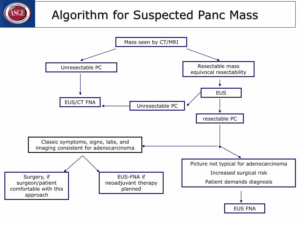
Algorithm for Suspected Panc Mass
Mass seen by CT/MRI
Unresectable PC Resectable mass equivocal resectability
EUS
EUS/CT FNAUnresectable PC
resectable PC
Classic symptoms, signs, labs, and imaging consistent for adenocarcinoma
Surgery, if surgeon/patient
comfortable with this approach
EUS-FNA if neoadjuvant therapy
planned
Picture not typical for adenocarcinoma
Increased surgical risk
Patient demands diagnosis
EUS FNA

Algorithm for Suspected Panc Mass
Mass seen by CT/MRI
Unresectable PC Resectable mass equivocal resectability
EUS/CT FNA

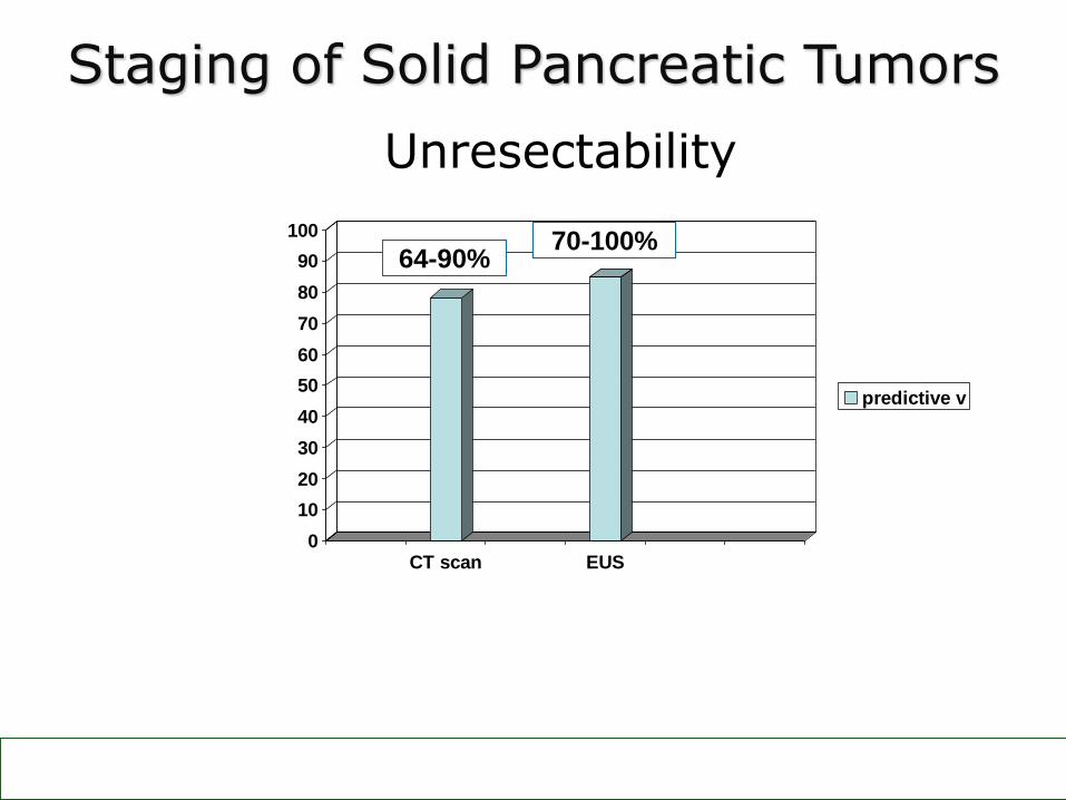
Staging of Solid Pancreatic Tumors
Resectability
0
10
20
30
40
50
60
70
80
90
100
CT scan EUS
predictive v
61-86%
•Detection Small P.T. < 2 cm
71-90%
DeWitt J et al. Ann Intern Med 2004;
0
10
20
30
40
50
60
70
80
90
100
CT scan EUS
predictive v
Unresectability
64-90%70-100%
Staging of Solid Pancreatic Tumors

Pancreatic Cancer
DYAGNOSTIC ACCURACY
Hunt, ,2002
EUS TC
Detection 97% 73%
Resecability 91% 83%
Vascular
Invasion91% 64%

Agarwal B, AM J Gastro 2004
MDHCT missed 60% of lesions < 2 cms
DeWitt J et al. Ann Intern Med 2004
MDHCT missed 47% lesions < 25 mm and 21% overall
EUS vs MDHCT
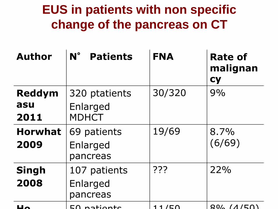
EUS in patients with non specific
change of the pancreas on CT
Author N° Patients FNA Rate of malignancy
Reddymasu
2011
320 ptatients
Enlarged MDHCT
30/320 9%
Horwhat
2009
69 patients
Enlarged pancreas
19/69 8.7% (6/69)
Singh
2008
107 patients
Enlarged pancreas
??? 22%
Ho 50 patients 11/50 8% (4/50)
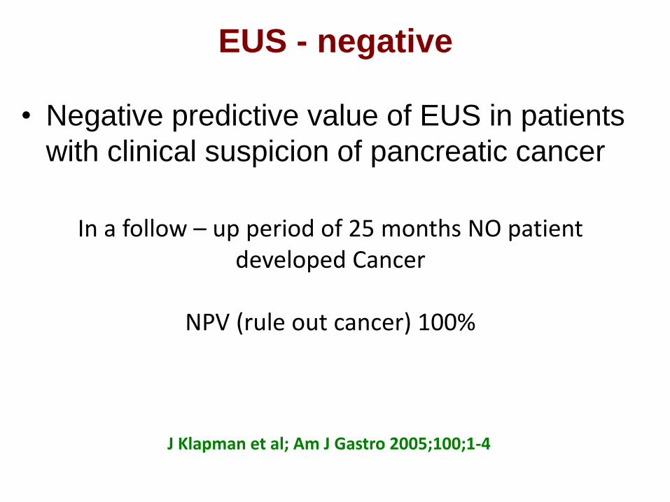
EUS - negative
• Negative predictive value of EUS in patients
with clinical suspicion of pancreatic cancer
J Klapman et al; Am J Gastro 2005;100;1-4
In a follow – up period of 25 months NO patient developed Cancer
NPV (rule out cancer) 100%

Pancreatic Cancer
Sensitivity:50-90%
Specificity: 90-100%
PV/confluence:EUS superiorSMV:Equivalent (~CT)Celiac trunk:Equivalent (~CT)HA, SMA:CT superior
Vascular Invasion
Rosch, 2002

Three-Dimensional Linear Endoscopic UltrasoundFeasibility of a Novel Technique Applied for the Detection of Vessel Involvement of Pancreatic Masses
Fritscher-Ravens A 2005

To document a diagnosis of malignancy in a patient with an unresectable mass as a prerequisite for adjuvant chemotherapy or radiation therapy
To exclude other tumor types
patients who are reluctant to undergo major surgery without a definitive diagnosis
To document the absence of malignancy when the pretest probability of malignancy is low
……………..Neoadviant therapy
Varadarajulu 2010
Indications for the use of EUS-FNA

Pancreatic Cancer Clinical Impact of EUS-FNA
99 patients elegible for surgery
Metastatic distant lymph nodes 6
Liver metastasis 4
Malignant ascites 1
Retroperitoneal infiltration 1•
Mortensen MB, et al. Endoscopy 2001.
EUS FNA influenced
Management in 12%

Celiac mesenteric region
Hepatic pedicule
Entire pancreas
Lymph nodes
Aspirate ascitic Fluid
Left liver nodeChang, GUT, 1997
Performance of EUS-FNA: Solid Pancreatic Tumors
CeliacLumboaorticRetroduodenopancreaticSuperior MesentericMediastinal
Linfonodo-mediastinico.avi

Malignant infiltration occurs in up to
30%<5 mm LNs,
lung, esophageal, gastric, pancreatic, rectal carcinoma.
Cui XW et al. World J Gastroenterol 2013; 19: 4850Hocke M et al. Endosc Ultrasound 2017; 6: 4
NODE

Celiac mesenteric region
Hepatic pedicule
Entire pancreas
Lymph nodes
Aspirate ascitic Fluid
Left liver node
Peritoneal
Carcinomatosis
Performance of EUS-FNA: Solid Pancreatic Tumors
Peter 2009

Celiac mesenteric region
Hepatic pedicule
Entire pancreas
Lymph nodes
Aspirate ascitic Fluid
Left liver node
Shah,Gatroint Endoscopy 2007
Small metastasisSensibility: 100%
Performance of EUS-FNA: Solid Pancreatic Tumors

Resectable TumorsShould FNA be performed?
Adenocarcinoma 78,5%
Mucinous cystic neoplasm 5,4%
Metastatic tumor 3,9%
Neuroendocrine tumor 3,8%
Poorly differentiated carcinoma 3,0%
Lymphoma 2,8%
Serous Cystadenoma 1,8%
Solid pseudopapillary tumor 0,8%
Volmar KE, et al. Gastrointest Endosc 2005
503 pts


Final diagnosis in benign disease after pancreatoduodenectomy for suspected malignancy.
Asbun HJ et al. Surgery 2014

Resectable TumorsShould FNA be performed?
Adenocarcinoma 78,5%
Mucinous cystic neoplasm 5,4%
Metastatic tumor 3,9%
Neuroendocrine tumor 3,8%
Poorly differentiated carcinoma 3,0%
Lymphoma 2,8%
Serous Cystadenoma 1,8%
Solid pseudopapillary tumor) 0,8%
Volmar KE, et al. Gastrointest Endosc 2005
Benign lesions
Pseudotumor Chronic pancreatitis
Groove pancreatitis
PAI tipo I e II

Paranganglioma Gangliocitico Ampollare
Fiscaletti et al 2011
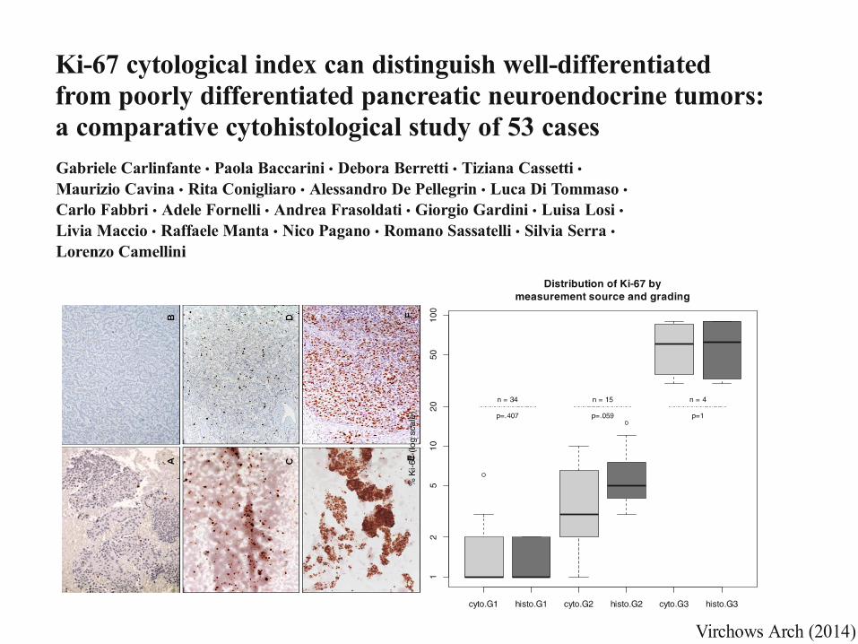
GEP-NETs


91%% 94%% 98% 72%%
Classif. 1
Classif. 2
Hewitt, GE, 2012
Sensibilità Specificità VPP VPN
85% 98% 99% 64%
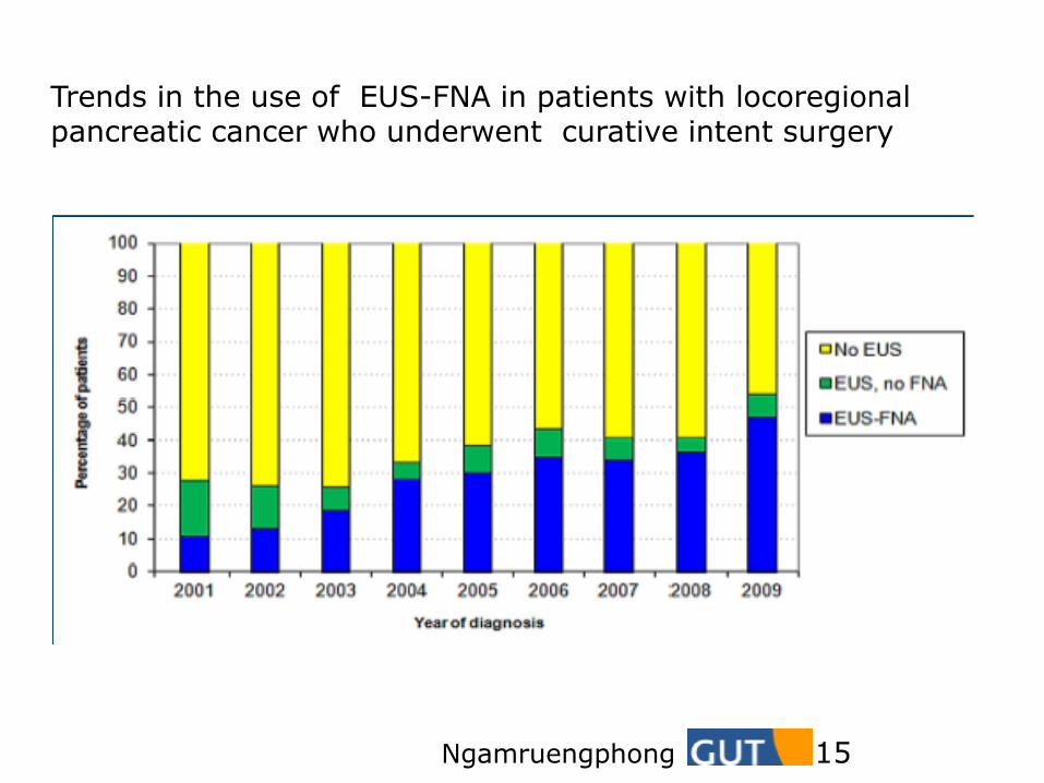
Trends in the use of EUS-FNA in patients with locoregional pancreatic cancer who underwent curative intent surgery
Ngamruengphong 15


1075 ptz
EUS-guided FNA: a benchmark for quality performance measurement
21 centers/41endoscopist
Savides 2008
85%
52%
85%
61%


Eloubedi 2007
Pancreatic cancer
Cholangiocarcinoma
Differential Diagnosis
ACCURACY
EUS 78%
IDUS 86%
P<0.002

Author (yr)No. Pts
Sen (%)
Spe (%)
PPV (%)
NPV (%)
Acc (%)
Saftoiu, ‘08 68 91.4 87.9 88.9 90.6 89.7
Hirche, ‘08 70 41 53 -- -- 45
Giovannini, ‘09 121 92.3 80 93.3 77.4 89.4
Iglesias-Garcia, ‘09 130 100 85.5 90.7 100 94
Iglesias-Garcia, ‘10* 86 100 92.9 96.7 100 97.7
Cancer vs Chronic PancreatitisElastosonography
* Second generation EUS elastography
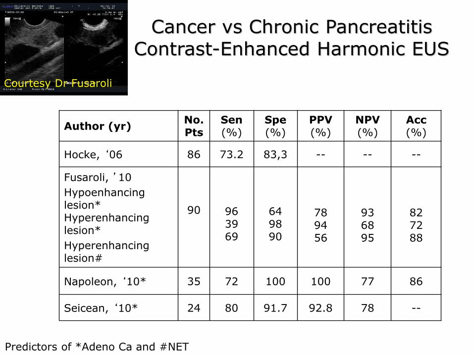
Cancer vs Chronic PancreatitisContrast-Enhanced Harmonic EUS
Author (yr)No. Pts
Sen (%)
Spe (%)
PPV (%)
NPV (%)
Acc (%)
Hocke, ‘06 86 73.2 83,3 -- -- --
Fusaroli, ’10
Hypoenhancing lesion* Hyperenhancing lesion*
Hyperenhancing lesion#
90 96 39 69
64 98 90
78 94 56
93 68 95
82 72 88
Napoleon, ‘10* 35 72 100 100 77 86
Seicean, ‘10* 24 80 91.7 92.8 78 --
Predictors of *Adeno Ca and #NET
Courtesy Dr Fusaroli

Identified pancreatic tumor
Increased detection ( difficult cases)
Help EUS-FNA
Rule out cancer
Fusaroli 2010
CHE-EUS in pancreatic tumors

NEDDLES
• 25 G• 22 G• 19 G• Tru-cut• Echo-Brush
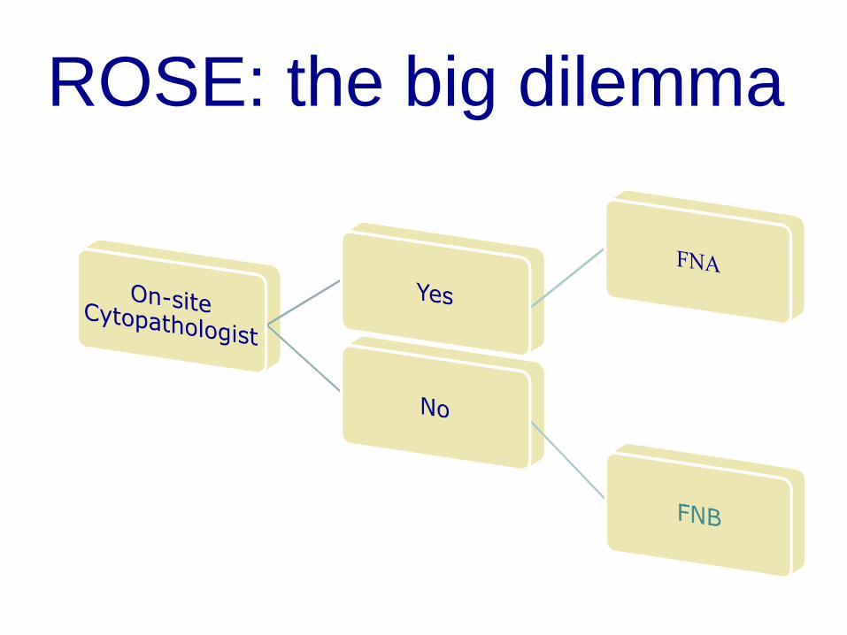
ROSE: the big dilemma

ROSE: the big dilemma
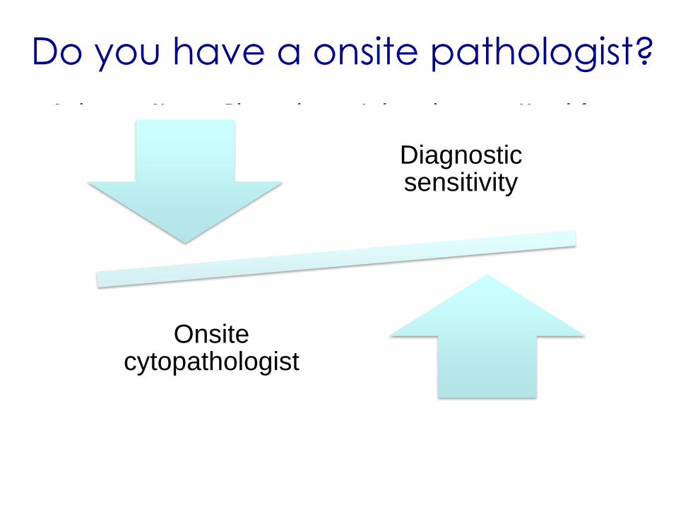
Do you have a onsite pathologist?
58
Author No. Diagnostic
Yield
Indeterminate Unsatisfactory
Klapman
Iglesias
Garcia
Alsohaibani
198
182
104
78% vs. 32%,
p=0.001
97% vs. 86%,
p=0.01
77% vs. 53%,
p=0.01
10% vs. 12%,
p=0.9
2.1% vs. 10.3%,
p=0.02
23% vs. 47%,
p=0.001
9% vs. 20%,
p=.003
1 vs. 13%,
p=0.002
0 vs. 17%, p=NS
Diagnostic sensitivity
Onsite cytopathologist

Survey
Dumonceau. 2012
Available Normal Selected cases
ROSE 28% 15.1%

19g
Rare Conditions
metastasis
IAP
lymphomas
NET
diffentiation

Sen Spe PPV NPV Acc
87.5%, 100%, 100%, 41.7%, 88.5%
J.W. Poley1, M.C. Petrone2, J.Iglesias-Garcia3, A. Larghi4, M. Giovannini5, G. Costamagna4, Paolo Arcidiaccono2, E. Bories5, M.J. Bruno1


Surgical outcomes
6%
20%
0
10
20
30
40
50 Stadio IV Stadio I
Stadio I
Stadio IV
The 5 years survival rate
15%–20%: candidates for pancreatectomy at the time of diagnosis
Groover 2010

C. Fabbri, C. Luigiano, A. Maimone, I. Tarantino, P. Baccarini, A.Fornelli, R. Liotta, A. Polifemo, L. Barresi, M. Traina, C. Virgilio, V. Cennamo
EUS-FNB of Small Solid Pancreatic Lesions using a 22-Gauge Needle with Side Fenestration
2014

2013

2014


5/7/2018
cutting edge
surface

5/7/2018
cutting edge
surface
Presence of Core
30-80%

a common-sense definition : it must contain
epithelium and stroma
Core tissue
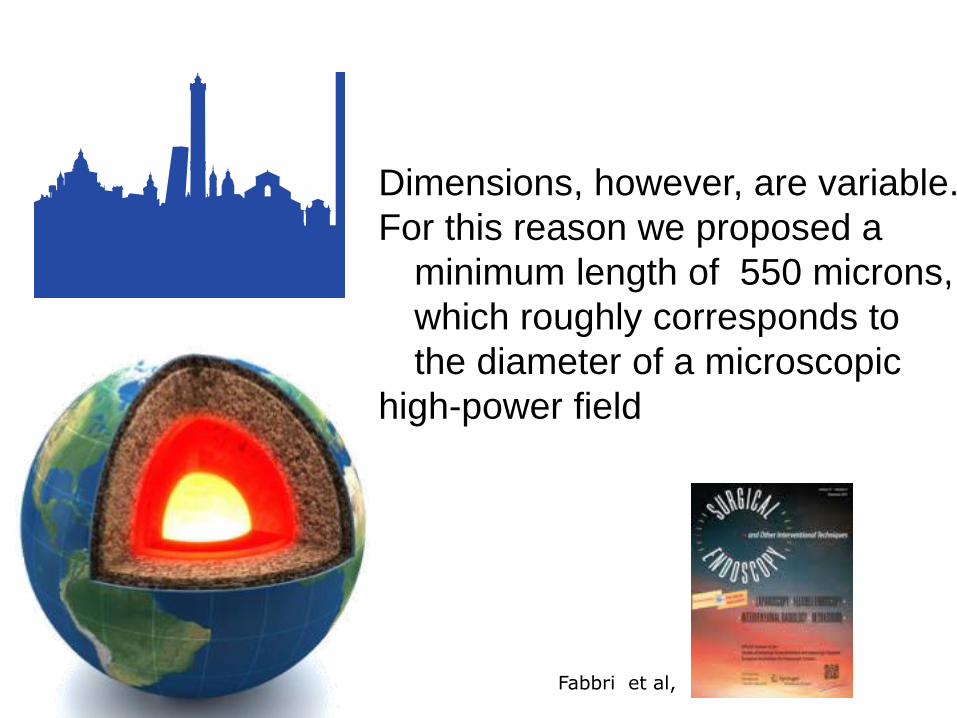
Dimensions, however, are variable.
For this reason we proposed a
minimum length of 550 microns,
which roughly corresponds to
the diameter of a microscopic
high-power field
Fabbri et al, 2014

Pancreatic endocrine tumor EUS-guided FNA DNAmicrosatellite loss and mortality
Differentiating neoplastic from benign lesions of thepancreas: translational techniques
Diagnostic approach to pancreatic tumors with thespecimens of endoscopic ultrasound-guided fine needleaspiration
EUS for pancreatic endocrine tumors: do we need toknow our pancreatic endocrine tumor's DNA?
Molecular Techniques
Fasanella 2009
Kalid 2009
Ito 2009
De Witt 2009

Molecular Techniques
K-ras analysis


STRENGTHS strong pointsSTRENGTHS strong points

2017

LO SCREENING DEL FUTURO
The MicroRNA(miRNAs) regulate the expression of oncogenes and tumors suppressor
La loro espressione è tessuto specifica e il loro profilo è alterato in tutte le
neoplasie
The miRNAs have characteristic expression profiles in some
carcinomas, and several species are dysregulated in Pancreatic Ductal Adenocarcinoma.
The Near Future
Zhang 2007

The MiRNA expression have
accurately predicted the
presence of malignancy in 89%
of cancer specimens in the
cellblock
Panarelli 2012
The Near Future


NET
Istotipo Caratteri lesione Procedura chirugica
Insulinoma < 2 cm, superficiale, No Wirs> 2 cm o coinvolg. Wirsung
EnucleoresezioneResezione
Gastrinoma Qualsiasi localizzazioneLocal. Esclusiva linfonodale
Resezione tipicaExeresi linfonodi
Tumori non funzionanti
< 2 cm, superficiale, No Wirs> 2 cm o coinvolg. Wirsung
EnucleoresezioneResezione tipica
VIPomaGlucagonomaSMSoma
Qualsiasi localizzazione Resezione tipica Falc
oni M
et al. 2
007,
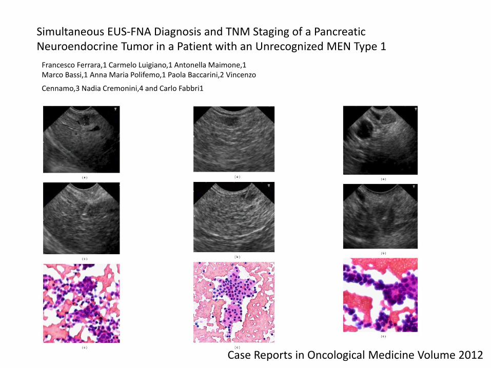
Simultaneous EUS-FNA Diagnosis and TNM Staging of a PancreaticNeuroendocrine Tumor in a Patient with an Unrecognized MEN Type 1
Francesco Ferrara,1 Carmelo Luigiano,1 Antonella Maimone,1Marco Bassi,1 Anna Maria Polifemo,1 Paola Baccarini,2 Vincenzo
Cennamo,3 Nadia Cremonini,4 and Carlo Fabbri1
Case Reports in Oncological Medicine Volume 2012

Tattooing before laparoscopic distal pancreatic resection
Lennon 2010

Pancreatology. 2013 Jul-Aug;13(4):449.Body tattooing: efficacy of a “new” practice.Fornelli A1, Fabbri C, Zanini N, Jovine E.


Goggins 2011
< 2 cm
Screening PC
SurgeryScreening/Follow up

Principles of screening
To be considered an important health problem, a disease need not necessary have a high degree of prevalence...
…..but also conditions with serious consequences to the individual may warrant relatively uneconomic screening measures.
Wilson WHO 1968

Brentnall
Canto
Canto
Ludwing
Poley
Verna
Kimmey
Canto
Al-Sukhni
LangerGut 2009
Gastroenterology 2012
Gastr Endosc 2002Ann Intern Med 1999
Clin Gastroen Hepatolog 2004
Clin Gastroen Hepatolog 2006
Am J Gastroent 2011
A J Gastroemter 2009
Clinical Cancer Contr 2010
Screening pancreatic cancer
J Gastrointest Surg 2012

Needle Forceps BiopsyEUS-TTNFB
19g
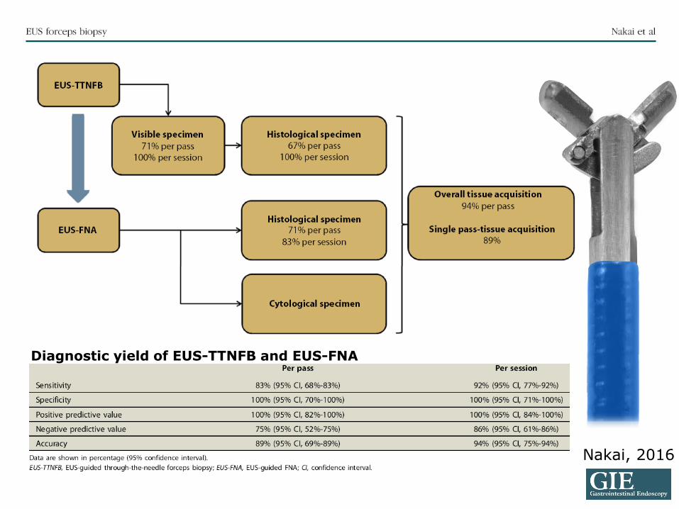
Nakai, 2016
Diagnostic yield of EUS-TTNFB and EUS-FNA

Nakai, 2016
Patient and tumor characteristics


5/7/20
18


Teamwork: Hard to get, easy to lose

Thanks
Recommended